Page 1
Instructions for use
Title Possible involvement of uncoupling protein 1 in appetite control by leptin
Author(s) Okamatsu-Ogura, Yuko; Nio-Kobayashi, Junko; Iwanaga, Toshihiko; Terao, Akira; Kimura, Kazuhiro; Saito, Masayuki
Citation Experimental Biology and Medicine, 236(11): 1274-1281
Issue Date 2011-11
Doc URL http://hdl.handle.net/2115/47824
Rights Exp Biol Med November 2011 vol. 236 no. 11 1274-1281, doi: 10.1258/ebm.2011.011143. This is the final draft, afterpeer-review, of a manuscript published in RSM journals: www.rsmpress.com.
Type article (author version)
File Information EBM236-11_1274-1281.pdf
Hokkaido University Collection of Scholarly and Academic Papers : HUSCAP
Page 2
1
Possible involvement of uncoupling protein 1 in appetite control by leptin
Yuko Okamatsu-Ogura1, Junko Nio-Kobayashi2, Toshihiko Iwanaga2, Akira Terao1, Kazuhiro
Kimura1, Masayuki Saito1,3
1 Department of Biomedical Sciences, Graduate School of Veterinary Medicine, Hokkaido University,
Sapporo 060-0818, Japan
2 Laboratory of Histology and Cytology, Graduate School of Medicine, Hokkaido University, Sapporo
060-8638, Japan
3 Department of Nutrition, Graduate School of Nursing and Nutrition, Tenshi College, Sapporo
065-0013, Japan
Running title:
UCP1 enhances leptin action
Corresponding author:
Yuko Okamatsu-Ogura, Ph.D.
Department of Biomedical Sciences, Graduate School of Veterinary Medicine, Hokkaido University,
Sapporo 060-0818, Japan
Tel: +81-11-706-5205
Fax: +81-11-757-0703
E-mail: [email protected]
Page 3
2
Abstract
Leptin reduces body fat by decreasing food intake and increasing energy expenditure. Uncoupling
protein (UCP) 1, a key molecule for brown adipose tissue (BAT) thermogenesis, was reported to
contribute to the stimulatory effect of leptin on energy expenditure. To clarify whether UCP1 is also
involved in the anorexigenic effect of leptin, in this study we examined the effect of leptin on food
intake using wild-type (WT) and UCP1-deficient (UCP1-KO) mice. Repeated injection of leptin
decreased food intake more markedly in WT mice than in UCP1-KO mice, while a single injection of
leptin showed similar effects in the two groups of mice. As chronic leptin stimulation induces UCP1
expression in BAT and ectopically in white adipose tissue (WAT), we mimicked the UCP1 induction
by repeated injection of CL316,243 (CL), a highly specific ß3-adrenoceptor agonist, and measured
food intake in response to a single injection of leptin. Two-week treatment with CL enhanced the
anorexigenic effect of leptin in WT mice, but not in UCP1-KO mice. Three-day treatment with CL
in WT mice also enhanced the anorexigenic effect of leptin and leptin-induced phosphorylation of
STAT3 in the arcuate nucleus of the hypothalamus, without any notable change in adiposity. These
results indicate that UCP1 enhances leptin action at the hypothalamus level, suggesting UCP1
contributes to the control of energy balance not only through the regulation of energy expenditure, but
also through appetite control by modulating leptin action.
Key words : uncoupling protein 1, brown adipose tissue, leptin, appetite control
Page 4
3
Introduction
Brown adipose tissue (BAT) is a tissue involved in metabolic heat production and has a
significant role in cold- and diet-induced thermogenesis 1,2. BAT thermogenesis is principally
dependent on the activation of uncoupling protein 1 (UCP1), which uncouples oxidative
phosphorylation in mitochondria to dissipate the electrochemical proton gradient as heat. The
activity of UCP1 is controlled by the sympathetic nerves to BAT, mainly through the ß-adrenergic
mechanism. The activation of the sympathetic nerve – ß-adrenergic receptor (ß-AR) pathway
induces lipolysis in BAT to produce fatty acids that activate UCP1 and are used simultaneously as a
substrate for thermogenesis. In addition, prolonged activation of this pathway induces hyperplasia of
BAT associated with an elevated UCP1 level.
Physiological roles and functional regulation of UCP1 mentioned above were assured by the
phenotype of UCP1-deficient (UCP1-KO) mice. UCP1-KO mice are unable to maintain body
temperature under cold circumstances 3, and get obese when housed at thermoneutrality 4. Brown
adipocyte of UCP1-KO mice shows the characteristics of brown adipocyte such as multilocular lipid
droplets and higher mitochondrial content compared to white adipocyte, however, accumulates larger
lipid droplets reflecting their functional defect 3. In UCP1-KO mice, injection of ß3-AR agonist
induces lipolysis from WAT as in wild-type (WT) mice, whereas it fails to show stimulatory effect on
BAT thermogenesis, such as increase in oxygen consumption and body temperature observed in WT
mice 5.
Leptin, a hormone secreted primarily by adipocytes, plays an important role in the regulation of
appetite and energy balance 6,7. Leptin inhibits appetite through action on the hypothalamus,
Page 5
4
especially the arcuate nucleus (ARC). Leptin secreted from adipose tissue enters the central nervous
system, binds to its receptor, and reduces food intake by stimulating anorexigenic peptides, such as
pro-opiomelanocortin, and by inhibiting orexigenic peptides, such as neuropeptide Y and agouti
gene-related protein. Besides its anorexigenic effect, leptin has been reported to increase energy
expenditure. For example, the peripheral or central administration of leptin increases oxygen
consumption in rats and mice 8,9. The involvement of BAT in the stimulatory effect of leptin on
energy expenditure was established by findings indicating that a single peripheral leptin injection
increases sympathetic nerve activity and temperature in BAT 10-12. Chronic leptin stimulation
increases UCP1 expression in BAT, and also induces it ectopically in white adipose tissue (WAT) 13,14.
Furthermore, chronic leptin treatment increases oxygen consumption and reduces body fat in WT mice,
but not in UCP1-KO mice, compared with pair-fed control mice 14,15, indicating that UCP1 is
indispensable for the effect of leptin on energy expenditure. Thus, leptin reduces food intake and
increases energy expenditure, thereby reducing body fat.
It is known that feeding conditions also affect the sympathetic nerve-BAT pathway 16, for
example, spontaneous overeating induced by high-fat diets or palatable foods increased
norepinephrine turnover rate 17, GDP-binding to mitochondria, an index of UCP1 activity 18, and
UCP1 expression in BAT 19, whereas all of these parameters were decreased by fasting 17,20. Such
physiological responses to food are referred to as diet-induced thermogenesis, and assumed to be a
mechanism to dissipate excess energy as heat. However, it is unknown whether or how leptin is
involved in this mechanism. In 1948, Brobeck 21 initially proposed a thermostatic hypothesis of food
intake that heat or body temperature is involved in appetite control: animals eat to keep warm and stop
Page 6
5
eating to stay cool. Subsequently, Himms-Hagen 22 suggested that the activation of BAT leads to an
increase in body temperature, which causes the termination of feeding. However, as far as we know,
there is no evidence for a BAT-related thermostatic mechanism for appetite control.
In a previous study, we observed a tendency to reduce food intake by chronic hyperleptinemia
more in WT mice than in UCP1-KO mice, despite the similar plasma leptin levels 14. This result
suggests the involvement of UCP1 in appetite control. To test this hypothesis, we examined the role
of UCP1 in the anorexigenic effect of leptin, particularly focusing on UCP1 ectopically expressed in
WAT.
Page 7
6
Materials and methods
Animals
UCP1-KO (ucp1-/-) mice on a congenic background of C57BL/6J were generated by backcross
matings of heterozygous (+/-) mice on a mixed 129/SvPas and C57BL/6J background with C57BL/6J
mice for 15 generations, and kindly provided by Dr. L. Kozak (Pennington Biomedical Research
Center, Baton Rouge, LA, USA) 3. All WT (ucp1+/+) mice were C57BL/6J. Mice were housed in
plastic cages placed in an air-conditioned room at 26 °C with a 12-hour light-dark cycle (lights on
07:00-19:00) and given free access to laboratory chow (MF: Oriental Yeast, Tokyo, Japan) and tap
water. Both male and female WT and UCP1-KO mice (20-30 weeks old) were used. The
experimental procedures and care of animals were approved by the Animal Care and Use Committee
of Hokkaido University.
Response to leptin injection
Mice were housed individually and allowed to acclimate for at least 7 days. Then, mice were
fasted for 24 hours and injected with recombinant mouse leptin (5 mg/kg, PeproTech, London, UK) or
saline intraperitoneally at 19:00 and given food. Food intake for 3 or 12 hours was estimated by
measuring the weight of remaining food and spillage in the cage. In another series of experiments,
mice were fed ad libitum and injected with leptin (1 mg/kg) subcutaneously twice a day at 07:00 and
19:00 for 4 days. Daily food intake was measured. Some mice were killed by cervical dislocation,
and fat pads from various regions (interscapular BAT, inguinal and perigonadal WAT) were quickly
removed and weighed. Tissue specimens were transferred into liquid nitrogen for Western blot
Page 8
7
analysis.
ß3-agonist CL316,243 treatment
WT and UCP1-KO mice were injected with ß3-AR agonist CL316,243 (CL: 0.1 mg/kg,
American Cyanamid, Pearl River, NY) or saline subcutaneously once a day at 19:00 for 3 or 14 days.
The mice were deprived of food at the time of the last injection. Twenty hours later, a blood sample
was taken for plasma leptin assay (Leptin ELISA kit; Morinaga, Yokohama, Japan). After 24-hour
fasting, mice were injected intraperitoneally with leptin (5 mg/kg) or saline, and food intake was
measured for 3 hours. Then, mice were killed by cervical dislocation, and fat pads from various
regions were quickly removed and weighed and transferred into liquid nitrogen for Western blot
analysis.
Western blotting
Tissue specimens were homogenized in Tris-EDTA buffer (10 mM Tris and 1 mM EDTA, pH
7.4). After centrifugation at 800 g for 10 minutes at 4°C, the obtained supernatant was centrifuged at
100,000 g for 1 hour at 4°C to obtain total membrane protein, and used to determine the content of
UCP1 by Western blotting. Briefly, membrane protein of BAT and inguinal WAT was separated by
SDS-PAGE and transferred onto polyvinylidine fluoride membranes (Immobilon; Millipore, Bedford,
MA). After blocking the membrane with 5% skimmed milk, it was incubated with polyclonal
antibody against UCP1 kindly provided by Drs. Teruo Kawada and Naohito Aoki (Kyoto University,
Kyoto, Japan) for 1 hour. The bound antibody was made visible using horseradish-peroxidase-linked
Page 9
8
goat anti-rabbit immunoglobulin (Zymed Laboratories, San Francisco, CA) and an enhanced
chemiluminescence system (Amersham, Little Chalfont, Bucks, UK).
Immunohistochemistry for phophoSTAT3 (pSTAT3) following leptin injection
Mice were fasted for 24 hours and injected with leptin (5 mg/kg) or saline intraperitoneally.
Thirty minutes after the injection, mice were perfused transcardially with 4% paraformaldehyde in 0.1
M phosphate buffer under anesthesia with sodium pentobarbital. Brains were postfixed,
cryoprotected with 30% sucrose, and sectioned coronally at 30 µm thickness with a freezing
microtome. The pSTAT3 was determined by immunohistochemical staining using rabbit
anti-pSTAT3 polyclonal antibody (Cell Signaling, MA, USA) and Histfine SAB-PO(R) kit (Nichirei,
Tokyo, Japan). pSTAT3 positive cells in the ARC and ventromedial hypothalamus (VMH) were
counted using 6 slices per animal.
Data analysis
Values are expressed as mean±SE. Statistical analysis was performed using analysis of
variance followed by post hoc testing by Tukey-Kramer test unless otherwise noted.
Page 10
9
Results
Responses to leptin injection
First, we investigated the acute response to a single injection of leptin in WT and UCP1-KO
mice. In WT mice, intraperitoneal injection of leptin reduced food intake to 68% (0~3 hour) and
78% (0~12 hour) of that of saline-injected control mice (Fig. 1A). Almost the same anorexigenic
effects of leptin were found in UCP1-KO mice.
We also examined the effect of repeated leptin injections on daily food intake (Fig. 1B). In
WT mice, repeated leptin injections reduced food intake to 91% on Day 1, and the reduction tended to
increase until Day 3 (79%) and then recover on Day 4 (87%). In UCP1-KO mice, repeated leptin
injection reduced food intake to 91% on Day 1, and the effect was sustained at a similar level until
Day 4. Two-way ANOVA revealed significant effect of Day (p<0.05), the genotype (p<0.05), and
interaction (p<0.05). The effect of leptin on food intake was more apparent in WT mice than in
UCP1-KO mice, particularly on Day 2 and Day 3. Thus, the effect of single leptin injection on food
intake was not different regardless of the absence or presence of UCP1, but that of repeated leptin
injection was greater in WT mice than in UCP1-KO mice. These results suggest that some
UCP1-dependent change induced by chronic, but not acute, leptin stimulation is involved in the
enhancement of the anorexigenic effect of leptin.
Previously, we and others showed that chronic leptin stimulation for 3-8 days not only increases
UCP1 expression in BAT, but also induces ectopic UCP1 expression in WAT 13,14. In this study, we
confirmed repeated leptin injection for only 2 days induced UCP1 in WAT, without notable effect on
UCP1 in BAT (Fig.1C).
Page 11
10
Effect of CL316,243 treatment
To investigate if the leptin-induced UCP1 expression is involved in the enhancement of the
leptin action, we mimicked the UCP1 induction by injecting the mice with CL, a highly specific
ß3-agonist. This treatment was done for 2 weeks before the effect of leptin was examined. As
reported previously 5, in WT mice, 2-week CL treatment resulted in significant increase of UCP1
expression in BAT and ectopic induction of UCP1 in WAT (Fig.2A). In WT mice, single leptin
injection reduced food intake in both the control (-27%) and the CL-treated (-48%) groups, and the
effect was greater in the CL-treated group (Fig.2B). In UCP1-KO mice, leptin injection reduced food
intake in the control (-26%) and CL-treated (-29%) groups, but there was no difference between the
two groups.
The anorexigenic effect of leptin is known to be influenced by adiposity, which is decreased by
chronic CL treatment 5. To induce UCP1 expression with minimum effect on body fat, next, we
examined the effect of short-term CL treatment. In WT mice, 3-day treatment with CL induced
UCP1 in WAT without notable effect on UCP1 expression in BAT, body weight, WAT weight, and
plasma leptin concentration (Figs. 3A, 3B). A single leptin injection reduced food intake in the
control group (-21%), but the reduction was greater in the CL-treated group (-47%) (Fig. 3C). In
UCP1-KO, the effect of leptin on food intake was not different between the control (-23%) and
CL-treated groups (-26%). These data indicate that the anorexigenic effect of leptin is enhanced by a
UCP1-dependent change induced by repeated CL injection.
To confirm the enhanced leptin action after the CL treatment, we also examined the
Page 12
11
leptin-induced phosphorylation of STAT3 in the hypothalamus as a marker of leptin signaling. In
saline-injected mice, pSTAT3 was not detected in any hypothalamic area in the control and CL-treated
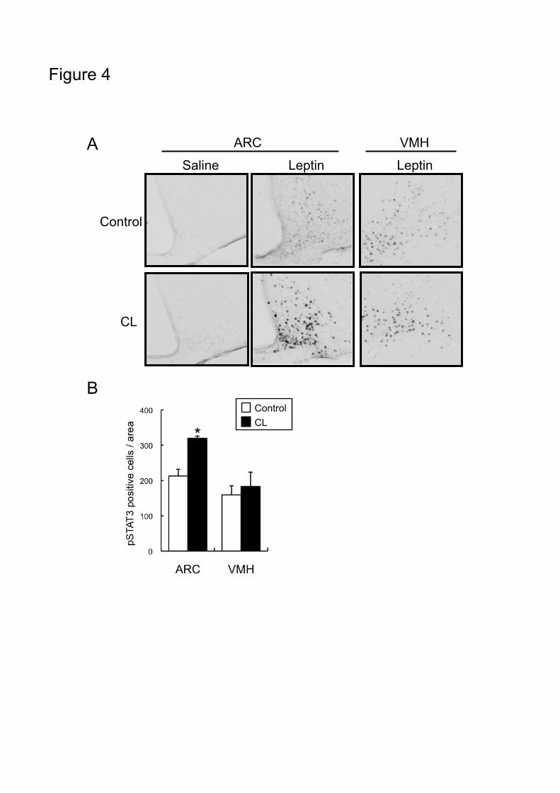
groups (Fig. 4A). Thirty minutes after leptin injection, pSTAT3-immunoreactive cells were detected
in both ARC and VMH. The number of pSTAT3-positive cells in the ARC was significantly higher
in the CL-treated group than the control group, but those in the VMH were almost the same in the two
groups (Fig.4B). Furthermore, the pSTAT3 immunoreactivity in the ARC of CL-treated group was
increased in intensity as compared with that in control group.
Page 13
12
Discussion
The purpose of the present study was to investigate the role of UCP1 in the anorexigenic effect
of leptin. The major findings were as follows: (1) repeated leptin injections for 2-3 days decreased
food intake more markedly in WT mice than in UCP1-KO mice, although a single injection of leptin
showed similar effects in the two groups of mice, (2) a two-day leptin treatment in WT mice induced
ectopic UCP1 expression in WAT, (3) a two-week treatment with CL increased UCP1 expression in
BAT, induced ectopic UCP1 in WAT, and enhanced the anorexigenic effect of leptin in WT mice, but
not in UCP1-KO mice, and (4) a three-day treatment with CL in WT mice also induced ectopic UCP1
in WAT, and enhanced the anorexigenic effect of leptin and leptin-induced phosphorylation of STAT3
in the ARC of hypothalamus.
A difference in the anorexigenic effect of leptin between WT and UCP1-KO mice was found
when leptin was injected repeatedly, whereas a single injection had similar effects on the two groups.
This was consistent with our previous report showing more reduced food intake by chronic
hyperleptinemia in WT mice than in UCP1-KO mice 14. These results indicate that the anorexigenic
effect of leptin is modulated by UCP1 and/or some UCP1-dependent changes induced by chronic, but
not acute, leptin stimulation. Chronic leptin stimulation was reported to increase UCP1 expression in
BAT and induces ectopic UCP1 in WAT 13,14. In this study, we found repeated leptin injection
showed no notable effect on UCP1 in BAT, but induced ectopic UCP1 in WAT on Day 2 when the
different leptin effect in WT and UCP1-KO mice were observed. These results raised the possibility
that UCP1 ectopically induced in WAT contributes to the enhancement of leptin action. To test this
idea, we injected CL, a highly specific β-3 AR agonist, which is known to increase UCP1 expression
Page 14
13
in BAT and to induce ectopic UCP1 in WAT. As predicted, a 2-week treatment with CL enhanced
the anorexigenic effect of leptin in WT mice, but not in UCP1-KO mice.
Previously, we reported that chronic treatment with CL reduces body fat in a UCP1-dependent
manner 5. Since the action of leptin is largely affected by adiposity 6,7, it is possible that the
enhancement of leptin action by the chronic CL treatment is secondary to the decreased adiposity.
However, a 3-day CL treatment in WT mice increased the anorexigenic effect of leptin without any
notable change in body weight, adiposity, UCP1 in BAT, and plasma leptin concentration, indicating
that the effect of CL treatment on the leptin action was not a consequence of decreased adiposity.
The CL-induced enhancement of leptin action was further confirmed by leptin-induced STAT3
phosphorylation, an important component of leptin signaling, in the ARC of hypothalamus 23,
suggesting an increase in leptin sensitivity at the hypothalamus level.
In this study, we injected CL to increase UCP1 expression in BAT and to induce ectopic UCP1
in WAT. Besides CL, overeating is an alternative stimulant of UCP1 expression. In mice and rats,
feeding on high-fat diets has been shown to increases UCP1 expression in BAT, and induces UCP1
expression in WAT. Recently, Feldmann et al. 4 reported that when fed with high-fat diets, food
intake was larger in UCP1-KO mice than in WT mice, while there was no difference when fed on a
normal diet. It is possible that the induction of UCP1 expression by high-fat feeding resulted in the
enhancement of leptin action in WT mice, and hence UCP1-KO mice lacking this pathway consumed
more food than WT mice.
The administration of CL has been reported to suppress food intake in rats and mice 24-26, but the
mechanism involved was not clear. It has been shown that the suppressive effect of CL on food
Page 15
14
intake is not exhibited in mice lacking ß3-AR 25, and rescued by the expression of ß3-AR in WAT and
BAT, but not in BAT alone 26. These results indicate the importance of ß3-AR in WAT in
CL-induced suppression of food intake. Considering that ectopic UCP1 in WAT is induced by the
ß-adrenergic mechanism 27, it is likely that the ectopic expression of UCP1 in WAT is involved in the
suppressive effect of CL on food intake, probably through the increased leptin sensitivity. This is
consistent with the findings of White et al. 28 that CL showed more reduced food intake in S5B/P1 rats
than in Osborne-Mendel rats, where UCP1 expression in WAT was induced more in the former than
the latter. These results also support the role of ectopic UCP1 in WAT in feeding control through the
modulation of leptin action.
The mechanism by which UCP1 in WAT modulates leptin action is not clear. Considering that
the expression level of UCP1 induced in WAT is as low as 1~2% of that in BAT, it is doubtful that
ectopic UCP1 in WAT contributes significantly to whole-body energy expenditure and thermogenesis.
However, ectopic UCP1 in WAT may have a role other than that in thermogenesis, such as acting as a
sensor to monitor local changes in WAT. The sensory innervation of WAT is suggested by the
existence of substance P and calcitonin gene-related peptide, typical marker peptides of primary
sensory neurons 29. Retrograde-tracing experiments also revealed that peripheral pseudounipolar
dorsal root ganglion cells innervate WAT 30. Song et al. 31 further showed that afferent nerves in
WAT project into many areas of brain, including the hypothalamus. Thus, it is possible that the local
change in metabolism or temperature induced by the ectopic expression of UCP1 in WAT is
transmitted to the central nervous system through the afferent nerves, and enhances leptin sensitivity
in the hypothalamus. In support of this, Yamada et al. 32 reported that afferent nerve signals from
Page 16
15
intra-abdominal fat tissue regulate food intake by modulating hypothalamic leptin sensitivity.
Collectively, it is most likely that UCP1 ectopically induced in WAT modulates the
anorexigenic action on leptin. However, it is to be noted that this conclusion does not necessarily
rule out the possible involvement of BAT in the UCP1-dependent enhancement of leptin action.
Leptin or CL not only chronically increases UCP1 expression both in WAT and BAT, but also acutely
activates UCP1 in BAT, leading to the secondary changes such as elevation of body temperature 5,10-12.
It is possible that such secondary changes may also alter leptin action on the ARC, and it takes 2-3
days to be manifested. Another possibility is that secretion of some humoral factor(s) is modified by
the induction or activation of UCP1. Leptin itself can be excluded from the candidates because we
found no difference in plasma leptin level between the control and CL-treated mice. Plasma
triglyceride (TG) was reported to induce leptin resistance at the blood-brain barrier 33. However, CL
decreased plasma TG level to the same extent in WT and UCP1-KO mice (Okamatsu-Ogura et al.,
unpublished observation), implying that plasma TG is not the causative factor in the enhancement of
leptin action induced by CL treatment. Further studies are needed to elucidate the precise mechanism
involved.
In conclusion, our results show the possible involvement of UCP1 in the enhancement of leptin
action. UCP1 may contribute to the control of energy balance via two pathways, directly through the
regulation of energy expenditure by its thermogenic activity and indirectly through appetite control by
modulating the anorexigenic effect of leptin.
Page 17
16
Author contributions
All authors participated in the design, interpretation of the studies and analysis of the data and review
of the manuscript; YOO performed the experiments and wrote the manuscript, JNK and IT assisted on
histological studies, AT and KK assisted with the design of experiments, MS conceived and designed
the study.
Page 18
17
ACKNOWLEDGEMENTS
We thank Dr. L. Kozak (Pennington Biomedical Research Center) for kindly providing us with
UCP1-KO mice, Drs. Teruo Kawada and Naohito Aoki (Kyoto University) for their kind gifts of the
anti-UCP1 antibody, and American Cyanamid Co. for providing CL316,243. This study was
supported in part by JSPS Research Fellowships for Young Scientists to Y.O.-O.
Page 19
18
REFERENCES
1. Cannon B, Nedergaard J. Brown adipose tissue: function and physiological significance. Physiol
Rev 2004;84(1):277-359
2. Lowell BB, Spiegelman BM. Towards a molecular understanding of adaptive thermogenesis.
Nature 2000;404(6778):652-60
3. Enerbäck S, Jacobsson A, Simpson EM, Guerra C, Yamashita H, Harper ME, Kozak LP. Mice
lacking mitochondrial uncoupling protein are cold-sensitive but not obese. Nature
1997;387(6628):90-4
4. Feldmann HM, Golozoubova V, Cannon B, Nedergaard J. UCP1 ablation induces obesity and
abolishes diet-induced thermogenesis in mice exempt from thermal stress by living at
thermoneutrality. Cell Metab 9 (2009) pp.203-209
5. Inokuma K, Okamatsu-Ogura Y, Omachi A, Matsushita Y, Kimura K, Yamashita H, Saito M.
Indispensable role of mitochondrial UCP1 for antiobesity effect of beta3-adrenergic stimulation.
Am J Physiol Endocrinol Metab 2006;290(5):E1014-21
6. Schwartz MW, Woods SC, Porte D, Seeley RJ, Baskin DG. Central nervous system control of
food intake. Nature 2000;404(6778):661-71
Page 20
19
7. Coll AP, Farooqi IS, O'Rahilly S. The hormonal control of food intake. Cell 2007;129(2):251-62
8. Mistry AM, Swick AG, Romsos DR. Leptin rapidly lowers food intake and elevates metabolic
rates in lean and ob/ob mice. J Nutr 1997;127(10):2065-72
9. van Dijk G. The role of leptin in the regulation of energy balance and adiposity. J
Neuroendocrinol 2001;13(10):913-21
10. Haynes WG, Morgan DA, Walsh SA, Mark AL, Sivitz WI. Receptor-mediated regional
sympathetic nerve activation by leptin. J Clin Invest 1997;100(2):270-8
11. Morrison SF. Activation of 5-HT1A receptors in raphe pallidus inhibits leptin-evoked increases
in brown adipose tissue thermogenesis. Am J Physiol Regul Integr Comp Physiol
2004;286(5):R832-7
12. Collins S, Kuhn CM, Petro AE, Swick AG, Chrunyk BA, Surwit RS. Role of leptin in fat
regulation. Nature 1996;380(6576):677
13. Commins SP, Watson PM, Padgett MA, Dudley A, Argyropoulos G, Gettys TW. Induction of
uncoupling protein expression in brown and white adipose tissue by leptin. Endocrinology
Page 21
20
1999;140(1):292-300
14. Okamatsu-Ogura, Y, Uozumi, A, Toda, C, Kimura, K, Yamashita, H, Saito, M. Uncoupling
protein 1 contributes to fatreducing effect of leptin. Obesity Res Clin Pract 2007;1:233-241
15. Commins SP, Watson PM, Frampton IC, Gettys TW. Leptin selectively reduces white adipose
tissue in mice via a UCP1-dependent mechanism in brown adipose tissue. Am J Physiol
Endocrinol Metab 2001;280(2):E372-7
16. Rothwell NJ, Stock MJ. A role for brown adipose tissue in diet-induced thermogenesis. Obes Res
1997;5(6):650-6
17. Young JB, Saville E, Rothwell NJ, Stock MJ, Landsberg L. Effect of diet and cold exposure on
norepinephrine turnover in brown adipose tissue of the rat. J Clin Invest 1982;69(5):1061-71
18. Nedergaard J, Raasmaja A, Cannon B. Parallel increases in amount of (3H)GDP binding and
thermogenin antigen in brown-adipose-tissue mitochondria of cafeteria-fed rats. Biochem
Biophys Res Commun 1984;122(3):1328-36
19. Margareto J, Marti A, Martinez JA. Changes in UCP mRNA expression levels in brown adipose
tissue and skeletal muscle after feeding a high-energy diet and relationships with leptin, glucose
Page 22
21
and PPARgamma. J Nutr Biochem 2001;12(3):130-137
20. Rothwell NJ, Stock MJ. Effect of chronic food restriction on energy balance, thermogenic
capacity, and brown-adipose-tissue activity in the rat. Biosci Rep 1982;2(8):543-9
21. Brobeck JR. Food intake as a mechanism of temperature regulation. Yale J Biol Med
1948;20(6):545-52
22. Himms-Hagen J. Role of brown adipose tissue thermogenesis in control of thermoregulatory
feeding in rats: a new hypothesis that links thermostatic and glucostatic hypotheses for control of
food intake. Proc Soc Exp Biol Med 1995 ;208(2):159-69
23. Vaisse C, Halaas JL, Horvath CM, Darnell JE Jr, Stoffel M, Friedman JM. Leptin activation of
Stat3 in the hypothalamus of wild-type and ob/ob mice but not db/db mice. Nat Genet
1996;14(1):95-7
24. Kumar MV, Moore RL, Scarpace PJ. Beta3-adrenergic regulation of leptin, food intake, and
adiposity is impaired with age. Pflugers Arch 1999;438(5):681-8
25. Susulic VS, Frederich RC, Lawitts J, Tozzo E, Kahn BB, Harper ME, Himms-Hagen J, Flier JS,
Lowell BB. Targeted disruption of the beta 3-adrenergic receptor gene. J Biol Chem
Page 23
22
1995;270(49):29483-92
26. Grujic D, Susulic VS, Harper ME, Himms-Hagen J, Cunningham BA, Corkey BE, Lowell BB.
Beta3-adrenergic receptors on white and brown adipocytes mediate beta3-selective
agonist-induced effects on energy expenditure, insulin secretion, and food intake. A study using
transgenic and gene knockout mice. J Biol Chem 1997;272(28):17686-93
27. Jimenez M, Barbatelli G, Allevi R, Cinti S, Seydoux J, Giacobino JP, Muzzin P, Preitner F. Beta
3-adrenoceptor knockout in C57BL/6J mice depresses the occurrence of brown adipocytes in
white fat. Eur J Biochem 2003;270(4):699-705
28. White CL, Ishihara Y, Dotson TL, Hughes DA, Bray GA, York DA. Effect of a beta-3 agonist on
food intake in two strains of rats that differ in susceptibility to obesity. Physiol Behav
2004;82(2-3):489-96
29. Shi H, Song CK, Giordano A, Cinti S, Bartness TJ. Sensory or sympathetic white adipose tissue
denervation differentially affects depot growth and cellularity. Am J Physiol Regul Integr Comp
Physiol 2005;288(4):R1028-37
30. Fishman RB, Dark J. Sensory innervation of white adipose tissue. Am J Physiol 1987;253(6 Pt
2):R942-4
Page 24
23
31. Song CK, Schwartz GJ, Bartness TJ. Anterograde transneuronal viral tract tracing reveals central
sensory circuits from white adipose tissue. Am J Physiol Regul Integr Comp Physiol
2009;296(3):R501-11
32. Yamada T, Katagiri H, Ishigaki Y, Ogihara T, Imai J, Uno K, Hasegawa Y, Gao J, Ishihara H,
Niijima A, Mano H, Aburatani H, Asano T, Oka Y. Signals from intra-abdominal fat modulate
insulin and leptin sensitivity through different mechanisms: neuronal involvement in food-intake
regulation. Cell Metab 2006;3(3):223-9
33. Banks WA, Coon AB, Robinson SM, Moinuddin A, Shultz JM, Nakaoke R, Morley JE.
Triglycerides induce leptin resistance at the blood-brain barrier. Diabetes 2004;53(5):1253-60
Page 25
24
Figure legends
Figure 1. Effects of leptin injection on food intake in WT and UCP1-KO mice
(A) WT and UCP1-KO mice were fasted for 24 hours and injected with leptin (5 mg/kg, i.p.) or saline.
Food intake for 3 and 12 hours after the injection was measured and expressed relative to the 3-hour
food intake of saline-injected controls (1.18±0.07 g in WT and 1.29±0.08 g in UCP1-KO mice).
Values are means±SE for 4 mice. *P<0.05 vs. saline-injected control of the same genotype. (B)
Mice were injected with leptin (1 mg/kg, s.c.) twice a day for 4 days. Daily food intake was
measured and expressed relative to that before injection on Day 0 (3.24±0.08 g in WT and 3.03±0.12 g
in UCP1-KO mice). Values are means±SE for 10 mice. Two-way ANOVA revealed significant
effect of Day (p<0.05), the genotype (p<0.05), and interaction (p<0.05). (C) UCP1 expression in
BAT and inguinal WAT (I-WAT) of WT mice were analyzed by Western blotting. To detect UCP1,
5 µg (BAT) or 30 µg (I-WAT) of membrane protein was used. UCP1 content was expressed as
relative to that in BAT of the Day0 group. *P<0.05 Day0 vs. Day2 by Student’s t-test.
Figure 2. Effects of 2-week treatment with CL316,243 in WT and UCP1-KO mice
WT and UCP1-KO mice were injected with CL316,243 (CL; 0.1 mg/kg, s.c.) or saline once a day for
2 weeks. (A) UCP1 expression in BAT and inguinal WAT (I-WAT) of WT mice were analyzed by
Western blotting. To detect UCP1, 5 µg (BAT) or 20 µg (I-WAT) of membrane protein was used.
UCP1 content was expressed as relative to that in BAT of the control group. Values are means ± SE
for 6 mice. *P<0.05 control group vs. CL group by Student’s t-test. (B) After 2-week treatment
Page 26
25
with CL316,243, WT and UCP1-KO mice were fasted for 24 hours and injected with leptin (5 mg/kg,
i.p.) or saline. Food intake for 3 hours after the injection was measured. Values are means±SE for
6 mice. *P<0.05 vs. saline-injected mice of the same group. †P<0.05 Leptin-injected control group
vs. CL group.
Figure 3. Effects of 3-day treatment with CL316,243
WT mice were injected with CL316,243 (CL; 0.1 mg/kg, s.c.) or saline once a day for 3 days. (A)
Body weight, adiposity, and plasma leptin concentration were measured. (B) UCP1 expression in
BAT and inguinal WAT (I-WAT) were analyzed by Western blotting. To detect UCP1, 5 µg (BAT)
or 30 µg (I-WAT) of membrane protein was used. UCP1 content was expressed as relative to that in
BAT of the control group. Values are means ± SE for 6 mice. *P<0.05 control group vs. CL group
by Student’s t-test. (C) After 3-day treatment with CL316,243, WT and UCP1-KO mice were fasted
for 24 hours and injected with leptin (5 mg/kg, i.p.) or saline. Food intake for 3 hours was measured.
Values are means±SE for 6 mice. *P<0.05 vs. saline-injected mice of the same group. †P<0.05
Leptin-injected control group vs. CL group
Figure 4. Effects of 3-day treatment with CL 316,243 on leptin-induced phosphorylation of
STAT3 in hypothalamus
WT mice were injected with CL316,243 (0.1 mg/kg, s.c.) or saline once a day for 3 days. Mice were
fasted for 24 hours and injected with leptin (5 mg/kg, i.p.) or saline, and sacrificed 30 minutes later.
(A) Phospho-STAT3 (pSTAT3) in hypothalamus was detected by immunostaining. (B) The numbers
Page 27
26
of pSTAT3-positive cells in the arcuate nucleus (ARC) and the ventromedial hypothalamus (VMH)
were counted. Values are means±SE for 3 mice. *P<0.05 vs. control group by Student’s t-test.
Page 28
Figure 1
A B Saline Leptin
0~3 h 0~12 h
WT KO WT KO
* *
* *
*
C
WT KO
5µg protein / lane�
30µg protein / lane�
BAT
I-WAT
Day0 + Day2
0.00
0.02
0.04
0.06
I-WAT
UC
P1
cont
ent /
pro
tein
I-WAT
Day0 Day2
*
0.0
0.5
1.0
1.5
BAT
UC
P1
cont
ent /
pro
tein
Day0 Day2
BAT
Page 29
0.00
0.05
0.10
0.15
0.20
I-WAT
UC
P1
cont
ent /
pro
tein
*
I-WAT
Cont CL
Figure 2
A
B Saline Leptin
*
†
* * *
Control CL
WT KO
Control CL
BAT
I-WAT
+Cont CL Cont CL
5µg protein / lane�
20µg protein / lane� 0.0
0.5
1.0
1.5
2.0
BAT
UC
P1
cont
ent /
pro
tein
*
Cont CL
BAT
Page 30
Figure 3
A
B
C
0
10
20
30
Control CL
Bod
y W
eigh
t (g)
0
250
500
750
1000
Control CL
Bod
y Fa
t (m
g)
0
5
10
Control CL
Pla
sma
Lept
in (n
g/m
l)
Control CL Control CL Control CL
BAT
I-WAT
Control CL +
5µg protein / lane�
30µg protein / lane� 0.00
0.05
0.10
0.15
I-WAT
UC
P1
cont
ent /
pro
tein
*
I-WAT
Control CL 0.0
0.5
1.0
1.5
BAT
UC
P1
cont
ent /
pro
tein
Control CL
BAT
Saline Leptin
0.0�
0.5�
1.0�
1.5�
Food intake (g)�
*
†
*
* *
Control CL
WT KO
Control CL
Page 31
Figure 4
A
B
*
Control CL
Control
CL
Saline Leptin Leptin
VMH ARC