2
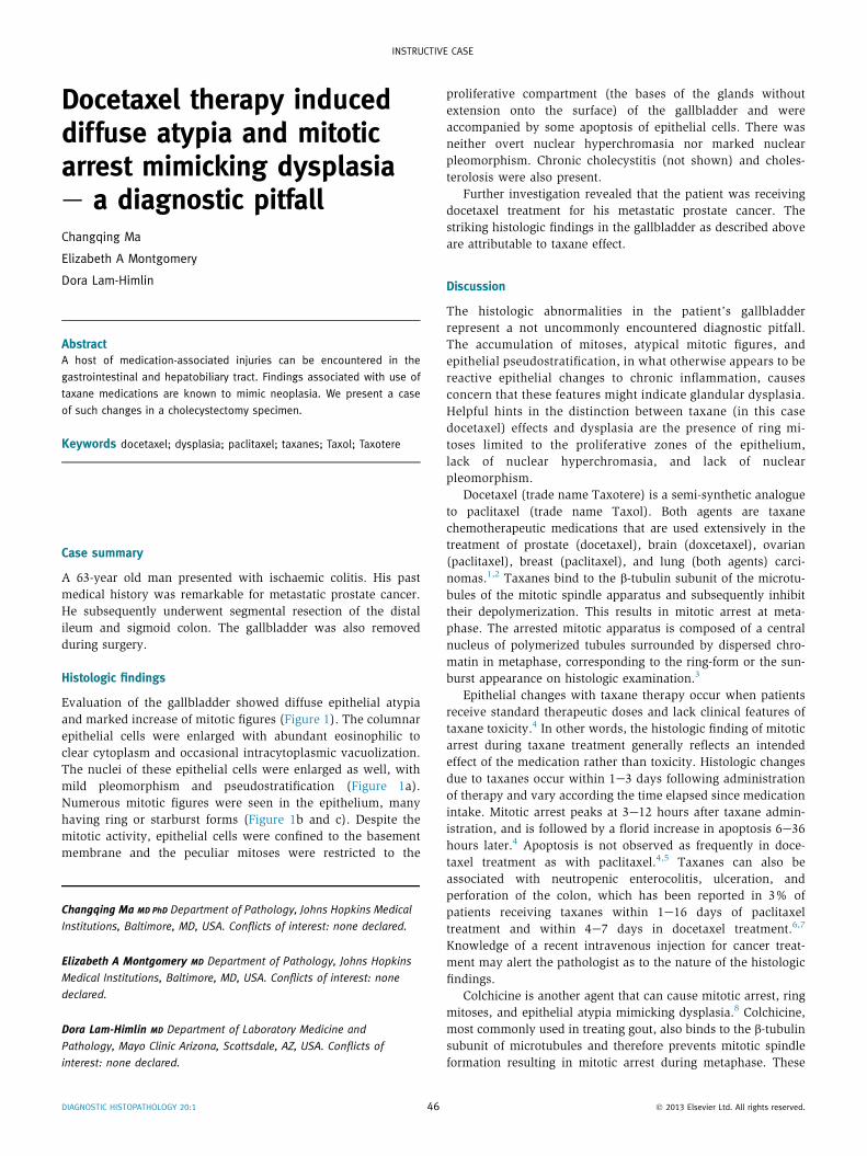
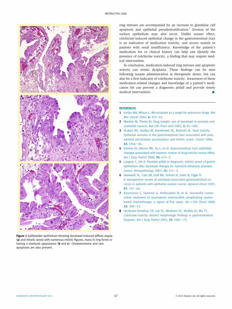
Docetaxel therapy induced diffuse atypia and mitotic arrest mimicking dysplasia e a diagnostic pitfall Changqing Ma Elizabeth A Montgomery Dora Lam-Himlin Abstract A host of medication-associated injuries can be encountered in the gastrointestinal and hepatobiliary tract. Findings associated with use of taxane medications are known to mimic neoplasia. We present a case of such changes in a cholecystectomy specimen. Keywords docetaxel; dysplasia; paclitaxel; taxanes; Taxol; Taxotere Case summary A 63-year old man presented with ischaemic colitis. His past medical history was remarkable for metastatic prostate cancer. He subsequently underwent segmental resection of the distal ileum and sigmoid colon. The gallbladder was also removed during surgery. Histologic findings Evaluation of the gallbladder showed diffuse epithelial atypia and marked increase of mitotic figures (Figure 1). The columnar epithelial cells were enlarged with abundant eosinophilic to clear cytoplasm and occasional intracytoplasmic vacuolization. The nuclei of these epithelial cells were enlarged as well, with mild pleomorphism and pseudostratification (Figure 1a). Numerous mitotic figures were seen in the epithelium, many having ring or starburst forms (Figure 1b and c). Despite the mitotic activity, epithelial cells were confined to the basement membrane and the peculiar mitoses were restricted to the proliferative compartment (the bases of the glands without extension onto the surface) of the gallbladder and were accompanied by some apoptosis of epithelial cells. There was neither overt nuclear hyperchromasia nor marked nuclear pleomorphism. Chronic cholecystitis (not shown) and choles- terolosis were also present. Further investigation revealed that the patient was receiving docetaxel treatment for his metastatic prostate cancer. The striking histologic findings in the gallbladder as described above are attributable to taxane effect. Discussion The histologic abnormalities in the patient’s gallbladder represent a not uncommonly encountered diagnostic pitfall. The accumulation of mitoses, atypical mitotic figures, and epithelial pseudostratification, in what otherwise appears to be reactive epithelial changes to chronic inflammation, causes concern that these features might indicate glandular dysplasia. Helpful hints in the distinction between taxane (in this case docetaxel) effects and dysplasia are the presence of ring mi- toses limited to the proliferative zones of the epithelium, lack of nuclear hyperchromasia, and lack of nuclear pleomorphism. Docetaxel (trade name Taxotere) is a semi-synthetic analogue to paclitaxel (trade name Taxol). Both agents are taxane chemotherapeutic medications that are used extensively in the treatment of prostate (docetaxel), brain (doxcetaxel), ovarian (paclitaxel), breast (paclitaxel), and lung (both agents) carci- nomas. 1,2 Taxanes bind to the b-tubulin subunit of the microtu- bules of the mitotic spindle apparatus and subsequently inhibit their depolymerization. This results in mitotic arrest at meta- phase. The arrested mitotic apparatus is composed of a central nucleus of polymerized tubules surrounded by dispersed chro- matin in metaphase, corresponding to the ring-form or the sun- burst appearance on histologic examination. 3 Epithelial changes with taxane therapy occur when patients receive standard therapeutic doses and lack clinical features of taxane toxicity. 4 In other words, the histologic finding of mitotic arrest during taxane treatment generally reflects an intended effect of the medication rather than toxicity. Histologic changes due to taxanes occur within 1e3 days following administration of therapy and vary according the time elapsed since medication intake. Mitotic arrest peaks at 3e12 hours after taxane admin- istration, and is followed by a florid increase in apoptosis 6e36 hours later. 4 Apoptosis is not observed as frequently in doce- taxel treatment as with paclitaxel. 4,5 Taxanes can also be associated with neutropenic enterocolitis, ulceration, and perforation of the colon, which has been reported in 3% of patients receiving taxanes within 1e16 days of paclitaxel treatment and within 4e7 days in docetaxel treatment. 6,7 Knowledge of a recent intravenous injection for cancer treat- ment may alert the pathologist as to the nature of the histologic findings. Colchicine is another agent that can cause mitotic arrest, ring mitoses, and epithelial atypia mimicking dysplasia. 8 Colchicine, most commonly used in treating gout, also binds to the b-tubulin subunit of microtubules and therefore prevents mitotic spindle formation resulting in mitotic arrest during metaphase. These Changqing Ma MD PhD Department of Pathology, Johns Hopkins Medical Institutions, Baltimore, MD, USA. Conflicts of interest: none declared. Elizabeth A Montgomery MD Department of Pathology, Johns Hopkins Medical Institutions, Baltimore, MD, USA. Conflicts of interest: none declared. Dora Lam-Himlin MD Department of Laboratory Medicine and Pathology, Mayo Clinic Arizona, Scottsdale, AZ, USA. Conflicts of interest: none declared. INSTRUCTIVE CASE DIAGNOSTIC HISTOPATHOLOGY 20:1 46 Ó 2013 Elsevier Ltd. All rights reserved.