Table of Contents Docking with Water in the Binding Site ............................................................................. 2 Case Study .......................................................................................................................... 3 Introduction.................................................................................................................... 3 Provided Input Files ........................................................................................................ 5 Preparing Protein for Docking Experiment ........................................................................ 6 Exploring Ligand for Docking Experiment .......................................................................... 7 Defining the Binding Site ................................................................................................ 7 Selecting Ligand for Docking .......................................................................................... 8 Setting Water in the Binding Site ....................................................................................... 9 Running GOLD Docking with water5 turned off ........................................................... 11 Running GOLD Docking with water5 toggled ............................................................... 13 Analysis of Results ............................................................................................................ 14 Water5: Off................................................................................................................... 14 Water5: toggled ........................................................................................................... 15 Conclusions................................................................................................................... 16 Docking with Water in the Binding Site using GOLD Version 2.0 – November 2017 GOLD v5.6
Transcript
Table of Contents Docking with Water in the Binding Site ............................................................................. 2
Case Study .......................................................................................................................... 3
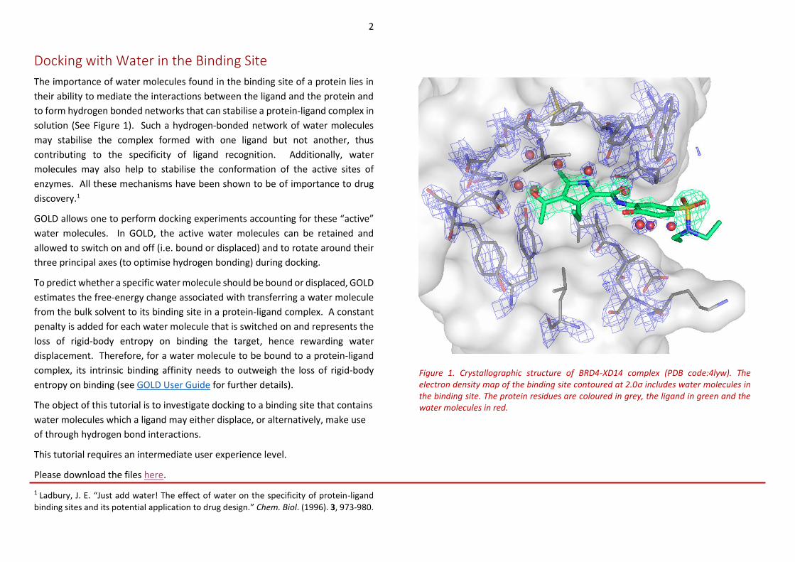
The importance of water molecules found in the binding site of a protein lies in
their ability to mediate the interactions between the ligand and the protein and
to form hydrogen bonded networks that can stabilise a protein-ligand complex in
solution (See Figure 1). Such a hydrogen-bonded network of water molecules
may stabilise the complex formed with one ligand but not another, thus
contributing to the specificity of ligand recognition. Additionally, water
molecules may also help to stabilise the conformation of the active sites of
enzymes. All these mechanisms have been shown to be of importance to drug
discovery.1
GOLD allows one to perform docking experiments accounting for these “active”
water molecules. In GOLD, the active water molecules can be retained and
allowed to switch on and off (i.e. bound or displaced) and to rotate around their
three principal axes (to optimise hydrogen bonding) during docking.
To predict whether a specific water molecule should be bound or displaced, GOLD
estimates the free-energy change associated with transferring a water molecule
from the bulk solvent to its binding site in a protein-ligand complex. A constant
penalty is added for each water molecule that is switched on and represents the
loss of rigid-body entropy on binding the target, hence rewarding water
displacement. Therefore, for a water molecule to be bound to a protein-ligand
complex, its intrinsic binding affinity needs to outweigh the loss of rigid-body
entropy on binding (see GOLD User Guide for further details).
The object of this tutorial is to investigate docking to a binding site that contains
water molecules which a ligand may either displace, or alternatively, make use
of through hydrogen bond interactions.
This tutorial requires an intermediate user experience level.
Please download the files here.
1 Ladbury, J. E. “Just add water! The effect of water on the specificity of protein-ligand
binding sites and its potential application to drug design.” Chem. Biol. (1996). 3, 973-980.
Figure 1. Crystallographic structure of BRD4-XD14 complex (PDB code:4lyw). The electron density map of the binding site contoured at 2.0σ includes water molecules in the binding site. The protein residues are coloured in grey, the ligand in green and the water molecules in red.
pyrrole-2-carboxamide) is a potent inhibitor of BRD4. The co-crystallised
structure of BRD4-XD14 (PDB code: 4lyw) reveals that XD14 binds in the acetyl-
lysine recognition site mimicking the acetyl-lysine residue interaction with BRD4
(see Figure 3). Positioning the 4-acyl substitution in the pyrrole ring towards the
highly conserved Asn140, XD14 engages in a direct hydrogen bond interaction.
2Lucas X et al. “4-Acyl Pyrrole: Mimicking Acetylated Lysines in Histone Code Reading.
Angew. Chem. Int. Ed. (2013). 52, 14055-14059
Figure 3. Co-crystallised BRD4-XD14 (PDB code: 4lyw). The protein is in light blue cartoon representation, the ligand in green and the water network is coloured in red. Additionally, we show the 2D interactions diagram of the ligand in complex with BRD4.
Figure 2. Secondary structure representation of BRD4. The key areas – acetyl-lysine site, WPL shelf and ZA loop – are highlighted in green, magenta and red, respectively.
4
The pyrrole ring is located deep inside the recognition pocket, and complements
the hydrophobic pocket defined by four conserved waters with a 5-methyl
substitution. In the co-crystallised structure of BRD4-XD14, an additional water
molecule is found to be directly involved in an H-bond with XD14.
Your task:
The object of this tutorial is to perform a docking experiment of XD14 to BRD4 in
the presence of several active water molecules. Here, we will illustrate the
requirements for setting up and running docking with active water. This example
assumes you are already familiar with how to setup protein(s) and ligands for
docking calculation. If not, please refer to the following sections of the GOLD
User Guide:
• Setting Up the Protein
• Essential Steps
Challenges:
Crystallographic structures of BRD4 in complex with different inhibitors have
shown that four water molecules are highly conserved in the bromodomains
binding site. Interestingly, the binding site of BRD4 in complex with XD14
inhibitor accommodates an additional water molecule that is directly involved in
an H-bond with XD14 (see Figure 4).
- In this tutorial, we will evaluate if the addition of the fifth water molecule
is required for a more accurate reproduction of the crystallographic
complex BRD4-XD14.
The example used here mimics the situation where a researcher has a crystal
structure of a protein binding site, and is unsure which and how many of the
waters in the binding site should be included in the model for use in an inhibitor
design effort.
Figure 4. Water network in the binding site of BRD4 complexed with XD14. Four conserved water molecules are represented as red spheres whereas, an additional water molecule specific to this complex and interacting via a H-bond with XD14 is shown as a purple sphere.
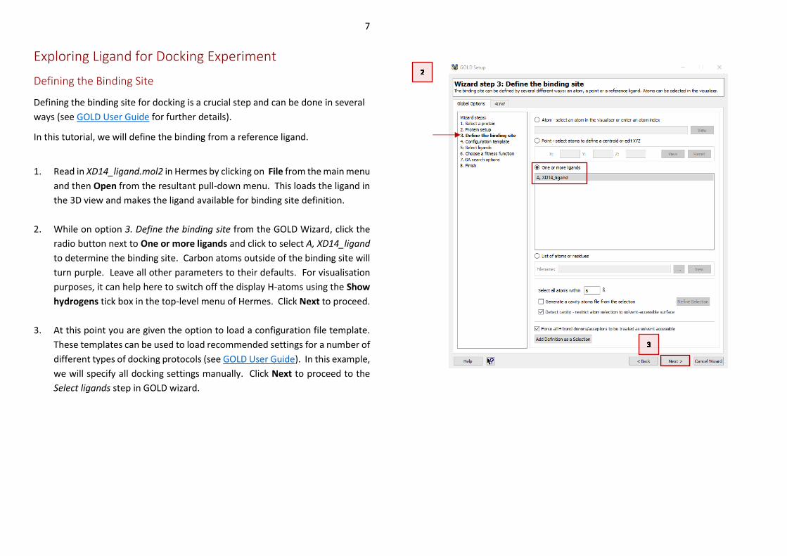
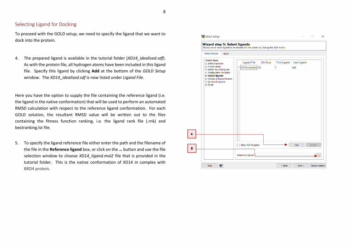
To proceed with the GOLD setup, we need to specify the ligand that we want to
dock into the protein.
4. The prepared ligand is available in the tutorial folder (XD14_idealised.sdf).
As with the protein file, all hydrogen atoms have been included in this ligand
file. Specify this ligand by clicking Add at the bottom of the GOLD Setup
window. The XD14_idealised.sdf is now listed under Ligand File.
Here you have the option to supply the file containing the reference ligand (i.e.
the ligand in the native conformation) that will be used to perform an automated
RMSD calculation with respect to the reference ligand conformation. For each
GOLD solution, the resultant RMSD value will be written out to the files
containing the fitness function ranking, i.e. the ligand rank file (.rnk) and
bestranking.lst file.
5. To specify the ligand reference file either enter the path and the filename of
the file in the Reference ligand box, or click on the … button and use the file
selection window to choose XD14_ligand.mol2 file that is provided in the
tutorial folder. This is the native conformation of XD14 in complex with
BRD4 protein.
9
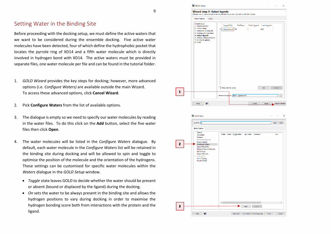
Setting Water in the Binding Site
Before proceeding with the docking setup, we must define the active waters that
we want to be considered during the ensemble docking. Five active water
molecules have been detected, four of which define the hydrophobic pocket that
locates the pyrrole ring of XD14 and a fifth water molecule which is directly
involved in hydrogen bond with XD14. The active waters must be provided in
separate files, one water molecule per file and can be found in the tutorial folder.
1. GOLD Wizard provides the key steps for docking; however, more advanced
options (i.e. Configure Waters) are available outside the main Wizard.
To access these advanced options, click Cancel Wizard.
2. Pick Configure Waters from the list of available options.
3. The dialogue is empty so we need to specify our water molecules by reading
in the water files. To do this click on the Add button, select the five water
files then click Open.
4. The water molecules will be listed in the Configure Waters dialogue. By
default, each water molecule in the Configure Waters list will be retained in
the binding site during docking and will be allowed to spin and toggle to
optimise the position of the molecule and the orientation of the hydrogens.
These settings can be customised for specific water molecules within the
Waters dialogue in the GOLD Setup window.
• Toggle state leaves GOLD to decide whether the water should be present
or absent (bound or displaced by the ligand) during the docking.
• On sets the water to be always present in the binding site and allows the
hydrogen positions to vary during docking in order to maximise the
hydrogen bonding score both from interactions with the protein and the
ligand.
10
• The Off water state option allows a water to be removed from
consideration during docking.
The orientation of the waters can be also changed.
• Activating the spin option makes GOLD automatically optimise the
orientation of the hydrogen atoms.
• Activating the trans_spin option and inserting a translation value into the
distance dialogue makes GOLD spin and translate the water molecule to
optimise the orientation of the hydrogen atoms as well as the water
molecule’s position within a defined radius. Note that the distance value
must be between 0 and 2 Å.
• Activating the fix option makes GOLD to use the orientation specified in
the input file.
Because in this tutorial we want to evaluate if the fifth water molecule
contributes to a better prediction of the experimental binding pose, four
conserved water molecules (water_1, water_2, water_3 and water_4) will be
always considered in the binding site, whereas the presence and displacement of
the water_5 will be evaluated.
11
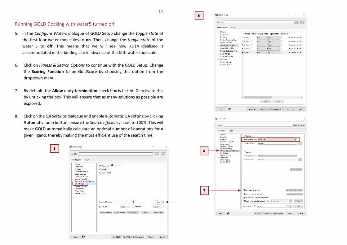
Running GOLD Docking with water5 turned off
5. In the Configure Waters dialogue of GOLD Setup change the toggle state of
the first four water molecules to on. Then, change the toggle state of the
water_5 to off. This means that we will see how XD14_idealised is
accommodated in the binding site in absence of the fifth water molecule.
6. Click on Fitness & Search Options to continue with the GOLD Setup. Change
the Scoring Function to be GoldScore by choosing this option from the
dropdown menu.
7. By default, the Allow early termination check box is ticked. Deactivate this
by unticking the box. This will ensure that as many solutions as possible are
explored.
8. Click on the GA Settings dialogue and enable automatic GA setting by clicking
Automatic radio button; ensure the Search efficiency is set to 100%. This will
make GOLD automatically calculate an optimal number of operations for a
given ligand, thereby making the most efficient use of the search time.
12
9. Before starting the docking, select Output Options. Click the … button next
to Output directory and either browse to or create an appropriate directory,
e.g. water5_off.
10. We have now finished setting up our docking, so click on the Run GOLD
button at the bottom of the GOLD interface. You will be presented with a
Finish GOLD Configuration window containing Save Files options.
11. Ensure the GOLD conf file box is ticked and type in the filename box to
rename the conf file as water5_off.conf.
12. Ensure that Protein(s) box is ticked. To save the edited 4LYW structure, click
Save to start the docking.
13. As the job progresses output will be displayed in several tabs in the Run
GOLD window. Note that the docking experiment will take several minutes.
14. Once the job is complete, the message Finished GOLD batch job, exiting…
will appear in the gold.log tab of the Run GOLD window. Additionally, the
View Solutions button in Run GOLD window will become available.
13
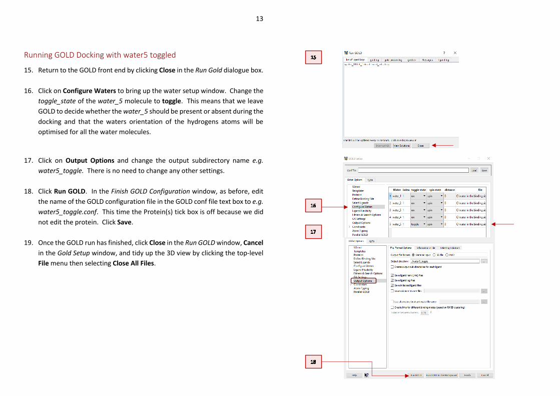
Running GOLD Docking with water5 toggled
15. Return to the GOLD front end by clicking Close in the Run Gold dialogue box.
16. Click on Configure Waters to bring up the water setup window. Change the
toggle_state of the water_5 molecule to toggle. This means that we leave
GOLD to decide whether the water_5 should be present or absent during the
docking and that the waters orientation of the hydrogens atoms will be
optimised for all the water molecules.
17. Click on Output Options and change the output subdirectory name e.g.
water5_toggle. There is no need to change any other settings.
18. Click Run GOLD. In the Finish GOLD Configuration window, as before, edit
the name of the GOLD configuration file in the GOLD conf file text box to e.g.
water5_toggle.conf. This time the Protein(s) tick box is off because we did
not edit the protein. Click Save.
19. Once the GOLD run has finished, click Close in the Run GOLD window, Cancel
in the Gold Setup window, and tidy up the 3D view by clicking the top-level
File menu then selecting Close All Files.
14
Analysis of Results
We will now evaluate how the different settings of the fifth water molecule will
affect the docking of XD14 to the BDR4 protein.
Note: In the following examples, you may wish to modify the representation style
or the colour of the reference ligand so that it is clearer. Once the files are loaded,
click on the Display tab in the Molecule Explorer, then right-click on the reference
ligand. From the dropdown menu, it is possible to modify the style and colour of
the reference ligand.
Water5: Off
1. Load the results of the water5_off docking by clicking File > Load GOLD
results then select the water5_off.conf file.
2. Load the reference ligand by clicking on File, Open, then navigate to the
tutorial directory which contains the XD14_ligand.mol2 file. Select the file
then click Open. Optionally you can also load the five water molecules.
3. The docking solutions are given in their docked order with their
corresponding fitness score under the column headed Goldscore.Fitness. If
desired, the solutions can be ordered by clicking on this Goldscore.Fitness
header to determine which is the highest scoring. We have obtained 10
docking solutions as this is the default number of how many times our ligand
was docked in the BRD4 protein.
4. Scroll through the docking solutions to check their poses against that of the
reference ligand. You should find that several docking solutions are found,
none of which closely resemble the correct binding mode.
5. The RMSD of the top ranked pose when compared to that of the co-
crystallised ligand is 2.73 Å. (Note that docking results and RMSD values may
15
vary due to the non-deterministic nature of GOLD). The top-ranking pose
matches the orientation of the pyrrole ring; however, it fails to match the
orientation of the diethyl sulphonamide and hydroxyphenyl groups.
6. By inspecting the binding mode, we can see that the hydrogen positions of
the four water molecules were optimised to form the water network
characteristic of the BRD4 binding site.
7. From Hermes click on File and select Close all files from the pull-down menu
to clear the 3D-viewer of Hermes.
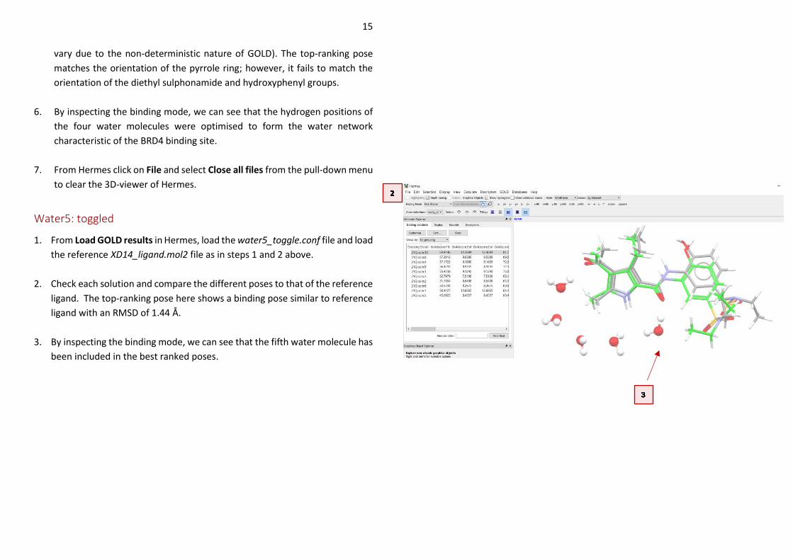
Water5: toggled
1. From Load GOLD results in Hermes, load the water5_toggle.conf file and load
the reference XD14_ligand.mol2 file as in steps 1 and 2 above.
2. Check each solution and compare the different poses to that of the reference
ligand. The top-ranking pose here shows a binding pose similar to reference
ligand with an RMSD of 1.44 Å.
3. By inspecting the binding mode, we can see that the fifth water molecule has
been included in the best ranked poses.
16
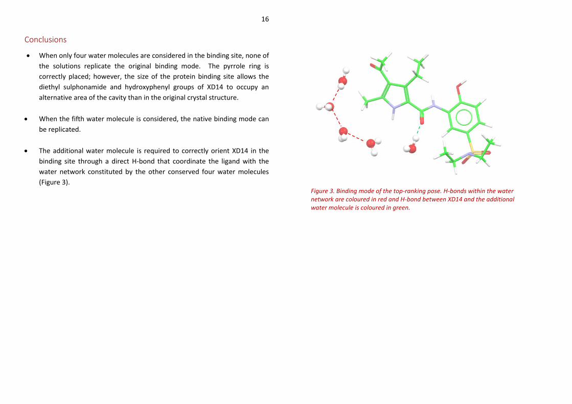
Conclusions
• When only four water molecules are considered in the binding site, none of
the solutions replicate the original binding mode. The pyrrole ring is
correctly placed; however, the size of the protein binding site allows the
diethyl sulphonamide and hydroxyphenyl groups of XD14 to occupy an
alternative area of the cavity than in the original crystal structure.
• When the fifth water molecule is considered, the native binding mode can
be replicated.
• The additional water molecule is required to correctly orient XD14 in the
binding site through a direct H-bond that coordinate the ligand with the
water network constituted by the other conserved four water molecules
(Figure 3). Figure 3. Binding mode of the top-ranking pose. H-bonds within the water network are coloured in red and H-bond between XD14 and the additional water molecule is coloured in green.