83
DOCTORAL THESES IN FOOD SCIENCES AT THE UNIVERSITY OF TURKU Food Chemistry
Early Life Intestinal Microbiota in Health and in Atopic Eczema
LOTTA NYLUND
Functional Foods Forum and Food Chemistry and Food Development
Department of Biochemistry
TURKU, FINLAND – 2015

Food Chemistry and Food Development Department of Biochemistry University of Turku, Finland
Supervised by
Professor Willem M de Vos Department of Veterinary Biosciences and RPU Immunobiology Faculty of Medicine, University of Helsinki, Helsinki, Finland and Laboratory of Microbiology Wageningen University Wageningen, the Netherlands Adjunct professor Reetta Satokari Department of Veterinary Biosciences University of Helsinki Helsinki, Finland
Professor Seppo Salminen Functional Foods Forum University of Turku Turku, Finland
Reviewed by
Adjunct Professor Anna Pelkonen Department of Allergology Helsinki University Central Hospital, Helsinki, Finland
Research Scientist, PhD Clara Gonzales de los Reyes-Gavilán Department of Microbiology and Biochemistry of Dairy Products Dairy Products Institute of Asturias Villaviciosa, Spain
Opponent Professor Glenn Gibson Department of Food and Nutritional Sciences University of Reading Reading, United Kingdom
Research director Seppo Salminen Functional Foods Forum University of Turku Turku, Finland
The originality of this dissertation has been checked in accordance with the University
of Turku quality assurance system using the Turnitin OriginalityCheck service
ISBN 978-951-29-6070-5 (print)
ISBN 978-951-29-6071-2 (pdf)
ISSN 2323-9395 (print)
ISSN 2323-9409 (pdf)
Painosalama Oy – Turku, Finland 2015

Table of Contents
TABLE OF CONTENTS
ABSTRACT .......................................................................................................... i
TIIVISTELMÄ .................................................................................................... ii
LIST OF ABBREVIATIONS ............................................................................. iii
LIST OF ORIGINAL PUBLICATIONS ............................................................ iv
1 INTRODUCTION ........................................................................................... 1
2 REVIEW OF THE LITERATURE ................................................................. 3 2.1 General characteristics and functions of the intestinal
microbiota ...................................................................................... 3 2.2 Microbial interactions with the human host .................................. 6 2.3 Importance of intestinal microbiota in human health and
disease ......................................................................................... 11 2.4 Development of healthy microbiota ............................................ 11 2.5 Factors affecting the microbiota development ............................ 16 2.6 Perturbations in the microbiota development and its
implications on health ................................................................. 22 2.7 Studying the early life microbiota –special challenges ............... 26 2.8 Overview of high-throughput microbiota analysis ..................... 26
3 AIMS OF THE STUDY ................................................................................ 31
4 MATERIALS AND METHODS .................................................................. 32 4.1 Subjects and study design ........................................................... 32 4.2 Methods ....................................................................................... 35
5 RESULTS ...................................................................................................... 41 5.1 Improvement of DNA extraction protocols for high-
throughput analyses (I) ................................................................ 41 5.2 Postpartum changes in microbiota composition of mothers (II) . 41 5.3 Temporal microbiota development in early childhood (II, III,
IV) and the effect of maternal microbiota (II)............................. 42 5.4 Microbiota signatures associated with eczema and its severity
(III, IV) ........................................................................................ 45 5.5 Changes in microbiota signatures related to the improvement
of eczema symptoms (IV) ........................................................... 47 5.6 Influence of probiotic supplementation on microbiota
composition (III, IV) ................................................................... 48
Table of Contents
6 DISCUSSION ................................................................................................ 49 6.1 Importance of DNA extraction in molecular based microbiota
analysis ........................................................................................ 49 6.2 Maternal microbiota during and after pregnancy ........................ 50 6.3 Microbiota development and the effect of maternal microbiota . 50 6.4 Microbiota signatures in infants with eczema and correlation
with the severity of symptoms .................................................... 52
7 SUMMARY AND FUTURE ASPECT ........................................................ 56
ACKNOWLEDGEMENTS ............................................................................... 57
REFERENCES ................................................................................................... 59
APPENDIX: ORIGINAL PUBLICATIONS .................................................... 71


Abstract
i
ABSTRACT
Decrease in microbial contacts in affluent societies is considered to lie behind the rise in allergic and other chronic inflammatory diseases during the last decades. Indeed, deviations in the intestinal microbiota composition and diversity have been associated with several diseases, such as atopic eczema. However, there is no consensus yet on what would constitute a beneficial or harmful microbiota. The aim of this thesis was to study the microbiota development in healthy infants and to characterize intestinal microbiota signatures associated with disease status and severity in infants with atopic eczema. The methodological aim was to compare and optimize methods for DNA extraction from fecal samples to be used in high-throughput microbiota analyses.
It was confirmed that the most critical step in successful microbial DNA extraction from fecal samples is the mechanical cell lysis procedure. Based on this finding, an efficient semi-automated extraction process was developed that can be scaled for use in high-throughput platforms such as phylogenetic microarray used in this series of studies. By analyzing a longitudinal mother-child cohort for 3 years it was observed that the microbiota development is a gradual process, where some bacterial groups reach the degree of adult-type pattern earlier than others. During the breast-feeding period, the microbiota appeared to be relatively simple, while major diversification was found to start during the weaning process. By the age of 3 years, the child’s microbiota composition started to resemble that of an adult, but the bacterial diversity has still not reached the full diversity, indicating that the microbiota maturation extends beyond this age. In addition, at three years of age, the child’s microbiota was more similar to mother’s microbiota than to microbiota of non-related women.In infants with atopic eczema, a high total microbiota diversity and abundance of butyrate-producing bacteria was found to correlate with mild symptoms at 6 months. At 18 months, infants with mild eczema had significantly higher microbiota diversity and aberrant microbiota composition when compared to healthy controls at the same age.
In conclusion, the comprehensive phylogenetic microarray analysis of early life microbiota shows the synergetic effect of vertical transmission and shared environment on the intestinal microbiota development. By the age of three years, the compositional development of intestinal microbiota is close to adult level, but the microbiota diversification continues beyond this age. In addition, specific microbiota signatures are associated with the existence and severity of atopic eczema and intestinal microbiota seems to have a role in alleviating the symptoms of this disease.
Abstract
ii
TIIVISTELMÄ
Allergiset sairaudet ovat yleistyneet nopeasti viime vuosikymmeninä etenkin länsimaisissa hyvinvointivaltioissa. Vähentyneen mikrobialtistuksen varhaislapsuudessa uskotaan olevan yksi näille sairauksille altistava riskitekijä. Poikkeava suolistomikrobisto onkin yhdistetty kohonneeseen riskiin sairastua esimerkiksi atooppiseen ekseemaan. Toistaiseksi spesifisiä mikrobiston komponentteja, jotka toimisivat joko atopialta suojaavina tai sille altistavina tekijöinä, ei kuitenkaan ole pystytty tunnistamaan. Tämän väitöskirjatyön tavoitteena oli tutkia suolistomikrobiston kehittymistä terveillä lapsilla sekä määrittää atooppiseen ekseemaan ja sen vakavuuteen yhteydessä olevat suolistomikrobiston tunnusmerkit. Tavoitteena oli myös vertailla ja optimoida DNA-eristysmenetelmiä, jotka soveltuisivat laajojen näytemäärien käsittelyyn ja niiden analysointiin uusimmilla molekulaarisilla menetelmillä.
Osoitimme, että mikrobisolujen mekaaninen hajoittaminen on kriittisin vaihe onnistuneessa DNA -eristyksessä ulostenäytteistä. Tähän havaintoon perustuen kehitettiin tehokas puoliautomatisoitu DNA- eristysprosessi, jota voidaan hyödyntää korkean suoritustehon menetelmissä, kuten esimerkiksi tässä tutkimussarjassa käytetyssä fylogeneettisessä mikrosiruanalyysissä. Tässä tutkimuksessa osoitettiin, että lasten suolistomikrobiston kehittyminen on vaiheittainen prosessi, jossa tietyt bakteeriryhmät saavuttavat aikuisenkaltaisen tason aiemmin kuin muut bakteeriryhmät. Imeväisiässä lapsen suolistomikrobisto on hyvin yksinkertainen, ja bakteerilajien monimuotoisuus kasvaa voimakkaasti kun vieroitus äidinmaidosta aloitetaan. Kolmeen ikävuoteen mennessä lapsen suolistomikrobiston lajikoostumus on melko ”aikuismainen” ja hyvin samankaltainen kuin äidillä. Mikrobiston kehittyminen kuitenkin jatkuu, sillä sen monimuotoisuus (diversiteetti) on tässä iässä vielä merkittävästi alhaisempi kuin aikuisilla. Atooppista ekseemaa sairastavilla, kuuden kuukauden ikäisillä lapsilla bakteeriston korkeampi diversiteetti ja korkeampi butyraattia tuottavien bakteerien määrä oli yhteydessä lievempiin oireisiin. Toisaalta 18 kuukauden iässä lievää atooppista ekseemaa sairastavilla lapsilla oli korkeampi suolistomikrobiston diversiteetti ja erilainen bakteerilajisto kuin terveillä lapsilla. Tässä väitöskirjatyössä osoitettiin fylogeneettisen mikrosiruanalyysin avulla äidin suolistomikrobiston ja yhteisen elinympäristön vaikutus lapsen suolistomikrobiston kehittymiseen varhaislapsuudessa. Lisäksi määritettiin atooppiseen ekseemaan ja sen vakavuuteen yhteydessä olevat suolistomikrobiston tunnusmerkit ja osoitettiin, että suolistomikrobisto saattaa olla osallisena atooppisen ihottuman oireiden lievittymisessä.
List of Abbreviations
iii
LIST OF ABBREVIATIONS
dsRNA Double stranded RNA DC Dendritic cell EPS Exopolysaccharide GOS Galacto-oligosaccharide GPCR G-protein coupled receptor HDAC Histone deacetylase HITChip Human Intestinal Tract chip HMO Human Milk Oligosaccharides IEC Intestinal Epithelial Cell Ig Immunoglobulin IL Interleukin KF KingFisher LPS Lipopolysaccharide MAMP Microbe-associated molecular pattern MCPP Monte Carlo Permutation Procedure NEC Necrotising enterocolitis NeM NucliSENS easyMAG NLR NOD-like receptor NOD Nucleotide-binding oligomerization domain OTU Operational Taxonomic Unit PCA Principal Component analysis PCR Polymerase chain reaction PUL Polysaccharide utilization loci RBB Repeated Bead Beating RDA Redundancy analysis SCFA Short-Chain Fatty Acid SCORAD SCORing Atopic Dermatitis spp. Species subsp. Subspecies Th Helper T cell Treg Regulatory T cell TLR Toll-Like Receptor 16S rRNA Bacterial ribosomal 16S RNA molecule
List of Original Publications
iv
LIST OF ORIGINAL PUBLICATIONS
I. Nylund L., Heilig H., Salminen S., de Vos WM. and Satokari R. Semi-automated extraction of microbial DNA from feces for qPCR and phylogenetic microarray analysis. J Microbiol Methods 2010; 83:231-235.
II. Nylund L., Grönlund M-M., Isolauri E., Salminen S., de Vos WM and Satokari R. High similarity between microbiota of three-year-old children and their mothers testify for vertical transmission and the impact of shared environment. Submitted
III. Nylund L., Satokari R., Nikkilä J., Rajilić-Stojanović M, Kalliomäki M, Isolauri E, Salminen S, de Vos WM. Microarray analysis reveals marked intestinal microbiota aberrancy in infants having eczema compared to healthy children in at-risk for atopic disease. BMC Microbiol. 2013; 13:12.
IV. Nylund L., Nermes M., Isolauri E., Salminen S., de Vos WM., Satokari R. Severity of atopic disease correlates with intestinal microbiota diversity and butyrate-producing bacteria. Allergy 2015; 70:241-244
ADDITIONAL PUBLICATION RELATED TO THE THESIS
The review of literature is partially based on the review article:
Nylund L., Satokari R., Salminen S. and de Vos WM. Intestinal microbiota during early life -impact on health and disease. Proc Nutr Soc 2014;73(4): 457-69.
Review of the Literature
1
1 INTRODUCTION
Although fetus is exposed to microbes and microbial structures already in utero, the major microbial colonization of the gastrointestinal tract starts at birth when an infant comes into contact with the microbes from the extrauterine environment. This initial exposure coincides with the ingestion of breast milk and its constituents, including oligosaccharides, immunoglobins and antimicrobial proteins, which facilitate the selective expansion of mutualistic microbes. This and other host factors are assumed to lead to a gradual compositional development and finally, to the establishment of a stable, individual-specific microbiota. The microbiota development process is affected by several factors including genetic background, mode of delivery, dietary pattern and medical procedures. These early colonization events and the establishing intestinal microbiota shape the immune system and have an effect on the development of variety of diseases later in life.
Atopic diseases are chronic, relapsing disorders usually starting at early childhood. During the last few decades, the incidences of these diseases (atopic eczema, asthma, allergic rhinoconjunctivitis and food allergy) have increased in epidemic proportions. In early life, the most common form of atopic disease is eczema, its prevalence being approximately 15 - 30 %, depending on the country studied (Deckers et al. 2012). According to the SCORAD (SCORing Atopic Dermatitis) assessment used to evaluate the severity of disease, the majority of patients are classified as “mild” (SCORAD <15), whereas 10-20 % of patients are “severe” (SCORAD >40) and this percentage seems to be higher in adult population with eczema (Mao et al. 2014).
The onset and severity of atopic diseases impair greatly the well-being and quality of life of patients and their family members (Alanne et al. 2011; Ricci et al. 2007). The pathogenesis of these diseases is thought to be a multifactorial process involving complex individual-specific interactions between microbial dysbiosis, genetic predisposition, impaired immune system and distinct environmental factors (Holloway et al. 2010; Hörmannsperger et al. 2012) (Figure 1).
Decrease in microbial contacts in affluent societies during last decades is considered to lie behind the increased prevalence of allergic and other chronic inflammatory diseases. Indeed, associations between the composition and diversity of intestinal microbiota and different diseases have been studied extensively and decreased total microbiota diversity has been linked to several disorders (de Vos et al. 2012). The reports on atopic eczema and microbiota in early life are partly contradictory, both decreased and increased microbiota diversity have been associated with the atopic eczema (Abrahamsson et al. 2012; Bisgaard et al. 2011; Forno et al. 2008; Gore et al. 2008; Stsepetova et al.

Review of the Literature 2
2007; Wang et al. 2008). Moreover, there is little overlap in the microbes that have been identified to be associated with atopic eczema in the different studies. This may be due to various factors such as low numbers of subjects, a great variation of ages of the studied infants and technical bias as the tools to characterize the microbiota are still developing. Hence, no consensus exists on what would constitute an “atopy-promoting” or “atopy-preventing” microbiota.
Previous studies have mainly addressed the microbiota composition preceding the development of atopic disease, although the age at the onset of atopic symptoms has been insufficiently clarified and the severity of eczema has not been considered (Abrahamsson et al. 2012; Bisgaard et al. 2011; Forno et al. 2008; Ismail et al. 2012; Kalliomäki et al. 2001a; Wang et al. 2008). Notably, the majority of these studies have been conducted by using traditional cultivation-based techniques or molecular techniques that only target a sub-set of the intestinal microbiota, thus limiting the detection of true microbiota diversity. In this study, we aimed to characterize the temporal changes in the diversity and community structure of early childhood intestinal microbiota and to identify specific microbiota signatures associated with eczema and its severity by using high-throughput phylogenetic microarray analysis.
Figure 1. Factors affecting the microbiota development and subsequently the later life health.
Review of the Literature 3
2 REVIEW OF THE LITERATURE
2.1 General characteristics and functions of the intestinal microbiota
Microbial communities inhabit a variety of body surfaces of human host. The most diverse population resides in the human gastrointestinal tract, which is densely inhabited by up to 1014 microbial cells, which outnumbers human cells by 10:1 (Human Microbiome Project Consortium 2012; Qin et al. 2010). In addition, the total amount of microbial genes in the intestine exceeds the human genes approximately by a 150-fold (Human Microbiome Project Consortium 2012; Qin et al. 2010). The intestinal microbiota composition is characterized by a high inter-individual variation with 500-1000 distinct bacterial species constituting the gut microbiota of an individual (Jalanka-Tuovinen et al. 2011; Tap et al. 2009). Despite of the high individuality of human intestinal microbiota, conserved set of colonizers are shared among individuals (Jalanka-Tuovinen et al. 2011; Turnbaugh et al. 2009) and may be required for the proper functioning of the gastrointestinal tract. Indeed, intestinal microbiota is involved in several functions crucial for the human host such as education of the immune system and the metabolism of dietary carbohydrates indigestible by the human host. Moreover, the commensal microbial population produces essential bioactive metabolites such as vitamins, amino acids and short-chain fatty acids and provides colonization resistance against invading pathogens.
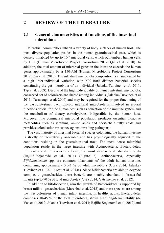
The vast majority of intestinal bacterial species colonizing the human intestine is strictly or facultatively anaerobic and has physiologically adjusted to the conditions residing in the gastrointestinal tract. The most dense microbial population reside in the large intestine with Actinobacteria, Bacteroidetes, Firmicutes and Proteobacteria being the most diverse and abundant phyla (Rajilić-Stojanović et al. 2014) (Figure 2). Actinobacteria, especially Bifidobacterium spp. are common inhabitants of the adult human intestine, comprising approximately 0.5-3 % of adult microbiota (Gura 2014; Jalanka-Tuovinen et al. 2011; Jost et al. 2014a). Since bifidobacteria are able to degrade complex oligosaccharides, these bacteria are notably abundant in breast-fed infants (up to 90 % of total microbiota) (Gura 2014; Yatsunenko et al. 2012).
In addition to bifidobacteria, also the growth of Bacteroidetes is supported by breast milk oligosaccharides (Marcobal et al. 2012) and these species are among the first colonizers of human infant intestine. In healthy adults, Bacteroidetes comprises 10-45 % of the total microbiota, shows high long-term stability (de Vos et al. 2012; Jalanka-Tuovinen et al. 2011; Rajilić-Stojanović et al. 2012) and
Review of the Literature 4
have been suggested to represent the phylogenetic core of the human intestinal tract (Tap et al. 2009).
Figure 2. Typical phylum-level microbiota composition in breast-fed infant, child and adult (data derived from studies II, III and IV).
The most diverse bacterial phylum is Firmicutes which constitutes approximately 50-80 % of the microbiota in the intestine of healthy adults (Rajilić-Stojanović et al. 2014). The most abundant members of this phylum are bacteria belonging to the Clostridium clusters IV and XIVa (Rajilić-Stojanović et al. 2014). These clusters contain bacterial species such as Faecalibacterium prausnitzii and Eubacterium rectale, which are able to produce butyrate from
Review of the Literature 5
sugars and Anaerostipes caccae and Eubacterium hallii, which can also use lactate and acetate to produce this short chain fatty acid that is crucial for the maintenance of intestinal homeostasis and health (Smith et al. 2013). Proteobacteria are common although low abundant members of the healthy human intestine, comprising approximately 1-5 % of the total microbiota in adults (Tap et al. 2009). However, these bacteria are among the first colonizers of the newborn intestine, being the dominant group in early days of life (Wopereis et al. 2014) (Figure 2). Akkermansia muciniphila is the sole representative of the Verrucomicrobia, one of the less abundant phyla in the gastrointestinal tract. It is present at low level in early life, but grows out later to become an important resident of the intestinal mucosa, where it produces propionate, another important signaling molecule in the gut (Derrien et al. 2011; Everard et al. 2013; van Passel et al. 2011).
Although fetus is exposed to microbes and microbial structures already in utero, the major microbial colonization of the gut starts during and after birth, when the infant comes into a contact with mother’s vaginal and intestinal microbiota and with the microbes from the surrounding environment. The early life microbiota development is a time-dependent process, where microbiota diversity and composition fluctuate in response to the nutritional behavior and major life events such as weaning, antibiotic treatments and starting at day-care (Wopereis et al. 2014).
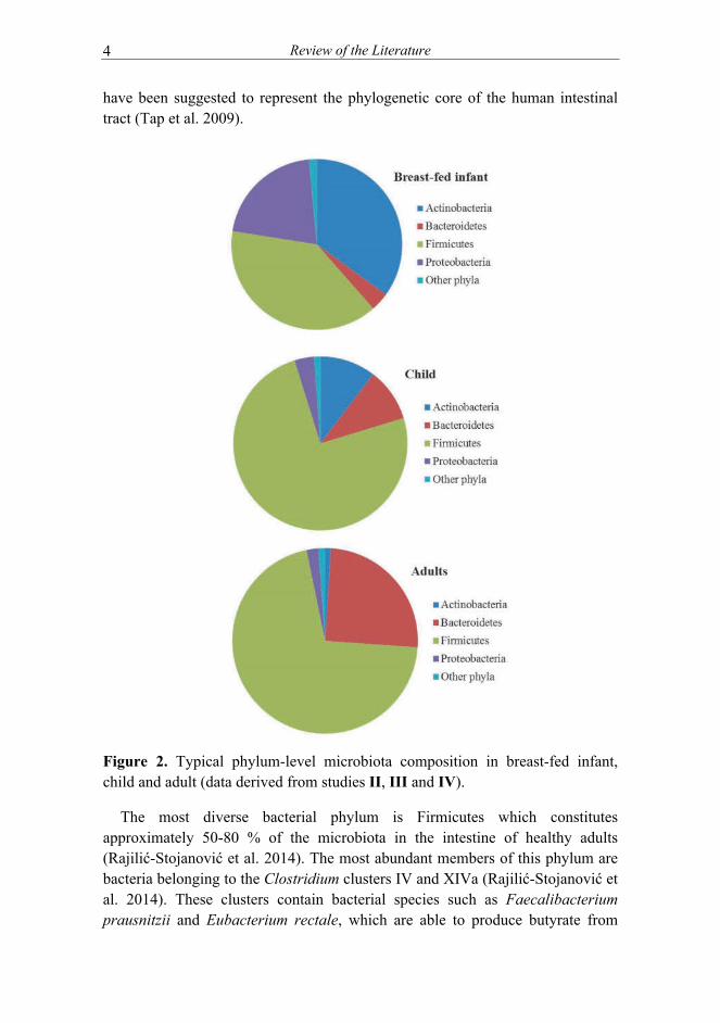
Diversity is a measure of the variety of organisms present in a given community, e.g. in the human intestine. Microbiota diversity consists of two components: species richness (how many species present in a sample) and species evenness (the relative abundance of the species). Higher microbiota diversity is associated with more stable community structure that is resilient towards external disturbances. Indeed, once reached its full diversity and complexity, the intestinal microbiota of healthy individuals remains relatively stable for at least a decade (Rajilić-Stojanović et al. 2012). In elderly, the microbiota may destabilize again with the increasing age and is characterized by decreasing diversity and decline in the complexity of species community (Biagi et al. 2010; Claesson et al. 2011) (Figure 3).

Review of the Literature 6
Figure 3. Schematic presentation of the development of microbiota diversity from birth to old age. See text for an explanation.
2.2 Microbial interactions with the human host
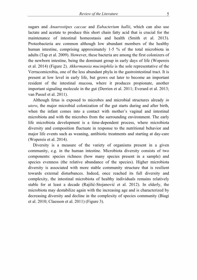
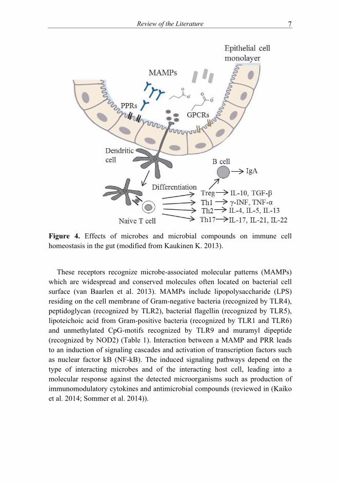
A dynamic and complex relationship exists between the intestinal microbiota, the immune system and the intestinal epithelium. Intestinal epithelial cells (IECs) reside at the interface between the human host and commensal microbiota and are crucial for the maintenance of immune homeostasis (Artis 2008). These cells form a physical barrier, sense microbe-derived signals and regulate the microbiota by secreting cytokines and antimicrobial compounds. The expression of antimicrobial proteins and epithelial permeability and proliferation are regulated by immune cells via the secretion of cytokines (Artis 2008). In turn, the intestinal microbiota affects immune cell homeostasis and has a crucial role in maturation and education of both innate and adaptive immune system (Maynard et al. 2012). The maturation of gut-associated lymphoid tissues starts at birth and is strictly dependent on microbial signaling (Cerf-Bensussan et al. 2010). Microbial colonization is required also for the production of secretory IgA and differentiation of T helper 1 (Th1), Th2 and Th17 cells and the development of regulatory T cells in the gut (Kosiewicz et al. 2014)(Figure 4).
2.2.1 Recognition of microbes by the immune system
Intestinal epithelial cells have a central role in innate immune recognition of commensal bacteria via pattern-recognition receptors (PRR), including transmembrane Toll-like receptors (TLRs) and cytosolic nucleotide-binding oligomerization domain (NOD) –like receptors (NLRs).
Review of the Literature 7
Figure 4. Effects of microbes and microbial compounds on immune cell homeostasis in the gut (modified from Kaukinen K. 2013).
These receptors recognize microbe-associated molecular patterns (MAMPs) which are widespread and conserved molecules often located on bacterial cell surface (van Baarlen et al. 2013). MAMPs include lipopolysaccharide (LPS) residing on the cell membrane of Gram-negative bacteria (recognized by TLR4), peptidoglycan (recognized by TLR2), bacterial flagellin (recognized by TLR5), lipoteichoic acid from Gram-positive bacteria (recognized by TLR1 and TLR6) and unmethylated CpG-motifs recognized by TLR9 and muramyl dipeptide (recognized by NOD2) (Table 1). Interaction between a MAMP and PRR leads to an induction of signaling cascades and activation of transcription factors such as nuclear factor kB (NF-kB). The induced signaling pathways depend on the type of interacting microbes and of the interacting host cell, leading into a molecular response against the detected microorganisms such as production of immunomodulatory cytokines and antimicrobial compounds (reviewed in (Kaiko et al. 2014; Sommer et al. 2014)).
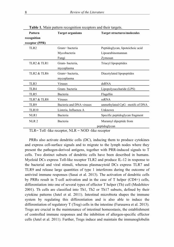
Review of the Literature 8
Table 1. Main pattern recognition receptors and their targets.
Pattern
recognition
receptor (PPR)
Target organisms Target structures/molecules
TLR2 Gram+ bacteria
Mycobacteria
Fungi
Peptidoglycan, lipoteichoic acid
Lipoarabinomannan
Zymosan
TLR2 & TLR1 Gram- bacteria,
mycoplasma
Triacyl lipopeptides
TLR2 & TLR6 Gram+ bacteria,
mycoplasma
Diacetylated lipopeptides
TLR3 Viruses dsRNA
TLR4 Gram- bacteria Lipopolysaccharide (LPS)
TLR5 Bacteria Flagellin
TLR7 & TLR8 Viruses ssRNA
TLR9 Bacteria and DNA viruses unmethylated CpG –motifs of DNA
TLR10 Listeria, Influenza A Unknown
NLR1 Bacteria Specific peptidoglycan fragment
NLR 2 Bacteria Muramyl dipeptide from
peptidoglycan
TLR= Toll -like receptor, NLR = NOD -like receptor
PRRs also activate dendritic cells (DC), inducing them to produce cytokines and express cell-surface signals and to migrate to the lymph nodes where they present the pathogen-derived antigens, together with PRR-induced signals to T cells. Two distinct subsets of dendritic cells have been described in humans. Myeloid DCs express Toll-like receptor TLR2 and produce IL-12 in response to the bacterial and viral stimuli, whereas plasmacytoid DCs express TLR7 and TLR9 and release large quantities of type 1 interferons during the outcome of antiviral immune responses (Sasai et al. 2013). The activation of dendritic cells by PRRs result in T-cell activation and in the case of T helper (CD4+) cells, differentiation into one of several types of effector T helper (Th) cell (Medzhitov 2001). Th cells are classified into Th1, Th2 or Th17 subsets, defined by their cytokine patterns (Jutel et al. 2011). Intestinal microbiota shapes the immune system by regulating this differentiation and is also able to induce the differentiation of regulatory T (Treg) cells in the intestine (Furusawa et al. 2013). Tregs are crucial in the maintenance of intestinal homeostasis, the establishment of controlled immune responses and the inhibition of allergen-specific effector cells (Jutel et al. 2011). Further, Tregs induce and maintain the immunoglobulin
Review of the Literature 9
A (IgA) –producing plasma cells in the intestine. The main role of Treg regulated secretory IgA seems to be the establishment and maintenance of homeostasis with the commensal microbiota (Cong et al. 2009).
2.2.2 Microbial metabolites and immune system
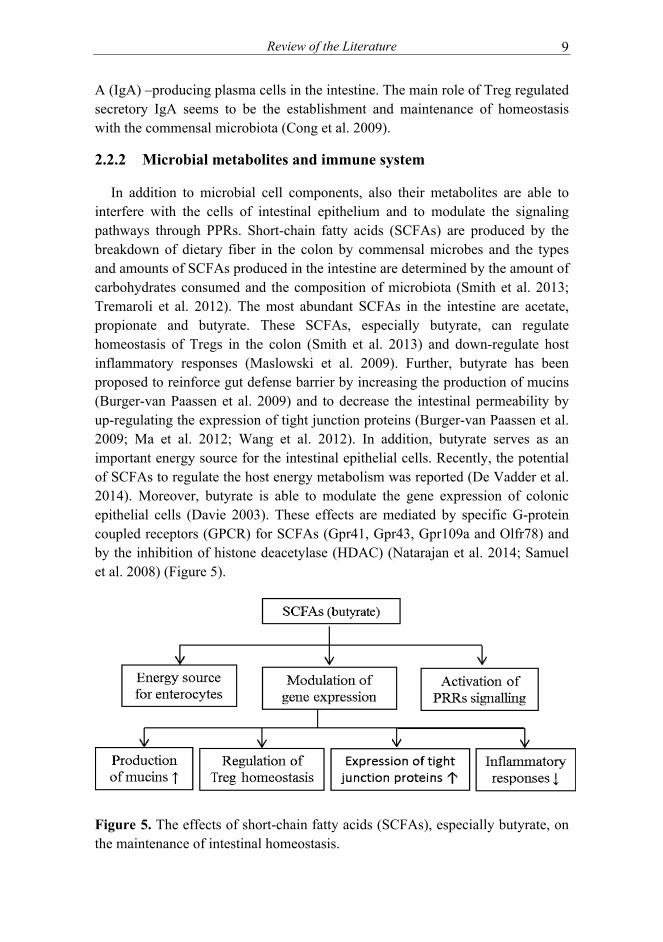
In addition to microbial cell components, also their metabolites are able to interfere with the cells of intestinal epithelium and to modulate the signaling pathways through PPRs. Short-chain fatty acids (SCFAs) are produced by the breakdown of dietary fiber in the colon by commensal microbes and the types and amounts of SCFAs produced in the intestine are determined by the amount of carbohydrates consumed and the composition of microbiota (Smith et al. 2013; Tremaroli et al. 2012). The most abundant SCFAs in the intestine are acetate, propionate and butyrate. These SCFAs, especially butyrate, can regulate homeostasis of Tregs in the colon (Smith et al. 2013) and down-regulate host inflammatory responses (Maslowski et al. 2009). Further, butyrate has been proposed to reinforce gut defense barrier by increasing the production of mucins (Burger-van Paassen et al. 2009) and to decrease the intestinal permeability by up-regulating the expression of tight junction proteins (Burger-van Paassen et al. 2009; Ma et al. 2012; Wang et al. 2012). In addition, butyrate serves as an important energy source for the intestinal epithelial cells. Recently, the potential of SCFAs to regulate the host energy metabolism was reported (De Vadder et al. 2014). Moreover, butyrate is able to modulate the gene expression of colonic epithelial cells (Davie 2003). These effects are mediated by specific G-protein coupled receptors (GPCR) for SCFAs (Gpr41, Gpr43, Gpr109a and Olfr78) and by the inhibition of histone deacetylase (HDAC) (Natarajan et al. 2014; Samuel et al. 2008) (Figure 5).
Figure 5. The effects of short-chain fatty acids (SCFAs), especially butyrate, on the maintenance of intestinal homeostasis.
Review of the Literature 10
2.2.3 Dysregulation of immune system
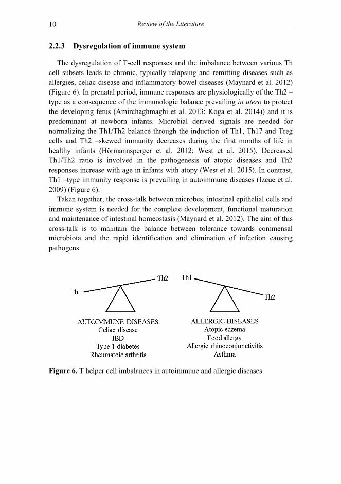
The dysregulation of T-cell responses and the imbalance between various Th cell subsets leads to chronic, typically relapsing and remitting diseases such as allergies, celiac disease and inflammatory bowel diseases (Maynard et al. 2012) (Figure 6). In prenatal period, immune responses are physiologically of the Th2 –type as a consequence of the immunologic balance prevailing in utero to protect the developing fetus (Amirchaghmaghi et al. 2013; Koga et al. 2014)) and it is predominant at newborn infants. Microbial derived signals are needed for normalizing the Th1/Th2 balance through the induction of Th1, Th17 and Treg cells and Th2 –skewed immunity decreases during the first months of life in healthy infants (Hörmannsperger et al. 2012; West et al. 2015). Decreased Th1/Th2 ratio is involved in the pathogenesis of atopic diseases and Th2 responses increase with age in infants with atopy (West et al. 2015). In contrast, Th1 –type immunity response is prevailing in autoimmune diseases (Izcue et al. 2009) (Figure 6).
Taken together, the cross-talk between microbes, intestinal epithelial cells and immune system is needed for the complete development, functional maturation and maintenance of intestinal homeostasis (Maynard et al. 2012). The aim of this cross-talk is to maintain the balance between tolerance towards commensal microbiota and the rapid identification and elimination of infection causing pathogens.
Figure 6. T helper cell imbalances in autoimmune and allergic diseases.
Review of the Literature 11
2.3 Importance of intestinal microbiota in human health and disease
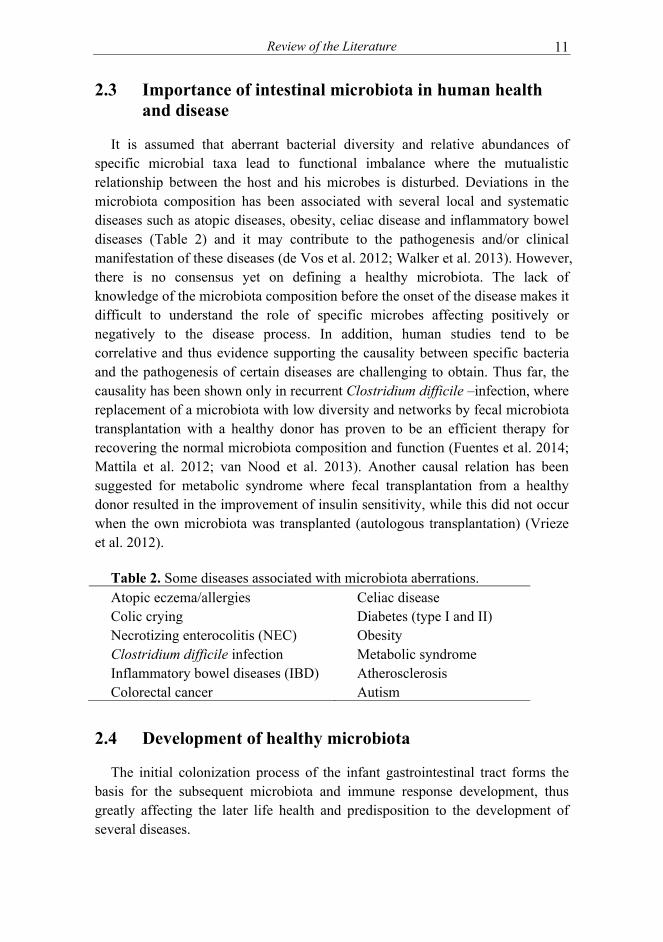
It is assumed that aberrant bacterial diversity and relative abundances of specific microbial taxa lead to functional imbalance where the mutualistic relationship between the host and his microbes is disturbed. Deviations in the microbiota composition has been associated with several local and systematic diseases such as atopic diseases, obesity, celiac disease and inflammatory bowel diseases (Table 2) and it may contribute to the pathogenesis and/or clinical manifestation of these diseases (de Vos et al. 2012; Walker et al. 2013). However, there is no consensus yet on defining a healthy microbiota. The lack of knowledge of the microbiota composition before the onset of the disease makes it difficult to understand the role of specific microbes affecting positively or negatively to the disease process. In addition, human studies tend to be correlative and thus evidence supporting the causality between specific bacteria and the pathogenesis of certain diseases are challenging to obtain. Thus far, the causality has been shown only in recurrent Clostridium difficile –infection, where replacement of a microbiota with low diversity and networks by fecal microbiota transplantation with a healthy donor has proven to be an efficient therapy for recovering the normal microbiota composition and function (Fuentes et al. 2014; Mattila et al. 2012; van Nood et al. 2013). Another causal relation has been suggested for metabolic syndrome where fecal transplantation from a healthy donor resulted in the improvement of insulin sensitivity, while this did not occur when the own microbiota was transplanted (autologous transplantation) (Vrieze et al. 2012).
Table 2. Some diseases associated with microbiota aberrations. Atopic eczema/allergies Celiac disease Colic crying Diabetes (type I and II) Necrotizing enterocolitis (NEC) Obesity Clostridium difficile infection Metabolic syndrome Inflammatory bowel diseases (IBD) Colorectal cancer
Atherosclerosis Autism
2.4 Development of healthy microbiota
The initial colonization process of the infant gastrointestinal tract forms the basis for the subsequent microbiota and immune response development, thus greatly affecting the later life health and predisposition to the development of several diseases.
Review of the Literature 12
2.4.1 Prenatal exposure to microbes and microbial compounds
This chapter has been published in Nylund L et al. Intestinal microbiota during early life –impact on health and disease. Proceedings of the Nutrition Society 2014 Jun 5:1-13.
Traditionally the fetus has been thought to be microbiologically sterile before birth. The presence of microbes in the amniotic fluid and placenta has mainly been associated with preterm deliveries due to maternal intrauterine infections and other pathological conditions (DiGiulio et al. 2008; Pararas et al. 2006; Wang et al. 2013) and the presence of bacterial DNA in amniotic fluid has been associated with lower gestational age and with low mean birth weight (DiGiulio et al. 2008; DiGiulio et al. 2010). However, recent studies utilizing molecular methods have shown that DNA of non-pathogenic bacteria can be detected in placenta and amniotic fluid samples in normal conditions (Rautava et al. 2012; Satokari et al. 2009). Hence, ingestion of amniotic fluid continuously during pregnancy exposes the fetus to bacteria and/or microbe-associated molecular patterns (MAMPs). The exact mechanism(s) of bacterial entry into the intrauterine environment remains elusive. However, ascension from the vagina, by retrograde spread from abdominal cavity, hematogeneously through placenta and contamination during medical procedures (such as amniocentesis) has been suggested as potential routes (Goldenberg et al. 2008). Also MAMPs can induce the immuno-stimulatory effects, for example via the stimulation of Toll-Like Receptors (TLRs), without the need for microbial cells to enter the amniotic cavity. This is supported by the study by Rautava and coworkers where the presence of bacterial DNA, indicative for the presence of other MAMPs too, in placenta and amniotic fluid was associated with the induction of expression profiles of TLRs, especially TLR2 and TLR5 in fetal intestine (Rautava et al. 2012).
Further, the expression of different TLRs, including TLR9, has been shown to change during the maturation of gut epithelial cells (Gribar et al. 2009; Nanthakumar et al. 2011). TLR9 recognizes unmethylated CpG motifs in bacterial DNA and its signaling maintains the gut epithelial homeostasis by improving the barrier functions and by inducing tolerance towards other MAMPs (Kant et al. 2013). In utero the intestinal expression of TLR9 of mouse embryos decreases from day 14 to 18 and then increases again during the postnatal period (Gribar et al. 2009). On the other hand, bacterial lipopolysaccharide (LPS) recognizing TLR4 follows an opposite expression pattern i.e. its expression decreases towards the end of pregnancy (Gribar et al. 2009). Thus, it appears that a full-term newborn is programmed to receive TLR9 stimulation, which will improve the tolerance towards commensal bacteria. Consistently with this,

Review of the Literature 13
necrotising enterocolitis (NEC) in preterm infants has been associated with decreased TLR9 and increased TLR4 expression of the intestinal epithelium (Gribar et al. 2009). Remarkably, TLR9 activation via CpG-DNA supplementation significantly reduced NEC severity (Gribar et al. 2009), suggesting that microbiota rich in CpG -motifs but poor in TLR4 ligands (such as lipopolysaccharide (LPS) produced by Gram-negative bacteria) could be optimal for the prevention or alleviation of NEC. In this respect human breast milk, which supports the growth of bifidobacteria, appears to be a “superfood” for the newborns. Bifidobacteria are organisms with high-G+C% content genomes that are especially rich in TLR9 –stimulating CpG-motifs (Kant et al. 2013). A number of strains of commercially produced lactobacilli have also been found to be rich in CpG-DNA (Kant et al. 2013) and probiotic interventions have shown some promising results in the prevention and alleviation of NEC (Downard et al. 2012). In a mouse model, TLR9 signaling was indeed observed to be an essential mediator of anti-inflammatory effects of probiotics (Rachmilewitz et al. 2004). Furthermore, DNA of Bifidobacterium and Lactobacillus spp., both rich in CpG motifs, have been found in human placenta (Satokari et al. 2009). Thus, it seems that prenatal exposure to MAMPs is an important step in programming the development of gut epithelium and immune system already in utero.
2.4.2 Microbiota development after birth
This chapter has been published in Nylund L et al. Intestinal microbiota during early life –impact on health and disease. Proceedings of the Nutrition Society 2014 Jun 5:1-13.
Meconium is the very first fecal specimen produced by the infant after birth. It consists mainly of amniotic fluid but includes also mucus, intestinal epithelial cells and concentrate of metabolites such as bile acids and pancreatic secretions (Kumagai et al. 2007). Several reports have described meconium microbiota composition providing further evidence for the suggestion that microbiological colonization may begin already in utero (Dominguez-Bello et al. 2010; Jiménez et al. 2008; Moles et al. 2013). However, most of the studies analyzing meconium bacteria have utilized 16S rRNA gene based methods and thus it remains to be assessed whether the presence of bacterial DNA in these samples is an indication of the real low level prenatal colonization or not.
Bacteria belonging to four major bacterial phyla in the intestine, Actinobacteria, Bacteroidetes, Firmicutes and Proteobacteria, are already detectable in the meconium. The predominant cultured bacteria seem to be bacilli within the Firmicutes phylum such as enterococci and staphylococci, or certain Proteobacteria such as Esherichia coli, Klebsiella and Enterobacter spp. (Dominguez-Bello et al. 2010; Favier et al. 2002; Gosalbes et al. 2013; Hu et al.
Review of the Literature 14
2013; Jiménez et al. 2008; Moles et al. 2013). This is in agreement with the reports that these facultative anaerobes are present in feces of healthy newborn infants (Adlerberth et al. 2009; Palmer et al. 2007; Scheepers et al. 2014). In addition, Enterococcus spp. are commonly present, approximately 40 % and 50 % of infants colonized at day 3, respectively (Adlerberth et al. 2009; Favier et al. 2002).
Following the colonization of facultative bacteria, anaerobic bacteria appear in the infant feces within the first weeks of life, decreasing the abundance of facultative anaerobes and thus introducing a shift in microbiota community structure (Avershina et al. 2014). It should be noted, however, that this shift may represent an outgrowth of specific groups of bacteria and does not preclude the fact that their colonization might have already occurred at low level. Especially the abundance of bifidobacteria increases rapidly from approximately 3.5 – 10 % in meconium (de Weerth et al. 2013; Jiménez et al. 2008; Moles et al. 2013) to 50-70 % and even up to 90 % in the feces of breast-fed infants at one month and 3 months of age, respectively (Bezirtzoglou et al. 2011; Fallani et al. 2010; Roger et al. 2010a; Turroni et al. 2012). However, large inter-individual variations are characteristic for infant microbiota and the abundance of bifidobacteria varies from 5 to 100 % in breast-fed infants (Roger et al. 2010a). Considering formula-fed infants, bifidobacteria (determined by fluorescent in situ hybridization (FISH)) may form a minor part of the microbiota, constituting approximately 25 % of the total microbiota (Roger et al. 2010a). In addition to the individual variation, the bifidobacterial abundance seems to vary greatly according to the geographic origin; infants from (northern) European countries harbor in general high numbers of bifidobacteria (Fallani et al. 2011; Roger et al. 2010a; Turroni et al. 2012), while these bacteria are less predominant in Asian and American infants (Fan et al. 2014; Koenig et al. 2011; Palmer et al. 2007). This observation can be mainly explained by demographic differences and by differences in the rate and duration of breast-feeding between the countries and potentially also the differences in use of antibiotics.
After the introduction of solid foods and weaning the relative abundance of bifidobacteria decreases gradually being approximately 60 % at 4 months, 25 % at 6 months and 10 % at 2 years (Avershina et al. 2014; Ringel-Kulka et al. 2013). Simultaneously, the relative abundances of lactobacilli decrease, whereas bacteria predominant in adult microbiota, such as Bacteroidetes and bacteria belonging to the Clostridium clusters XIVa and IV increase (Koenig et al. 2011; Ringel-Kulka et al. 2013; Roger et al. 2010a). However, the early life microbiota composition is characterized by high inter-individual (Avershina et al. 2014; Avershina et al. 2013). The bacterial abundances in healthy infant microbiota vary greatly in a subject-wise manner and fluctuate further in response to the changes in different life events such as antibiotic treatments and introduction of
Review of the Literature
15
solid foods (Koenig et al. 2011; Palmer et al. 2007). During and after weaning major changes occur in microbiota diversity and composition, this transitional phase being more pronounced in breast-fed than in formula-fed infants (Roger et al. 2010a). The succession of Bacteroides spp. and bacteria belonging to the Clostridium clusters XIVa and IV proceeds rapidly while the relative proportion of bifidobacteria decreases (Koenig et al. 2011; Roger et al. 2010a).
Previously, it has been suggested that the microbiota diversity and composition stabilize and reach the level of adult microbiota within the first year or two (Mackie et al. 1999; Palmer et al. 2007; Yatsunenko et al. 2012) . However, recent studies have shown that microbiota maturation will continue longer (Agans et al. 2011; Ringel-Kulka et al. 2013). Interestingly, the establishment of bacteria belonging to Clostridium cluster XIVa at a level similar to adults has been observed already in young children (1-4 years) (Ringel-Kulka et al. 2013), while other bacterial groups still remain at low-level abundance. This indicates that the microbiota development is a gradual process, where some bacterial groups may reach the degree of stabilization earlier than others. However, considering the major physiological changes taking place in the human body within the childhood and adolescence it may be argued that the development of the intestinal microbiota continues throughout this time period and is not finished until the human host reaches adulthood. This is supported by the first studies on adolescent microbiota which reported significant differences between the microbiota composition of adolescent children (11-18 years) and adults (Agans et al. 2011). The most striking difference was the almost two-fold higher abundance of bifidobacteria in adolescent subjects (9 % vs 5.5 % of total microbiota, respectively) (Agans et al. 2011) .
Review of the Literature 16
2.5 Factors affecting the microbiota development
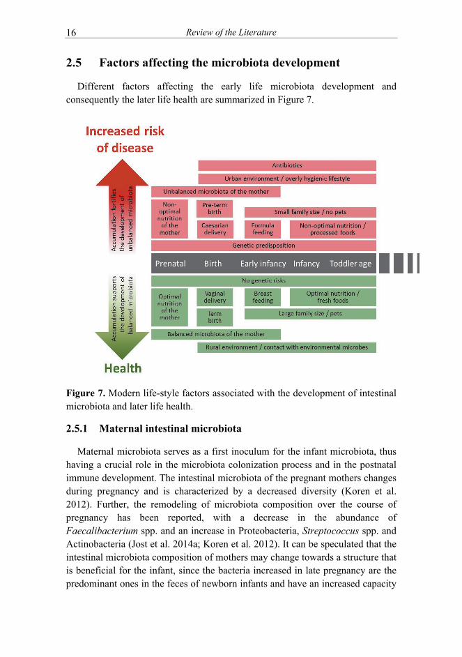
Different factors affecting the early life microbiota development and consequently the later life health are summarized in Figure 7.
Figure 7. Modern life-style factors associated with the development of intestinal microbiota and later life health.
2.5.1 Maternal intestinal microbiota
Maternal microbiota serves as a first inoculum for the infant microbiota, thus having a crucial role in the microbiota colonization process and in the postnatal immune development. The intestinal microbiota of the pregnant mothers changes during pregnancy and is characterized by a decreased diversity (Koren et al. 2012). Further, the remodeling of microbiota composition over the course of pregnancy has been reported, with a decrease in the abundance of Faecalibacterium spp. and an increase in Proteobacteria, Streptococcus spp. and Actinobacteria (Jost et al. 2014a; Koren et al. 2012). It can be speculated that the intestinal microbiota composition of mothers may change towards a structure that is beneficial for the infant, since the bacteria increased in late pregnancy are the predominant ones in the feces of newborn infants and have an increased capacity
Review of the Literature 17
to harvest energy to the host (Koren et al. 2012). Similarities between the microbiotas of child and his/her mother increase with the age of the children, indicating the importance of shared environment and diet on shaping the microbiota composition (Koren et al. 2012). In addition, maternal microbiota shapes the specificities of secretory IgA secreted to breast-milk (Maynard et al. 2012). Transfer of these maternal sIgA to the infant promotes the establishment of regulatory immune system and supports the mutualistic relationship with the commensal microbiota (Maynard et al. 2012).
The vertical transmission of specific bacterial species or strains from mother to infant has been shown in studies utilizing conventional culturing methods (Keski-Nisula et al. 2013; Makino et al. 2013) and with molecular methods (Takahashi et al. 2010), whereas comprehensive studies considering the transmission of whole microbial community are scarce. The strongest evidence exists for the vertical transmission of lactobacilli and bifidobacteria strains from mother to her infant (Gueimonde et al. 2006; Makino et al. 2011; Makino et al. 2013). Moreover, each family appears to harbor their own unique set of these species (Makino et al. 2013). In addition, the mother-derived Bifidobacterium strains that were shared between mother-infant pairs showed higher growth in the presence of galacto-oligosaccharides (GOS) and at a higher redox potential than bifidobacteria originating from other sources (Takahashi et al. 2010). This indicates the ability of these strains to utilize breast- milk GOS as an energy source and the high adaptation to the intestinal conditions of newborn infant. Moreover, vaginally born infants have been shown to share significantly more similar proportion of 16S rRNA gene sequence types with their own mother than with other mothers, whereas no significant sequence overlapping was observed in infants born via Caesarean section (C-section) and their mothers (Jakobsson et al. 2013).
2.5.2 Breast -milk
This chapter has been published in Nylund L et al. Intestinal microbiota during early life –impact on health and disease. Proceedings of the Nutrition Society 2014 Jun 5:1-13.
Human milk oligosaccharides (HMO) have an essential role in the promotion of the development of normal physiology of intestine and immune system in infants. Human milk contains a complex mixture of oligosaccharides, their exact composition varying according to different extrinsic and intrinsic factors. These factors include the genetic background of the mother, maternal health status, diet, secretor status and Lewis blood group type (Albrecht et al. 2011; Bode 2012; Thurl et al. 2010). Oligosaccharide molecules participate in the maintenance of a healthy gut microbiota of the infant in three ways. 1) They block the colonization
Review of the Literature 18
of pathogenic bacteria by acting as receptor analogs and binding to the bacterial surface, thus preventing the pathogens from binding to their target oligosaccharides on the epithelial cell surface (Zivkovic et al. 2011). 2) They act as prebiotic substrates promoting the growth of beneficial bacteria, notably bifidobacteria, concurrently preventing the adherence of potentially harmful bacteria via colonization resistance (Bode 2012). 3) They have also been suggested to stimulate intestinal epithelial cells, lymphocyte cytokine production and leukocyte rolling and adhesion (Bode 2012).
HMOs are the third most abundant component in human milk after lactose and lipids and the abundance of oligosaccharides is more than ten times higher in human than in cow’s milk (5-16 g/l vs 0.03-0.06 g/l, respectively) (Kunz et al. 2000). However, human infants lack the extensive set of enzymes needed for the digestion of glycan residues of human milk oligosaccharides. Thus, these molecules are both non-nutritive and non-digestible, necessitating they have some other biological function which entitles their presence in human breast milk (Zivkovic et al. 2011). Indeed, since HMOs pass undigested to the lower part of the intestinal tract, they can be consumed by the specific members of infant gut microbiota (Marcobal et al. 2012). Since a wide repertoire of enzymes are needed for the degradation and utilization of the intricate structures of both human milk oligosaccharides and plant polysaccharides, such processes most likely involve several different commensal bacteria acting synergistically.
Only two bacterial genera, Bifidobacterium and Bacteroides, have been described to have the capability for milk oligosaccharide utilization (Marcobal et al. 2010; Yu et al. 2013). Bifidobacteria, such as B. longum subsp. infantis and B. bifidum, typically abundant in infant microbiota, harbour a complex set of genes specifically involved in HMO utilization (Sela et al. 2012). The B. longum subsp. infantis genome harbors entire gene clusters controlling the expression of glycosidases, membrane-spanning transporters and other proteins dedicated to human milk oligosaccharide utilization (Sela et al. 2008; Sela et al. 2012) and is the only bacterial species able to digest all HMO structures (Sela et al. 2014; Underwood et al. 2015). In contrast, B. longum subsp. longum, which is more abundant in adult microbiota, is unable to use diverse HMOs, but have the capability to utilize short chain oligosaccharides (Sela et al. 2012) . However, HMOs have reported to up-regulate the expression of several pathways in B. longum subsp. longum such as genes involved in carbohydrate degradation and cell adherence (Gonzalez et al. 2008). Possibly, B. longum subsp. longum relies on cross-feeding with other bacteria, which first degrade complex polysaccharides to shorter units and thereby can also use HMOs as a nutrient source.
Bacteroides spp. genomes harbour a specific gene cluster termed polysaccharide utilization loci (PUL), enabling a wide range of saccharolytic

Review of the Literature 19
ability (Marcobal et al. 2012; Martens et al. 2011). For example B. thetaiotaomicron can degrade more than a dozen different types of glycans (Marcobal et al. 2012), most likely also HMOs. In addition, in vitro utilization of HMOs by B. fragilis and B. vulgatus has been reported (Marcobal et al. 2012). Consistently, B. fragilis and B. vulgatus are the predominant Bacteroides spp. found in breast-fed infants (Tannock et al. 2013). The abundance of bacterial groups, which have restricted capacity to utilize different polysaccharide compounds, are likely to fluctuate more in response to the type of incoming carbohydrates, whereas bacteria with a wide glycan-degrading capability may have a competitive advantage in the gut. Bacteroides spp. are among the first groups colonizing the gut (Palmer et al. 2007; Penders et al. 2006), increasing further after the introduction of solid food and weaning (Koenig et al. 2011; Roger et al. 2010a), and are part of the common core microbiota in adults (Huse et al. 2012; Qin et al. 2010; Rajilić-Stojanović et al. 2009). In addition, the ability of Bacteroides spp. to switch substrate specificity in response to the changing ingestion of nutrients indicates that they are adapted to the symbiotic life with human host and are permanent colonizers of the gut.
Human milk is also a source of bacteria to the infant and together with the bacterial transfer during birth they constitute the essential pioneer colonizers of the neonatal gut. The presence of commensal microbes in breast milk of healthy mothers was first acknowledged only a decade ago (Martin et al. 2003) and since then, more than 200 different species belonging to 50 different genera have been described in human milk (Hunt et al. 2011). The predominant bacteria observed in human milk samples are Bacilli, such as Streptococcus spp. and Staphylococcus spp. (Hunt et al. 2011; Jost et al. 2013). In addition, Bifidobacterium spp. are present and Bacteroidetes and specific clostridia such as butyrate-producing bacteria Faecalibacterium and Roseburia spp. have been detected (Hunt et al. 2011; Jost et al. 2013; Martin et al. 2009) . As the latter bacterial species are only found in the intestine, this strongly points to a fecal origin. The bacterial composition of breast milk may vary depending on the genetic background, maternal dietary habits and demographic differences between the mothers. For example, European mothers commonly harbour Lactobacillus and Bifidobacterium spp. in their breast milk, whereas these bacteria were rarely detected in mothers from the USA. However, it can be speculated that methodological drawbacks in DNA extraction may have impacted these results (Jost et al. 2013; Ward et al. 2013). Further, the mode of delivery has been shown to affect the milk microbiota composition (Cabrera-Rubio et al. 2012; Khodayar-Pardo et al. 2014; Simsek et al. 2014). Milk samples from mothers who delivered their infants vaginally contained more Leuconostocaceae and less Carnobacteriaceae (Cabrera-Rubio et al. 2012) and bifidobacteria
Review of the Literature 20
(Khodayar-Pardo et al. 2014) than milk samples from mothers who had gone through an elective C-section.
Maternal health status seems to have a major effect on the milk microbiota composition. For example, milk microbiota of overweight mothers differs from that of normal weight mothers (Cabrera-Rubio et al. 2012; Collado et al. 2012). The bacterial composition of breast milk seems to be stable at intra-individual level over time, while representing a great inter-individual variation. This suggests that human milk microbiota is highly personalized, in a manner similar to intestinal microbiota (Costello et al. 2009; Human Microbiome Project Consortium 2012; Ursell et al. 2012).
Recently, the existence of a ”core” milk microbiota has been suggested (Hunt et al. 2011). The milk core microbiota consisted of nine OTUs (operational taxonomic unit), corresponding to Staphylococcus, Streptococcus (Firmicutes), Corynebacterium, Propionibacterium (Actinobacteria) and Serratia, Pseudomonas, Ralstonia, Sphingomonas and Bradyrhizobiaceae (Proteobacteria), constituting approximately half of the total bacterial community. It is noteworthy that many of the core microbiota genera are typically found from the skin and it seems likely that some part of the breast milk microbiota originates from the skin. Another origin of bacteria in human milk may be the intestinal tract of the mother. While fecal contamination is a likely avenue, it also has been suggested that intestinal bacteria could transfer within the phagocytosing cells from the gut to human milk via entero-mammary circulation of immune cells (Grönlund et al. 2007). Interestingly, Bifidobacterium breve is one of the most commonly detected bifidobacterial species in human milk samples (Alp et al. 2010; Boesten et al. 2011; Martin et al. 2009; Roger et al. 2010b; Solis et al. 2010; Turroni et al. 2011) and it produces exopolysaccharide (EPS), which masks other surface antigens and presents an ability to remain immunologically “silent” (Fanning et al. 2012). The production of EPS seems to be important for the persistence of B. breve in the gut (Fanning et al. 2012). Speculatively, EPS may also play a role in the survival of this bacterium within immune cells, enabling its transfer via the entero-mammary circulation route. Moreover, B. breve and other bifidobacteria are known to produce specific pili that are assumed to play a role in colonization (O'Connell Motherway et al. 2011). While fecal and skin contamination and selection by the milk environment are ecologically plausible routes, other origins of bacteria in human milk cannot be excluded. Whatever the route, human milk bacteria should be considered as an important source of bacteria for breast-fed infants.

Review of the Literature
21
2.5.3 Other factors affecting the microbiota development
Other factors affecting the early life microbiota development are presented in Table 3.
Table 3. Environmental factors affecting the microbiota development in children.
Environmental
factor
Effect on intestinal microbiota in
children
References
Mode of delivery Vaginal bacteria are prevalent in
vaginally born neonates (lactobacilli and
Prevotella spp.) vs. skin bacteria prevail in
neonates born via CS (Staphylococcus,
Propionibacterium and Corynebacterium
spp.)
Dominguez-Bello et al.
2010;
Goldani et al. 2011
Delayed colonization and decreased
abundance of bifidobacteria and
Bacteroidetes in infants born via CS
Biasucci et al. 2010;
Jakobsson et al. 2013
High diversity in infants born via
emergency CS vs. very low diversity in
infants born via elective CS
Azad et al. 2013
Gestational age Very low microbial diversity, delayed
bacterial colonization, especially that of
anaerobic bacteria in pre-term infants
Rouge et al. 2010;
Barrett et al. 2013;
Greenwood et al. 2014; La
Rosa et al. 2014; Moles et
al. 2013; Normann et al.
2013
Geographical
factors: country, its
climate and
environment, hygienic
conditions, typical
dietary habits and other
cultural issues
Bifidobacterium spp. highly dominant
in infants from (northern) European
countries, whereas less predominant in
Asian and American infants
Fallani et al. 2011;
Roger et al. 2010a;
Turroni et al. 2012; Fan et
al. 2013; Koenig et al.
2011; Palmer et al. 2007
Antibiotic
treatment(s)
Decreased microbiota diversity Dethlefsen et al. 2011;
Fouhy et al. 2012
Higher proportion of Proteobacteria
and reduction of bifidobacteria,
lactobacilli and Bacteroidetes
Fouhy et al. 2012;
Fallani et al. 2010; Hussey
et al. 2011; Mangin et al.
2012; Rea et al. 2011;
Tanaka et al. 2009a
CS =Caesarean section
Review of the Literature 22
2.6 Perturbations in the microbiota development and its implications on health
Exposure to the microbes from natural environment is thought to be crucial for the normal microbiota development and the optimal maturation of immune system. Modern life style factors such as hygienic conditions, reduced contact with animals and extended use of broad spectrum antibiotics lead to a decreased exposure to these microbes. Since sufficient microbial exposure is needed for the development of balanced microbiota community and consequently, for the optimal maturation of immune system, any factors causing deviations in this process may have a long-lasting impact on later life health. For example, early life antibiotic treatment(s) have been associated with the increased risk for health problems later in life such as the risk for celiac disease development (Mårild et al. 2013), allergic diseases (Foliaki et al. 2009) and the increased risk of obesity at school age (Ajslev et al. 2011). Interestingly, maternal antibiotic use during pregnancy have been associated with an increased risk of cow’s milk allergy, asthma, eczema and hay fever in their infants (McKeever et al. 2002; Metsälä et al. 2013), highlighting the importance of balanced maternal microbiota in providing a beneficial inoculum for the infant microbiota development and subsequently health later in life.
2.6.1 Atopic diseases
This chapter has been modified from Nylund L et al. Intestinal microbiota during early life –impact on health and disease. Proceedings of the Nutrition Society 2014 Jun 5:1-13.
Atopic diseases are chronic and relapsing disorders usually starting in early childhood. Atopy has been characterized as a genetic disposition to develop an allergic reaction and produce elevated levels of immunoglobulin E (IgE) upon exposure to an environmental antigen (Bieber 2010). Atopic diseases include eczema (atopic dermatitis), allergic rhinitis (hay fever), allergic conjunctivitis and allergic asthma. In early life, the most common form of atopic disease is eczema, its prevalence being approximately 15-30 % depending on the country studied (Deckers et al. 2012). During the last decades, associations between the composition of intestinal microbiota and atopic diseases have been studied intensively.
Previous studies have mainly addressed the microbiota composition preceding the development of atopic disease, although the age at the onset of atopic symptoms has been insufficiently clarified and the severity of eczema has not been considered. Reduced diversity at early life (i.e. at 1 week, 1 month or 4 months of age) has been associated with an increased risk of developing atopy or
Review of the Literature
23
allergic disease (Abrahamsson et al. 2012; Bisgaard et al. 2011; Forno et al. 2008; Ismail et al. 2012; Kalliomäki et al. 2001a; Wang et al. 2008) (Table 4). However, after 1 year of age the total microbiota diversity in children either developing or having eczema is comparable or even higher than that of healthy children (Abrahamsson et al. 2012). Notably, the majority of these studies have been conducted by using traditional cultivation-based techniques or molecular techniques that only target a sub-set of the intestinal microbiota, thus limiting the detection of true microbiota diversity.
The results on specific bacteria either increasing or decreasing the risk of developing atopic diseases or associated with their onset are still conflicting (Gore et al. 2008; Johansson et al. 2011; Mah et al. 2007; Penders et al. 2006; Sepp et al. 2005; Stsepetova et al. 2007). Aberrancies in bifidobacterial community have been associated with children with atopic diseases, most often characterized by either reduced total abundance or shifts in species community (Gore et al. 2008; Mah et al. 2007; Sepp et al. 2005; Stsepetova et al. 2007). Further, decreased amounts of Bacteroides spp. and increased amounts of specific Firmicutes such as Staphylococcus aureus and different clostridial groups, have been associated with the development and onset of allergic diseases (Abrahamsson et al. 2012; Bisgaard et al. 2011; Björksten et al. 2001; Sjögren et al. 2009a; Storrø et al. 2011; Thompson-Chagoyan et al. 2011). Interestingly, both Bifidobacterium spp. and Bacteroides spp. have been reported to have anti-inflammatory properties via their ability to direct the cellular and physical maturation of the developing immune system (Chiclowski et al. 2012; Hooper et al. 2001; Pagnini et al. 2010). For example, polysaccharide A from B. fragilis is able to direct the development of CD4+ T cells, thus inducing the differentiation of Th1 lineage and correction of the Th1/Th2 imbalance (Mazmanian et al. 2005). Further, this polysaccharide has been shown to promote immunologic tolerance through induction of regulatory T (Treg) cells, resulting in the suppression of IL-17 responses (Round et al. 2011). Moreover, both Bifidobacterium and Bacteroides spp. have high frequency of immunostimulatory CpG motifs in their genomes, thus being rich in TLR9 ligands (Kant et al. 2013). TLR9 stimulation is known to both enhance epithelial integrity and direct immune responses towards Th1 type (reviewed in Kant et al 2013 (Kant et al. 2013)). These effects may be diminished in allergic subjects, who have reduced numbers of Bifidobacterium and Bacteroides spp.
In recent studies, increased levels of IL-17 have been associated with asthma (Alyasin et al. 2013; Ramirez-Velazquez et al. 2013). Furthermore, one of the most important defense mechanisms in the epithelial barrier is IgA, which is present at high concentrations in the intestinal mucus layer (Brandtzaeg 2009). Low levels of IgA predisposes infant to increased binding of antigens to mucosal membrane, to increased mucosal leakiness and an increased uptake of dietary

Review of the Literature
24
antigens (Johansen et al. 1999). Low levels of IgA have also been associated with increased risk for the development of IgE-mediated allergic diseases in children (Kukkonen et al. 2010). Further, it has been suggested that high numbers of Clostridium spp. may be associated with degradation of antigen-specific IgA, which could debilitate the immature gut barrier (Pärtty et al. 2013). The protective role of specific bacteria and their compounds against atopy and allergic diseases is further supported by several clinical studies reporting the effects of probiotic strains on the alleviation of allergic symptoms even when the probiotics failed to modify the microbiota composition or diversity (Elazab et al. 2013; Nermes et al. 2011). These effects can be related to the probiotic effects on the hosts’ immunological functions such as improvement of the barrier function and increasing allergen-specific IgA levels, which are essential for the development of tolerance and can be considered as a marker for immune maturation (Di Mauro et al. 2013; Kukkonen et al. 2010; Mantis et al. 2011; Nermes et al. 2011; Rautava et al. 2006; Rosenfeldt et al. 2004). Further, probiotics have been suggested to have immunomodulatory impacts that affect the Th1/Th2 balance such as stimulation of Th1 type immune responses, induction of apoptosis of Th2 cells and induction of Treg and dendritic cells (Kant et al. 2013; Kwon et al. 2010; Lyons et al. 2010; Marschan et al. 2008; Rautava et al. 2006; Torii et al. 2011; West et al. 2009).

Tab
le 4
. Sum
mar
y of
stu
dies
ana
lyzi
ng th
e as
soci
atio
ns b
etw
een
inte
stin
al m
icro
biot
a di
vers
ity
and
the
risk
of
deve
lopi
ng a
topi
c ec
zem
a
Ref
eren
ce
Stu
dy
pop
ula
tion
Fec
al
sam
ple
s
anal
yzed
Fol
low
-up
Met
hod
use
d
Mai
n f
ind
ings
Abr
aham
sson
et
al. 2
012
Infa
nts
wit
h Ig
E –
asso
ciat
ed e
czem
a (n
=20
) an
d
heal
thy
cont
rols
(n=
20)
1 w
eek,
1
mon
th a
nd 1
2
mon
ths
2 ye
ars
454-
pyro
sequ
enci
ng
Infa
nts
wit
h Ig
E –
asso
ciat
ed e
czem
a ha
d
low
er d
iver
sity
of
tota
l mic
robi
ota
and
Bac
tero
idet
es a
t 1 m
onth
and
low
er d
iver
sity
of P
rote
obac
teri
a at
12
mon
ths
of a
ge.
Bis
gaar
d et
al.
2011
Chi
ldre
n w
ho
deve
lope
d
atop
ic
ecze
ma
(n=
127)
,
alle
rgic
rh
init
is
(n=
28)
or
asth
ma
(n=
27)
and
heal
thy
cont
rols
(n=
71)
1 an
d 12
mon
ths
6 ye
ars
PC
R-D
GG
E
and
conv
enti
onal
cult
urin
g
Red
uced
bac
teri
al d
iver
sity
at
1 an
d 12
mon
ths
of a
ge w
as a
ssoc
iate
d w
ith
incr
ease
d
risk
of
al
lerg
ic
sens
itiz
atio
n (S
PT
+
or
spec
ific
Ig
E)
and
alle
rgic
rh
init
is
but
not
atop
ic e
czem
a
For
no e
t al.
2008
In
fant
s w
ith
ecze
ma
(n=
9)
and
heal
thy
cont
rols
(n=
12)
1 an
d 4
mon
ths
6
mon
ths
PC
R-D
GG
E
Infa
nts
wit
h ec
zem
a ha
d lo
wer
div
ersi
ty a
t
1 an
d 4
mon
ths.
Ism
ail e
t al.
2012
In
fant
s w
ho
deve
lope
d
ecze
ma
(n=
33)
and
heal
thy
cont
rols
(n=
65)
1 w
eek
12
mon
ths
T-R
FL
P
Mic
robi
al d
iver
sity
at 1
wee
k of
age
low
er
in i
nfan
ts w
ho h
ad e
czem
a at
12
mon
ths
of
age.
Kal
liom
äki e
t al.
2001
a
Infa
nts
who
de
velo
ped
atop
ic
ecze
ma
(n=
22)
and
heal
thy
cont
rols
(n=
54).
3 w
eeks
and
3 m
onth
s
2 ye
ars
FIS
H
Chi
ldre
n w
ith
atop
ic e
czem
a ha
d m
ore
clos
trid
ia a
nd te
nden
cy to
hav
e lo
wer
bifi
doba
cter
ia
Wan
g et
al.
2008
In
fant
s w
ho
deve
lope
d
atop
ic
ecze
ma
(n=
15)
and
heal
thy
cont
rols
(n=
20).
1 w
eek
18
mon
ths
T-R
FL
P a
nd
TT
GE
Infa
nts
wit
h at
opic
ecz
ema
had
redu
ced
dive
rsit
y
PC
R-D
GG
E
=
PC
R
com
bine
d w
ith
Den
atur
ing
Gra
dien
t G
el
Ele
ctro
phor
esis
, T
-RF
LP
=
T
erm
inal
R
estr
icti
on
Fra
gmen
t L
engt
h P
olym
orph
ism
, FIS
H =
Flu
ores
cenc
e In
Sit
u H
ybri
diza
tion
, TT
GE
= T
empo
ral T
empe
ratu
re G
radi
ent G
el E
lect
roph
ores
is
Review of the Literature 25
Review of the Literature
26
2.7 Studying the early life microbiota –special challenges
Although the intestinal microbiota of infants is dominated by fewer bacterial groups than that of adults, the inter-individual variability is significantly higher than in adults (Yatsunenko et al. 2012). Microbiota in children under 3 years of age fluctuates substantially and is extremely prone to environmental factors (Koren et al. 2012). Especially during and after weaning process, the income of new substrates promotes the selection of different microbial species (Arrieta et al. 2014; Fallani et al. 2011). In addition, the conditions of the gastrointestinal tract changes along with the introduction of a variety of non-digestible carbohydrates other than those present in breast-milk (Fallani et al. 2011). Furthermore, the behavior of infants encourage the significant exposure to microbes from their environment, since they are constantly introducing hands, feet, toys etc. to their mouth (Arrieta et al. 2014). In addition, they often suffer from infectious diseases. Thus, when the microbial community structure is constantly fluctuating and changing in an individual-specific manner, it is a great challenge to assess the effect of different factors such as antibiotic treatment or intervention procedures on this development process.
2.8 Overview of high-throughput microbiota analysis
Most of the bacterial species colonizing the human intestine are strictly or facultatively anaerobic and have physiologically adjusted to the conditions residing in the gastrointestinal tract. These conditions are difficult to mimic in laboratory conditions. In spite of the fact that over 1000 species have been cultured from the human intestinal tract (Rajilić-Stojanović et al. 2014), an important fraction (estimated to be approximately 70 %) of the intestinal microbes have not been cultured or identified yet (Fraher et al. 2012). Therefore, the true diversity of intestinal microbiota has remained incompletely covered by using traditional cultivation strategies or molecular methods with low resolution, whereas the rapid development of culture-independent, high-throughput molecular applications during the recent years has enabled the deep and comprehensive analyses of total microbiota diversity. These high-throughput techniques are based on either direct sequencing of nucleic acids in the sample or on their detection by using high-density oligonucleotide arrays.
Phylogenetic microarray is a high-throughput platform designed for the simultaneous detection of thousands of DNA or RNA sequences. Further, it allows the concurrent analysis of numerous samples, making microarrays a fast, cheap and user-friendly technology. As other molecular methods for microbiota

Review of the Literature
27
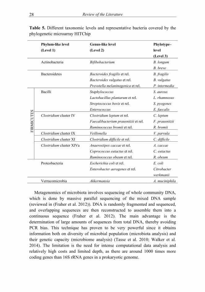
community analysis, this technology is most often based on sequences of variable regions of the 16S rRNA gene, denoting that the sequence knowledge is a prerequisite for the array design. Phylogenetic microarrays have been shown to have higher sensitivity than high-throughput sequencing strategies (Claesson et al. 2009; Tottey et al. 2013) and to detect bacterial DNA as low as 0.00025 % of the sample (Paliy et al. 2009). The major challenge in this technology is the possibility of cross-hybridizations, i.e. that probes hybridize also to highly similar non-target sequences (Fraher et al. 2012). Several phylogenetic microarrays targeting the human intestinal microbiota have been developed (Centanni et al. 2013; Paliy et al. 2009; Rajilić-Stojanović et al. 2009; Tottey et al. 2013), the Human Intestinal Tract Chip (HITChip) being one of the most intensively used. The HITChip enables rapid and sensitive profiling of the microbiota diversity and allow the relative quantification of all bacterial groups at different taxonomic levels simultaneously (Rajilić-Stojanović et al. 2009) (Table 5). The so-called next-generation sequencing methods deliver sequences from 16S rRNA gene amplicons or from total community DNA. Direct sequencing of partial 16S rRNA gene amplicons (‘massively parallel sequencing’) is able to sequence massive amount of sequences at the same time in same reaction. Due to the sequencing of short reads (typically around 100 bp), it is possible to increase the amount of sequences analyzed, thus enabling also the detection of low abundant bacteria (reviewed in (Fraher et al. 2012)). This method gives phylogenetic and quantitative information and enables detection of unknown bacteria. Massively parallel sequencing is fast but requires intense computational data analysis that includes the removal of chimeric sequences (Fraher et al. 2012). One overlooked issue is the generation of errors as is illustrated by the need for developing a low error 16S rRNA amplicon sequencing method to be able to monitor the microbiota of subjects over time (difficulties in identifying subjects based on their fecal microbiota (Faith et al. 2013), while this was done easily with the HITChip (Rajilić-Stojanović et al. 2012).
Review of the Literature
28
Table 5. Different taxonomic levels and representative bacteria covered by the phylogenetic microarray HITChip
Phylum-like level
(Level 1)
Genus-like level
(Level 2)
Phylotype–
level
(Level 3)
Actinobacteria Bifibobacterium
B. longum
B. breve
Bacteroidetes Bacteroides fragilis et rel.
Bacteroides vulgatus et rel.
Prevotella melaninogenica et rel.
B. fragilis
B. vulgatus
P. intermedia
FIR
MIC
UT
ES
Bacilli Staphylococcus
Lactobacillus plantarum et rel.
Streptococcus bovis et rel.
Enterococcus
S. aureus
L. rhamnosus
S. pyogenes
E. faecalis
Clostridium cluster IV Clostridium leptum et rel.
Faecalibacterium prausnitzii et rel.
Ruminococcus bromii et rel.
C. leptum
F. prausnitzii
R. bromii
Clostridium cluster IX Veillonella V. parvula
Clostridium cluster XI Clostridium difficile et rel. C. difficile
Clostridium cluster XIVa Anaerostipes caccae et rel.
Coprococcus eutactus et rel.
Ruminococcus obeum et rel.
A. caccae
C. eutactus
R. obeum
Proteobacteria Escherichia coli et rel.
Enterobacter aerogenes et rel.
E. coli
Citrobacter
werkmanii
Verrucomicrobia Akkermansia A. muciniphila
Metagenomics of microbiota involves sequencing of whole community DNA,
which is done by massive parallel sequencing of the mixed DNA sample (reviewed in (Fraher et al. 2012)). DNA is randomly fragmented and sequenced, and overlapping sequences are then reconstructed to assemble them into a continuous sequence (Fraher et al. 2012). The main advantage is the determination of large amounts of sequences from total DNA, thereby avoiding PCR bias. This technique has proven to be very powerful since it obtains information both on diversity of microbial population (microbiota analysis) and their genetic capacity (microbiome analysis) (Tasse et al. 2010; Walker et al. 2014). The limitation is the need for intense computational data analysis and relatively high costs and limited depth, as there are around 1000 times more coding genes than 16S rRNA genes in a prokaryotic genome.
Review of the Literature
29
While advances in molecular microbiological techniques allow a high-throughput analysis of the microbiota, several problems are related to inappropriate use of these 16S rRNA gene sequence -based technologies (Table 6). Sufficient pretreatment of fecal samples such as sample collection procedures and storage conditions, efficient nucleic acid extraction and unbiased PCR amplification are crucial in order to cover the full microbial diversity (Maukonen et al. 2012). In addition, identification of sequences based on very short reads is not always reliable (Werner et al. 2012). Consequently, quantitative PCR (qPCR) and other molecular techniques targeting specific taxonomic groups are still relevant in verifying the results obtained by using more sophisticated methods described above. Quantitative PCR is one of the most accurate molecular methods currently available for microbiota analysis. It allows also the detection of the low abundant members of the intestinal microbiota, which may be missed by high-throughput techniques (Werner et al. 2012).
Despite the powerful molecular techniques for microbiota characterization, culturing is still utmost valuable as it is a necessary step for the detailed physiological and biochemical identification of individual isolates. Experimental characterization of isolates and their metabolic activities are needed to expand the reference databases used for the annotation of metagenomic sequence data, because currently a significant proportion of obtained data is lacking a close match in the reference databases (Le Chatelier et al. 2013; Walker et al. 2014). The recent development of advanced, high-throughput culturing has proven to be a powerful tool in the discovery of the unknown members of human intestine (Dubourg et al. 2013; Lagier et al. 2012; Pfleiderer et al. 2013)). This ‘microbial culturomics’ employs a large set of growth media and a wide variety of culturing conditions and is combined with 16S rRNA gene sequencing and mass spectrometry for the identification of obtained colonies (Lagier et al. 2012). In future, high-throughput cultivation of new species in combination with metagenomics will give us new insights into microbiota composition and function (Rajilić-Stojanović et al. 2014).
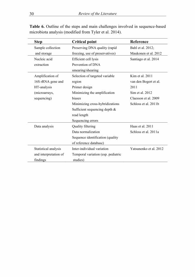
Review of the Literature
30
Table 6. Outline of the steps and main challenges involved in sequence-based microbiota analysis (modified from Tyler et al. 2014).
Step Critical point Reference Sample collection
and storage
Preserving DNA quality (rapid
freezing, use of preservatives)
Bahl et al. 2012;
Maukonen et al. 2012
Nucleic acid
extraction
Efficient cell lysis
Prevention of DNA
smearing/shearing
Santiago et al. 2014
Amplification of
16S rRNA gene and
HT-analysis
(microarrays,
sequencing)
Selection of targeted variable
region
Primer design
Minimizing the amplification
biases
Minimizing cross-hybridizations
Sufficient sequencing depth &
read length
Sequencing errors
Kim et al. 2011
van den Bogert et al.
2011
Sim et al. 2012
Claesson et al. 2009
Schloss et al. 2011b
Data analysis Quality filtering
Data normalization
Sequence identification (quality
of reference database)
Haas et al. 2011
Schloss et al. 2011a
Statistical analysis
and interpretation of
findings
Inter-individual variation
Temporal variation (esp. pediatric
studies)
Yatsunenko et al. 2012

Aims of the Study
31
3 AIMS OF THE STUDY
The main purpose of the present series was to analyze the presence of specific groups of bacteria that relate to the health status in babies. The specific aims were:
1. To compare and optimize DNA extraction to facilitate fecal DNA
extraction in large cohort studies (I).
2. To characterize the long-term effects of maternal microbiota on infant
intestinal microbiota composition and development (II).
3. To characterize the diversity and temporal changes of intestinal
microbiota in early childhood (II, III, IV)
4. To identify intestinal microbiota characterisitcs and specific bacterial
groups associated with eczema and its severity (III, IV).

Materials and Methods
32
4 MATERIALS AND METHODS
4.1 Subjects and study design
4.1.1 Subject characteristics
Baseline characteristics of study subjects (studies II-IV) are summarized in Table 7.
Table 7. Summary of baseline characteristics of subjects from studies II-IV.
Study II Study III Study IV
Children Mothers
Number of
subjects 10 10 19 39
Diagnosed
atopic disease 0
10
(100 %) 15 (44 %) 28 (72 %)
Fecal samples
analyzed
1, 3, 6, 12,
24 and 36
months of
age
3rd
trimester of
pregnancy, 1
and 36 mo
postpartum
6 and 18
months of
age
All infants 6
months of age.
Additional
sample at 9
months of age
from subjects
with more
severe disease
Birth weeks of
gestation 39.3 (1.5) NA 39.2 (1.3) 39.3 (1.5)
Caesarean
section 0 NA 0 5 (13 %)
Male gender 5 (50 %) NA 16 (47 %) 20 (51 %)
Birth weight, g3589
(339) NA
3638
(416) 3530 (439)
Has older
sibling(s) 5 (50 %) NA 12 (35 %) 23 (59 %)
Probiotic
group 0 0 15 (44 %) 19 (49 %)
Values are mean (SD) or n (%). NA = not applicable
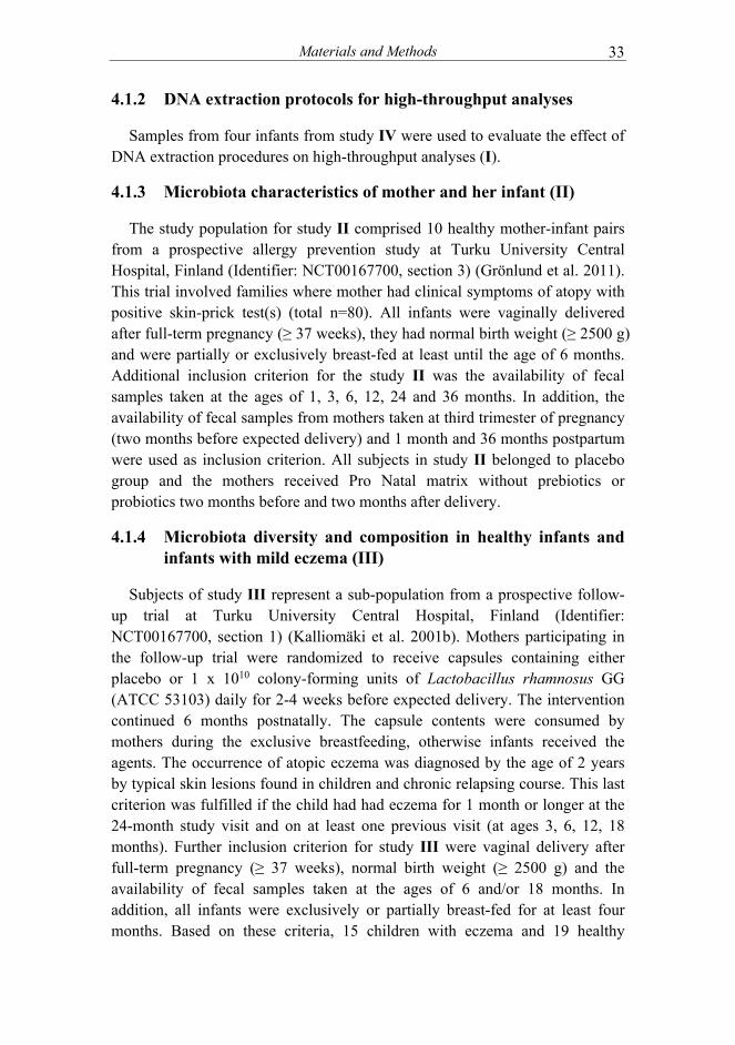
Materials and Methods
33
4.1.2 DNA extraction protocols for high-throughput analyses
Samples from four infants from study IV were used to evaluate the effect of DNA extraction procedures on high-throughput analyses (I).
4.1.3 Microbiota characteristics of mother and her infant (II)
The study population for study II comprised 10 healthy mother-infant pairs from a prospective allergy prevention study at Turku University Central Hospital, Finland (Identifier: NCT00167700, section 3) (Grönlund et al. 2011). This trial involved families where mother had clinical symptoms of atopy with positive skin-prick test(s) (total n=80). All infants were vaginally delivered after full-term pregnancy (≥ 37 weeks), they had normal birth weight (≥ 2500 g) and were partially or exclusively breast-fed at least until the age of 6 months. Additional inclusion criterion for the study II was the availability of fecal samples taken at the ages of 1, 3, 6, 12, 24 and 36 months. In addition, the availability of fecal samples from mothers taken at third trimester of pregnancy (two months before expected delivery) and 1 month and 36 months postpartum were used as inclusion criterion. All subjects in study II belonged to placebo group and the mothers received Pro Natal matrix without prebiotics or probiotics two months before and two months after delivery.
4.1.4 Microbiota diversity and composition in healthy infants and infants with mild eczema (III)
Subjects of study III represent a sub-population from a prospective follow-up trial at Turku University Central Hospital, Finland (Identifier: NCT00167700, section 1) (Kalliomäki et al. 2001b). Mothers participating in the follow-up trial were randomized to receive capsules containing either placebo or 1 x 10¹⁰ colony-forming units of Lactobacillus rhamnosus GG (ATCC 53103) daily for 2-4 weeks before expected delivery. The intervention continued 6 months postnatally. The capsule contents were consumed by mothers during the exclusive breastfeeding, otherwise infants received the agents. The occurrence of atopic eczema was diagnosed by the age of 2 years by typical skin lesions found in children and chronic relapsing course. This last criterion was fulfilled if the child had had eczema for 1 month or longer at the 24-month study visit and on at least one previous visit (at ages 3, 6, 12, 18 months). Further inclusion criterion for study III were vaginal delivery after full-term pregnancy (≥ 37 weeks), normal birth weight (≥ 2500 g) and the availability of fecal samples taken at the ages of 6 and/or 18 months. In addition, all infants were exclusively or partially breast-fed for at least four months. Based on these criteria, 15 children with eczema and 19 healthy

Materials and Methods 34
controls (Figure 8) from the original study population (n= 132) (Kalliomäki et al. 2001b) were included in study III.
Figure 8. Study III design
4.1.5 Microbiota signatures and severity of eczema (IV)
Subjects for this study were selected from two prospective follow-up trials at Turku University Central Hospital, Finland (Identifiers: NCT00167700, section 1 and NCT01148667) (Kalliomäki et al. 2001b; Nermes et al. 2011). Infants with moderate/severe atopic eczema represent a sub-population from a probiotic intervention study where infants were randomized in a double-blind design to receive extensively hydrolyzed casein formula supplemented with or without Lactobacillus rhamnosus GG (ATCC 53103). The duration of intervention was 3 months and fecal samples were collected at the study entry and at the end of the intervention. The extent and severity of atopic eczema was assessed using the SCORAD index (Kunz et al. 1997; Nermes et al. 2011). The severity of atopic eczema was classified in three categories: mild (SCORAD <15), moderate (15<SCORAD >40) and severe eczema (SCORAD >40) (Kunz et al. 1997). Five infants were born by caesarean section and 33 were delivered vaginally. All infants were born full-term and were at least partially breast-fed before the study entry. In addition, they all received solid food(s) at the time of fecal sample collection. Infants with other diseases except atopic eczema were excluded. Additional inclusion criterion for the present study was the availability of two fecal samples taken at baseline and after the intervention. Based on these criteria, 16 children from the original study population (n= 37) (Nermes et al. 2011) were included (Figure 9). Healthy infants (n=11), infants with mild atopic eczema (n=10) and two infants with moderate atopic eczema are a sub-population from another follow-up trial (Kalliomäki et al. 2001b). The selection criteria and other characteristics of these children were as described in study III.
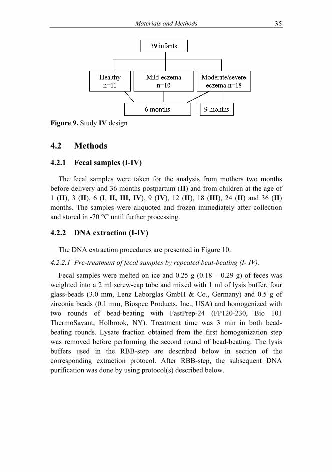
Materials and Methods 35
Figure 9. Study IV design
4.2 Methods
4.2.1 Fecal samples (I-IV)
The fecal samples were taken for the analysis from mothers two months before delivery and 36 months postpartum (II) and from children at the age of 1 (II), 3 (II), 6 (I, II, III, IV), 9 (IV), 12 (II), 18 (III), 24 (II) and 36 (II) months. The samples were aliquoted and frozen immediately after collection and stored in -70 °C until further processing.
4.2.2 DNA extraction (I-IV)
The DNA extraction procedures are presented in Figure 10.
4.2.2.1 Pre-treatment of fecal samples by repeated beat-beating (I- IV).
Fecal samples were melted on ice and 0.25 g (0.18 – 0.29 g) of feces was weighted into a 2 ml screw-cap tube and mixed with 1 ml of lysis buffer, four glass-beads (3.0 mm, Lenz Laborglas GmbH & Co., Germany) and 0.5 g of zirconia beads (0.1 mm, Biospec Products, Inc., USA) and homogenized with two rounds of bead-beating with FastPrep-24 (FP120-230, Bio 101 ThermoSavant, Holbrook, NY). Treatment time was 3 min in both bead-beating rounds. Lysate fraction obtained from the first homogenization step was removed before performing the second round of bead-beating. The lysis buffers used in the RBB-step are described below in section of the corresponding extraction protocol. After RBB-step, the subsequent DNA purification was done by using protocol(s) described below.

Materials and Methods 36
Figure 10. DNA extraction protocols used in the studies.
4.2.2.2 Manual DNA extraction utilizing the Qiagen column (I, III).
The lysis buffer used in the RBB-step was 4% (w/v) sodium dodecyl sulphate (SDS), 50 mM Tris-HCl, 500 mM NaCl and 50 mM EDTA. The RBB-step was followed by DNA precipitation with ammonium acetate and isopropanol. DNA was further purified by RNase A and proteinase K treatments and by using the purification columns and buffers from the QIAamp DNA Mini Kit (Qiagen, Hilden, Germany).
4.2.2.3 Semi-automated DNA extraction by using KingFisher (I, II, IV)
The commercially available InviMag® Stool DNA kit (Invitek GmbH, Berlin, Germany) was used for the extraction of DNA in the KingFisher magnetic particle processor (Thermo Electron, Vantaa, Finland). KingFisher makes use of magnetic beads as a solid carrier of nucleic acids (or other substances) and performs magnetic bead-based protocols in an automated fashion in a 96-well plate format.
The repeated bead beating -step was performed with lysis buffer P of the kit and after the bead-beating the samples were centrifuged at +4 °C for 5 min at full speed and the supernatant was collected. DNA was extracted by following the kit manufacturer’s instructions for bacterial DNA extraction involving proteinase K treatment and subsequent purification by using the KingFisher device. The KingFisher protocol included binding of nucleic acids on magnetic beads, five washing steps and elution.
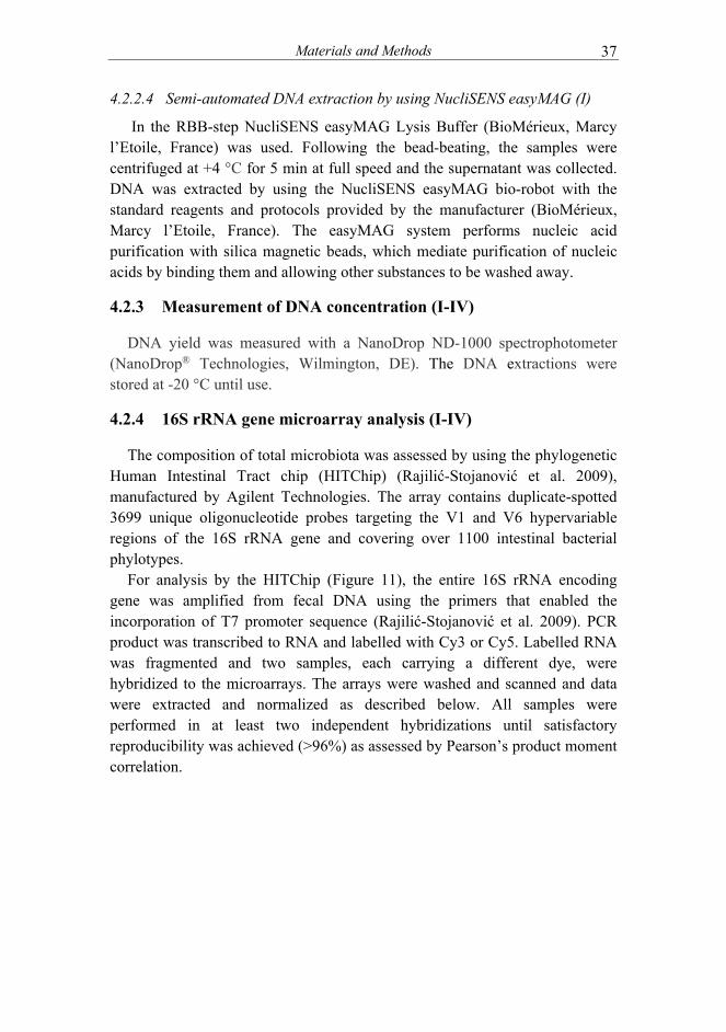
Materials and Methods 37
4.2.2.4 Semi-automated DNA extraction by using NucliSENS easyMAG (I)
In the RBB-step NucliSENS easyMAG Lysis Buffer (BioMérieux, Marcy l’Etoile, France) was used. Following the bead-beating, the samples were centrifuged at +4 °C for 5 min at full speed and the supernatant was collected. DNA was extracted by using the NucliSENS easyMAG bio-robot with the standard reagents and protocols provided by the manufacturer (BioMérieux, Marcy l’Etoile, France). The easyMAG system performs nucleic acid purification with silica magnetic beads, which mediate purification of nucleic acids by binding them and allowing other substances to be washed away.
4.2.3 Measurement of DNA concentration (I-IV)
DNA yield was measured with a NanoDrop ND-1000 spectrophotometer (NanoDrop® Technologies, Wilmington, DE). The DNA extractions were stored at -20 °C until use.
4.2.4 16S rRNA gene microarray analysis (I-IV)
The composition of total microbiota was assessed by using the phylogenetic Human Intestinal Tract chip (HITChip) (Rajilić-Stojanović et al. 2009), manufactured by Agilent Technologies. The array contains duplicate-spotted 3699 unique oligonucleotide probes targeting the V1 and V6 hypervariable regions of the 16S rRNA gene and covering over 1100 intestinal bacterial phylotypes.
For analysis by the HITChip (Figure 11), the entire 16S rRNA encoding gene was amplified from fecal DNA using the primers that enabled the incorporation of T7 promoter sequence (Rajilić-Stojanović et al. 2009). PCR product was transcribed to RNA and labelled with Cy3 or Cy5. Labelled RNA was fragmented and two samples, each carrying a different dye, were hybridized to the microarrays. The arrays were washed and scanned and data were extracted and normalized as described below. All samples were performed in at least two independent hybridizations until satisfactory reproducibility was achieved (>96%) as assessed by Pearson’s product moment correlation.
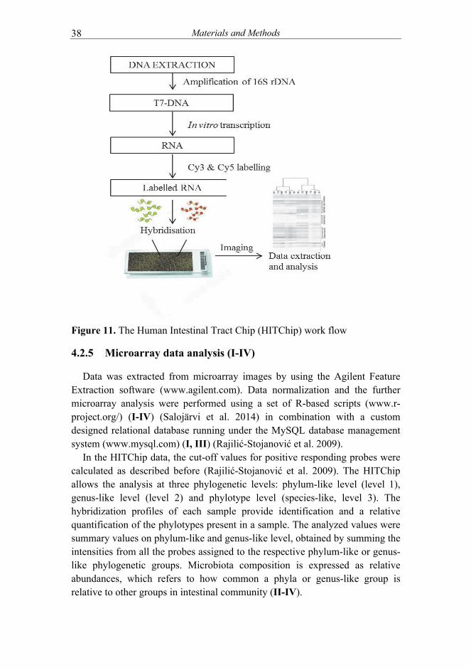
Materials and Methods 38
Figure 11. The Human Intestinal Tract Chip (HITChip) work flow
4.2.5 Microarray data analysis (I-IV)
Data was extracted from microarray images by using the Agilent Feature Extraction software (www.agilent.com). Data normalization and the further microarray analysis were performed using a set of R-based scripts (www.r-project.org/) (I-IV) (Salojärvi et al. 2014) in combination with a custom designed relational database running under the MySQL database management system (www.mysql.com) (I, III) (Rajilić-Stojanović et al. 2009).
In the HITChip data, the cut-off values for positive responding probes were calculated as described before (Rajilić-Stojanović et al. 2009). The HITChip allows the analysis at three phylogenetic levels: phylum-like level (level 1), genus-like level (level 2) and phylotype level (species-like, level 3). The hybridization profiles of each sample provide identification and a relative quantification of the phylotypes present in a sample. The analyzed values were summary values on phylum-like and genus-like level, obtained by summing the intensities from all the probes assigned to the respective phylum-like or genus-like phylogenetic groups. Microbiota composition is expressed as relative abundances, which refers to how common a phyla or genus-like group is relative to other groups in intestinal community (II-IV).
Materials and Methods
39
The diversity of the microbial community assessed by the HITChip (I-IV) was expressed as Simpson’s reciprocal index of diversity (1/D) by using the probe-level data. This index was calculated using the equation D = ∑ Pi^2 where Pi is the proportion of ith taxon. The proportion of each taxon was calculated as a proportion of each probe signal compared to the total signal (Rajilić-Stojanović et al. 2009). Simpson’s reciprocal index of diversity takes into account the number of microbiota members as well as their abundance. A higher index value indicates a more diverse microbial community.
4.2.6 Quantitative PCR (III)
Accuracy of HITChip analysis on phylotype level was shown in study III by using strategic quantitative PCRs (qPCR) to analyze the composition of bifidobacterial community in fecal samples by using genus and species-specific primers. All qPCR reactions were carried out in an Applied Biosystems 7300 Fast Real-Time PCR System in a 96-well format and by using SYBR Green chemistry (SYBR Green PCR Master Mix, Applied Biosystems, USA). The PCR reactions and thermocycling conditions were as reported earlier (Gueimonde et al. 2004; Rinne et al. 2005). The fluorescence products were detected in the last step of each cycle and a melting curve analysis was made after amplification. The bacterial concentration in each sample was calculated by comparing the Ct values to those obtained from standard curves. A standard curve was made from serial dilutions of DNA isolated from each pure culture of the different reference strains. Samples were analyzed in duplicate in at least two independent runs. The primers, their specificities and reference strains have been published previously (Nylund et al. 2013).
4.2.7 Statistical analyses.
Statistical analyses of both HITChip and qPCR data were carried out with log-transformed data. In qPCR data, non-detected values were imputed with the half of the theoretical detection limit (II). In microarray analysis, genus-like level data was used in correlation analysis (IV) and in the RDA (II, III, IV) and PCA (II) clustering. In principal component analysis (PCA), any variable that best explains the variance in the data set is searched, followed by the transformation of data in a way that the greatest variance comes to lie on the first axis. Relative abundances are presented as medians with interquartile ranges (II, III) or as mean with standard deviation values (I-IV). The overall statistical differences between the groups were determined by using Student’s t-test or One-way ANOVA, and subsequent contrast tests (I, III, IV). Microbiota development was assessed using ANOVA for repeated measurements (II) Hierarchical cluster analysis, computed with Pearson
Materials and Methods 40
correlation distance metric was used to compare the microbiota profiles obtained with the HITChip (I). Correlations between diversity indexes, bacterial groups and SCORAD values were calculated using Pearson’s coefficient (r) (IV).
Redundancy analysis (RDA) was performed by using the multivariate statistical analysis software Canoco version 4.5 (III) or 5 (II, IV) for Windows (Microcomputer Power, Ithaca, USA). RDA plot shows bacterial groups principally contributing to the difference between the groups of subjects. The Monte Carlo Permutation Procedure from Canoco program was used to assess the statistical significance of the variation in large datasets. Statistical analyses were performed by using IBM SPSS Statistics versions 18 (I) and 21 (II, IV) and by using R (www.r-project.org/) (III) and a P value < 0.05 was considered statistically significant.
4.2.8 Ethical aspects
The study protocols of original interventions were approved by the Ethics Committee of the Hospital District of South-Western Finland and subjects were enrolled in the study after written informed consent was obtained from the parents. Clinical trials are registered at ClinicalTrials.gov (Identification codes: NCT00167700 (II, III, IV) and NCT01148667 (IV)).

Results 41
5 RESULTS
5.1 Improvement of DNA extraction protocols for high-throughput analyses (I)
5.1.1 The yield and purity of nucleic acids obtained with different extraction methods.
The yield of total nucleic acids (both DNA and RNA) obtained with the manual method utilizing the Qiagen kit was ten-fold lower than that derived from the semi-automated methods. The KingFisher with InviMag® Stool DNA kit (KF) -method provided nucleic acids with best purity as assessed by the ratio of optical density (OD) at 260 nm to OD at 280 nm. The NucliSENS easyMAG (NeM) -method yielded in less pure nucleic acids as compared to the other two methods most likely because it does not involve a proteinase step. All three extraction methods generated DNA that was sufficiently pure for subsequent molecular analyses.
5.1.2 Analysis of microbiota diversity and composition by phylogenetic microarray profiling.
All three extraction methods gave comparable microbiota profiles as assessed by the microbiota composition analysis based on the HITChip hybridization signals. The average hybridization signals at the phylum (level 1) and genus-like levels (level 2) were comparable with all methods used. In addition, the diversity indices of analyzed samples varied from 54 to 88 and the indices were comparable between the profiles obtained by different extraction methods.
5.2 Postpartum changes in microbiota composition of mothers (II)
Maternal microbiota composition showed only minor changes when samples taken during the third trimester of pregnancy (T3) were compared to those obtained 1 and 36 months postpartum (p=0.38). At the phylum-level comparison, a significantly decreased Bacteroidetes abundance was observed at late pregnancy when compared to 36 months postpartum sample (10.93 % ± 11.26 % vs 23.12 % ± 15.68 %, respectively, p=0.048). In addition, there was a tendency towards a decreased microbiota diversity in late pregnancy when compared to 36 months postpartum samples (132.2 ± 53.7 and 168.55 ± 32.5, respectively, p=0.07).
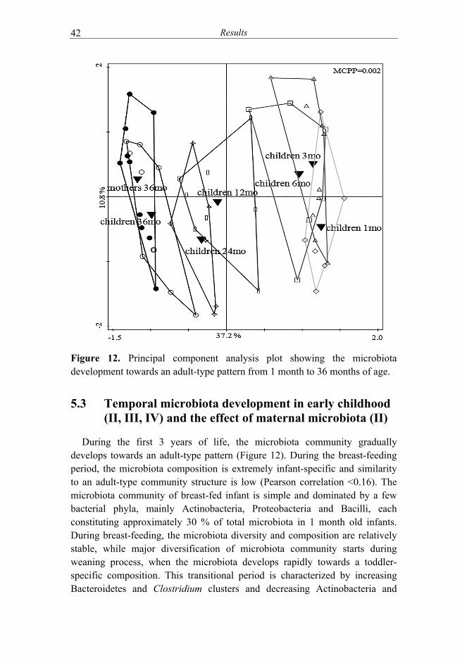
Results 42
Figure 12. Principal component analysis plot showing the microbiota development towards an adult-type pattern from 1 month to 36 months of age.
5.3 Temporal microbiota development in early childhood (II, III, IV) and the effect of maternal microbiota (II)
During the first 3 years of life, the microbiota community gradually develops towards an adult-type pattern (Figure 12). During the breast-feeding period, the microbiota composition is extremely infant-specific and similarity to an adult-type community structure is low (Pearson correlation <0.16). The microbiota community of breast-fed infant is simple and dominated by a few bacterial phyla, mainly Actinobacteria, Proteobacteria and Bacilli, each constituting approximately 30 % of total microbiota in 1 month old infants. During breast-feeding, the microbiota diversity and composition are relatively stable, while major diversification of microbiota community starts during weaning process, when the microbiota develops rapidly towards a toddler-specific composition. This transitional period is characterized by increasing Bacteroidetes and Clostridium clusters and decreasing Actinobacteria and
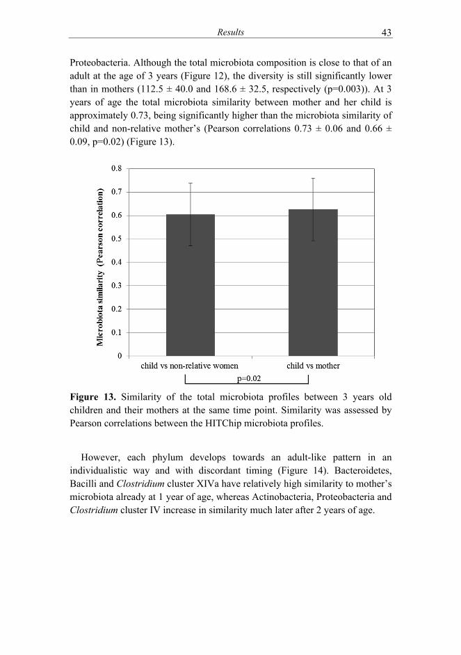
Results 43
Proteobacteria. Although the total microbiota composition is close to that of an adult at the age of 3 years (Figure 12), the diversity is still significantly lower than in mothers (112.5 ± 40.0 and 168.6 ± 32.5, respectively (p=0.003)). At 3 years of age the total microbiota similarity between mother and her child is approximately 0.73, being significantly higher than the microbiota similarity of child and non-relative mother’s (Pearson correlations 0.73 ± 0.06 and 0.66 ± 0.09, p=0.02) (Figure 13).
Figure 13. Similarity of the total microbiota profiles between 3 years old children and their mothers at the same time point. Similarity was assessed by Pearson correlations between the HITChip microbiota profiles.
However, each phylum develops towards an adult-like pattern in an individualistic way and with discordant timing (Figure 14). Bacteroidetes, Bacilli and Clostridium cluster XIVa have relatively high similarity to mother’s microbiota already at 1 year of age, whereas Actinobacteria, Proteobacteria and Clostridium cluster IV increase in similarity much later after 2 years of age.

Results 44
Figure 14. The similarities of infants´ phylum-level microbiota to mothers´ third trimester (A) and 3 years postpartum (B) samples.
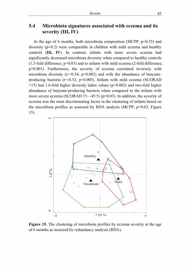
Results
45
5.4 Microbiota signatures associated with eczema and its severity (III, IV)
At the age of 6 months, both microbiota composition (MCPP, p=0.35) and diversity (p=0.2) were comparable in children with mild eczema and healthy controls (III, IV). In contrast, infants with more severe eczema had significantly decreased microbiota diversity when compared to healthy controls (1.5-fold difference, p=0.03) and to infants with mild eczema (2-fold difference, p=0.001). Furthermore, the severity of eczema correlated inversely with microbiota diversity (r=-0.54, p=0.002) and with the abundance of butyrate-producing bacteria (r=-0.52, p=0.005). Infants with mild eczema (SCORAD <15) had 1.6-fold higher diversity index values (p=0.002) and two-fold higher abundance of butyrate-producing bacteria when compared to the infants with more severe eczema (SCORAD 15—45.5) (p=0.03). In addition, the severity of eczema was the most discriminating factor in the clustering of infants based on the microbiota profiles as assessed by RDA analysis (MCPP, p=0.03, Figure 15).
Figure 15. The clustering of microbiota profiles by eczema severity at the age of 6 months as assessed by redundancy analysis (RDA).
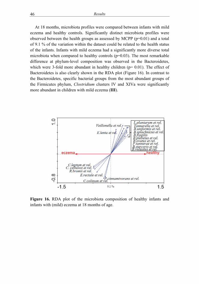
Results 46
At 18 months, microbiota profiles were compared between infants with mild eczema and healthy controls. Significantly distinct microbiota profiles were observed between the health groups as assessed by MCPP (p=0.01) and a total of 9.1 % of the variation within the dataset could be related to the health status of the infants. Infants with mild eczema had a significantly more diverse total microbiota when compared to healthy controls (p=0.03). The most remarkable difference at phylum-level composition was observed in the Bacteroidetes, which were 3-fold more abundant in healthy children (p= 0.01). The effect of Bacteroidetes is also clearly shown in the RDA plot (Figure 16). In contrast to the Bacteroidetes, specific bacterial groups from the most abundant groups of the Firmicutes phylum, Clostridium clusters IV and XIVa were significantly more abundant in children with mild eczema (III).
Figure 16. RDA plot of the microbiota composition of healthy infants and infants with (mild) eczema at 18 months of age.

Results 47
5.5 Changes in microbiota signatures related to the improvement of eczema symptoms (IV)
The alleviation of eczema symptoms and microbiota changes within the more severe eczema group (SCORAD >15, n=15) was analyzed. These infants participated in the probiotic intervention study where they were randomized in a double-blind design to receive extensively hydrolyzed casein formula supplemented with or without Lactobacillus rhamnosus GG (ATCC 53103). Microbiota composition showed significant development within the study period as assessed by RDA analysis (MCPP, P= 0.002), but there were no differences in the microbiota composition between the intervention groups (RDA and MCPP, p=0.54). During the intervention, SCORAD values decreased (p<0.001) and microbiota diversity increased (P<0.001) in all infants. The decrease in SCORAD values correlated with the increase in bacteria related to Coprococcus eutactus (r= -0.59, p=0.02), the predominating butyrate-producing bacteria (Figure 17).
Figure 17. The decrease in SCORAD values correlates with the increase in bacteria related to the butyrate-producer Coprococcus eutactus.
Results 48
5.6 Influence of probiotic supplementation on microbiota composition (III, IV)
When comparing the levels of HITChip signals between children from the placebo group and those who or whose mothers had received L. rhamnosus GG supplementation, no statistically significant differences in microbiota diversity or composition were observed at the age of 6 months (III, IV). However, the supplementation with L. rhamnosus GG showed effects on three genus-like (level 2) bacterial groups at the age of 18 months i.e. a year after the cessation of the probiotic supplementation. The children that had received L. rhamnosus GG had higher levels of the butyrate-producing groups related to Anaerostipes caccae(LGG 2.89 ± 2.13 % and placebo 1.18 ± 0.91 % of the total microbiota, p=0.03) and Eubacterium ventriosum(LGG 0.17 ± 0.11 % and placebo 0.11 ± 0.07 of the total microbiota, p=0.04) than those of placebo group. Moreover, the placebo group children had higher levels of bacteria related to Clostridium difficile at 18 months of age as compared to the probiotic group children (1.19 ± 0.85 % and 0.78 ± 0.60 %, respectively, p=0.047) (III).
Discussion 49
6 DISCUSSION
6.1 Importance of DNA extraction in molecular based microbiota analysis
While advances in molecular microbiological methods allow a high-throughput analysis of the microbiota, the laborious manual nucleic acid extraction from fecal and other clinical samples remains a bottleneck in clinical trials with large number of samples. In addition, when selecting the protocol(s) to be used it should be noted that even small differences in experimental techniques at different steps of molecular based microbiota analysis may affect the structure of obtained community and lead to contradictory results from seemingly similar studies (Tyler et al. 2014).
A combination of mechanical and chemical disruption is typically used to lyse the microbial cells prior to DNA purification and analysis by using molecular techniques. The relative efficiency of disrupting different organisms is highly dependent on the cell wall structure of the microbial cell and the selection of a specific technique (Maukonen et al. 2012; Rosewarne et al. 2011; Salonen et al. 2010; Tyler et al. 2014; Zoetendal et al. 2001). Together with previous observations, our results show that the most critical step in successful microbial DNA extraction from fecal samples is the mechanical cell lysis by bead-beating, which enables the detection of highest bacterial numbers and full coverage of microbial diversity (Study I, de Boer et al. 2010; Maukonen et al. 2012; Salonen et al. 2010). However, a too rigorous lysis protocol will shear the DNA of easily disrupted Gram-negative bacterial cells while being too mild for the hard-to-lyse Gram-positive bacteria. In repeated bead-beating, the physical damage of DNA is prevented by employing two successive rounds of bead-beating. After the first round of mechanical lysis the lysate is drawn off before performing the second beating step in order to lyse the remaining intact cells (Salonen et al. 2010).
The protocol used to purify the obtained nucleic acids after mechanical cell lysis has only a minor effect on the yield and microbial community composition as assessed by high-throughput phylogenetic microarray (I). We showed that the automated purification methods are efficient for microbial DNA extraction from fecal samples after the mechanical disruption of cells. In addition, the size of the extracted DNA was shown to be compatible with high -throughput analysis systems such as next-generation sequencing and phylogenetic microarrays. Importantly, the semi-automated extraction methods evaluated in study I could be performed in half of time required for the reference manual protocol and the hands-free time allows the next set of
Discussion
50
samples to be pre-treated by bead-beating for DNA extraction, thereby significantly speeding up the DNA extraction from large numbers of samples.
6.2 Maternal microbiota during and after pregnancy
By analysing the fecal samples taken at 3rd trimester of pregnancy and early lactation (1 month postpartum), we detected only minor differences in the maternal microbiota composition , which is in line with the previous results (Jost et al. 2014a; Koren et al. 2012). The intestinal microbiota of the mothers-to-be are reported to shift over the course of pregnancy, being characterized by a decreased diversity and compositional changes occurring in late pregnancy (Collado et al. 2008; Koren et al. 2012). These changes in intestinal microbiota community coincide with the appearance of bacteria in milk ducts and both of these processes seem to be driven by hormonal signaling (Fernandez et al. 2013). However, we observed the increase in relative abundance of Bacteroidetes and slightly increased diversity at 3 years after delivery (II). These results show, for the first time long-term perinatal microbiota changes, which may reflect the microbiota recovery from the physiological demands of the pregnancy and lactation.
6.3 Microbiota development and the effect of maternal microbiota
During and after the birth process, microbes from the mother and surrounding environment colonize the gastrointestinal tract of the infant. By 1 month, the rapid initial acquisition of microbes after birth is thought to be over and to have developed into a breast milk adapted composition that will remain relatively stable during the exclusive breastfeeding (Hesla et al. 2014; Jost et al. 2014b; Roger et al. 2010a). All the infants analyzed in this series of studies were breast-fed at least until the age of 4 months and indeed, no major changes were detected in the microbiota community structure until the introduction of solid foods (II). Our results showed that the microbiota compositions of breast-fed infants are characterized by low diversity and the dominance of a few bacterial groups. This infant-specificity was highlighted by the extremely low similarity to maternal microbiota (Pearson correlation <0.16) during the first 6 months of life (II).
In a longitudinal analysis of 10 mother-child pairs we showed that at 3 years of age, the child´s microbiota is more similar to his/her own mother’s microbiota than to the non-relative women’s microbiota (II). This may indicate that the mother continues to share her microbiota with the child even after birth.
Discussion
51
It has been suggested that during the breast-feeding, human milk acts as an intermediary between the maternal intestinal microbiota and the infant (Newburg et al. 2014). In addition, microbial components and metabolites such as short chain fatty acids are transported across the placenta and secreted into breast milk, providing an additional point of interaction between maternal microbiota and infant immune system (Thorburn et al. 2014). Thus, the breast-milk may represent a post-natally extended bond between mother and her infant, providing one mechanism by which microbial communities tend to cluster in family members and are more similar in twins (Maynard et al. 2012; Tims et al. 2013).
Although the mother serves as the main source of bacteria in early life due to the constant and intimate contact with her infant during delivery and lactation, the lack of long-term maintenance of maternal microbes in the infant gut has been suggested (Vaishampayan et al. 2010). Recently, the direct impact of family members in general on the microbial colonization process have been highlighted (Song et al. 2013; Yatsunenko et al. 2012). In study II, this became evident after the cessation of exclusive breast-feeding, when an infant comes into contact with novel sources of microbes and to nutrients other than those present in breast milk (II). Introduction of solid foods leads to a rapid compositional development, when microbiota is prone to the influence of external factors such as microbial diversity in surrounding environment (Hanski et al. 2012), dietary preferences and other lifestyle factors (Dominguez-Bello et al. 2010; Song et al. 2013). Interestingly, in a recent study, the microbiota composition of teenagers was found to be as similar to their father’s as to their mother’s microbiota, although the number of studied subjects was relatively low (Yatsunenko et al. 2012). In addition, the skin (Song et al. 2013) and fecal (Yatsunenko et al. 2012) microbiota of cohabiting couples were more similar to each other’s than to adults from different families (Song et al. 2013), further highlighting the influence of shared household on microbiota composition. This is supported by our finding that microbiota of 3 years old children was more similar to own mother’s microbiota than to microbiotas of nonrelative women (II).
Even though the major diversification of microbiota takes place during the weaning period, its development continues even after the transition to consume full adult-type diet. Despite the highly significant increase of microbiota diversity with age, the HITChip diversity indexes at 3 years of age were observed to be relatively low (~110) when compared to the indexes (150-200) detected in healthy adults (II). Previously, it has been suggested that by the age of 2 to 3 years the microbiota resembles that of an adult (Koenig et al. 2011; Louis et al. 2009; Palmer et al. 2007). However, our results obtained by a deep and longitudinal analysis indicate that the microbiota maturation continues at

Discussion
52
least until the age of 3 years and most likely even further, because the bacterial diversity has still not reached the diversity of an adult person. This is supported by recent cross-sectional studies where significantly lower diversity indexes at 4 years old children (Ringel-Kulka et al. 2013) and differences in the microbiota composition of adolescent children aged 11-18 years (Agans et al. 2011) were reported when compared to healthy adults. Considering the major physiological changes occurring within childhood and adolescence it may be speculated that the microbiota development is not finished until the human host reaches adulthood.
The microbiota development is a gradual process, where some phyla reach the level of maturation earlier than the others (II). Notably, Bacteroidetes was found to be more similar to mother’s microbiota than the microbiota in general in all time points analyzed in study II. Together with previous studies, our finding suggests that anaerobic species may become dominant early in the development process (Jost et al. 2014b; Ringel-Kulka 2013.)
To conclude, although the maternal inoculum forms the basis of microbiota composition, other factors involved in the microbiota development process such as environment, diet, medications and host related factors create a selection pressure and thus greatly affect the formation of individual –specific adult microbiota (Kerr et al. 2014).
6.4 Microbiota signatures in infants with eczema and correlation with the severity of symptoms
In newborn infant, the immune system and mucosal epithelium of the intestine are anatomically and functionally immature and the barrier function is still developing (Rouwet et al. 2002). At birth, intestinal permeability is high while declining rapidly after birth in the process called gut closure (Drozdowski et al. 2010). This maturation process is tightly regulated by growth factors, hormones, breast milk, mucus layer composition and commensal microbiota to promote infant growth and to avoid severe diseases (Drozdowski et al. 2010). Increased permeability can have beneficial effects in early life such as uptake of nutrients and development of systemic tolerance (van Elburg et al. 2003). However, in subjects developing atopic eczema this increased permeability extends beyond neonatal period and the persistent loss of epithelial integrity is suggested to be the main mechanism leading to atopic eczema (De Benedetto et al. 2011; Faura Tellez et al. 2014; Hon et al. 2013).
In study IV, milder symptoms of eczema were found to correlate with the higher abundance of butyrate –producing bacteria which coincided with the higher microbiota diversity. Previous studies have mainly reported associations
Discussion 53
between reduced diversity in early life and the development of atopic eczema. However, in these studies the age at the onset of atopic symptoms has not been clarified and the severity of eczema has not been considered. If subjects with different severity of disease are analysed as a one group, it may result in biased conclusions on the associations between microbiota signatures and atopic eczema. Moreover, the majority of these studies have been conducted by molecular techniques with incomprehensive throughput capacity, thus limiting the detection of true microbiota diversity.
The total microbiota of children with mild eczema was found to become more diverse than the microbiota of healthy controls by 18 months of age (III). High diversity of the intestinal microbiota in childhood is considered to be beneficial, since decreased microbiota diversity has been associated with increased risk of allergic diseases. In addition, the pathogenesis of atopic diseases is associated with an impaired gut barrier function and increased intestinal permeability and gastrointestinal symptoms are common among the patients (Rosenfeldt et al. 2004). It seems that a certain level of diversity of microbiota in the early infancy is essential for modulation of the expression of genes involved in the normal pattern of intestinal development such as postnatal intestinal maturation and maintenance of mucosal barrier (Hooper et al. 2010; Sjögren et al. 2009b). This repeated exposure to a variety of bacterial antigens potentially enhances the development of immune regulation through inhibition of responses to inappropriate targets, such as gut contents and allergens (Munyaka et al. 2014). However, it has been shown that only a few bacterial species may contribute to the differences in microbiota diversity (Le Chatelier et al. 2013). Interestingly, butyrate-producing bacteria Faecalibacterium prausnitzii (Clostridium cluster IV) and Coprococcus eutactus (Clostridium cluster XIVa) were shown to significantly distinguish between individuals with high and low bacterial richness (Le Chatelier et al. 2013).
Butyrate is the main energy source for colonocytes and it also has numerous other functions which are crucial for the host physiology (Schilderink et al. 2013). One of the primary functions of butyrate is its ability to enhance the intestinal barrier function by promoting mucin production, by up-regulating the expression of tight-junction proteins and by inhibiting pro-inflammatory signaling pathways such as nuclear factor kappa B (NF-κB) activation (Lakhdari et al. 2011). SCFAs may also support the intestinal integrity by promoting the IgA secretion by B cells (Ishikawa and Nanjo 2009) and they contribute to the gut motility and wound healing (Tremaroli et al. 2012). Moreover, butyrate promotes the proliferation and activation of regulatory T cell (Treg) in the colon (Furusawa et al. 2013) and was shown to increase the capacity of Tregs to suppress the proliferation of effector CD4+ T cells in a

Discussion
54
mouse model (Smith et al. 2013). This may be an important function through which the butyrate is able to alleviate the eczema symptoms, since Tregs have an important role in the early life immune programming and in shaping the immune system toward tolerant state. In a recent study by Ismail and coworkers (Ismail et al. 2014), deficient Treg responses to microbial stimuli at birth were associated with the development of eczema by the age of 1 year. Moreover, intestinal epithelial cells exposed to TLR9 ligand Bifidobacterium breve m16-V were shown to induce Treg and Th1 polarization which reduced the severity of eczema symptoms in infants with moderate or severe eczema (de Kivit et al. 2013).
Higher levels of bacteria related to the butyrate-producing Anaerostipes caccae and Eubacterium ventriosum were observed in 18 months old infants who or whose mothers had received the L. rhamnosus GG supplementation (III). Although the number of subjects in this study was insufficient for a more comprehensive analysis on the effects of probiotic supplementation on microbiota composition, this is result is of interest, since Lactobacillus and Bifidobacterium strains produce acetate and lactate as their carbohydrate breakdown products. Acetate and lactate then serve as substrates in metabolic cross-feeding interactions between intestinal bacteria and are subsequently metabolized to butyrate and other SCFAs (De Vuyst et al. 2011). These specific groups of bacteria may be affected by changes in daily carbohydrate intakes. For example, consumption of inulin or resistant starch increases the levels of bifidobacteria and subsequently the levels of butyrate-producing Faecalibacterium prausnitzii or Ruminococcus bromii and Eubacterium rectale, respectively (Ramirez-Farias et al. 2009; Walker et al. 2011). Thus, supporting the growth of butyrate-producing bacteria may have an important role in preventing and alleviating eczema symptoms in early childhood (IV). Further comparison between healthy children and children with mild eczema was carried out at the age of 18 months (III). A decreased relative abundance of bacteria belonging to Bacteroidetes was observed in children with mild eczema at 18 months of age when compared to healthy children. Previous studies have reported an association between decreased amounts Bacteroides spp. and the development of atopy and increased risk for atopic sensitization (Abrahamsson et al. 2012; Björksten et al. 2001; Storrø et al. 2011). Bacteroides spp. are specialized in the breakdown of complex plant polysaccharides (Xu et al. 2003) and their abundance has been associated with increased short-chain fatty acid concentrations in the infant gut after introduction of first solid foods (Koenig et al. 2011). Furthermore, B. fragilis polysaccharide has been shown to direct the cellular and physical maturation of the developing immune system in mice model (Mazmanian et al. 2005) by inducing the differentiation of Th1 lineage and thus correcting the Th1/Th2 imbalance (Mazmanian et al. 2005). In

Discussion
55
addition, a decrease in the levels of Bacteroidetes was observed in mice deficient in production of active alpha -defensins (Salzman et al. 2010). Furthermore, decreased alpha-defensin levels and increased beta-defensin levels have been associated with increased risk of developing atopy (Savilahti et al. 2012). Altogether, the significance of Bacteroides spp. in the development and maintenance of healthy infant gut and balanced mucosal immunity needs to be considered in future studies.

Summary and Future Aspect
56
7 SUMMARY AND FUTURE ASPECT
The microbiota development is a gradual process, which begins during early phases of life. During the succession of microbes some bacterial groups reach the degree of adult-type pattern earlier than others. The development and diversification of the intestinal microbiota is likely to continue throughout childhood and adolescence and may not be fully completed until the human host reaches adulthood. The course of microbiota succession is affected by both life-style factors and medical practices that direct the intestinal colonisation and have an impact on health later in life. These pre- and postnatal exposures to microbes, microbial structures and their metabolites are crucial for the maturation and education of immune system. Unlike our own genome, our intestinal microbiota can be modified by a variety of foods, food components, pharmaceutical treatments and other practices targeting microbiota composition, stability and activity. Due to its plastic nature and susceptibility towards external modulations, the intestinal microbiota modification potentially provides new complementary therapeutic approach to alleviate symptoms of a variety of diseases. However, the microbial species composition does not necessarily reflect the functional profile in the intestine (Arrieta 2014) and thus, possible mechanisms explaining the observed associations between differences in microbial communities and specific disease states are difficult to determine. While 16S rRNA gene analysis of the microbial communities gives a survey of the bacteria present, it does not provide any functional information. In future, the decreasing costs of metagenomic sequencing will make it a more feasible tool for microbiome analysis in clinical studies with large amounts of samples. This will allow the definition of the composition and essential functional capability of the microbiomes associated with a normal physiological condition, as well as with different disease processes. However, the host-microbiota interactions are complex and most likely individual-specific thus, it is unlikely that “one-model-fits-all” mechanistic explanations of the disease etiologies exist. The exploration of these interactions will provide new patient-specific strategies to be developed as an era of personalized medicine is emerging.
Acknowledgements 57
ACKNOWLEDGEMENTS
The work for this thesis was carried out at Functional Foods Forum, University of
Turku and at the Department of Veterinary Biosciences, University of Helsinki.
Finnish Graduate School on Applied Bioscience: Bioengineering, Food & Nutrition,
Environment, Finnish Funding agency for Technology and Innovation (TEKES),
Academy of Finland, University of Turku Doctoral Programme of Clinical
Investigation, Turku University Foundation and August and Lydia Heino’s
Scholarship Fund are acknowledged for their financial support.
I owe my sincere gratitude to my supervisors Professor Willem de Vos, Professor
Seppo Salminen and Adjunct Professor Reetta Satokari. Thank you, Willem for your
support and advices during these years. It has been a privilege to work with such a
genuine expert in research. You always knew when I needed a gentle push in order to
get things ready, but you also gave me space when I needed it most. Thank you for
your encouragement and for showing such an incredible patience to me during these
years. I want to thank Seppo for his continuous support and encouragement during my
studies. Words are not enough to express my gratitude to Reetta. Without you this
thesis would have never been finished. You are the most optimistic person I have ever
known and I deeply admire your positive attitude towards life. I’ll try to keep in mind
the sentence you once said me: “diamonds are created by high-pressure”.
Docent Anna Pelkonen and PhD Clara Gonzales de los Reyes-Gavilán are warmly
acknowledged for their excellent and constructive review of this thesis. I warmly
thank Ms Kristiina Aittokallio-Korkeaniemi for revising the Finnish abstract of this
thesis.
My steering group members Jaana Mättö and Anne Salonen are sincerely thanked for
their advice and interest in my thesis work. I wish to express my gratitude to all my
coauthors for their invaluable contributions. Erika Isolauri is thanked for providing
the samples for these studies.
In addition, the whole HITChip team from University of Wageningen and from
University of Helsinki is warmly thanked for their excellent technical assistance in
microarray experiments and for their valuable help in data management and analysis.
Special thank goes to Carolin Kolmeder for all the nice moments we have had, both in
work and in free time.
Acknowledgements
58
I want to express my sincere gratitude to all present and past employees of Functional
Foods Forum for providing the most enjoyable working atmosphere and sharing you
expertise in all kind of matters during morning coffee and lunch breaks. There is no
such matter that could not be solved during morning coffee. Special thanks go to Ulla
Hoppu, Henna Röytiö, Pirkko Aakko, Satu Leinonen and Satu Vesterlund for sharing
uncountable number of lunch breaks with me. In addition, Satu Tölkkö is greatly
acknowledged for keeping the lab working and for her supporting discussions
throughout the years I’ve been working at FFF.
I am privileged to have so many good friends around me. Thank you all for sharing
the joys and sorrows of life with me! Annina Markula, Hanna Turta and Maria
Tiainen, you are my “Charlie’s Angels”. Although our lives have taken us to separate
directions, you have always been there for me. “Terbio –girls” Anniina Peter, Heidi
Härmä, Henriikka Salomäki and Mira Rantasalo are thanked for being such a good
friends. Special thanks goes to Henriikka for providing me the most important peer-
support, both in case of doing PhD research and being a footballer’s wife.
I wish to thank the furry and not-so-furry members of Lounais-Suomen Bernit ry for
giving me something else to think about.
My warm thanks go to my mum Minna, sister Emmi and parents-in-law Pekka and
Marjut for their countless baby-sitting hours.
Dear Reijo, thank you for your unconditional love and support during all these years, you really are the ground beneath my feet! Without your encouragement and sense of humor, I would never have managed to finish this thesis. Our gorgeous little children Veera and Otto, you two are the light of my life! Thank you for reminding me daily what is truly important in life.
Turku, March 2015
Lotta Nylund

References
59
REFERENCES
Abrahamsson TR, Jakobsson HE,
Andersson AF, Björksten B, Engstrand L, Jenmalm MC. 2012. Low diversity of the gut microbiota in infants with atopic eczema. J Allergy Clin Immunol 129(2):434,440.e2.
Adlerberth I and Wold AE. 2009. Establishment of the gut microbiota in western infants Acta Paediatr 98(2):229-38.
Agans R, Rigsbee L, Kenche H, Michail S, Khamis HJ, Paliy O. 2011. Distal gut microbiota of adolescent children is different from that of adults. FEMS Microbiol Ecol 77(2):404-12.
Ajslev TA, Andersen CS, Gamborg M, Sörensen TI, Jess T. 2011. Childhood overweight after establishment of the gut microbiota: The role of delivery mode, pre-pregnancy weight and early administration of antibiotics. Int J Obes (Lond) 35(4):522-9.
Alanne S, Nermes M, Söderlund R, Laitinen K. 2011. Quality of life in infants with atopic dermatitis and healthy infants: A follow-up from birth to 24 months. Acta Paediatr 100(8):e65-70.
Albrecht S, Schols HA, van den Heuvel EG, Voragen AG, Gruppen H. 2011. Occurrence of oligosaccharides in feces of breast-fed babies in their first six months of life and the corresponding breast milk. Carbohydr Res 346(16):2540-50.
Alp G, Aslim B, Suludere Z, Akca G. 2010. The role of hemagglutination and effect of exopolysaccharide production on bifidobacteria adhesion to caco-2 cells in vitro. Microbiol Immunol 54(11):658-65.
Alyasin S, Karimi MH, Amin R, Babaei M, Darougar S. 2013. Interleukin-17 gene expression and serum levels in children with severe asthma. Iran J Immunol 10(3):177-85.
Amirchaghmaghi E, Taghavi SA, Shapouri F, Saeidi S, Rezaei A, Aflatoonian R. 2013. The role of toll like receptors in pregnancy. Int J Fertil Steril 7(3):147-54.
Arrieta MC, Stiemsma LT, Amenyogbe N, Brown EM, Finlay B. 2014. The intestinal microbiome in early life: Health and disease. Front Immunol 5:427.
Artis D. 2008. Epithelial-cell recognition of commensal bacteria and maintenance of immune homeostasis in the gut. Nat Rev Immunol 8(6):411-20.
Avershina E, Storrø O, Oien T, Johnsen R, Pope P, Rudi K. 2014. Major faecal microbiota shifts in composition and diversity with age in a geographically restricted cohort of mothers
and their children. FEMS Microbiol Ecol 87(1):280-290..
Avershina E, Storrø O, Oien T, Johnsen R, Wilson R, Egeland T, Rudi K. 2013. Bifidobacterial succession and correlation networks in a large unselected cohort of mothers and their children. Appl Environ Microbiol 79(2):497-507.
Azad MB, Konya T, Maughan H, Guttman DS, Field CJ, Chari RS, Sears MR, Becker AB, Scott JA, Kozyrskyj AL, et al. 2013. Gut microbiota of healthy canadian infants: Profiles by mode of delivery and infant diet at 4 months. CMAJ 185(5):385-94.
Bahl MI, Bergström A, Licht TR. 2012. Freezing fecal samples prior to DNA extraction affects the firmicutes to bacteroidetes ratio determined by downstream quantitative PCR analysis. FEMS Microbiol Lett 329(2):193-7.
Barrett E, Guinane CM, Ryan CA, Dempsey EM, Murphy BP, O'Toole PW, Fitzgerald GF, Cotter PD, Ross RP, Stanton C. 2013. Microbiota diversity and stability of the preterm neonatal ileum and colon of two infants. MicrobiologyOpen 2(2):215-25.
Bezirtzoglou E and Stavropoulou E. 2011. Immunology and probiotic impact of the newborn and young children intestinal microflora. Anaerobe 17(6):369-74.
Biagi E, Nylund L, Candela M, Ostan R, Bucci L, Pini E, Nikkilä J, Monti D, Satokari R, Franceschi C, et al. 2010. Through ageing, and beyond: Gut microbiota and inflammatory status in seniors and centenarians. PLoS One 5(5):e10667.
Biasucci G, Rubini M, Riboni S, Morelli L, Bessi E, Retetangos C. 2010. Mode of delivery affects the bacterial community in the newborn gut. Early Hum Dev 86 Suppl 1:13-5.
Bieber T. 2010. Atopic dermatitis. Ann Dermatol 22(2):125-37.
Bisgaard H, Li N, Bonnelykke K, Chawes BL, Skov T, Paludan-Muller G, Stokholm J, Smith B, Krogfelt KA. 2011. Reduced diversity of the intestinal microbiota during infancy is associated with increased risk of allergic disease at school age. J Allergy Clin Immunol 128(3):646,52.e1-5.
Björksten B, Sepp E, Julge K, Voor T, Mikelsaar M. 2001. Allergy development and the intestinal microflora during the first year of life. 108(4):516-20.
Bode L. 2012. Human milk oligosaccharides: Every baby needs a sugar mama. Glycobiology 22(9):1147-62.
Boesten R, Schuren F, Ben Amor K, Haarman M, Knol J, de Vos WM. 2011. Bifidobacterium population analysis in the infant gut by direct mapping of genomic

References
60
hybridization patterns: Potential for monitoring temporal development and effects of dietary regimens. Microb Biotechnol 4(3):417-27.
Brandtzaeg P. 2009. Mucosal immunity: Induction, dissemination, and effector functions. Scand J Immunol 70(6):505-15.
Burger-van Paassen N, Vincent A, Puiman PJ, van der Sluis M, Bouma J, Boehm G, van Goudoever JB, van Seuningen I, Renes IB. 2009. The regulation of intestinal mucin MUC2 expression by short-chain fatty acids: Implications for epithelial protection. Biochem J 420(2):211-9.
Cabrera-Rubio R, Collado MC, Laitinen K, Salminen S, Isolauri E, Mira A. 2012. The human milk microbiome changes over lactation and is shaped by maternal weight and mode of delivery. Am J Clin Nutr 96(3):544-51.
Centanni M, Turroni S, Biagi E, Severgnini M, Consolandi C, Brigidi P, Candela M. 2013. A novel combined approach based on HTF-microbi.array and qPCR for a reliable characterization of the bifidobacterium-dominated gut microbiota of breast-fed infants. FEMS Microbiol Lett 343(2):121-6.
Cerf-Bensussan N and Gaboriau-Routhiau V. 2010. The immune system and the gut microbiota: Friends or foes? Nat Rev Immunol 10(10):735-44.
Chichlowski M, De Lartigue G, German JB, Raybould HE, Mills DA. 2012. Bifidobacteria isolated from infants and cultured on human milk oligosaccharides affect intestinal epithelial function. J Pediatr Gastroenterol Nutr 55(3):321-7.
Claesson MJ, O'Sullivan O, Wang Q, Nikkilä J, Marchesi JR, Smidt H, de Vos WM, Ross RP, O'Toole PW. 2009. Comparative analysis of pyrosequencing and a phylogenetic microarray for exploring microbial community structures in the human distal intestine. PLoS One 4(8):e6669.
Claesson MJ, Cusack S, O'Sullivan O, Greene-Diniz R, de Weerd H, Flannery E, Marchesi JR, Falush D, Dinan T, Fitzgerald G, et al. 2011. Composition, variability, and temporal stability of the intestinal microbiota of the elderly. Proc Natl Acad Sci U S A 108 Suppl 1:4586-91.
Collado MC, Laitinen K, Salminen S, Isolauri E. 2012. Maternal weight and excessive weight gain during pregnancy modify the immunomodulatory potential of breast milk. Pediatr Res 72(1):77-85.
Collado M, Isolauri E, Laitinen K, Salminen S. 2008. Distinct composition of gut microbiota during pregnancy in overweight and normal-weight women. Am J Clin Nutr 88(4):894-9.
Cong Y, Feng T, Fujihashi K, Schoeb TR, Elson CO. 2009. A dominant, coordinated T regulatory cell-IgA response to the intestinal microbiota. Proc Natl Acad Sci U S A 106(46):19256-61.
Costello EK, Lauber CL, Hamady M, Fierer N, Gordon JI, Knight R. 2009. Bacterial community variation in human body habitats across space and time. Science 326(5960):1694-7.
Davie JR. 2003. Inhibition of histone deacetylase activity by butyrate. J Nutr 133(7 Suppl):2485S-93S.
De Benedetto A, Rafaels NM, McGirt LY, Ivanov AI, Georas SN, Cheadle C, Berger AE, Zhang K, Vidyasagar S, Yoshida T, et al. 2011. Tight junction defects in patients with atopic dermatitis. J Allergy Clin Immunol 127(3):773,86.e1-7.
de Boer R, Peters R, Gierveld S, Schuurman T, Kooistra-Smid M, Savelkoul P. 2010. Improved detection of microbial DNA after bead-beating before DNA isolation. J Microbiol Methods 80(2):209-11.
de Kivit S, Kraneveld AD, Knippels LM, van Kooyk Y, Garssen J, Willemsen LE. 2013. Intestinal epithelium-derived galectin-9 is involved in the immunomodulating effects of nondigestible oligosaccharides. J Innate Immun 5(6):625-38.
De Vadder F, Kovatcheva-Datchary P, Goncalves D, Vinera J, Zitoun C, Duchampt A, Backhed F, Mithieux G. 2014. Microbiota-generated metabolites promote metabolic benefits via gut-brain neural circuits. Cell 156(1-2):84-96.
de Vos WM and de Vos EA. 2012. Role of the intestinal microbiome in health and disease: From correlation to causation. Nutr Rev 70 Suppl 1:S45-56.
De Vuyst L and Leroy F. 2011. Cross-feeding between bifidobacteria and butyrate-producing colon bacteria explains bifdobacterial competitiveness, butyrate production, and gas production. Int J Food Microbiol 149(1):73-80.
de Weerth C, Fuentes S, Puylaert P, de Vos WM. 2013. Intestinal microbiota of infants with colic: Development and specific signatures. Pediatrics 131(2):e550-8.
Deckers IA, McLean S, Linssen S, Mommers M, van Schayck CP, Sheikh A. 2012. Investigating international time trends in the incidence and prevalence of atopic eczema 1990-2010: A systematic review of epidemiological studies. PLoS One 7(7):e39803.
Derrien M, Van Baarlen P, Hooiveld G, Norin E, Muller M, de Vos WM. 2011. Modulation of mucosal immune response,

References
61
tolerance, and proliferation in mice colonized by the mucin-degrader Akkermansia muciniphila. Front Microbiol 2:166.
Dethlefsen L and Relman DA. 2011. Incomplete recovery and individualized responses of the human distal gut microbiota to repeated antibiotic perturbation. Proc Natl Acad Sci U S A 108 Suppl 1:4554-61.
Di Mauro A, Neu J, Riezzo G, Raimondi F, Martinelli D, Francavilla R, Indrio F. 2013. Gastrointestinal function development and microbiota. Ital J Pediatr 39:15,7288-39-15.
DiGiulio DB, Romero R, Amogan HP, Kusanovic JP, Bik EM, Gotsch F, Kim CJ, Erez O, Edwin S, Relman DA. 2008. Microbial prevalence, diversity and abundance in amniotic fluid during preterm labor: A molecular and culture-based investigation. PLoS One 3(8):e3056.
DiGiulio DB, Romero R, Kusanovic JP, Gomez R, Kim CJ, Seok KS, Gotsch F, Mazaki-Tovi S, Vaisbuch E, Sanders K, et al. 2010. Prevalence and diversity of microbes in the amniotic fluid, the fetal inflammatory response, and pregnancy outcome in women with preterm pre-labor rupture of membranes. Am J Reprod Immunol 64(1):38-57.
Dominguez-Bello MG, Costello EK, Contreras M, Magris M, Hidalgo G, Fierer N, Knight R. 2010. Delivery mode shapes the acquisition and structure of the initial microbiota across multiple body habitats in newborns. Proc Natl Acad Sci U S A 107(26):11971-5.
Downard CD, Renaud E, St Peter SD, Abdullah F, Islam S, Saito JM, Blakely ML, Huang EY, Arca MJ, Cassidy L, et al. 2012. Treatment of necrotizing enterocolitis: An american pediatric surgical association outcomes and clinical trials committee systematic review. J Pediatr Surg 47(11):2111-22.
Drozdowski LA, Clandinin T, Thomson AB. 2010. Ontogeny, growth and development of the small intestine: Understanding pediatric gastroenterology. World J Gastroenterol 16(7):787-99.
Dubourg G, Lagier JC, Armougom F, Robert C, Hamad I, Brouqui P, Raoult D. 2013. The proof of concept that culturomics can be superior to metagenomics to study atypical stool samples. Eur J Clin Microbiol Infect Dis 32(8):1099.
Elazab N, Mendy A, Gasana J, Vieira ER, Quizon A, Forno E. 2013. Probiotic administration in early life, atopy, and asthma: A meta-analysis of clinical trials. Pediatrics 132(3):e666-76.
Everard A, Belzer C, Geurts L, Ouwerkerk JP, Druart C, Bindels LB, Guiot Y, Derrien M,
Muccioli GG, Delzenne NM, et al. 2013. Cross-talk between akkermansia muciniphila and intestinal epithelium controls diet-induced obesity. Proc Natl Acad Sci U S A 110(22):9066-71.
Faith JJ, Guruge JL, Charbonneau M, Subramanian S, Seedorf H, Goodman AL, Clemente JC, Knight R, Heath AC, Leibel RL, et al. 2013. The long-term stability of the human gut microbiota. Science 341(6141):1237439.
Fallani M, Young D, Scott J, Norin E, Amarri S, Adam R, Aguilera M, Khanna S, Gil A, Edwards CA, et al. 2010. Intestinal microbiota of 6-week-old infants across europe: Geographic influence beyond delivery mode, breast-feeding, and antibiotics. J Pediatr Gastroenterol Nutr 51(1):77-84.
Fallani M, Amarri S, Uusijarvi A, Adam R, Khanna S, Aguilera M, Gil A, Vieites JM, Norin E, Young D, et al. 2011. Determinants of the human infant intestinal microbiota after the introduction of first complementary foods in infant samples from five European centres. Microbiology 157(Pt 5):1385-92.
Fan W, Huo G, Li X, Yang L, Duan C. 2014. Impact of diet in shaping gut microbiota revealed by a comparative study in infants during the six months of life. J Microbiol Biotechnol 24(2):133-43.
Fanning S, Hall LJ, Cronin M, Zomer A, MacSharry J, Goulding D, Motherway MO, Shanahan F, Nally K, Dougan G, et al. 2012. Bifidobacterial surface-exopolysaccharide facilitates commensal-host interaction through immune modulation and pathogen protection. Proc Natl Acad Sci U S A 109(6):2108-13.
Faura Tellez G, Nawijn MC, Koppelman GH. 2014. Protocadherin-1: Epithelial barrier dysfunction in asthma and eczema. Eur Respir J 43(3):671-4.
Favier CF, Vaughan EE, De Vos WM, Akkermans AD. 2002. Molecular monitoring of succession of bacterial communities in human neonates. Appl Env Microbiol 68(1):219-26.
Fernandez L, Langa S, Martin V, Maldonado A, Jimenez E, Martin R, Rodriguez JM. 2013. The human milk microbiota: Origin and potential roles in health and disease. Pharmacol Res 69(1):1-10.
Foliaki S, Pearce N, Björksten B, Mallol J, Montefort S, von Mutius E, International Study of Asthma and Allergies in Childhood Phase III Study Group. 2009. Antibiotic use in infancy and symptoms of asthma, rhinoconjunctivitis, and eczema in children 6 and 7 years old: International study of asthma and allergies in childhood phase III. J Allergy Clin Immunol 124(5):982-9.
References
62
Forno E, Onderdonk AB, McCracken J, Litonjua AA, Laskey D, Delaney ML, Dubois AM, Gold DR, Ryan LM, Weiss ST, et al. 2008. Diversity of the gut microbiota and eczema in early life. Clin Mol Allergy 6:11.
Fouhy F, Guinane CM, Hussey S, Wall R, Ryan CA, Dempsey EM, Murphy B, Ross RP, Fitzgerald GF, Stanton C, et al. 2012. High-throughput sequencing reveals the incomplete, short-term recovery of infant gut microbiota following parenteral antibiotic treatment with ampicillin and gentamicin. Antimicrob Agents Chemother 56(11):5811-20.
Fraher MH, O'Toole PW, Quigley EM. 2012. Techniques used to characterize the gut microbiota: A guide for the clinician. Nat Rev Gastroenterol Hepatol 9(6):312-22.
Fuentes S, van Nood E, Tims S, Heikamp-de Jong I, ter Braak CJ, Keller JJ, Zoetendal EG, de Vos WM. 2014. Reset of a critically disturbed microbial ecosystem: Faecal transplant in recurrent Clostridium difficile infection. ISME J 8(8):1621-33.
Furusawa Y, Obata Y, Fukuda S, Endo TA, Nakato G, Takahashi D, Nakanishi Y, Uetake C, Kato K, Kato T, et al. 2013. Commensal microbe-derived butyrate induces the differentiation of colonic regulatory T cells. Nature 504(7480):446-50.
Goldani HA, Bettiol H, Barbieri MA, Silva AA, Agranonik M, Morais MB, Goldani MZ. 2011. Cesarean delivery is associated with an increased risk of obesity in adulthood in a Brazilian birth cohort study. Am J Clin Nutr 93(6):1344-7.
Goldenberg RL, Culhane JF, Iams JD, Romero R. 2008. Epidemiology and causes of preterm birth. Lancet 371(9606):75-84.
Gonzalez R, Klaassens ES, Malinen E, de Vos WM, Vaughan EE. 2008. Differential transcriptional response of bifidobacterium longum to human milk, formula milk, and galactooligosaccharide. Appl Env Microbiol 74(15):4686-94.
Gore C, Munro K, Lay C, Bibiloni R, Morris J, Woodcock A, Custovic A, Tannock GW. 2008. Bifidobacterium pseudocatenulatum is associated with atopic eczema: A nested case-control study investigating the fecal microbiota of infants. J Allergy Clin Immunol 121(1):135-40.
Gosalbes MJ, Llop S, Valles Y, Moya A, Ballester F, Francino MP. 2013. Meconium microbiota types dominated by lactic acid or enteric bacteria are differentially associated with maternal eczema and respiratory problems in infants. Clin Exp Allergy 43(2):198-211.
Greenwood C, Morrow AL, Lagomarcino AJ, Altaye M, Taft DH, Yu Z, Newburg DS,
Ward DV, Schibler KR. 2014. Early empiric antibiotic use in preterm infants is associated with lower bacterial diversity and higher relative abundance of enterobacter. J Pediatr 165(1):23-9.
Gribar SC, Sodhi CP, Richardson WM, Anand RJ, Gittes GK, Branca MF, Jakub A, Shi XH, Shah S, Ozolek JA, et al. 2009. Reciprocal expression and signaling of TLR4 and TLR9 in the pathogenesis and treatment of necrotizing enterocolitis. J Immunol 182(1):636-46.
Grönlund MM, Grzeskowiak L, Isolauri E, Salminen S. 2011. Influence of mother's intestinal microbiota on gut colonization in the infant. Gut Microbes 2(4):227-33.
Grönlund MM, Gueimonde M, Laitinen K, Kociubinski G, Grönroos T, Salminen S, Isolauri E. 2007. Maternal breast-milk and intestinal bifidobacteria guide the compositional development of the bifidobacterium microbiota in infants at risk of allergic disease. Clin Exp Allergy 37(12):1764-72.
Gueimonde M, Tölkkö S, Korpimäki T, Salminen S. 2004. New real-time quantitative PCR procedure for quantification of bifidobacteria in human fecal samples. Appl Env Microbiol 70(7):4165-9.
Gueimonde M, Sakata S, Kalliomäki M, Isolauri E, Benno Y, Salminen S. 2006. Effect of maternal consumption of lactobacillus GG on transfer and establishment of fecal bifidobacterial microbiota in neonates. J Pediatr Gastroenterol Nutr 42(2):166-70.
Gura T. 2014. Nature's first functional food. Science 345(6198):747-9.
Haas BJ, Gevers D, Earl AM, Feldgarden M, Ward DV, Giannoukos G, Ciulla D, Tabbaa D, Highlander SK, Sodergren E, et al. 2011. Chimeric 16S rRNA sequence formation and detection in sanger and 454-pyrosequenced PCR amplicons. Genome Res 21(3):494-504.
Hanski I, von Hertzen L, Fyhrquist N, Koskinen K, Torppa K, Laatikainen T, Karisola P, Auvinen P, Paulin L, Mäkelä MJ, et al. 2012. Environmental biodiversity, human microbiota, and allergy are interrelated. Proc Natl Acad Sci U S A 109(21):8334-9.
Hesla HM, Stenius F, Jaderlund L, Nelson R, Engstrand L, Alm J, Dicksved J. 2014. Impact of lifestyle on the gut microbiota of healthy infants and their mothers - the ALADDIN birth cohort. FEMS Microbiol Ecol 90(3):791-801.
Holloway JW, Yang IA, Holgate ST. 2010. Genetics of allergic disease. J Allergy Clin Immunol 125(2 Suppl 2):S81-94.
References
63
Hon KL, Leung AK, Barankin B. 2013. Barrier repair therapy in atopic dermatitis: An overview. Am J Clin Dermatol 14(5):389-99.
Hooper LV and Macpherson AJ. 2010. Immune adaptations that maintain homeostasis with the intestinal microbiota Nat Rev Immunol 10(3):159-69.
Hooper LV, Wong MH, Thelin A, Hansson L, Falk PG, Gordon JI. 2001. Molecular analysis of commensal host-microbial relationships in the intestine. Science 291(5505):881-4.
Hörmannsperger G, Clavel T, Haller D. 2012. Gut matters: Microbe-host interactions in allergic diseases. J Allergy Clin Immunol 129(6):1452-9.
Hu J, Nomura Y, Bashir A, Fernandez-Hernandez H, Itzkowitz S, Pei Z, Stone J, Loudon H, Peter I. 2013. Diversified microbiota of meconium is affected by maternal diabetes status. PLoS One 8(11):e78257.
Human Microbiome Project Consortium. 2012. Structure, function and diversity of the healthy human microbiome. Nature 486(7402):207-14.
Hunt KM, Foster JA, Forney LJ, Schutte UM, Beck DL, Abdo Z, Fox LK, Williams JE, McGuire MK, McGuire MA. 2011. Characterization of the diversity and temporal stability of bacterial communities in human milk. PLoS One 6(6):e21313.
Huse SM, Ye Y, Zhou Y, Fodor AA. 2012. A core human microbiome as viewed through 16S rRNA sequence clusters. PLoS One 7(6):e34242.
Hussey S, Wall R, Gruffman E, O'Sullivan L, Ryan CA, Murphy B, Fitzgerald G, Stanton C, Ross RP. 2011. Parenteral antibiotics reduce bifidobacteria colonization and diversity in neonates. Int J Microbiol 2011:10.1155/2011/130574.
Ismail IH, Boyle RJ, Mah LJ, Licciardi PV, Tang ML. 2014. Reduced neonatal regulatory T cell response to microbial stimuli associates with subsequent eczema in high risk infants. Pediatr Allergy Immunol 25(7):674-84.
Ismail IH, Oppedisano F, Joseph SJ, Boyle RJ, Licciardi PV, Robins-Browne RM, Tang ML. 2012. Reduced gut microbial diversity in early life is associated with later development of eczema but not atopy in high-risk infants Pediatr Allergy Immunol .
Izcue A, Coombes JL, Powrie F. 2009. Regulatory lymphocytes and intestinal inflammation. Annu Rev Immunol 27:313-38.
Jakobsson HE, Jernberg C, Andersson AF, Sjölund-Karlsson M, Jansson JK, Engstrand L. 2010. Short-term antibiotic treatment has differing long-term impacts on the human
throat and gut microbiome. PLoS One 5(3):e9836.
Jakobsson HE, Abrahamsson TR, Jenmalm MC, Harris K, Quince C, Jernberg C, Björksten B, Engstrand L, Andersson AF. 2013. Decreased gut microbiota diversity, delayed bacteroidetes colonisation and reduced Th1 responses in infants delivered by caesarean section. Gut 63(4):559-66.
Jalanka-Tuovinen J, Salonen A, Nikkilä J, Immonen O, Kekkonen R, Lahti L, Palva A, de Vos WM. 2011. Intestinal microbiota in healthy adults: Temporal analysis reveals individual and common core and relation to intestinal symptoms. PLoS One 6(7):e23035.
Jiménez E, Marin ML, Martin R, Odriozola JM, Olivares M, Xaus J, Fernandez L, Rodriguez JM. 2008. Is meconium from healthy newborns actually sterile? Res Microbiol 159(3):187-93.
Johansen FE, Pekna M, Norderhaug IN, Haneberg B, Hietala MA, Krajci P, Betsholtz C, Brandtzaeg P. 1999. Absence of epithelial immunoglobulin A transport, with increased mucosal leakiness, in polymeric immunoglobulin receptor/secretory component-deficient mice. J Exp Med 190(7):915-22.
Johansson MA, Sjögren YM, Persson JO, Nilsson C, Sverremark-Ekström E. 2011. Early colonization with a group of lactobacilli decreases the risk for allergy at five years of age despite allergic heredity. PLoS One 6(8):e23031.
Jost T, Lacroix C, Braegger C, Chassard C. 2014a. Stability of the maternal gut microbiota during late pregnancy and early lactation. Curr Microbiol 68(4):419-27.
Jost T, Lacroix C, Braegger C, Chassard C. 2013. Assessment of bacterial diversity in breast milk using culture-dependent and culture-independent approaches. Br J Nutr 110(7):1253-62.
Jost T, Lacroix C, Braegger CP, Rochat F, Chassard C. 2014b. Vertical mother-neonate transfer of maternal gut bacteria via breastfeeding. Environ Microbiol 16(9):2891-904.
Jutel M and Akdis CA. 2011. T-cell subset regulation in atopy. Curr Allergy Asthma Rep 11(2):139-45.
Kaiko GE and Stappenbeck TS. 2014. Host-microbe interactions shaping the gastrointestinal environment. Trends Immunol 35(11):538-48.
Kalliomäki M, Kirjavainen P, Eerola E, Kero P, Salminen S, Isolauri E. 2001a. Distinct patterns of neonatal gut microflora in infants in whom atopy was and was not developing. J Allergy Clin Imm 107(1):129-34.
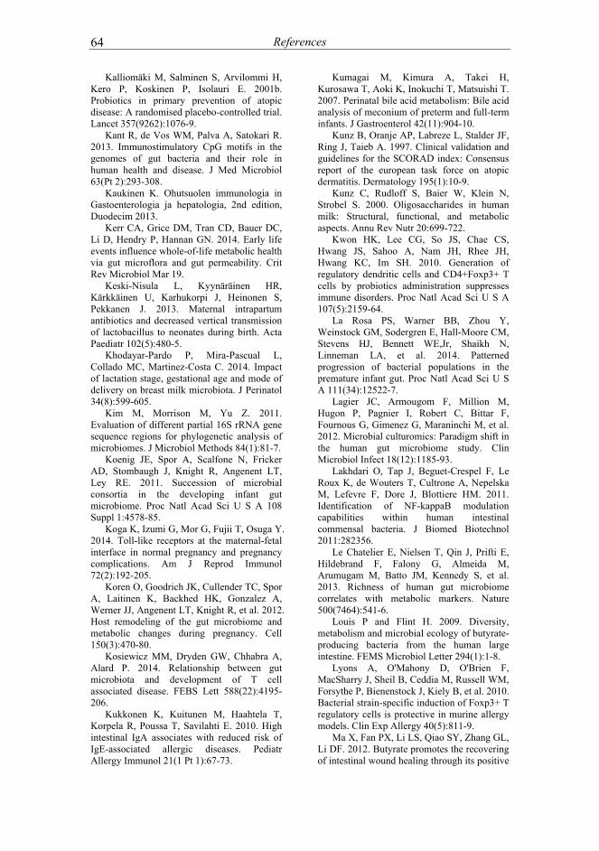
References
64
Kalliomäki M, Salminen S, Arvilommi H, Kero P, Koskinen P, Isolauri E. 2001b. Probiotics in primary prevention of atopic disease: A randomised placebo-controlled trial. Lancet 357(9262):1076-9.
Kant R, de Vos WM, Palva A, Satokari R. 2013. Immunostimulatory CpG motifs in the genomes of gut bacteria and their role in human health and disease. J Med Microbiol 63(Pt 2):293-308.
Kaukinen K. Ohutsuolen immunologia in Gastoenterologia ja hepatologia, 2nd edition, Duodecim 2013.
Kerr CA, Grice DM, Tran CD, Bauer DC, Li D, Hendry P, Hannan GN. 2014. Early life events influence whole-of-life metabolic health via gut microflora and gut permeability. Crit Rev Microbiol Mar 19.
Keski-Nisula L, Kyynäräinen HR, Kärkkäinen U, Karhukorpi J, Heinonen S, Pekkanen J. 2013. Maternal intrapartum antibiotics and decreased vertical transmission of lactobacillus to neonates during birth. Acta Paediatr 102(5):480-5.
Khodayar-Pardo P, Mira-Pascual L, Collado MC, Martinez-Costa C. 2014. Impact of lactation stage, gestational age and mode of delivery on breast milk microbiota. J Perinatol 34(8):599-605.
Kim M, Morrison M, Yu Z. 2011. Evaluation of different partial 16S rRNA gene sequence regions for phylogenetic analysis of microbiomes. J Microbiol Methods 84(1):81-7.
Koenig JE, Spor A, Scalfone N, Fricker AD, Stombaugh J, Knight R, Angenent LT, Ley RE. 2011. Succession of microbial consortia in the developing infant gut microbiome. Proc Natl Acad Sci U S A 108 Suppl 1:4578-85.
Koga K, Izumi G, Mor G, Fujii T, Osuga Y. 2014. Toll-like receptors at the maternal-fetal interface in normal pregnancy and pregnancy complications. Am J Reprod Immunol 72(2):192-205.
Koren O, Goodrich JK, Cullender TC, Spor A, Laitinen K, Backhed HK, Gonzalez A, Werner JJ, Angenent LT, Knight R, et al. 2012. Host remodeling of the gut microbiome and metabolic changes during pregnancy. Cell 150(3):470-80.
Kosiewicz MM, Dryden GW, Chhabra A, Alard P. 2014. Relationship between gut microbiota and development of T cell associated disease. FEBS Lett 588(22):4195-206.
Kukkonen K, Kuitunen M, Haahtela T, Korpela R, Poussa T, Savilahti E. 2010. High intestinal IgA associates with reduced risk of IgE-associated allergic diseases. Pediatr Allergy Immunol 21(1 Pt 1):67-73.
Kumagai M, Kimura A, Takei H, Kurosawa T, Aoki K, Inokuchi T, Matsuishi T. 2007. Perinatal bile acid metabolism: Bile acid analysis of meconium of preterm and full-term infants. J Gastroenterol 42(11):904-10.
Kunz B, Oranje AP, Labreze L, Stalder JF, Ring J, Taieb A. 1997. Clinical validation and guidelines for the SCORAD index: Consensus report of the european task force on atopic dermatitis. Dermatology 195(1):10-9.
Kunz C, Rudloff S, Baier W, Klein N, Strobel S. 2000. Oligosaccharides in human milk: Structural, functional, and metabolic aspects. Annu Rev Nutr 20:699-722.
Kwon HK, Lee CG, So JS, Chae CS, Hwang JS, Sahoo A, Nam JH, Rhee JH, Hwang KC, Im SH. 2010. Generation of regulatory dendritic cells and CD4+Foxp3+ T cells by probiotics administration suppresses immune disorders. Proc Natl Acad Sci U S A 107(5):2159-64.
La Rosa PS, Warner BB, Zhou Y, Weinstock GM, Sodergren E, Hall-Moore CM, Stevens HJ, Bennett WE,Jr, Shaikh N, Linneman LA, et al. 2014. Patterned progression of bacterial populations in the premature infant gut. Proc Natl Acad Sci U S A 111(34):12522-7.
Lagier JC, Armougom F, Million M, Hugon P, Pagnier I, Robert C, Bittar F, Fournous G, Gimenez G, Maraninchi M, et al. 2012. Microbial culturomics: Paradigm shift in the human gut microbiome study. Clin Microbiol Infect 18(12):1185-93.
Lakhdari O, Tap J, Beguet-Crespel F, Le Roux K, de Wouters T, Cultrone A, Nepelska M, Lefevre F, Dore J, Blottiere HM. 2011. Identification of NF-kappaB modulation capabilities within human intestinal commensal bacteria. J Biomed Biotechnol 2011:282356.
Le Chatelier E, Nielsen T, Qin J, Prifti E, Hildebrand F, Falony G, Almeida M, Arumugam M, Batto JM, Kennedy S, et al. 2013. Richness of human gut microbiome correlates with metabolic markers. Nature 500(7464):541-6.
Louis P and Flint H. 2009. Diversity, metabolism and microbial ecology of butyrate-producing bacteria from the human large intestine. FEMS Microbiol Letter 294(1):1-8.
Lyons A, O'Mahony D, O'Brien F, MacSharry J, Sheil B, Ceddia M, Russell WM, Forsythe P, Bienenstock J, Kiely B, et al. 2010. Bacterial strain-specific induction of Foxp3+ T regulatory cells is protective in murine allergy models. Clin Exp Allergy 40(5):811-9.
Ma X, Fan PX, Li LS, Qiao SY, Zhang GL, Li DF. 2012. Butyrate promotes the recovering of intestinal wound healing through its positive
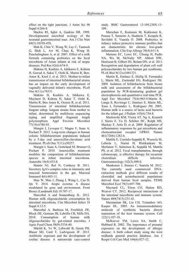
References
65
effect on the tight junctions. J Anim Sci 90 Suppl 4:266-8.
Mackie RI, Sghir A, Gaskins HR. 1999. Developmental microbial ecology of the neonatal gastrointestinal tract. Am J Clin Nutr 69(5):1035S-45S.
Mah K, Chin V, Wong W, Lay C, Tannock G, Shek L, Aw M, Chua K, Wong H, Panchalingham A, et al. 2007. Effect of a milk formula containing probiotics on the fecal microbiota of Asian infants at risk of atopic diseases. Ped Res 62(6):674-9.
Makino H, Kushiro A, Ishikawa E, Kubota H, Gawad A, Sakai T, Oishi K, Martin R, Ben-Amor K, Knol J, et al. 2013. Mother-to-infant transmission of intestinal bifidobacterial strains has an impact on the early development of vaginally delivered infant's microbiota. PLoS One 8(11):e78331.
Makino H, Kushiro A, Ishikawa E, Muylaert D, Kubota H, Sakai T, Oishi K, Martin R, Ben Amor K, Oozeer R, et al. 2011. Transmission of intestinal bifidobacterium longum subsp. longum strains from mother to infant, determined by multilocus sequencing typing and amplified fragment length polymorphism. Appl Environ Microbiol 77(19):6788-93.
Mangin I, Leveque C, Magne F, Suau A, Pochart P. 2012. Long-term changes in human colonic bifidobacterium populations induced by a 5-day oral amoxicillin-clavulanic acid treatment. PLoS One 7(11):e50257.
Mangin I, Suau A, Gotteland M, Brunser O, Pochart P. 2010. Amoxicillin treatment modifies the composition of bifidobacterium species in infant intestinal microbiota. Anaerobe 16(4):433-8.
Mantis NJ, Rol N, Corthesy B. 2011. Secretory IgA's complex roles in immunity and mucosal homeostasis in the gut. Mucosal Immunol 4(6):603-11.
Mao W, Mao J, Zhang J, Wang L, Cao D, Qu Y. 2014. Atopic eczema: A disease modulated by gene and environment. Front Biosci (Landmark Ed) 19:707-17.
Marcobal A and Sonnenburg JL. 2012. Human milk oligosaccharide consumption by intestinal microbiota. Clin Microbiol Infect 18 Suppl 4:12-5.
Marcobal A, Barboza M, Froehlich JW, Block DE, German JB, Lebrilla CB, Mills DA. 2010. Consumption of human milk oligosaccharides by gut-related microbes. J Agric Food Chem 58(9):5334-40.
Mårild K, Ye W, Lebwohl B, Green PH, Blaser MJ, Card T, Ludvigsson JF. 2013. Antibiotic exposure and the development of coeliac disease: A nationwide case-control
study. BMC Gastroenterol 13:109,230X-13-109.
Marschan E, Kuitunen M, Kukkonen K, Poussa T, Sarnesto A, Haahtela T, Korpela R, Savilahti E, Vaarala O. 2008. Probiotics in infancy induce protective immune profiles that are characteristic for chronic low-grade inflammation. Clin Exp Allergy 38(4):611-8.
Martens EC, Lowe EC, Chiang H, Pudlo NA, Wu M, McNulty NP, Abbott DW, Henrissat B, Gilbert HJ, Bolam DN, et al. 2011. Recognition and degradation of plant cell wall polysaccharides by two human gut symbionts. PLoS Biol 9(12):e1001221.
Martin R, Jiménez E, Heilig H, Fernandez L, Marin ML, Zoetendal EG, Rodriguez JM. 2009. Isolation of bifidobacteria from breast milk and assessment of the bifidobacterial population by PCR-denaturing gradient gel electrophoresis and quantitative real-time PCR. Appl Env Microbiol 75(4):965-9.Martin R, Langa S, Reviriego C, Jiménez E, Marin ML, Xaus J, Fernandez L, Rodriguez JM. 2003. Human milk is a source of lactic acid bacteria for the infant gut. J Pediatr 143(6):754-8.
Maslowski KM, Vieira AT, Ng A, Kranich J, Sierro F, Yu D, Schilter HC, Rolph MS, Mackay F, Artis D, et al. 2009. Regulation of inflammatory responses by gut microbiota and chemoattractant receptor GPR43. Nature 461(7268):1282-6.
Mattila E, Uusitalo-Seppälä R, Wuorela M, Lehtola L, Nurmi H, Ristikankare M, Moilanen V, Salminen K, Seppälä M, Mattila PS, et al. 2012. Fecal transplantation, through colonoscopy, is effective therapy for recurrent clostridium difficile infection. Gastroenterology 142(3):490-6.
Maukonen J, Simoes C, Saarela M. 2012. The currently used commercial DNA-extraction methods give different results of clostridial and actinobacterial populations derived from human fecal samples. FEMS Microbiol Ecol 79(3):697-708.
Maynard CL, Elson CO, Hatton RD, Weaver CT. 2012. Reciprocal interactions of the intestinal microbiota and immune system. Nature 489(7415):231-41.
Mazmanian SK, Liu CH, Tzianabos AO, Kasper DL. 2005. An immunomodulatory molecule of symbiotic bacteria directs maturation of the host immune system. Cell 122(1):107-18.
McKeever TM, Lewis SA, Smith C, Hubbard R. 2002. The importance of prenatal exposures on the development of allergic disease: A birth cohort study using the west midlands general practice database. Am J Respir Crit Care Med 166(6):827-32.

References
66
Medzhitov R. 2001. Toll-like receptors and innate immunity. Nat Rev Immunol 1(2):135-45.
Metsälä J, Lundqvist A, Virta LJ, Kaila M, Gissler M, Virtanen SM. 2013. Mother's and offspring's use of antibiotics and infant allergy to cow's milk. Epidemiology 24(2):303-9.
Moles L, Gomez M, Heilig H, Bustos G, Fuentes S, de Vos W, Fernandez L, Rodriguez JM, Jiménez E. 2013. Bacterial diversity in meconium of preterm neonates and evolution of their fecal microbiota during the first month of life. PLoS One 8(6):e66986.
Munyaka PM, Khafipour E, Ghia JE. 2014. External influence of early childhood establishment of gut microbiota and subsequent health implications. Front Pediatr 2:109.
Nanthakumar N, Meng D, Goldstein AM, Zhu W, Lu L, Uauy R, Llanos A, Claud EC, Walker WA. 2011. The mechanism of excessive intestinal inflammation in necrotizing enterocolitis: An immature innate immune response. PLoS One 6(3):e17776.
Natarajan N and Pluznick JL. 2014. From microbe to man: The role of microbial short chain fatty acid metabolites in host cell biology. Am J Physiol Cell Physiol :ajpcell.00228.2014.
Nermes M, Kantele JM, Atosuo TJ, Salminen S, Isolauri E. 2011. Interaction of orally administered lactobacillus rhamnosus GG with skin and gut microbiota and humoral immunity in infants with atopic dermatitis. Clin Exp Allergy 41(3):370-7.
Newburg DS and Morelli L. 2014. Human milk and infant intestinal mucosal glycans guide succession of the neonatal intestinal microbiota. Pediatr Res 77(1-2):115-120.
Normann E, Fahlen A, Engstrand L, Lilja HE. 2013. Intestinal microbial profiles in extremely preterm infants with and without necrotizing enterocolitis. Acta Paediatr 102(2):129-36.
Nylund L, Satokari R, Nikkila J, Rajilic-Stojanovic M, Kalliomäki M, Isolauri E, Salminen S, de Vos WM. 2013. Microarray analysis reveals marked intestinal microbiota aberrancy in infants having eczema compared to healthy children in at-risk for atopic disease. BMC Microbiol 13:12.
O'Connell Motherway M, Zomer A, Leahy SC, Reunanen J, Bottacini F, Claesson MJ, O'Brien F, Flynn K, Casey PG, Munoz JA, et al. 2011. Functional genome analysis of bifidobacterium breve UCC2003 reveals type IVb tight adherence (tad) pili as an essential and conserved host-colonization factor. Proc Natl Acad Sci U S A 108(27):11217-22.
Pagnini C, Saeed R, Bamias G, Arseneau KO, Pizarro TT, Cominelli F. 2010. Probiotics
promote gut health through stimulation of epithelial innate immunity. Proc Natl Acad Sci U S A 107(1):454-9.
Paliy O, Kenche H, Abernathy F, Michail S. 2009. High-throughput quantitative analysis of the human intestinal microbiota with a phylogenetic microarray. Appl Env Microbiol 75(11):3572-9.
Palmer C, Bik EM, Digiulio DB, Relman DA, Brown PO. 2007. Development of the human infant intestinal microbiota. PLoS ONE 5(7):e177.
Pararas MV, Skevaki CL, Kafetzis DA. 2006. Preterm birth due to maternal infection: Causative pathogens and modes of prevention. Eur J Clin Microbiol Infect Dis 25(9):562-9.
Pärtty A, Luoto R, Kalliomäki M, Salminen S, Isolauri E. 2013. Effects of early prebiotic and probiotic supplementation on development of gut microbiota and fussing and crying in preterm infants: A randomized, double-blind, placebo-controlled trial. J Pediatr 163(5):1272,7.e1-2.
Penders J, Stobberingh E, Thijs C, Adams H, Vink C, van Ree R, van den Brandt P. 2006. Molecular fingerprinting of the intestinal microbiota of infants in whom atopic eczema was or was not developing. Clin Exp Allergy 36(12):1602-8.
Pfleiderer A, Lagier JC, Armougom F, Robert C, Vialettes B, Raoult D. 2013. Culturomics identified 11 new bacterial species from a single anorexia nervosa stool sample. Eur J Clin Microbiol Infect Dis 32(11):1471-81.
Qin J and members of MetaHIT Consortium. 2010. A human gut microbial gene catalog established by deep metagenomic sequencing. Nature 464(7285):59-65.
Rachmilewitz D, Katakura K, Karmeli F, Hayashi T, Reinus C, Rudensky B, Akira S, Takeda K, Lee J, Takabayashi K, et al. 2004. Toll-like receptor 9 signaling mediates the anti-inflammatory effects of probiotics in murine experimental colitis. Gastroenterology 126(2):520-8.
Rajilić-Stojanović M, Heilig H, Molenaar D, Kajander K, Smidt H, de Vos W. 2009. Development and application of the human intestinal tract chip (HITChip), A phylogenetic microarray: Absence of universally conserved phylotypes in the abundant microbiota of young and elderly adults. Environ Microbiol 11:1736-1743.
Rajilic-Stojanovic M and de Vos WM. 2014. The first 1000 cultured species of the human gastrointestinal microbiota. FEMS Microbiol Rev 38(5):996-1047.
Rajilic-Stojanovic M, Heilig HG, Tims S, Zoetendal EG, de Vos WM. 2012. Long-term

References
67
monitoring of the human intestinal microbiota composition. Environ Microbiol Oct 15.
Ramirez-Farias C, Slezak K, Fuller Z, Duncan A, Holtrop G, Louis P. 2009. Effect of inulin on the human gut microbiota: Stimulation of bifidobacterium adolescentis and faecalibacterium prausnitzii. Br J Nutr 101(4):541-50.
Ramirez-Velazquez C, Castillo EC, Guido-Bayardo L, Ortiz-Navarrete V. 2013. IL-17-producing peripheral blood CD177+ neutrophils increase in allergic asthmatic subjects. Allergy Asthma Clin Immunol 9(1):23,1492-9-23.
Rautava S, Arvilommi H, Isolauri E. 2006. Specific probiotics in enhancing maturation of IgA responses in formula-fed infants. Pediatr Res 60(2):221-4.
Rautava S, Collado MC, Salminen S, Isolauri E. 2012. Probiotics modulate host-microbe interaction in the placenta and fetal gut: A randomized, double-blind, placebo-controlled trial. Neonatology 102(3):178-84.
Rea MC, Dobson A, O'Sullivan O, Crispie F, Fouhy F, Cotter PD, Shanahan F, Kiely B, Hill C, Ross RP. 2011. Effect of broad- and narrow-spectrum antimicrobials on clostridium difficile and microbial diversity in a model of the distal colon. Proc Natl Acad Sci U S A 108 Suppl 1:4639-44.
Ricci G, Bendandi B, Bellini F, Patrizi A, Masi M. 2007. Atopic dermatitis: Quality of life of young italian children and their families and correlation with severity score. Pediatr Allergy Immunol 18(3):245-9.
Ringel-Kulka T, Cheng J, Ringel Y, Salojärvi J, Carroll I, Palva A, de Vos WM, Satokari R. 2013. Intestinal microbiota in healthy U.S. young children and adults--a high throughput microarray analysis. PLoS One 8(5):e64315.
Rinne M, Gueimonde M, Kalliomäki M, Hoppu U, Salminen S, Isolauri E. 2005. Similar bifidogenic effects of prebiotic-supplemented partially hydrolyzed infant formula and breastfeeding on infant gut microbiota. 43(1):59-65.
Roger LC and McCartney AL. 2010a. Longitudinal investigation of the faecal microbiota of healthy full-term infants using fluorescence in situ hybridization and denaturing gradient gel electrophoresis. Microbiology 156(Pt 11):3317-28.
Roger LC, Costabile A, Holland DT, Hoyles L, McCartney AL. 2010b. Examination of faecal bifidobacterium populations in breast- and formula-fed infants during the first 18 months of life. Microbiology 156(Pt 11):3329-41.
Rosenfeldt V, Benfeldt E, Valerius NH, Paerregaard A, Michaelsen KF. 2004. Effect of probiotics on gastrointestinal symptoms and small intestinal permeability in children with atopic dermatitis. J Pediatr 145(5):612-6.
Rosewarne CP, Pope PB, Denman SE, McSweeney CS, O'Cuiv P, Morrison M. 2011. High-yield and phylogenetically robust methods of DNA recovery for analysis of microbial biofilms adherent to plant biomass in the herbivore gut. Microb Ecol 61(2):448-54.
Rouge C, Goldenberg O, Ferraris L, Berger B, Rochat F, Legrand A, Gobel UB, Vodovar M, Voyer M, Roze JC, et al. 2010. Investigation of the intestinal microbiota in preterm infants using different methods. Anaerobe 16(4):362-70.
Round JL, Lee SM, Li J, Tran G, Jabri B, Chatila TA, Mazmanian SK. 2011. The toll-like receptor 2 pathway establishes colonization by a commensal of the human microbiota. Science 332(6032):974-7.
Rouwet EV, Heineman E, Buurman WA, ter Riet G, Ramsay G, Blanco CE. 2002. Intestinal permeability and carrier-mediated monosaccharide absorption in preterm neonates during the early postnatal period. Pediatr Res 51(1):64-70.
Salojärvi J and Lahti L. 2014. Microbiome R package (v.0.99.31). .
Salonen A, Nikkilä J, Jalanka-Tuovinen J, Immonen O, Rajilić-Stojanović M, Kekkonen R, Palva A, de Vos W. 2010. Comparative analysis of fecal DNA extraction methods with phylogenetic microarray: Effective recovery of bacterial and archaeal DNA using mechanical cell lysis. J Microbiol Methods 81(2):127-34.
Salzman NH, Hung K, Haribhai D, Chu H, Karlsson-Sjoberg J, Amir E, Teggatz P, Barman M, Hayward M, Eastwood D, et al. 2010. Enteric defensins are essential regulators of intestinal microbial ecology. Nat Immunol 11(1):76-83.
Samuel BS, Shaito A, Motoike T, Rey FE, Backhed F, Manchester JK, Hammer RE, Williams SC, Crowley J, Yanagisawa M, et al. 2008. Effects of the gut microbiota on host adiposity are modulated by the short-chain fatty-acid binding G protein-coupled receptor, Gpr41. Proc Natl Acad Sci U S A 105(43):16767-72.
Santiago A, Panda S, Mengels G, Martinez X, Azpiroz F, Dore J, Guarner F, Manichanh C. 2014. Processing faecal samples: A step forward for standards in microbial community analysis. BMC Microbiol 14:112,2180-14-112.
Sasai M and Yamamoto M. 2013. Pathogen recognition receptors: Ligands and signaling pathways by toll-like receptors. Int Rev Immunol 32(2):116-33.

References
68
Satokari R, Grönroos T, Laitinen K, Salminen S, Isolauri E. 2009. Bifidobacterium and lactobacillus DNA in the human placenta. Lett Appl Microbiol 48(1):8-12.
Savilahti EM, Kukkonen AK, Haahtela T, Tuure T, Kuitunen M, Savilahti E. 2012. Intestinal defensin secretion in infancy is associated with the emergence of sensitization and atopic dermatitis. Clin Exp Allergy 42(3):405-11.
Scheepers LE, Penders J, Mbakwa CA, Thijs C, Mommers M, Arts IC. 2014. The intestinal microbiota composition and weight development in children: The KOALA birth cohort study. Int J Obes (Lond) 39(1):16-25.
Schilderink R, Verseijden C, de Jonge WJ. 2013. Dietary inhibitors of histone deacetylases in intestinal immunity and homeostasis. Front Immunol 4:226.
Schloss PD and Westcott SL. 2011a. Assessing and improving methods used in operational taxonomic unit-based approaches for 16S rRNA gene sequence analysis. Appl Environ Microbiol 77(10):3219-26.
Schloss PD, Gevers D, Westcott SL. 2011b. Reducing the effects of PCR amplification and sequencing artifacts on 16S rRNA-based studies. PLoS One 6(12):e27310.
Sela DA and Mills DA. 2014. The marriage of nutrigenomics with the microbiome: The case of infant-associated bifidobacteria and milk. Am J Clin Nutr 99(3):697S-703S.
Sela DA, Garrido D, Lerno L, Wu S, Tan K, Eom HJ, Joachimiak A, Lebrilla CB, Mills DA. 2012. Bifidobacterium longum subsp. infantis ATCC 15697 alpha-fucosidases are active on fucosylated human milk oligosaccharides. Appl Environ Microbiol 78(3):795-803.
Sela DA, Chapman J, Adeuya A, Kim JH, Chen F, Whitehead TR, Lapidus A, Rokhsar DS, Lebrilla CB, German JB, et al. 2008. The genome sequence of bifidobacterium longum subsp. infantis reveals adaptations for milk utilization within the infant microbiome. Proc Natl Acad Sci 105(48):18964-9.
Sepp E, Julge K, Mikelsaar M, Bjorksten B. 2005. Intestinal microbiota and immunoglobulin E responses in 5-year-old estonian children. 35(9):1141-6.
Sim K, Cox MJ, Wopereis H, Martin R, Knol J, Li MS, Cookson WO, Moffatt MF, Kroll JS. 2012. Improved detection of bifidobacteria with optimised 16S rRNA-gene based pyrosequencing. PLoS One 7(3):e32543.
Simsek Y, Karabiyik P, Polat K, Duran Z, Polat A. 2014. Mode of delivery changes oxidative and antioxidative properties of human milk: A prospective controlled clinical investigation. J Matern Fetal Neonatal Med :1-5.
Sjögren YM, Jenmalm MC, Böttcher MF, Björkstén B, Sverremark-Ekström E. 2009a. Altered early infant gut microbiota in children developing allergy up to 5 years of age. Clinical & Experimental Allergy 39(4):518-26.
Sjögren YM, Tomicic S, Lundberg A, Böttcher MF, Björkstén B, Sverremark-Ekström E, Jenmalm MC. 2009b. Influence of early gut microbiota on the maturation of childhood mucosal and systemic immune responses Clinical & Experimental Allergy 39(12):1842-51.
Smith PM, Howitt MR, Panikov N, Michaud M, Gallini CA, Bohlooly-Y M, Glickman JN, Garrett WS. 2013. The microbial metabolites, short-chain fatty acids, regulate colonic treg cell homeostasis. Science 341(6145):569-73.
Solis G, de Los Reyes-Gavilan CG, Fernandez N, Margolles A, Gueimonde M. 2010. Establishment and development of lactic acid bacteria and bifidobacteria microbiota in breast-milk and the infant gut Anaerobe 16(3):307-10.
Sommer F and Backhed F. 2014. The gut microbiota engages different signaling pathways to induce Duox2 expression in the ileum and colon epithelium. Mucosal Immunol Aug 27.
Song SJ, Lauber C, Costello EK, Lozupone CA, Humphrey G, Berg-Lyons D, Caporaso JG, Knights D, Clemente JC, Nakielny S, et al. 2013. Cohabiting family members share microbiota with one another and with their dogs. Elife 2:e00458.
Storrø O, Oien T, Langsrud O, Rudi K, Dotterud C, Johnsen R. 2011. Temporal variations in early gut microbial colonization are associated with allergen-specific immunoglobulin E but not atopic eczema at 2 years of age. Clin Exp Allergy 41(11):1545-54.
Stsepetova J, Sepp E, Julge K, Vaughan E, Mikelsaar M, de Vos W. 2007. Molecularly assessed shifts of bifidobacterium ssp. and less diverse microbial communities are characteristic of 5-year-old allergic children. FEMS Immunol Med Microbiol 51(2):260-9.
Takahashi H, Mikami K, Nishino R, Matsuoka T, Kimura M, Koga Y. 2010. Comparative analysis of the properties of bifidobacterial isolates from fecal samples of mother-infant pairs. J Pediatr Gastroenterol Nutr 51(5):653-60.
Tanaka S, Kobayashi T, Songjinda P, Tateyama A, Tsubouchi M, Kiyohara C, Shirakawa T, Sonomoto K, Nakayama J. 2009b. Influence of antibiotic exposure in the early postnatal period on the development of intestinal microbiota. FEMS Immunol Med Microbiol 56(1):80-7.
References
69
Tannock GW, Lawley B, Munro K, Gowri Pathmanathan S, Zhou SJ, Makrides M, Gibson RA, Sullivan T, Prosser CG, Lowry D, et al. 2013. Comparison of the compositions of the stool microbiotas of infants fed goat milk formula, cow milk-based formula, or breast milk. Appl Environ Microbiol 79(9):3040-8.
Tap J, Mondot S, Levenez F, Pelletier E, Caron C, Furet J, Ugarte E, Muñoz-Tamayo R, Paslier D, Nalin R, et al. 2009. Towards the human intestinal microbiota phylogenetic core. Environ Microbiol 11(10):2547-84
Tasse L, Bercovici J, Pizzut-Serin S, Robe P, Tap J, Klopp C, Cantarel BL, Coutinho PM, Henrissat B, Leclerc M, et al. 2010. Functional metagenomics to mine the human gut microbiome for dietary fiber catabolic enzymes. Genome Res 20(11):1605-12.
Thompson-Chagoyan OC, Fallani M, Maldonado J, Vieites JM, Khanna S, Edwards C, Dore J, Gil A. 2011. Faecal microbiota and short-chain fatty acid levels in faeces from infants with cow's milk protein allergy. Int Arch Allergy Immunol 156(3):325-32.
Thorburn AN, Macia L, Mackay CR. 2014. Diet, metabolites, and "western-lifestyle" inflammatory diseases. Immunity 40(6):833-42.
Thurl S, Munzert M, Henker J, Boehm G, Muller-Werner B, Jelinek J, Stahl B. 2010. Variation of human milk oligosaccharides in relation to milk groups and lactational periods. Br J Nutr 104(9):1261-71.
Tims S, Derom C, Jonkers DM, Vlietinck R, Saris WH, Kleerebezem M, de Vos WM, Zoetendal EG. 2013. Microbiota conservation and BMI signatures in adult monozygotic twins. ISME J 7(4):707-17.
Torii S, Torii A, Itoh K, Urisu A, Terada A, Fujisawa T, Yamada K, Suzuki H, Ishida Y, Nakamura F, et al. 2011. Effects of oral administration of lactobacillus acidophilus L-92 on the symptoms and serum markers of atopic dermatitis in children. Int Arch Allergy Immunol 154(3):236-45.
Tottey W, Denonfoux J, Jaziri F, Parisot N, Missaoui M, Hill D, Borrel G, Peyretaillade E, Alric M, Harris HM, et al. 2013. The human gut chip "HuGChip", an explorative phylogenetic microarray for determining gut microbiome diversity at family level. PLoS One 8(5):e62544.
Tremaroli V and Backhed F. 2012. Functional interactions between the gut microbiota and host metabolism. Nature 489(7415):242-9.
Turnbaugh P, Hamady M, Yatsunenko T, Cantarel B, Duncan A, Ley R, Sogin M, Jones W, Roe B, Affourtit J, et al. 2009. A core gut microbiome in obese and lean twins. Nature 457(7228):480-4.
Turroni F, Foroni E, Serafini F, Viappiani A, Montanini B, Bottacini F, Ferrarini A, Bacchini PL, Rota C, Delledonne M, et al. 2011. Ability of bifidobacterium breve to grow on different types of milk: Exploring the metabolism of milk through genome analysis. Appl Environ Microbiol 77(20):7408-17.
Turroni F, Peano C, Pass DA, Foroni E, Severgnini M, Claesson MJ, Kerr C, Hourihane J, Murray D, Fuligni F, et al. 2012. Diversity of bifidobacteria within the infant gut microbiota. PLoS One 7(5):e36957.
Tyler AD, Smith MI, Silverberg MS. 2014. Analyzing the human microbiome: A "how to" guide for physicians. Am J Gastroenterol 109(7):983-93.
Underwood MA, German JB, Lebrilla CB, Mills DA. 2015. Bifidobacterium longum subspecies infantis: Champion colonizer of the infant gut. Pediatr Res 77(1-2):229-235.
Ursell LK, Clemente JC, Rideout JR, Gevers D, Caporaso JG, Knight R. 2012. The interpersonal and intrapersonal diversity of human-associated microbiota in key body sites. J Allergy Clin Immunol 129(5):1204-8.
Vaishampayan PA, Kuehl JV, Froula JL, Morgan JL, Ochman H, Francino MP. 2010. Comparative metagenomics and population dynamics of the gut microbiota in mother and infant. Genome Biol Evol 2:53-66.
van Baarlen P, Wells JM, Kleerebezem M. 2013. Regulation of intestinal homeostasis and immunity with probiotic lactobacilli. Trends Immunol 34(5):208-15.
van den Bogert B, de Vos WM, Zoetendal EG, Kleerebezem M. 2011. Microarray analysis and barcoded pyrosequencing provide consistent microbial profiles depending on the source of human intestinal samples. Appl Environ Microbiol 77(6):2071-80.
van Elburg RM, Fetter WP, Bunkers CM, Heymans HS. 2003. Intestinal permeability in relation to birth weight and gestational and postnatal age. Arch Dis Child Fetal Neonatal Ed 88(1):F52-5.
van Nood E, Vrieze A, Nieuwdorp M, Fuentes S, Zoetendal EG, de Vos WM, Visser CE, Kuijper EJ, Bartelsman JF, Tijssen JG, et al. 2013. Duodenal infusion of donor feces for recurrent Clostridium difficile. N Engl J Med 368(5):407-15.
van Passel MW, Kant R, Zoetendal EG, Plugge CM, Derrien M, Malfatti SA, Chain PS, Woyke T, Palva A, de Vos WM, et al. 2011. The genome of Akkermansia muciniphila, a dedicated intestinal mucin degrader, and its use in exploring intestinal metagenomes. PLoS One 6(3):e16876.
Vrieze A, Van Nood E, Holleman F, Salojarvi J, Kootte RS, Bartelsman JF,
References
70
Dallinga-Thie GM, Ackermans MT, Serlie MJ, Oozeer R, et al. 2012. Transfer of intestinal microbiota from lean donors increases insulin sensitivity in individuals with metabolic syndrome. Gastroenterology 143(4):913,6.e7.
Walker AW and Lawley TD. 2013. Therapeutic modulation of intestinal dysbiosis. Pharmacol Res 69(1):75-86.
Walker AW, Duncan SH, Louis P, Flint HJ. 2014. Phylogeny, culturing, and metagenomics of the human gut microbiota. Trends Microbiol 22(5):267-74.
Walker AW, Ince J, Duncan SH, Webster LM, Holtrop G, Ze X, Brown D, Stares MD, Scott P, Bergerat A, et al. 2011. Dominant and diet-responsive groups of bacteria within the human colonic microbiota. ISME J 5(2):220-30.
Wang HB, Wang PY, Wang X, Wan YL, Liu YC. 2012. Butyrate enhances intestinal epithelial barrier function via up-regulation of tight junction protein claudin-1 transcription. Dig Dis Sci 57(12):3126-35.
Wang M, Karlsson C, Olsson C, Adlerberth I, Wold AE, Strachan DP, Martricardi PM, Aberg N, Perkin MR, Tripodi S, et al. 2008. Reduced diversity in the early fecal microbiota of infants with atopic eczema. J Allergy Clin Immunol 121(1):129-34.
Wang X, Buhimschi CS, Temoin S, Bhandari V, Han YW, Buhimschi IA. 2013. Comparative microbial analysis of paired amniotic fluid and cord blood from pregnancies complicated by preterm birth and early-onset neonatal sepsis. PLoS One 8(2):e56131.
Ward TL, Hosid S, Ioshikhes I, Altosaar I. 2013. Human milk metagenome: A functional capacity analysis. BMC Microbiol 13:116,2180-13-116.
Werner JJ, Zhou D, Caporaso JG, Knight R, Angenent LT. 2012. Comparison of illumina paired-end and single-direction sequencing for microbial 16S rRNA gene amplicon surveys. ISME J 6(7):1273-6.
West CE, Jenmalm MC, Prescott SL. 2015. The gut microbiota and its role in the development of allergic disease: A wider perspective. Clin Exp Allergy 45(1):43-53.
West CE, Hammarstrom ML, Hernell O. 2009. Probiotics during weaning reduce the incidence of eczema. Pediatr Allergy Immunol 20(5):430-7.
Wopereis H, Oozeer R, Knipping K, Belzer C, Knol J. 2014. The first thousand days - intestinal microbiology of early life: Establishing a symbiosis. Pediatr Allergy Immunol 25(5):428-38.
Xu J, Bjursell MK, Himrod J, Deng S, Carmichael LK, Chiang HC, Hooper LV,
Gordon JI. 2003. A genomic view of the human-bacteroides thetaiotaomicron symbiosis. Science 299(5615):2074-6.
Yatsunenko T, Rey FE, Manary MJ, Trehan I, Dominguez-Bello MG, Contreras M, Magris M, Hidalgo G, Baldassano RN, Anokhin AP, et al. 2012. Human gut microbiome viewed across age and geography. Nature 486(7402):222-7.
Yu ZT, Chen C, Newburg DS. 2013. Utilization of major fucosylated and sialylated human milk oligosaccharides by isolated human gut microbes. Glycobiology 23(11):1281-92.
Zivkovic AM, German JB, Lebrilla CB, Mills DA. 2011. Human milk glycobiome and its impact on the infant gastrointestinal microbiota. Proc Natl Acad Sci U S A 108 Suppl 1:4653-8.
Zoetendal EG, Ben-Amor K, Akkermans AD, Abee T, de Vos WM. 2001. DNA isolation protocols affect the detection limit of PCR approaches of bacteria in samples from the human gastrointestinal tract. Syst Appl Microbiol 24(3):405-10.

Appendix: Original Publications
71
APPENDIX: ORIGINAL PUBLICATIONS
I. Reprinted from Journal of Microbiological Methods 2010, 83, 231-235 with permission from Elsevier Limited.
II. Submitted manuscript.
III. Reprinted from BMC Microbiology 2013, 13, 12 with permission
from BioMed Central. IV. Reprinted from Allergy 2015, 70, 241-244 with permission from John
Wiley and Sons. ADDITIONAL PUBLICATION RELATED TO THESIS Reprinted from Proceedings of the Nutrition Society 2014, 73, 457-69 with permission from Cambridge University Press.


