Does Lactic Acid Cause Muscular Fatigue? Ernest W. Maglischo, Ph.D. 1970 Lazy Meadow Lane Prescott, AZ 86303 USA [email protected]Abstract. Until recently, lactic acid accumulation and the acidosis resulting from it were believed to be the cause of muscular fatigue in most athletic events. However, some early pieces of research that supported this contention suffered from a flaw in design. They were conducted with excised muscle fibers that were cooled below body temperature. More recent studies have demonstrated little or no effect of lactic acid accumulation and acidosis on muscle contraction force and velocity when the fibers were warmed near body temperature. This finding has caused many in the scientific community to doubt acidosis is the cause of muscular fatigue. Some are now suggesting that fatigue results from creatine phosphate (CP) depletion and, specifically, the increases of inorganic phosphate and ADP that occur when the muscle supply of CP is reduced. Despite these findings, a very strong case for acidosis has been put forward by Knuth and associates (2006). They showed that acidosis resulted in a loss of muscle power even when fibers were warmed to 30 0 C (86 0 F) before testing. Evidence both for and against acidosis as a cause of muscular fatigue has been presented in this paper so the reader can form his or her own opinion on this controversial issue. Introduction. When people are asked to define fatigue, they frequently use words like “exhaustion” and “pain”. These terms describe only two of several forms that muscular fatigue can take. Exhaustion refers to the kind of fatigue that that occurs in events lasting for several hours. That fatigue is caused by depletion of carbohydrate sources in muscles. Pain and reduced performance are terms associated with fatigue in events lasting one to several minutes. That type of muscular fatigue is the subject of this paper. In 1924, Nobel Laureate A.V. Hill and his associates suggested that muscular fatigue in events lasting one to several minutes was caused by lactic acid accumulation in the muscles. They proposed, that during intense exercise an inadequate oxygen supply caused muscles to release energy anaerobically (without oxygen), and, in doing so, they produced lactic acid which lowered their pH and caused them to fatigue (Hill, Long, and Lupton, 1924). This model was widely accepted and continues today, with only minor modifications, as the classic explanation for muscular fatigue in most athletic activities. However, doubt has been cast on this premise with the report in 1995 that high levels of lactic acid did not interfere with muscular contraction at near-normal body temperatures (Pate, et al, 1995). This finding has led to a re-examination of the role of lactic acid in muscular fatigue The purpose of this paper will be to describe the proposed role of lactic acid in muscular fatigue as well as other possible causes of fatigue that have been suggested by recent research so that readers may form their own opinion on this issue. Let’s set the stage for that information by first presenting a brief review of the metabolic process with an emphasis on how energy is made available for muscular contraction and the ways in which lactic acid and other potential causes of fatigue are both produced and utilized during that process. A Brief Review of Energy Metabolism. Energy is defined as the capacity for doing work. Metabolism refers to the breakdown and synthesis of chemical compounds in the body that both store and release the energy that is needed to perform that work. These chemical compounds are derived from the foods we eat after they have been digested to carbohydrate, fat, and protein. Metabolism of these chemicals is aided by the activity of enzymes that catalyze (speed-up) the process. The most eminent of these compounds is ATP (adenosine triphosphate) and it will be discussed first.
Transcript
Does Lactic Acid Cause Muscular Fatigue?
Ernest W. Maglischo, Ph.D. 1970 Lazy Meadow Lane Prescott, AZ 86303 USA [email protected] Abstract. Until recently, lactic acid accumulation and the acidosis resulting from it were believed to be the cause of muscular fatigue in most athletic events. However, some early pieces of research that supported this contention suffered from a flaw in design. They were conducted with excised muscle fibers that were cooled below body temperature. More recent studies have demonstrated little or no effect of lactic acid accumulation and acidosis on muscle contraction force and velocity when the fibers were warmed near body temperature. This finding has caused many in the scientific community to doubt acidosis is the cause of muscular fatigue. Some are now suggesting that fatigue results from creatine phosphate (CP) depletion and, specifically, the increases of inorganic phosphate and ADP that occur when the muscle supply of CP is reduced. Despite these findings, a very strong case for acidosis has been put forward by Knuth and associates (2006). They showed that acidosis resulted in a loss of muscle power even when fibers were warmed to 300C (860 F) before testing. Evidence both for and against acidosis as a cause of muscular fatigue has been presented in this paper so the reader can form his or her own opinion on this controversial issue.
Introduction. When people are asked to define fatigue, they frequently use words like “exhaustion” and “pain”. These terms describe only two of several forms that muscular fatigue can take. Exhaustion refers to the kind of fatigue that that occurs in events lasting for several hours. That fatigue is caused by depletion of carbohydrate sources in muscles. Pain and reduced performance are terms associated with fatigue in events lasting one to several minutes. That type of muscular fatigue is the subject of this paper. In 1924, Nobel Laureate A.V. Hill and his associates suggested that muscular fatigue in events lasting one to several minutes was caused by lactic acid accumulation in the muscles. They proposed, that during intense exercise an inadequate oxygen supply caused muscles to release energy anaerobically (without oxygen), and, in doing so, they produced lactic acid which lowered their pH and caused them to fatigue (Hill, Long, and Lupton, 1924). This model was widely accepted and continues today, with only minor modifications, as the classic explanation for muscular fatigue in most athletic activities. However, doubt has been cast on this premise with the report in 1995 that high levels of lactic acid did not interfere with muscular contraction at near-normal body temperatures (Pate, et al, 1995). This finding has led to a re-examination of the role of lactic acid in muscular fatigue The purpose of this paper will be to describe the proposed role of lactic acid in muscular fatigue as well as other possible causes of fatigue that have been suggested by recent research so that readers may form their own opinion on this issue. Let’s set the stage for that information by first presenting a brief review of the metabolic process with an emphasis on how energy is made available for muscular contraction and the ways in which lactic acid and other potential causes of fatigue are both produced and utilized during that process. A Brief Review of Energy Metabolism. Energy is defined as the capacity for doing work. Metabolism refers to the breakdown and synthesis of chemical compounds in the body that both store and release the energy that is needed to perform that work. These chemical compounds are derived from the foods we eat after they have been digested to carbohydrate, fat, and protein. Metabolism of these chemicals is aided by the activity of enzymes that catalyze (speed-up) the process. The most eminent of these compounds is ATP (adenosine triphosphate) and it will be discussed first.
2
ATP, the universal supplier of energy for muscular contraction. During exercise of any intensity, the energy for muscular contraction is provided by the breakdown of ATP that is stored in the muscles. ATP is composed of a protein, adenosine, and three phosphates molecules. It can be regenerated in the body from the metabolism of carbohydrate, fat and protein, although carbohydrate is the preferred source during exercise. Each of the phosphates is bound to the entire compound by chemical energy. When a nerve impulse reaches a muscle fiber and signals it to contract, one of these phosphates will be split from each available ATP molecule and the energy binding it will be set free to power the contraction. The substance remaining is ADP (adenosine diphosphate), which is composed of adenosine and two phosphate molecules. This process is diagrammed in figure 1. ATP + ATPase ADP + Pi + energy Muscle Contraction Figure 1. ATP (adenosine triphosphate) releases energy for muscular contraction when it loses one of its phosphate molecules and become ADP (adenosine diphosphate). The splitting of phosphate from adenosine and the subsequent release of energy is catalyzed by the enzyme Adenosinetriphosphatase (ATPase).
Some of the energy released from ATP will be used to do the mechanical work of muscle contraction and even more will be lost as heat energy. The freed inorganic phosphate (Pi) will accumulate in the muscle fiber. ATP is the only source of energy the muscles can use for contraction. All other chemical sources in the muscles must first be converted to ATP before their energy can be used for that purpose. Other sources of energy for regenerating ATP. While ATP is the direct source of energy for muscular contraction, it is in short supply in muscles. They contain 2 to 6 mmols per kilogram of tissue. This is only enough to sustain contraction for a few seconds. In order for exercise to continue for any length of time, ATP must quickly and continually be reformed. This is accomplished by finding other sources of inorganic phosphate and energy so ADP can be reconverted back to ATP. The reconversion of ADP to ATP is indicated by the double arrow in figure 1. It demonstrates that the release of energy from ATP and the regeneration of ATP is a reversible process. The sources of inorganic phosphate and energy that can be used to regenerate ATP in the muscles are creatine phosphate (CP), muscle glycogen, blood glucose, fat and even muscle protein. Creatine phosphate, glycogen, fat and protein are stored in the muscle fibers. Blood glucose diffuses into muscle cells from the blood during exercise. Of these five substances, creatine phosphate can regenerate ATP most rapidly but only for a few additional seconds. After that, muscle glycogen becomes the preferred source for replacing ATP. While their supply in muscles is much greater, the processes of regenerating ATP from blood glucose, fat and protein are slower because they must first be transported into muscle fibers or converted to byproducts of glucose metabolism before they can be used. As a result, ATP regeneration from these substances is too slow to meet the demand for energy during most athletic activities unless they are very long (in excess of one to two hours) or performed at a very slow pace. Therefore, these substances can provide only small amounts of supplemental energy during hard training and competition. This explains why muscle glycogen becomes the major source of energy for replacing ATP when the creatine phosphate supply declines. The metabolic process of regenerating ATP takes place in several stages. When ATP is regenerated from the metabolism of creatine phosphate, the process has been termed the ATP-CP reaction. When muscle glycogen is used to regenerate ATP, the process is termed glycolysis. Some ATP can be regenerated in first few steps in glycolysis. That process is termed anaerobic glycolysis because it does not involve oxygen. After that, additional ATP can be regenerated aerobically (with oxygen), from the byproducts of glucose metabolism although this metabolic process, termed aerobic
3
glycolysis, involves considerably more steps and, therefore, requires more time before ATP can be regenerated. ATP can also be regenerated from fat and protein although these processes are even longer, more complex, and, therefore, slower than that of metabolizing muscle glycogen aerobically. This is because fat and protein must first be converted to byproducts of glucose metabolism before they can begin to participate in the glycolytic process that will ultimately lead to the regeneration of ATP. As a consequence, this slows the procedure considerably right from the start. It is prolonged even more once they enter the glycolytic process because they can only be metabolized aerobically and, as mentioned, that requires a great number of steps before any energy and phosphate can be liberated to reform ATP. For example, the rate of ATP regeneration from fat is more than two times slower than the aerobic metabolism of glycogen. Although blood glucose can be metabolized anaerobically, this process is somewhat slower than that of metabolizing muscle glycogen aerobically because the glucose must be transported from the blood into the muscles before glycolysis can begin. The term aerobic metabolism is often used in preference to aerobic glycolysis to describe the aerobic process of regenerating ATP because it involves the simultaneous metabolism of fat, protein and blood glucose as well as glycogen. Let’s look more closely at the ATP-CP reaction and the processes of anaerobic and aerobic glycolysis since they are the major ways that ATP is regenerated during intense exercise. We will begin with the ATP-CP reaction. The ATP-CP reaction. As mentioned, the most rapid method for regenerating ATP is through the breakdown of creatine phosphate. Creatine phosphate is made up of two chemical compounds, creatine and phosphate, and, because of this, only one step is needed to split them. Splitting allows the release of inorganic phosphate and the free energy that bound the two substances together. That inorganic phosphate and free energy supply the necessary ingredients to regenerate ATP from ADP. This reaction is illustrated in figure 2. CP + ADP ATP + Cr Figure 2. CP (creatine phosphate) is a chemical compound containing creatine and a phosphate molecule that is bound to the creatine by chemical energy. When CP splits, it releases energy and phosphate that can combine with ADP to regenerate ATP. As indicated earlier, ADP is so-named because it contains two phosphate molecules and needs only one more to become ATP.
ATP can be regenerated very rapidly when CP is the source, reaching a rate of 9 mmols of ATP produced, per kilogram of muscle, per second, almost immediately after exercise begins. However, creatine phosphate is also stored in small quantities in muscles, only 18 to 20 mmols per kilogram. Consequently, during very intense exercise there is only enough available to regenerate ATP for 5 to 10 additional seconds if no other sources of ATP regeneration were available. The process of anaerobic and aerobic glycolysis are available, however. They will be described next. Glycolysis. Glycolysis refers to the breakdown of muscle glycogen to carbon dioxide (CO2) and water (H20). ATP can be regenerated and used as a source of energy for muscular contraction at several points during the glycolytic process. One common misconception is that glycolysis is the second stage of ATP regeneration, and that it does not begin until the muscle’s creatine-phosphate has been depleted. Actually, both the ATP-CP reaction and glycolysis are regenerating ATP, even at rest. The rates of both increase dramatically when exercise begins. However, when the demand for energy is increased, the ATP-CP reaction, because it is faster, will be the major contributor to ATP regeneration for the first few seconds. After that, the muscle’s creatine phosphate supply will be reduced to the point where glycolysis takes over as the major source of phosphate and energy for ATP resysthesis from ADP.
4
Although glucose metabolism (glycolysis) is one continuous process, it is usually thought of as having two distinct phases, an anaerobic and an aerobic phase. The earliest stage is termed anaerobic glycolysis because it does not require oxygen to regenerate ATP. The later stage, because it requires oxygen, is termed aerobic. The anaerobic phase is shorter and thus, faster than the aerobic, consisting of only 11 or 12 steps to regenerate ATP. The aerobic phase is actually a continuation of the anaerobic phase, requiring a much greater number of additional steps and an adequate supply of oxygen to replace ATP. It is during the aerobic phase that fat and protein can also be used to regenerate ATP. The process of regenerating ATP anaerobically is approximately ten times slower than the ATP-CP reaction. However, it can replace ATP 3 times faster than it can be restored aerobically. Thus, a rapid rate of anaerobic glycolysis is essential to success in any physical exercise that is performed at high rates of effort. The anaerobic and aerobic phases of glycolysis will be described next. First, however, I should mention that muscle glycogen must be converted to its simpler form, glucose, before it can be metabolized. This is a rapid process termed glycogenolysis and it is the first step in replacing ATP, whether it be by anaerobic and aerobica means. Anaerobic glycolysis. Once glycogen is split to glucose, the latter substance will be metabolized to pyruvate and NADH (nicotinamide adenine dinucleotide) in a very rapid process that requires only 11 steps. At that point, the pyruvate and the hydrogen ions from NADH can take one of two paths. The first path they can take is to enter the mitochondria of muscles where they will be metabolized aerobically to their final products, carbon dioxide (CO2) and water (H2O). In the second path, they can combine to form lactic acid and NAD+ (a coenzyme of NADH that has lost a hydrogen ion). As you are probably aware, the early belief was that lactic acid was an end product of anaerobic metabolism and remained in the muscles, reducing their pH and causing fatigue. More recently evidence has accumulated that the body can be trained to remove, metabolize and buffer lactic acid, thus delaying fatigue. As mentioned, there are now some experts who believe that lactic acid simply accompanies fatigue but does not cause it. Regardless, of the position taken on this issue, some energy and inorganic phosphate (Pi) will be released to regenerate small amounts of ATP regardless whether pyruvate and NADH or lactic acid and NAD+ are the byproducts of anaerobic glycolysis (see figure 4). Anaerobic glycolysis takes place in the cytoplasm (protoplasm) of individual muscle fibers. As mentioned, ATP can be regenerated quickly through anaerobic glycolysis. The process is not very efficient, however, because it regenerates only 3 ATP per mol of glycogen used (Spriet and Hargreaves, 2006: Westerblad and Allen, 2009). Nevertheless, the anaerobic process is essential when a rapid source of energy is needed for muscular contraction. Some consider the formation of pyruvate and NADH part of anaerobic glycolysis because no oxygen is involved while others believe it is part of the aerobic process. This is because the metabolism of pyruvate and NADH to carbon dioxide and water is a continuation of the process of glycolysis that began with the formation of glucose from glycogen and because oxygen is necessary to continue that process. On the other hand, when lactic acid is produced from pyruvate and NADH the process is definitely considered anaerobic because it does not involve oxygen. However, you will learn later that the metabolism of lactic acid, like that of pyruvate, may also continue in the mitochondria of muscles during exercise. Thus, the terms aerobic and anaerobic are somewhat archaic. This represents the difficulty in describing such a complex and interrelated process as metabolism in a step by step manner with universally accepted terms that we find are no longer completely accurate.
5
Aerobic metabolism. As indicated, pyruvate and NADH, (and, perhaps, lactic acid), must be absorbed from the cytoplasm into the mitochondria of muscle fibers in order to continue being metabolized. Mitochondria are rod-shaped structures found in the cytoplasm of cells (see figure 4). They have been likened to little chemical factories in the muscles where aerobic metabolism takes place because they contain the key enzymes and can take up the oxygen needed for that process. Pyruvate enters the mitochondria easily and is oxidized to carbon dioxide (CO2) by means of Krebs cycle (also known as the citric acid cycle. NADH cannot pass through the mitochondrial membrane but the hydrogen ions (H+) it contains can be shuttled in via the Glycerol phosphate and Malate-Aspartate shuttles where they will be reduced to water (H2O) in the electron transport chain. Hydrogen ions are protons that are very acidic and capable of lowering the pH of muscles if they are allowed to accumulate. Thus, their removal from NADH into the mitochondria and their subsequent reduction to water via the electron transport chain is important to prolonging most types of exercise. Some additional hydrogen ions will also be released from pyruvate during its passage through Krebs cycle. These will also be transferred to the electron transport chain and metabolized to water. Aerobic metabolism is much more efficient than anaerobic glycolysis because it produces more ATP from each molecule of muscle glycogen and because the end products, carbon dioxide (CO2) and water (H2O) are easily eliminated from the body (Westerblad and Allen, 2009). Thirty-nine (39) ATP molecules will be produced when molecules of pyruvate and hydrogen ions are metabolized aerobically in the mitochondria. It was mentioned earlier that, while aerobic metabolism is more efficient, it is much slower than anaerobic glycolysis because of the greater number of steps required before ATP can be regenerated. As a result, only moderate efforts could be sustained if the energy and phosphate needed to reform ATP came from aerobic metabolism alone. In most athletic events lasting less than one hour, ATP that is supplied by aerobic metabolism must be supplemented with ATP that is released anaerobically in order to meet the demand for energy to power muscular contraction. As compared to the rate at rest, the energy consumption of skeletal muscles can increase 100 fold during high intensity exercise (Westerblad, Allen and Lannergan (2002). When that happens the capacity to regenerate ATP aerobically will be greatly exceeded and a large portion of the energy will have to be supplied through anaerobic ATP regeneration. So it is good that the rate of anaerobic ATP regeneration can increase markedly beyond that of aerobic metabolism. Obviously, the advantage of regenerating ATP through anaerobic metabolism is that energy needed for muscular contraction can be supplied rapidly so that exercise can proceed at a faster rate. The disadvantage is that certain byproducts are produced, i.e. lactic acid and others to be described later, that are believed to cause fatigue. The advantage of regenerating ATP via aerobic metabolism is that no potentially harmful byproducts are produced so that the only limitation is the amount of glycogen, fat and protein available for the aerobic regeneration of ATP. The aerobic process proceeds until carbon dioxide (CO2) and water (H2O) have been produced. Both of which can be easily eliminated from the body in exhaled air and sweat so that fatigue does not occur. The disadvantage is that aerobic metabolism is a slow process that cannot regenerate ATP rapidly enough to supply all of the energy needed to be competitive in most athletic activities unless they are quite long and completed at a slow pace Maximal oxygen consumption (VO2max). As you can see from their chemical formulas, the end products of aerobic metabolism, CO2 and H2O, require a generous supply of oxygen for their production. Because of this, experts on training have reasoned that increasing oxygen consumption during exercise will increase the energy supplied aerobically and reduce the need for anaerobic ATP regeneration during exercise. Hence, the reason that maximum oxygen consumption (VO2max) has attained considerable prominence as a predictor of endurance performance over the years. The
6
importance of VO2max may have been overstated, however, because the mitochondria in muscle fibers probably play an even more important role than oxygen in regulating the rate of aerobic metabolism It has been well documented that muscles never use all of the oxygen available to them (Noakes, 2000). So, the factor limiting the rate of aerobic metabolism in muscles may not be the amount of oxygen available, but, instead, the size and number of mitochondria on hand to absorb that oxygen. With more and larger mitochondria, muscles should be able to take in greater amounts of the pyruvate, hydrogen ions, lactic acid, and NAD+ that were formed during exercise. Perhaps, also, aerobic metabolism in the mitochondria may slow the rate of CP depletion as well as the rates of increase of inorganic phosphate (Pi) and ADP, which, as you will learn later, may also contribute to muscular fatigue during exercise. Thus, the potential of all of these substances for creating fatigue should be lessened. Regardless of the metabolites involved, muscle fatigue is associated with events that occur during the anaerobic phase of glycolysis. Therefore, the endurance of athletes should be extended if their reliance on that phase is reduced in favor of deriving more energy from aerobic metabolism Consequently, VO2max may simply be a byproduct of the number of mitochondria available to take up oxygen from the circulation. As such, it may not be important in and of itself but only as an indicator that more chemical factories (mitochondria) are available to dispose of pyruvate, hydrogen ions, lactic acid and other metabolites during exercise. Mitochondrial density is the term used to designate the percent volume of a muscle cell occupied by mitochondria. Consequently, this term reflects their size and number. While VO2max is related to a rapid rate of aerobic metabolism, it is probably mitochondrial density that determines that rate. Proper training has been reported to double mitochondrial density (Hood, 2001), allowing athletes to generate more ATP aerobically during exercise. In two related studies, VO2max had only a moderate relationship (0.74) with endurance in groups of endurance-trained and untrained rats while the number and size of their mitochondria had a much higher relationship of 0.92 (Davies, Packer and Brooks 1981 and 1982). Endurance training improved the size and number of mitochondria in the rats’ muscle fibers by 100%, which resulted in a 400% increase in their running time to exhaustion. At the same time, their VO2max improved only 15%. A control group of rats also increased their VO2max by 15% with sprint training, but they did not increase mitochondria nor did they improve their running endurance. Antioxidants are also involved in maintaining mitochondrial density because it can be compromised by the presence of free radicals during exercise. Free radicals are produced during aerobic metabolism as a result of the oxygen used during that process. Some of the more prominent free radicals are superoxide, hydrogen peroxide, and nitric oxide. Their presence has been shown to damage the DNA and other proteins of mitochondria, thereby, reducing the number functioning during exercise (Davies et al., 1982). Antioxidants in the body protect the mitochondria from damage by free radicals. Therefore, increasing the intake of antioxidants such as Vitamin E and beta carotene, through the diet or by supplementation should help maintain mitochondrial density. Lactic acid formation. As stated earlier, it happens, that during moderate to high intensity exercise, pyruvate and NADH will be produced faster than they can be transported into the mitochondria and oxidized no matter how numerous and large those structures may be. When this occurs, the remaining portions of those substances will take the second path open to them and combine to form lactic acid in the cytoplasm of muscle fibers. This occurs when pyruvate takes up a hydrogen ion (H+) from NADH, and becomes lactic acid while NADH is oxidized to NAD+. Lactic acid can be removed from the muscles by a variety of means that will be discussed later. Nevertheless, during intense exercise some will remain to accumulate in the muscle fibers, and this will lower muscle pH and lead to acidosis. The rate of lactic acid accumulation will depend upon the demand placed on ATP to release energy for muscular contraction, and the ability of creatine phosphate and aerobic metabolism to meet that
7
demand. As indicated earlier, after the first several seconds of exercise the muscle supply of creatine phosphate will be reduced considerably and that source will no longer be viable. If at that time, the demand for ATP continues to exceed the ability of aerobic metabolism to replace it, anaerobic glycolysis will “take up the slack”, increasing its rate and producing pyruvate and NADH in excess of the rate at which they can be absorbed into the mitochondria. Consequently, these byproducts will accumulate in the cytoplasm of the muscle fiber where they will be transformed to lactic acid and NAD+. The formation of lactic acid and NAD+ from pyruvate and NADH is diagrammed in figure 3. Pyruvate + NADH = Lactic Acid (pyruvate + H+) + NAD+
Figure 3. Lactic acid is formed when NADH donates its H+ ion to pyruvate. The NADH then becomes NAD+.
The processes of anaerobic glycolysis and aerobic metabolism that have been described are illustrated by the drawings in figure 4.
Figure 4. The drawing on the left depicts the process of anaerobic glycolysis that takes place in the cytoplasm of muscle fibers. During this process, glycogen is metabolized to pyruvate and NADH and three (3) molecules of ATP will be regenerated. The drawing on the right shows a mitochondrion where pyruvate, and the hydrogen ions from NADH can be metabolized aerobically. Pyruvate enters the mitochondrion and is metabolized to carbon dioxide in Krebs cycle. The hydrogen ions from NADH (and pyruvate) are metabolized to water in the electron transport chain. Thirty-nine (39) ATP molecules can be regenerated by aerobic metabolism. Any pyruvate and NADH that are not absorbed into the mitochondria will be converted to lactic acid and NAD+ as shown in the drawing of a muscle fiber on the left.
Fallacies about Lactic Acid: There are many misconceptions about lactic acid that have been repeated so often they are accepted as fact. One of these is that lactic acid is not produced until the exercise intensity reaches a point where the amount of oxygen available is insufficient to admit all of the pyruvate and the hydrogen ions from NADH into the mitochondria. The truth is that some lactic acid is always being produced in the muscles, even at rest. Also, some pyruvate and NADH will combine to form lactic acid and NAD+ during mild exercise even when the muscles have an adequate supply of oxygen.
8
Another popular misconception is that lactic acid is an end product of anaerobic glycolysis that remains in the muscles until exercise is completed. We have known for some time now that lactic acid can be removed from muscles and reconverted to glycogen and stored for later use in the liver while exercise is ongoing (Wilmore, Costill and Kenney, 2008). We have also been aware that lactic acid produced in muscles could be carried by the blood to the heart when it can be used as energy for contraction of cardiac fibers. As you will learn later, lactic acid can also be transported to non-working muscle fibers where it can be converted to glucose and stored for later use. There is also some evidence now that lactic acid can be transported into the mitochondria of working muscle fibers where it can be used to regenerate ATP (Brooks, et al., 1999). Finally, the erroneous concept of the oxygen debt needs to be addressed. This is, perhaps, the most recognized and misunderstood term associated with exercise. As you probably know, the oxygen debt refers to the excess amount of oxygen that is consumed during recovery after exercise by athletes who continue to breathe deeply and rapidly for a period of time. The excess oxygen is believed to be used for removal of the lactic acid that accumulated in the body during exercise. Supposedly, that lactic acid accumulated because athletes were not able to consume enough oxygen to prevent its formation while exercise was ongoing. Thus, lactic acid came to represent the “debt” incurred during exercise and oxygen the “currency” used to repay it after exercise. In the first half of the 21st. century, researchers were not able to measure muscle lactic acid accurately. Consequently, the excess oxygen consumed during recovery, the so-called oxygen debt, came to represent the magnitude of lactic acid produced, but not eliminated, during exercise. When this amount was added to the amount of oxygen consumed during exercise, and subtracted from the amount of oxygen that would have been consumed at rest during the same period of time, the quantity remaining was calculated as the oxygen cost of that exercise (see figure 5). 02 cost = O2 consumed + O2 debt – resting O2 consumption Figure 5. The oxygen cost of exercise is equal to the oxygen consumed during exercise, plus the oxygen debt repaid after exercise, minus the amount of oxygen that would have been consumed at rest during the same period.
This calculation had many practical uses. It allowed exercise scientists to estimate the number of calories burned during exercise, (each liter of oxygen cost equaled approximately 5 kilocalories burned). It also permitted speculation as to what proportion of the total cost of exercise was paid by anaerobic (the oxygen debt after exercise) and aerobic metabolism (the oxygen consumed during exercise), respectively. This information was used by coaches and trainers to suggest the proportions of anaerobic and aerobic training needed by athletes in various sports. We now realize that the function of the oxygen debt is not so easily explained. Research has repeatedly shown that lactic acid remains in muscles during recovery even after oxygen consumption has returned to resting levels. Certainly some of the excess oxygen that was consumed during recovery aids the removal of lactic acid. However, it remains a mystery why oxygen consumption returned to resting levels before all of the lactic acid in muscles was removed. What we do know is that the oxygen debt represents only a portion of the anaerobic energy production during exercise and this makes it difficult for scientists to accurately quantify the amount of energy that was contributed anaerobically during a particular exercise bout. The Acidosis Model of Muscular Fatigue With this introduction, it is now time to describe the so-called lactic acid theory of muscular fatigue that was proposed by A.V. Hill and others. These scientists proposed that lactic acid accumulates in muscles during exercise producing a condition called acidosis that caused fatigue. This process can be further explained as follows.
9
Very little lactic acid is produced in the muscles at rest and during very mild exercise and that which is produced can be removed quite readily so no excess accumulation occurs. However, when the production rate is high, as it usually is during most athletic contests, the mechanisms for removing lactic acid will be stressed to the limit and become incapable of removing all that is produced. This results in the accumulation of excess lactic acid in the muscles. As lactic acid accumulates in working muscles, their (the muscle’s) pH, which is normally slightly alkaline (7.04), will be reduced to some acidic value between 6.9 and 6.4 . When this happens, a condition called acidosis exists. As acidosis progressed in muscles it supposedly slowed their contraction force and velocity so that their power output was reduced and athletes were no longer able to maintain a desired level of performance. Supposedly acidosis inhibits the activity of two rate limiting enzymes of metabolism, phosphorylase, which is involved in glycogenolysis), and phosphofructokinase (PFK) (Sahlin, 1986). In addition, acidosis is thought to interfere with the release of calcium from the sarcoplasmic reticulum, which, in turn, reduces the amount that can bind with troponin on the actin molecule. This binding of calcium with troponin is believed to initiate muscular contraction by allowing the myosin cross bridges to bind with actin and exert an inward pull (see figure 10). Therefore, acidosis could conceivably reduce the contraction force of muscle fibers by both slowing the rate of ATP regeneration and by reducing the number and/or rate of formation of strong bonds between myosin and actin. Many experts believe the effects of acidosis are progressive because reductions of muscle force and contraction velocity became more prominent with greater declines in muscle pH until at a certain level, thought to be in the neighborhood of 6.4, muscles became unable to regenerate ATP and thus, the energy they need for contraction (Sahlin, et al., 1976). Later, researchers presented evidence in support of this theory when they showed that acidosis reduced muscle force and power by slowing the rate of anaerobic ATP production, and by interfering with coupling of the actin and myosin cross-bridges. Both of these effects were shown to reduce muscular power in several studies where groups of fibers and single muscle fibers were tested. In addition, through the use of muscle biopsies, increased amounts of lactic acid and subsequent acidosis were reported in the muscles and blood of athletes when they became fatigued. However, you will learn later that the results of these studies may have been in error where exercising muscle was concerned. Lactate and lactic acid – how are they related. In the interest of accuracy, it should be noted that lactic acid is not the direct cause of acidosis. The actual culprit is an increase of hydrogen ions (H+) that are released when lactic acid dissociates to lactate in muscles. Lactate is lactic acid that has lost a hydrogen ion (H+) and gained a sodium (Na+) or potassium (K+) ion (see figure 6). Lactic acid (pyruvate + H+) + Na+ = Lactate (pyruvate + Na+) + H+
NADH + H+ = NAD+ Figure 6. Lactic acid must first be converted to lactate before it can leave the working muscles. The H+ ions that are left behind by this process combine with NADH and are oxidized to NAD+ so they can also enter the mitochondria and be removed from the body as water (H2O).
Lactic acid must first be converted to lactate before it can leave the muscles because the membranes of muscle fibers will not allow lactic acid to pass through or be pumped from them. On the other hand, lactate can both diffuse and be actively transported out of the muscle by substances termed lactate transporters. When lactic acid is converted to lactate, it’s H+ ion will be left behind. As indicated earlier, H+ ions are very acidic and it is they, and not lactic acid, that lower muscle pH. Nevertheless, the accumulation of lactic acid is, involved, if only indirectly, in lowering muscle pH. Lactate and hydrogen ions accumulate at nearly the same rate during exercise, and, because of this an increase of the former substance in the blood has been used to estimate the magnitude of
10
hydrogen ions and, consequently, the extent of acidosis in the muscles. This is the rationale for the popular practice of blood lactate testing. As you will see, the magnitudes of anaerobic metabolism and acidosis can only be estimated by the amount of lactate in blood because muscles have several ways of removing this metabolite during exercise, both before and after it enters the bloodstream.
Thus, the rate and extent of H+ accumulation (pH reduction) depends on the interplay between three factors. (1) the rate of lactic acid production in the working muscles, (2) the rate of removal of lactic acid from those same muscles as lactate and (3) the buffering ability of those muscles. Obviously, muscle pH will become acidic when the rate of production exceeds the removal and buffering rates, a situation that occurs in all athletic events except the very shortest and longest. The Fate of Lactic Acid During Exercise. I indicated earlier that, until recently, lactic acid had been considered an end product of anaerobic metabolism that was very slow to leave the muscles. Consequently, it was believed that the only way its accumulation in muscles could be reduced during exercise was by not producing it in the first place. That is, by moving pyruvate and NADH+ into the mitochondria before they combined to form lactic acid. However, recent research has demonstrated that the human body has many ways it can reduce the accumulation of lactic acid even after it has been produced. These methods involve the combined effects of improving the blood supply to muscles and increasing the activity of a group of lactate transporters known collectively as monocarboxylate transporters (MCT’s). Training has been shown to increase the quantity of lactate transporters in muscle fibers (Donovan and Pagliassotti, 1990) As indicated previously, lactic acid must be converted to lactate before it can be transported from working muscles (see figure 6). The method of removal we are most familiar with is via the bloodstream. Lactate can both diffuse and be transported into the capillaries around muscle fibers. From there it can enter the bloodstream, and, as indicated earlier, be transported to other areas of the body where it will be used directly for energy, (the heart), and to areas where it can be reconverted to glycogen and stored for later use (the liver and non-working muscles). For example, during arm-dominant exercise, lactic acid may be removed from the working arm muscles and transported to the non-working leg muscles where its effect on lowering pH will not be so immediate. Another method for removing lactic acid, that is less well known, occurs when it, once again as lactate, bypasses the bloodstream and is transported directly to nonworking muscle fibers which lie adjacent to the working fibers. These are usually slow twitch fibers within the same muscle group. This procedure allows lactic acid to be transferred from working fast twitch fibers where it is being produced rapidly to slow twitch fibers with a higher pH and greater capacity for metabolizing lactate. Finally, there is evidence that lactic acid can actually be used to provide energy in muscles where it was produced during exercise. There is the possibility that lactic acid, as lactate, can be shuttled into the mitochondria of the muscle fibers where it was produced and be reconverted to pyruvate, and metabolized to CO2 while regenerating ATP and, thus, energy for contraction of those same fibers (Brooks, et al., 1999). The hydrogen ions remaining from the conversion of lactic acid to lactate will, in combination with NADH, be oxidized to NAD+ so they can also enter the mitochondria. (The term oxidation, in this case, indicates that hydrogen ions (H+) have been transferred from NADH, leaving the coenzyme NAD+). Once there, they can enter the electron transport chain where they will be degraded to H2O and ATP before being eliminated from the body (Brooks, Fahey and Baldwin, 2005). The so-called intracellular lactate shuttle is illustrated in figure 7.
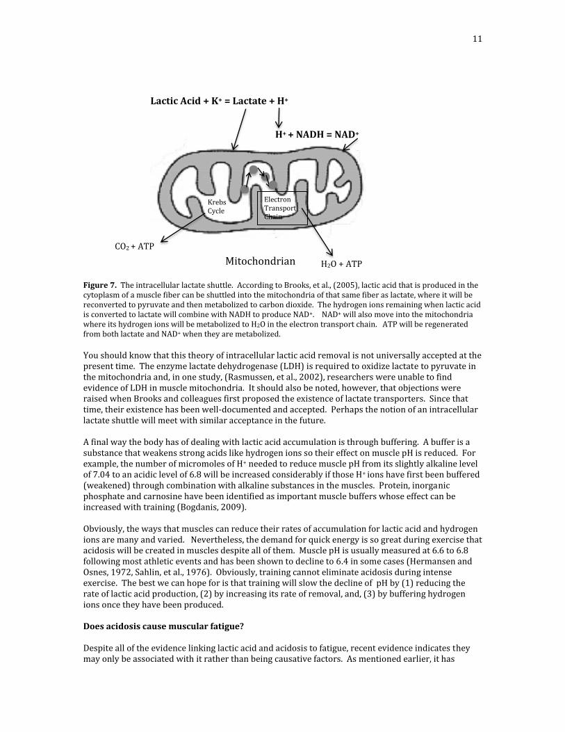
11
Figure 7. The intracellular lactate shuttle. According to Brooks, et al., (2005), lactic acid that is produced in the cytoplasm of a muscle fiber can be shuttled into the mitochondria of that same fiber as lactate, where it will be reconverted to pyruvate and then metabolized to carbon dioxide. The hydrogen ions remaining when lactic acid is converted to lactate will combine with NADH to produce NAD+. NAD+ will also move into the mitochondria where its hydrogen ions will be metabolized to H2O in the electron transport chain. ATP will be regenerated from both lactate and NAD+ when they are metabolized.
You should know that this theory of intracellular lactic acid removal is not universally accepted at the present time. The enzyme lactate dehydrogenase (LDH) is required to oxidize lactate to pyruvate in the mitochondria and, in one study, (Rasmussen, et al., 2002), researchers were unable to find evidence of LDH in muscle mitochondria. It should also be noted, however, that objections were raised when Brooks and colleagues first proposed the existence of lactate transporters. Since that time, their existence has been well-documented and accepted. Perhaps the notion of an intracellular lactate shuttle will meet with similar acceptance in the future. A final way the body has of dealing with lactic acid accumulation is through buffering. A buffer is a substance that weakens strong acids like hydrogen ions so their effect on muscle pH is reduced. For example, the number of micromoles of H+ needed to reduce muscle pH from its slightly alkaline level of 7.04 to an acidic level of 6.8 will be increased considerably if those H+ ions have first been buffered (weakened) through combination with alkaline substances in the muscles. Protein, inorganic phosphate and carnosine have been identified as important muscle buffers whose effect can be increased with training (Bogdanis, 2009). Obviously, the ways that muscles can reduce their rates of accumulation for lactic acid and hydrogen ions are many and varied. Nevertheless, the demand for quick energy is so great during exercise that acidosis will be created in muscles despite all of them. Muscle pH is usually measured at 6.6 to 6.8 following most athletic events and has been shown to decline to 6.4 in some cases (Hermansen and Osnes, 1972, Sahlin, et al., 1976). Obviously, training cannot eliminate acidosis during intense exercise. The best we can hope for is that training will slow the decline of pH by (1) reducing the rate of lactic acid production, (2) by increasing its rate of removal, and, (3) by buffering hydrogen ions once they have been produced. Does acidosis cause muscular fatigue? Despite all of the evidence linking lactic acid and acidosis to fatigue, recent evidence indicates they may only be associated with it rather than being causative factors. As mentioned earlier, it has
Lactic Acid + K+ = Lactate + H+
H+ + NADH = NAD+
CO2 + ATP
Mitochondrian H2O + ATP
Krebs Cycle
Electron Transport Chain
12
recently been discovered that earlier studies supporting acidosis as the precipitating cause of muscular fatigue may have suffered from a serous flaw. They were conducted with excised muscle tissue that had been frozen and then thawed to a temperature of only 150 C (590 F) before being tested. Several researchers have now reported that the effects of acidosis are significantly reduced or non-existent in muscle fibers that are warmed nearer normal body temperature. These scientists developed a technique for warming the tissue to 300 C (860F) and beyond before testing. As a result, they showed that acidosis did not reduce force and velocity when muscle tissue was first warmed near body temperature (300 C and 320C /900F), (Pate, et al., 1995, Bruton, et al., 1998, Westerblad, et al., 1997, Westerblad, et al., 2002, Zhang, et al., 2006). In one of these studies (Westerblad et al., 1997), acidosis caused a 28% decline in peak force for a single fast twitch fiber of mouse muscle at a lower temperature of 120C (540F) but only a 10% decline at 320C. In the same study, contraction velocity was reduced 20% at 120C while at 320C acidosis did not slow muscle contraction speed at all. Muscle relaxation speed was also reduced by acidosis but the effect was considerably greater at 120 versus 320. These results are summarized in Table 1. As a result, of these and similar findings, the belief that lactic acid accumulation and subsequent acidosis cause muscular fatigue is now being questioned. Nevertheless, there are still some researchers who subscribe to the acidosis theory. Table 1. The effect of acidosis on single muscle fiber contraction force and velocity at temperatures of 120 and 320. Muscle temperature
Contraction force
Contraction velocity
120 C (540 F) -28% -20% 320 C (900 F) -10% 0%
Data from: Westerblad, H., J.D. Burton, and H. Lannergren. (1997). The effect of intracellular pH of contractile function in intact, single fibers of mouse muscle declines with increasing temperature. Journal of Physiology, 500: 193-204.
One such group is Knuth and associates (2006). They set out to determine if acidosis and muscle temperature might affect slow and fast twitch muscle fibers differently. Previous studies on warmed muscle had been conducted with isolated muscle fibers of only one type. Pate and colleagues used the single fibers from the psoas muscle of rabbits in their study. This is a predominantly fast twitch muscle. Westerblad and colleagues also studied the effects of high temperatures on single fast twitch muscle fibers of mice. In other studies, mixed samples of fast and slow twitch muscle fibers were used where the effects on individual fibers types were not reported (Wiseman, Beck, and Chase, 1996). It is easy to see that the effect of acidosis at high temperature on a single fiber of could be misleading if fiber types differed in their response to these conditions. It is also possible that a combination of high muscle temperature and acidosis could reduce the contraction force and/or velocity for one type of fiber and that this result could be hidden by a compensatory effect on the other fiber type within a mixed group of fibers so that there appeared to be no loss of function. This is to say nothing of the fact that the detrimental effect of acidosis at high muscle temperatures on a particular fiber type, either fast or slow, might not show up in a mixed group of fibers that contained a preponderance of one type over the other. Therefore, Knuth and colleagues tested the effect of low pH (6.2) on both the force and contraction velocity of single fast- and slow-twitch muscle fibers at temperatures of 150C (590F) and 300C. Further, they looked at the effects of acidosis on the contracting power of each fiber type. As in previous studies, rats were used as subjects. A summary of their results is presented in Table 2. Following is a description of those results.
13
Table 2. The effect of low and high muscle temperatures on the peak force, contraction velocity, peak power, and loaded force and contraction velocity for slow twitch (ST) and fast twitch (FT) single muscle fibers of rats. Peak Force Contraction Peak Power Loaded force/velocity Velocity relationships Force Velocity
Muscle temperature
ST
FT
ST
FT
ST
FT
ST
FT
ST
FT
150 C (590F) -30% -30% -9% -27% -17% -37% -25% 0% -28% -14% 300 C (860F) -12% -4 to
11% -25% -32% -34% -18% -16% -22% --8% -11%
Data from: Knuth, S.T., H. Dave, J.R. Peters and R.H. Fitts. (2006). Low cell pH depresses peak power in rat skeletal muscle fibers at both 300 C and 150C: implications for muscle fatigue. Journal of Physiology, 575(3): 887-899.
Peak force. At 150C acidosis caused a 30% decrease in peak contraction force for both slow- and fast-twitch muscle fibers. These results were in agreement with those cited previously by Westerblad and associates (1997) for single fast twitch fibers of mouse muscle at a temperature of 120C. At a temperature of 300C, the peak force of slow twitch muscle fibers declined only 12%. This finding also agreed with that of Westerblad and associates who reported a 10% reduction for their sample of single fast twitch fibers at the higher temperature. The decline in peak force was also very similar for fast twitch fibers. It was 4% to 11%, in this study versus 10% in the study by Westerblad and colleagues. Contraction velocity. The effect of low pH on muscle contraction velocity was very different from that reported by Westerblad and associates. In their study, the contraction velocity of single fast twitch fibers of mouse muscle was affected very little if at all at higher muscle temperatures. However, Knuth and colleagues reported that slow twitch muscle fibers contracted 25% more slowly at 300C and 9% slower at 150C. These results were statistically significant. Apparently, the inhibiting effect of acidosis on the contraction velocity of slow twitch muscle fibers was increased considerably at a temperature closer to that of the human body during exercise. Also, in contrast to the earlier report, fast twitch fibers contracted significantly slower at both high and low muscle temperatures (27% slower at 150C vs. 32% slower at 300C) in the study by Knuth and colleagues. In this case, it appears that the contraction velocities of fast twitch fibers were reduced considerably by acidosis regardless of whether the temperature was low or high. The effect of low temperatures on the muscle contraction velocity of fast twitch fibers was similar to that reported by Westerfield and associates for single fast twitch fibers of mouse muscle at 120C (-20%). The result at a higher temperature was very different, however. Westerblad and colleagues reported no change in contraction velocity at a temperature of 320C whereas Knuth and co-workers reported a decrease of 32% at 300C. Peak power. In the next part of their study, Knuth and associates combined their results on contraction force and velocity into a measure of peak power. The effect of acidosis on peak power was found to be different for each fiber type although it was detrimental for both. The detrimental effect was greater on slow twitch muscle fibers at the higher temperature. Contractile power declined 34% in slow twitch fibers at a pH of 6.2 and a temperature of 300 C but only 17% at the same pH but a lower temperature of 150C. The opposite effect was observed with fast twitch fibers. Their peak power declined only 18% at the higher temperature compared to 37% at the lower. It should be noted that the decline in peak power for fast twitch fibers at the higher temperature was more marked than their decline in peak force
14
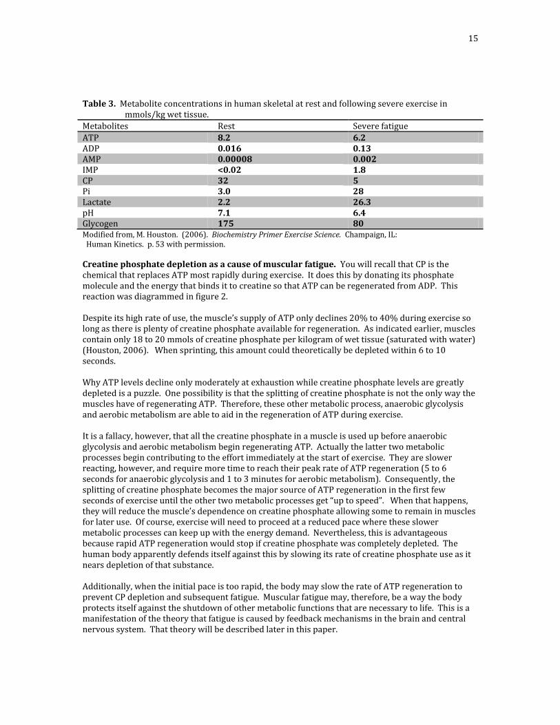
(-18% for power vs -4% to -5% for force). This indicates that, for fast twitch muscle fibers, a reduction of contraction velocity has a greater effect on muscle power than does a reduction in force (force x velocity = power). Thus, it appears that, in the study by Knuth and colleagues, acidosis caused a considerable loss of contractile power in both fiber types at temperatures near those of the human body (-34% for slow twitch and -18% for fast twitch). As you can see, the loss was greater for slow twitch fibers at the high compared to the lower muscle temperature. By contrast, peak power declined less at the higher temperature for fast twitch muscle fibers, although the drop of 18% was still quite significant. Force/velocity relationships. The usual procedure in studies of this type is to measure maximal muscle contraction velocity with no load on the muscle. Although valuable, measuring the velocity of unloaded muscle fibers is not as practical as measuring the effects on force and velocity when muscles are contracting against resistance. For this reason, Knuth and co-workers also measured the effects of three different loads on muscular force and velocity at both high and low temperatures during acidosis. Slow twitch fibers lost similar amounts of force at 150C (-25%) and 300C (-16%) when contracting against resistance during acidosis. At the same time, the contraction velocity of those fibers was not reduced at 150C during acidosis while it declined 22% at a temperature of 300C. Consequently, slow twitch fibers lost both contraction force and velocity at high temperatures when working against resistance during acidosis while losing only contraction force at the lower temperature. Both the force and velocity of fast twitch fibers declined when they worked against resistance during acidosis regardless of whether the muscle temperature was low or high. The loss of force was considerably less at the higher temperature, however, (-28% at 150C vs. -8% at 300C). The contraction velocity of fast twitch fibers declined similarly at both low and high temperatures (-14% at 150C and -11% at 300C). Therefore, fast twitch fibers exhibited declines in force and velocity at both high and low muscle temperatures, although, the effect on force was much less at the higher temperature while the effect on velocity was similar at both low and high temperatures. These results probably account for the fact that the power of fast twitch fibers decreased less at the higher temperature (-18% at 300C vs. -37% at 150C). The finding by Knuth and colleagues that acidosis does produce a detrimental effect on fast and slow twitch muscle fibers at near-normal body temperatures stands in marked contrast to those cited earlier single fast twitch fibers. The effect appears to be more pronounced in slow twitch fibers because they contract slower and with less force. Fast twitch fibers appear to maintain their force during acidosis but, like their slow twitch counterparts, contraction velocity declines, causing a smaller, but still significant, loss of power at higher temperatures. Obviously, the possibility that acidosis does not cause muscular fatigue is far from resolved. Let’s examine some of the other metabolic products that researchers are now associating with fatigue to determine their feasibility. Other possible causes of muscular fatigue. The use of non-invasive NMR techniques has enabled scientists to look at reductions and increases of substances such as ATP, CP, Pi, and H+, immediately following exercise. This research strongly suggests that CP depletion, and muscle calcium changes brought on by increases of inorganic phosphate (Pi) and ADP may be major causative factors for muscular fatigue. The concentrations at rest and after severe exercise of these and other metabolic products that may be associated with muscular fatigue are listed in Table 3. Since many scientists believe that creatine phosphate (CP) is the leading candidate, it will be discussed first.
15
Table 3. Metabolite concentrations in human skeletal at rest and following severe exercise in mmols/kg wet tissue.
Metabolites Rest Severe fatigue
ATP 8.2 6.2 ADP 0.016 0.13 AMP 0.00008 0.002 IMP <0.02 1.8 CP 32 5 Pi 3.0 28 Lactate 2.2 26.3 pH 7.1 6.4 Glycogen 175 80 Modified from, M. Houston. (2006). Biochemistry Primer Exercise Science. Champaign, IL: Human Kinetics. p. 53 with permission.
Creatine phosphate depletion as a cause of muscular fatigue. You will recall that CP is the chemical that replaces ATP most rapidly during exercise. It does this by donating its phosphate molecule and the energy that binds it to creatine so that ATP can be regenerated from ADP. This reaction was diagrammed in figure 2. Despite its high rate of use, the muscle’s supply of ATP only declines 20% to 40% during exercise so long as there is plenty of creatine phosphate available for regeneration. As indicated earlier, muscles contain only 18 to 20 mmols of creatine phosphate per kilogram of wet tissue (saturated with water) (Houston, 2006). When sprinting, this amount could theoretically be depleted within 6 to 10 seconds. Why ATP levels decline only moderately at exhaustion while creatine phosphate levels are greatly depleted is a puzzle. One possibility is that the splitting of creatine phosphate is not the only way the muscles have of regenerating ATP. Therefore, these other metabolic process, anaerobic glycolysis and aerobic metabolism are able to aid in the regeneration of ATP during exercise. It is a fallacy, however, that all the creatine phosphate in a muscle is used up before anaerobic glycolysis and aerobic metabolism begin regenerating ATP. Actually the latter two metabolic processes begin contributing to the effort immediately at the start of exercise. They are slower reacting, however, and require more time to reach their peak rate of ATP regeneration (5 to 6 seconds for anaerobic glycolysis and 1 to 3 minutes for aerobic metabolism). Consequently, the splitting of creatine phosphate becomes the major source of ATP regeneration in the first few seconds of exercise until the other two metabolic processes get “up to speed”. When that happens, they will reduce the muscle’s dependence on creatine phosphate allowing some to remain in muscles for later use. Of course, exercise will need to proceed at a reduced pace where these slower metabolic processes can keep up with the energy demand. Nevertheless, this is advantageous because rapid ATP regeneration would stop if creatine phosphate was completely depleted. The human body apparently defends itself against this by slowing its rate of creatine phosphate use as it nears depletion of that substance. Additionally, when the initial pace is too rapid, the body may slow the rate of ATP regeneration to prevent CP depletion and subsequent fatigue. Muscular fatigue may, therefore, be a way the body protects itself against the shutdown of other metabolic functions that are necessary to life. This is a manifestation of the theory that fatigue is caused by feedback mechanisms in the brain and central nervous system. That theory will be described later in this paper.
16
Creatine phosphate declines in two stages during exercise, a fast stage at the start of exercise, followed by a slower phase until the end of exercise. The two phases of CP decline are illustrated by the graph in figure 8. The rapidity of the initial drop and the severity of the final drop are both dependent upon the intensity of work. Athletes can reduce the rate of creatine phosphate use, by regulating their effort (pacing) so the muscle’s supply will not be depleted before the exercise is completed.
Figure 8. Creatine phosphate is depleted in two stages during exercise. A rapid stage, from the beginning of exercise until the rates of anaerobic and aerobic metabolism are increased (from 0 to 4 minutes in the figure), and a slower phase until the end of exercise (from 4 to 10 minutes in this figure). Adapted from G.A. Brooks, T.D. Fahey, and K.M. Baldwin. (2005). Exercise Physiology: Human Bioenergetics and Its Applications. Boston, MA: McGraw Hill Book Co., p. 854 with permission. One of the reasons creatine loading has become so popular is a belief that the rate of creatine phosphate depletion can be delayed during exercise by increasing the muscles’ supply prior to exercise. A number of studies have suggested this practice will increase performance and muscular strength. The most commonly used procedure for creatine loading is to consume 20 grams daily for 4 to 6 days. The 20 grams are ingested in four doses of 5 grams daily, separated by 4 to 5 hours. This has been shown to increase the CP content of muscles by an average of 11% (Houston, 2006). Athletes with the lowest initial levels of CP tend to store more while trained athletes and those with high initial levels will store less. Creatine supplementation appears to be most effective for improving performance in events lasting up to 3 minutes” (Houston, 2006). It also seems to improve performance when subsequent efforts are repeated with short recovery periods. Numerous reports from athletes indicate that creatine supplementation enables them to train harder because they recover faster. Many also report increases of muscle and strength with creatine loading. This effect has not been universally supported, however. Louis and associates (2003) found no such effect in their research, while, at the same time, Safdar and colleagues (2008) showed that creatine supplementation increased the expression of genes involved in muscle growth. On a negative note, it has been
17
suggested that sustained creatine supplementation will reduce its uptake by muscle cells over time so that the performance enhancing effect will become minimal at best (Snow and Murphy, 2003). Increases of Pi, ADP, and muscle temperature as causes of fatigue. Other possible causes of muscular fatigue have been suggested to result from increases of inorganic phosphate (Pi) and adenosine diphosphate (ADP) during muscular contraction. It should be mentioned that creatine phosphate metabolism is also associated with increases of inorganic phosphate because it (CP) is a major source of Pi during exercise. In addition, inorganic phosphate also increases when ATP is split and ADP is formed. As shown in figure 9 the splitting of ATP and release of its energy results in the formation of ADP (adenosine diphosphate) as well as inorganic phosphate which probably accounts for the fact that both are associated with muscular fatigue (Sahlin, 2009). ATP ADP + Pi Figure 9. The splitting of ATP and subsequent formation of ADP and Pi. Accumulation of the latter two substances may be responsible for reducing calcium release. This, in turn, results in a weak attachment of myosin to actin that reduces contraction force. The amount of free inorganic phosphate in muscles will increase when CP and glycogen are used to regenerate ATP. ADP also increases because the rate of ATP regeneration
slows as the amount of creatine phosphate in muscles is reduced. The manner in which increases of inorganic phosphate and ADP may contribute to fatigue is through their effect on calcium release in muscle fibers. Calcium, stored in the sarcoplasmic reticulum of muscle fibers, is essential to muscular contraction because it allows their myosin cross-bridges to attach to their actin filaments and pull them inward. This causes the fiber to shorten or contract. This is a partial description of the popular sliding filament theory of muscular contraction. This process is pictured and described further in figure 10.
Figure 10 The drawing on the left depicts a muscle fiber showing the myofibrils, the sarcoplasmic reticulum where calcium is stored, and the transverse tubules. The drawing on the right shows the arrangement of myosin and actin filaments within a myofibril. It also depicts the cross-bridges from the thicker myosin filament that allow it to attach to the thinner actin filament and exert an inward pull that causes the fiber to contract. When a nerve impulse, called an action potential, stimulates a muscle fiber, its charge spreads by way of the transverse tubules to the sarcoplasmic reticulum causing the release of calcium. Calcium spreads to the myofibrils allowing the cross-bridges from myosin to attach to actin and exert an inward pull. Adapted from Wilmore, Costill and Kenney (2008), p. 32, with permission.
Muscle fatigue can manifest itself in several different ways during contraction, all of which involve the muscles’ calcium supply. These are, (1) by a reduction in the rate of ATP regeneration, thereby, lessening the release of energy that powers contraction; (2) by a reduction in contraction force; (3) by a reduction in contraction velocity; and (4) by a reduction of contraction rate.
18
Contraction force may be reduced when an increase of inorganic phosphate reduces the release of calcium in muscles. This, in turn, interferes with the coupling of actin and myosin so that fewer and weaker bonds are formed. When this happens contraction force will be characterized as “weak” rather than “strong”. Obviously, when force declines, more fibers will have to contract to maintain a particular level of performance and fatigue will soon follow. Increases of inorganic phosphate, like those of hydrogen ions, are temperature and fiber type dependent. In one study, an increase of inorganic phosphate reduced muscle contractile force by 54% and 50% in slow and fast twitch muscle fibers respectively at a temperature of 150C. This effect was reduced to 19% in slow twitch muscle fibers at a temperature of 300C. Fast twitch muscle fibers were much less affected with force declining only 5% at 300C. Muscle contraction velocity was not reduced by a high level of inorganic phosphate at either low or high temperatures. On the other hand, peak power was reduced 49% for slow twitch and 40% for fast twitch fibers at 150probably because of the effect of increased inorganic phosphate on reducing muscle contractile force. Muscle contractile power was reduced only 16% for slow twitch and 18% for fast twitch fibers at 300
(Debold, et al., 2004). This result was obviously due to a smaller reduction of force and no reduction of contractile velocity at the higher temperature. Apparently, slow twitch fibers lose a significant amount of force at near-body temperature when inorganic phosphate increases, while the loss of force was much less pronounced but still significant in fast twitch muscle fibers. These results suggest to some, that an increase of inorganic phosphate may be the actual cause of muscle fatigue. In their opinion, acidosis, may, at most, reduce muscle contraction force somewhat more, or, it may simply accompany the burgeoning inorganic phosphate without, of itself, contributing to a loss of contraction force. In support of an enhancing effect, the results of one study showed that an increase of hydrogen ions and inorganic phosphate together caused muscles to fatigue more rapidly than an increase of Pi alone (Nosek, et al., 1987). It should be noted that the loss of peak power caused by high levels of inorganic phosphate was less for slow twitch fibers in the study by Debold and co-workers than the loss resulting from acidosis in the study by Knuth and colleagues (-16% Debold, et al., vs. -34% for Knuth et al.). It seems, therefore that acidosis may have a greater effect on slow twitch muscle fatigue at high temperatures than an increase of inorganic phosphate although both factors probably contribute. On the other hand, acidosis and an increase of inorganic phosphate appeared to contribute equally to reduced power of fast twitch fibers in the studies of Debold and co-workers and Knuth and colleagues. It declined by 18% in both cases. Another way an increase of inorganic phosphate can compromise muscle contraction force is by increasing the amount of calcium needed to produce an attachment of myosin with actin. Apparently, considerably more calcium is required to initiate contraction when inorganic phosphate accumulates in muscles. With less calcium released and more needed to maintain a particular level of force, it is easy to see why muscular force could be reduced by an increase of inorganic phosphate. In 2006, Debold and associates measured the calcium needed to produce strong contractions of both the slow twitch (soleus) and fast twitch (gastrocnemius) muscle fibers of rats at both high and low temperatures when inorganic phosphate was increased to an unusually high value of 30 mmols. Slow twitch fibers required more calcium to produce strong contractions regardless of whether the muscle temperature was 150 or 300.. However, they needed twice as much calcium at the higher temperature. By contrast, fast twitch fibers only required more calcium when their muscle temperature was increased to 300. As an aside to this topic, there is evidence that the stimulant caffeine can reduce the weakening effect of Pi on muscle contraction force. This is because caffeine stimulates calcium release allowing stronger contractions even when fibers are fatigued (Lannergren and Westerblad, 1989). Caffeine is
19
almost 100% absorbed into the bloodstream within 5 minutes, and ingestion of 2 to 3 mg of caffeine per kilogram of body weight has been shown to increase muscle power by 7% (Kovaks, et al., 1998). An increase of ADP has also been implicated in muscle fatigue because it reduces muscle contraction velocity. In one study, an increase of ADP decreased the contraction velocity of the toe muscles of rats (Westerblad, Dahlstadt, and Lannergren, 1998). In another, an increase of ADP reduced contraction velocity by more than 50% at a muscle temperature of 300C (Coupland, et al., 2005). A reduction of contraction velocity may be particularly detrimental in activities requiring fast, compared to slower movements. This is because a decrease of contraction velocity becomes more important to power output when the exercise requires fast movements (Westerblad and Allen, 2009). Consequently, a reduction of contraction velocity will reduce power considerably in activities where speed of movement is a major factor. By the same token, a reduction of contractile force will have a more limiting effect on power than a reduction of contractile velocity when exercise requires the exertion of a great amount of force. As indicated earlier, an increased amount of inorganic phosphate seems to have no effect on muscle contraction velocity (Cooke and Pate, 1985, Westerblad, et al., 1998). One way the body has of reducing the rate of ADP accumulation while increasing the energy supply is by using two ADP molecules to regenerate one molecule of ATP. Two ADP molecules can interact so that one phosphate and energy that binds it to adenosine will be transferred from one to the other, leaving one ATP and one AMP (adenosine monophosphate) molecule. This reaction is diagrammed in figure 11. Westerblad and co-workers (2002) have suggested that the effects of acidosis on muscle contraction can be overridden when the rate of ADP accumulation is reduced by this reaction. ADP + ADP ATP + AMP Figure 11. ATP, can be regenerated when two ADP molecules combine and produce one ATP and one AMP (adenosine monophosphate) molecule.
Increasing the time required for a muscle fiber to relax after contraction is another way that an increase of ADP may cause muscle fatigue. ADP accumulation (and acidosis) have been shown to reduce the rapidity of successive muscle contractions by increasing the time it takes for a fiber to relax between those contractions (Westerblad and Lannergren, 1991). Apparently an increase of ADP slows the time required for calcium to leave the contraction site and return to its place of storage in the sarcoplasmic reticulum in preparation for the next release. Consequently, it takes longer for the myosin cross-bridges to detach from actin. In one study, the half-time for muscle relaxation following contraction increased from 39 milliseconds to 128 milliseconds when the fibers were fatigued (Sjoholm, et al., 1983.) As would be expected, a decrease in the muscle fiber contraction rate will reduce muscle power. Increases of both inorganic phosphate and ADP can also inhibit the activity of enzymes that are involved in the regeneration of ATP, slowing it, and, thus, reducing the rate of energy release for muscular contraction (Sahlin et al., 1998). This has been shown to cause muscle fatigue at low muscle temperatures. Whether this effect is also present at body temperatures during exercise remains to be seen. Finally, high muscle temperatures during exercise may also precipitate muscle fatigue. An increase of muscle temperature may contribute to reduced contractile force because more calcium appears to be needed to produce strong myosin/actin bonds when temperatures are nearer those of the human body. As mentioned earlier, the amount of calcium required to effect contraction was increased markedly at 300C for both ST and FT muscle fibers compared to the amount required to produce the same contraction force at 150C (Debold et al., 2006). This was particularly true for slow twitch fibers where the increase was nearly double that required by fast twitch fibers.
20
Conclusions and implications for training and research. Lactic acid has been touted as the primary cause of muscular fatigue in most athletic activities for several decades now. This was based on the fact that the hydrogen ions released from it during exercise caused a movement of muscle pH from neutral (higher pH) to acidic (lower pH). Low muscle pH, known as acidosis, was believed to reduce performance by slowing the rate of energy release and by reducing the force, velocity and rate of muscle contraction. However, early research into this matter may have been flawed because it was conducted with groups of muscle fibers and single fibers that had been frozen and thawed to a temperature that was far below that of the human body. With improved techniques it has become possible to study the effect of acidosis on fibers that have been warmed to near-normal body temperature. The results of studies where these higher temperatures were used showed that the effects of pH on muscle performance were reduced considerably or disappeared entirely. This has prompted a re-evaluation of the role of lactic acid in muscular fatigue. At the present time many prominent experts in exercise science no longer believe that acidosis causes muscular fatigue while others continue to assert its importance as a causative factor. Let me present a summary of the cases for and against acidosis using information that was discussed in this paper. The case against acidosis will be summarized first since it has generated the most controversy during the past several years. The Case Against Acidosis. We have known for decades that lactic acid accumulates rapidly in muscles during hard exercise and that this was accompanied by a reduction of performance. Consequently, it was quite natural to assume a causative relationship between lactic acid accumulation and muscular fatigue. However, the finding that muscle contraction force and velocity were not greatly reduced by acidosis at higher temperatures, has led many researchers to doubt the causative role of lactic acid in muscular fatigue. They believe the relationship between the two may have been coincidental rather than causative. That is, the accumulation of lactic acid may simply accompany or, perhaps, enhance the effect of other metabolic products that are the primary culprits where muscular fatigue is concerned. As mentioned, Westerblad and co-workers (1998) were able to fatigue the toe muscle of rats without acidosis occurring. Even those who think lactic acid plays a minor role in muscular fatigue do not believe it is the most potent causative factor because its effect on muscle pH can be overridden by the action of other metabolic substances. For example, although acidosis is believed to reduce the rate of energy release via anaerobic glycolysis, there is evidence that this effect can be overridden by increases in muscle ADP and AMP (Sahlin, 1986, Westerblad, et al., 2002). Thus, muscles might still be capable of regenerating ATP despite a low pH. Other research that was cited earlier in this paper also indicates the possibility that the effects of low pH are eliminated, reduced or “overridden” by factors such as increases of muscle temperature, inorganic phosphate, and ADP, as well as by a decrease of creatine phosphate in muscles. This may be particularly true of fast twitch muscle fibers, because a much greater nervous stimulation is needed to activate them and, because their rates of anaerobic metabolism and, thus, their muscle power output are much higher during exercise. The rate of creatine phosphate depletion gets top billing by many as the event that causes muscular fatigue. This may happen because fail-safe mechanisms in the central nervous system and/or the muscles of the body slow muscle contraction speed when they sense uncommon distress. For example, one or both may sense that the muscle supply of CP is being depleted too rapidly and, therefore, will slow muscle force and speed in order to slow the rate of ATP regeneration so the demand for energy can be met through anaerobic and aerobic metabolism. Other possible causes of muscular fatigue that have been mentioned are increases of inorganic phosphate and ADP. Free inorganic phosphate may slow the rate of calcium release from the
21
sarcoplasmic reticulum so that muscle force declines. It is possible that this effect can be overridden during acidosis but not when inorganic phosphate increases in muscles. At the same time, an increase of ADP may reduce muscle contraction velocity when CP is nearing depletion. Support for creatine phosphate depletion, high body temperature and increases of inorganic phosphate and ADP as causes of muscular fatigue comes from many sources. The fact that muscle contraction velocity is unaffected by acidosis at high temperatures is one of these (Pate et al., 1995, Westerblad, et al., 1997, Bruton, et al., 1998). Although these results have been refuted by Knuth and associates (2006), for both slow and fast twitch muscle fibers, the possibility exists that some unforeseen factor caused the discrepancy in that study and that acidosis is associated with but does not cause muscle fatigue. Other arguments against lactic acid come from the results of several studies where the rate of fatigue development in single muscle fibers was not increased when acidosis was exaggerated (Bruton, 1998, Zhang, 2006). Also, Lamb and co-workers (1992) reported that acidosis did not reduce calcium release in muscle fibers while Sahlin and Ren (1989) reported that muscle force was restored within a few minutes of recovery following exercise despite the fact that muscle pH was still below 7.0. Further evidence against lactic acid accumulation and subsequent acidosis as a major cause of muscular fatigue comes from the results of several studies where an increase of inorganic phosphate produced reductions in force and power in both slow and fast twitch muscle fibers at high muscle temperatures that are greater than those produced by acidosis. (Debold, et al., 2004 and 2006). In one of these, an increase of inorganic phosphate reduced muscle force by 26% in slow twitch fibers and 18% in fast twitch fibers. By contrast, acidosis reduced muscular force by only 12% in slow twitch and by 4% to 5% in fast twitch fibers in the study by Knuth and colleagues. Additionally, if the results of different studies can be compared, muscle contraction power, as reflected by a loss of force at different contraction velocities was reduced more by an increase of inorganic phosphate than by acidosis. Power was reduced 38% in slow and 30% in fast twitch fibers when the inorganic phosphate content of those fibers was increased (Debold, et al., 2004). The loss of power was similar to that during acidosis (34%) in slow twitch muscle fibers. Fast twitch fibers exhibited a much smaller loss of power (18% ) during acidosis, however (Knuth, et al., 2006). Debold and colleagues believed their results were due to the effect of inorganic phosphate on slowing fiber relaxation time. They also reported, in 2006, that an increase of inorganic phosphate increased the amount of calcium needed for muscle contraction to a greater extent at high compared to low muscle temperatures and that this effect was two times greater for fast twitch than it was for slow twitch muscle fibers. Where muscle contraction velocity is concerned, increases of muscle inorganic phosphate have not been found to affect it at either high or low temperatures (Fitts, 2008). An increase of ADP has been reported to slow contraction velocity considerably, however, (Cooke and Pate, 1985; Westerblad, et al., 1998). Whether the differences in these percentage performance decrements indicate greater causation by inorganic phosphate and ADP compared to acidosis remains to be seen. Nevertheless, the possibility must be considered that an increase of one or both may have a more powerful influence on muscular fatigue than does acidosis. We will have to await further research for an answer. The Case For Acidosis. Perhaps the strongest evidence that acidosis plays a major role in muscular fatigue has been provided by the research of Knuth and colleagues (2006). Contradicting the results of other studies that showed little or no effect of acidosis on muscle performance at temperatures near those of the human body, they demonstrated that muscle contractile force, velocity and power were reduced by acidosis when muscle temperatures were 300C. Where muscle contraction force
22
was concerned their results showed, in agreement with other studies, that it was reduced less at higher, compared to lower temperatures, however. The magnitude of that reduction differed by fiber type, however. The contraction force of slow twitch fibers was significantly reduced by 12% at a higher temperature compared to a 30% reduction at a lower temperature. In fast twitch muscle fibers, contractile force was not reduced significantly by acidosis at the higher temperature. The decrease was only 4% to 11% at the higher temperature (300C) versus 30% at a lower temperature (150C). The fact that the deleterious effect of acidosis on the contraction force of muscle fibers did not disappear entirely at higher temperatures in this and earlier research should not be dismissed lightly. Reductions ranging from 4% to 11% (Knuth et al., 2006) and 18% (Pate, et al., 1995) for fast twitch fibers in two studies should have a detrimental effect on performance when the margin of victory is often measured in portions of an inch and hundredths of a second. Consequently, acidosis may have a significant detrimental effect on performance even if that effect is not as debilitating as once believed. According to Knuth and associates, acidosis definitely had a detrimental effect on muscle contractile velocity at higher temperatures. In fact, performance was reduced more for slow twitch muscle fibers at a high versus a low muscle temperature. They lost 27% of their contraction speed at a higher temperature and only 9% at a lower temperature. Reductions of contractile velocity were considerable for fast twitch fibers at both high and low temperatures although they were slightly, but non-significantly greater, at the high temperature (27% at 150C versus 32% at 300C). Reductions of these magnitudes should be sufficient to reduce muscle performance at both high or low temperatures, therefore, they support the contention that acidosis can cause fatigue at typical exercise muscle temperatures. This finding was in stark contrast to the results of Westerblad and associates (1997), who found no slowing of fast twitch contraction velocity at higher temperatures. Knuth and associates also looked at aspects of muscle performance that were not included in previous studies. They measured the effect of acidosis on the amount of muscular force at various contraction velocities and they measured the effect of acidosis on muscle power (the product of force and velocity). As you would expect, the force exerted by slow twitch muscle fibers was reduced more at each different contraction velocitiy when the muscle temperature was 300C than it was reduced at a temperature of 150C. Fast twitch muscle fibers demonstrated less reduction in force at a higher temperature and a similar reduction of contraction velocity at both high and low muscle temperatures. Peak power is considered the most discriminating measure of muscle performance because it reflects the ability to do work. Knuth and associates reported that power declined for both fiber types at high temperatures during acidosis, although the effect on fast twitch fibers was less pronounced. Their power diminished by 18% at 300C versus 37% at 150C. At the same time, slow twitch muscle fibers demonstrated a significantly greater loss of power at the high, compared to the lower temperature. They lost 34% of their contraction power at 300C versus 17% at 150C Contrary to the results of others, Knuth and co-workers (2006) demonstrated that acidosis had a profound detrimental effect on muscle performance when that effect was differentiated according to fiber type. While the effect was more pronounced in slow twitch muscle fibers it was, nevertheless, significant in fast twitch fibers as well. The 18% reduction in their muscular power at a higher temperature, although less than the 37% reduction at a lower temperature, should be of sufficient magnitude to exert a detrimental effect on performance. Why did Knuth and co-workers find that acidosis caused muscular fatigue at high temperatures when the results of other researchers indicated otherwise? Perhaps it was because studies that demonstrated little if any effect of acidosis on muscle contraction force and velocity were done with
23
groups of mixed muscle fibers where the proportions of fast and slow twitch fibers were not identified, or with single fast twitch fibers. As mentioned earlier, this may have allowed fast twitch fibers to compensate for the reduced performance of slow twitch fibers in mixed samples so that muscle performance did not appear to be affected in an adverse manner. Whereas the profound effect of acidosis on single slow twitch muscle fibers was missed in studies where only single fast twitch fibers were used. Measures of muscle contraction power left no doubt that acidosis had a detrimental effect on performance of both slow and fast twitch muscle fibers in the study by Knuth and co-workers, however. There was a significant loss of muscular power in both fiber types at high muscle temperatures, 34% for slow twitch muscle fibers and 18% for fast twitch fibers. It was mentioned earlier that a reduced rate of calcium release, and not acidosis, is thought by some to be the cause of muscle fatigue. Another compelling bit of support for acidosis as the actual cause of fatigue is that peak force decreases during acidosis even when the rate of calcium release is high in muscle fibers. If an increase of inorganic phosphate was the culprit, we would expect to see an inhibition of calcium release accompanying the loss of muscular force. This suggests that an increase of hydrogen ions will cause fatigue even when the muscle release of inorganic phosphate is high. Along this same line, the finding that the reduction in calcium release that accompanies an increase of inorganic phosphate is intensified by an increase of hydrogen ions is another point in support of acidosis as a major factor in muscular fatigue. Further support for acidosis is found in the fact that increased amounts of calcium are required to produce a particular level of force when muscle pH is low (Westerblad, et al., 2000). Acidosis has also been demonstrated to cause a slowing of muscle relaxation time even at near-body temperatures (Bruton, et al., 1998). In addition, Westerblad and Lannergren (1991) reported muscle fibers did not take longer to relax between fatiguing contractions when acidosis was prevented. It seems, therefore, that acidosis is a major causative factor even though calcium sensitivity and muscle relaxation time may be associated with muscular fatigue. In another attempt to substantiate the role of acidosis in muscular fatigue, Bangsbo and colleagues (1996) showed that the endurance of leg muscles was reduced if the lactate concentration of the legs was increased by arm exercise prior to testing the legs. Of course, it could be argued that the increase of lactate was coincidental in that, exercise may have also increased other metabolites that were responsible for reducing leg endurance. Nevertheless, this is one more indication that acidosis may cause muscular fatigue. Finally, there is a theory that muscle fatigue is the result of feedback mechanisms that reside in the central nervous system , (Noakes, 2000). A precept of this theory is that the central nervous system (the brain and autonomic nervous system) sense unusual bodily distress during exercise and try to lessen that distress by reducing (inhibiting) the rate of muscular contraction and, perhaps, by changing the pattern of muscle activation toward greater recruitment of enduring slow twitch muscle fibers in preference to fast twitch fibers. The brain can sense distress from several different sources during exercise. A low muscle pH, and an unusually high heart rate are two of the more prominent sources of distress as are an unusually high respiratory rate and body temperature. The brain is predisposed to a certain level of distress during a particular exercise and will send out inhibiting messages to the heart, lungs and muscles when that level is reached. Noakes believes that improved performances occur when those inhibiting effects are removed during training by “re-programming” the brain to accept a previously inhibiting level of distress as manageable. In this respect, acidosis may be the most prominent distressing event that stimulates the feedback mechanisms of pain, nausea, breathlessness, and high heart rates that are responsible for both reducing the contraction rate and changing the recruitment pattern toward greater reliance on slower and less powerful fiber types. It is also possible that the effect of acidosis on performance may be mitigated by psychological factors as much or more so than by those that are physiological in nature. That is, athletes may consciously
24
slow their efforts and their paces as acidosis progresses because they lose the motivation to push on, or because they become frightened that they will not be able to complete the event at a respectable level of effort. In the meantime, an important question to consider is “What are the implications of this information for training athletes?”. Implications for training. It appears now that muscular fatigue may be more complicated than we once thought. Lactic acid accumulation and the acidosis it produces may not be the only cause of muscular fatigue nor even the most crippling cause. The rates of creatine phosphate use and the rate of inorganic phosphate and ADP increases during exercise may fatigue muscles even more. Whereas, lactic acid accumulation and acidosis may only accompany or, perhaps, enhance their effect. Based on this information, “Have we been training the wrong physiological processes all these years? For decades, our approach to training has been concerned with improving the physiological mechanisms that reduce lactic acid production and accumulation. Training methods that were proven to increase cardiac output, VO2max, and buffering capacity were front and center in this effort. Recent additions to this list have been training that increases mitochondrial density and lactate transporters. Do these methods require a radical change or only some modifications? I don’t believe a radical change is required. Our training methods have worked even though their reasons for doing so may be different than we once thought. It is certainly evident that fatigue results from the need for high rates of energy release and the increased amount of anaerobic metabolism required to accomplish that end. Therefore, training methods that have been shown to reduce the demand for anaerobic metabolism or to mitigate its deleterious effects, i.e. improved buffering, should continue to be justified even if increases and decreases of byproducts of the anaerobic process other than lactic acid prove to cause muscular fatigue. That being said, some modifications to our traditional training methods may be in order while some suggestions for future research certainly are. More emphasis should be placed on training that will increase mitochondrial density, rather than VO 2max (although the latter will also increase as a result of that training). The ways that VO2max can be improved by training have been studied extensively. However, evidence is mounting that the size and number of mitochondria in muscle fibers and not oxygen consumption may actually determine the rate of aerobic metabolism during exercise and, therefore, the contribution it can make to the total energy supply during exercise. An increase in mitochondrial density should certainly reduce an athlete’s reliance on anaerobic metabolism even if the actual reason for doing so is to reduce the rate of creatine phosphate use and the rates of increase of inorganic phosphate and ADP rather than reducing the rate of lactic acid accumulation. Training methods that will increase cardiac output should continue to occupy a “front and center” role in training. A substantial increase of cardiac output will continue to carry more nutrients to the muscles and increase the rate of metabolite removal even if reducing the muscle content of certain of those metabolites is more important to maintaining performance than the removal of lactate. Buffering capacity is the questionable process. If acidosis is not a cause of muscular fatigue, perhaps, improving buffering capacity is not important to performance. Yet, there is a reasonable amount of scientific evidence that indicates improved performance and increased buffering capacity go hand in hand. (This is an important point in support of acidosis as a major cause of muscular fatigue). Consequently, until we know for sure that acidosis is not involved, it would be wise to continue training to improve buffering capacity. In the meantime, research on reducing the rate of creatine phosphate use during exercise, its rate of restoration after exercise, and the effects of supplementation of this substance on performance should be accelerated. Research on ways that the accumulation of inorganic phosphate and ADP can
25
be reduced, mediated and/or removed from the working muscles during exercise should also become a priority. Likewise, new or different training techniques that may achieve these effects should also be investigated. We should also consider the possibility that muscular fatigue results from feedback mechanisms that elicit inhibiting responses by the brain and central nervous system. Therefore, coaches and athletes should pay more attention to training at race pace while gently and progressively removing inhibitions that allow them to increase that pace or sustain it for a longer period of time. This requires applying the principle of progressive overload to training. That is, (1) swimming progressively more repeats at race pace, (2) swimming at race pace with progressively less rest between repeats and sets of repeats, and, (3) swimming standard sets of repeats at progressively faster average speeds. Finally, we should not dismiss the role of lactic acid as inconsequential. After all, at the present time, acidosis not been absolutely discredited as a cause of muscular fatigue References: Bangsbo, J., K. Madsen, B. Kiens, and E.A. Richter. (1996). Effect of muscle acidity on muscle Metabolism and fatigue during intense exercise in man. Journal of Physiology (London), 495: 587-596. Bogdanis, G.C. (2009). Fatigue and training status. In C. Williams and S. Ratel (eds). Human Muscle Fatigue. New York, NY: Routledge, pp. 164-204. Brooks, G.A., T.D. Fahey, and K.M. Baldwin. (2005). Exercise Physiology: HumanBioenergetic and Its Application. New York, NY: McGraw-Hill. Brooks, G.A., H. Dubouchaud, M. Brown, J.P. Sicurello, and C.E. Butz. (1999). Role of mitochondrial lactic dehydrogenase and lactate oxidation in the “intra-cellular lactate shuttle.” Proceedings of the National Academy of Science. 96: 1129-1134. Bruton, J.D., J. Linnergren, and H. Westerblad. (1998). Effects of CO2-induced acidification on the fatigue resistance of single mouse muscle fibers at 280C. Journal of Applied Physiology. 85: 478-483. Coggan, A.R., S.C. Swanson, L.A. Mendenhall, D.L. Habash, and C.I. Kien. (1995). Effect of endurance training on hepatic glycogenolysis and gluconeogenesis during prolonged exercise in men. American Journal of Physiology. 268: E375- E383. Cooke, R. and E. Pate. (1985). The effects of ADP and phosphate on the contraction of muscle fibers. Biophysical Journal, 48: 789-798. Coupland, M.E., G.J. Pinniger and K.W. Ranatunga. (2005). Endothermic force generation, Temperature-jump experiments and effect of increased (MgADP) in rabbit psoas muscle. Journal of Physiology, 567(Pt 2): 471-492. Davies, K.J.A., L. Packer and G.A. Brooks. (1981). Biochemical adaptation of mitochondria, muscle, and whole-animal respiration to endurance training. Archives of Biochemistry and Biophysiology. 209: 539-554. Davies, K.J.A., L. Packer and G.A. Brooks. (1982). Exercise bioenergetics following sprint training. Archives of Biochemistry and Biophysiology. 215: 260-265. Debold, E.P., H. Dave, and R.H. Fitts. (2004). Fiber type and temperature dependence of inorganic phosphate: implications for fatigue. American Journal Physiology and Cell Physiology, 287: C673-C681. Debold, E.P., J. Romatowski, and R.H. Fitts. (2006). The depressive effect of Pi on the force calcium relationship in skinned single muscle fibers is temperature dependent. American Journal of Physiology and Cell Physiology, 290: C1041- C1050. C.M, Donovan & M.J. Pagliassotti. 1990. Enhanced efficiency of lactate removal after endurance training. J. Appl. Physiol. 68(3):1053-1058. Fitts, R.H. (2008). The cross-bridge cycle and skeletal muscle fatigue. Journal of Applied Physiology, 104: 551-558.
26
Hermansen, L. and J. Osnes. 1972). Blood and muscle pH after maximal exercise in man. Journal of Applied Physiology, 32: 304-308. Hill, A.V, C.N.H. Long and H. Lupton. (1924). Muscular exercise, lactic acid, and the supply and utilization of oxygen (Pt. I to VIII). Procedures Royal Society (London) Ser. B, 96: 438-475; 97: 84-138; 97: 155-176. Holloszy, J.O. (1975). Adaptation of skeletal muscle to endurance exercise. Medicine and Science in Sports. 7(3): 155-164. Hood, D.A. (2001). Contractile activity-induced mitochondrial biogenesis in skeletal muscle. Journal of Applied Physiology, 90: 1137-1157. Houston, M. (2006). Biochemistry Primer for Exercise Science. Champaign, IL: Human Kinetics. Kovaks, E., J. Stegan, and F. Brouns. (1998). Effect of caffeinated drinks on substrate metabolism, caffeine excretion, and performance. Journal of Applied Physiology, 85: 709-715. Knuth, S.T., H. Dave, J.R. Peters and R.H. Fitts. (2006). Low cell pH depresses peak power in rat skeletal muscle fibers at both 300 C and 150C: implications for muscle fatigue. Journal of Physiology, 575(3): 887-899. Lannergren J. and H. Westerblad. (1989). Maximum tension and force-velocity properties of fatigued, single Xenopus muscle fibers studied by caffeine and high K+. Journal of Physiology (London), 409: 473- 490. Lamb, G.D., E. Recupero, and D.G. Stephenson. (1992). Effect of myoplasmic pH on excitation- contraction coupling in skeletal muscle fibers of the toad. Journal of Physiology (London), 409: 211 - 224. Louis, M., J.R. Poortmans, M. Franceaux, E. Hultman, J. Berre, N. Boisseau, V.R. Young, K. Smith, J.A. Babraj, T. Waddell, and M.J. Rennie. (2003). No effect of creatine supplementation on human myofibrillar and sarcoplasmic protein synthesis after resistance exercise. American Journal of Physiology, 285: E1089 - E1094. Martyn, D.A. and A.M. Gordon. (1992). Force and stiffness in glycerinated rabbit psoas fibers. Effects of calcium and elevated phosphate. Journal of General Physiology, 99: 795-816. Noakes, T.D. (2000). Physiological models to understand exercise fatigue and the adaptations that predict or enhance athletic performance. Scandanavian Journal of Medicine and Science in Sports, 10(3): 123-145. Nosek, T.M., K.Y. Fender, and R.E. Godt. (1987). It is dipronated inorganic phosphate that depresses force in skinned skeletal muscle fibers. Science, 236: 191-193. Pate, E., M. Bhimani, K. Franks-Skiba, and R. Cooke. (1995). Reduced effect of pH on skinned rabbit psoas muscle mechanics at high temperatures: implications for fatigue. Journal of Applied Physiology, 486: 689-694. Rasmussen, H.N., G. van Hall, and U.F. Rasmussen. (2002). Lactate dehydrogenae is not a mitochondrial enzyme in human and mouse vastus lateralis muscle. Journal of Applied Physiology, 541: 575-580. Safdar, A., N.J Yardley, R. Snow, S. Melov, and M.A. Tarnopolsky. (2008). Global and targeted gene expression and protein content in skeletal muscle of young men following short-term creatine monohydrate supplementation. Physiological Genomics, 32: 219 – 228. Sahlin, K. (1986). Muscle fatigue and lactic acid accumulation. Acta Physiologica Scandanavica (Supplement), 556: 83–91. Sahlin, K. (2009). Metabolic factors in fatigue. In M. Hargreaves and L. Spriet (eds.). Exercise Metabolism. Champaign, IL.: Human Kinetics, pp. 163-186. Sahlin, K. and J.M. Ren. 1989). Relationship of contraction capacity to metabolic changes during recovery from a fatiguing contraction. Journal of Applied Physiology, 67: 648-654 Sahlin, K., M. Tonkonogi, and K. Soderlund. (1998). Energy supply and muscle fatigue in humans. Acta Physiologica Scandanavica, 162: 261-266. Sahlin, K., R.C. Harris, B. Nylind, and E. Hultman. (1976). Lactate content and pH in muscle samples obtained after dynamic exercise. Pflugers Archives, 367: 143 - 149. Sjoholm, H. K. Sahlin, . Edstrom, and E. Hultman. (1983). Quantative estimation of anaerobic and oxidative energy metabolism and contraction characteristics in intact human skeletal muscle in response to electrical stimulation. Clinical Physiology, 3: 227-239 Snow, R.J. and R.M. Murphy. (2003). Factors influencing creatine loading into human skeletal
27
muscle. Exercise and Sport Sciences Reviews. 31: 154-158. Spriet, L.L. and M. Hargreaves. (2006). Overview of exercise metabolism. In M. Hargreaves and L. Spriet (eds.). Exercise Metabolism. Champaign, IL.: Human Kinetics, pp. 1-6 Westerblad, H., and D.G. Allen. (2009). Cellular mechanisms of muscular fatigue. In C. Williams and S. Ratel (eds). Human Muscle Fatigue. New York, NY: Routledge, pp. 48-75. Westerblad, H. and J. Lannergren. (1991). Slowing of relaxation during fatigue in single mouse muscle fibers. Journal of Physiology, 434: 323-336. Westerblad, H., J.D. Burton, and H. Lannergren. (1997). The effect of intracellular pH of contractile function in intact, single fibers of mouse muscle declines with increasing temperature. Journal of Physiology, 500: 193-204. Westerblad, H., A.J. Dahlstadt, and J. Lannergren. (1998). Mechanisms underlying reduced maximum shortening velocity during fatigue of intact, single fibers of mouse muscle. Journal of Physiology, 510:269-277 Westerblad, H., J.D. Bruton, D.G. Allen, and J. Lannergren. (2000). Functional of Ca2+ in long-lasting fatigue of skeletal muscle. European Journal of Applied Physiology, 83: 166-174. Westerblad, H., D.G. Allen, and J. Lannergren. (2002). Muscle fatigue: lactic acid or inorganic phosphate the major cause? News in Physiological Sciences, 17: 17- 21. Wilmore, J.H., D.L. Costill, and W. L. Kenney. (2008). Physiology of Sport and Exercise. Champaign, IL: Human Kinetics. Wilson, J.R., K.K. McCully, D.M. Mancini, B. Boden, and B. Chance, (1988). Relationship of muscular fatigue to pH and deprotonated Pi in humans: A 31P- NMR study. Journal of Applied Physiology, 64: 2333-2339. Wiseman, R.W., T.W. Beck, and P.B. Chase. (1996). Effect of intracellular pH on force development Depends on temperature in intact skeletal muscles from mouse. American Journal of Physiology, 271: C878-C886. Zhang, S.J., J.D. Bruton, A. Katz, and H. Westerblad. (2006). Limited oxygen diffusion accelerates fatigue development in mouse skeletal muscle. Journal of Physiology, 572: 551-559.