pH-Dependent mismatch discrimination of oligonucleotide duplexes containing 2 0 -deoxytubercidin and 2- or 7-substituted derivatives: protonated base pairs formed between 7-deazapurines and cytosine Xiaohua Peng 1,2 , Hong Li 1,2 and Frank Seela 1,2, * 1 Laboratorium fu ¨r Organische und Bioorganische Chemie, Institut fu ¨r Chemie, Universita ¨t Osnabru ¨ck, Barbarastrasse 7, D-49069 Osnabru ¨ck, Germany and 2 Laboratory of Bioorganic Chemistry and Chemical Biology, Center for Nanotechnology (CeNTech), Heisenbergstrasse 11, D-48149 Mu ¨nster, Germany Received June 20, 2006; Revised and Accepted September 18, 2006 ABSTRACT Oligonucleotides incorporating 2 0 -deoxytubercidin (1a), its 2-amino derivative 2a and related 2-, or 7-substituted analogs (1d, 2b–d, 3 and 4) are synthe- sized. For this purpose, a series of novel phospho- ramidites are prepared and employed in solid-phase synthesis. Hybridization experiments performed with 12mer duplexes indicate that 7-halogenated nucleosides enhance the duplex stability both in antiparallel and parallel DNA, whereas 2-fluorinated 7-deaza-2 0 -deoxyadenosine residues destabilize the duplex structure. The 7-deazaadenine nucleo- sides 1a, 1d and their 2-amino derivatives 2a–d form stable base pairs with dT but also with dC and dG. The mispairing with dC is pH-dependent. Ambiguous base pairing is observed at pH 7 or under acid conditions, whereas base discrimination occurs in alkaline medium (pH 8.0). This results from protonated base pairs formed between 1a or 2a and dC under neutral or acid condition, which are destroyed in alkaline medium. It is underlined by the increased basicity of the pyrrolo[2,3-d]pyrimidine nucleosides over that of the parent purine com- pounds (pK a values: 1a = 5.30; 2a = 5.71; dA = 3.50). INTRODUCTION Mutations in the DNA molecule are the basis of evolution. It is widely accepted that tautomerism of the canonical nucle- obases and the formation of wobble base pairs play an impor- tant role in this phenomenon (1,2). To keep the number of errors low, enzymatic proof reading during nucleoside triphosphates incorporation takes place with the help of polymerases (3). DNA mutation is caused by mismatches of the normal bases because of a failure of proofreading during DNA replication (4). DNA is also damaged continuously by oxidation, by depurination, by light or other processes occur- ring within the cellular environment. The daily number of errors in a human is estimated to be several thousands. This damage is removed by repair enzymes (5). Several diagnostic tools have been developed to detect such single nucleotide polymorphisms (SNPs) by hybridiza- tion in solution or on polymer surfaces (biochips). Modified nucleosides are used in these protocols as fluorescent dyes to be anchored to them without disturbing the DNA structure (6,7). 7-Deazapurine nucleoside triphosphates are commonly used for these purposes (6–8). Thus, the knowledge about their recognition properties and base discrimination is of mutual interest. Mismatch discrimination is evaluated from the difference in melting temperatures (T m ) between matched and mismatched base pairs within an oligonucleotide duplex. For a given mismatch, the properties of the modified nucleo- sides incorporated in the DNA chain and the environmental conditions are of utmost importance for the stability of base pairs. Although studies on the mispairing of modified nucleo- sides have been performed, little attention has been paid to the influence of the pH values of the reaction medium on the recognition of canonical and modified nucleosides. Among the modified nucleosides, 7-deazapurine (pyrrolo [2,3-d]pyrimidine) nucleosides and 7-substituted derivatives (purine numbering is used throughout the discussion) have attracted attention because they closely resemble the structure of purine nucleosides and are therefore ideal shape mimics of the canonical DNA constituents. They are well accepted by DNA polymerases and made a significant contribution to DNA and RNA sequencing and diagnostics (6–9). Reporter groups that are necessary to generate high-sensitivity probes are usually introduced at the 7-position of a 7-deazapurine giving them steric freedom in duplex DNA. Substituents of *To whom correspondence should be addressed. Tel: +49 541 969 2791; Fax:+49 541 969 2370; Email: [email protected]Ó 2006 The Author(s). This is an Open Access article distributed under the terms of the Creative Commons Attribution Non-Commercial License (http://creativecommons.org/licenses/ by-nc/2.0/uk/) which permits unrestricted non-commercial use, distribution, and reproduction in any medium, provided the original work is properly cited. Published online 27 October 2006 Nucleic Acids Research, 2006, Vol. 34, No. 20 5987–6000 doi:10.1093/nar/gkl719
Transcript
pH-Dependent mismatch discriminationof oligonucleotide duplexes containing20-deoxytubercidin and 2- or 7-substitutedderivatives: protonated base pairs formedbetween 7-deazapurines and cytosineXiaohua Peng1,2, Hong Li1,2 and Frank Seela1,2,*
1Laboratorium fur Organische und Bioorganische Chemie, Institut fur Chemie, Universitat Osnabruck,Barbarastrasse 7, D-49069 Osnabruck, Germany and 2Laboratory of Bioorganic Chemistry and Chemical Biology,Center for Nanotechnology (CeNTech), Heisenbergstrasse 11, D-48149 Munster, Germany
Received June 20, 2006; Revised and Accepted September 18, 2006
ABSTRACT
Oligonucleotides incorporating 20-deoxytubercidin(1a), its 2-amino derivative 2a and related 2-, or7-substituted analogs (1d, 2b–d, 3 and 4) are synthe-sized. For this purpose, a series of novel phospho-ramidites are prepared and employed in solid-phasesynthesis. Hybridization experiments performedwith 12mer duplexes indicate that 7-halogenatednucleosides enhance the duplex stability both inantiparallel and parallel DNA, whereas 2-fluorinated7-deaza-20-deoxyadenosine residues destabilizethe duplex structure. The 7-deazaadenine nucleo-sides 1a, 1d and their 2-amino derivatives 2a–dform stable base pairs with dT but also with dCand dG. The mispairing with dC is pH-dependent.Ambiguous base pairing is observed at pH 7 orunder acid conditions, whereas base discriminationoccurs in alkaline medium (pH 8.0). This resultsfrom protonated base pairs formed between 1a or 2aand dC under neutral or acid condition, which aredestroyed in alkaline medium. It is underlined by theincreased basicity of the pyrrolo[2,3-d]pyrimidinenucleosides over that of the parent purine com-pounds (pKa values: 1a = 5.30; 2a = 5.71; dA = 3.50).
INTRODUCTION
Mutations in the DNA molecule are the basis of evolution. Itis widely accepted that tautomerism of the canonical nucle-obases and the formation of wobble base pairs play an impor-tant role in this phenomenon (1,2). To keep the number oferrors low, enzymatic proof reading during nucleosidetriphosphates incorporation takes place with the help of
polymerases (3). DNA mutation is caused by mismatches ofthe normal bases because of a failure of proofreading duringDNA replication (4). DNA is also damaged continuously byoxidation, by depurination, by light or other processes occur-ring within the cellular environment. The daily number oferrors in a human is estimated to be several thousands. Thisdamage is removed by repair enzymes (5).
Several diagnostic tools have been developed to detectsuch single nucleotide polymorphisms (SNPs) by hybridiza-tion in solution or on polymer surfaces (biochips). Modifiednucleosides are used in these protocols as fluorescent dyesto be anchored to them without disturbing the DNA structure(6,7). 7-Deazapurine nucleoside triphosphates are commonlyused for these purposes (6–8). Thus, the knowledge abouttheir recognition properties and base discrimination is ofmutual interest. Mismatch discrimination is evaluated fromthe difference in melting temperatures (Tm) between matchedand mismatched base pairs within an oligonucleotide duplex.For a given mismatch, the properties of the modified nucleo-sides incorporated in the DNA chain and the environmentalconditions are of utmost importance for the stability of basepairs. Although studies on the mispairing of modified nucleo-sides have been performed, little attention has been paid tothe influence of the pH values of the reaction medium onthe recognition of canonical and modified nucleosides.
Among the modified nucleosides, 7-deazapurine (pyrrolo[2,3-d]pyrimidine) nucleosides and 7-substituted derivatives(purine numbering is used throughout the discussion) haveattracted attention because they closely resemble the structureof purine nucleosides and are therefore ideal shape mimics ofthe canonical DNA constituents. They are well accepted byDNA polymerases and made a significant contribution toDNA and RNA sequencing and diagnostics (6–9). Reportergroups that are necessary to generate high-sensitivity probesare usually introduced at the 7-position of a 7-deazapurinegiving them steric freedom in duplex DNA. Substituents of
*To whom correspondence should be addressed. Tel: +49 541 969 2791; Fax:+49 541 969 2370; Email: [email protected]
� 2006 The Author(s).This is an Open Access article distributed under the terms of the Creative Commons Attribution Non-Commercial License (http://creativecommons.org/licenses/by-nc/2.0/uk/) which permits unrestricted non-commercial use, distribution, and reproduction in any medium, provided the original work is properly cited.
Published online 27 October 2006 Nucleic Acids Research, 2006, Vol. 34, No. 20 5987–6000doi:10.1093/nar/gkl719
moderate size incorporated into the DNA chain have shownto increase duplex stability with the potential of a bettermismatch discrimination (10–14).
20-Deoxytubercidin (1a) and its 2-amino derivative 2a cansubstitute 20-deoxyadenosine (dA) without significantlychanging the base pair stability with dT (15,16) (see alsoTable 2). Studies on the pKa values of 7-deazapurine nucleo-sides show that compared to the parent dA (pKa ¼ 3.50) (17)compounds 1a (pKa ¼ 5.30) (18) and 2a (pKa ¼ 5.71; Supple-mentary Data) are much more easily protonated. Moreover, itis shown that the pKa values of nucleobases present in stackedoligonucleotides can be significantly higher due to the attrac-tive force of the phosphodiester backbone for the protons(17). Thus, the pKa values of nucleobases are shifted byone or two pKa units towards neutral conditions. This indi-cates that 7-deazaadenine nucleosides such as 1a or 2a asconstituents of oligonucleosides might be protonated alreadyunder neutral conditions. In order to investigate this matterin more detail, 20-deoxytubercidin (1a) as well as 2-, or7-substituted derivatives (1d, 2a–d, 3) or 4 were incorporatedinto oligonucleotides and their hybridization propertieswere studied (Scheme 1). For this, the phosphoramidites(5a–d and 6) were synthesized and the base pair stability aswell as the pH-dependent mismatch discrimination ofoligonucleotides were investigated.
MATERIALS AND METHODS
General
All chemicals were purchased from Sigma-Aldrich ChemieGmbH (Taufkirchen, Germany). Solvents were of laboratorygrade. Snake-venom phosphodiesterase (EC 3.1.15.1, Crotal-lus adamanteus) and alkaline phosphatase (EC 3.1.3.1,Escherichia coli) were gifts from Roche Diagnostics GmbH(Germany). The phosphoramidites related to compounds 1a,1d and 4 were synthesized as described previously: 4-ben-zoylamino-7-[2-deoxy-5-O-(4,40-dimethoxytriphenylmethyl)-b-D-erythro-pentofuranosyl]-7H-pyrrolo[2,3-d]pyrimidine30-(2-cyanoethyl)-N,N-diisopropylphosphoramidite (19), 7-[2-deoxy-5-O-(4,40-dimethoxytriphenylmethyl)-b-D-erythro-pentofuranosyl]-4-[(dimethylamino)methylidene]amino-5-iodo-7H-pyrrolo[2,3-d]pyrimidine 30-(2-cyanoethyl)-N,N-diisopropylphosphoramidite (20) and 7-[2-deoxy-5-O-(4,40-dimethoxytriphenylmethyl)-b-D-erythro-pentofuranosyl]-2-
formylamino-7H-pyrrolo[2,3-d]pyrimidine 30-(2-cyanoethyl)-N,N-diisopropylphosphoramidite (21). The standard phos-phoramidites are commercial materials bought from Proligo(Hamburg, Germany). Thin-layer chromatography (TLC)was performed on TLC aluminium sheets covered with silicagel 60 F254 (0.2 mm, VWR International, Darmstadt, Ger-many). Column flash chromatography (FC): silica gel 60(VWR International, Darmstadt, Germany) at 0.4 bar. UVSpectra were recorded on a U-3200 spectrophotometer(Hitachi, Japan). NMR spectra were measured on an Avance-250 or AMX-500 spectrometers (Bruker, Rheinstetten,Germany). Chemical shifts (d) are given in p.p.m. relativeto internal Me4Si or external H3PO4 (31P). The J-values aregiven in Hz. Elemental analyses were performed by theMikroanalytisches Laboratorium Beller, Gottingen, Germany.
Oligodeoxyribonucleotides
The oligonucleotide synthesis was performed on an ABI392-08 synthesizer (Applied Biosystems, Weiterstadt,Germany) on a 1.0 mmol scale using the phosphoramidites5a–d, 6 as well as those of the canonical 20-deoxyribo-nucleosides (Proligo, Hamburg, Germany) following the syn-thesis protocol for 30-cyanoethyl phosphoramidite chemistry(22). The phosphoramidites related to compounds 1a (19),1d (20) and 4 (21) were also employed. The average couplingyield of the modified phosphoramidites was always >98%.After cleavage from the solid-support, the oligonucleotideswere deprotected in 25% aq. NH3 for 16–18 h at 60�C. Thesynthesis of oligonucleotides incorporating the 2-fluoronucleoside 3 used tBPA-protected (tert-butylphenoxyacetyl)canonical phosphoramidites and employing ultra mild depro-tection conditions (25% aq. NH3, room temperature, 2 h). Ifthe deprotection was performed at elevated temperature (25%aq. NH3, 60�C, 20–24 h), the 2-fluoro substituent was dis-placed by an amino group. This conversion can be used tosynthesize oligonucleotides containing the diamino nucleo-side 2a using the ‘fluoro’ phosphoramidite 6 instead of 5a.The oligonucleotides were purified by reversed-phase HPLC.The detailed procedure for oligonucleotide purification isshown in Supplementary Data.
The compositions of oligonucleotides was determined byreversed-phase HPLC (RP-18) after tandem enzymatic hydrol-ysis with snake-venom phosphodiesterase (EC 3.1.15.1,C.adamanteus) followed by alkaline phosphatase (EC 3.1.3.1,E.coli from Roche Diagnostics GmbH, Germany) (11)
Scheme 1. The structures of nucleosides and phosphoramidites.
(Supplementary Data). The absorbances were quantified at260 nm by measuring the peak areas under consideration ofthe molar extinction coefficient of each monomer. Quantifica-tion of the material was made as shown in (11,14). These datafit with the calculated data, which shows that the oligonu-cleotides were completely hydrolyzed. The molecular massesof all synthesized oligonucleotides were determined byMALDI-TOF mass spectra measured on a Biflex-III spec-trometer in the reflector mode (Bruker Saxonia, Leipzig,Germany). They were in agreement with the calculated values.A table of MALDI-TOF mass data for characterization isshown in Supplementary Data.
UV thermal denaturation curves were acquired on a Cary-1/3 UV/VIS spectrophotometer (Varian, Australia) equippedwith a Cary thermoelectrical controller. All thermal measure-ments were conducted in 0.1 M NaCl, 10 mM MgCl2 and10 mM sodium cacodylate buffer, with 5 mM single-strandconcentration. The extinction coefficients at 260 nm ofoligonucleotides were calculated from the sum of the extinc-tion coefficients of the monomeric 20-deoxyribonucleosidescorrected by the hypochromicity. The hypochromicity (h ¼[(emonomer � epolymer) · (emonomer)
�1] · 100%) was deter-mined from the absorbance before and after enzymatic diges-tion with snake-venom phosphodiesterase (EC 3.1.15.1,C.adamanteus) [for details see (11)]. The hypochromicitywas �20% for all oligonucleotides (e260 of monomers:Cl7c7iGd 6350, dA 15400, dT 8800, dG 11700, dC 7600,m5iCd 6300, 2a 8100, 2b 8200, 2c 7700, 2d 7800, 3 9800,4 4100). Absorbance versus temperature spectra were col-lected at 260 nm over a range of 10–85�C with 0.1�C incre-ments and a heating rate of 1.0�C/min. Samples wereannealed by heating rapidly to 85�C for 10–15 min, followedby cooling slowly to 10�C. The thermodynamic data werecalculated with the program Meltwin 3.0 (23).
7-(2-Deoxy-b-D-erythro-pentofuranosyl)-4-[(dimethylamino)methylidene]amino-2-formylamino-7H-pyrrolo[2,3-d]pyrimi-dine (7a). A solution of 7-(2-deoxy-b-D-erythro-pentofuranosyl)-7H-pyrrolo[2,3-d]pyrimidin-2,4-diamine (2a)(24) (400 mg, 1.51 mmol) in MeOH (15 ml) was stirredwith N,N-dimethylformamide dimethylacetal (2.0 ml, 14.9mmol) for 24 h at 40–50�C. After removal of the solvent,the residue was redissolved in MeOH (20 ml) and twodrops of water were added. The solution was stirred at30–40�C for 48 h and adsorbed on a small amount (5.0 g)of silica gel. This material was loaded on the top of a silicagel column (4 · 10 cm), and the product was eluted stepwisewith CH2Cl2/MeOH (98:2, 300 ml) and CH2Cl2/MeOH (95:5,600 ml). The product-containing fractions were combinedand evaporated to give a colorless foam (390 mg, 74%).TLC (silica gel, CH2Cl2/MeOH, 9:1): Rf 0.33. UV (MeOH):lmax ¼ 233, 271, 325 nm (e ¼ 20 100, 15 000, 8000).1H NMR (DMSO-d6): d ¼ 2.11–2.17, 2.42–2.50 [2m, 2 H,H-C(20)]; 3.11, 3.18 (2s, 6 H, Me2N); 3.48–3.54 [m, 2 H,H-C(50)]; 3.76 3.80 [m, 1 H, H-C(40)]; 4.31–4.34 [m, 1 H,H-C(30)]; 4.91 [‘t’, 1 H, J ¼ 5.3 Hz, OH-C(50)]; 5.28 [d, 1H, J ¼ 3.6 Hz, OH-C(30)]; 6.46–6.51 [m, 2 H, H-C(5),H-C(10)]; 7.33 [d, 1 H, J ¼ 3.6 Hz, H-C(6)]; 8.83 (s, 1 H,N¼CH); 9.48 (d, 1 H, J ¼ 9.9 Hz, NH); 10.39 (d, J ¼ 9.9Hz, 1 H, COH). Anal. calc. for C15H20N6O4 (348.36):C, 51.72; H, 5.79; N, 24.12; found: C, 51.72; H, 5.70;N, 23.95.
7-[2-Deoxy-5-O-(4,40-dimethoxytriphenylmethyl)-b-D-eryt-hro-pentofuranosyl]-4-[(dimethylamino)methylidene]amino-2-formylamino-7H-pyrrolo[2,3-d]pyrimidine 30-(2-cyanoethyl)-N,N-diisopropylphosphoramidite (5a). Compound 8a (200 mg,0.31 mmol) dissolved in anhydrous CH2Cl2 (3.0 ml) under Arwas reacted with 2-cyanoethyl-N,N-diisopropylchlorophos-phoramidite (100 ml, 0.42 mmol) in the presence of (iPr)2NEt(100 ml) at room temperature. After 30 min, the reaction mix-ture was diluted with CH2Cl2 and the solution was washedwith a 5% aqueous NaHCO3 solution, followed by brine.The organic solution was dried over anhydrous Na2SO4, fil-trated and concentrated. The residue was submitted to FC(column 3 · 9 cm, CH2Cl2/acetone, 95:5) yielding a colorlessfoam (206 mg, 78%). TLC (silica gel, CH2Cl2/acetone, 9:1):Rf 0.2, 0.26. 31P NMR (CDCl3): 149.6, 149.8.
5-Chloro-7-[2-deoxy-5-O-(4,40-dimethoxytriphenylmethyl)-b-D-erythro-pentofuranosyl]-4-[(dimethylamino)methylidene]-amino-2-formylamino-7H-pyrrolo[2,3-d]pyrimidine 30-(2-cyanoethyl)-N,N-diisopropylphosphoramidite (5b). Com-pound 8b (400 mg, 0.58 mmol) was treated with (iPr)2NEt(157 ml) and 2-cyanoethyl-N,N-diisopropylchlorophospho-ramidite (157 ml, 0.70 mmol) as described for 5a. FC (column3 · 9 cm, CH2Cl2/acetone, 95:5) resulted in a colorless foam(363 mg, 71%). TLC (CH2Cl2/acetone, 9:1): Rf 0.23, 0.30.31P NMR (CDCl3): 149.7, 149.9.
5-Bromo-7-[2-deoxy-5-O-(4,40-dimethoxytriphenylmethyl)-b-D-erythro-pentofuranosyl]-4-[(dimethylamino)methylidene]-amino-2-formylamino-7H-pyrrolo[2,3-d]pyrimidine 30-(2-cyanoethyl)-N,N-diisopropylphosphoramidite (5c). Compound8c (400 mg, 0.55 mmol) was treated with (iPr)2NEt (157 ml)and 2-cyanoethyl-N,N-diisopropylchlorophosphoramidite(157 ml, 0.70 mmol) as described for 5a. FC (column 3 ·9 cm, CH2Cl2/acetone, 95:5) resulted in a colorless foam(384 mg, 75%). TLC (CH2Cl2/acetone, 9:1): Rf 0.23, 0.30.31P NMR (CDCl3): 149.8, 150.0.
7-[2-Deoxy-5-O-(4,40-dimethoxytriphenylmethyl)-b-D-erythro-pentofuranosyl]-4-[(dimethylamino)methylidene]amino-2-formy-lamino-5-iodo-7H-pyrrolo[2,3-d]pyrimidine 30-(2-cyanoethyl)-N,N-diisopropylphosphoramidite (5d). Compound 8d (250 mg,0.32 mmol) was treated with (iPr)2NEt (80 ml) and 2-cyanoethyl-N,N-diisopropylchlorophosphoramidite (100 ml, 0.42 mmol) asdescribed for 5a. FC (column 3 · 9 cm, CH2Cl2/acetone,95:5) resulted in a colorless foam (249 mg, 80%). TLC(CH2Cl2/acetone, 9:1): Rf 0.23, 0.30. 31P NMR (CDCl3):149.9, 150.1.
7H-Pyrrolo[2,3-d]pyrimidin-2,4-diamine (10). A suspen-sion of 4-chloro-7H-pyrrolo[2,3-d]pyrimidin-2-amine (24)
(9: 4.0 g, 23.73 mmol) in dioxane (60 ml) and 25% aq. NH3
(160 ml) was introduced into an autoclave and stirred at100�C for 24 h. The clear solution was evaporated to removeammonia (! half the volume). The solution was applied to aSerdolit AD-4 column (4 · 20 cm, resin 0.1–0.2 mm; Serva,Germany), the column was washed with H2O (200 ml) andthe product was eluted with H2O/iPrOH (5:1, 500 ml). Theproduct-containing fractions were combined and the solventwas evaporated to give compound 10 as yellowish foam(3.22 g, 91%). TLC (CH2Cl2/MeOH, 5:1): Rf 0.25. 1HNMR (DMSO-d6): d ¼ 6.51 [d, 1 H, J ¼ 3.4 Hz, H-C(5)];6.70 (br. s, 2 H, NH2); 6.81 [d, 1 H, J ¼ 3.4 Hz, H-C(6)];7.85 (br. s, 2 H, NH2); 11.43 (s, 1 H, NH). 13C-NMR(DMSO-d6): d ¼ 154.3, 153.8, 149.6, 119.4, 100.7, 95.0.Anal. calc. for C6H7N5 (149.15): C, 48.32; H, 4.73; N,46.95; found: C, 48.30; H, 4.80; N, 46.80.
2-Fluoro-7H-pyrrolo[2,3-d]pyrimidin-4-amine (11). Into astirred solution of 10 (2.0 g, 13.41 mmol) in HF/pyridine(15 ml) (a tefelon flask), which was cooled to �50�C,tBuNO2 (1.78 ml) was dropwise in 30 min. The reaction mix-ture was stirred at �60 to �50�C for 7 h, poured on a stirredice-cooled CaCO3 powder (30 g) and allowed to stay over-night. The product was extracted with MeOH (100 ml) andwas further purified by FC (column 4 · 9 cm, elution withCH2Cl2/MeOH, 98:2 ! 9:1) to give compound 10 as a yel-lowish solid (612 mg, 30%) [small amounts of by-productswere separated (Supplementary Data)]. TLC (silica gel,CH2Cl2/MeOH, 9:1): Rf 0.43. 1H NMR (DMSO-d6): d ¼6.53 [d, 1 H, J ¼ 2.4 Hz, H-C(5)]; 7.01 [d, 1 H, J ¼ 2.4Hz, H-C(6)]; 7.41 (br. s, 2 H, NH2); 11.56 (s, 1 H, NH).19F NMR (DMSO-d6): �55.1. Anal. calc. for C6H5FN4
(152.13): C, 47.37; H, 3.31; N, 36.83; found: C, 47.10; H,3.43; N, 36.66.
7-[2-Deoxy-3,5-di-O-(p-toluoyl)-b-D-erythro-pentofurano-syl]-2-fluoro-7H-pyrrolo[2,3-d]pyrimidin-4-amine (13). Intoa suspension of powdered KOH (494 mg, 85%, 7.4 mmol)and TDA-1 (0.2 ml, 0.63 mmol) in MeCN (30 ml), compound11 (456 mg, 3.00 mmol) was added. After the mixture wasstirred for 5 min, sugar halide 12 (26) (1.38 g, 3.55 mmol)was added during 5 min and the stirring was continued for10 min. Insoluble material was filtered off, the precipitatewas washed with MeCN and the filtrate was evaporated todryness. The residue was applied to FC (silica gel column4 · 12 cm, elution with CH2Cl2/MeOH, 100:0 ! 99:1).The product-containing fractions were combined and evapo-rated to give a colorless foam (1.12 g, 74%). TLC (silica gel,CH2Cl2/MeOH, 98:2): Rf 0.23. 1H NMR (CDCl3): d ¼ 2.42,2.44 (2s, 6 H, 2Me); 2.72–2.77 [m, 2 H, H-C(20)]; 4.57–4.71[m, 3H, H-C(40), H-C(50)]; 5.50 (br. s, 2 H, NH2); 5.69–5.71[m, 1 H, H-C(30)]; 6.36 [d, 1 H, J ¼ 3.7 Hz, H-C(5)]; 6.70 [‘t’,1 H, J ¼ 7.0 Hz, H-C(10)]; 7.09 [d, 1 H, J ¼ 3.7 Hz, H-C(6)];7.23–7.30, 7.93–7.99 (2m, 8 H, 2C6H4). 19F NMR (DMSO-d6): �97.4. Anal. calc. for C27H25FN4O5 (504.51): C,64.28; H, 4.99; N, 11.11; found: C, 64.10; H, 4.83; N, 11.10.
7-(2-Deoxy-b-D-erythro-pentofuranosyl)-2-fluoro-7H-pyrrolo[2,3-d]pyrimidin-4-amine (3). Compound 13 (0.9 g,1.78 mmol) was dissolved in NH3/MeOH (methanol saturatedwith ammonia at 0�C, 50 ml) and stirred at room temperaturefor 16 h. After removal of the solvent, the residue was dis-solved in MeOH and adsorbed on a small amount (4.0 g) ofsilica gel. This material was loaded on the top of a silica gel
column (4 · 9 cm), and the product was eluted stepwisewith CH2Cl2/MeOH (98:2, 300 ml) and CH2Cl2/MeOH(9:1, 600 ml). The product-containing fractions were com-bined and the solvent was evaporated to give a yellowishsolid (405 mg, 85%). TLC (silica gel, CH2Cl2/MeOH, 9:1):Rf 0.27. UV (MeOH): lmax ¼ 221, 269 (e ¼ 15 400,10 800). 1H NMR (DMSO-d6): d ¼ 2.11–2.18, 2.39–2.50[2m, 2 H, H-C(20)]; 3.48–3.59 [m, 1 H, H-C(50)]; 3.79–3.81[m, 1 H, H-C(40)]; 4.31–4.33 [m, 1 H, H-C(30)]; 4.94 [‘t’,1 H, J ¼ 5.4 Hz, OH-C(50)]; 5.28 [d, 1 H, J ¼ 4.0 Hz,OH-C(30)]; 6.33 [‘t’, 1 H, J ¼ 7.0 Hz, H-C(50)]; 6.60 [d,1 H, J ¼ 3.5 Hz, H-C(5)]; 7.30 [d, 1 H, J ¼ 3.5 Hz, H-C(6)]; 7.55 (br. s, 2 H, NH2). 19F NMR (DMSO-d6):�54.2. Anal. calc. for C11H13FN4O3 (268.24): C, 49.25; H,4.88; N, 20.89; found: C, 49.33; H, 4.89; N, 21.04.
7-(2-Deoxy-b-D-erythro-pentofuranosyl)-4-[(dimethylamino)-methylidene]amino-2-fluoro-7H-pyrrolo[2,3-d]pyrimidine (14).A solution of compound 3 (268 mg, 1.0 mmol) in MeOH(10 ml) was stirred with N,N-dimethylformamide dimethylac-etal (2.0 ml, 14.9 mmol) for 18 h at room temperature. Afterevaporation, the residue was applied to FC (silica gel, column4 · 10 cm, elution with CH2Cl2/MeOH, 95:5) yielding a yel-lowish foam (272 mg, 84 %). TLC (silica gel, CH2Cl2/MeOH, 9:1): Rf 0.44. UV (MeOH): lmax ¼ 223, 261, 320nm (e ¼ 14 400, 11 000, 20 100). 1H NMR (DMSO-d6):d ¼ 2.15–2.22, 2.43–2.50 [2m, 2 H, H-C(20)]; 3.13, 3.20(2s, 6 H, Me2N); 3.49–3.58 [m, 2 H, H-C(50)]; 3.79–3.81[m, 1 H, H-C(40)]; 4.33–4.34 [m, 1 H, H-C(30)]; 4.96 [‘t’,1 H, J ¼ 5.2 Hz, OH-C(50)]; 5.31 [d, 1 H, J ¼ 4.0 Hz,OH-C(30)]; 6.41 [‘t’, 1 H, J ¼ 7.0 Hz, H-C(10)], 6.55[d, 1 H, J ¼ 3.5 Hz, H-C(5)]; 7.47 [d, 1 H, J ¼ 3.5 Hz,H-C(6)]; 8.76 (s, 1 H, N ¼ CH). 19F NMR (DMSO-d6):�53.6. Anal. calc. for C14H18FN5O3 (323.32): C, 52.01; H,5.61; N, 21.66; found: C, 51.62; H, 5.57; N, 21.63.
7-[2-Deoxy-5-O-(4,40-dimethoxytriphenylmethyl)-b-D-erythro-pentofuranosyl]-4-[(dimethylamino)methylidene]amino-2-fluoro-7H-pyrrolo[2,3-d]pyrimidine (15). Compound 15 (323 mg,1.0 mmol) was co-evaporated with anhydrous pyridine (threetimes) and then dissolved in pyridine (4.0 ml). To this solutionDMT-Cl (405 mg, 1.2 mmol) was added and the mixture wasstirred at room temperature for 3 h. The reaction was quenchedby the addition of MeOH and the mixture was evaporated to dry-ness. It was dissolved in CH2Cl2 (3.0 ml) and subjected to FC(column 4 · 12 cm, elution with CH2Cl2/MeOH, 98:2) to givea colorless foam (438 mg, 70%). TLC (silica gel, CH2Cl2/MeOH, 95:5): Rf 0.40. UV (MeOH): lmax ¼ 235, 263, 326(e ¼ 34 900, 14 400, 23 000). 1H NMR (DMSO-d6):d ¼ 2.23–2.28 [m, 1 H, H-C(20)]; 2.50–2.58 [m, 1 H,H-C(20)]; 3.12–3.20 [m, 8 H, Me2N, H-C(50)]; 3.71 (s, 6 H,2OMe); 3.91–3.93 [m, 1 H, H-C(40)]; 4.36–4.37 [m, 1 H,H-C(30)]; 5.38 [d, 1 H, J ¼ 4.4 Hz, OH-C(30)]; 6.43 [‘t’, 1 H,J ¼ 6.4 Hz, H-C(10)]; 6.53 [d, 1 H, J ¼ 3.5 Hz, H-C(5)];6.79–6.84 (m, 4 H, arom H); 7.20–7.48 [m, 10 H, arom H,H-C(6)]; 8.78 (s, 1 H, N ¼ CH). 19F NMR (DMSO-d6):�53.3. Anal. calc. for C35H36FN5O5 (625.69): C, 67.19; H,5.80; N, 11.19; found: C, 66.81; H, 5.74; N, 11.01.
7-[2-Deoxy-5-O-(4,40-dimethoxytriphenylmethyl)-b-D-ery-thro-pentofuranosyl]-4-[(dimethylamino)methylidene]amino-2-fluoro-7H-pyrrolo[2,3-d]pyrimidine 30-(2-cyanoethyl)-N,N-diisopropylphosphoramidite (6). Compound 15 (313 mg,0.50 mmol) was treated with (iPr)2NEt (157 ml) and
2-cyanoethyl-N,N-diisopropylchlorophosphoramidite (134 ml,0.60 mmol) as described for 5a. FC (column 3 · 9 cm,CH2Cl2/acetone, 95:5) resulted in a colorless foam (289 mg,70%). TLC (CH2Cl2/acetone, 95:5): Rf 0.32, 0.41. 31P NMR(CDCl3): 149.7, 149.9.
RESULTS AND DISCUSSION
Monomers
The phosphoramidites of 20-deoxytubercidin (1a) and its7-iodo-derivative (1d) as well as that of compound 4 havealready been described (19–21). Compound 2a was preparedaccording to (24) and the synthesis of its 7-halogenatedderivatives 2b–d refers to (25). As the N,N-dialky-laminomethylidene protecting groups had already been suc-cessfully employed in the case of related 2,6-diaminopurinenucleosides (13,16,27), now a similar strategy was chosenfor 2a–d. Nucleosides 2a–d were treated with N,N-dimethyl-formamide dimethylacetal in methanol yielding thebis-amidine [7a–d] (Scheme 2). After work-up, the N,N-dimethylaminomethylidene residue was partially hydrolyzedduring silica gel FC resulting in a mixture of compounds[7a–d] and 7a–d. This was established on the basis of TLCand NMR data. As a typical example, the analytical data ofcompound [7c] is given in Supplementary Data. A completeand selective conversion of the amidine residue at position-2to a formyl group was accomplished by the addition of tracesof water to the methanolic solution of [7a–d] while stirring at30–40�C for 48 h. This resulted in compounds 7a–d. As prob-lems regarding the stability of amidine protection werereported for 2-amino-7-deaza-20-deoxy-7-propynyladenosine(28), the half-lives of deprotection were measured for com-pounds 7a–d UV-spectrophotometrically (25% aq. NH3 at40�C). The apparent values for the complete deprotectionare 53 min for 7a, 35 min for 7b, 36 min for 7c and 50min for 7d. Subsequently, the 50-hydroxyl groups wereprotected with the 4,40-dimethoxytrityl (DMT) residues togive nucleosides 8a–d. Phosphitylation of the latter per-formed in anhydrous CH2Cl2 in the presence of iPr2EtNand 2-cyanoethyl-N,N-diisopropylchlorophosphoramidite,furnished the phosphoramidites 5a–d.
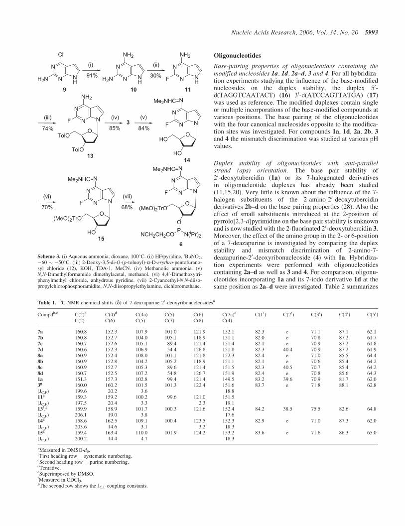
Next, the syntheses of the 2-fluoro-7-deazaadenine nucleo-side 3 and its phosphoramidite were performed (Scheme 3).As the diazotization/fluorination reaction required strongacid conditions (HF/pyridine) (29), it resulted in the
decomposition of the 2,6-diamino nucleoside 2a. Thus, thenucleobase 10 was used instead of the nucleoside. As a pre-cursor the 2-amino-6-chloro-7-deazapurine (9) (24) wasemployed, which was converted to the diamino compound10 in aqeous ammonia (autoclave, 100�C). The diazotization/fluorination reaction was performed under the same condi-tions as done for 2a by dropwise addition of tBuNO2 afford-ing the 2-fluoro base 11. The low yield of 11 (30%) is causedby the partial fluorination at the 6-position giving2,6-difluoro-7-deazapurine, in which the 6-fluoro group wasdisplaced by nucleophiles such as MeOH (!2-fluoro-6-methoxy-7-deazapurine) (Supplementary Data). Nucleobase-anion glycosylation of 11 with 2-deoxy-3,5-di-O-(p-toluoyl)-a-D-erythro-pentofuranosyl chloride (12) yieldedthe toluoyl-protected b-D-nucleoside 13 (74% yield;Scheme 3). Compound 13 was converted to the 2-fluoronucleoside 3 in methanolic ammonia at room temperature.The 2-amino group of 3 was protected by the N,N-dimethy-laminomethylidene residue to give 14, which shows ahalf-life value of 19 min (25% aqeous ammonia, room tem-perature). Subsequently, compound 14 was converted intothe 50-O-DMT-derivative 15 under standard conditions. Phos-phitylation of 15 was performed in anhydrous CH2Cl2 in thepresence of iPr2EtN and 2-cyanoethyl-N,N-diisopropy-lchlorophosphoramidite, furnishing the phosphoramidite 6.The reactions performed with the 2-fluoro nucleosides werecarried out at room temperature in order to avoid the dis-placement of the 2-fluoro substituent. As 20-deoxy-2-fluorotubercidin is a convertible nucleoside, it can be usedto generate DNA containing modified nucleosides with vari-ous substituents at the 2-position by using elevated tempera-ture and/or different deprotection conditions leading tofluorine displacement.
All compounds were characterized by 1H-, 13C-, 31P- or19F-NMR spectra as well as by elemental analysis.13C-NMR shift assignment was made according to gated-decoupled spectra and to those of the free nucleosides(Table 1) (24,25). Compared with the non-functionalizedcompounds 7a or 8a, the C-7 signal is shifted upfield �12p.p.m. upon bromination (7c and 8c) and �50 p.p.m. upon iod-ination (7d and 8d), but locates downfield upon chlorination(�3 p.p.m. for 7b and 8b). In comparison to the parent com-pound 1a (24), the 2-fluoro substituent of nucleoside 3 causesa downfield shift (�9 p.p.m.) at C-2 in the 13C-NMR spectrumwith a 1JC,F coupling constant of 200 Hz, two 3JC,F couplingsof 15–20 p.p.m. for C-4 or C-6 and �4 p.p.m. for C-5.
Scheme 2. (i) N,N-Dimethylformamide dimethylacetal, methanol, 40–50�C, 24 h. (ii) water, 30–40�C, 48 h. (iii) 4,40-Dimethoxytriphenylmethyl chloride,anhydrous pyridine. (iv) 2-Cyanoethyl-N,N-diisopropylchlorophosphoramidite, N,N-diisopropylethylamine, dichloromethane.
Base-pairing properties of oligonucleotides containing themodified nucleosides 1a, 1d, 2a–d, 3 and 4. For all hybridiza-tion experiments studying the influence of the base-modifiednucleosides on the duplex stability, the duplex 50-d(TAGGTCAATACT) (16) 30-d(ATCCAGTTATGA) (17)was used as reference. The modified duplexes contain singleor multiple incorporations of the base-modified compounds atvarious positions. The base pairing of the oligonucleotideswith the four canonical nucleosides opposite to the modifica-tion sites was investigated. For compounds 1a, 1d, 2a, 2b, 3and 4 the mismatch discrimination was studied at various pHvalues.
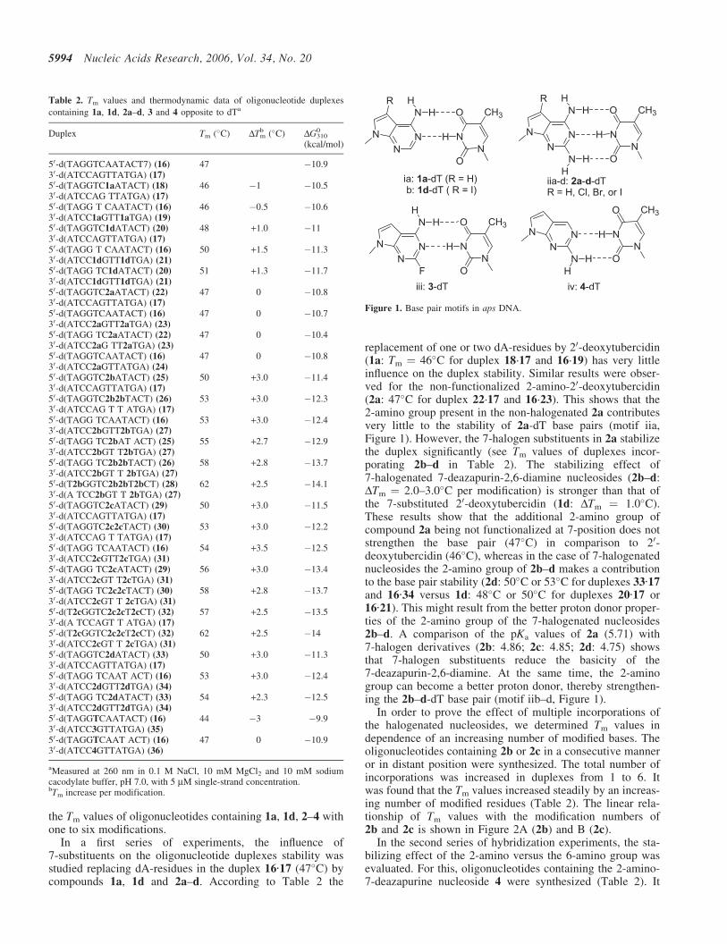
Duplex stability of oligonucleotides with anti-parallelstrand (aps) orientation. The base pair stability of20-deoxytubercidin (1a) or its 7-halogenated derivativesin oligonucleotide duplexes has already been studied(11,15,20). Very little is known about the influence of the 7-halogen substituents of the 2-amino-20-deoxytubercidinderivatives 2b–d on the base pairing properties (28). Also theeffect of small substituents introduced at the 2-position ofpyrrolo[2,3-d]pyrimidine on the base pair stability is unknownand is now studied with the 2-fluorinated 20-deoxytubercidin 3.Moreover, the effect of the amino group in the 2- or 6-positionof a 7-deazapurine is investigated by comparing the duplexstability and mismatch discrimination of 2-amino-7-deazapurine-20-deoxyribonucleoside (4) with 1a. Hybridiza-tion experiments were performed with oligonucleotidescontaining 2a–d as well as 3 and 4. For comparison, oligonu-cleotides incorporating 1a and its 7-iodo derivative 1d at thesame position as 2a–d were investigated. Table 2 summarizes
g 159.4 163.4 110.0 101.9 124.2 153.2 83.6 e 71.6 86.3 65.0(JC,F) 200.2 14.4 4.7 18.3
aMeasured in DMSO-d6.bFirst heading row ¼ systematic numbering.cSecond heading row ¼ purine numbering.dTentative.eSuperimposed by DMSO.fMeasured in CDCl3.gThe second row shows the JC,F coupling constants.
the Tm values of oligonucleotides containing 1a, 1d, 2–4 withone to six modifications.
In a first series of experiments, the influence of7-substituents on the oligonucleotide duplexes stability wasstudied replacing dA-residues in the duplex 16·17 (47�C) bycompounds 1a, 1d and 2a–d. According to Table 2 the
replacement of one or two dA-residues by 20-deoxytubercidin(1a: Tm ¼ 46�C for duplex 18·17 and 16·19) has very littleinfluence on the duplex stability. Similar results were obser-ved for the non-functionalized 2-amino-20-deoxytubercidin(2a: 47�C for duplex 22·17 and 16·23). This shows that the2-amino group present in the non-halogenated 2a contributesvery little to the stability of 2a-dT base pairs (motif iia,Figure 1). However, the 7-halogen substituents in 2a stabilizethe duplex significantly (see Tm values of duplexes incor-porating 2b–d in Table 2). The stabilizing effect of7-halogenated 7-deazapurin-2,6-diamine nucleosides (2b–d:DTm ¼ 2.0–3.0�C per modification) is stronger than that ofthe 7-substituted 20-deoxytubercidin (1d: DTm ¼ 1.0�C).These results show that the additional 2-amino group ofcompound 2a being not functionalized at 7-position does notstrengthen the base pair (47�C) in comparison to 20-deoxytubercidin (46�C), whereas in the case of 7-halogenatednucleosides the 2-amino group of 2b–d makes a contributionto the base pair stability (2d: 50�C or 53�C for duplexes 33·17and 16·34 versus 1d: 48�C or 50�C for duplexes 20·17 or16·21). This might result from the better proton donor proper-ties of the 2-amino group of the 7-halogenated nucleosides2b–d. A comparison of the pKa values of 2a (5.71) with7-halogen derivatives (2b: 4.86; 2c: 4.85; 2d: 4.75) showsthat 7-halogen substituents reduce the basicity of the7-deazapurin-2,6-diamine. At the same time, the 2-aminogroup can become a better proton donor, thereby strengthen-ing the 2b–d-dT base pair (motif iib–d, Figure 1).
In order to prove the effect of multiple incorporations ofthe halogenated nucleosides, we determined Tm values independence of an increasing number of modified bases. Theoligonucleotides containing 2b or 2c in a consecutive manneror in distant position were synthesized. The total number ofincorporations was increased in duplexes from 1 to 6. Itwas found that the Tm values increased steadily by an increas-ing number of modified residues (Table 2). The linear rela-tionship of Tm values with the modification numbers of2b and 2c is shown in Figure 2A (2b) and B (2c).
In the second series of hybridization experiments, the sta-bilizing effect of the 2-amino versus the 6-amino group wasevaluated. For this, oligonucleotides containing the 2-amino-7-deazapurine nucleoside 4 were synthesized (Table 2). It
Table 2. Tm values and thermodynamic data of oligonucleotide duplexes
aMeasured at 260 nm in 0.1 M NaCl, 10 mM MgCl2 and 10 mM sodiumcacodylate buffer, pH 7.0, with 5 mM single-strand concentration.bTm increase per modification.
was observed that the duplex 16·36 (47�C) incorporating4 shows the same stability as that containing 1a having anamino function in the 6-position (46�C for duplex 18·17).This indicates that the amino group in the 2- (4) or 6-position(1a) of 7-deazapurines plays a similar role on the base pairstability when pairing with dT. Obviously, a bidentate basepair is formed between 4 and dT (motif iv, Figure 1).
The effect of 2-halogen substituent on the duplex stabilitywas also investigated. For this purpose the 7-deaza-2-fluoro-20-deoxyadenosine (3) was incorporated at exactly the samepositions as the other modified derivatives. According toTable 2, compound 3 decreases the duplex stability by 3�Cper modification (duplex 16·35). Owing to its negative effect,multiple incorporations of 3 were not undertaken. Thedestabilization of the nucleoside 3 results from the presenceof the 2-fluoro group. Its electron-withdrawing propertydecreases the proton acceptor ability of nitrogen-1 causingweaker hydrogen bonding within the base pair (motif iii,Figure 1), which is underlined by the pKa-value (<1.5) of
3 (Supplementary Data). In addition, the 2-fluoro substituentinduces steric strain with the 2-oxo group of dT.
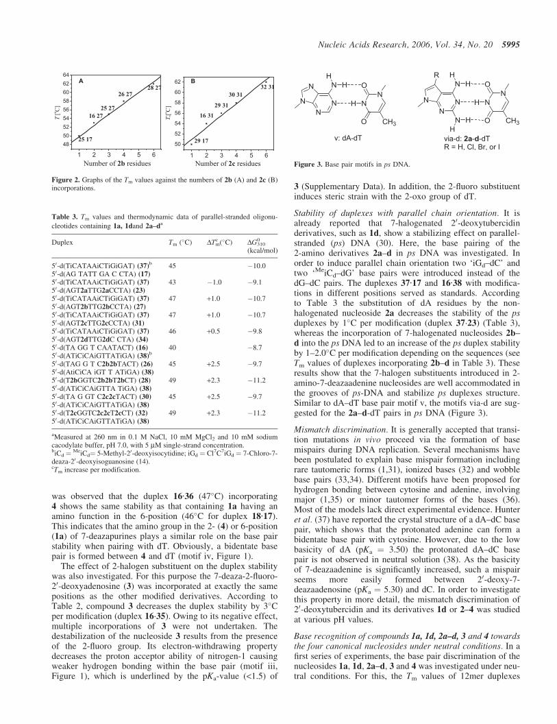
Stability of duplexes with parallel chain orientation. It isalready reported that 7-halogenated 20-deoxytubercidinderivatives, such as 1d, show a stabilizing effect on parallel-stranded (ps) DNA (30). Here, the base pairing of the2-amino derivatives 2a–d in ps DNA was investigated. Inorder to induce parallel chain orientation two ‘iGd–dC’ andtwo ‘MeiCd–dG’ base pairs were introduced instead of thedG–dC pairs. The duplexes 37·17 and 16·38 with modifica-tions in different positions served as standards. Accordingto Table 3 the substitution of dA residues by the non-halogenated nucleoside 2a decreases the stability of the psduplexes by 1�C per modification (duplex 37·23) (Table 3),whereas the incorporation of 7-halogenated nucleosides 2b–d into the ps DNA led to an increase of the ps duplex stabilityby 1–2.0�C per modification depending on the sequences (seeTm values of duplexes incorporating 2b–d in Table 3). Theseresults show that the 7-halogen substituents introduced in 2-amino-7-deazaadenine nucleosides are well accommodated inthe grooves of ps-DNA and stabilize ps duplexes structure.Similar to dA–dT base pair motif v, the motifs via-d are sug-gested for the 2a–d-dT pairs in ps DNA (Figure 3).
Mismatch discrimination. It is generally accepted that transi-tion mutations in vivo proceed via the formation of basemispairs during DNA replication. Several mechanisms havebeen postulated to explain base mispair formation includingrare tautomeric forms (1,31), ionized bases (32) and wobblebase pairs (33,34). Different motifs have been proposed forhydrogen bonding between cytosine and adenine, involvingmajor (1,35) or minor tautomer forms of the bases (36).Most of the models lack direct experimental evidence. Hunteret al. (37) have reported the crystal structure of a dA–dC basepair, which shows that the protonated adenine can form abidentate base pair with cytosine. However, due to the lowbasicity of dA (pKa ¼ 3.50) the protonated dA–dC basepair is not observed in neutral solution (38). As the basicityof 7-deazaadenine is significantly increased, such a mispairseems more easily formed between 20-deoxy-7-deazaadenosine (pKa ¼ 5.30) and dC. In order to investigatethis property in more detail, the mismatch discrimination of20-deoxytubercidin and its derivatives 1d or 2–4 was studiedat various pH values.
Base recognition of compounds 1a, 1d, 2a–d, 3 and 4 towardsthe four canonical nucleosides under neutral conditions. In afirst series of experiments, the base pair discrimination of thenucleosides 1a, 1d, 2a–d, 3 and 4 was investigated under neu-tral conditions. For this, the Tm values of 12mer duplexes
Figure 2. Graphs of the Tm values against the numbers of 2b (A) and 2c (B)incorporations.
Table 3. Tm values and thermodynamic data of parallel-stranded oligonu-
aMeasured at 260 nm in 0.1 M NaCl, 10 mM MgCl2 and 10 mM sodiumcacodylate buffer, pH 7.0, with 5 mM single-strand concentration.biCd ¼ MeiCd¼ 5-Methyl-20-deoxyisocytidine; iGd ¼ Cl7C7iGd ¼ 7-Chloro-7-deaza-20-deoxyisoguanosine (14).cTm increase per modification.
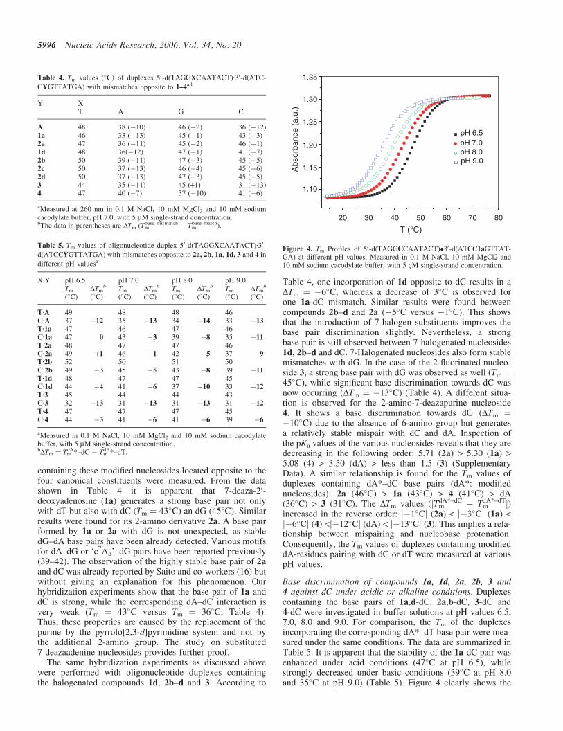
containing these modified nucleosides located opposite to thefour canonical constituents were measured. From the datashown in Table 4 it is apparent that 7-deaza-20-deoxyadenosine (1a) generates a strong base pair not onlywith dT but also with dC (Tm ¼ 43�C) an dG (45�C). Similarresults were found for its 2-amino derivative 2a. A base pairformed by 1a or 2a with dG is not unexpected, as stabledG–dA base pairs have been already detected. Various motifsfor dA–dG or ‘c7Ad’–dG pairs have been reported previously(39–42). The observation of the highly stable base pair of 2aand dC was already reported by Saito and co-workers (16) butwithout giving an explanation for this phenomenon. Ourhybridization experiments show that the base pair of 1a anddC is strong, while the corresponding dA–dC interaction isvery weak (Tm ¼ 43�C versus Tm ¼ 36�C; Table 4).Thus, these properties are caused by the replacement of thepurine by the pyrrolo[2,3-d]pyrimidine system and not bythe additional 2-amino group. The study on substituted7-deazaadenine nucleosides provides further proof.
The same hybridization experiments as discussed abovewere performed with oligonucleotide duplexes containingthe halogenated compounds 1d, 2b–d and 3. According to
Table 4, one incorporation of 1d opposite to dC results in aDTm ¼ �6�C, whereas a decrease of 3�C is observed forone 1a-dC mismatch. Similar results were found betweencompounds 2b–d and 2a (�5�C versus �1�C). This showsthat the introduction of 7-halogen substituents improves thebase pair discrimination slightly. Nevertheless, a strongbase pair is still observed between 7-halogenated nucleosides1d, 2b–d and dC. 7-Halogenated nucleosides also form stablemismatches with dG. In the case of the 2-fluorinated nucleo-side 3, a strong base pair with dG was observed as well (Tm ¼45�C), while significant base discrimination towards dC wasnow occurring (DTm ¼ �13�C) (Table 4). A different situa-tion is observed for the 2-amino-7-deazapurine nucleoside4. It shows a base discrimination towards dG (DTm ¼�10�C) due to the absence of 6-amino group but generatesa relatively stable mispair with dC and dA. Inspection ofthe pKa values of the various nucleosides reveals that they aredecreasing in the following order: 5.71 (2a) > 5.30 (1a) >5.08 (4) > 3.50 (dA) > less than 1.5 (3) (SupplementaryData). A similar relationship is found for the Tm values ofduplexes containing dA*–dC base pairs (dA*: modifiednucleosides): 2a (46�C) > 1a (43�C) > 4 (41�C) > dA(36�C) > 3 (31�C). The DTm values (jTm
dA*–dC � TmdA*–dTj)
increased in the reverse order: j�1�Cj (2a) < j�3�Cj (1a) <j�6�Cj (4) <j�12�Cj (dA) < j�13�Cj (3). This implies a rela-tionship between mispairing and nucleobase protonation.Consequently, the Tm values of duplexes containing modifieddA-residues pairing with dC or dT were measured at variouspH values.
Base discrimination of compounds 1a, 1d, 2a, 2b, 3 and4 against dC under acidic or alkaline conditions. Duplexescontaining the base pairs of 1a,d-dC, 2a,b-dC, 3-dC and4-dC were investigated in buffer solutions at pH values 6.5,7.0, 8.0 and 9.0. For comparison, the Tm of the duplexesincorporating the corresponding dA*–dT base pair were mea-sured under the same conditions. The data are summarized inTable 5. It is apparent that the stability of the 1a-dC pair wasenhanced under acid conditions (47�C at pH 6.5), whilestrongly decreased under basic conditions (39�C at pH 8.0and 35�C at pH 9.0) (Table 5). Figure 4 clearly shows the
Table 4. Tm values (�C) of duplexes 50-d(TAGGXCAATACT)·30-d(ATC-
aMeasured at 260 nm in 0.1 M NaCl, 10 mM MgCl2 and 10 mM sodiumcacodylate buffer, pH 7.0, with 5 mM single-strand concentration.bThe data in parentheses are DTm (Tm
base mismatch � Tmbase match).
Table 5. Tm values of oligonucleotide duplex 50-d(TAGGXCAATACT)·30-d(ATCCYGTTATGA) with mismatches opposite to 2a, 2b, 1a, 1d, 3 and 4 in
different pH valuesa
X·Y pH 6.5 pH 7.0 pH 8.0 pH 9.0Tm
(�C)DTm
b
(�C)Tm
(�C)DTm
b
(�C)Tm
(�C)DTm
b
(�C)Tm
(�C)DTm
b
(�C)
T·A 49 48 48 46C·A 37 �12 35 �13 34 �14 33 �13
T·1a 47 46 47 46C·1a 47 0 43 �3 39 �8 35 �11
T·2a 48 47 47 46C·2a 49 +1 46 �1 42 �5 37 �9
T·2b 52 50 51 50C·2b 49 �3 45 �5 43 �8 39 �11
T·1d 48 47 47 45C·1d 44 �4 41 �6 37 �10 33 �12
T·3 45 44 44 43C·3 32 �13 31 �13 31 �13 31 �12
T·4 47 47 47 45C·4 44 �3 41 �6 41 �6 39 �6
aMeasured in 0.1 M NaCl, 10 mM MgCl2 and 10 mM sodium cacodylatebuffer, with 5 mM single-strand concentration.bDTm ¼ Tm
dA*–dC � TmdA*–dT.
20 30 40 50 60 70 80
1.10
1.15
1.20
1.25
1.30
1.35
pH 8.0pH 7.0
Abs
orba
nce
(a.u
.)
T (°C)
pH 6.5
pH 9.0
Figure 4. Tm Profiles of 50-d(TAGGCCAATACT)�30-d(ATCC1aGTTAT-GA) at different pH values. Measured in 0.1 M NaCl, 10 mM MgCl2 and10 mM sodium cacodylate buffer, with 5 cM single-strand concentration.
relationship between the stability of 1a-dC base pair and pHvalue of the buffer solution. Similar results were found for 2a(Table 5). When the pH value is 6.5, the duplex with a 2a-dCpair (49�C) is even more stable than that incorporating a 2a-dT base pair (48�C).
Similar results as found for non-halogenated nucleosides1a or 2a are observed for 7-halogenated compounds 1d and2b. The stability of duplexes incorporating 1d-dC or 2b-dCpairs decreased with increasing pH values: Tm
pH 6.5 > TmpH 7.0
> TmpH 8.0 > Tm
pH 9.0. This demonstrates that the 7-deazapurinenucleosides 1a,d and 2a,b show an enhanced base discrimina-tion towards dC under alkaline conditions. This phenomenoncan be clearly seen from Figure 5A–D. It is apparentthat compounds 1a,d and 2a,b show much stronger discrim-ination towards dC under alkaline condition than that in neu-tral or acidic medium as indicated from the order of �DTm
(�DTmpH 6.5< �DTm
pH 7.0< �DTmpH 8.0 < �DTm
pH 9.0). The variousTm values of the corresponding duplexes with 1a, 1d, 2a or2b opposite to dC at various pH values were visualized inthe inserted figures, which reflects the destabilizing effectof alkaline buffer solution on ‘c7Ad*’–dC base pair and thestabilizing effect of mild acid conditions. However, whenthe pH values are in the range of 6.5–9.0, such a behaviouris not observed for the dA–dC or 3-dC pairs as indicatedfrom the similar DTm values (��13�C) obtained in acidic,neutral or alkaline medium (Table 5 and Figure 5E, F).
According to the pKa values of the monomeric nucleosides1a (5.30) and 2a (5.71), one might argue that the pKa valuesare too low to cause protonation under neutral or weak alkal-ine conditions. However, from pKa studies on oligonu-cleotides it is known that the pKa values of nucleobaseswithin stacked oligonucleotides can be raised significantlyby the attractive force of the phosphodiester backbone forthe protons, and by the stabilization caused by hydrogen
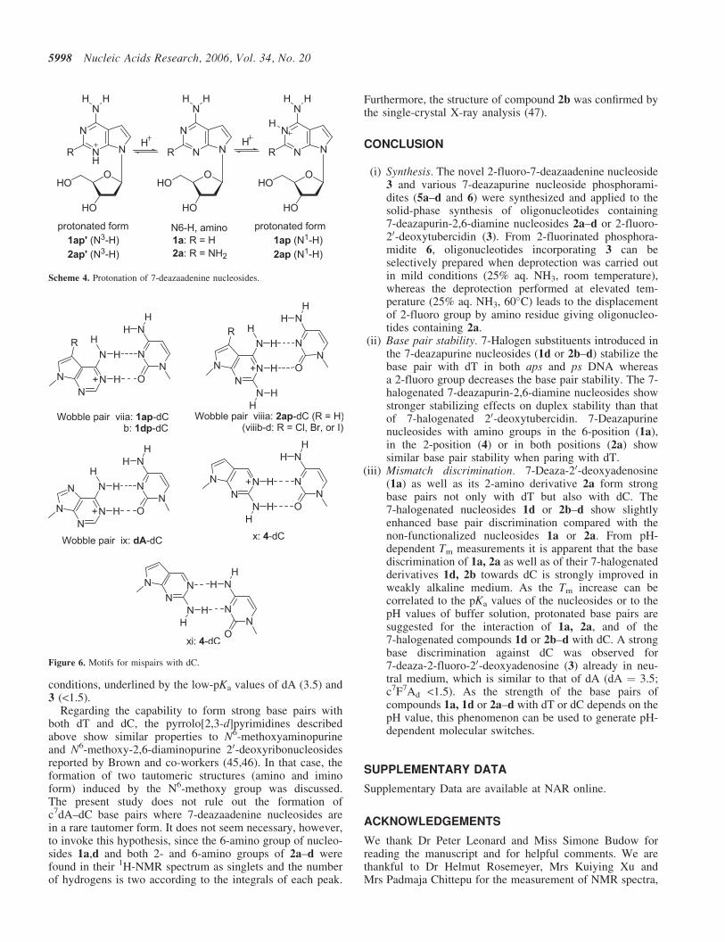
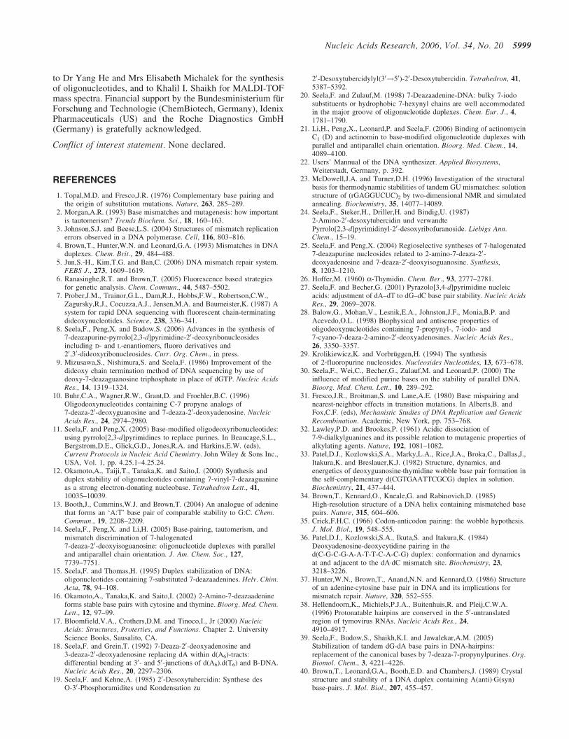
bonding (17). In our case, a pKa increase of 1–2 pK valuesis likely to occur. Thus, the protonated base pairs are easilyformed between the 7-deazapurine nucleosides 1a or 2a anddC under neutral conditions when they are as constituents ofoligonucleotides. According to the 7-deazapurine structurea protonation in the five-member ring can be excluded. Pos-sible protonation sites are only nitrogen-1 or nitrogen-3(Scheme 4). Rosemeyer and Seela (43) reported that15N-NMR studies on 7-deazaadenine nucleosides show thatthe protonation site of 1a is N-1 (1ap; Scheme 4, right).Therefore, the 1a-dC or 1d-dC base pairs should formaccording to bidentate motifs viia,b with nucleosides 1a or1d in the N-1 protonated state (Figure 6). Similarly, bidentatebase pair motifs viiia–d are suggested for 2a–d-dC mispairs(Figure 6). Our findings are consistent with earlier observa-tions reported by Hunter et al. (37) on a protonated dA–dCbase pair (motif ix) existing in the crystal structure. Also,Saito and co-workers have observed that a wobble base pairof 4-amino-6-methoxy-9-(20-deoxy-b-D-erythro-pentofurano-syl)-7H-pyrimido[4,5-b]indole and dC (44).
The 2-amino-7-deazapurine nucleoside 4 also generatesstable base pair with dC under acidic condition (pH 6.5),whereas the stability of 4-dC base pair is kept in the samerange in neutral or basic buffer solution (pH 7.0, 8.0 and9.0) (Table 5). This might be due to the formation of differentbase pair motifs under acid or neutral and basic conditions.The protonated base pair (motif x) is likely to be generatedin acid condition, whereas a wobble base pair xi is presentin neutral and basic conditions (Figure 6). In the case of 2-fluoro-7-deazaadenine nucleoside 3 and dA, rather strongbase discrimination was observed against dC keeping theTm values constant at different pH values (6.5, 7.0, 8.0, 9.0)(Table 5 and Figure 5E, F). This results from the properties ofdA or compound 3 which are not protonated under these
dA*�dC) of the duplexes 50-d(TAGGXCAATACT)�30-d(ATCCYGTTATGA)incorporating 1a (A), 2a (B), 1d (C), 2b (D), dA (E) or 3 (F) in the position Y, while dC or dT are in the position X (data from Table 5). The inserts shows the Tm
values of the corresponding duplexes with 1a, 1d, 2a, 2b, 3 and dA opposite to dC at various pH values.
conditions, underlined by the low-pKa values of dA (3.5) and3 (<1.5).
Regarding the capability to form strong base pairs withboth dT and dC, the pyrrolo[2,3-d]pyrimidines describedabove show similar properties to N6-methoxyaminopurineand N6-methoxy-2,6-diaminopurine 20-deoxyribonucleosidesreported by Brown and co-workers (45,46). In that case, theformation of two tautomeric structures (amino and iminoform) induced by the N6-methoxy group was discussed.The present study does not rule out the formation ofc7dA–dC base pairs where 7-deazaadenine nucleosides arein a rare tautomer form. It does not seem necessary, however,to invoke this hypothesis, since the 6-amino group of nucleo-sides 1a,d and both 2- and 6-amino groups of 2a–d werefound in their 1H-NMR spectrum as singlets and the numberof hydrogens is two according to the integrals of each peak.
Furthermore, the structure of compound 2b was confirmed bythe single-crystal X-ray analysis (47).
CONCLUSION
(i) Synthesis. The novel 2-fluoro-7-deazaadenine nucleoside3 and various 7-deazapurine nucleoside phosphorami-dites (5a–d and 6) were synthesized and applied to thesolid-phase synthesis of oligonucleotides containing7-deazapurin-2,6-diamine nucleosides 2a–d or 2-fluoro-20-deoxytubercidin (3). From 2-fluorinated phosphora-midite 6, oligonucleotides incorporating 3 can beselectively prepared when deprotection was carried outin mild conditions (25% aq. NH3, room temperature),whereas the deprotection performed at elevated tem-perature (25% aq. NH3, 60�C) leads to the displacementof 2-fluoro group by amino residue giving oligonucleo-tides containing 2a.
(ii) Base pair stability. 7-Halogen substituents introduced inthe 7-deazapurine nucleosides (1d or 2b–d) stabilize thebase pair with dT in both aps and ps DNA whereasa 2-fluoro group decreases the base pair stability. The 7-halogenated 7-deazapurin-2,6-diamine nucleosides showstronger stabilizing effects on duplex stability than thatof 7-halogenated 20-deoxytubercidin. 7-Deazapurinenucleosides with amino groups in the 6-position (1a),in the 2-position (4) or in both positions (2a) showsimilar base pair stability when paring with dT.
(iii) Mismatch discrimination. 7-Deaza-20-deoxyadenosine(1a) as well as its 2-amino derivative 2a form strongbase pairs not only with dT but also with dC. The7-halogenated nucleosides 1d or 2b–d show slightlyenhanced base pair discrimination compared with thenon-functionalized nucleosides 1a or 2a. From pH-dependent Tm measurements it is apparent that the basediscrimination of 1a, 2a as well as of their 7-halogenatedderivatives 1d, 2b towards dC is strongly improved inweakly alkaline medium. As the Tm increase can becorrelated to the pKa values of the nucleosides or to thepH values of buffer solution, protonated base pairs aresuggested for the interaction of 1a, 2a, and of the7-halogenated compounds 1d or 2b–d with dC. A strongbase discrimination against dC was observed for7-deaza-2-fluoro-20-deoxyadenosine (3) already in neu-tral medium, which is similar to that of dA (dA ¼ 3.5;c7F7Ad <1.5). As the strength of the base pairs ofcompounds 1a, 1d or 2a–d with dT or dC depends on thepH value, this phenomenon can be used to generate pH-dependent molecular switches.
SUPPLEMENTARY DATA
Supplementary Data are available at NAR online.
ACKNOWLEDGEMENTS
We thank Dr Peter Leonard and Miss Simone Budow forreading the manuscript and for helpful comments. We arethankful to Dr Helmut Rosemeyer, Mrs Kuiying Xu andMrs Padmaja Chittepu for the measurement of NMR spectra,
Scheme 4. Protonation of 7-deazaadenine nucleosides.
to Dr Yang He and Mrs Elisabeth Michalek for the synthesisof oligonucleotides, and to Khalil I. Shaikh for MALDI-TOFmass spectra. Financial support by the Bundesministerium furForschung and Technologie (ChemBiotech, Germany), IdenixPharmaceuticals (US) and the Roche Diagnostics GmbH(Germany) is gratefully acknowledged.
Conflict of interest statement. None declared.
REFERENCES
1. Topal,M.D. and Fresco,J.R. (1976) Complementary base pairing andthe origin of substitution mutations. Nature, 263, 285–289.
2. Morgan,A.R. (1993) Base mismatches and mutagenesis: how importantis tautomerism? Trends Biochem. Sci., 18, 160–163.
3. Johnson,S.J. and Beese,L.S. (2004) Structures of mismatch replicationerrors observed in a DNA polymerase. Cell, 116, 803–816.
4. Brown,T., Hunter,W.N. and Leonard,G.A. (1993) Mismatches in DNAduplexes. Chem. Brit., 29, 484–488.
5. Jun,S.-H., Kim,T.G. and Ban,C. (2006) DNA mismatch repair system.FEBS J., 273, 1609–1619.
6. Ranasinghe,R.T. and Brown,T. (2005) Fluorescence based strategiesfor genetic analysis. Chem. Commun., 44, 5487–5502.
7. Prober,J.M., Trainor,G.L., Dam,R.J., Hobbs,F.W., Robertson,C.W.,Zagursky,R.J., Cocuzza,A.J., Jensen,M.A. and Baumeister,K. (1987) Asystem for rapid DNA sequencing with fluorescent chain-terminatingdideoxynucleotides. Science, 238, 336–341.
8. Seela,F., Peng,X. and Budow,S. (2006) Advances in the synthesis of7-deazapurine-pyrrolo[2,3-d]pyrimidine-20-deoxyribonucleosidesincluding D- and L-enantiomers, fluoro derivatives and20,30-dideoxyribonucleosides. Curr. Org. Chem., in press.
9. Mizusawa,S., Nishimura,S. and Seela,F. (1986) Improvement of thedideoxy chain termination method of DNA sequencing by use ofdeoxy-7-deazaguanosine triphosphate in place of dGTP. Nucleic AcidsRes., 14, 1319–1324.
10. Buhr,C.A., Wagner,R.W., Grant,D. and Froehler,B.C. (1996)Oligodeoxynucleotides containing C-7 propyne analogs of7-deaza-20-deoxyguanosine and 7-deaza-20-deoxyadenosine. NucleicAcids Res., 24, 2974–2980.
11. Seela,F. and Peng,X. (2005) Base-modified oligodeoxyribonucleotides:using pyrrolo[2,3-d]pyrimidines to replace purines. In Beaucage,S.L.,Bergstrom,D.E., Glick,G.D., Jones,R.A. and Harkins,E.W. (eds),Current Protocols in Nucleic Acid Chemistry. John Wiley & Sons Inc.,USA, Vol. 1, pp. 4.25.1–4.25.24.
12. Okamoto,A., Taiji,T., Tanaka,K. and Saito,I. (2000) Synthesis andduplex stability of oligonucleotides containing 7-vinyl-7-deazaguanineas a strong electron-donating nucleobase. Tetrahedron Lett., 41,10035–10039.
13. Booth,J., Cummins,W.J. and Brown,T. (2004) An analogue of adeninethat forms an ‘A:T’ base pair of comparable stability to G:C. Chem.Commun., 19, 2208–2209.
14. Seela,F., Peng,X. and Li,H. (2005) Base-pairing, tautomerism, andmismatch discrimination of 7-halogenated7-deaza-20-deoxyisoguanosine: oligonucleotide duplexes with paralleland antiparallel chain orientation. J. Am. Chem. Soc., 127,7739–7751.
15. Seela,F. and Thomas,H. (1995) Duplex stabilization of DNA:oligonucleotides containing 7-substituted 7-deazaadenines. Helv. Chim.Acta, 78, 94–108.
16. Okamoto,A., Tanaka,K. and Saito,I. (2002) 2-Amino-7-deazaadenineforms stable base pairs with cytosine and thymine. Bioorg. Med. Chem.Lett., 12, 97–99.
17. Bloomfield,V.A., Crothers,D.M. and Tinoco,I., Jr (2000) NucleicAcids: Structures, Proterties, and Functions. Chapter 2. UniversityScience Books, Sausalito, CA.
18. Seela,F. and Grein,T. (1992) 7-Deaza-20-deoxyadenosine and3-deaza-20-deoxyadenosine replacing dA within d(A6)-tracts:differential bending at 30- and 50-junctions of d(A6).d(T6) and B-DNA.Nucleic Acids Res., 20, 2297–2306.
19. Seela,F. and Kehne,A. (1985) 20-Desoxytubercidin: Synthese desO-30-Phosphoramidites und Kondensation zu
20. Seela,F. and Zulauf,M. (1998) 7-Deazaadenine-DNA: bulky 7-iodosubstituents or hydrophobic 7-hexynyl chains are well accommodatedin the major groove of oligonucleotide duplexes. Chem. Eur. J., 4,1781–1790.
21. Li,H., Peng,X., Leonard,P. and Seela,F. (2006) Binding of actinomycinC1 (D) and actinomin to base-modified oligonucleotide duplexes withparallel and antiparallel chain orientation. Bioorg. Med. Chem., 14,4089–4100.
22. Users’ Mannual of the DNA synthesizer. Applied Biosystems,Weiterstadt, Germany, p. 392.
23. McDowell,J.A. and Turner,D.H. (1996) Investigation of the structuralbasis for thermodynamic stabilities of tandem GU mismatches: solutionstructure of (rGAGGUCUC)2 by two-dimensional NMR and simulatedannealing. Biochemistry, 35, 14077–14089.
24. Seela,F., Steker,H., Driller,H. and Bindig,U. (1987)2-Amino-20-desoxytubercidin und verwandtePyrrolo[2,3-d]pyrimidinyl-20-desoxyribofuranoside. Liebigs Ann.Chem., 15–19.
25. Seela,F. and Peng,X. (2004) Regioselective syntheses of 7-halogenated7-deazapurine nucleosides related to 2-amino-7-deaza-20-deoxyadenosine and 7-deaza-20-deoxyisoguanosine. Synthesis,8, 1203–1210.
29. Krolikiewicz,K. and Vorbruggen,H. (1994) The synthesisof 2-fluoropurine nucleosides. Nucleosides Nucleotides, 13, 673–678.
30. Seela,F., Wei,C., Becher,G., Zulauf,M. and Leonard,P. (2000) Theinfluence of modified purine bases on the stability of parallel DNA.Bioorg. Med. Chem. Lett., 10, 289–292.
31. Fresco,J.R., Broitman,S. and Lane,A.E. (1980) Base mispairing andnearest-neighbor effects in transition mutations. In Alberts,B. andFox,C.F. (eds), Mechanistic Studies of DNA Replication and GeneticRecombination. Academic, New York, pp. 753–768.
32. Lawley,P.D. and Brookes,P. (1961) Acidic dissociation of7-9-dialkylguanines and its possible relation to mutagenic properties ofalkylating agents. Nature, 192, 1081–1082.
33. Patel,D.J., Kozlowski,S.A., Marky,L.A., Rice,J.A., Broka,C., Dallas,J.,Itakura,K. and Breslauer,K.J. (1982) Structure, dynamics, andenergetics of deoxyguanosine·thymidine wobble base pair formation inthe self-complementary d(CGTGAATTCGCG) duplex in solution.Biochemistry, 21, 437–444.
34. Brown,T., Kennard,O., Kneale,G. and Rabinovich,D. (1985)High-resolution structure of a DNA helix containing mismatched basepairs. Nature, 315, 604–606.
36. Patel,D.J., Kozlowski,S.A., Ikuta,S. and Itakura,K. (1984)Deoxyadenosine-deoxycytidine pairing in thed(C-G-C-G-A-A-T-T-C-A-C-G) duplex: conformation and dynamicsat and adjacent to the dA·dC mismatch site. Biochemistry, 23,3218–3226.
37. Hunter,W.N., Brown,T., Anand,N.N. and Kennard,O. (1986) Structureof an adenine·cytosine base pair in DNA and its implications formismatch repair. Nature, 320, 552–555.
38. Hellendoorn,K., Michiels,P.J.A., Buitenhuis,R. and Pleij,C.W.A.(1996) Protonatable hairpins are conserved in the 50-untranslatedregion of tymovirus RNAs. Nucleic Acids Res., 24,4910–4917.
39. Seela,F., Budow,S., Shaikh,K.I. and Jawalekar,A.M. (2005)Stabilization of tandem dG-dA base pairs in DNA-hairpins:replacement of the canonical bases by 7-deaza-7-propynylpurines. Org.Biomol. Chem., 3, 4221–4226.
40. Brown,T., Leonard,G.A., Booth,E.D. and Chambers,J. (1989) Crystalstructure and stability of a DNA duplex containing A(anti)·G(syn)base-pairs. J. Mol. Biol., 207, 455–457.
41. Greene,K.L., Jones,R.L., Li,Y., Robinson,H., Wang,A.H.-J., Zon,G.and Wilson,W.D. (1994) Solution structure of a GA mismatchDNA sequence, d(CCATGAATGG)2, determined by 2D NMRand structural refinement methods. Biochemistry, 33, 1053–1062.
42. Li,Y., Zon,G. and Wilson,W.D. (1991) NMR and molecular modelingevidence for a G·A mismatch base pair in a purine-rich DNA duplex.Proc. Natl Acad. Sci. USA, 88, 26–30.
43. Rosemeyer,H. and Seela,F. (1988) Stereoselective synthesis ofpyrrolo[2,3-d]pyrimidine a- and b-D-ribonucleosides fromanomerically pure D-ribofuranosyl chlorides: solid-liquidphase-transfer glycosylation and 15N-NMR spectra. Helv. Chim. Acta,71, 1573–1584.
44. Okamoto,A., Tanaka,K. and Saito,I. (2004) DNA logic gates. J. Am.Chem. Soc., 126, 9458–9463.
45. Brown,D.M. and Kong Thoo Lin,P. (1991) Synthesis and duplexstability of oligonucleotides containing adenine-guanine analogues.Carbohydr. Res., 216, 129–139.
46. Hill,F., Williams,D.M., Loakes,D. and Brown,D.M. (1998)Comparative mutagenicities of N6-methoxy-2,6-diaminopurine andN6-methoxyaminopurine 20-deoxyribonuclesides and their50-triphosphates. Nucleic Acids Res., 26, 1144–1149.
47. Seela,F., Peng,X., Eickmeier,H. and Reuter,H (2006)2-Amino-7-chloro-20-deoxytubercidin. Acta Cryst., C62,593–595.