28
22.01 – Intro to Ionizing Radiation Dose & Dosimetry, Slide 1 Slides for Dose and Dosimetry 22.01 – Intro to Radiation November 18 th , 2015
22.01 – Intro to Ionizing Radiation Dose & Dosimetry, Slide 1
Slides for Dose and Dosimetry
22.01 – Intro to Radiation November 18th, 2015
22.01 – Intro to Ionizing Radiation Dose & Dosimetry, Slide 2
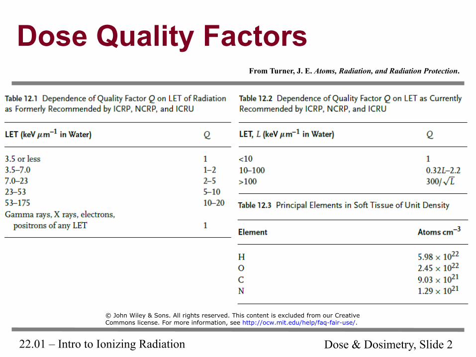
Dose Quality Factors From Turner, J. E. Atoms, Radiation, and Radiation Protection.
© John Wiley & Sons. All rights reserved. This content is excluded from our CreativeCommons license. For more information, see http://ocw.mit.edu/help/faq-fair-use/.
22.01 – Intro to Ionizing Radiation Dose & Dosimetry, Slide 3
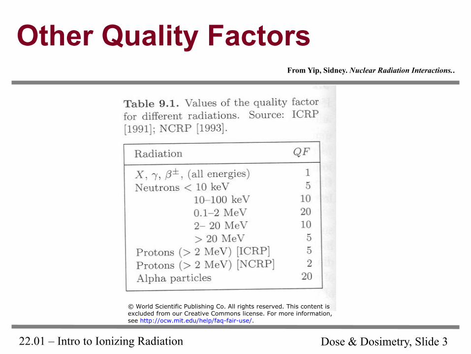
Other Quality Factors From Yip, Sidney. Nuclear Radiation Interactions..
© World Scientific Publishing Co. All rights reserved. This content isexcluded from our Creative Commons license. For more information,see http://ocw.mit.edu/help/faq-fair-use/.
22.01 – Intro to Ionizing Radiation Dose & Dosimetry, Slide 4
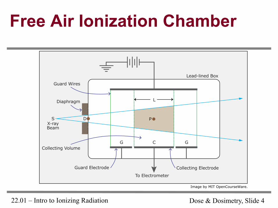
Free Air Ionization Chamber
Image by MIT OpenCourseWare.
To Electrometer
Diaphragm
Lead-lined Box
Guard Wires
Collecting ElectrodeGuard Electrode
Collecting Volume
X-rayBeam
P
GCG
S D
L
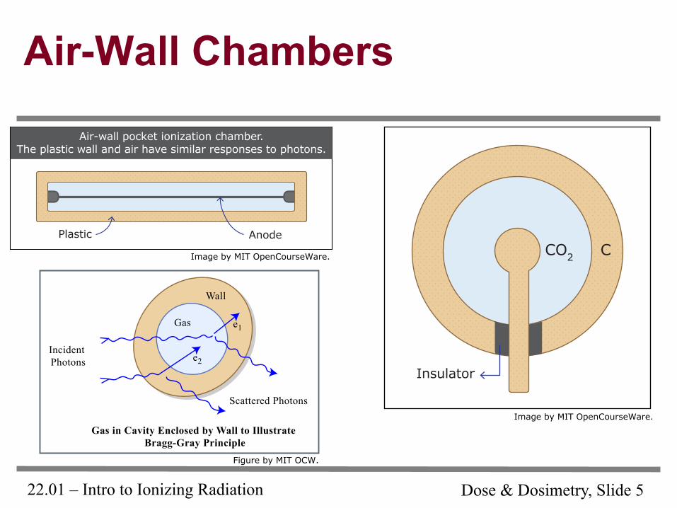
22.01 – Intro to Ionizing Radiation Dose & Dosimetry, Slide 5
Air-Wall Chambers
Figure by MIT OCW.
Image by MIT OpenCourseWare.
Image by MIT OpenCourseWare.
Gas
Wall
Incident Photons
Gas in Cavity Enclosed by Wall to Illustrate Bragg-Gray Principle
Scattered Photons
e1
e2
CCO2
Insulator
Air-wall pocket ionization chamber.The plastic wall and air have similar responses to photons.
Plastic Anode
22.01 – Intro to Ionizing Radiation Dose & Dosimetry, Slide 6
Air Wall Chambers – Civil Defense
Image removed due to copyright restrictions. Schematic diagram of a pocket ion dosimeter. Figure 8.23 in Yip, Sidney. Nuclear Radiation Interactions. For more information, see https://www.orau.org/ptp/collection/dosimeters/pocketchamdos.htm
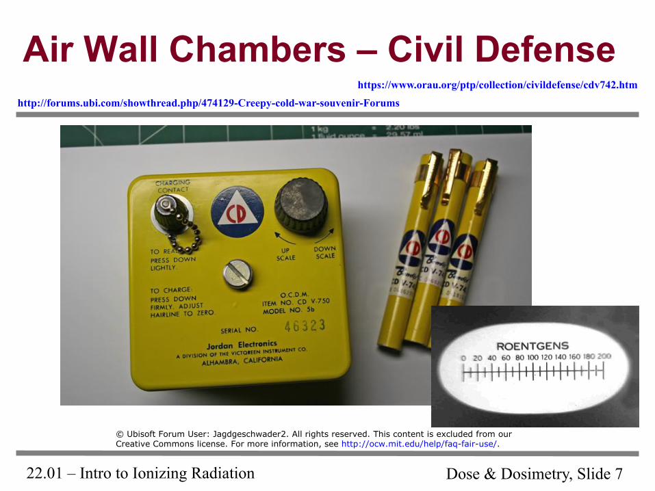
22.01 – Intro to Ionizing Radiation Dose & Dosimetry, Slide 7
Air Wall Chambers – Civil Defense http://forums.ubi.com/showthread.php/474129-Creepy-cold-war-souvenir-Forums
https://www.orau.org/ptp/collection/civildefense/cdv742.htm
© Ubisoft Forum User: Jagdgeschwader2. All rights reserved. This content is excluded from ourCreative Commons license. For more information, see http://ocw.mit.edu/help/faq-fair-use/.
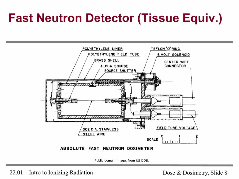
22.01 – Intro to Ionizing Radiation Dose & Dosimetry, Slide 8
Fast Neutron Detector (Tissue Equiv.)
Public domain image, from US DOE.
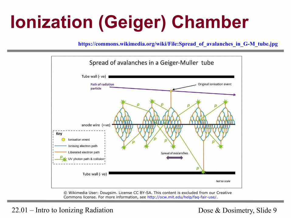
22.01 – Intro to Ionizing Radiation Dose & Dosimetry, Slide 9
Ionization (Geiger) Chamber https://commons.wikimedia.org/wiki/File:Spread_of_avalanches_in_G-M_tube.jpg
© Wikimedia User: Dougsim. License CC BY-SA. This content is excluded from our CreativeCommons license. For more information, see http://ocw.mit.edu/help/faq-fair-use/.
22.01 – Intro to Ionizing Radiation Dose & Dosimetry, Slide 10
Gas Detector Cutaway
Image removed due to copyright restrictions. Schematic diagram of coaxial gas detector, commonly used for Geiger-Müller tubes. Figure 8.2 in Yip, Sidney. Nuclear Radiation Interactions.
22.01 – Intro to Ionizing Radiation Dose & Dosimetry, Slide 11
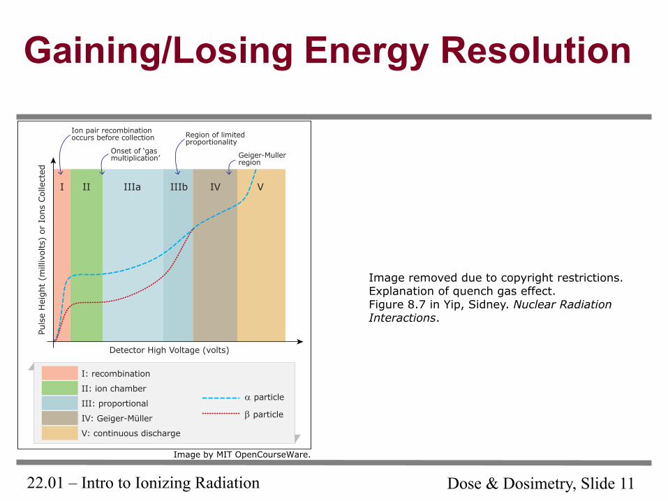
Gaining/Losing Energy Resolution
Image removed due to copyright restrictions. Explanation of quench gas effect. Figure 8.7 in Yip, Sidney. Nuclear Radiation Interactions.
Image by MIT OpenCourseWare.
I: recombination
II: ion chamber
III: proportional
IV: Geiger-Müller
V: continuous discharge
α particle
β particle
Detector High Voltage (volts)
Puls
e H
eigh
t (m
illiv
olts
) or
Ion
s Col
lect
ed
I II IIIa IIIb IV V
Ion pair recombinationoccurs before collection
Onset of ‘gasmultiplication’
Region of limitedproportionality
Geiger-Mullerregion
22.01 – Intro to Ionizing Radiation Dose & Dosimetry, Slide 12
Combined Gamma/Neutron Detector
Image removed due to copyright restrictions. Cross-section diagram of compensated ion chamber. Figure 8.3 in Yip, Sidney. Nuclear Radiation Interactions.
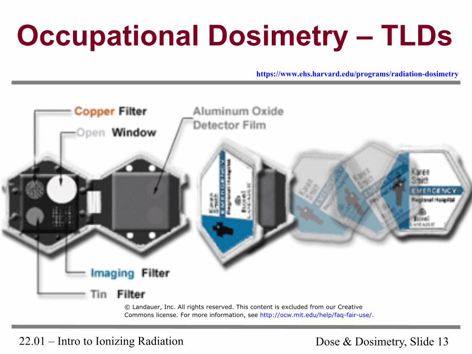
22.01 – Intro to Ionizing Radiation Dose & Dosimetry, Slide 13
Occupational Dosimetry – TLDs
© Landauer, Inc. All rights reserved. This content is excluded from our CreativeCommons license. For more information, see http://ocw.mit.edu/help/faq-fair-use/.
https://www.ehs.harvard.edu/programs/radiation-dosimetry
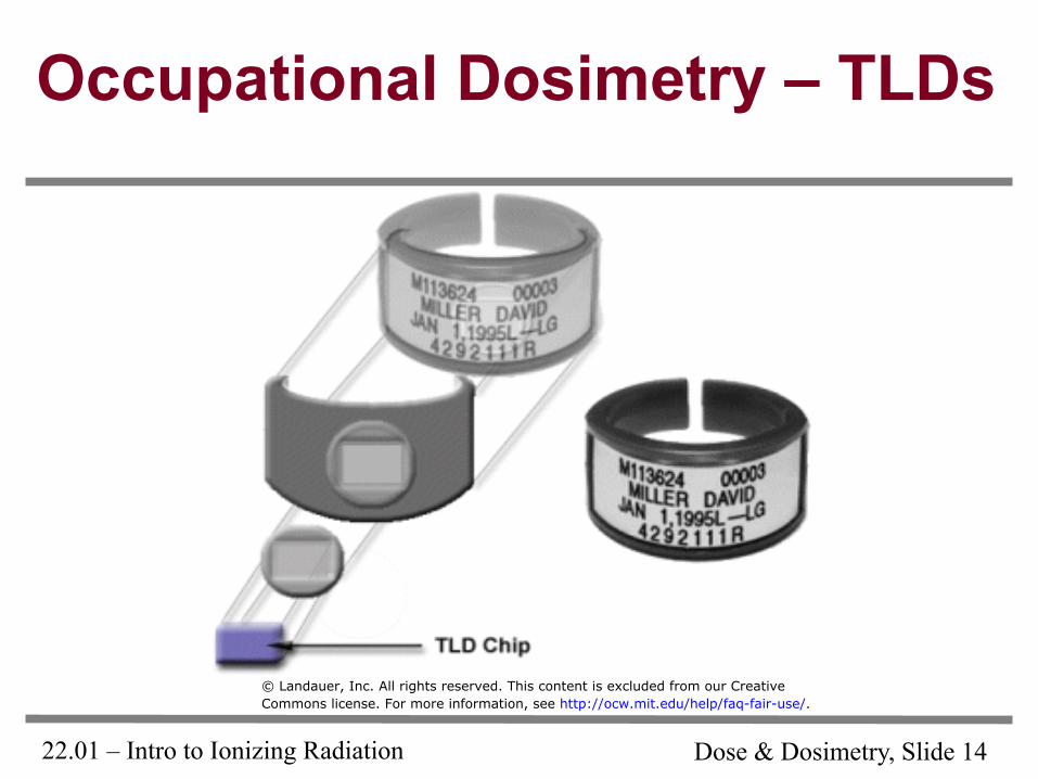
22.01 – Intro to Ionizing Radiation Dose & Dosimetry, Slide 14
Occupational Dosimetry – TLDs
© Landauer, Inc. All rights reserved. This content is excluded from our CreativeCommons license. For more information, see http://ocw.mit.edu/help/faq-fair-use/.
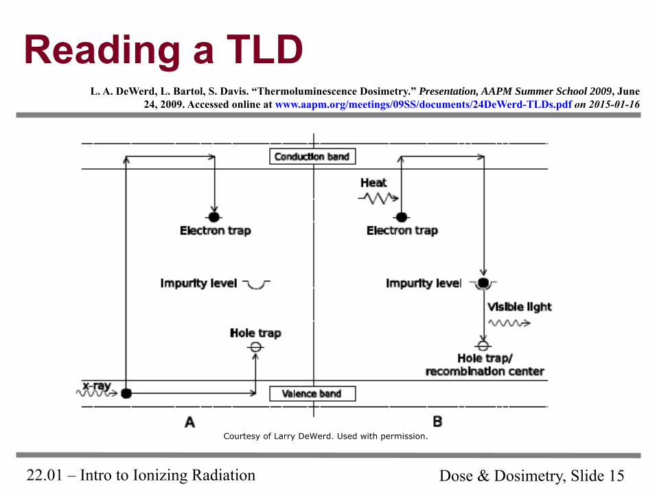
22.01 – Intro to Ionizing Radiation Dose & Dosimetry, Slide 15
Reading a TLD L. A. DeWerd, L. Bartol, S. Davis. “Thermoluminescence Dosimetry.” Presentation, AAPM Summer School 2009, June
24, 2009. Accessed online at www.aapm.org/meetings/09SS/documents/24DeWerd-TLDs.pdf on 2015-01-16
Courtesy of Larry DeWerd. Used with permission.
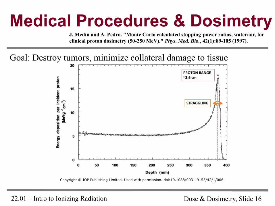
22.01 – Intro to Ionizing Radiation Dose & Dosimetry, Slide 16
Medical Procedures & Dosimetry
Goal: Destroy tumors, minimize collateral damage to tissue
J. Medin and A. Pedro. "Monte Carlo calculated stopping-power ratios, water/air, for clinical proton dosimetry (50-250 MeV)." Phys. Med. Bio., 42(1):89-105 (1997).
Copyright © IOP Publishing Limited. Used with permission. doi:10.1088/0031-9155/42/1/006.
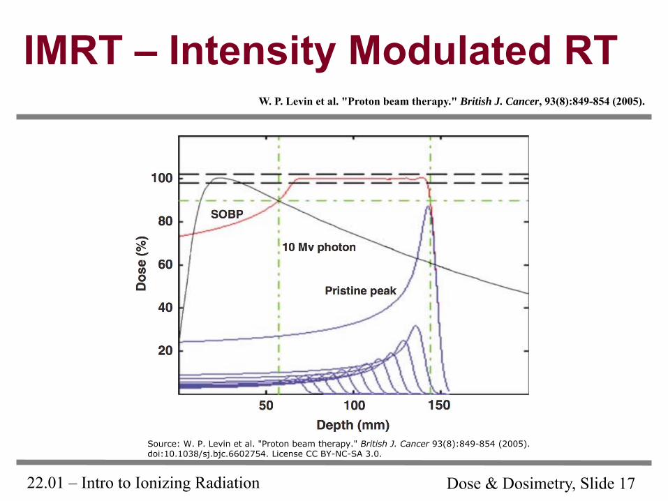
22.01 – Intro to Ionizing Radiation Dose & Dosimetry, Slide 17
IMRT – Intensity Modulated RT W. P. Levin et al. "Proton beam therapy." British J. Cancer, 93(8):849-854 (2005).
Source: W. P. Levin et al. "Proton beam therapy." British J. Cancer 93(8):849-854 (2005).doi:10.1038/sj.bjc.6602754. License CC BY-NC-SA 3.0.
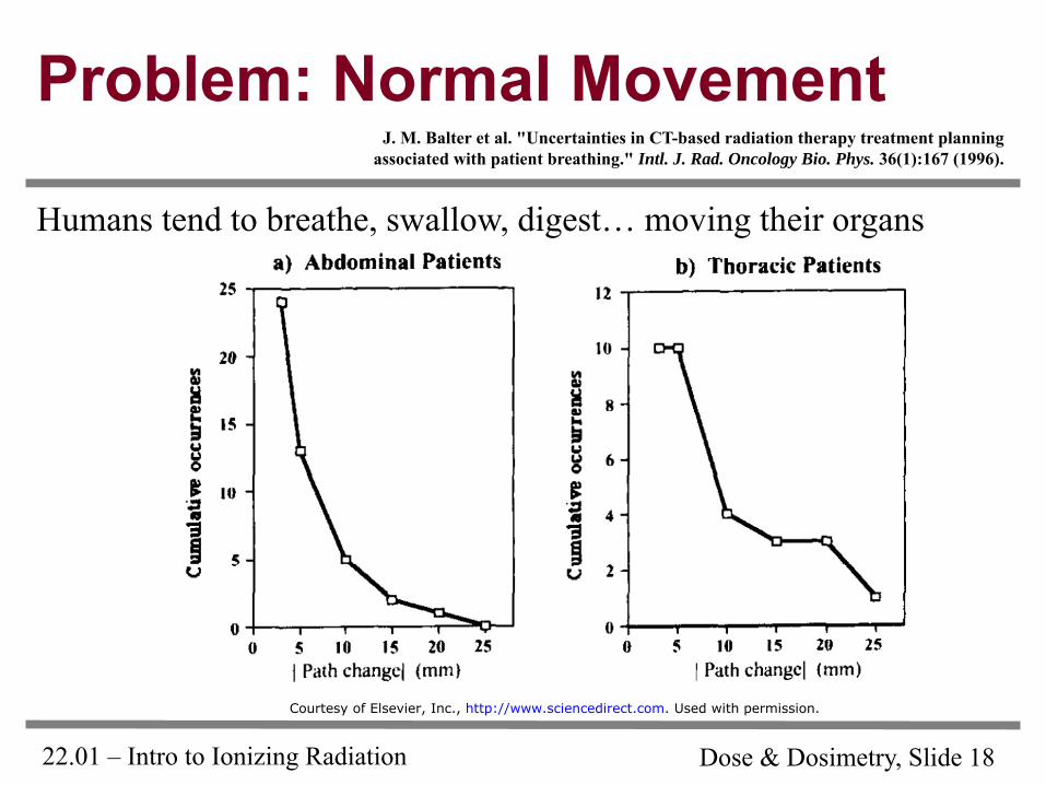
22.01 – Intro to Ionizing Radiation Dose & Dosimetry, Slide 18
Problem: Normal Movement J. M. Balter et al. "Uncertainties in CT-based radiation therapy treatment planning
associated with patient breathing." Intl. J. Rad. Oncology Bio. Phys. 36(1):167 (1996).
Humans tend to breathe, swallow, digest… moving their organs
Courtesy of Elsevier, Inc., http://www.sciencedirect.com. Used with permission.
22.01 – Intro to Ionizing Radiation Dose & Dosimetry, Slide 19
The Ideal IMRT Dosimeter
The dosimeter can determine absolute dose
The dosimeter can provide three-dimensional data
The dosimeter’s response isn’t orientation-dependent
The dosimeter is well-calibrated, and the interpretation of its readout is rigorously supported by data
The dosimeter’s ability to measure absolute dose is insensitive to dose rate and energy of the radiation
The dosimeter is non-toxic
The dosimeter’s cost to build and maintain is reasonable
22.01 – Intro to Ionizing Radiation Dose & Dosimetry, Slide 20
Existing Dosimetry Methods
Monte Carlo calculations
Conventional port films
Electronic portal imaging devices (EPID)
Gel dosimetry
Electron spin resonance spectroscopy
Thermoluminescent dosimetry
Silicon diodes
Scintillation fibers
Prompt gamma monitoring
PET scans
MOSFET dosimeters
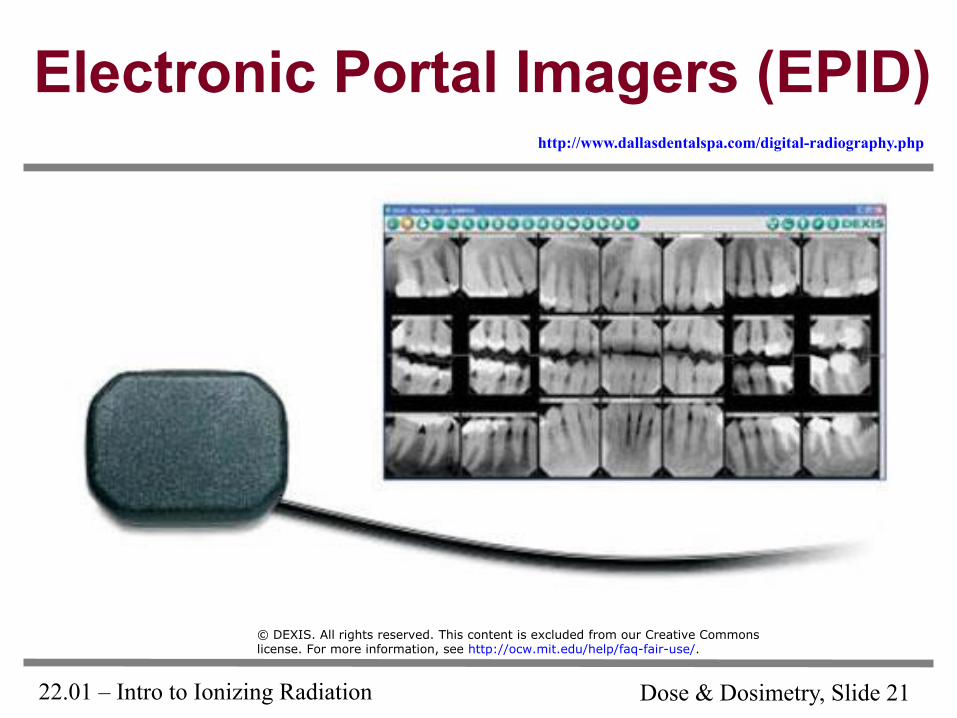
22.01 – Intro to Ionizing Radiation Dose & Dosimetry, Slide 21
Electronic Portal Imagers (EPID) http://www.dallasdentalspa.com/digital-radiography.php
© DEXIS. All rights reserved. This content is excluded from our Creative Commonslicense. For more information, see http://ocw.mit.edu/help/faq-fair-use/.
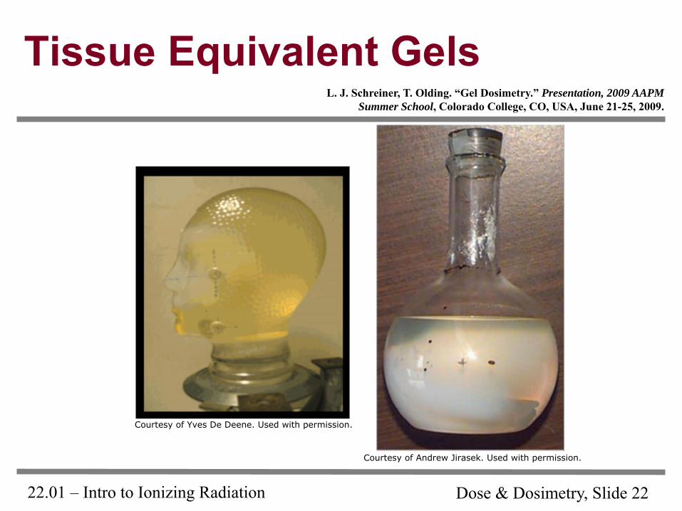
22.01 – Intro to Ionizing Radiation Dose & Dosimetry, Slide 22
Tissue Equivalent Gels L. J. Schreiner, T. Olding. “Gel Dosimetry.” Presentation, 2009 AAPM
Summer School, Colorado College, CO, USA, June 21-25, 2009.
Courtesy of Andrew Jirasek. Used with permission.
Courtesy of Yves De Deene. Used with permission.
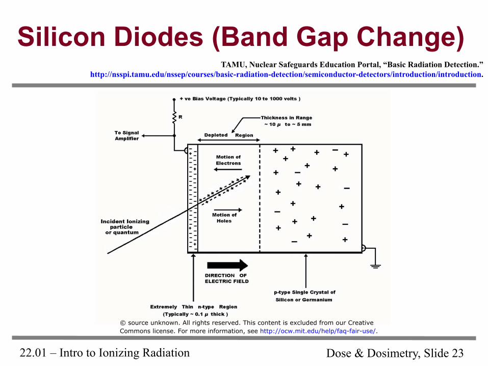
22.01 – Intro to Ionizing Radiation Dose & Dosimetry, Slide 23
Silicon Diodes (Band Gap Change) TAMU, Nuclear Safeguards Education Portal, “Basic Radiation Detection.”
http://nsspi.tamu.edu/nssep/courses/basic-radiation-detection/semiconductor-detectors/introduction/introduction.
© source unknown. All rights reserved. This content is excluded from our CreativeCommons license. For more information, see http://ocw.mit.edu/help/faq-fair-use/.
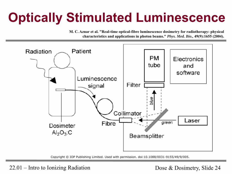
22.01 – Intro to Ionizing Radiation Dose & Dosimetry, Slide 24
Optically Stimulated Luminescence M. C. Aznar et al. "Real-time optical-fibre luminescence dosimetry for radiotherapy: physical
characteristics and applications in photon beams." Phys. Med. Bio., 49(9):1655 (2004).
Copyright © IOP Publishing Limited. Used with permission. doi:10.1088/0031-9155/49/9/005.
22.01 – Intro to Ionizing Radiation Dose & Dosimetry, Slide 25
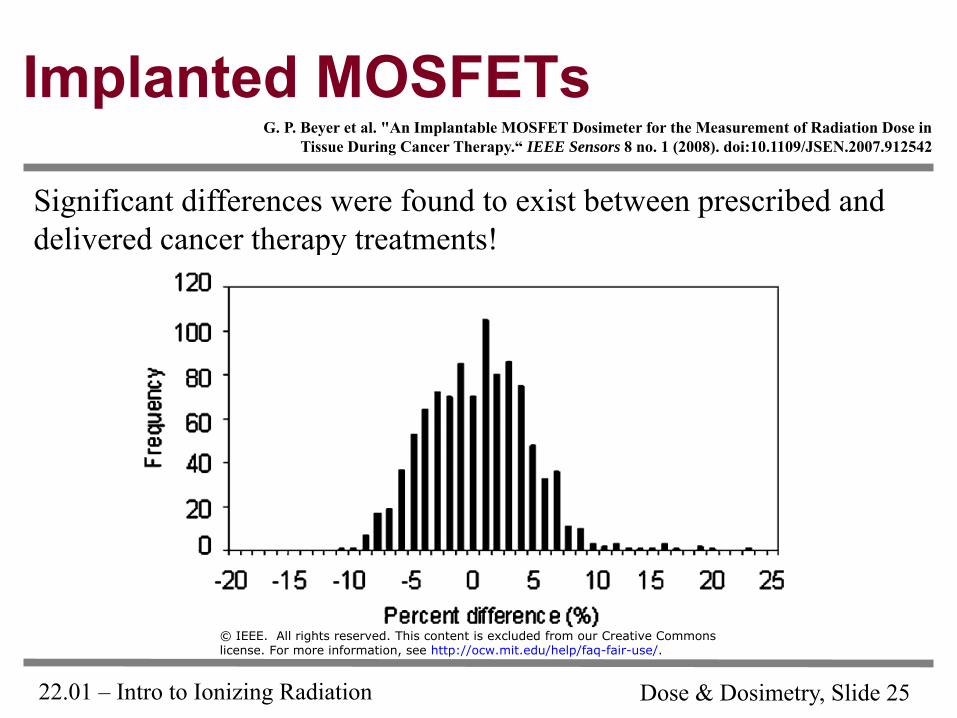
Implanted MOSFETs
Significant differences were found to exist between prescribed and
delivered cancer therapy treatments!
G. P. Beyer et al. "An Implantable MOSFET Dosimeter for the Measurement of Radiation Dose in Tissue During Cancer Therapy.“ IEEE Sensors 8 no. 1 (2008). doi:10.1109/JSEN.2007.912542
© IEEE. All rights reserved. This content is excluded from our Creative Commonslicense. For more information, see http://ocw.mit.edu/help/faq-fair-use/.
22.01 – Intro to Ionizing Radiation Dose & Dosimetry, Slide 26
Problems
• Don’t know the real dose to the tumor
• Don’t know the dose to surrounding tissue
• Can’t control the proton accelerator in real time
• Don’t know the dose rate vs. time
• In-situ methods haven’t worked well
• Ex-situ methods don’t tell you real-time information
22.01 – Intro to Ionizing Radiation Dose & Dosimetry, Slide 27
Our Idea…
… I will present it once our provisional patent is filed!!!
MIT OpenCourseWarehttp://ocw.mit.edu
22.01Introduction to Nuclear Engineering and Ionizing RadiationFall2015
For information about citing these materials or our Terms of Use, visit: http://ocw.mit.edu/terms.


