In South Korea, where avian influenza virus subtypes H3N2, H5N1, H6N1, and H9N2 circulate or have been de- tected, 3 genetically similar canine influenza virus (H3N2) strains of avian origin (A/canine/Korea/01/2007, A/canine/ Korea/02/2007, and A/canine/Korea/03/2007) were iso- lated from dogs exhibiting severe respiratory disease. To determine whether the novel canine influenza virus of avian origin was transmitted among dogs, we experimentally in- fected beagles with this influenza virus (H3N2) isolate. The beagles shed virus through nasal excretion, seroconverted, and became ill with severe necrotizing tracheobronchitis and bronchioalveolitis with accompanying clinical signs (e.g., high fever). Consistent with histologic observation of lung lesions, large amounts of avian influenza virus bind- ing receptor (SAα 2,3-gal) were identified in canine tracheal, bronchial, and bronchiolar epithelial cells, which suggests potential for direct transmission of avian influenza virus (H3N2) from poultry to dogs. Our data provide evidence that dogs may play a role in interspecies transmission and spread of influenza virus. I nfluenza A virus, a member of the genus Orthomyxovirus, is an economically important virus that causes disease in humans, pigs, horses, and fowl (1). A crucial feature in the ecology and epidemiology of influenza virus is interspecies transmission (2). The emergence of new virus subtypes and their interspecies transmission is of great concern; measures to counteract their spread are vital for preventing influenza epidemics and pandemics. One of the basic mechanisms of interspecies transmission of influenza virus is direct transfer of an essentially unaltered virus from 1 species to another (3); however, some factors restrict this transfer. In particu- lar, the presence or absence of host species–specific influ- enza virus binding receptors in the upper and lower respira- tory tracts serves to prevent such cross-species or zoonotic transmission. Human influenza viruses bind to glycolipids or glycans that contain terminal sialyl-galactosyl residues with α 2,6-gal linkages (SAα 2,6-gal), whereas avian influ- enza viruses bind to residues with SAα 2,3-gal linkages (4). Examples of interspecies transmission of influenza viruses include recent human infections with the H5N1 subtype of avian influenza virus, and in canine infections with the H3N8 subtype of equine influenza virus (3,5). In addition, influenza infections were recently reported in species (ca- nine, feline) that historically do not carry influenza virus (6). However, most directly transmitted infections of entire influenza viruses from a natural host species to a new host species do not result in sustained transmission in the new host species (3). Therefore, establishing new, long-lived in- fluenza virus lineage is uncommon and difficult (7). We report interspecies transmission of a complete avi- an influenza virus (H3N2) to dogs and the emergence of a new canine influenza virus associated with acute respira- tory disease in South Korea, where avian influenza viruses (H3N2, H5N1, H6N1, and H9N2) currently circulate or have been previously detected (8). We investigated patho- genicity of the isolated virus in experimental dogs and eval- uated localization of SAα 2,6-gal and SAα 2,3-gal linkages in upper and lower canine respiratory tracts. Materials and Methods Outbreak Histories From May through September 2007, cases of severe respiratory disease occurred in animals at 3 veterinary clinics located 10–30 km apart in Kyunggi Province and 1 kennel located in Jeolla Province (southern part of South Transmission of Avian Influenza Virus (H3N2) to Dogs Daesub Song,* 1 Bokyu Kang,* 1 Chulseung Lee,* Kwonil Jung,† Gunwoo Ha,‡ Dongseok Kang,‡ Seongjun Park,§ Bongkyun Park,§ and Jinsik Oh‡ Emerging Infectious Diseases • www.cdc.gov/eid • Vol. 14, No. 5, May 2008 741 1 These authors contributed equally to this article. *Green Cross Veterinary Products Company, Ltd., Yong-in, South Korea; †Daewoong Pharmaceutical Company, Ltd., Yong-in, South Korea; ‡Animal Genetics, Inc., Suwon, South Korea; and §Seoul National University, Seoul, South Korea
Transcript
In South Korea, where avian infl uenza virus subtypes H3N2, H5N1, H6N1, and H9N2 circulate or have been de-tected, 3 genetically similar canine infl uenza virus (H3N2) strains of avian origin (A/canine/Korea/01/2007, A/canine/Korea/02/2007, and A/canine/Korea/03/2007) were iso-lated from dogs exhibiting severe respiratory disease. To determine whether the novel canine infl uenza virus of avian origin was transmitted among dogs, we experimentally in-fected beagles with this infl uenza virus (H3N2) isolate. The beagles shed virus through nasal excretion, seroconverted, and became ill with severe necrotizing tracheobronchitis and bronchioalveolitis with accompanying clinical signs (e.g., high fever). Consistent with histologic observation of lung lesions, large amounts of avian infl uenza virus bind-ing receptor (SAα 2,3-gal) were identifi ed in canine tracheal, bronchial, and bronchiolar epithelial cells, which suggests potential for direct transmission of avian infl uenza virus (H3N2) from poultry to dogs. Our data provide evidence that dogs may play a role in interspecies transmission and spread of infl uenza virus.
Infl uenza A virus, a member of the genus Orthomyxovirus, is an economically important virus that causes disease in
humans, pigs, horses, and fowl (1). A crucial feature in the ecology and epidemiology of infl uenza virus is interspecies transmission (2). The emergence of new virus subtypes and their interspecies transmission is of great concern; measures to counteract their spread are vital for preventing infl uenza epidemics and pandemics. One of the basic mechanisms of interspecies transmission of infl uenza virus is direct transfer of an essentially unaltered virus from 1 species to another (3); however, some factors restrict this transfer. In particu-lar, the presence or absence of host species–specifi c infl u-
enza virus binding receptors in the upper and lower respira-tory tracts serves to prevent such cross-species or zoonotic transmission. Human infl uenza viruses bind to glycolipids or glycans that contain terminal sialyl-galactosyl residues with α 2,6-gal linkages (SAα 2,6-gal), whereas avian infl u-enza viruses bind to residues with SAα 2,3-gal linkages (4). Examples of interspecies transmission of infl uenza viruses include recent human infections with the H5N1 subtype of avian infl uenza virus, and in canine infections with the H3N8 subtype of equine infl uenza virus (3,5). In addition, infl uenza infections were recently reported in species (ca-nine, feline) that historically do not carry infl uenza virus (6). However, most directly transmitted infections of entire infl uenza viruses from a natural host species to a new host species do not result in sustained transmission in the new host species (3). Therefore, establishing new, long-lived in-fl uenza virus lineage is uncommon and diffi cult (7).
We report interspecies transmission of a complete avi-an infl uenza virus (H3N2) to dogs and the emergence of a new canine infl uenza virus associated with acute respira-tory disease in South Korea, where avian infl uenza viruses (H3N2, H5N1, H6N1, and H9N2) currently circulate or have been previously detected (8). We investigated patho-genicity of the isolated virus in experimental dogs and eval-uated localization of SAα 2,6-gal and SAα 2,3-gal linkages in upper and lower canine respiratory tracts.
Materials and Methods
Outbreak HistoriesFrom May through September 2007, cases of severe
respiratory disease occurred in animals at 3 veterinary clinics located 10–30 km apart in Kyunggi Province and 1 kennel located in Jeolla Province (southern part of South
Transmission of Avian Infl uenza Virus (H3N2) to Dogs
1These authors contributed equally to this article.
*Green Cross Veterinary Products Company, Ltd., Yong-in, South Korea; †Daewoong Pharmaceutical Company, Ltd., Yong-in, South Korea; ‡Animal Genetics, Inc., Suwon, South Korea; and §Seoul National University, Seoul, South Korea
RESEARCH
Korea). The fi rst case, which occurred in May, was identi-fi ed in a 5-year-old miniature schnauzer that had nasal dis-charge for 3 days and sneezing for 2 days, after which the signs subsided and the dog recovered. In August, another case was identifi ed in a 3-year-old cocker spaniel that had fever, cough, nasal discharge, and anorexia and died after the onset of clinical signs. In September, severe respiratory disease was identifi ed in 2 Jindo dogs (a Korean breed of hunting dog that originated on Jindo Island) and a 3-year-old Yorkshire terrier. These animals had severe cough, fever, and nasal discharge and died 2 days after visiting the same animal hospital. Finally, an outbreak of canine infl uenza occurred in an animal clinic in which all 13 dogs housed in a shelter facility were found to be infected with the same virus; their clinical signs were nasal discharge, cough, and high fever. Of the dogs in the affected kennel in Jeolla Province, paired serum samples showed that 47 (90%) of 52 were seropositive for canine infl uenza virus (H3N2) at the fi rst sampling and that 100% were seroposi-tive by the second sampling.
Nasal swabs from the miniature schnauzer, cocker spaniel, and Yorkshire terrier were submitted to Animal Genetics, Inc. (Suwon, South Korea) for reverse transcrip-tion–PCR (RT-PCR) and testing with a commercial rapid infl uenza virus antigen detection kit (Animal Genetics, Inc.). Hemagglutinin inhibition (HI) tests were performed according to the World Organization for Animal Health recommendations; commercial nucleocapsid protein (NP)–based ELISA (Animal Genetics, Inc.) was used for sero-logic testing.
RT-PCR and Sequencing Nasal swabs from the above-mentioned 3 dogs were
also used to isolate the infl uenza A virus by inoculation into 11-day-old chicken eggs. After 3–4 days of incubation, allantoic fl uids were clarifi ed by low speed centrifugation, and these fl uids were shown to agglutinate chicken eryth-rocytes. Virus RNA was extracted from allantoic fl uids by using Trizol LS (Molecular Research Center, Inc., Cincin-nati, OH, USA) according to the manufacturer’s instruc-tions. RT-PCR was performed under standard conditions with random hexamer primers. Isolated infl uenza virus was subtyped by RT-PCR analysis by using primers specifi c for canine, swine, and avian hemagglutinin 3 (H3) genes. Primers for the detection of viral genes H3, neuraminidase 2 (N2), polymerase basic protein (PB) 1, PB2, polymerase acidic protein (PA), NP, matrix protein (M), and nonstruc-tural protein (NS) were designed by using the Primer 3 program with modifi cations (Whitehead Institute, Massa-chusetts Institute for Technology Center for Genome Re-search, Boston, MA, USA).
For PCR, pairs of primers were used to detect target genes. cDNA (2 μL) was mixed with a reaction mixture
containing 2.5 μL of 10× Taq DNA polymerase buffer, 1.5 mmol/L MgCl2, 2.0 μL of dNTPs (2.5 mmol/L/μL), 1 μL of each specifi c primer (10 pmol/L each), and 1 μL of Taq DNA polymerase (Promega, Madison, WI, USA). Distilled water was added to make a fi nal volume of 25 μL. PCR was performed by reaction initiation at 94°C for 10 min, amplifi cation for 32 cycles at 94°C for 30 s, 55°C for 30 s, and 72°C for 30 s, and by fi nal extension at 72°C for 10 min. The reaction was held at 4°C until further use. PCR products were analyzed by electrophoresis in 1.5% agarose gel containing ethidium bromide. Sequences of the isolat-ed virus genes were edited and analyzed by using Bioedit software (www.mbio.ncsu.edu/BioEdit/bioedit.html). Phy-logenetic trees were generated by using the MEGALIGN program (DNASTAR, Madison, WI, USA) with the Clust-alX alignment algorithm (www.megasoftware.net).
Experimental Infection with Isolated VirusWe experimentally reproduced viral infection in 10-
week-old conventional beagle puppies that had been divid-ed into inoculated (I) and noninoculated (NI) groups. Group I puppies (n = 9) were inoculated intranasally with 2 mL of virus isolate with a titer of 106.9 50% egg infectious dose (EID50)/0.1 mL; group NI puppies (n = 6) were inoculated intranasally with 2 mL of sterile phosphate buffered saline. Before they were inoculated, the animals were sedated by intramuscular injection of 0.1 mg/kg acepromazine malate (Bayer, Seoul, South Korea). Clinical signs of infection were monitored for 7 days after inoculation, and feces and nasal discharge were examined for virus shedding by RT-PCR for 10 days after inoculation. To detect antibodies against nucleoprotein and HI for hemagglutinin, we ana-lyzed convalescent-phase serum samples from 3 puppies in each group for virus-specifi c antibodies by ELISA (Animal Genetics, Inc.). HI tests were performed according to World Organization for Animal Health–recommended procedures (9). At 3, 6, and 9 days postinoculation (dpi), 3 group I puppies and 2 group NI puppies were humanely euthanized for gross and histopathologic examination. All necropsy procedures were performed by veterinary pathologists. All organs from dogs and pigs (positive control) were rapidly immersed in 10% neutral formalin buffer to prevent autoly-sis and stored overnight. To detect infl uenza A virus anti-gens in group I or group NI tissues, we performed immu-nohistochemical examination by using goat anti–infl uenza A virus antibody (1:100; Chemicon, Temecula, CA, USA). To determine the presence or absence of SA α2,3-gal link-ages comprising avian infl uenza virus receptors and SA α2,6-gal linkages comprising human infl uenza receptors in the respiratory tracts of noninfected puppies, lectin-based staining was performed as previously reported (10). Por-cine tissue served as a positive control. All experimental procedures were approved by an independent animal care
Transmission of Avian Infl uenza Virus (H3N2) to Dogs
and use committee, and the guidelines of National Veteri-nary Research and Quarantine Service for the reproduction of pathogenesis in dogs were respected.
Results
Isolation of VirusNasal swabs from the miniature schnauzer, cocker
spaniel, and Yorkshire terrier were positive for infl uenza virus and negative for other pathogens, including canine distemper virus, canine parainfl uenza-2 virus, and Borde-tella bronchiseptica. The isolated viruses were designated A/canine/Korea/01/2007 (H3N2), A/canine/Korea/02/2007 (H3N2), and A/canine/Korea/03/2007 (H3N2).
M and NS) of each isolated canine infl uenza virus were sequenced (EU127500, H gene; EU127501, N gene), and homologous sequences were sought in GenBank (Table). Sequences from avian infl uenza viruses that displayed ho-mologies from 95.5% to 98.9% were identifi ed for all 8 gene segments from 1 of the 3 subtype H3N2 canine iso-lates (A/canine/Korea/01/2007). The HA and NA genes of this isolate showed greatest identity with those of Korean avian infl uenza virus isolate S11, and the NS gene showed greatest identity to that of avian infl uenza virus (A/chick-en/Nanchang/7-010/2000 [H3N6]) isolated from Chinese chickens. All the other genes, including PB1, PB2, PA, NP and M, were closely related to those of avian infl uenza vi-rus isolated from ducks in Hong Kong, Japan, and China.
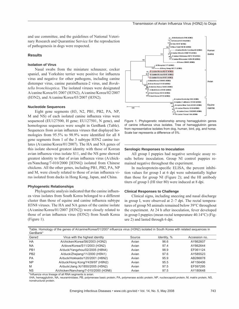
Phylogenetic Relationships Phylogenetic analysis indicated that the canine infl uen-
za virus isolates from South Korea belonged to a different cluster than those of equine and canine infl uenza subtype H3N8 viruses. The HA and NA genes of the canine isolate (A/canine/Korea/01/2007 [H3N2]) were closely related to those of avian infl uenza virus (H3N2) from South Korea (Figure 1).
Serologic Responses to InoculationAll group I puppies had negative serologic assay re-
sults before inoculation. Group NI control puppies re-mained negative throughout the experiment.
In nucleoprotein-specifi c ELISA, the percent inhibi-tion values for group I at 6 dpi were substantially higher than those for group NI (Figure 2); and the HI antibody titers of group I (HI titer 80) were induced at 8 dpi.
Clinical Responses to ChallengeClinical signs, including sneezing and nasal discharge
in group I, were observed at 2–7 dpi. The rectal tempera-tures of group NI animals remained below 39°C throughout the experiment. At 24 h after inoculation, fever developed in group I puppies (mean rectal temperature 40.14°C) (Fig-ure 2) and lasted through 6 dpi.
Table. Homology of the genes of A/canine/Korea/01/2007 influenza virus (H3N2) isolated in South Korea with related sequences inGenBank*Gene† Virus with the highest identity Source Identity, % Accession no. HA A/chicken/Korea/S6/2003 (H3N2) Avian 96.6 AY862607 NA A/dove/Korea/S11/2003 (H3N2) Avian 97.4 AY862644 PB1 A/duck/Yangzhou/02/2005 (H8N4) Avian 98.9 EF061124PB2 A/duck/Zhejiang/11/2000 (H5N1) Avian 97.6 AY585523 PA A/duck/Hokkaido/120/2001 (H6N2) Avian 95.9 AB286878NP A/duck/Hong Kong/Y439/97 (H9N2) Avian 95.5 AF156406M A/duck/Jiang Xi/1850/2005 (H5N2) Avian 97.5 EF597295NS A/chicken/Nanchang/7-010/2000 (H3N6) Avian 97.5 AY180648 *Influenza virus lineage of all RNA segments is avian. †HA, hemagglutinin, NA, neuraminidase; PB, polymerase basic protein; PA, polymerase acidic protein; NP, nucleocapsid protein; M, matrix protein, NS, nonstructural protein.
Figure 1. Phylogenetic relationship among hemagglutinin genes of canine infl uenza virus isolates. Tree of hemagglutinin genes from representative isolates from dog, human, bird, pig, and horse. Scale bar represents a difference of 5%.
RESEARCH
Virus Shedding Infl uenza virus was not detected in feces. However, for
group I puppies, virus shedding in nasal discharge began at 1 dpi and continued to 6 dpi; the highest titers, 106.1(EID50/0.1 mL), were reached by 4 dpi. RT-PCR products generated from shed viruses were sequenced and identifi ed as identi-cal to the inoculated virus.
Histopathologic Findings Gross lesions were limited to the lungs and were char-
acterized by multifocal to coalescing reddish consolidation. In tissues collected on 3, 6 and 9 dpi, histopathologic lesions were observed in the trachea and lungs, and extrapulmonary lesions were absent in puppies infected with the isolate (A/canine/Korea/01/2007 [H3N2]). Severe virus-induced ne-crosis and infl ammation of the upper (trachea and bronchi) and lower (bronchiole and alveoli) respiratory tracts were noted on histologic examination. Although minor differ-ences in the severity of the histologic fi ndings were ob-served among the 9 infected dogs, all infected dogs shared the following histopathologic features regardless how long after inoculation tissues were collected: 1) moderate to se-vere multilobular or diffuse necrotizing tracheobronchitis with suppurative infl ammation in the lumina and squamous metaplasia of the tracheobronchial epithelium (Figure 3, panel B); 2) moderate to severe multilobular or diffuse ne-crotizing bronchiolitis and alveolitis (i.e., bronchioalveoli-tis, occasionally accompanied by chronic peribronchiolar and perivascular infl ammation) (Figure 3, panels D and E); and 3) mild to moderate multilobular or diffuse thickening of alveolar septa by infi ltrates of infl ammatory cells, such as interstitial pulmonary macrophages. At 3, 6, and 9 dpi, large amounts of infl uenza A virus antigen were found in
bronchial and bronchiolar epithelium and lumens (Figure 3, panel F).
Receptor Binding AssayConsistent with the histologic lung lesions, large
amounts of SAα 2,3-gal were found on the surface of bron-chial and bronchiolar epithelial cells of group NI puppies and were rarely found on tracheal epithelial cells (Figure 4). In contrast, SAα 2,6-gal was not detected in tracheal, bronchial, or bronchiolar epithelial cells, which suggests that canine species may have a lesser role as intermediate hosts for transmission of human infl uenza viruses to dogs than for avian infl uenza viruses.
DiscussionBecause all genes of the canine isolates were of avian
infl uenza virus origin, we concluded that the entire genome of the avian infl uenza virus had been transmitted to the dogs. Transmission of avian infl uenza A virus to a new mamma-lian species is of great concern, because it potentially al-lows the virus to adapt to a new mammalian host, cross new species barriers, and acquire pandemic potential.
Transmission of an entire avian infl uenza virus to an unrelated mammalian species is a rare event. Several out-breaks of avian infl uenza infection have occurred in mam-mals. Infl uenza virus (H7N7) of avian origin was isolated from the lungs and brains of dead seals. In addition, it was replicated to high titers in ferrets, cats, and pigs and caused conjunctivitis in humans (11,12). Avian origin infl uenza vi-rus (H4N5) was reported as the cause of infection and death in harbor seals along the New England coastline (13), and avian origin infl uenza (H5N1) infection was identifi ed in a dog after ingestion of a duck infected with subtype H5N1 during an outbreak in Thailand in 2004 (14).
Previously, outbreaks of hemorrhagic pneumonia caused by equine infl uenza virus (H3N8) were noted in racing dogs, and a human infl uenza virus (H3N2) was iso-lated from dogs (15,16). However, these reports provide limited serologic and virologic evidence for infl uenza virus infection in dogs. We report the emergence of a new canine infl uenza virus strain that causes acute respiratory disease in dogs and that differs from previous outbreaks of equine infl uenza virus (H3N8) infections.
Concerning the possible mechanism of avian infl u-enza virus transmission to dogs, we posit that this trans-mission results from feeding dogs untreated minced meats of ducks or chickens. In South Korea, untreated duck and chicken meats, including internal organs and heads, have been widely used to feed dogs for fattening in local canine farms or kennels. In a previous study, avian infl uenza virus (H3N2) was isolated from ducks and chickens sold at live-bird markets in South Korea. Live-bird markets are thought to constitute “a missing link in the epidemiology of avian
Figure 2. Body temperature, virus shedding, and antibody seroconversion after challenge with canine infl uenza virus. Body temperature was increased from 1 day postinoculation (dpi) and slowly decreased to normal temperature by 7 dpi. Virus shedding was detected from 1 dpi to 6 dpi by reverse transcription–PCR. However, the ELISA antibody titers increased after 6 dpi. Antibody titers were regarded as positive if percent inhibition (PI) was >50.
Transmission of Avian Infl uenza Virus (H3N2) to Dogs
infl uenza viruses” because they bring together numerous hosts, such as chickens, ducks, turkeys, geese, and doves, in a high-density setting, which represents an ideal envi-ronment for virus interspecies transmission (17,18). The S11 strain, whose HA and NA genes showed the greatest identity to those of the A/canine/Korea/01/2007 (H3N2) isolates from dogs, was isolated from a tracheal swab of a healthy chicken and is nonpathogenic in poultry (8). These
observations support the hypothesis that avian infl uenza vi-rus (H3N2) strains could be transmitted by feeding infected poultry by-products to dogs (2).
It is also possible that cross-species transmission of infl uenza virus occurs directly by aerosol transmission from infected birds to susceptible dogs as a consequence of close contact between the 2 species. Lectin-staining results showed that canine upper (trachea and bronchi) and lower
Figure 3. Histopathologic lesions in the trachea and lungs of control (A and C) or experimentally infected (B, D–F) dogs (A/canine/Korea/01/2007 [H3N2]) at different days postinoculation (dpi). A) Control dog at 9 dpi, showing normal pseudostratifi ed columnar epithelium lining of the trachea; original magnifi cation ×400. Hematoxylin and eosin (HE) stain. B) Infl uenza-infected dog at 9 dpi, showing necrotizing tracheitis characterized by necrosis (n), squamous metaplasia (s), and hyperplasia of the epithelium and nonsuppurative infl ammation (c) in the connective tissue; original magnifi cation ×400. HE stain. C) Control dog at 3 dpi, showing normal alveoli; original magnifi cation ×200. HE stain. D) Infl uenza-infected dog at 3 dpi, showing severe diffuse necrotizing bronchitis and bronchiolitis with suppurative infl ammation in the lumina; original magnifi cation ×100. HE stain. E) Infl uenza-infected dog at 6 dpi, showing severe necrotizing bronchiolitis; original magnifi cation ×200. HE stain. F) Infl uenza-infected dog at 6 dpi (serial section of E), showing large amounts of infl uenza A virus antigens (red stain; arrows) in the bronchiolar epithelium and lumen. Immunohistochemistry; Red Substrate (Dako, Carpinteria, CA, USA); Mayer’s hematoxylin counterstain. G) Infl uenza-infected dog at 9 dpi, showing severe necrotizing alveolitis with accumulation of necrotic cells in terminal bronchioles (tb) and alveoli (a); original magnifi cation ×200. HE stain.
Figure 4. Lectin staining (red stain) for SAα 2,3-gal (avian infl uenza virus receptors) and SAα 2,6-gal (human infl uenza virus receptors) in canine trachea, bronchus, and bronchioles, together with porcine tissues as a positive control. Original magnifi cation all x300. −, no staining; ±, rare or few positive cells; +, moderate numbers of positive cells; and ++, many positive cells.
RESEARCH
(bronchiole) respiratory tract epithelium cells display SAα 2,3-gal, to which avian infl uenza viruses bind, making pos-sible a direct transmission of avian infl uenza viruses from poultry to dogs. Additionally, according to the animal hos-pital veterinarian, this outbreak was traced to a Jindo dog purchased at a live-animal market in Kyunggi Province that sold chicken, duck, pheasant, rabbit, cats, pet dogs, and other dogs. The Jindo dog was hospitalized at the local animal hospital and may have infected the other pet dogs at the hospital. This epidemiologic result also suggests that the novel canine infl uenza virus of avian origin was trans-mitted within canine species.
Antigenic and phylogenetic analyses showed that the HA and NA genes of the A/canine/Korea/01/2007 (H3N2) isolate are closely related to isolates identifi ed in 2003 from chickens and doves in South Korea. Furthermore, HA genes of canine infl uenza isolates were different from recent isolates from swine in South Korea (19). The other genes of the canine infl uenza isolate are more closely re-lated to those of the subtype H9N2 isolate found in ducks from Hong Kong, the subtype H6N2 isolate from ducks in Japan, and several other avian infl uenza strains from southeastern China in 2000 through 2005. This fi nding suggests that multiple variants of subtype H3 infl uenza viruses may be circulating in these regions and causing disease in pet dogs.
Our experimental reproduction of the disease caused by this isolate induced severe pathologic changes and showed that infected dogs excreted infl uenza virus (H3N2) in nasal discharge but not in feces. This fi nding suggests that dog-to-dog transmission of subtype H3N2 could occur through the nasal route and that dog-to-dog transmission of the virus could play an important role in the epizootiology of the disease.
In our study, virologic, serologic, pathologic, and phy-logenetic analyses showed cross-species infection of an en-tire avian infl uenza A virus (H3N2) to another mammalian species, dogs. Evidence of avian infl uenza virus infection in pet dogs raises the concern that dogs may be become a new source of transmission of novel infl uenza viruses, especially where avian infl uenza viruses are circulating or have been detected.
AcknowledgmentsWe thank the staff of Green Cross Veterinary Products,
South Korea, and Animal Genetics, South Korea, for their as-sistance. We also thank Patrick Hughes for technical editing and H.Y. Kim for samples from the fi rst case.
Dr Song is a virologist at Green Cross Veterinary Products, Yong-in, South Korea. His research interests include swine virol-ogy, viral enteritis of pigs, and viral diseases of animals.
3. Crawford PC, Dubovi EJ, Castleman WL, Stephenson I, Gibbs EPJ, Chen L, et al. Transmission of equine infl uenza virus to dogs. Sci-ence. 2005;310:482–5.
4. Suzuki Y. Sialobiology of infl uenza: molecular mechanism of host range variation of infl uenza viruses. Biol Pharm Bull. 2005;28:399–408.
5. Guan Y, Poon LL, Cheung CY, Ellis TM, Lim W, Lipatov AS, et al. H5N1 infl uenza: a protean pandemic threat. Proc Natl Acad Sci U S A. 2004;101:8156–61.
6. Keawcharoen J, Oraveerakul K, Kuiken T, Fouchier RA, Amonsin A, Payungporn S, et al. Avian infl uenza H5N1 in tigers and leopards. Emerg Infect Dis. 2004;10:2189–91.
7. Webster RG, Bean WJ, Gorman OT, Chambers TM, Kawakowa Y. Evolution and ecology of infl uenza viruses. Microbiol Rev. 1992;56:152–79.
8. Choi YK, Seo SH, Kim JA, Webby RJ, Webster RG. Avian infl uenza viruses in Korean live poultry markets and their pathogenic poten-tial. Virology. 2005;332:529–37.
9. World Organization of Animal Health. Manual of diagnostic tests and vaccines for terrestrial animals. 5th ed. Paris: The Organization; 2005.
10. Wan H, Perez DR. Quail carry sialic acid receptors compat-ible with binding of avian and human infl uenza viruses. Virology. 2006;346:278–86.
11. Webster RG, Geraci J, Petursson G, Skirnisson K. Conjunctivitis in human beings caused by infl uenza A virus of seals. N Engl J Med. 1981;304:911.
12. Webster RG, Hinshaw VS, Bean WJ, Van Wyke KL, Geraci JR, St Aubin DJ, et al. Characterization of an infl uenza A virus from seals. Virology. 1981;113:712–24.
13. Hinshaw VS, Bean WJ, Webster RG, Rehg JE, Fiorelli P, Early G, et al. Are seals frequently infected with avian infl uenza viruses? J Virol. 1984;51:863–5.
14. Songserm T, Amonsin A, Jam-on R, Sae-Heng N, Pariyothorn N, Payungporn S, et al. Fatal avian infl uenza A H5N1 in a dog. Emerg Infect Dis. 2006;12:1744–7.
15. Chang CP, New AE, Taylor JF, Chang HS. Infl uenza virus isola-tions from dogs during a human epidemic in Taiwan. Int J Zoon. 1976;3:61–4.
16. Houser RE, Heuschele WP. Evidence of prior infection with infl u-enza A/Texas/77 (H3N2) virus in dogs with clinical parainfl uenza. Can J Comp Med. 1980;44:396–402.
17. Liu H, Liu X, Cheng J, Peng D, Jia L, Huang Y. Phylogenetic analy-sis of the hemagglutinin genes of twenty-six avian infl uenza viruses of subtype H9N2 isolated from chickens in China during 1996–2001. Avian Dis. 2003;47:116–27.
18. Liu M, He S, Walker D, Zhou N, Perez DR, Mo B, et al. The in-fl uenza virus gene pool in a poultry market in south central China. Virology. 2003;305:267–75.
19. Song DS, Lee JY, Oh JS, Lyoo KS, Yoon KJ, Park YH, et al. Isola-tion of H3N2 swine infl uenza virus in South Korea. J Vet Diagn Invest. 2003;15:30–4.
Address for correspondence: Jinsik Oh, Animal Genetics, Inc., 404-5, Wonchun-dong, Youngtong-gu, Suwon-si, Kyunggi-do, 443-823, South Korea; email: [email protected]