102
1 DOTTORATO DI RICERCA IN Biotecnologie Odontostomatologiche CICLO XXVIII Regenerative Endodontics: a review of clinical protocols. Valentina Giuliani
1
DOTTORATO DI RICERCA IN
Biotecnologie Odontostomatologiche
CICLO XXVIII
Regenerative Endodontics:
a review of clinical protocols.
Valentina Giuliani
2
DOTTORATO DI RICERCA IN
Biotecnologie Odontostomatologiche
CICLO XXVIII
COORDINATORE Prof. Gabriella Pagavino
Regenerative Endodontics:
a review of clinical protocols.
Settore Scientifico Disciplinare MED /28
Dottorando Tutore
Valentina Giuliani Gabriella Pagavino
Coordinatore
Prof. Pagavino Gabriella
3
Anni 2012/2015
Accademic year 2015/16
March 16th 2016
Florence, Italy
Committee:
Promoter Prof. Gabriella Pagavino
Prof. Marco Ferrari
Prof. Lucio Montebugnoli
Dott.ssa Chiara Baroni
TITLE:
Regenerative endodontics: review of clinical
protocols.
CANDIDATE:
Giuliani Valentina
4
Chapter 1
1.1 Apexification with Calcium Hydroxide
Chapter 2
2.1 Mineral Trioxide Aggregate: Chemical composition and setting
reaction
2.2 Biocompatibility and bioactivity of Mineral Trioxide Aggregate
2.3 Clinical use of Mineral Trioxide aggregate as apical plug
Chapter 3
3.1 Dental stem cell; apexogenesis and apexification.
3.2 Dental pulp stem cells
3.3 Regenerative endodontic procedure: revascularization,
revitalization or maturogenesis?
3.4 Nature of tissues present in the canals of these teeth treated with
regenerative endodontics
Chapter 4
Regenerative endodontics: review of clinical protocols.
4.1 Introduction
4.2 Matherial and Methods
4.3 Results
4.4 Discussion
Summary and conclusion
5
Chapter 1
1.1 Apexification with Calcium Hydroxide
Traumatic injury or a depth caries penetration, during the developing
of dentition, could lead to a premature pulp necrosis of immature
permanent tooth. Dental injuries are most common in young patients
(8-14 years), when children are most active. In these cases
endodontics treatment aimed to keep the dentition in physiologically
functional state for the maintenance of oral and systemic health. Teeth
with immature root development present challenges in cleaning,
shaping and obturation of large canals with open apices (1-3). In these
cases conventional root canal treatment can be overcome using
Calcium Hydroxide Ca(OH)2 apexification approach (4) or Mineral
Trioxide Aggregate (MTA) apical plug technique (5). In both clinical
approaches the risk of future root fracture and tooth mobility, due to a
poor root-crown ratio, still remains.
Apexification is defined as ‘‘a method of inducing a calcified barrier
in a root with an open apex or the continued apical development of an
incompletely formed root in teeth with necrotic pulp”. Teeth treated
with this material require the placement of long-term calcium
hydroxide in the root canal to induce formation of an apical hard
6
tissue barrier. The formation of the apical barrier is necessary to allow
the filling of the root canal system without risk of overfilling
The high pH and antimicrobial properties (6) of Ca(OH)2 combined
with the permeability of dentin (7) guided to a common and well
established use of calcium hydroxide in dentistry.
Hermann introduced the use of calcium hydroxide in 1920 (8); it was
often applied within the root canal system as intracanal medicament
and in apexification procedure. In 1964 Kaiser (9) proposed the use of
calcium hydroxide mixed with camphorate parachlorophenol to
induce the formation of calcified barrier across the apex.
Frank (5), who emphasized the importance of reducing contamination
within the root canal by instrumentation, medication and dressing the
canal space temporarily with a reasonable paste seal, popularized this
procedure.
The use of calcium hydroxide mixed with saline solution (10), sterile
water (11), or distilled water (12) has been investigate with good
clinical success; the mechanism of action of calcium hydroxide in
induction of an apical barrier remain controversial; two different
aspects could play an important role in osteogenetic potential of
calcium hydroxide: mineralized and antibacterial actions. Ca(OH)2
induced a multi-layered necrosis (13) with subjacent mineralization;
the necrosis generates a low grade of irritation of underlying tissue
sufficient to produce a matrix that mineralized. Calcium was attracted
to this area and the mineralization of newly formed collagenous
matrix is initiated from the calcified foci. The apical barrier formation
is more successful in the absence of microorganism, the release of
hydroxyl ions cause the damage to bacterial cytoplasmic membrane,
protein denaturation and damage to bacterial DNA (14-19). The hard
tissue barrier formed at the end of the root has been histologically
described as follow: an outer layer composed of a dense acellular
cementum-like tissue and a more central mix of irregular dense fibro-
collagenous connective tissue containing foreign material highly
mineralized (20). By using repeated Ca(OH)2 dressings, during a 3 to
7
6 months period, it demonstrated that it was possible not only to
induce healing of the apical lesion but also to induce closure of the
root apex with calcified tissue (apexification).
When Ca(OH)2 is used in apexification procedure, the therapy could
extend from months to years before achieve the desired effect (21,22).
In a review Sheehy and Robert reported an average duration for apical
barrier formation ranging from 5 to 20 mouths (21); the high
variability could be due to: presence or absence of periradicular
lesion, and /or stage of root development and consequent apical width
(23). Immature teeth treated with Ca(OH)2 showed an high failure rate
because of an unusual incidence of root fracture. This might be the
direct consequence of changes in the physical properties of dentin due
to the use of Ca(OH)2 medicament (1, 24, 25). Cvek (1), investigated
885 luxated non-vital teeth over a period ranging from 3 to 54 months,
reported that 168 teeth suffered a cervical root fracture within the
follow-up period, ranged from 3.5 to 5 years. Each failed teeth showed
a cervical resorptive defect near the fracture; this may be a result of a
change in the organic matrix (2, 25). Calcium hydroxide dissolves
pulp tissue because of denaturation and hydrolysis of dentin proteins,
furthermore the high pH reduce the organic support of the dentin
matrix; both effects result in a loose of connection between collagen
fibrils and hydroxyapatite crystals, that could negatively influence
mechanical properties of dentin (2, 25, 26, 27). The weakening of the
root structure in term of on micro-tensile fracture strength of dentine
tissue after prolonged used of calcium hydroxide, was supported by in
vitro studies too (28, 29).
Outcome assessment of calcium hydroxide in apexification treatments,
showed a success rates ranged from 79% to 100% (1,30-34). Based on
a meta-analysis and systematic review the rate of clinical success and
apical barrier formation mineral trioxide aggregate and calcium
hydroxide as material used for the endodontic management of
immature teeth had no perceivable discrepancy (35).
8
References
1. Cvek M. Prognosis of luxated non-vital maxillary incisors treated with
calcium hydroxide and filled with gutta-percha. A retrospective
clinical study. Endod Dent Traumatol 1992;8:45.
2. Andreasen JO, Farik B, Munksgaard EC. Long-term calcium
hydroxide as a root canal dressing may increase risk of root fracture.
Dent Traumatol 2002;18:134.
3. Trope M. Treatmengt of immature tooth with non vital pulp and apical
periodontitis. Dent Clin North Am 2010;54:313-24.
4. Frank AL. Therapy for the divergent pulpless tooth by continued
apical formation..J Am Dent Assoc 1966;72:87–93.
5. Torabinejad M, Chivian N. Clinical applications of mineral trioxide
aggregate.J Endod 1999;25:197–205.
6. Tamburic SD, Vuleta GM, Ognjanovic JM. In vitro release of calcium
hydroxil ions from two types ofcalcium hydroxide preparation. Int
Endod J 1993;26:125-30.
7. Foreman PC, Barnes IE. Review of Calcium hydroxide. Int Endod J
1990;23:283-97.
8. Hermann BW. Calcium hydroxid als Mittel Zum Behandel and Fullen
Von Zahnwurzelkana len. Wuzburg: Med Diss, 1920.
9. Kaiser HJ. Management of wide open apex canals with calcium
hydroxide. Presented at the 21st Annual Meeting of the American
Association of Endodontists, Washington DC April 17 1964.
9
10. Citrome GP, Kaminski EJ, Heuer MA. A comparative study of tooth
apexification in the dog. J Endod 1979;5:290–7. 44.
11. Michanowicz J, Michanowicz A. A conservative approach and
procedure to fill an incompletely formed root using calcium hydroxide
as an adjunct. J Dent Child 1967;32:42–7.
12. Binnie WH, Rowe AHR. A histologic study of the periapical tissues
of incompletely formed pulpless teeth filled with calcium hydroxide. J
Dent Res 1973;52:1110–6.
13. Schroder U, Granath L. Early reaction of intact human teeth to
calcium hydroxide following experimental pulpotomy and its
significance to the development of hard tissue barrier. Odontol Revy
1971;22:379–95.
14. Barthel CR, Levin LG, Reisner HM, Trope M. TNFalpha release in
monocytes after exposure to calcium hydroxide treated E. coli LPS.
Int Endod J 1997;30:155–9. 60.
15. Bystrom RH, Claesson R, Sundqvist G. The antibacterial effect of
calcium hydroxide in the treatment of infected root canals. Endod
Dent Traumatol 1985;1:170–5. 61.
16. Estrela C, Pimento FC, Ito IY, Bammann LL. In vitro determination
of direct antimicrobial effect of calcium hydroxide. J Endod
1998;24:15–7. 62.
17. Jiang J, Zuo J, Chen SH, Holliday LS. Calcium hydroxide reduces
lipopolysaccharide-stimulated osteoclast formation. Oral Surg Oral
Med Oral Pathol Oral Radiol Endod 2003;95:348–54. 63.
18. Kontakiotis E, Nakou M, Georgopoulou M. In vitro study of the
indirect action of calcium hydroxide on the anaerobic flora of the root
canal. Int Endod J 1995;28:285–9. 64.
19. Safavi KE, Nicholls FC. Alteration of biological properties of
bacterial lipopolysaccharide by calcium hydroxide. J Endod
1994;20:127–9.
10
20. Baldassari-Cruz LA, Walton RE, Johnson WT. Scanning electron
microscopy and histologic analysis of an apexifi- cation ‘cap’. Oral
Surg Oral Med Oral Pathol Oral Radiol Endod 1998;86:465–8.
21. Sheehy EC, Robert GJ. Use of calcium hydroxide for apical barrier
formation and healing in non-vital immature permanent teeth: a
review. Br Dent J 1997;183:241-6.
22. Dannemberg JL. Pedodontic endodontic. Dent Clin North Am
1974;18:367-77.
23. Rafter M. Apexification: a review. Dent Traumatol 2005;21:1-8.
24. Stormer K, Jacobsen I, Attramadal A. Hvor funkjonsdyktige blir
rottfylte unge permanente incisiver? In: Nordisk forening for
pedodonti. Bergen, Norway: Aarsmote, 198
25. Doyon GE, Dumsha T, von Fraunhofer JA. Fracture Resistance of
Human Root Dentin Exposed to Intracanal Calcium Hydroxide. J
Endod 2005;31:895-97.
26. Anderson M, Lund A, Andreasen JO, Andreasen FM. In vitro
solubility of human pulp tissue in calcium hydroxide and sodium
hypochlorite. Endod Dent Traumatol 1992; 8:104–8.
27. Hasselgren G, Olsson B, Cvek M. Effects of calcium hydroxide and
sodium hypochlorite on the dissolution of necrotic porcine muscle
tissue. J Endod 1988;7:17–2.
28. Rosemberg B, Murray PE, Namerow K. The effect of calcium
hydroxide root filling on dentine fracture strenght. Dent Traumatol
2007;23:26-29.
29. White JD, Lacefield WR, Chavers LS, Eleazer PD. The effect of three
commonly used endodontic materials on the strenght and hardness of
root dentine. J Endod 2002;28:828-30.
30. Kerekes K, Heide S, Jacobsen I. Follow-up examination of endodontic
treatment in traumatized juvenile incisors. J Endod 1980;6:744 – 8.
11
31. Witherspoon DE, Small JC, Regan JD, Nunn M. Retrospective
Analysis of Open Apex Teeth Obturated with Mineral Trioxide
Aggregate. J Endod 2008;34:1171–1176.
32. Dominguez Reyes A, Munoz Muǹoz L, Aznar Martın T. Study of
calcium hydroxide apexification in 26 young permanent incisors. Dent
Traumatol 2005;21(3):141-5.
33. Walia T, Chawla HS, Gauba K. Management of wide open apices in
non-vital permanent teeth with Ca(OH)2 paste. J Clin Pediatr Dent
2000;25(1):51-6.
34. Finucane D, Kinirons MJ. Non-vital immature permanent incisors:
factors that may influence treatment outcome. Endod Dent Traumatol
1999;15(6):273-7.
35. Chala S, Abouqal R, Rida S Apexification of immature teeth with
calcium hydroxide or mineral trioxide aggregate: systematic review
and meta-analysis. Oral Surg Oral Med Oral Pathol Oral Radiol Endod
2011;112:e36-e42.
12
Chapter 2
Despite the high success rate of long term calcium hydroxide
apexification (mid 90%), the disadvantage of this technique (1),
multiple appointment during an extended period of time, susceptibility
to root fracture, risk of coronal micro-leakage during the therapy; lead
to alternative treatment that might be offer a more predictable and
better long-term prognosis for necrotic teeth with incomplete root
development.
Artificial apical barriers with a variety of materials have been
suggested as an alternative to traditional Ca(OH)2 apexification (2-7).
In 1999 Torabinejad and Chivian proposed the use of a new material,
Mineral Trioxide Aggregate (MTA), as artificial apical barrier for the
treatment of immature necrotic teeth (8).
2.1 Mineral Trioxide Aggregate: Chemical composition and setting
reaction
Mineral trioxide aggregate (MTA) was developed at Loma Linda
University, in the 1990s, as a root-end filling material. It was used
primarily to seal lateral root perforations (8,9) and as root-end filling
material (10-13). In 1998 Mineral trioxide aggregate (MTA) received
acceptance by the US Federal Drug Administration and became
commercially available as ProRoot MTA (Tulsa Dental Products,
Tulsa, OK, USA).
The literature widely documented the physical, chemical and
biological characteristics of MTA; it’s consist in a powder contains
50-75% calcium oxide (CaO) and 15-25% silicon dioxide (SiO2).
13
These two components together comprise 70-95% of the cement (11,
14, 19, 20, 21).
Scanning electron microscope (SEM) of polished sections of un-
hydrated MTA embedded in resin, show distinctive cement grains and
bismuth oxide particles, which are separated from one another. The
mean value of the prisms was 87% calcium and 2.47% silica, the
remainder being oxygen. In areas of amorphous structure, there
seemed to be 33% calcium, 49% phosphate, 2% carbon, 3% chloride,
and 6% silica.
The elemental composition of MTA as shown by energy dispersive
spectroscopy (EDS) indicates the presence of calcium, silicon and
oxygen with minor peaks for aluminium, potassium, magnesium and
bismuth. The phases present in un-hydrated MTA, determined by X-
ray diffraction analysis, exhibits peaks for tricalcium silicate,
dicalcium silicate and bismuth oxide. Each phase has a particular
pattern that can be subsequently searched and matched with data
derived from the International Centre of Diffraction. MTA contains
other phases such as dicalcium silicate and tricalcium aluminate in
minimal quantities. In the first publication on MTA composition,
calcium phosphate was the main constituent of MTA (9). Further
analysis demonstrated that the former appeared as discrete crystals
and the latter as an amorphous structure with no apparent crystal
growth but a granular appearance.
When these raw materials are blended, they produce tricalcium
silicate, dicalcium silicate, tricalcium aluminate, and tetracalcium
aluminoferrite. Various methods have been used to examine MTA
composition including energy dispersive analysis with x-ray (EDAX),
inductively coupled plasma optical emission spectroscopy (ICP-OES),
x-ray diffraction analyses (XRD), x-ray fluorescence spectrometry
(XRF), energy x-ray spectrometry, and energy dispersive
spectroscopy (9–18).
Mineral Trioxide Aggregate is available in two commercial form grey
MTA (GMTA), the oldest formula, and white MTA (WMTA), the
14
newest formula. The difference between the grey and the white
materials is the presence of iron in the grey material, which makes up
the phase tetracalcium alumina-ferrite. This phase is absent in white
MTA.
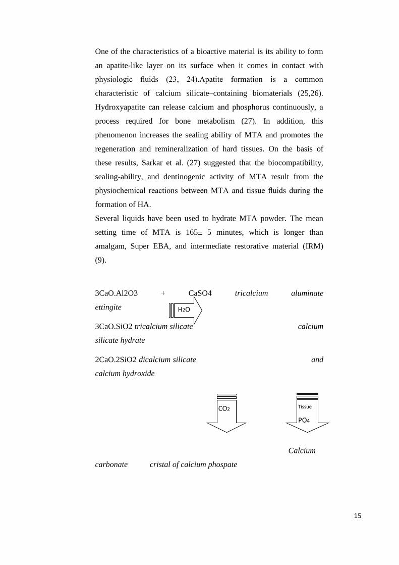
Mixing MTA powder with sterile water in a 3:1 powder-to-liquid ratio
(22) we obtain a colloidal gel that solidifies into a hard structure.
During the initial stages of the reaction of MTA after hydratation,
calcium silicate hydrate is formed, coating the cement particles and
preventing further reactions. Tricalcium aluminate dissolves and
reacts with the calcium and sulfate ions present in the liquid phase to
produce ettringite, which also precipitates on the cement particle
surface. The initial phase is followed by a dormant period, wherein the
hydrate coating on the cement grains prevents further hydration. The
dormant period lasts for 1-2 hours, which is a period of relative
inactivity and the cement is plastic and workable. Following the
completion of the dormant period, setting of the cement proceeds to
the acceleration stage, where the hydration process accelerates again.
Sulfate ions are depleted and monosulfate forms from ettringite.
Crystalline calcium hydroxide also precipitates from the liquid phase
(20).
The hydration reaction takes several years to complete, although the
cement mass would have achieved the final hardening and maximum
physical and mechanical properties by 28 days.
If MTA is left exposed to the environment, the calcium hydroxide
reacts with the atmospheric carbon dioxide resulting in the deposition
of calcium carbonate on the cement surface. These deposits are
commonly mistaken as being an integral part of the cement
microstructure. When in contact with tissue fluids and synthetic tissue
fluids, which contain phosphate ions, the calcium hydroxide produced,
as a by-product of MTA hydration, reacts to form calcium phosphate
and is deposited on the cement surface. Calcium phosphate is crystal
on the material surface has been reported to be the reason for the
bioactivity shown by MTA.
15
One of the characteristics of a bioactive material is its ability to form
an apatite-like layer on its surface when it comes in contact with
physiologic fluids (23, 24).Apatite formation is a common
characteristic of calcium silicate–containing biomaterials (25,26).
Hydroxyapatite can release calcium and phosphorus continuously, a
process required for bone metabolism (27). In addition, this
phenomenon increases the sealing ability of MTA and promotes the
regeneration and remineralization of hard tissues. On the basis of
these results, Sarkar et al. (27) suggested that the biocompatibility,
sealing-ability, and dentinogenic activity of MTA result from the
physiochemical reactions between MTA and tissue fluids during the
formation of HA.
Several liquids have been used to hydrate MTA powder. The mean
setting time of MTA is 165± 5 minutes, which is longer than
amalgam, Super EBA, and intermediate restorative material (IRM)
(9).
3CaO.Al2O3 + CaSO4 tricalcium aluminate
ettingite
3CaO.SiO2 tricalcium silicate calcium
silicate hydrate
2CaO.2SiO2 dicalcium silicate and
calcium hydroxide
Calcium
carbonate cristal of calcium phospate
H2O
CO2 Tissue
PO4
16
2.2 Biocompatibility and bioactivity of Mineral Trioxide Aggregate
The MTA cement reflect a current requirement to have materials for
endodontic therapy that are able to stimulate the healing process of
periapical tissues, instead of merely biocompatible or inert materials.
In the past decade, the two major characteristics that justify the
successful use of MTA in endodontics were the excellent seal ability
and biocompatibility. In case of necrotic teeth with immature root
development, MTA has been advocated as an apexification material
because it permits an adequate seal of the canal preventing bacterial
leakage, and a poor inflammatory reaction in periodontal tissue.
Nowadays, it’s well-documented the favourable biologic response
stimulated by MTA in human periapical tissue; cell from periradicular
healing tissue migrate to the apex and differentiate in cells capable of
secreting a cementum, osteocementum or osteodentin organic matrix
under the influence of specific cellular signals (28). Bone and
periodontal healing/regeneration is a complex event that involves the
different stages (30), in which the migration and invasion of
multipotent mesenchymal stem cells are required (31).
When MTA has been placed in direct contact with human tissues, the
following reaction have been observed:
Releases calcium ions and facilitates cell attachment and
proliferation. The ability of MTA to moderate the migration of cells
should probably be considered to be an important stage in the
induction of tissue repair. Apical barrier formation can occur even in
the presence of gaps between the MTA plug and the root canal walls.
This observation was probably related to the ability of MTA to
enhance cell migration of human bone marrow stem cells (29).
Currently human PDL cells were used to simulate the root-end
environment; these cells are responsible for the formation and
17
maintenance of periodontal ligament fiber attachments as well as
repair, remodelling, and regeneration of the adjacent alveolar bone
and cementum. In a human PDL cell culture study, cell attachment to
MTA was observed; PDL fibroblasts showed proliferation on MTA
and survival.
Creates antibacterial environment by its alkaline pH. The bioactivity
of MTA has been attribute to its ability to produce hydroxyapatite
(Ca5(PO4)3(OH)) in presence of phosphate buffered saline. The
hydroxyapatite is a crystal presents in form of calcium carbonate,
calcium phosphate and calcium floruro. Initially an amorphous
calcium phosphate phase is formed, which acts as a precursor to the
secondary phase during which carbonated apatite is formed.
Carbonated apatite is also known as biologic apatite and represents the
mineral phase of hard tissue (bone, dentin, and cementum) (32, 33).
The bioactivity of MTA could be attributed to its capacity to form
carbonated apatite, which is important in formation and maintenance
of the bone tissue biomaterial interface. The apatite formed by the
cement-Phosfate Buffered Saline (PBF) system was deposited within
collagen fibrils, promoting controlled mineral nucleation on dentin as
reported by Reyes-Carmona et al (34). The possible biologic and
clinical significance of their findings includes the following:
(1) the interaction of MTA and Portland cement with dentin in a
phosphate-containing fluid promotes a biomineralization process,
(2) This process could be significant in minimizing leakage,
(3) The formation of the interfacial layer and the intratubular
mineralization process could influence the push-out bond strength,
and
(4) The formation of carbonated apatite precipitates could be
responsible for the ability of the cements to stimulate repair and
dentinogenesis or cementogenesis.

18
Modulates cytokine production. Basing on In vitro and in vivo animal
study, MTA seems to play a role on the production of signalling
molecules. Macrophages produce different types of cytokines,
signalling molecules and inflammatory products. Up regulation of
various types of cytokines and biologic markers has been reported in
the presence of MTA in several cell culture studies when compared
with control or other tested materials. These cytokines and biologic
markers include interleukin (IL)-1a (36, 37), IL-1 b (36-38), IL-2(39),
IL-4(39), IL-6(36, 38, 40), IL-8 (41), IL-10 (39), IL-18 (36),
osteocalcin (36, 39, 42-44), alkaline phosphatase (42, 44, 45), bone
sialoprotein (44), osteopontin (44), and BMP-2 (45).
Encourages differentiation and migration of hard tissue producing
cells. MTA can induce osteoblastic/cementoblastic differentiation of
human periodontal ligament cells, which express calcium sensing
receptors (CaSR) and bone morphogenetic protein-2 (BMP-2)
receptors that are potentially involved in osteogenesis. BMP induce
the production of bone when injected into ectopic sites. PDL human
fibroblast attached to MTA produce an osteogenic phenotype, which
reflects up-regulation of the expression of alkaline phosphatase,
osteonidogen, osteonectin, and osteopontin (42).
Forms hydroxyapatite (or carbonated apatite) on the surface of MTA
and provides a biologic seal.
2.3 Clinical use of Mineral Trioxide aggregate as apical plug
An MTA plug in the apical portion of the root forms a barrier that
prevent the extrusion of the root filling material, the ensuing
permanent bonded restoration increases fracture resistance of
immature teeth and enhances the retention of natural dentition (46,47).
Previous case series and prospective studies reported a high
percentage of successful outcomes at one or two years follow-ups
19
when MTA was used as the apical plug in necrotic teeth with open
apices (48-52). Different techniques for delivering MTA to the apical
portion, time of therapy (one or more appointments), and use of
intermediate medication with calcium hydroxide as intracanal dressing
material have been proposed. The lack of both consensus regarding
techniques and the limited follow-ups has encouraged the
development of new studies (50-53). Only a few studies discuss at
long time the clinical outcomes of the treatment of immature and
necrotic teeth using MTA as an artificial apical barrier. The
percentage of clinical and radiographic success of MTA apexification
range is variable from 68.4% to 100% with a maximum median
follow-up of 30.9 months (48,49,54, 55).
The long term outcome of this treatment was documented in a clinical
study performed, in the last ten years, at department of Endodontics at
University of Florence (56).
The clinical success of the apical plug technique was, in general,
judge using PAI score in association to clinical signs and symptoms:
PAI score ≤ 2 and the absence of signs and symptoms was associated
to a heling case;
PAI 3 or 4 with score improved at follow-up from immediate post-
treatment radiograph without signs and symptoms was associate to an
healing case;
failure was diagnosed when signs or symptoms were present or the
PAI was > 4 ( 7-10).
Sometimes the results basing on clinical and radiographic criteria
were dichotomized as healed or disease (55).
The clinical protocol of the treatment of immature necrotic teeth could
have different approach but in all cases, the necrosis of the teeth imply
the presence of infected pulp. In immature teeth cleaning and shaping
of the root canal system challenging because of the thin dentinal walls,
20
the disinfection could be achieved with calcium hydroxide and
currently root canal irrigation; sodium hypoclorite and EDTA (49).
Antibacterial action of calcium hydroxide is directly proportional its
strong alkalinity; Sjӧgren at al. (57) showed that calcium hydroxide
for seven days was highly effective in killing root canal flora, but for
long time it may denature the carboxylate and phosphate groups
leading to a collapse in the dentine structure. Until Cvek in 1992
underlined as long-term apexification with calcium hydroxide
reduction the root strength make the teeth more susceptibility to the
fracture (58). Andreasen et al. reported that immature roots that had
Ca(OH)2 placed within the root canal for 100 days showed a
significant reduction in fracture resistance versus control; but up to 4
weeks of calcium hydroxide did not adversely affect the fracture
resistant (59). The dentinal strength is determined by the link between
hydroxyapatite and collagenous fibrils. The high alkalinity of calcium
hydroxide may denature the carboxylate and phosphate groups leading
to a collapse in the dentin structure. The pre-treatment use of calcium
hydroxide before the application of apical plug of MTA could
adversely influenced the formation of apical barrier. In in vivo animal
study, Felippe et al. showed no significant differences in the formation
of apical tissue barrier, bone and root resorption, and the presence of
microorganisms between the two experimental groups: teeth treated
with CH pretreatment and apical plug with MTA and teeth treated
without CH pretreatment. In addition, their findings determined that
placing MTA alone results in more complete apical barrier formation
compared and they further demonstrated that the amount of MTA
extrusion was significantly higher in samples pretreated with CH
compared with those without CH pretreatment (60).
The main clinical drawbacks of MTA, when used as apical plug,
include a difficult handling characteristics, long setting time, an
absence of a known solvent for this material, and the difficulty of its
removal after curing. The long setting time of MTA is one of the
reasons that MTA should not be applied in 1 visit. This has been cited
21
as one of the shortcomings of this material. There is no known solvent
for set MTA; presumably, MTA cannot be removed from the root
canal when it is used as an apical barrier or root canal filling material.
An investigation using both rotary file and ultrasonic devices for
retreating root canals filled with WMTA as a root canal filling
material demonstrated the inability of these devices to completely
remove set MTA. Finally handling of MTA is not simple for some of
its clinical applications and requires practice; in particular in case of
long immature root where the carrier can not arrive in the apical
portion; for a correct adaptation of MTA to the canal walls the use of
microscopic device is usually required.
References
1. Cvek M. Treatment of non-vital permanent incisors with calcium
hydroxide: I-follow-up period of periapical repair and apical closure
of immature roots. Odontol Revy 1972; 23:27-44.
2. Brandell DW, Torabinejad M, Bakland LK, Lessard GM.
Demineralized dentin, hydroxyapatite and dentine chips as apical
plugs. Endod dent Traumatol 1986;2:210-4.
3. Coviello J, Brilliant JD. A preliminary clinical study on the use of
tricalcium phosphate as an apical barrier. J Endod 1979; 5:6-13.
4. Pitts DL, Jones JE, Oswald RJ. A histological comparison of calcium
hydroxide plugs and dentin plugs used for the control of gutta-percha
root canal filling material. J Endod 1984;10:283-93.
5. Rossmeisl R, Reader A, Melfi R, Marquard J. A study of freeze-dried
(lyophilized) cortical bone used as an apical barrier in adult monkey
teeth. Oral Surg Oral Med Oral Pathol 1982;53:219-26.
22
6. Rossmeisl R, Reader A, Melfi R, Marquard J. A study of freeze-dried
(lyophilized) dentin used as an apical barrier in adult monkey teeth.
Oral Surg Oral Med Oral Pathol 1982;53:303-10.
7. Shumacher JW, Rutledge RF. An alternative to apexification J Endod
1993:19:529-31.
8. Lee SJ, Monsef M, Torabinejad M. Sealing ability of a mineral
trioxide aggregate for repair of lateral root perforations. J
Endod. 1993;19:541–4.
9. Pitt Ford TR, Torabinejad M, McKendry DJ, Hong CU, Kariyawasam
SP. Use of mineral trioxide aggregate for repair of furcal
perforations. Oral Surg Oral Med Oral Pathol Oral Radiol
Endod. 1995;79:756–63.
10. Torabinejad M, Hong CU, Lee SJ, Monsef M, Pitt Ford TR.
Investigation of mineral trioxide aggregate for root-end filling in
dogs. J Endod. 1995;21:603–8.
11. Torabinejad M, Pitt Ford TR, McKendry DJ, Abedi HR, Miller DA,
Kariyawasam SP. Histologic assessment of mineral trioxide aggregate
as a root-end filling in monkeys. J Endod.1997;23:225–8.
12. Chong BS, Pitt Ford TR, Hudson MB. A prospective clinical study of
Mineral Trioxide Aggregate and IRM when used as root-end filling
materials in endodontic surgery. Int Endod J.2003;36:520–6.
13. Saunders WP. A prospective clinical study of periradicular surgery
using mineral trioxide aggregate as a root-end filling. J
Endod. 2008;34:660–5.
14. Torabinejad M, Chiavian N. Clinical application of mineral trioxide
aggregate. J Endod 1999;25:197-205.
15. Torabinejad M, Hong CU, McDonald F, Pitt Ford TR. Physical and
chemical properties of a new root-end filling material. J Endod
1995;21:349–53.
23
16. Asgary S, Parirokh M, Eghbal MJ, Brink F. A comparative study of
white mineral trioxide aggregate and white Portland cements using X-
ray microanalysis. Aust Endod J 2004;30:89–92.
17. Asgary S, Parirokh M, Eghbal MJ, Brink F. Chemical differences
between white and gray mineral trioxide aggregate. J Endod
2005;31:101–3.
18. Dammaschke T, Gerth HU, Zu¨chner H, Scha¨fer E. Chemical and
physical surface and bulk material characterization of white ProRoot
MTA and two Portland cements. Dent Mater 2005;21:731–8.
19. Camilleri J, Montesin FE, Di Silvio L, Pitt Ford TR. The chemical
constitution and biocompatibility of accelerated Portland cement for
endodontic use. Int Endod J 2005;38:834–42.
20. Camilleri J, Montesin FE, Brady K, Sweeney R, Curtis RV, Ford TR.
The constitution of mineral trioxide aggregate. Dent Mater
2005;21:297–303.
21. Song JS, Mante FK, Romanow WJ, Kim S. Chemical analysis of
powder and setforms of Portland cement, gray ProRoot MTA, white
ProRoot MTA, and gray MTA-Angelus. Oral Surg Oral Med Oral
Pathol Oral Radiol Endod 2006;102: 809–15.
22. Islam I, Chng HK, Yap AU. X-ray diffraction analysis of mineral
trioxide aggregateand Portland cement. Int Endod J 2006;39:220–5.
23. Hench LL, Wilson J. Surface-active biomaterials. Science
1984;226:630–6.
24. Ducheyne P, El-Ghannam A, Shapiro IM. Effect of bioactive glass
template on osteoblast proliferation and in vitro synthesis of bone-like
tissue. J Cell Biochem 1994; 56:162–7.
25. Gou Z, Chang J, Zhai W, Wang J. Study on the self-setting property
and the in vitro bioactivity of beta-Ca2SiO4. J Biomed Mater Res B
Appl Biomater 2005;73: 244–51. 241.
26. Zhao W, Wang J, Zhai W, Wang Z, Chang J. The self-setting
properties and in vitro bioactivity of tricalcium silicate. Biomaterials
2005;26:6113–21.
24
27. Sarkar NK, Caicedo R, Ritwik P, Moiseyeva R, Kawashima I.
Physicochemical basis of the biologic properties of mineral trioxide
aggregate. J Endod 2005;31:97–100.
28. Ripamonti U, Reddi AH. Tissue engeneering, morphogenesis, and
regeneration of the periodontal tissue by bone morphogenetic proteins.
Crit rev Oral Bio Med 1997;8:154-63.
29. Schindeler A, McDonald MM, Bokko P, Little DG. Bone remodeling
during fracture repair: the cellular picture. Semin Cell Dev Biol
2008;19:459-466.
30. Bonson S, Jeansonne BG, Lallier TE. Root-end filling materials alter
fibroblastdifferentiation. J Dent Res 2004;83:408–13.
31. Yasuda Y, Ogawa M, Arakawa T, Kadowaki T, Saito T. The effect of
mineral trioxide aggregate on the mineralization ability of rat dental
pulp cells: an in vitro study. J Endod 2008;34:1057–60.
32. Tay FR, Pashley DH, Rueggeberg FA, Loushine RJ, Weller RN.
Calcium phosphate phase transformation produced by the interaction
of the Portland cement compo-nent of white mineral trioxide
aggregate with a phosphate-containing fluid. J Endod 2007;33:1347–
51
33. Tadic D, Peters F, Epple M. Continuous synthesis of amorphous
carbonated apatites. Biomaterials 2002;23:2553–9.
34. Reyes-Carmona JF, Felippe MF, WT Felippe.Biomineralization
Ability and Interaction of Mineral Trioxide Aggregate and White
Portland Cement With Dentin in a Phosphate-containing Fluid. J
Endod 2009;35:731–736.
35. Pelliccioni GA, Ciapetti G, Cenni E, et al. Evaluation of osteoblast-
like cell response to Proroot MTA (mineral trioxide aggregate)
cement. J Mater Sci Mater Med 2004; 15:167–73.
36. Koh ET, Torabinejad M, PittFord TR, Brady K, McDonald F. Mineral
trioxide aggregate stimulates a biological response in human
osteoblasts. J Biomed Mater Res 1997;37:432–9.
25
37. Abdullah D, Ford TR, Papaioannou S, Nicholson J, McDonald F. An
evaluation of accelerated Portland cement as a restorative material.
Biomaterials 2002;23: 4001–10.
38. Huang TH, Yang CC, Ding SJ, Yeng M, Kao CT, Chou MY.
Inflammatory cytokines reaction elicited by root-end filling materials.
J Biomed Mater Res B Appl Biomater 2005;73:123–8.
39. Mitchell PJ, Pitt Ford TR, Torabinejad M, McDonald F. Osteoblast
biocompatibility of mineral trioxide aggregate. Biomaterials
1999;20:167–73.
40. Deller-QuinnM,Perinpanayagam H.Osteoblastexpression ofcytokines
is altered on MTA surfaces. Oral Surg Oral Med Oral Pathol Oral
Radiol Endod 2009;108:302–7.
41. Thomson TS, Berry JE, Somerman MJ, Kirkwood KL. Cementoblasts
maintain expression of osteocalcin in the presence of mineral trioxide
aggregate. J Endod 2003;29:407–12.
42. Tani-Ishii N,HamadaN, WatanabeK, TujimotoY, TeranakaT,
UmemotoT. Expression of bone extracellular matrix proteins on
osteoblast cells in the presence of mineral trioxide. J Endod
2007;33:836–9.
43. Chen CL, Huang TH, Ding SJ, Shie MY, Kao CT. Comparison of
calcium and silicate cement and mineral trioxide aggregate biologic
effects and bone markers expression in MG63 cells. J Endod
2009;35:682–5.
44. Min KS, Yang SH, Kim EC. The combined effect of mineral trioxide
aggregate and enamel matrix derivative on odontoblastic
differentiation in human dental pulp cells. J Endod 2009;35:847–51.
45. Guven G, Cehreli ZC, Ural A, Serdar MA, Basak F. Effect of mineral
trioxide aggregate cements on transforming growth factor beta1 and
bone morphogenetic protein production by human fibroblasts in vitro.
J Endod 2007;33:447–50.
46. Hachmeister DR, Schindler WG, Walker WA 3rd, Thomas DD. The
sealing ability and retention characteristics of mineral trioxide
aggregate in a model of apexification. J Endod 2002;28:386.
26
47. Fleiss JL. The design and analysis of clinical experiments. New York:
John Wiley & Sons 1986 p. 7.
48. Giuliani V, Baccetti T, Pace R, Pagavino G. The use of MTA in teeth
with necrotic pulps and open apices. Dent Traumatol 2002;18:217–21.
49. Pace R, Giuliani V, Pini Prato L, Baccetti T, Pagavino G. Apical plug
technique using mineral trioxide aggregate: results from a case series.
Int Endod J 2007:40:478–84.
50. Sarris S, Tahmassebi JF, Duggal MS, Cross IA. A clinical evaluation
of mineral trioxide aggregate for root-end closure of non-vital
immature permanent incisors in children: a pilot study. Dent
Traumatol 2008:24:79–85.
51. Simon S, Rilliard F, Berdal A, Machtou P. The use of mineral trioxide
aggregate in one-visit apexification treatment: a prospective study. Int
Endod J 2007;40:186 –97.
52. Holden DT, Schwartz SA, Kirkpatrick TC, Schindler WG. Clinical
outcomes of artificial root end barriers with mineral trioxide aggregate
in teeth with immature apices. J Endod 2008;34:812–7.
53. Witherspoon DE, Small JC, Regan JD, et al. Retrospective analysis of
open apex teeth obturated with mineral trioxide aggregate. J Endod
2008;34:1171–6.
54. Jeeruphan T, Jantarat J, Yanpiset K, Suwannapan L, Khewsawai P,
Hargreaves KM. Mahidol study 1: comparison of radiographic and
survival outcome of immature teeth treated with either regenerative
endodontic or apexification methods: a retrospective study. J Endod
2012,38:1330-36.
55. Mente J, Hage N, Pfefferle T, Koch MJ, Dreyhaupt J, Staehle HJ,
FriedmanS. Mineral Trioxide Aggregate Apical Plugs in Teeth with
Open Apical Foramina: A Retrospective Analysis of Treatment
Outcome. J Endod 2009; 35: 1354-58.
56. Pace R, Giuliani V, Nieri M, Di Nasso L, Pagavino G. Mineral
trioxide aggregate as apical plug in teeth with necrotic pulp and
immature apices: a 10-year case series. J Endod 2014; 40;1250-4.
27
57. Sjӧgren U, Figdor D, Spangberg I, Sundqvist G. the antimicrobial
effect of calcium hydroxide as a short-term intracanal dressing. Int
Endod J 1991;24:119-25.
58. Cvek M. prognosis of luxated non-vital incisors treated with calcium
hydroxide and filled with gutta-percha: a retrospective clinical study.
Endod dent traumaol 1992;8:45-55.
59. Andreasen JO, Munksgaard EC, Bakland LK. Comparison of fracture
resistence in root canals of immature sheep teeth after filling with
calcium hydroxide or MTA. Dent Traumatol 2006;22:154-6.
60. Felippe WT, Felippe MC, Rocha MJ. The effect of mineral trioxide
aggregate on the apexification and periapical healing of teeth with
incomplete root formation. Int Endod J 2006;39:2–9.
28
Chapter 3
3.1 Dental stem cell; apexogenesis and apexification
Necrosis of dental pulp in immature permanent teeth before complete
root development poses a clinical challenge. As previously described
apexification with calcium hydroxide and/or MTA apical plug
technique enable the formation of a calcified barrier or an artificial
barrier, respectively, at the root apex of canal system. Both technique
is able to stimulate regeneration of pulp tissue, and continued root
development, so the risk of fracture in a tooth with thin dentine walls
remains. Cervical root fracture was markedly higher in endodontic
treated immature teeth than in mature teeth and the range of incidence
recorded is from 27% to 77% basing to the stage of root development
(1). Postnatal stem cells with the capacity to self-replicate and
differentiate into specialized tissue types have been identified in
dental tissue. The tooth with immature apices, by regenerating tissue,
could restore the physiologically functional dentition.
Regeneration of dentin relies on having vital pulps; however,
regeneration of pulp tissue has been difficult as the tissue is encased in
dentin without collateral blood supply except from the root apical end.
Attempts to regenerate pulp tissue have been a long quest. With the
advent of modern tissue engineering concept and the discovery of
dental stem cells, regeneration of pulp and dentin has been tested.
Moony and Rutherford conduct the first team that initiated the testing
of pulp tissue engineering (36-38). This attempt arrested due to the
lack of isolation and characterization of pulp stem cells that
potentially may differentiate into odontoblasts. Regenerated pulp
tissue should be functionally competent, e.g., capable of forming
dentin to repair lost structure.
29
Gronthos et al. demonstrated in vivo the ability of pulp cells to
generate dentin, human pulp/dentin complex can be formed
ectopically in immunocompromised mice (39). This discovery has
promoted the investigation on the stem cell-based regenerating
pulp/dentin for clinical applications.
In the early 1960s, Nygard Ostby (2) showed that new vascularized
tissue could be induced in the apical third of the root canal of
endodontically treated mature teeth with necrotic pulps and apical
lesions. This was accomplished by the creation of a blood clot in the
apical third of a cleaned and disinfected root canal by using an
apically extended root canal file just before root canal filling. He
proposed that through formation of a clot (scaffold), a vasculature
could be established to support growth of new tissue into the unfilled
portion of the root canal. Teeth that had been treated following this
procedure provide provided histologic evidence in support of his
concept. In order to validate this hypothesis Myers & Fountain, in
1974, reported an increased root length and calcified material in
necrotic canals of monkey canines with immature apices after
disinfection with NaOCl and filled the canals with citrated whole
blood or gel foam (3). The hard deposition of hard tissue in the root
canals has also been shown in reimplanted teeth after traumatically
avulsion (4,5).
It appeared that the non-vital pulp acted as a matrix into which the
new blood vessels and tissue could grow (5,6).
In 2001, Iwaya et al. (7) described a procedure, which they termed
revascularization used in a necrotic immature mandibular second
premolar with a chronic apical abscess. After 30 months they
observed thickening of the root canal walls by mineralized tissue and
continued root development. Subsequent even Banchs and Trope
reported a successful case of revascularization procedure for the
treatment of a necrotic teeth with large apical lesion (8).
30
3.2 Dental pulp stem cells
The first element of tissue engineering is a source of cells capable of
differentiating into the desired tissue component. Interestingly, stem
cells are found in dental pulp (17, 18), in the apical papilla (19, 20),
and even in the inflamed periapical tissue collected during endodontic
surgical procedures (inflamed periapical progenitor cells) (21). These
findings suggest an opportunity for harvesting stem cells during
clinical procedures. Indeed, the evoked bleeding during endodontic
regenerative procedures conducted on immature teeth with pulpal
necrosis reveals a massive influx of mesenchymal stem cells into the
root canal space (22).
The dental pulp is soft tissue of ectomesenchymal and mesenchymal
origin that develops from the dental papilla. Mesenchymal Stem Cells
(MSCs) are defined by the International Society for Cellular Therapy
as cells that express the molecular markers CD73, CD90, and CD105
and lack expression of CD45, CD34, CD14 or CD11b, CD79a or
CD19 and HLA-DR surface molecules (14). It has been shown that
stem cells are a heterogeneous population of cells, and their molecular
profile is very dynamic because of their ability to express a plethora of
other markers depending on their differentiation, activation, or
passage (Tab. 1). Dental-tissue-derived MSC-like populations are:
dental pulp stem cells (DPSCs),
stem cells from human exfoliated teeth (SHED),
stem cells of the apical papilla (SCAP),
periodontal ligament stem cells (PDLSCs), and
dental follicle progenitor stem cells (DFPSc),
31
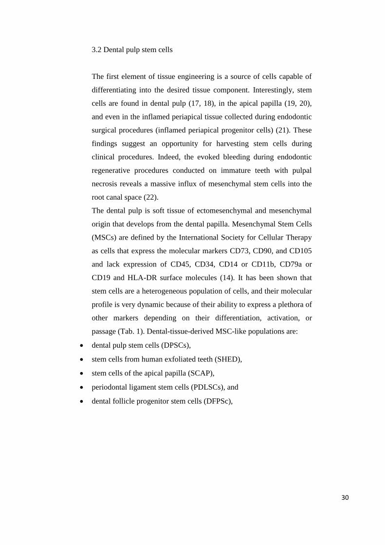
Cell Type In Vitro Activity
DPSCs Osteo/Dentinogenic
Adipogenic
Chondrogenic
Myogenic
Neurogenic
SHED Dentinogenic
Adipogenic
Chondrogenic
Myogenic
Neurogenic
Osteo-inductive
SCAP Dentinogenic
Adipogenic
Chondrogenic
Myogenic
Neurogenic
PDLS Osteo/Cementogenic
Adipogenic
Chondrogenic
Myogenic
Neurogenic
DFPC Cementogenic
Odontogenic
Adipogenic-
Chondrogenic
Myogenic
Neurogenic
Table 1. Multiple differentiation property of Human Dental Stem
Cells.
32
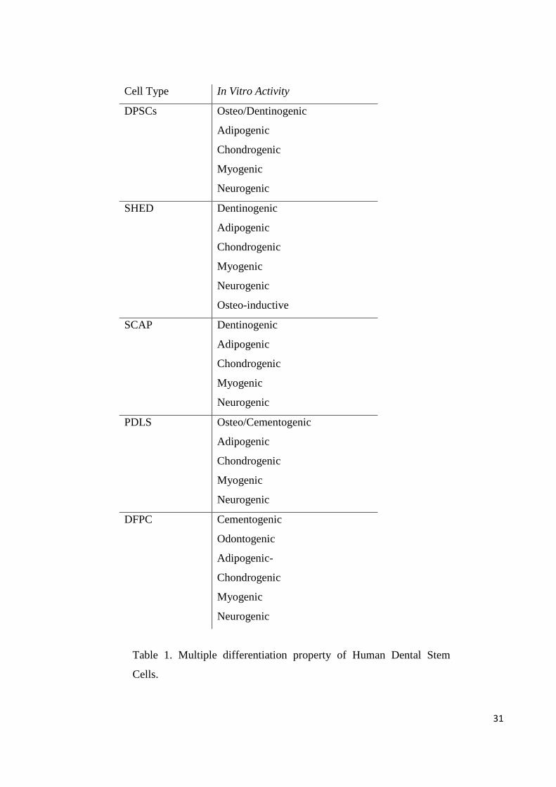
Figure 1. Histologic section of human apical papilla and dental pulp.
Huang GTJ et al. Mesenchymal Stem Cells derived from dental tissues vs. those from other sources: their
biology and role in regenerative medicine. J Dent Res 2009;88:792-806.
33
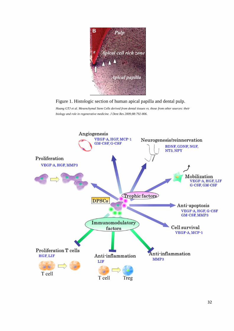
Figure 2. Potential for secretion of trophic and immunomodulatory
factors and from stem cells/progenitors.
Nakashima M. et al Dental pulp stem cells and regeneration. Endod Topics 2013;28:38–50.
During the development of root and the dental pulp, the dental papilla,
located apically, demonstrated the capacity to developing pulp.
SCAP are a population of dental stem cells that hold great potential in
regenerative endodontics (20). These cells are believed to be the main
source of undifferentiated cells in the process of root development
(20), have greater proliferation rates than dental pulp stem cells (15,
20), and have been previously differentiated in odontoblast-like cells
resulting in the de novo production of dentin in vivo (16). Apical
papilla tissue is clinically gelatinous and easily detached from the
apex. Histologically, the apical papilla is distinctive from the pulp by
a cell-rich zone and for a minor contents of cellular and vascular
components. SCAP stem cells are present in a quiescent stage when
present in their niche; once activated or released from their niche,
these cells undergo significant changes, adopting a molecular profile
dictated by environmental cues.
The dental follicle surrounding the developing tooth root contains
progenitor cell for the developing of periodontum (cementum,
alveolar bone and PDL); whilst the inner and outer enamel epithelia
fuse form the Hertwig epithelial root sheath (HERS).
Postnatal population of human dental pulp stem cells has been
identified and isolated, they have the ability to differentiate into
odontoblast-like cell which express the early odontoblast cell marker,
dentine sialophosfoprotein, and can form a dentin complex (17).
DPSCs were capable to regenerate a dental-pulp-like complex
composed of soft-fibrous tissue, mineralized matrix and odontoblast-
like layer able to deposit reparative dentin-like structure on the surface
of human dentin (30-32). A stem cell-based approach in regenerative
Endodontics needs to full fill the requirement of regenerating dental
pulp in the whole three-dimensional geometry of root canals. In case
34
of necrotic and immature teeth the perivascular stem cell in niches
located in the apical papilla, suspected of giving to new hard tissue
and root formation (33, 34), moreover other authors suggest the
possibility that stem and progenitor cell could came from the
periodontal ligament and when bleeding occur could be enter in the
root canal (35).
Laceration of the apical papilla in patients triggers an in flow of blood
into the root canal space that has a concentration of mesenchymal
stem cell markers (CD73 and CD105) from 400-fold to 600-fold
greater as compared with concentrations of these cells circulating in
the patient’s systemic blood. Thus, several local sources of stem cells
are available for clinical dental procedures, and stem cells can be
delivered into the root canal system of patients. When bleeding
occurs, mesenchymal stem cells from the bone marrow and
periodontal ligament may transplanted into the root canal (20). After
bleeding a blood clot that forms inside the root canal represents a rich
source of growth factors that may play an important role in the
regeneration process: differentiation, growth and maturation of
fibroblast, odontoblast and cementoblast.
Infact the second element of tissue engineering focuses on growth
factors or other tissue-inducing mediators. Stem cells have the
capacity to differentiate into a number of cell phenotypes depending
on their lineage and exposure to environmental stimuli such as growth
factors, extracellular matrix, hypoxia, or other conditions (18,20-26).
Thus, the environment is a critical factor in regulating tissue
differentiation. When lacerating the apical papilla appended a high
local concentration of stem cells into the root canal space may not be
sufficient to guide their differentiation into cells of the pulp-dentin
complex. Instead, growth factors should be considered as important
adjuncts. This is an important concept to remember when interpreting
histologic studies after regenerative procedures.
Growth factors are proteins that bind to receptors on the cell and
induce cellular proliferation and/or differentiation. Many growth

35
factors are quite versatile, stimulating cellular division in numerous
cell types, while others are more cell specific. Growth factors play a
role in signalling many events in pulp-dentine regeneration. Two
important families of growth factor that play a vital role are
transforming growth factor (TGF) and bone morphogenetic protein
(BMP). TGF-β1 and β3 are important in cellular signalling for
odontoblast differentiation and stimulation of dentin matrix secretion.
These growth factors are secreted by odontoblasts and are deposited
within the dentin matrix, where they remain protected in an active
form through interaction with other components of the dentin matrix.
The addition of purified dentin protein fractions stimulates an increase
in tertiary dentin matrix secretion suggesting that TGF-β1 is involved
in injury signalling and tooth-healing reaction.
BMPs induce higher quantity and more homogeneous reparatory
dentin with the presence of many tubes with defined odontoblastic
process as compared to that with calcium hydroxide.
BMP-2, BMP-4 and BMP-7 have been shown to direct stem cell
differentiation into odontoblasts and result in dentin formation making
the BMP family the most likely candidate as growth factors.
The third element of tissue engineering is a scaffold. A scaffold is
much more important than simply forming a three-dimensional tissue
structure. In addition, scaffolds play a key role in regulating stem cell
differentiation by local release of growth factors or by the signalling
cascade triggered when stem cells bind to the extracellular matrix and
to each other in a three-dimensional environment (40,41).
Scaffolds may be endogenous (eg, collagen, dentin, PRP, PRF) or
synthetic substances (eg, hydrogels, MTA, or other compounds) (77,
78). This principle may play a very important role in interpreting
clinical regenerative studies. For example, instrumentation of dentin
cylinders that was followed by irrigation with 5.25% NaOCl and
extensive washing led to a dentin surface that promoted differentiation
of cells into clastic-like cells capable of resorbing dentin (71). In
contrast, irrigation of dentin cylinders with 17% EDTA either alone or
36
after NaOCl treatment produced a dentin surface that promoted cell
differentiation into cells expressing an appropriate marker for a
mineralizing phenotype (eg, dentin sialoprotein) (41). Accordingly,
the selection of irrigants and their sequence (EDTA last) may play
critical roles in conditioning dentin into a surface capable of
supporting differentiation of a desired cell phenotype.
3.3 Regenerative endodontic procedure: revascularization,
revitalization or maturogenesis?
The American Association of Endodontics define the term
Regenerative Endodontic Procedure (REP) as follow:
“Regenerative endodontic procedures are biologically based
procedures designed to physiologically replace damage tooth
structure including dentin and root structure as well as cell of the pulp
dentin complex.” (Glossary of Endodontic Terms).
REP are all the procedure aimed to restore damage pulp by
stimulation of existing stem and progenitor cell present in the root
canal and/or the introduction and stimulation of new stem and pulp
progenitor cell into the root canal under condition that are favourable
to their differentiation and reestablishment of function.
The literature reports high number of studies with heterogenic term,
the newest in general report “regenerative endodontics” as key word
or in the title, but before it was not unusual find title with the term
revitalization or revasculation.
Considering that the nature of the tissue formed posttreatment was
unpredictable and that the only certainty was the presence of blood
Trope chosen the term revascularization (9). Huang and Lin (10)
supported the use of revascularization only in case of traumatized
teeth (10); in case of no traumatic necrotic teeth the term induced or
guided tissue generation and regeneration has been suggested.
Subsequently Lenzi & Trope suggest the term revitalization and
37
Weisleder & Benitez maturogeneis (11, 12); the last definition was in
accord to Hargreaves who explained the importance to describe
continued root development in contrast to apexogenesis (13).
Regenerative endodontic protocols, also referred to as
revascularization processes in infected, immature teeth with necrotic
pulps contemplate the continuation of full root development and
thickening of the root walls in immature permanent teeth with pulp
necrosis (51).
The successful revascularization of immature teeth with apical
periodontitis is mainly dependent upon:
Canal disinfection:
The development of an endodontic infection plays a critical role in
treatment considerations and the success of regenerative procedures.
The knowledge of bacterial biofilm, the microbial virulence, adhesion
characteristics, and the antibiotic sensitivity of the organisms involved
would assist in identifying the best antibacterial strategies. For most of
the research performed in the 20th century, culturing of root canal
microflora was the state of the art, and clinical decisions were
frequently based on cultivation results. Nowadays several generations
of molecular technologies have led to a dramatic improvement in
knowledge of endodontic microbiology. Older molecular studies
merely investigated the presence of organisms that had been identified
by culturing or used the inefficient and expensive cloning and
sequencing methodologies (52).
Three endodontic microbiome in three different locations could play
an important role in the endodontic infection: microflora in normal
oral cavity, in necrotic root canal space, and in apical abscess. For
example it can be seen from the differential abundance of microbiota
how the proportion of streptococci and Veillonella spp., which are
very abundant in the oral cavity, decrease in endodontic infections and
the abundance of gram-negative anaerobes such as Fusobacterium
38
spp., Prevotella spp., and Porphyromonas spp. and the gram-positive
Parvimonas spp. increase.
There should be some selectivity in the choice of antimicrobial agents,
given what is known about the nature of endodontic infections.
In case of treatment of teeth with immature apex a very thin dentinal
wall, minimal mechanical instrumentation is advocated so as not to
further weaken the tooth structure. However, it is important to note
that without the frictional force applied by a file to dentinal wall,
bacterial biofilms remain intact and are much more resistant to
antimicrobial agents than if they were rendered planktonic by this
mechanical disruption. Therefore, a small amount of filing is
performed, the intent of which is not to shape the root canal (such in
mature teeth) but rather to create inroads through the biofilm to allow
maximum permeation by the antimicrobials.
Moreover although maximum antimicrobial efficacy is needed to
prevent bacterial irritation of the revascularized/regenerated tissue,
minimal toxicity of these antimicrobials on the soft and hard tissues
surrounding this newly formed tissue is critical. For example, it is
known that 2.5%–5.25% sodium hypochlorite and 2% chlorhexidine
are among the most effective antimicrobials in nonsurgical endodontic
treatment of teeth with mature apex. However, in vitro and animal
model studies have shown that these materials at these concentrations
may be toxic to stem cells of the apical papilla (33), may prevent
adhesion of stem cells to dentin (53), and may abrogate the bioactivity
of growth factors sequestered in dentin (52). Therefore, of these
agents, current clinical guidelines advocate only the use of 1.25%
sodium hypochlorite at the first clinical appointment.
The use of antibiotics becomes the obvious next choice because of
their selectivity, their relatively reduced toxicity, and their potential
residual effect while the tissue is growing. Several different antibiotics
and antibiotic combinations have been proposed. The most widely
used is triple antibiotic paste (ciprofloxacin, metronidazole, and
minocycline), which was historically introduced after trials on root
39
canal cultivable microflora (54-56). Triple antibiotic paste has been
found in an animal in situ study to disinfect 70% of root canals
compared with only 10% disinfected by 1% sodium hypochlorite (57).
However, because of the staining effect of minocycline, it was
replaced with cefaclor (51) or eliminated altogether (7). Augmentin
(GlaxoSmithKline, Philadelphia, PA) was used in a recent report (58)
because it has been shown to be most effective against root canal flora
(59, 60), it has the clavulanic acid that inactivates beta-lactamases that
are prevalent in endodontic infections (59), and Augmentin does not
discolour teeth.
A creamy mix of antibiotics in a powder form with water or another
sterile fluid, as is commonly advocated, results in high concentrations
of the antibiotics. These high concentrations have been recently found
to be toxic to the stem cells of the apical papilla (61). Therefore, lower
concentrations of the antibiotics need to be used, and work is currently
underway to determine the concentrations that would achieve effective
disinfection with the least toxicity to the apical regenerative tissue.
Interestingly, calcium hydroxide was not found to be toxic in the same
study (61). This medicament has been found to provide clinically
acceptable results in many case reports and case series (62, 63), and so
it provides an important alternative to be considered.
Scaffold placement in the canal for the growing tissues.
Once canal disinfection has been completed, the apex is mechanically
irritated to induce clot formation, which will serve as a scaffold for
tissue generation (8, 51). In any tissue engineering procedure, the cell
growth and differentiation are related to an apposite scaffold (75-78).
Extracellular matrix molecules (79) control the differentiation of stem
cells. In this regard, it is anticipated that a suitable scaffold that
contains growth factors might be promising tool to enrich the rate of
40
tissue differentiation as it would selectively bind and localize cells and
undergo biodegradation over time (13).
Intracanal blood (vs circulating blood) obtained from the laceration of
apical tissue have high levels of stem cell markers (22). In addition,
the blood clot may serve as a matrix for the growth of new tissue (8,
64) as well as a source of growth and differentiation factors (65–67).
Alternatives to a blood clot include platelet rich plasma (PRP) (7) and
autologous fibrin matrix (AFM) (68). PRP and AFM contain growth
factors that, along with other beneficial actions, initiate vascular
ingrowth, induce cell differentiation, and improve soft- and hard tissue
wound healing (69–72).
The platelets release growth factors that are trapped inside the fibrin
matrix following activation. These are considered to be the stimulant
for response in the periosteum and are responsible for bone repair
during normal wound healing. Nevertheless, there is still concern
linked to the procedures for production of autologous fibrin adhesives.
Besides, legal restrictions on blood handling with concentrated
platelet rich plasma have coexisted. In an effort to overcome these
problems, it was contemplated to develop a new family of platelet
concentrates, which came to be recognized as the platelet rich fibrin
(PRF). PRF consists of an intimate assembly of cytokines, glycan
chains, structural glycoproteins enmeshed within a slowly
polymerized fibrin network.17 These biochemical components have
well known synergistic effects on healing processes (80) Fibrin is the
natural guide of angiogenesis. Fibrin constitutes a natural support to
immunity (81).
Keswani et al. reported that PRF might serve as a potentially ideal
scaffold in revascularization of immature permanent teeth with
necrotic pulps as it is rich in growth factors, enhances cellular
proliferation and differentiation, and acts as a matrix for tissue
ingrowth (82).
Shivashankar et al. reported a case of revitalization of tooth with
necrotic pulp and open apex using PRF (83). They described evidence
41
of continued thickening of the dentinal walls, root lengthening,
regression of the periapical lesion and apical closure with use of PRF.
The authors considered PRF to be an excellent biomaterial for
pulpdentin complex regeneration. Analogously, Rudagi et al. also
reported a case demonstrating the successful healing and apexification
with combined use of MTA as an apical barrier, and autologus platelet
rich fibrin membrane as an internal matrix (84).
A potential disadvantage of using either PRP or AFM is that they
require a blood draw, which may be intimidating to practitioners and
patients. It is worth mentioning that some authors have reported
continued root growth in cases in which they were not able to achieve
a blood clot in the canal space (74,75). This suggests that although a
blood clot may increase the likelihood of favourable outcomes, it may
not be necessary.
Bacteria-tight sealing of the access aperture:
The access cavity is restored using a material that seals it against
bacteria. In most studies, the materials of choice are ProRoot mineral
trioxide aggregate (MTA) glass–ionomer resin. MTA has been shown
to prevent coronal bacterial filtration, is biocompatible with the
adjacent pulp tissue, induces the proliferation of pulp cells, raises the
pH during prolonged periods of time and allows exceptional marginal
adaptation, finally it can set in the presence of blood and, once set, is
highly resistant to penetration by bacteria (85).
However other materials has been used for access sealing, such as
glass–ionomer or silver amalgam and recently calcium-enriched
mixture (CEM) cement, placed over the blood clot instead of MTA.
42
3.4 Nature of tissues present in the canals of these teeth treated with
regenerative endodontics
Root development consists of 3 parts: an increase in root wall
thickness, an increase in root length, and the narrowing of the canal
apically leading to the formation of the root apex. Vital pulp tissue
inside the root canal is presumably necessary for an increase in root
wall thickness because the canal becomes thinner. An increase in root
length and the formation of the apex are functions of the apical papilla
and Hertwig epithelial root sheath.
Based on these guidelines, many success in vitro and in vivo studies
have been reported in literature (43, 44). Recently, Torabinejad and
Faras (45) presented clinical, radiographic, and histologic findings
showing "pulp-like vital connective tissue" from a tooth after
regenerative endodontic treatment done using platelet rich plasma
(PRP) as a scaffold. Examinations of hematoxylin-eosin–stained
sections revealed the presence of a mildly cellular fibrous connective
tissue, fibroblasts, and blood vessels. A few lymphocytes were
observed in the specimens, and there was no evidence of odontoblasts
in the sections examined. The specimens contained some small
scattered round to irregular-shaped granular basophilic material
partially surrounded by a few flattened multinucleated foreign body–
type giant cells.
Examination of the soft tissue removed from the canal showed the
absence of any signs of severe pathology. The presence of a few
inflammatory cells in the periphery of the specimens and scattered
small calcific materials could be because of the reaction of the pulp-
like tissue to the external irritants.
The removal of the soft tissue without its surrounding hard tissues
such as dentin does not allow good orientation and the identification
of cells that had thickened the root of this tooth after regenerative
endodontic procedures. Cells (such as odontoblasts or odontoblasts-
like cells) that had thickened the root after regenerative endodontic
43
procedures could have been left on the surfaces of the hard tissue
during tissue extirpation using a barbed broach. Animal studies are
needed to confirm these speculations.
These findings indicated that these types of tissues are not of pulpal
origin and the whole revitalization process is not tissue regeneration
but tissue repair.
Similar histological report was presented by Shimizu et al. from a
tooth extracted after the completion of regenerative endodontic
treatment in which more than one half of the canal was found filled
with pulp-like loose connective tissue (46). Positive response to cold
and/or electric pulp tests occurs in some cases (47). These findings
indicate the success of regenerative endodontic procedures.
In contrast to this, literature also reports some cases in which despite
following proper protocol, pulp regeneration and root development
failed. Lenzi and Trope (48) found empty root canal space after
treatment of an immature maxillary central incisor with a necrotic
pulp. Nosrat et al.(42) showed the absence of vital tissue inside the
root canal space of treated immature maxillary incisors with necrotic
pulps after 6 years. Nosrat et al (49) presented a case where root
maturation occurred in a maxillary central incisor, even though a
regenerative endodontic procedure resulted in an empty root canal
space. Even after using tissue engineering strategies, cementum-like
hard tissue was deposited on root canal walls, and bony islands were
found throughout the root canals.
Formation of a hard-tissue barrier inside the canal between the coronal
MTA plug and the root apex (50) is another reported unfavourable
outcome.
Results from in vivo animal studies using similar protocols with an
induced blood clot in the canal suggest that the regenerated tissue is
not pulp tissue but, in fact, repair tissue consisting of bone, cementum,
and inflammatory tissue (67–70). Even in case of failure of
regenerative endodontic in vivo human studies, the histology analysis
44
no pulp-like tissue characterized by the presence of odontoblast like
cells lining the mineralized tissue was observed.
References
1. Cvek M. Prognosis of luxated non-vital maxillary incisors treated with
calcium hydroxide and filled with gutta-percha: a retrospective
clinical study. Endod Dent Traumatol 1992;8:45–55.
2. Nygaard-Ostby B, Hjortdal O. Tissue formation in the root canal
following pulp removal. Scand J Dent Res 1971;79:333–49.
3. Myers MC, Fountain SB. Dental pulp regeneration aided by blood and
blood substitutes after experimentally induced periapical infection.
Oral Surg Oral Med Oral Pathol 1974;37:441–50.
4. Cvek M, Cleaton-Jones P, Austin J, et al. Pulp revascularization in re
implanted immature monkey incisors: predictability and the effect of
antibiotic systemic prophylaxis. Endod Dent Traumatol 1990;6:157–
169.
5. Kling M, Cvek M, Mejare I. Rate and predictability of pulp
revascularization in therapeutically reimplanted permanent incisors.
Endod Dent Traumatol 1986;2:83–89.
6. Skoglund A, Tronstad L. Pulpal changes in replanted and auto
transplanted immature teeth of dogs. J Endod 1981;7:309–316.
7. Iwaya SI, Ikawa M, Kubota M. Revascularization of an immature
permanent tooth with apical periodontitis and sinus tract. Dent
Traumatol 2001;17:185–187.
8. Banchs F, Trope M. Revascularization of immature permanent teeth
with apical periodontitis: new treatment protocol? J Endod
2004;30:196–200.
45
9. Trope M. Regenerative potential of dental pulp. J Endod
2008;34:S13–7.
10. Huang GT, Lin LM. Letter to the editor: comments on the use of the
term ‘‘revascularization’’ to describe root regeneration. J Endod
2008;34:511. author reply 511–2.
11. Lenzi R, Trope M. Revitalization procedures in two traumatized
incisors withdifferent biological outcomes. J Endod 2012;38:411–4.
12. Weisleder R, Benitez CR. Maturogenesis: is it a new concept? J
Endod 2003;29: 776–8.
13. Hargreaves KM, Giesler T, Henry M, Wang Y. Regeneration potential
of the youngpermanent tooth: what does the future hold? J Endod
2008;34:S51–6.
14. Dominici M, Le Blanc K, Mueller I, et al. Minimal criteria for
defining multipotent mesenchymal stromal cells. The International
Society for Cellular Therapy position statement. Cytotherapy
2006;8:315–7.
15. Sonoyama W, Liu Y, Yamaza T, et al. Characterization of the apical
papilla and itsresiding stem cells from human immature permanent
teeth: a pilot study. J Endod 2008;34:166–71.
16. Huang GT, Yamaza T, Shea LD, et al. Stem/progenitor cell-mediated
de novo regeneration of dental pulp with newly deposited continuous
layer of dentin in an in vivo model. Tissue Eng Part A 2010;16:605–
15.
17. Nakashima M, Akamine A. The application of tissue engineering to
regeneration of pulp and dentin in endodontics. J Endod 2005;31:711–
8.
18. Alongi DJ, Yamaza T, Song Y, et al. Stem/progenitor cells from
inflamed humandental pulp retain tissue regeneration potential. Regen
Med 2010;5:617–31.
19. Huang GT, Sonoyama W, Liu Y, et al. The hidden treasure in apical
papilla: the potential role in pulp/dentin regeneration and bioroot
engineering. J Endod 2008;34:645–51.
46
20. Sonoyama W, Liu Y, Fang D, et al. Mesenchymal stem cell-mediated
functionaltooth regeneration in swine. PLoS ONE 2006;1:e79.
21. Liao J, Al Shahrani M, Al-Habib M, Tanaka T, Huang GT. Cells
isolated from inflamed periapical tissue express mesenchymal stem
cell markers and are highly osteogenic. J Endod 2011;37:1217–24.
22. Lovelace TW, Henry MA, Hargreaves KM, Diogenes A. Evaluation
of the delivery ofmesenchymal stem cells into the root canal space of
necrotic immature teeth after clinical regenerative endodontic
procedure. J Endod 2011;37:133–8.
23. Gronthos S, Mankani M, Brahim J, Robey PG, Shi S. Postnatal human
dental pulp stem cells (DPSCs) in vitro and in vivo, Proc. Natl. Acad.
Sci. USA 97 (2000) 13625–13630.
24. Wei X, Ling J, Wu L, et al. Expression of mineralization markers in
dental pulp cells.J Endod 2007;33:703–8.
25. Li L, Zhu YQ, Jiang L, et al. Hypoxia promotes mineralization of
human dental pulpcells. J Endod 2011;37:799–802.
26. Huang GT, Shagramanova K, Chan SW. Formation of odontoblast-
like cells fromcultured human dental pulp cells on dentin in vitro. J
Endod 2006;32:1066–73.
27. Sun HH, Jin T, Yu Q, Chen FM. Biological approaches toward dental
pulp regeneration by tissue engineering. J Tissue Eng Regen Med
2011;5:e1–16.
28. Galler KM, D’Souza RN, Federlin M, et al. Dentin conditioning
codetermines cellfate in regenerative endodontics. J Endod
2011;37:1536–41.
29. Prescott RS, Alsanea R, Fayad MI, et al. In vivo generation of dental
pulp-like tissueby using dental pulp stem cells, a collagen scaffold,
and dentin matrix protein 1 after subcutaneous transplantation in mice.
J Endod 2008;34:421–6.
30. Kim SG, Zhou J, Solomon C, et al. Effects of growth factors on dental
bilateralbicuspids using a modified novel technique to eliminate
potential coronal discolouration: a case report. Int Endod J
2009;42:84–92.
47
31. Gronthos S, Brahim J, Li W, Fisher LW, Cherman N, Boyde A, et al.,
Stem cell properties of human dental pulp stem cells. J. Dental Res. 81
(2002) 531–535.
32. Batouli S, Miura M, Brahim J, Tsutsui TW, Fisher LW, Gronthos S, et
al., Comparison of stem-cell-mediated osteogenesis and
dentinogenesis, J. Dental Res. 82 (2003) 976–981.
33. Trevino EG, Patwardhan AN, Henry MA, et al. Effect of irrigants on
the survival ofhuman stem cells of the apical papilla in a platelet-rich
plasma scaffold in human root tips. J Endod 2011;37:1109–15.
34. Huang GT. A paradigm shift in endodontic management of immature
teeth: conservation of stem cells for regeneration. J Dent
2008;36:379–86.
35. Shah N, Logani A, Bhaskar U, Aggarwal V. Efficacy of
revascularization to induceapexification/apexogensis in infected,
nonvital, immature teeth: a pilot clinical study. J Endod 2008;34:919–
25.
36. Mooney DJ, Powell C, Piana J, Rutherford B. Engineering dental
pulp-like tissue in vitro. Biotechnol Prog. 1996;12:865–8.
37. Bohl KS, Shon J, Rutherford B, Mooney DJ. Role of synthetic
extracellular matrix in development of engineered dental pulp. Journal
of Biomaterials Science Polymer Edition. 1998;9:749–64.
38. Buurma B, Gu K, Rutherford RB. Transplantation of human pulpal
and gingival fibroblasts attached to synthetic scaffolds. European
Journal of Oral Sciences. 1999;107:282–9.
39. Gronthos S, Mankani M, Brahim J, Robey PG, Shi S. Postnatal human
dental pulp stem cells (DPSCs) in vitro and in vivo. Proceedings of
the National Academy of Sciences. 2000;97:13625–13630.
40. Discher DE, Mooney DJ, Zandstra PW. Growth factors, matrices, and
forces combine and control stem cells. Science 2009;324:1673–7.
41. Kim JK, Shukla R, Casagrande L, et al. Differentiating dental pulp
cells via RGDdendrimer conjugates. J Dent Res 2010;89:1433–8.
42. Nosrat A, Homayounfar N, Oloomi K. Drawbacks and unfavorable
outcomes of regenerative endodontic treatments of necrotic immature
48
teeth: A literature review and report of a case. J Endod 2012;38:1428-
34.
43. Thibodeau B. Case report: Pulp revascularization of a necrotic,
infected, immature, permanent tooth. Pediatr Dent 2009;31:145-8.
44. Thomson A, Kahler B. Regenerative endodontics-biologically-based
treatment for immature permanent teeth: A case report and review of
the literature. Aust Dent J 2010;55:446-52.
45. Torabinejad M, Faras H. A clinical and histological report of a tooth
with an open apex treated with regenerative endodontics using
platelet-rich plasma. J Endod 2012;38:864-8.
46. Shimizu E, Jong G, Partridge N, Rosenberg PA, Lin LM. Histologic
observation of a human immature permanent tooth with irreversible
pulpitis after revascularization/regeneration procedure. J Endod
2012;38:1293-7.
47. Law AS. Considerations for regeneration procedures. J Endod
2013;39:S44-56.
48. Lenzi R, Trope M. Revitalization procedures in two traumatized
incisors with different biological outcomes. J Endod 2012;38:411-4.
49. Nosrat A, Li KL, Vir K, Hicks ML, Fouad AF. Is pulp regeneration
necessary for root maturation? J Endod 2013;39:1291-5.
50. Chen MY, Chen KL, Chen CA, Tayebaty F, Rosenberg PA, Lin LM.
Responses of immature permanent teeth with infected necrotic pulp
tissue and apical periodontitis/abscess to revascularization procedures.
Int Endod J 2012;45:294-305.
51. Thibodeau B, Trope M. Pulp revascularization of a necrotic infected
immature permanent tooth: case report and review of the literature.
Pediatric Dentistry 2007; 29: 47–50.
52. Ring K, Murray P, Namerow K, et al. The comparison of the effect of
endodontic irrigation on cell adherence to root canal dentin. J Endod
2008;34:1474–9.
53. Casagrande L, Demarco FF, Zhang Z, et al. Dentin-derived BMP-2
and odontoblast differentiation. J Dent Res 2010;89:603–8.
49
54. Hoshino E, Kurihara-Ando N, Sato I, et al. In-vitro antibacterial
susceptibility of bacteria taken from infected root dentine to a mixture
of ciprofloxacin, metronidazole and minocycline. Int Endod J
1996;29:125–30.
55. Sato I, Ando-Kurihara N, Kota K, et al. Sterilization of infected root-
canal dentine by topical application of a mixture of ciprofloxacin,
metronidazole and minocycline in situ. Int Endod J 1996;29:118–24.
56. Sato T, Hoshino E, Uematsu H, et al. In vitro antimicrobial
susceptibility to combinations of drugs on bacteria from carious and
endodontic lesions of human deciduous teeth. Oral Microbiol
Immunol 1993;8:172–6.
57. Windley W, Teixeira F, Levin L, et al. Disinfection of immature teeth
with a triple
antibiotic paste. J Endod 2005;31:439–43.
58. Jungermann GB, Burns K, Nandakumar R, et al. Antibiotic resistance
in primary and persistent endodontic infections. J Endod
2011;37:1337–44.
59. Baumgartner JC, Xia T. Antibiotic susceptibility of bacteria associated
with endodontic abscesses. J Endod 2003;29:44–7.
60. Ruparel NB, Teixeira FB, Ferraz CC, et al. Direct effect of intracanal
medicaments on survival of stem cells of the apical papilla. J Endod
2012;38:1372–5.
61. Bose R, Nummikoski P, Hargreaves K. A retrospective evaluation of
radiographic outcomes in immature teeth with necrotic root canal
systems treated with regenerative endodontic procedures. J Endod
2009;35:1343–9.
62. Jeeruphan T, Jantarat J, Yanpiset K, et al. Mahidol study 1:
comparison of radiographic and survival outcomes of immature teeth
treated with either regenerative endodontic or apexification methods: a
retrospective study. J Endod 2012;38:1330–6.
63. Witherspoon DE, Small JC, Harris GZ. Mineral trioxide aggregate
pulpotomies: a case series outcomes assessment. J Am Dent Assoc
2006;137:610–8.
50
64. Thibodeau B, Teixeira F, Yamauchi M, et al. Pulp revascularization of
immature dog teeth with apical periodontitis. J Endod 2007;33:680–9.
65. Maeda H, Wada N, Nakamuta H, Akamine A. Human periapical
granulation tissue contains osteogenic cells. Cell Tissue Res
2004;315:203–8.
66. Marx RE. Platelet-rich plasma: evidence to support its use. J Oral
Maxillofac Surg 2004;62:489–96.
67. Freymiller EG, Aghaloo TL. Platelet-rich plasma: ready or not? J Oral
Maxillofac Surg 2004;62:484–8.
68. Geisler TM. Clinical considerations for regenerative endodontic
procedures. Dent Clin North Am 2012;56:603–26.
69. Hiremath H, Gada N, Kini Y, et al. Single-step apical barrier
placement in immature teeth using mineral trioxide aggregate and
management of periapical inflammatory lesion using platelet-rich
plasma and hydroxyapatite. J Endod 2008;34:1020–4.
70. Sharkawy HE, Kantarci A, Deady J, et al. Platelet-rich plasma: growth
factors and pro-and anti inflammatory properties. J Periodontol
2007;78:661–9.
71. Anitua E, S anchez M, Nurden AT, et al. New insights into and novel
applications for platelet-rich fibrin therapies. Trends Biotechnol
2006;24:227–34.
72. Mullane EM, Dong Z, Sedgley CM, et al. Effects of VEGF and FGF2
on th revascularization of severed human dental pulps. J Dent Res
2008;87:1144–8.
73. Chueh LH, Huang GT. Immature teeth with periradicular periodontitis
or abscess undergoing apexogenesis: a paradigm shift. Journal of
Endodontics 2006; 32:1205–13.
74. Chueh LH, Ho YC, Kuo TC, Lai WH, Chen YH, Chiang CP.
Regenerative endodontic treatment for necrotic immature permanent
teeth. Journal of Endodontics 2009;35:160–4.
75. Howard D, Buttery LD, Shakesheff KM, Roberts SJ. Tissue
engineering: strategies, stem cells and scaffolds. J Anat 2008;213:66-
72.
51
76. Mikos AG, Herring SW, Ochareon P, et al. Engineering complex
tissues. Tissue Eng 2006;12:3307-3339.
77. Yoo YJ, Baek SH, Son HH. Pulp tissue regeneration and root
formation of permanent teeth with pulpal/periapical deseases. J
Korean Acad Conserv Dent 2010; 35:238-245.
78. Forghani M, Parisay I, Maghsoudlou A. Apexogenesis and
revascularization treatment procedures for two traumatized immature
permanent maxillary incisors: a case report. Restor Dent Endod
2013;38:178-181.
79. Chan BP, Leong KW. Scaffolding in tissue engineering: general
approaches and tissue-specific considerations. Eur Spine J
2008;17(Supplement 4):467-479.
80. Dohan DM, Choukroun J, Diss A, Dohan SL, Dohan AJ, Mouhyi J,
Gogly B. Platelet-rich fibrin (PRF): a second generation platelet
concentrate. Part III: leucocyte activation: a new feature for platelet
concentrates? Oral Surg Oral Med Oral Pathol Oral Radiol Endod
2006;101: e51-55.
81. Choukroun J, Diss A, Simonpieri A, et al. Platelet-rich fibrin (PRF): a
second-generation platelet concentrate. Part IV: clinical effects on
tissue healing. Oral Surg Oral Med Oral Pathol Oral Radiol Endod
2006;101:e56-60.
82. Keswani D, Pandey RK. Revascularization of an immature tooth with
a necrotic pulp using platelet-rich fibrin: a case report. Int Endod J
2013;46:1096-1104.
83. Shivashankar VY, Johns DA, Vidyanath S, Kumar MR. Platelet rich
fibrin in the revitalization of tooth with necrotic pulp and open apex. J
Conserv Dent 2012;15: 395-398.
84. Rudagi KB, Rudagi B. One-step apexification in immature tooth using
grey mineral trioxide aggregate as an apical barrier and autologus
platelet rich fibrin membrane as an internal matrix. J Conserv Dent
2012; 15:196-199.

52
85. Parirokh M, Torabinejad M (2010) Mineral trioxide aggregate: a
comprehensive literature review–Part I: chemical, physical, and
antibacterial properties. Journal of Endodontics 36, 16–27.
53
Chapter 4
Regenerative endodontics: review of clinical protocols
4.1 Introduction
Regenerative endodontics can be defined as biologically based
procedures aimed to promote the regeneration of damaged tissues,
including dentine and root structure as well as cells of the pulp-
dentine complex (1).
Successful operative protocol of regenerative endodontics is
revascularization (1, 2). In case of non-vital, immature tooth with a
wide apical opening this approach is performed to re-establish the
vitality of the pulp. This allows the complete maturation of the root in
term of wall thickening, root lengthening, apical closure and healing
of periradicular lesions if present.
The critical components identified to contribute to the successful
outcome of this procedure are the stem cells, signalling molecules, and
a 3-dimensional physical scaffold (3), without which an empty canal
space would not support ingrowth of new tissues from the periapical
area. Blood clot and collagen have been recommended and used as
scaffold materials for revascularization (4).
The literature reports different disinfection protocols with and without
the use of a combination of antibiotic paste or intra-canal medication
as calcium hydroxide.
The necrosis of an immature teeth could be caused by carie, trauma or
anatomical reasons as dens evaginatus/invaginatus. All these events
and clinical conditions lead to an intra-canal infection.
In 2013 Moreno-Hidalgo et al. (5) identified and reviewed the
scientific evidence regarding regenerative endodontic protocols. Four
human studies were included after applying the inclusion and
exclusion criteria; they showed a substantially omogeneous results

54
concerning the irrigation (sodium hypochlorite with different
concentration) and disinfection of the canals (Triple antibiotic paste);
the use of different scaffold was not investigated.
When an excellent disinfection of the canal was reached the grown,
development and differentiation of stem cells and vasculature are
active by a providing a framework scaffold that can also be infused
with a variety of grown factors. Induced bleeding from the periapical
area and the following blood clot commonly represents a natural
scaffold with grown factors. Until 2001 for Iwaya S. blood
represented a natural scaffold to promote the regeneration of the pulp
tissue (6).
Since a blood clot scaffold is not always possible, some research have
begun examining other three-dimensional scaffold that can be
constructed with or without bleeding. After that different scaffold
have been used in the regenerative treatment: Platelet-rich plasma
(PRP), platelet-rich fibrin (PRF), plasma-rich in grow factors (PRGF),
crosslinked collagen-hydroxyapatite scaffold and polymeric scaffold
as poly (lactide co-glycolide)-polyethylene glycol (PLGA-PEG)
nanoparticles developed for bone tissue engineering (7-10).
As suggested in a recent pilot retrospective cohort study, Alobaid et
al. advocated the need of more studies to better understand the true
incidence of continued root development after revascularization
procedures and the influence of patient and tooth factors such as the
aetiology of pulpal necrosis (eg, trauma vs caries), age, size of apical
foramen, presence, size and temporal persistence of periapical
pathology, and so on (11).
On the basis of these studies and case reports, this systematic review
aimed to critically summarized the results of all human studies and
compare the efficacy of the regenerative protocols for necrotic
immature permanent teeth.
Moreover a correlation between the cause of necrosis, the element
type and the outcome of treatment has been investigated.
Using PICOS terms, the following objective has been formulated:
55
P (Population): adults with at least immature necrotic teeth with or
without periapical lesion.
I (Interventions): any regenerative protocol used in necrotic teeth with
immature root development to promote root development and healing
of periradicular lesion if present. For example regenerative endodontic
treatment with Platelet-rich plasma (PRP), or with Blood Clot and
MTA etc.
C (Comparison): no root development procedures or different root
development procedures with blood clot (BC) and with the use of
additional scaffold and growth factors.
O (Outcomes): length root and thickness of dentinal walls, apical
diameter.
S (Study design): all human studies (case report, case series, Cohort
study, Case Control Study and Randomized Controlled Trials RCT).
This study has been written in accordance with the PRISMA statement
for reporting systematic reviews and meta-analysis of studies that
evaluate health care interventions.
4.2 Material and Methods
Eligibility criteria
All human studies described the regenerative procedure in necrotic
teeth with incomplete root development were included in this study;
review articles, opinion articles and letters were excluded. All studies
dealing only with the treatment of necrotic immature teeth with no
procedure aimed to a continue root development (regeneration), apical
plug technique and apexification procedure were excluded.
Treatment protocol adopted were classified as follow basing the
scaffold and grown factors used:
Blood clot (BC) and coronal seal with MTA (group 0);
PRP with or without BC and coronal sealing (group 1);
PRF with or without BC and coronal sealing (group 2);
56
Other treatment protocol (group 3).
Primary outcome variables were: root development defined as
increase of root length and/or increase of thickness of dentin walls of
the root and/or apical closure.
Source and Search strategy
The basis for this research were the PRISMA guidelines
(www.prisma-statement.org), using the MEDLINE database until
April 2015, and the Medical Subject Headings (MeSH). In addition,
the OneSearch SBART databases were explored to find possible
papers matching our established selection criteria. Hand searching on
Journal of Endodontics and International Endodontic Journal from
1999 to 2015 was conducted. The reference section of previous review
articles, identified by electronic search, were scanned searched to find
additional relevant articles. The search was limited to the English
language.
Eligibility of the articles, and the assessment of the list of reference
scanned from hand and electronic search were performed by the same
operators.
Afterthought the full text was retrieved and the relevant studies
selected were analysed following inclusion criteria; all the remained
studies were discarded.
The search strategy used in MEDLINE (Entrez PubMed, www.
ncbi.nim.nih.gov) was: ‘regenerative endodontics’ or ‘regenerative
endodontic’ or ‘revascularization procedure’.
Data collection
Data from eligible studies were extracted by the reviewer (VG)
according to guidelines outlined by the Cochrane Collaboration.
List of information extracted:
Authors
Years of publication
Sample size
Necrosis causes
Scaffold and source of growth factors used
57
The variable was registered at patient level when reported.
The authors of the original studies were not contacted for clarification.
Summery measurement and planned method of analysis
Since not all studies reported the increase of root length, root
thickness and apical closure in real measure (mm) a scale was
arbitrarily designed by the author as follow:
Root canal length
complete development of root length LLT (0),
increase of root length LL + (1),
no increase of root length LL - (2).
Dentinal walls thickness
complete development of dentinal walls thickness Ø T (0),
increase of dentinal walls thickness Ø + (1),
no increase of dentinal walls thickness Ø - (2).
Apical root closure
complete apical closure AT (0),
increase of apical closure A+ (1),
no increase of apical closure A - (2).
The frequency of each variable and the relative percentage were
recorded overall and for each experimental group.
The successful outcome of endodontic regeneration procedure were
considered complete when the follow items were recorded:
absence of sign and symptoms;
absence of periradicular pathological radiolucency area;
root apical closure AT;
complete development of dentinal thickness ØT;
complete development of root length could recorded LLT.
No failure results classified as satisfactory results were considered
when the follow items were recorded:
absence of sign and symptoms;
absence of periradicular pathological radiolucency area;
improve or complete dentinal thickness (ØT, Ø+);
58
improve or complete root length (LLT, LL+).
The percentage of successful outcame has been calculated for each
operative protocols using different scaffold and grown factors.
The presence of correlation coefficient between the successful
outcome and the type of treated teeth, and the causes of necrosis has
been calculated using the Pearson correlation coefficient.
The correlation coefficient where calculated between the type of teeth
and the satisfactory, and cause of necrosis and satisfactory results (as
before).
4.3 Results
Study selection
The electronic search identified 293 titles, hand search 6 titles and 8
titles were identified by through cross-referencing, for a total of 307
articles.
After the reading of title and abstract 247 articles were excluded
because 2 of them were not inherent, and 245 were animal or in vitro
studies. The full-text of sixty-two articles were investigated basing on
the inclusion criteria (PRISMA) and classified as follows: 8 revision,
3 RCT, 5 Cohort studies, 1 case control studies, 34 case report and 11
case series studies.
Three case report were excluded because one study was not available
on line (Cehreli ZC1, Sara S, Aksoy B. Revascularization of
immature permanent incisors after severe extrusive luxation injury.
Tex Dent J. 2012;7:675-81); one retrospective case series was not
inherent to regenerative procedure but reported the outcome of cases
treated with apexification with calcium hydroxide and the outcome of
teeth treated with apical plug technique with MTA (Cheuch LH et al.
Regenerative Endodontic Treatment for Necrotic Immature Permanent
Teeth. J Endod 2009;35:160-64); and one case report has the purpose
to evaluate whether regenerative endodontic procedures are able to
deliver stem cells into the canal space of immature teeth in young
patients and to identify the possible tissue origin for these cells
59
(Lovelace TW et a. Evaluation of the Delivery of Mesenchymal Stem
Cells into the Root Canal Space of Necrotic Immature Teeth after
Clinical Regenerative Endodontic Procedure. J Endod 2011;37:133–
138). An overview of the selection process was provided by flow
diagram (Fig. 1).
The 8 revision identified (5, 58-64) were excluded; a total of 51
studies were analysed (2,3,6,7,9, 11-56).
The causes of necrosis (trauma, caries, and dens
evaginatus/invaginatus), and the type of element (anterior, premolar
and molar) were recorded for all teeth treated included in the selected
articles.
Study characteristics
In the 51 studies selected on a total of 357 treated teeth were analysed.
For 20 teeth the authors (12) did not specified the type (anterior,
premolar or posterior), for the remaining 337 teeth 24.3% were
premolar (n°82), 2,4% were molar (n°8) and 73.3% were anterior
teeth (n°247).
In group 0 the results of apical closure were reported for 152 teeth, the
results of root canal length were reported for 139 teeth and the results
for dentinal walls thickness were reported for 160 teeth. 45 teeth
(29.6%), after regenerative endodontics procedure with BC as
scaffold, reported a complete apical closure; 79 teeth (52%) an
improve of apical closure and 28 teeth (18.4%) an absence of apical
closure. Four teeth (2.5%) reported a complete root length
development; 131 teeth (82%) an improve root length development
and 25 teeth (15.5%) an absence of root length development. Eight
teeth (5%) reported a complete dentinal walls thickness development;
130 teeth (81.3%) an improve of dentinal walls thickness and 22 teeth
(13.7%) no improve of dentinal walls thickness.
A complete successful outcome was recorded only in four teeth on a
total of 151 (table 2). A satisfactory outcome was recorded in 135
(89.4%).
60
In group 1 a total of 25 teeth were treated; the results of apical closure
root canal length and dentinal walls thickness were reported for all
teeth. 13 teeth (52%), after regenerative endodontics procedure with
PRP as scaffold, reported a complete apical closure; 12 teeth (48%) an
improve of apical closure and no teeth showed an absence of apical
closure. Six teeth (24%) reported a complete root length and dentinal
thickness development; a total of 18 teeth (72%) an improve root
length and dentinal thickness development and one teeth (4%) an
absence of root length and dentinal thickness development.
A complete successful outcome was recorded in 5 teeth on a total of
25 (table 2). A satisfactory outcome was recorded in 24.
In group 2 the results of apical closure, root canal length and dentinal
walls thickness were reported for 10 teeth. 7 teeth (70%), after
regenerative endodontic procedure with PRF as scaffold, reported a
complete apical closure and root length development; a total of 3 teeth
(30%) an improve of apical closure and root length development, no
teeth with an absence of apical closure and root length development
were recorded. Four teeth (40%) reported a complete dentinal walls
thickness development; a total of 6 teeth (60%) an improve of dentinal
walls thickness no teeth with no improve of dentinal walls thickness
were recorded.
A complete successful outcome was recorded only in one tooth on a
total of 10 teeth, but a satisfactory results was judge in all the treated
teeth (table 2).
The causes of pulp necrosis was specified only for a total of 259 teeth
on a total of 357 teeth. In 10.4% of treated teeth, the necrosis was a
consequence of caries, in 23.2% of treated teeth the necrosis has been
observed in teeth with dens evaginatus/invaginatus, and finally in
66.4% of treated teeth the necrosis happened after a dental trauma
(table 3).
No correlation has been recorded between the successful outcome of
the therapy and the tooth type or the causes of pulp necrosis (table 4).
61
No correlation has been observed between a satisfactory results and
tooth type or causes of pulp necrosis (table 5).
4.4 Discussion
Regenerative approaches gained the advantage over the apexification
and MTA apical plug technique because they can allow for further
root maturation in length and thickness by regenerated vital tissue.
The present review aimed to identify if the use of different scaffold
(BC, PRP, PRF) could significantly influence the outcome of
regenerative endodontics treatment in necrotic immature teeth. In a
review dealing with the outcome of endodontic regeneration
treatments, Law (57) reported several challenges in interpreting the
results.
These challenges included variability in technique and recall period, a
potential bias of only successful cases being reported, different
methods to judge the successful outcome and the lack of consistent
radiographic angulation between pre-treatment and follow-up
radiographs.
The standardization of radiographic images enabled the calculation of
development root length, dentin wall thickness and apical closure.
The major part of the articles included in this review are case report
and case series studies with low level of evidence, one was a case
control study and five cohort studies, only three RCT were found.
Even selecting only the three RCT (9,17,35) the risk of bias following
the Cochrane risk of bias tool are high: no study reported a low risk
for all items.
All the three RCT described a randomized assigned teeth for each arm
of the study but only one (Bezgin et al. 17) reported the randomization
methods; they assigned all odd numbered teeth to the PRP group and
all the even numbered teeth to BC group. This method was judge by
the author with high risk of bias.
62
Allocation concealment and blinding of participant and personnel
were never mentioned.
The blinding outcome assessment was judged with low risk of bias in
Nagy et al. study (9) because two authors performed the measurement
blindly but the article did not report the indication about the operator.
In Bezgin et al. (17) study, the operator was blinded to the scaffold
used in the treatment but it did not report who performed the
measurement.
Finally in Jadhav et al. (35) study, the outcome assessment was judge
by two endodontists not involved in the study so the risk of bias was
judged low.
Incomplete outcome reporting was judged at low risk of bias only in
Nagy et al. (9) study because they reported change in root length,
dentinal wall thickness and apical closure in mm.
Bezgin et al. 1(7) reported the outcome in term of % RRA
(radiographic root area) without reported the initial and the final
measure. This value did not give information in term of increase or
decrease of length of the root and wall thickness.
Jahvad et al. (35) judge satisfactory, good and excellent the apical
closure, the root lengthening and the dentinal wall thickening without
give a specific meaning at these categories.
For this reason in order not to lose the information given by all articles
selected in this review an arbitrarily classification of results collected
were adopted and used for qualitative analysis. The outcome was
judge successfully when a complete apical closure, root length and
development of dentinal walls thickness (A 0, LL 0, Ø 0) has been
reported. The total amount of successful outcome in all teeth collected
in the present review was very low: 4.9%. This could be due to the
incomplete data reported, and to a too strictly categories that identified
the successful outcome. Similar percentage of success were recorded
for all experimental group 1.5% when BL was used as scaffold, 1.9%
when PRP was used as scaffold and 0.4% when PRF was used as
scaffold. It is important to underline that this data could not be
63
significant because in a few number of teeth PRP and PRF has been
used as scaffold (25 and 10 teeth respectively) respect the BC (151
teeth).
Scaffolds are used in regenerative procedures to provide a framework
through which cells and a vasculature can grow (31). Several report
have been demonstrated good success with BC scaffold in association
with MTA (33, 50,51); besides it not always possible invoke bleeding
in the root canals researchers have begun examining other three-
dimensional scaffold.
Plated-rich plasma (PRP) and plated-rich fibrin (PRF) form a three-
dimensional network of fibrin which acts as a scaffold with
concentrated growth micro molecules.
PRF consists of an intimate assembly of cytokines, glycan chains,
structural glycoproteins enmeshed within a slowly polymerized fibrin
network. These biochemical components have well known synergistic
effects on healing processes. Fibrin is the natural guide of
angiogenesis.
PRF can be considered as an appropriate scaffold for regenerative
endodontics as it full fills all the properties as mentioned above.
Satisfactory outcome where obtained in the few studies collected in
this review dealing PRF has been used as scaffold (2, 3, 20, 40,36).
Even when PRP were used as scaffold in the studies selected for this
review (1,2,12,13,17,19,42,35) not failure were recorded. PRP has
been used as scaffold in two RCT studies: Bezgin et al. (17) compared
the use of PRP and BC as scaffold in necrotic single-rooted immature
teeth, no significant difference in treatment outcome was found
between the two protocols. Previously Jadhav et al. (35) reported
significant better results in term of periapical healing, apical closure
and dentinal walls thickness when the use of PRP was compared with
the use of BL.
A 30% of studies in which neither intracanal bleeding was evocated
nor PRP or PRF was used, concern the use of different and
heterogeneous scaffolds. Nevis & Cymerman (4) showed successfully
64
results when cross-linked collagen-hydroxyapatite material has been
used as scaffold. It is an implant material that contained a three
dimensional network of collagen fibrils covered with nanocrystals of
hydroxyapatite. The presence of branching microchannels, within a
solid collagen hydroxyapatite spong,e allows osteogenic cells, new
blood vessels, and growth factors to better migrate into the scaffold
and the healing infrabony defect.
Bakhtiar et al. (22) showed the positive effects of plasma-rich in
growth factor (PRGF) on development of pulp and apical closure in
three cases reports. PRGF stimulate the Hertwig’s epithelial root
sheath in addition to increasing phosphatase activity and promote the
osteogenic differentiation. A newer class of biocompatible and
biodegradable poly (lactide-co glycolide)-polyethylene glycol (PLGA-
PEG) nanoparticles developed for bone tissue engineering as a
scaffold for SCAP cells. These hydrogels are injectable scaffolds that
can be delivered by syringe in the apical portion of necrotic teeth with
immature root development. Shiehzadeh V. et al. (10) reported the
results of three cases. More simple procedure involved the use of
Calcium Hydroxide alone. CaOH2 promote the recruitment, migration,
proliferation and mineralization of dental stem cells. Park & Ahn (25),
in necrotic teeth with open apices, after a copious irrigation with
sodium hypochlorite and an intracanal medication with calcium
hydroxide, limit to the coronal or half part of the canal, reported an
apical closure and an increase of root canal thickness.
The literature was very inconsistent on indications dealing with the
use of different scaffold depending on different clinical condition.
Only few in vitro study support the hypothesis that PRP may enhance
wound healing only if the parenchymal tissue has not completely
destroyed, due to the fast degradation of growth factors in PRP (60-
61).
Finally, in the present review no correlation were found between the
successfull outcome and type of element treated or causes of necrosis.
Trauma is clearly an important contributory factor to the incidence of
65
immature permanent teeth with necrotic pulps because an estimated
22% of children suffer trauma to the permanent dentition, with an age
range peaking between 7 and 10 years, more frequently in males, and
most commonly involving the maxillary central and lateral incisors
(66).The situation is very different for a case with necrotic pulp and an
established infection manifesting clinically as pulp necrosis with an
acute or chronic abscess or a radiographically visible apical lesion. In
these cases, the infection has been established for a sufficient duration
to allow the development of bacterial biofilms inside the root canal.
When the necrosis of the pulp is due to a trauma, after the beginning
there is no infection or minimal contamination without an established
bacterial biofilm. In this clinical circumstance host, responses will
allow sufficient connective tissue to revascularize the pulp space
through the relatively large apical foramen and continue
mineralization, leading to increases in the width and length of the root
(57). In case of traumatic injuries of immature teeth with no infection
or minimal contamination without an established bacterial biofilm, the
host responses will allow sufficient connective tissue to revascularize
the pulp space through the relatively large apical foramen and
continue mineralization, leading to increases in the width and length
of the root. The success of revascularization after traumatic injuries of
immature teeth has been shown to increase with the size of the apical
foramen (65).
In general anterior teeth are more susceptible to traumatic injury, carie
are most common in posterior teeth.
The correlation has been calculated between a satisfactory outcome
(improve o complete root development, absence of clinical signs and
symptoms, absence of periradicular lesion) and the tooth type
involved or causes of necrosis. The use of only two variables,
changing in root length and dentinal walls thickness, could be justified
because these are the parameters that differentiate the outcome of
regenerative procedure from apexification with calcium hydroxide or
MTA apical plug technique. Apical root closure could be reach with
66
traditional apexification technique or with MTA apical plug
technique.
As previously reported by Jeeruptan et al. (38) and Bose et al. (62) a
statistically significant difference in increase of wall dentinal
thickness and root length between teeth treated with regenerative
terapy and apexification with calcium hydroxide or MTA apical plug
technique were found. Jeeruptan et al. (38) and Bose et al. (62)
reported an increase of root length of 14.9% and 48% in immature
necrotic teeth treated with regenerative endodontic respectively.
In the present study in teeth without signs and symptoms, and
periradicular pathology, treated with regenerative endodontic
procedure no correlation were found between the increase of dentinal
walls thickness or root length and the type of treated tooth or the
causes of necrosis.
The type of teeth, anterior posterior or premolar, the causes of
necrosis seems not influenced the outcome of regenerative endodontic
treatment. No difference were found in terms of outcome using
different scaffold in regenerative endodontic treatment.
In conclusion, this review has several limitation: first of all the quality
of the studies is very low. The section materials and methods often
present bias for the description of PICOS terms. The major obstacle
met has been represented by incomplete outcome data reported. RCTs
adhering to the Consort guidelines could be encouraged to better guide
the clinicians in the decision-making process.
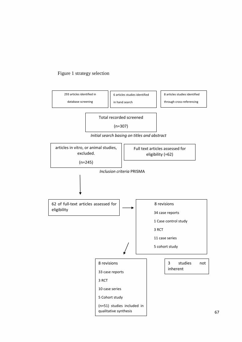
67
Figure 1 strategy selection
Initial search basing on titles and abstract
Inclusion criteria PRISMA
293 articles identified in
database screening
6 articles studies identified
in hand search
8 revisions
34 case reports
1 Case control study
3 RCT
11 case series
5 cohort study
62 of full-text articles assessed for
eligibility
8 revisions
33 case reports
3 RCT
10 case series
5 Cohort study
(n=51) studies included in
qualitative synthesis
8 articles studies identified
through cross-referencing
articles in vitro, or animal studies,
excluded.
(n=245)
Full text articles assessed for
eligibility (=62)
Total recorded screened
(n=307)
3 studies not
inherent
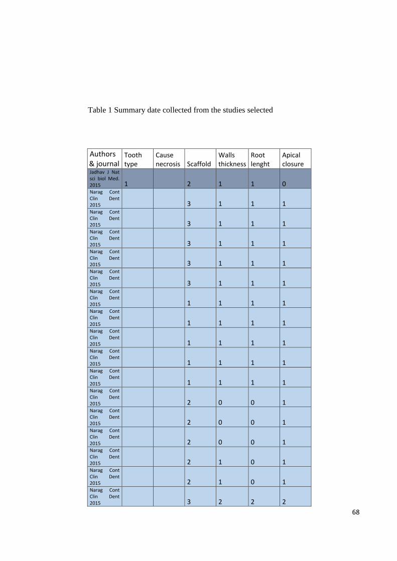
68
Table 1 Summary date collected from the studies selected
Authors & journal
Tooth type
Cause necrosis Scaffold
Walls thickness
Root lenght
Apical closure
Jadhav J Nat sci biol Med. 2015 1
2 1 1 0 Narag Cont Clin Dent 2015
3 1 1 1 Narag Cont Clin Dent 2015
3 1 1 1 Narag Cont Clin Dent 2015
3 1 1 1 Narag Cont Clin Dent 2015
3 1 1 1 Narag Cont Clin Dent 2015
3 1 1 1 Narag Cont Clin Dent 2015
1 1 1 1 Narag Cont Clin Dent 2015
1 1 1 1 Narag Cont Clin Dent 2015
1 1 1 1 Narag Cont Clin Dent 2015
1 1 1 1 Narag Cont Clin Dent 2015
1 1 1 1 Narag Cont Clin Dent 2015
2 0 0 1 Narag Cont Clin Dent 2015
2 0 0 1 Narag Cont Clin Dent 2015
2 0 0 1 Narag Cont Clin Dent 2015
2 1 0 1 Narag Cont Clin Dent 2015
2 1 0 1 Narag Cont Clin Dent 2015
3 2 2 2

69
Narag Cont Clin Dent 2015
3 2 2 2 Narag Cont Clin Dent 2015
3 2 2 2 Narag Cont Clin Dent 2015
3 2 2 2 Narag Cont Clin Dent 2015
3 2 2 2 Nagaveni Pediatr Dent 2015 0 1 2 1 0 0 Nevis J Endod 2015 1 0 3 2 2 2 Nevis J Endod 2015 1 0 3 2 0 0 Nevis J Endod 2015 0 0 3 2 2 2 Nevis J Endod 2015 0 0 3 2 2 2 Nevis J Endod 2015 0 1 3 2 0 0 Büyükbayram Case Rep Dent 2014 0 2 0 1 2 0 Bezgin J Endod 2015 1 0 1 1 1 0 Bezgin J Endod 2015 0 1 1 1 1 0 Bezgin J Endod 2015 1 0 1 1 1 0 Bezgin J Endod 2015 1 0 1 1 1 0 Bezgin J Endod 2015 0 1 1 1 1 0 Bezgin J Endod 2015 1 0 1 1 1 0 Bezgin J Endod 2015 0 1 1 2 2 1 Bezgin J Endod 2015 0 1 1 1 1 1 Bezgin J Endod 2015 0 1 1 1 1 0 Bezgin J Endod 2015 0 1 1 1 1 1 Bezgin J Endod 2015 1 0 0 1 1 0 Bezgin J Endod 2015 0 1 0 1 1 0 Bezgin J Endod 2015 0 1 0 0 0 0 Bezgin J Endod 2015 0 1 0 1 1 0 Bezgin J Endod 2015 0 1 0 1 1 1 Bezgin J Endod 2015 0 1 0 1 1 1 Bezgin J Endod 2015 0 1 0 1 1 1 Bezgin J Endod 2015 1 0 0 1 1 0 Bezgin J Endod 2015 0 1 0 2 2 1 Bezgin J Endod 2015 0 1 0 1 1 0

70
Saud J Endod 2014 0 1 0 1 1 0 Saud J Endod 2014 0 1 0 1 1 0 Saud J Endod 2014 0 1 0 1 1 0 Saud J Endod 2014 0 1 0 1 1 0 Saud J Endod 2014 0 1 0 1 1 0 Saud J Endod 2014 0 1 0 1 1 0 Saud J Endod 2014 0 1 0 1 1 0 Saud J Endod 2014 0 1 0 1 1 0 Saud J Endod 2014 0 1 0 1 1 0 Saud J Endod 2014 0 1 0 1 1 0 Saud J Endod 2014 0 1 0 1 1 0 Saud J Endod 2014 0 1 0 1 1 0 Saud J Endod 2014 0 1 0 1 1 0 Saud J Endod 2014 0 1 0 1 1 0 Saud J Endod 2014 0 1 0 1 1 0 Saud J Endod 2014 0 1 0 1 1 0 Saud J Endod 2014 0 1 0 1 1 0 Saud J Endod 2014 0 1 0 1 1 0 Saud J Endod 2014 0 1 0 1 1 0 Saud J Endod 2014 0 1 0 1 1 0 Sachdeva Int J Endod 2014 0 1 1 1 1 1 Johns J Cons Dent 2014 0
2 0 1 1
Johns J Cons Dent 2014 0
2 0 1 1
Saud J Endod 2014 0 1 0 1 1 1 Saud J Endod 2014 0 1 0 1 1 1 Bakhtiar NY State Dent 2014 0 1 1 0 0 0 Bakhtiar NY State Dent 2014 0 1 1 0 0 0 Bakhtiar NY State Dent 2014 0 1 1 0 0 0 Bakhtiar NY State Dent 2014 0 1 1 0 0 0 Polat Rest Dent Endod 2014 1
1 0 0 0
Allobaid J Endod 2014 0 0 0 2 2 2 Allobaid J Endod 2014 0 0 0 2 2 2

71
Allobaid J Endod 2014 0 0 0 2 2 2 Allobaid J Endod 2014 0 0 0 2 2 2 Allobaid J Endod 2014 0 2 0 1 1 1 Allobaid J Endod 2014 0 2 0 1 1 1 Allobaid J Endod 2014 0 2 0 1 1 1 Allobaid J Endod 2014 0 1 0 1 1 1 Allobaid J Endod 2014 0 1 0 1 1 1 Allobaid J Endod 2014 0 1 0 1 1 1 Allobaid J Endod 2014 0 1 0 1 1 1 Allobaid J Endod 2014 0 1 0 1 1 1 Allobaid J Endod 2014 0 1 0 1 1 1 Allobaid J Endod 2014 0 1 0 1 1 1 Allobaid J Endod 2014 0 1 0 1 1 1 Allobaid J Endod 2014 0 1 0 1 1 1 Allobaid J Endod 2014 0 1 0 1 1 1 Allobaid J Endod 2014 0 1 0 1 1 1 Allobaid J Endod 2014 0 1 0 1 1 1 Shiehzadeh Indian J dent Res 2014 1 1 3 2 2 0 iehzadeh Indian J dent Res 2014 0 0 3 2 2 1 Park M Pediatr Dent 2014 1 2 3 0 0 0 Park M Pediatr Dent 2014 1 2
0 0 0
Kumar J Endod 2014 0 2 3 1 1 0 Kumar J Endod 2014 0 2 3 1 1 0 Jadhav Case Rep Dent 2014 0 1 1 0 0 1 Kahler J Endod 2014 1 2 0
Kahler J endod 2014 0 1 0 1 1 1 Kahler J Endod 2014 0 1 0 1 2 1 Kahler J Endod 2014 0 1 0 1 1 1 Kahler J endod 2014 0 1 0 2 2 1 Kahler J Endod 2014 1 2 0 1 1 1 Kahler J Endod 2014 1 2 0 1 2 1 Kahler J Endod 2014 0
0 1 2 1

72
Kahler J Endod 2014 0
0 1 2 2
Kahler J Endod 2014 0 1 0 1 1 2 Kahler J Endod 2014 0 1 0
2 Kahler J Endod 2014 0 1 0
Kahler J Endod 2014 0 1 0
Kahler J Endod 2014 0 1 0
Kahler J Endod 2014 0 1 0
Kahler J Endod 2014 0 1 0
Nagy J Endod 2014 0
0 1 1 1
Nagy J Endod 2014 0
0 1 1 1
Nagy J Endod 2014 0
0 1 1 1
Nagy J Endod 2014 0
0 1 1 1
Nagy J Endod 2014 0
0 1 1 1
Nagy J Endod 2014 0
0 1 1 1
Nagy J Endod 2014 0
0 1 1 1
Nagy J Endod 2014 0
0 1 1 1
Nagy J Endod 2014 0
0 1 1 1
Nagy J Endod 2014 0
0 1 1 1
Nagy J Endod 2014 0
0 1 1 1
Nagy J Endod 2014 0
0 1 1 1
Nagy J Endod 2014 0
3 1 1 1
Nagy J Endod 2014 0
3 1 1 1
Nagy J Endod 2014 0
3 1 1 1
Nagy J Endod 2014 0
3 1 1 1
Nagy J Endod 2014 0
3 1 1 1
Nagy J Endod 2014 0
3 1 1 1
Nagy J Endod 2014 0
3 1 1 1
Nagy J Endod 2014 0
3 1 1 1
Nagy J Endod 2014 0
3 1 1 1
Nagy J Endod 2014 0
3 1 1 1
Nagy J Endod 2014 0
3 1 1 1
Nagy J Endod 2014 0
3 1 1 1
Nagy J Endod 2014 0
3 2 2 2
Nagy J Endod 2014 0
3 2 2 2

73
Nagy J Endod 2014 0
3 2 2 2
Nagy J Endod 2014 0
3 2 2 2
Nagy J Endod 2014 0
3 2 2 2
Nagy J Endod 2014 0
3 2 2 2
Nagy J Endod 2014 0
3 2 2 2
Nagy J Endod 2014 0
3 2 2 2
Mishira Cont Clin Dent 2013 0 2 1 1 1 1 Noy Pediatr Dent 2013 0
0 2 2 2
Paryani J Endod 2013 0 1 3 1 1 1 Paryani J Endod 2013 0 0 3 2 2 2 McTigue Pediatr Dent 2013 0 1 0 1 1 1 McTigue Pediatr Dent 2013 0 1 0 1 1 1 McTigue Pediatr Dent 2013 0 1 0 1 1 1 McTigue Pediatr Dent 2013 0 1 0 1 1 1 McTigue Pediatr Dent 2013 0 1 0 1 1 1 McTigue Pediatr Dent 2013 0 1 0 1 1 1 McTigue Pediatr Dent 2013 0 1 0 1 1 1 McTigue Pediatr Dent 2013 0 1 0 1 1 1 McTigue Pediatr Dent 2013 0 1 0 1 1 1 McTigue Pediatr Dent 2013 0 1 0 1 1 1 McTigue Pediatr Dent 2013 0 1 0 1 1 1 McTigue Pediatr Dent 2013 0 1 0 1 1 1 McTigue Pediatr Dent 2013 0 1 0 1 1 1 McTigue Pediatr Dent 2013 0 1 0 1 1 1 McTigue Pediatr Dent 2013 0 1 0 1 1 2 McTigue Pediatr Dent 2013 0 1 0 1 1 2
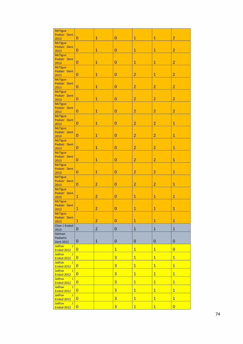
74
McTigue Pediatr Dent 2013 0 1 0 1 1 2 McTigue Pediatr Dent 2013 0 1 0 1 1 2 McTigue Pediatr Dent 2013 0 1 0 1 1 2 McTigue Pediatr Dent 2013 0 1 0 2 1 2 McTigue Pediatr Dent 2013 0 1 0 2 2 2 McTigue Pediatr Dent 2013 0 1 0 2 2 2 McTigue Pediatr Dent 2013 0 1 0 2 2 2 McTigue Pediatr Dent 2013 0 1 0 2 2 1 McTigue Pediatr Dent 2013 0 1 0 2 2 1 McTigue Pediatr Dent 2013 0 1 0 2 2 1 McTigue Pediatr Dent 2013 0 1 0 2 2 1 McTigue Pediatr Dent 2013 0 1 0 2 2 1 McTigue Pediatr Dent 2013 0 2 0 2 2 1 McTigue Pediatr Dent 2013 1 2 0 1 1 1 McTigue Pediatr Dent 2013 1 2 0 1 1 1 McTigue Pediatr Dent 2013 1 2 0 1 1 1 Chen J Endod 2013 0 2 0 1 1 1 Gelman Pediatric Dent 2012 0 1 0 0 0 0 Jadhav J Endod 2012 0
1 1 1 0
Jadhav J Endod 2012 0
3 1 1 1
Jadhav J Endod 2012 0
3 1 1 1
Jadhav J Endod 2012 0
3 1 1 1
Jadhav J Endod 2012 0
3 1 1 1
Jadhav J Endod 2012 0
3 1 1 1
Jadhav J Endod 2012 0
3 1 1 1
Jadhav J Endod 2012 0
3 1 1 0

75
Jadhav J Endod 2012 0
3 1 1 1
Jadhav J Endod 2012 0
3 1 1 1
Jadhav J Endod 2012 0
3 1 0 0
Jadhav J Endod 2012 0
3 1 1 0
Jadhav J Endod 2012 0
3 1 0 0
Jadhav J Endod 2012 0
3 0 0 0
Jadhav J Endod 2012 0
3 0 1 0
Jadhav J Endod 2012 0
3 1 1 1
Jadhav J Endod 2012 0
3 1 1 1
Jadhav J Endod 2012 0
3 1 0 0
Jadhav J Endod 2012 0
3 1 1 1
Jadhav J Endod 2012 0
3 0 1 0
Shivashankar J Conserv dent 2012 0 1 2 0 0 0 Nosrat J Endo 2012 0 0 0 2 2 0 Nosrat J Endod 2012 0 1
Jeruphan J Endod 2012 0 0 3 1 1
Jeruphan J Endod 2012 0 0 3 1 1
Jeruphan J Endod 2012 0 0 3 1 1
Jeruphan J Endod 2012 0 0 3 1 0
Jeruphan J Endod 2012 0 0 3 1 0
Jeruphan J Endod 2012 0 2 3 0 0
Jeruphan J Endod 2012 0 2 3 0 0
Jeruphan J Endod 2012 0 2 3 0 0
Jeruphan J Endod 2012 0 2 3 0 0
Jeruphan J Endod 2012 0 2 3 0 0
Jeruphan J Endod 2012 0 2 3 0 0
Jeruphan J Endod 2012 0 2 3 0 0
Jeruphan J Endod 2012 0 2 3 0 0
Jeruphan J Endod 2012 0 2 3 0 0
Jeruphan J Endod 2012 0 2 3 0 0
Jeruphan J Endod 2012 0 2 3 0 0
Jeruphan J Endod 2012 0 2 3 0 0
Jeruphan J Endod 2012 0 2 3 0 0
Jeruphan J Endod 2012 0 2 3 0 0

76
Jeruphan J Endod 2012 0 2 3 0 0
Jeruphan J Endod 2012 0 2 3 0 0
Jeruphan J Endod 2012 0 2 3 0 0
Jeruphan J Endod 2012 0 2 3 0 0
Jeruphan J Endod 2012 0 2 3 0 0
Jeruphan J Endod 2012 0 2 3 0 0
Jeruphan J Endod 2012 0 1 3 0 0
Jeruphan J Endod 2012 0 1 3 0 0
Jeruphan J Endod 2012 0 1 3 0 0
Jeruphan J Endod 2012 0 1 3 0 0
Jeruphan J Endod 2012 0 1 3 0 0
Jeruphan J Endod 2012 0 1 3 0 0
Jeruphan J Endod 2012 0 1 3 0 0
Jeruphan J Endod 2012 0 1 3 0 0
Jeruphan J Endod 2012 0 1 3 0 0
Jeruphan J Endod 2012 0 1 3 0 0
Jeruphan J Endod 2012 0 1 3 0 0
Jeruphan J Endod 2012 1 1 3 0 0
Jeruphan J Endod 2012 1 1 3 0 0
Jeruphan J Endod 2012 1 1 3 0 0
Jeruphan J Endod 2012 1 1 3 0 0
Jeruphan J Endod 2012 1 1 3 0 0
Jeruphan J Endod 2012 1 1 3 0 0
Jeruphan J Endod 2012 1 1 3 0 0
Jeruphan J Endod 2012 1 1 3 0 0
Jeruphan J Endod 2012 1 1 3 0 0
Jeruphan J Endod 2012 1 1 3 0 0
Jeruphan J Endod 2012 1 1 3 0 0
Jeruphan J Endod 2012 1 1 3 0 0
Jeruphan J Endod 2012 1 1 3 0 0
Jeruphan J Endod 2012 1 1 3 0 0
Jeruphan J Endod 2012 1 1 3 0 0
Jeruphan J Endod 2012 1 1 3 0 0
Jeruphan J Endod 2012 1 1 3 0 0
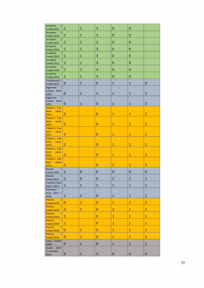
77
Jeruphan J Endod 2012 1 1 3 0 0
Jeruphan J Endod 2012 1 1 3 0 0
Jeruphan J Endod 2012 1 1 3 0 0
Jeruphan J Endod 2012 1 1 3 0 0
Jeruphan J Endod 2012 1 1 3 0 0
Jeruphan J Endod 2012 1 1 3 0 0
Jeruphan J Endod 2012 1 1 3 0 0
Jeruphan J Endod 2012 1 1 3 0 0
Torabinejad J Endod 2012 0 1 0 1 1 0 Aggarwal J Conser Dent 2012 0 1 3 1 1 1 Aggarwal J Conser Dent 2012 0 1 0 1 1 2 Chereli J Can dent assoc 2011 2
0 1 1 1
Chereli J Can dent assoc 2011 2
0 1 1 1
Chereli J Can dent assoc 2011 2
0 1 1 1
Chereli J Can dent assoc 2011 2
0 1 1 1
Chereli J Can dent assoc 2011 2
0 1 1 1
Chereli J Can dent assoc 2011 2
0 1 1 1
Nosrat J Endod 2011 2 0 0 0 0 0 Nosrat J Endod 2011 2 0 0 1 1 1 Lovelace Aust Dent J 2011 1 1 1 1 1 1 Thomson Aust Dent J 2010 1 2 0 1 1 1 Petrino J Endod 2010 0 1 0 1 1 1 Petrino J Endod 2010 0 1 0 2 1 2 Petrino J Endod 2010 1
0 1 1 1
Petrino J Endod 2010 1
0 1 1 1
Petrino J Endod 2010 0 1 0 1 1 1 Petrino J Endod 2010 0 1 0 1 1 2 Cotti J Endod 2008 0 1 0 1 1 1 Iwaya Dent Traumatol 2011 0 1 3 0 0 0

78
Chen J Endod 2012 0 0 0 1 1 2 Chen J Endod 2012 0 0 0 1 1 2 Chen J Endod 2012 0 0 0 1 1 2 Chen J Endod 2012 0 2 0 1 1 2 Chen J Endod 2012 0 2 0 1 1 2 Chen J Endod 2012 0 2 0 1 1 0 Chen J Endod 2012 0 2 0 1 1 0 Chen J Endod 2012 0 2 0 1 1 0 Chen J Endod 2012 0 2 0 1 1 0 Chen J Endod 2012 0 2 0 1 1 0 Chen J Endod 2012 1 1 0 1 1 0 Chen J Endod 2012 1 1 0 1 1
Chen J Endod 2012 1 1 0 1 1
Chen J Endod 2012 1 1 0 1 1
Chen J Endod 2012 1 1 0 1 1
Chen J Endod 2012 1 1 0 1 1
Chen J Endod 2012 1 1 0 1 1
Chen J Endod 2012 1 1 0 1 1
Chen J Endod 2012 1 1 0 1 1
Chen J Endod 2012 1 1 0 1 1 0 Chereli J Endod 2011 1 2 3 0 1 1 Chereli J Endod 2011 1 2 3 1 2 1 Chereli J Endod 2011 1 2 3 1 2 1 Chereli J Endod 2011 1 2 3 2 2 0 Chereli J Endod 2011 1 2 0 1 1 0 Chereli J Endod 2011 1 2 0 1 1 2 Kim J Endod 2010 0 1 0 2 2 0 Shin Int Endod J 2009 1 2 3 1 1 1 Reynols Int Endod J 2009 1 2 0 0 1 0 Reynols Int Endod J 2009 1 2 0 0 1 0 Shah J Endod 2008 0 1 3 0 0
Shah J Endod 2008 0 1 3 0 0
Shah J Endod 2008 0 1 3 0 0
Shah J Endod 2008 0 1 3 0 0
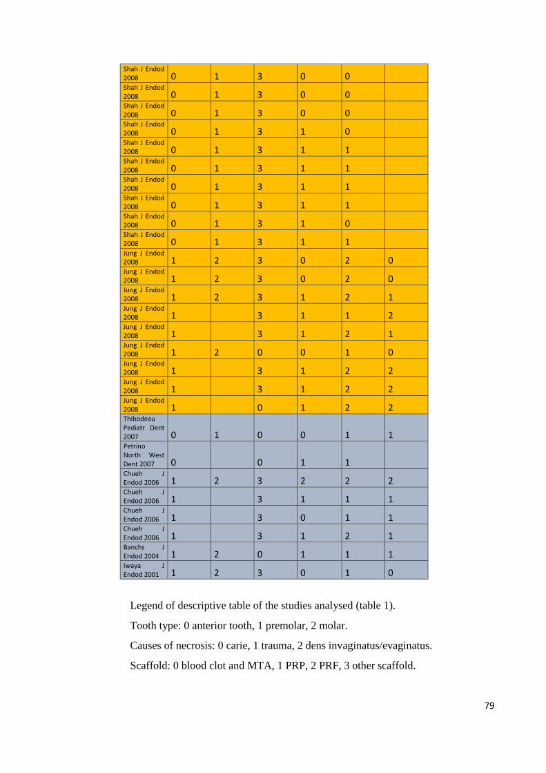
79
Shah J Endod 2008 0 1 3 0 0
Shah J Endod 2008 0 1 3 0 0
Shah J Endod 2008 0 1 3 0 0
Shah J Endod 2008 0 1 3 1 0
Shah J Endod 2008 0 1 3 1 1
Shah J Endod 2008 0 1 3 1 1
Shah J Endod 2008 0 1 3 1 1
Shah J Endod 2008 0 1 3 1 1
Shah J Endod 2008 0 1 3 1 0
Shah J Endod 2008 0 1 3 1 1
Jung J Endod 2008 1 2 3 0 2 0 Jung J Endod 2008 1 2 3 0 2 0 Jung J Endod 2008 1 2 3 1 2 1 Jung J Endod 2008 1
3 1 1 2
Jung J Endod 2008 1
3 1 2 1
Jung J Endod 2008 1 2 0 0 1 0 Jung J Endod 2008 1
3 1 2 2
Jung J Endod 2008 1
3 1 2 2
Jung J Endod 2008 1
0 1 2 2
Thibodeau Pediatr Dent 2007 0 1 0 0 1 1 Petrino North West Dent 2007 0
0 1 1
Chueh J Endod 2006 1 2 3 2 2 2 Chueh J Endod 2006 1
3 1 1 1
Chueh J Endod 2006 1
3 0 1 1
Chueh J Endod 2006 1
3 1 2 1
Banchs J Endod 2004 1 2 0 1 1 1 Iwaya J Endod 2001 1 2 3 0 1 0
Legend of descriptive table of the studies analysed (table 1).
Tooth type: 0 anterior tooth, 1 premolar, 2 molar.
Causes of necrosis: 0 carie, 1 trauma, 2 dens invaginatus/evaginatus.
Scaffold: 0 blood clot and MTA, 1 PRP, 2 PRF, 3 other scaffold.
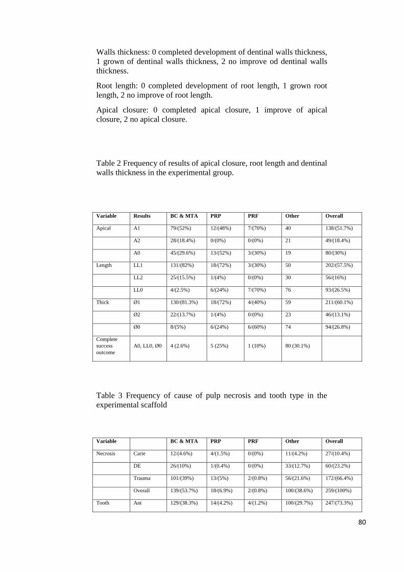
80
Walls thickness: 0 completed development of dentinal walls thickness,
1 grown of dentinal walls thickness, 2 no improve od dentinal walls
thickness.
Root length: 0 completed development of root length, 1 grown root
length, 2 no improve of root length.
Apical closure: 0 completed apical closure, 1 improve of apical
closure, 2 no apical closure.
Table 2 Frequency of results of apical closure, root length and dentinal
walls thickness in the experimental group.
Variable Results BC & MTA PRP PRF Other Overall
Apical A1 79/(52%) 12/(48%) 7/(70%) 40 138/(51.7%)
A2 28/(18.4%) 0/(0%) 0/(0%) 21 49/(18.4%)
A0 45/(29.6%) 13/(52%) 3/(30%) 19 80/(30%)
Length LL1 131/(82%) 18/(72%) 3/(30%) 50 202/(57.5%)
LL2 25/(15.5%) 1/(4%) 0/(0%) 30 56/(16%)
LL0 4/(2.5%) 6/(24%) 7/(70%) 76 93/(26.5%)
Thick Ø1 130/(81.3%) 18/(72%) 4/(40%) 59 211/(60.1%)
Ø2 22/(13.7%) 1/(4%) 0/(0%) 23 46/(13.1%)
Ø0 8/(5%) 6/(24%) 6/(60%) 74 94/(26.8%)
Complete
success
outcome
A0, LL0, Ø0 4 (2.6%) 5 (25%) 1 (10%) 80 (30.1%)
Table 3 Frequency of cause of pulp necrosis and tooth type in the
experimental scaffold
Variable BC & MTA PRP PRF Other Overall
Necrosis Carie 12/(4.6%) 4/(1.5%) 0/(0%) 11/(4.2%) 27/(10.4%)
DE 26/(10%) 1/(0.4%) 0/(0%) 33/(12.7%) 60/(23.2%)
Trauma 101/(39%) 13/(5%) 2/(0.8%) 56/(21.6%) 172/(66.4%)
Overall 139/(53.7%) 18/(6.9%) 2/(0.8%) 100/(38.6%) 259/(100%)
Tooth Ant 129/(38.3%) 14/(4.2%) 4/(1.2%) 100/(29.7%) 247/(73.3%)
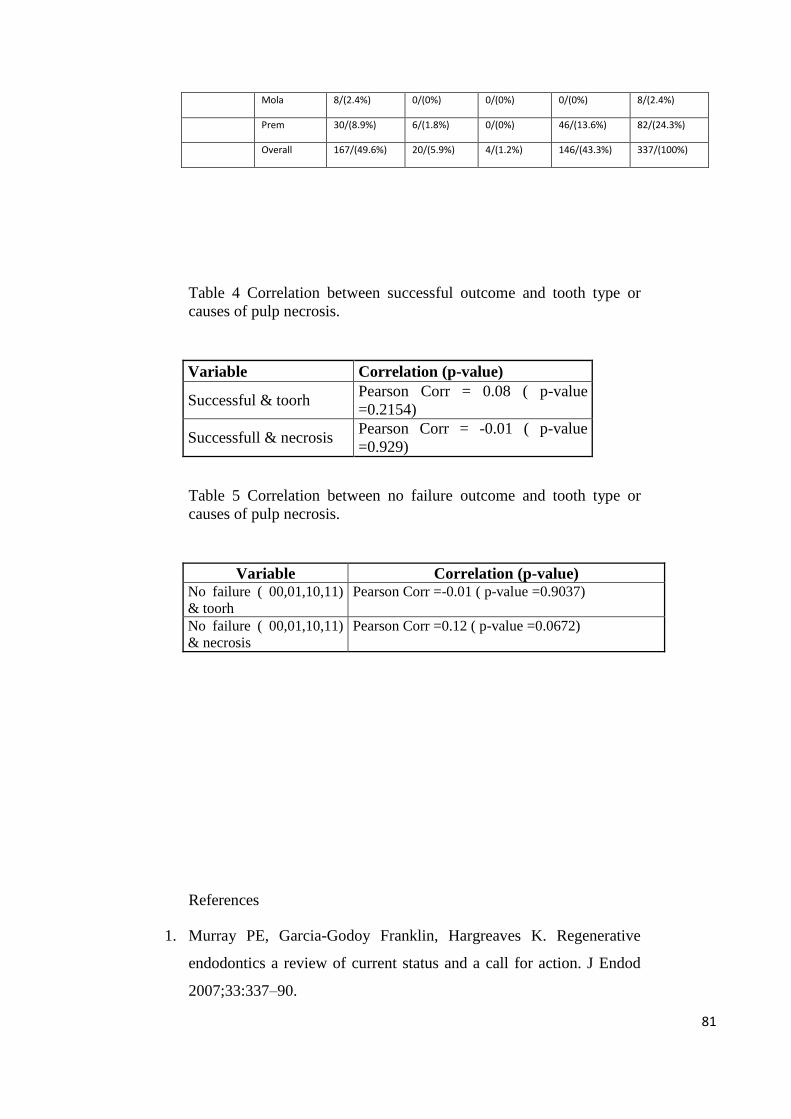
81
Mola 8/(2.4%) 0/(0%) 0/(0%) 0/(0%) 8/(2.4%)
Prem 30/(8.9%) 6/(1.8%) 0/(0%) 46/(13.6%) 82/(24.3%)
Overall 167/(49.6%) 20/(5.9%) 4/(1.2%) 146/(43.3%) 337/(100%)
Table 4 Correlation between successful outcome and tooth type or
causes of pulp necrosis.
Variable Correlation (p-value)
Successful & toorh Pearson Corr = 0.08 ( p-value
=0.2154)
Successfull & necrosis Pearson Corr = -0.01 ( p-value
=0.929)
Table 5 Correlation between no failure outcome and tooth type or
causes of pulp necrosis.
Variable Correlation (p-value) No failure ( 00,01,10,11)
& toorh
Pearson Corr =-0.01 ( p-value =0.9037)
No failure ( 00,01,10,11)
& necrosis
Pearson Corr =0.12 ( p-value =0.0672)
References
1. Murray PE, Garcia-Godoy Franklin, Hargreaves K. Regenerative
endodontics a review of current status and a call for action. J Endod
2007;33:337–90.
82
2. Shah N, Logani A, Bhaskar U, Aggarwal V. Efficacy of
revascularization to induce apexification or apexogenesis in infected,
nonvital, immature teeth: a pilot clinical study. J Endod 2008;34:919–
25.
3. Hargreaves K, Geisler T, Henry M. Regeneration potential of the
permanent tooth: what does the future hold? J Endod 2008;34:51–6.
4. Thibodeau B, Teixeira F, Yamauchi M, et al. Pulp revascularization of
immature dog teeth with apical periodontitis. J Endod 2007;33:680–9.
5. Moreno-Hidalgo MC. Caleza-Jmenez C. Mendoza-Mendoza A.
Iglesias-Linares. Revascularization of immature permanent teeth with
apical periodontitis. Int Endod J 2013;47:321-331.
6. Iwaya S, Ikawa M, Kubota M. Revascularization of an immature
permanent tooth with apical periodontitis and sinus tract. Dental
Traumatology 2001; 17: 185–187.
7. Hotwani K, Sharma K. Platelet rich fibrin - a novel acumen into
regenerative endodontic therapy. Restor Dent Endod 2014;39:1-6.
8. Narang I, Mittal N, Mishra N. A comparative evaluation of the blood
clot, platelet-rich plasma, and platelet-rich fibrin in regeneration of
necrotic immature permanent teeth: A clinical study. Contemp Clin
Dent. 2015 Jan-Mar; 6(1): 63–68.
9. Nagy MN, Tawfik HE, Hashem AAR, and Abu-Seida AM.
Regenerative Potential of Immature Permanent Teeth with Necrotic
Pulps after Different Regenerative Protocols. J Endod 2014;40:192–
198.
10. Shiehzadeh V, Aghmasheh F, Shiehzadeh F, Joulae M, Kosarieh E,
Shiehzadeh F. Healing of large periapical lesions following delivery
of dental stem cells with an injectable scaffold: New method and three
case reports. Case report 2014;25:248-53.
11. Alobaid AS, Cortes LM, Lo J, Nguyen TT, DDS, Albert J, Abu-Melha
AS, Lin LM, Gibbs JL. Radiographic and clinical outcomes of the
treatment of immature permanent teeth by revascularization or
83
apexification: A Pilot Retrospective Cohort Study. J Endod
2014;40:1063–1070.
12. Polat GG, Yildimir C, Akgun OM et al. The use of platelet rich
plasma in the treatment of immature tooth with periapical lesion: a
case report. Restor Dent Endod 2014;39:230-234.
13. Jadhav GR, Shah D, Raghvendra SSJ. Autologus Platelet Rich Fibrin
aided Revascularization of an immature, non-vital permanent tooth
with apical periodontitis: A case report. Nat Sci Biol Med. 2015 Jan-
Jun; 6(1): 224–225.
14. Nagaveni NB, Poornima P, Joshi JS, Pathak S, Nandini DB.
Revascularization of Immature, Nonvital Permanent Tooth Using
Platelet-rich Fibrin in Children. Pediatric dentistry 2015;37:1-6.
15. Nevins AJ & Cymerman JJ. Revitalization of Open Apex Teeth with
Apical Periodontitis Using a Collagen-hydroxyapatite Scaffold. J
Endod 2015;-:1–8.
16. Büyükbayram IG,Özalp F, Aytugar E, Aydemir S. Regenerative
Endodontic Treatment of an Infected Immature Dens Invaginatus with
the Aid of Cone-Beam Computed Tomography. Case Reports in
Dentistry 2014; 5.
17. Bezgin T, Yilmaz AD, Celik BN, Kolsuz ME, Sonmez H. Efficacy of
platelet-rich plasma as a scaffold in regenerative endodontic
treatment. J Endod 2015:41:54-44.
18. Saoud TM, Zaazou A, Nabil A, Moussa S, Lin LM, Jibbs JM. and
Radiographic Outcomes of Traumatized Immature Permanent
Necrotic Teeth after Revascularization/Revitalization Therapy. J
Endod 2014;40:1946–195.
19. Sachdeva GS, Sachdeva LT, Goel M & Bala S. Regenerative
endodontic treatment of an immature tooth with a necrotic pulp and
apical periodontitis using platelet-rich plasma (PRP) and mineral
trioxide aggregate (MTA): a case report. Int Endod J 2014;49:902-10.
84
20. Johns DA, Shivashankar VY, Krishnamma S, Johns M. Use of
photoactivated disinfection and platelet-rich fibrin in regenerative
Endodontics. J Conserv Dent. 2014;17: 487–490.
21. Saoud TMA, Sigurdsson A, Rosenberg PA, Lin LM, Ricucci D.
Treatment of a Large Cyst like Inflammatory Periapical Lesion
Associated with Mature Necrotic Teeth Using Regenerative
Endodontic Therapy J Endod 2014;40:2081–2086.
22. Bakhtiar H,Vatanpour M Rayani A, Navi F, ;Asna Ashari E;Ahmadi-
Anahid J. The plasma-rich in growth factor as a suitable matrix in
regenerative endodontics: a case series. NY State Dent J 2014;80:49-
53.
23. Güven Polat G, Yıldırım C, Akgün OM, Altun C, Dinçer D, Özkan
CK. The use of platelet rich plasma in the treatment of immature tooth
with periapical lesion: a case report. Restor Dent Endod 2014;39:230-
234.
24. Shiehzadeh V, Aghmasheh F, Shiehzadeh F, Joulae M, Kosarieh E,
Shiehzadeh F. Healing of large periapical lesions following delivery
of dental stem cells with an injectable scaffold: New method and three
case reports. Indian J Dent Res 2014;25:248-53.
25. Park M, Ahn BD. Immature permanent teeth with apical periodontitis
and abscess treated byregenerative endodontic treatment using
calcium hydroxide and MTA. Pediatr Dent 2014;36:107-10.
26. Kumar H, Al-Ali M, Parashos P, and Manton DJ. Management of 2
Teeth Diagnosed with Dens Invaginatus with Regenerative
Endodontics and Apexificationin the Same Patient: A Case Report and
Review. J Endod 2014;40:725–731.
27. Jadhav GR, Shah N, and Logani A. Platelet-Rich Plasma
Supplemented Revascularization of an Immature Tooth Associated
with a Periapical Lesion in a 40-Year-Old Man. Case Rep Dent 2014;
2014:479584
85
28. Kahler B, Mistry S, Moule A, Ringsmuth AK, Case P, Thomson A,
Holcombe T. Revascularization Outcomes: A Prospective Analysis of
16 Consecutive Cases. J Endod 2014;40:333–338.
29. Mishra N, Narang I, Mittal N. Platelet-rich fibrin-mediated
revitalization of immature necrotic tooth. Contemp Clin Dent.
2013;4:412-5.
30. Noy AF, Nuni E, Moskovitz M Regenerative endodontic treatment of
an immature permanent canine following infant oral mutilation Pediatr
Dent 2013:35:355-59.
31. Paryani K, Kim SG- Regenerative endodontic treatment of permanent
teeth after completion of rootdevelopment: a report of 2 cases. J
Endod 2013;39:929-34.
32. McTigue DJ, Subramanian K, Kumar A. Case series: management of
immature permanent teeth with pulpal necrosis: a case series. Pediatr
Dent 2013; 35:55-60.
33. Chen X, Bao ZF, Liu Y, Liu M, MS, Jin XQ, Xu XB Regenerative
Endodontic Treatment of an Immature Permanent Tooth at an Early
Stage of Root Development: A Case Report. J Endod 2013;39:719–
722.
34. Gelman R, Park H Pulp revascularization in an immature necrotic
tooth: a case report Pediatr Dent 2012;34:496-99.
35. Jadhav G, Shah N, Logani A. Revascularization with and without
Platelet-rich Plasma in Nonvital, Immature, Anterior Teeth: A Pilot
Clinical Study. J Endod 2012;38:1581–1587.
36. Shivashankar VY, Johns DA, Vidyanath S, Kumar MR. Platelet Rich
Fibrin in the revitalization of tooth with necrotic pulp and open apex. J
Conserv Dent. 2012;15: 395–398.
37. Nosrat A, Homayounfar N, Oloomi K.Drawbacks and Unfavorable
Outcomes of Regenerative Endodontic Treatments of Necrotic
Immature Teeth: A Literature Review and Report of a Case J Endod
2012;38:1428–1434.
86
38. Jeeruphan T, Jantarat J, Yanpiset K, Suwannapan L, Khewsawai P,
and Hargreaves KM. Mahidol Study 1: Comparison of Radiographic
and Survival Outcomes of Immature Teeth Treated with Either
Regenerative Endodontic or Apexification Methods: A Retrospective
Study. J Endod 2012;38:1330–1336.
39. Torabinejad M & Faras H. A clinical and histological report of a tooth
with an open apex treated with regenerative endodontics using
platelet-rich plasma. J Endod. 2012;38:864-8.
40. Aggarwal V, Miglani S, Singla M. Conventional apexification and
revascularization induced maturogenesis of two non-vital, immature
teeth in same patient: 24 months follow up of a case. J Consev Dent
2012;15:68-72.
41. Cehreli ZC, Isbitiren B, Sara S, Erbas G. Regenerative Endodontic
Treatment (Revascularization) of Immature Necrotic Molars
Medicated with Calcium Hydroxide: A Case Series. J Endod
2011;37:1327–1330.
42. Torabinejad M, Turman M. Revitalization of Tooth with Necrotic
Pulp and Open Apex by Using Platelet-rich Plasma: A Case Report. J
Endod 2011;37:265–268.
43. Thomson A, Kahler B. Regenerative endodontics – biologically-based
treatment for immature permanent teeth: a case report and review of
the literature. Aust Dent J 2010; 55: 446–452.
44. Petrino JA, Boda KK, Shambarger S, Bowles WR, and McClanahan
SB. Challenges in Regenerative Endodontics: A Case Series. J Endod
2010;36:536–541.
45. Cotti E, Mereu M, Lusso D. Regenerative Treatment of an Immature,
Traumatized Tooth With Apical Periodontitis: Report of a Case. J
Endod 2008;34:611– 616.
46. Chen MYH, Chen KL, Chen CA, Tayebaty F, Rosenber PA, Lin LM.
Responses of immature permanent teeth with infected necrotic pulp
87
tissue and apical periodontitis/abscess to revascularization procedures.
Int Endod J 2012;45:294–305.
47. Cehreli ZC, Isbitiren B, Sara S, Erbas G. Regenerative Endodontic
Treatment (Revascularization) of Immature Necrotic Molars
Medicated with Calcium Hydroxide: A Case Series. J Endod
2011;37:1327–1330.
48. Kim JH, Kim K, Shin SJ, Park JW, Jung Y.Tooth Discoloration of
Immature Permanent Incisor Associated with Triple Antibiotic
Therapy: A Case Report. J Endod 2010;36:1086–109.
49. Shin SY, Albert JS & Mortman RS. One step pulp revascularization
treatment of an immature permanent tooth with chronic apical
abscess: a case report. Int Endod J 2009;42:1118–1126.
50. Reynolds K, Johnson JD & Cohenca N. Pulp revascularization of
necrotic bilateral bicuspids using a modified novel technique to
eliminate potential coronal discolouration: a case report. Int Endod J
2009;42: 84–92.
51. Ding RY, Cheung GS,Chen J, Yin XZ, Wang .QQ, Zhang CF. Pulp
Revascularization of Immature Teeth With Apical Periodontitis: A
Clinical Study. J Endod 2009;35:745–749.
52. Jung J, Lee SJ, Hargreaves KM. Teeth with Pulpal Necrosis: A Case
Series. J Endod 2008;34:876 – 887.
53. Thibodeau B & Trope M. Pulp revascularization of a necrotic infected
immature permanent tooth: case report and review of the literature.
Pediatr Dent. 2007;29:47-50.
54. Petrino JA. Revascularization of necrotic pulp of immature teeth with
apical periodontitis. Northwest Dent 2007.
55. Chueh LH, Huang GTJ Immature Teeth With Periradicular
Periodontitis or Abscess Undergoing Apexogenesis: A Paradigm
Shift. J Endod 2006;32: 1205–1213.
88
56. Banchs F, Trope M. Revascularization of Immature Permanent Teeth
With Apical Periodontitis: New Treatment Protocol? J Endod
2004;30:196-200.
57. Fouad AF & Prashant V. Healing after Regenerative Procedure with
and without Pulpal Infection. J Endod 2014;40:S58–S64.
58. Law AS. Considerations for Regeneration Procedures. J Endod
2013;39:S44–S56.
59. Wigler R, Kaufman AY, Lin S, Steinbock N, Hazan-Molina H,
Torneck CD. Revascularization: A Treatment for Permanent Teeth
with Necrotic Pulp and Incomplete Root Development. J Endod
2013;39:319–326.
60. Law AS. Outcomes of Regenerative Endodontic Procedures. Dent
Clin N Am 2012;56: 627–637.
61. Garcia-Godoy M & Murray PE. Recommendations for using
regenerative endodontic procedures in permanent immature
traumatized teeth. Dental Traumatology 2012; 28: 33–41.
62. Bose R, Nummikoski P, Hargreaves K. A Retrospective Evaluation of
Radiographic Outcomes in Immature Teeth With Necrotic Root Canal
Systems Treated With Regenerative Endodontic Procedures. J Endod
2009;35:1343–1349.
63. Martin G, Ricucci D, Gibbs JL, Lin LM. Histological finding of
revascularizated/revitalizated immature permanent molar with apical
periodontitis using platelet-rich-plasma. J Endod 2013;39:138-44.
64. Nikolidakis D, Jansen JA. The biology of platelet-rich plasma and its
application in oral surgery: literature review. Tissue Eng Part B Rev
2008;14:249-58.
65. Fouad AF & Verma P Healing after regenerative procedures with and
without pulpal infection. J Endod 2014;40:S58-S64.
66. Bastone EB, Freer TJ, McNamara JR. Epidemiology of dental trauma:
a review of literature. Aust Endod J 2000;45:2-9.
89
Summary and conclusion
Chapters 1 and 2.
The pulpal necrosis and arrest of root development makes the
endodontic treatment in immature permanent teeth especially
challenging (1). In this circumstance, in children the incorrect
management can result in loss of permanent teeth, disorders of
mandibular growth and masticatory function, and even speech
disorders and facial cosmetic impairment (1,2). Apexification is a
nonsurgical method of inducing a calcific barrier at the open root apex
of necrotic teeth (1,2). The barrier prevents toxins and bacteria from
entering periradicular tissue, and facilitates placement of a root canal
sealant and filling material (3). Calcium hydroxide is commonly used
for apexification, it has predictable results, and can be mixed with a
number of different substances (camphorated mono chlorophenol,
distilled water, saline, anesthetic solutions, chlorhexidine, and
cresatin) to induce apical closure (1,2). Hermann introduced the use
of calcium hydroxide in endodontics in 1920 (4). Calcium hydroxide
is a slow-acting antiseptic intracanal medication that needs a 24-hour
contact period for the complete killing of enterococci and a 1-week
contact period to achieve a bacterial reduction rate of 92.5% (5,6). In
addition, calcium hydroxide has the ability to hydrolyze the lipid
portion of bacterial lipopolysaccharide, resulting in inactivation of the
biologic activity of lipopolysaccharide and subsequent cessation of
lipopolysaccharide-induced inflammation in the periradicular tissue
(7). In case of teeth treated with calcium hydroxide, a prolonged
90
period needed to form the apical barrier, and during the treatment
period the teeth are susceptible to fracture. Besides risk of tooth
fracture, calcium hydroxide has a number of limitations, the variables
which can affect treatment outcomes are: treatment time ranging from
5 months to 20 months; the apical closure in relationship to treatment
time is unpredictable; long patient compliance due to the extended
treatment time (1).
To overcome these disadvantage of apexification with calcium
hydroxide Mineral Trioxide Aggregate (MTA) has been
recommended for such usage. MTA was introduced in 1993 by
Torabinejad et al (8); it has excellent biocompatibility as well as
sealing, osteogenesis, and cementogenesis abilities (9-11). MTA
promote the formation of apatite-like crystals on the outer contact
surface when exposed to body fluid (12); this ability was useful to seal
the apical broad foramen in case of necrotic teeth with incomplete root
development. Apexification with MTA, known as MTA apical plug
technique, can be completed in one or two treatment sessions, making
it possible to restore the tooth within a short timeframe while avoiding
reliance on patient compliance and prolonged exposure of root dentin
to calcium hydroxide (13-16). Clinical studies reported a percentage
of healing range between 77% to 85% in necrotic teeth with
incomplete root development treated with apical plug technique (13-
16); moreover the results obtained is stable at long term follow-up
(17). As documented in our recent study complete healing was
observed in 7 out of a total of 17 teeth at the one year follow-up, the
cases of complete healing increased at five years (13 out of 16 teeth)
and the results remained stable at 10 years (15 out of 16 teeth) without
statistically significant differences. As per previous studies (16, 18,
19) longer recall intervals (more than one year) could be an important
factor for a better treatment outcome, particularly in cases of necrotic
teeth with large periapical lesions (19, 20). Pace et al (17) obserevd a
change in outcome from the time of initial therapy and the 1 and 5
year recalls; the results remained substantially stable between the 5
91
and 10 year follow-ups. Only one case was documented as a failure
(6%), this percentage could be significant in case studies with bigger
sample sizes. The failure case, that was documented ten years after
treatment, was associated with a longitudinal root fracture that could
have been due to an immature thin and fragile root. As previously
reported, dentin thickness that is correlated with the stage of root
development, is one of the most important incidence factors in the
frequency of tooth fracture (21).
Despide the difference in steps protocol between apexification with
calcium hydroside and MTA apical plug technique a recent meta-
analysis showed that both materials had similar clinical success rates,
radiographic success rates, and apical barrier formation rates (22).
However, MTA was associated with a significantly shorter time to
achieve apical barrier formation than the Calcium Hydroxide. This is
of great significance because many failures with calcium hydroxide
are due to poor patient follow-up because of the extensive treatment
time. Finally the same study revealed no statistical difference in term
of radiographic success rate between the MTA group and the calcium
hydroxide group, even only two studies, collected and examinated,
reported radiographic success rate data.
Chapter 3
Regenerative endodontic procedures are defined as biologically based
procedures aimed to replace damaged dentin and cells of the pulp–
dentin complex (23). Regenerative endodontics currently has two
major concepts: guided tissue regeneration and tissue engineering
(24). Guided tissue regeneration involved the tissue regeneration
through the formation of a blood clot: revascularization; this concept
was introduced for the first time by Nygaard-Ostby in the year 1961
(25). Tissue engineering with stem cells is still developing. Both these
concepts had the same goal: promote physiological pulp formation by
activation of stem cells.
92
Iwaya et al. documented a successful response to vitality tests in
treated necrotic immature tooth as well as root development after
regenerative intervention (26). In this study a double anti-biotic paste,
ciprofloxacin and metronidazole, has been used to reach a
debridement of root canal and to promote the regeneration of the
pulpal tissue. Afterwards Banch and Trope (27) developed a triple
antibiotic paste consisting of ciprofloxacin, metronidazole, and
minocycline placed in the canal for 28 days, after debridement with
sodium hypochlorite (5.25%). At the second visit, after removed the
antibiotic paste, bleeding was induced, and a filling was placed
coronally to blood clot. The treatment demonstrated root development
in terms of lenghing and thickness of the dentin root and a positive
response to vitality testing. This studies opened a new era to the
treatment of necrotic immature permanent teeth.
Pulp Tissue regenerative procedure involved three essenzial
components: (A) stem cells, (B) growth factors, and scaffolds (C).
Stem cells from the dental pulp were identified based on their ability
to regenerate a pulp dentin like complex (28); these were identified as
dental pulp stem cells (DPSCs). The DPSCs included Stem Cells of
the Apical Papilla (SCAP), Stem Cells of Human Exfoliated
Deciduous teeth (SHED), and Periodontal Ligament Stem Cells
(PDLSCs); all of these cells have potential for regeneration of pulpal
tissue. DPSCs are multipotent cells with an ability to differentiate into
adipocytes, osteoblasts, melanocytes, myoblasts and endothelial cells,
produce mineralized tissue, and demonstrate neurogenic potential
(29).
SCAPs in the apical papilla of immature teeth have been postulated to
differentiate into cells responsible for continued root development in
pulpally damaged immature teeth with a retained apical papilla (30).
Stem cells isolated from the remnant pulp of exfoliated deciduous
teeth (SHED) have a higher proliferation rate and mineralization
potential compared with DPSCs (30). They also demonstrate a higher
93
osteo-inductive capacity in vivo and have a higher neurogenic
potential compared with DPSCs (31).
Cells isolated from the periodontal ligament (PDLSCs) displayed
osteogenic and differentiation potential in vitro (32), besides the
potential to regenerate into cementoblast like cells, adipocytes, and
collagen forming cells. They are present in all age groups but their
regenerative potential, migration, and proliferation capacity decreases
with age (33). With regenerative procedures, the growth and
development of cell and vasculature are achieved by providing a
network scaffold that can also infused with a variety of factors.
Growth factors play a vital role in endodontic regeneration procedure
they influence stem cell activity such as them proliferation, and
differentiation into different lineage. They play an important role in
the formation and repair of the dentin pulp complex. The
Transforming Growth Factors (TGF) b are implicated in odontoblast
differentiation, dentin matrix secretion, tooth development, and tissue
repair (34). Bone morphogenic proteins (BMPs) play an important
role in the pulp: BMP4 and BMP5 are expressed during ameloblast
differentiation and BMP2, BMP4, BMP6, BMP7, and GDF11 (growth
differentiation factor) during odontoblast differentiation (35).
Angiogenesis is critical to the development and survival of the
regenerated pulp. Examination of the dentin matrix shows high
concentration of platelet derived growth factor (PDGF-AB), vascular
endothelial growth factor (VEGF), fibro-blast growth factor (FGF),
placenta growth factor (PIGF), and low concentration of epidermal
growth factor (EGF).
Finally for tissue engineering strategies, the choice of an appropriate
scaffold is a crucial step; the role of the scaffold has changed from
passive carrier toward a bioactive matrix, which can induce a desired
cellular behavior.
In dental- pulp complex regeneration procedure scaffolds are three
dimensional network that serve as a template aiding development of
tissue by providing regulatory molecules and mechanical support.
94
Scaffolds may be endogenous (eg, collagen, dentin) or synthetic
substances (eg, hydrogels, MTA, or other compounds). The more
common scaffold used in regenerative endodontic procedure include:
the blood clot developed during revascularization and dentin chips
(37-39); platelet rich plasma (PRP) and platelet rich fibrin (PRF).
Chapter 4
Background
In case of necrotic teeth with incomplete root formation, the
regenerative endodontics procedures aim to replace damage pulp-
dentin complex and normal pulpal physiologic functions that include
root development and immunocompetency. These procedures are
based on three principle of tissue engineering: appropriate source of
stem cell; presence of growth factors for stem cell differentiation and
an appropriate scaffold.
Aim: the aim of the present study was to systematically analysed the
Regenerative Endodontic protocols and the relative results of in vivo
human studies.
Methods
An electronic search was conducted in PubMed using appropriate
Medical Subject Heading terms (regenerative endodontics,
regenerative endodontic) covering a period until to April 2015.
Additional hand searching was conducted, and reference section of
each relevant articles were included in the search. In order to
identified the relevant articles the scanning of titles, abstracts and
ultimately full texts were performed. Treatment protocol adopted were
classified as follow basing the scaffold used: Blood clot (BC) and
coronal seal with MTA (group 0); PRP with or without BC and
coronal sealing (group 1); PRF with or without BC and coronal
sealing (group 2); and other treatment protocols (group 3). The
95
frequency of the following variables: rooth canal length, dentin walls
thickness and apical closure, and the relative percentage were
recorded overall and for each experimental group.
The successful outcome of endodontic regeneration procedure were
considered complete when the follow items were recorded: absence of
sign and symptoms; absence of periradicular pathological
radiolucency area; root apical closure; complete development of
dentinal thickness; complete development of root length.
Satisfactory results were considered when the follow items were
recorded: absence of sign and symptoms; absence of periradicular
pathological radiolucency area; improve or complete dentinal
thickness; improve or complete root length.
The percentage of successful outcame has been calculated for each
operative protocols using different scaffold.
The presence of correlation coefficient between the successful
outcome and the type of treated teeth, and the causes of necrosis has
been calculated using the Pearson correlation coefficient.
The correlation coefficient were calculated between the type of teeth
and the satisfactory results, and the cause of necrosis and satisfactory
results (as before).
Results
In the 51 studies selected a total of 357 treated teeth were analysed.
152 teeth were classified in group 0; a complete successful outcome
was recorded only in four teeth on a total of 151, and satisfactory
outcome was recorded in 135.
In group 1 a total of 25 teeth were treated, a complete successful
outcome was recorded in 5 teeth on a total of 25, and a satisfactory
results in 24 studies.
In group 2 a complete successful outcome was recorded only in one
tooth on a total of 10 teeth, but the outcome was judge satisfactory in
all teeth.
96
The causes of pulp necrosis was specified only for 259 teeth on a total
of 357 teeth. In 10.4% of treated teeth, the necrosis was a consequence
of caries, in 23.2% of treated teeth the necrosis has been observed in
teeth with dens evaginatus/invaginatus, and finally in 66.4% of treated
teeth the necrosis was the result of a trauma.
For 20 teeth the authors (12) did not specified the type (anterior,
premolar or posterior), for the remaining 337 teeth 24.3% were
premolar (n°82), 2,4% were molar (n°8) and 73.3% were anterior
teeth (n°247).
No correlation has been recorded between the successful outcome of
the therapy and the tooth type or the causes of pulp necrosis.
No correlation has been observed between a satisfactory results and
tooth type or causes of pulp necrosis.
Discussion
The present review aimed to identify if the use of different scaffold
(BC, PRP, PRF) could significantly influence the outcome of
regenerative endodontics treatment in necrotic immature teeth.
The major finding recorded by this research is the low level of
evidence of the studies dealing regenerative endodontics and the wide
and different methods used to judge the outcome of the therapy. For
this reason in order not to lose the information given by all articles
selected in this review, an arbitrarily classification of results collected
were adopted and used for qualitative analysis. The outcome was
judge successfully when a complete apical closure, root length and
development of dentinal walls thickness (A 0, LL 0, Ø 0) has been
reported. The total amount of successful outcome in all teeth collected
in the present review was very low: 4.9%. This could be due to the
incomplete data reported, and to a too strictly categories that identified
the successful outcome. Similar percentage of success were recorded
for all experimental group 1.5% when BL was used as scaffold, 1.9%
when PRP was used as scaffold and 0.4% when PRF was used as
scaffold. It is important to underline that this data could not be
97
significant because in a few number of teeth PRP and PRF has been
used as scaffold (25 and 10 teeth respectively) respect the BC (151
teeth).
Scaffolds are used in regenerative procedures to provide a framework
through which cells and a vasculature can grow (39). Several report
have been demonstrated good success with BC scaffold in association
with MTA (40-43); since it not always possible invoke bleeding in the
root canals researchers have begun examining other three-dimensional
scaffold.
On the basis of current studies more and more case reports and case
series show favourable outcomes of regenerative endodontic
procedures, but it is still difficult to predict the outcome and identify
which steps of the protocol could be potential for optimization of the
therapy. This could be due to both low level of studies pubblished,
only 3 RCT were identified in the present study, and to etherogeneus
methods adopted to judge the outcome of the treatment.
However, how can a standardized protocol be developed in the
absence of randomized controlled clinical trials? To address this issue,
the American Association of Endodontists (AAE) formed a standing
committee on regenerative endodontics in 2007. This committee has
developed initiatives for forming an online clinical registry of
regenerative cases and developing continuing education materials,
new insurance treatment codes, and a standardized clinical protocol.
This is a nagging problem of international community of researchs so
much so a recent review addresses this problem by focusing on recent
strategies for developing standardized clinical protocols, and the
selection of proper outcome measures, as well as reviewing the
fundamental role of paradigms in designing and interpreting
regenerative studies (43).
98
References
1. Hargreaves KM, Diogenes A, Teixeira FB. Treatment options:
biological basis of regenerative endodontic procedures. Pediatr Dent
2013;35:129e40.
2. Shabahang S. Treatment options: apexogenesis and apexification. J
Endod 2013;39:S26e9.
3. Mackie IC. Management and root canal treatment of non-vital
immature permanent teeth. UK National Clinical Guidelines in
Pediatric Dentistry. Int J Paediat Dent 1998;8:289e93.
4. Hermann B.W. Calcium hydroxide als mittel zum behandel und fullen
von zahnwurzelkanalen Dissertation University of Wuzburg, Med
Diss V, Germany (1920) [in German]
5. Safavi KE, Spangberg LS, Langeland K. Root canal dentinal tubule
disinfection. J Endod, 16 (1990), pp. 207–210
6. Saleh IM, Ruyter IE, Haapasalo M, Ørstavik D. The effects of dentine
pretreatment on the adhesion of root-canal sealers. Int Endod J, 35
(2002), pp. 859–866
7. Safavi KE, Nichols FC. Effect of calcium hydroxide on bacterial
lipopolysaccharide. J Endod, 19 (1993), pp. 76–78
8. Torabinejad M, Watson TF, Pitt Ford TR Sealing ability of mineral
trioxide aggregate when used as a root end filling material J Endod, 19
(1993), pp. 591–599.
9. Mitcheli PJ, Pitt Ford TR, Torabinejad M.Osteoblast biocompability
of mineral trioxide aggregate Acta Biomater, 20 (1999), pp. 167–173.
10. Dreger LA, Felippe WT, Reyes-CarmonaJF, Felippe GS, Bortoluzzi
EA, Felippe MC. Mineral trioxide aggregate and Portland cement
promote biomineralization in vivo. J Endod, 38 (2012), pp. 324–329.
11. Lawley GR, Schindler WG, Walker WA, Kolodrubetz D. Evaluation
of ultrasonically placed MAT and fracture resistance with intracanal
composite resin in a model of apexification. J Endod, 30 (2004), pp.
167–172
99
12. Bird DC, Komabayashi T, Guo L, Opperman LA, Spears R. In vitro
evaluation of dentinal tubule penetration and biomineralization ability
of a new root-end filling material. J Endod, 38 (2012), pp. 1093–1096
13. Simon S, Rilliard F, Berdal A, et al. The use of mineral trioxide
aggregate in one-visit apexification treatment: a prospective study. Int
Endod J 2007;40:186–97.
14. Holden DT, Schwartz SA, Kirkpatrick TC, et al. Clinical outcomes of
artificial rootend barriers with mineral trioxide aggregate in teeth with
immature apices. J Endod 2008;34:812–7.
15. Sarris S, Tahmassebi JF, Duggal MS, et al. A clinical evaluation of
mineral trioxide aggregate for root-end closure of non-vital immature
permanent incisors in children-a pilot study. Dent Traumatol
2008;24:79–85.
16. Witherspoon DE, Small JC, Regan JD, et al. Retrospective analysis of
open apex teeth obturated with mineral trioxide aggregate. J Endod
2008;34:1171–6.
17. Pace R, Giuliani V, Nieri M, Di Nasso L, Pagavino G. Mineral
trioxide aggregate as apical plug in teeth with necrotic pulp and
immature apices: a 10-year case series. J Endod. 2014
Aug;40(8):1250-4.
18. Sjőgren U, Hagglund B, Sundqvist G, Wing K. Factors affecting the
long-term results of endodontic treatment. J Endod 1990;16:498–504.
19. Pace R, Giuliani V, Pini Prato L, Baccetti T, Pagavino G. Apical plug
technique using mineral trioxide aggregate: results from a case series.
Int Endod J 2007;40:478-84.
20. Bogen G, Kuttler S. Mineral trioxide aggregate obturation: a review
and case series. J Endod 2009;35:777-90.
21. Cvek M. Prognosis of luxated non-vital maxillary incisors treated with
calcium hydroxide and filled with gutta-percha: a retrospective
clinical study. Endod Dent Traumatol 1992;8:45-55.
22. Jia-Cheng Lin, Jia-Xuan Lu, Qian Zeng, Wei Zhao, Wen-Qing Li,
Jun-Qi Ling. Comparison of mineral trioxide aggregate and calcium
hydroxide for apexification of immature permanent teeth: A
100
systematic review and meta-analysis. Journal of the Formosan
Medical Association 2016 in press. DOI:
http://dx.doi.org/10.1016/j.jfma.2016.01.010.
23. Murray PE, Garcia-Godoy F, Hargreaves KM. Regenerative endo-
dontics: a review of current status and a call for action. J Endod
2007;33:377–390.3.
24. Yang M. Regenerative Endodontics: A New Treatment Modalityfor
Pulp Regeneration. JSM Dent 2013;1:1011.4.
25. Nygaard-Østby B. The role of the blood clot in endodontic ther-apy.
An experimental histologic study. Acta Odontol Scand 1961;19:324–
353.
26. Iwaya SI, Ikawa M, Kubota M. Revascularization of an immature
permanent tooth with apical periodontitis and sinus tract. Dent
Traumatol 2001;17:185–187.9.
27. Banchs F, Trope M. Revascularization of immature permanent teeth
with apical periodon titis: new treatment protocol? J Endod
2004;30:196–200.
28. Gronthos S, Mankani M, Brahim J, Robey PG, Shi S. Postnatalhuman
dental pulp stem cells (DPSCs) in vitro and in vivo. ProcNatl Acad
Sci U S A 2000;97:13625–13630.
29. Zhang W, Walboomers XF, Van Kuppevelt TH, Daamen WF,
VanDamme PA, Bian Z, Jansen JA. In vivo evaluation of human
dental pulp stem cells differentiated towards multiple lineages. J
Tissue Eng Regen Med 2008;2:117–125.
30. Huang GT-J, Sonoyama W, Liu Y, Liu H, Wang S, Shi S. The hid-den
treasure in apical papilla: the potential role in pulp/dentin regeneration
and bio root engineering. J Endod 2008;34:645–651.
31. Miura M, Gronthos S, Zhao M, Lu B, Fisher LW, Gehron RobeyP,
Shi S. SHED: stem cells from human exfoliated deciduous teeth. Proc
Natl Acad Sci USA 2003;100:5807–5812.
32. Adams AM, Soames JV, Searle RF. Cultural and morphological
characteristics of human periodontal ligament cells in vitro. Arch Oral
Biol. 1993;38:6572-662.
101
33. Zhanga J, Ana Y, Gaoa L-N, Zhangb Y-J, Jinb Y, Chena F-M. The
effect of aging on the pluripotential capacity and regenerative
potential of human periodontal ligament stem cells. Biomaterials
2012;33:6974–6986.
34. Stevens MM, Marini RP, Schaefer D, Aronson J, Langer R,Shastri
VP. In vivo engineering of organs: the bone bioreactor. Proc Natl
Acad Sci USA 2005;102:11450–11455. Smith AJ, Matthews JB, Hall
RC. Transforming growth factor-beta1(TGF-beta1) in dentine matrix.
Ligand activation and receptorexpression. Eur J Oral Sci
1998;106:179–184.55.
35. Aberg T, Wozney J, Thesleff I. Expression patterns of bone
morphogenetic proteins (Bmps) in the developing mouse tooth suggest
roles in morphogenesis and cell differentiation. Dev Dyn
1997;210:3832-396.
36. Roberts-Clark DJ, Smith AJ. Angiogenic growth factors in
humandentine matrix. Arch Oral Biol 2000;45:1013–1016.
37. Mjor IA, Dahl E, Cox CF. Healing of pulp exposures: an
ultrastructural study. J Oral Pathol Med 1991;20:496–501.84.
38. Silva TA, Rosa AL, Lara VS. Dentin matrix proteins and
solublefactors: intrinsic regulatory signals for healing and resorption
ofdental and periodontal tissues? Oral Dis 2004;10:63–74.
39. Bezgin T, Yilmaz A, Celik B, Kolsuz M, Sonmez H. Efficacy of
platelet-rich plasma as a scaffold in regenerative endodontic
treatment. J Endod, 2015;41:36-44.
40. Paryani K, Kim SG- Regenerative endodontic treatment of permanent
teeth after completion of root development: a report of 2 cases. J
Endod 2013;39:929-34.
41. Chen X, Bao ZF, Liu Y, Liu M, MS, Jin XQ, Xu XB Regenerative
Endodontic Treatment of an Immature Permanent Tooth at an Early
Stage of Root Development: A Case Report. J Endod 2013;39:719–
722.
42. Reynolds K, Johnson JD & Cohenca N. Pulp revascularization of
necrotic bilateral bicuspids using a modified novel technique to

102
eliminate potential coronal discolouration: a case report. Int Endod J
2009;42: 84–92.
43. Chueh LH, Huang GTJ Immature Teeth With Periradicular
Periodontitis or Abscess Undergoing Apexogenesis: A Paradigm
Shift. J Endod 2006;32: 1205–1213.


