Page 1
0
AAllmmaa MMaatteerr SSttuuddiioorruumm –– UUnniivveerrssiittàà ddii BBoollooggnnaa
DOTTORATO DI RICERCA IN SCIENZE
FARMACOLOGICHE,TOSSICOLOGICHE, DELLO
SVILUPPO E DEL MOVIMENTO
Ciclo XXVI
Settore Concorsuale di afferenza: 05/G1
Settore Scientifico disciplinare: BIO/14
EVALUATION OF ANTITUMORAL ACTIVITY OF BONE TARGETED
DRUGS/CONVENTIONAL CHEMOTHERAPIES AND
IDENTIFICATION OF BIOMARKERS FOR THE SELECTION OF
PATIENTS WITH BREAST CANCER FOR THE BONE TARGETED
THERAPY IN ADJUVANT SETTING
Presentata da: Dott.ssa Laura Mercatali
Coordinatore Dottorato Relatore
Prof. Giorgio Cantelli Forti Prof Patrizia Hrelia
Co-relatore: Dr Wainer Zoli
Esame finale anno 2014
Page 2
1
INDEX
1 Introduction………………………………………………………………….3
1.1 Breast cancer……………………………………………………….………3
1.1.1 Epidemiology ……………………………………….…..3
1.1.2 Pathological classification and clinical parameters...........3
1.1.3 Prognostic and predictive factors…………..……………5
1.1.4 Antitumoral treatment……………………..……………..6
1.1.5 Follow-up............................................................................9
1.2 Bone Metastases.............................................................................................9
1.2.1 Phisiopathology of bone metastases.............. .................12
1.2.2 Metastases process...........................................................12
1.2.3 Local invasiveness and EMT...........................................16
1.2.4 Blood and lynphatic dissemination.................................17
1.2.5 Diffusion and colonization of secondary tissues.............19
1.2.6 Types of metastases..........................................................19
1.2.6.1 Osteolytic Metastases ....................................................19
1.2.7 Complications of Bone Metastases....................................24
1.2.8Bone targeted therapy……………………………………24
1.2.8.1 Zoledronic Acid…………..…….………………...25
1.28.2 Denosumab …………………………….…………27
1.3 Tumor markers………………………………………… …………………..29
1.3.1 β2-microglobulin (B2M)…………… ………………………….29
Page 3
2
1. 3.2 Connective tissue growth factor (CTGF)…………………………30
1.3.3 Heparanase (HPSE)……………………………………………….31
1.3.4 Osteonectin (SPARC)………………….…………..………………31
1.3.5 Trefoil factor 1 (TFF1)………………………………………….…32
1.3.6 TNFRSF11A (RANK)…………………………………….………32
1.3.7 Chemokine receptor type 4 (CXCR4)……………..….…………...32
1.3.8 Bone sialoprotein (IBSP)..................................................................33
1.4 Multidisciplinary aspproach and ttranslational Research.................................33
1.5 Aims.........................................................................................................................34
2 Materials and Methods.............................................................................................35
2.A Preclinical study.................................................................................35
2.B Clincal study.....................................................................................40
3 Results.........................................................................................................................43
3 A Preclinical study....................................................................................43
3 B Clinical study........................................................................................57
4 Discussion....................................................................................................................66
5 Conclusions.................................................................................................................72
6 References......................................................................................... .........................74
Publications in the three years of PhD……………… ……………………………. .94
Aknowledgment………………………………………… ……………………………96
Page 4
3
1 Introduction
1. 1Breast Cancer
1.1.1 Epidemiology
In 2008, the estimated age-adjusted annual incidence of breast cancer in Europe (40
countries) was 88.4/100 000 and the mortality 24.3/100 000. The incidence increased after
the introduction of mammography screening and continues to do so with the aging of the
population. The most important riskfactors include genetic predisposition, exposure to
estrogens (endogenous and exogenous) and ionising radiation, low parity and history of
atypical hyperplasia. The Western-style diet, obesity and consumption of alcohol also
contribute to the rising incidence of breast cancer [2]. There is a steep age gradient, with
about a quarter of breast cancers occurring before age 50, and <5% before age 35. The
estimated prevalence of breast cancer in Europe in 2010 was 3 763 070 cases [3] and is
increasing, both as a consequence of increased incidence and of improvements in treatment
outcomes. In most Western countries, the mortality rate has decreased in recent years,
especially in younger age groups because of improved treatment and earlier detection [4].
However, breast cancer is still the leading cause of cancer-related deaths in European
women.
1.1.2 Pathologial classification and clinical parameters
Final pathological diagnosis should be made according to the World Health Organization
(WHO) classification [5] and the tumour–node–metastases (TNM) staging system
analysing all tissue removed (Table 1). to include number, locationand maximum diameter
of tumours removed, the total number of removed and number of positive lymph nodes,
and the extent of metastases in the lymph nodes [isolated tumour cells, micrometastases
(0.2–2 mm), macrometastases].
Page 5
4
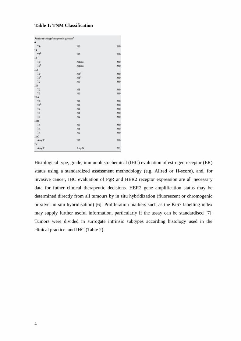
Table 1: TNM Classification
Histological type, grade, immunohistochemical (IHC) evaluation of estrogen receptor (ER)
status using a standardized assessment methodology (e.g. Allred or H-score), and, for
invasive cancer, IHC evaluation of PgR and HER2 receptor expression are all necessary
data for futher clinical therapeutic decisions. HER2 gene amplification status may be
determined directly from all tumours by in situ hybridization (fluorescent or chromogenic
or silver in situ hybridisation) [6]. Proliferation markers such as the Ki67 labelling index
may supply further useful information, particularly if the assay can be standardised [7].
Tumors were divided in surrogate intrinsic subtypes according histology used in the
clinical practice and IHC (Table 2).
Page 6
5
Tab2: surrogate definitions of intrinsic subtypes of breast cancer according to the
2013 St Gallen Consensus Conference and recommended by the ESMO Clinical
Practice Guidelines
1.1.3 Prognostic and predictive factors
The most important prognostic factors in early breast cancer are expression of ER/PgR,
HER2 and proliferation markers, number of involved regional lymph nodes, tumour
histology, size, grade and presence of peritumoural vascular invasion. Clinical parameters
(age, tumour stage, ER expression and histological grade) have been incorporated into
scoring systems that permit a relatively accurate estimation of the probability of recurrence
and death from breast cancer; [8]. Gene expression profiles such as MammaPrint®
(Agendia, Amsterdam, the Netherlands) or Oncotype DX® Recurrence Score (Genomic
Health, Redwood City, USA) may be used to gain additional prognostic and/or predictive
information to complement pathology assessment and to predict response to adjuvant
chemotherapy. This is particularly true in patients with ER-positive early breast cancer;
however, their true clinical utility is still being evaluated in large randomised clinical trials
such as MINDACT, TAILORx and RxPONDER.
Page 7
6
ER/PgR and HER2 are the only validated predictive factors, allowing for selection of
patients for endocrine therapies (ETs) and anti-HER2 treatments, respectively. High ER
expression is also usually associated with lesser absolute benefit of chemotherapy. After
neoadjuvant systemic treatment, the response to treatment and quantity of residual disease
are important prognostic factors but need as much standardisation as any of the other
biological markers, and no uniform guidelines exist for the evaluation of response to
neoadjuvant treatment, although some guidance is provided by the FDA recommendation
for accelerated drug approval in neoadjuvant treatment of breast cancer [9].
1.1.4 Antitumoral Treatment
Adjuvant systemic treatment
After surgery, the decision on systemic adjuvant treatment is based on predicted sensitivity
to particular treatment methods and their use and individual risk of relapse. According to
the 2011 and 2013 St Gallen guidelines, the decision on systemic adjuvant therapies should
be based on the surrogate intrinsic phenotype determined by ER/PgR, HER2 and Ki67
assessment with the selective help of first-generation genomic tests when available (such
as MammaPrint® or Oncotype DX®) for luminal cases with unclear chemotherapy
indications [10,11]. All luminal cancers should be treated with Endocrine Therapy. Most
luminal A tumours, except those with highest risk of relapse (extensive nodal
involvement), require no chemotherapy, whereas luminal B HER2-negative cancers
constitute a population of the highest uncertainty regarding chemotherapy indications.
Indications for chemotherapy within this subtype depend on the individual risk of relapse,
taking into account the tumour extent and features suggestive of its aggressiveness (grade,
proliferation, vascular invasion), presumed responsiveness to ET and patient preferences.
Features associated with lower endocrine responsiveness include low steroid receptor
expression, lack of PgR expression, high tumour grade and high expression of proliferation
markers.
Luminal B HER2(+)tumours are treated with chemotherapy, ET and trastuzumab; no
randomised data exist to support omission of chemotherapy in this group; however, in
cases of contraindications for chemotherapy or patient refusal, it is acceptable to offer the
combination of targeted agents (ET and trastuzumab). Triple-negative tumours benefit
from adjuvant chemotherapy, with the possible exception of low-risk ‘special histological
subtypes’ such as medullary or adenoidcystic carcinomas. HER2 (non-luminal) cancers,
apart from selected cases with very low risk, such as T1aN0, are treated with
chemotherapy plus trastuzumab.
Page 8
7
Trastuzumab may routinely be combined with non-anthracycline-based chemotherapy and
ET; concomitant use with anthracyclines is not routinely recommended outside of clinical
trials, although may be considered in selected patients treated in experienced centres. For
most patients, the use of a sequential anthracycline-based followed by taxane-trastuzumab-
based regimen is the preferred choice. RT may be delivered safely during trastuzumab, ET
and nonanthracycline- based chemotherapy. If chemotherapy and RT are to be used
separately, chemotherapy usually precedes RT.
Endocrine therapy
ET is indicated in all patients with detectable ER expression, defined as ≥1% of invasive
cancer cells, irrespective of chemotherapy and/or targeted therapy [12]. The choice of
medication is primarily determined by patient’s menopausal status. Other factors include
(minor) differences in efficacy and side effect profile. Permenopausal patients are treated
with Tamoxifen
Premenopausal patients. Tamoxifen 20 mg/day for 5–10 years. In patients becoming
postmenopausal during the first 5 years of tamoxifen, a switch to letrozole, an aromatase
inhibitor (AI), seems to be particularly beneficial [13]. The value of addition of ovarian
suppression [by gonadotropin-releasing hormone (GnRH) agonists or ovarian ablation] is
not well-defined, in particular in chemotherapytreated patients, who frequently develop
ovarian failure as a consequence of cytotoxic treatment [14] failure is not well-established
and contradictory data exist.
Postmenopausal patients. Aromatase Inhibitors (AIs) (both non-steroidal and steroidal) and
tamoxifen are valid options. AIs allow for prolongation of the DFS, with no significant
impact on OS (1%–2%, depending if upfront or sequential strategy) [15-16].
The use of tamoxifen is associated with increased risk of thromboembolic complications
and endometrial hyperplasia(including endometrial cancer). Caution should be exercised in
patients with conditions predisposing to these sequelae and appropriate diagnostic tests
carried out in those presenting with symptoms suggestive of these complications. Although
there are no unequivocal data on their detrimental effects, patients on tamoxifen should be
advised to avoid the use of strong and moderate CYP2D6 inhibitors or, if such drugs
cannot be replaced, a switch to alternative treatment, i.e. AIs, should be considered [17].
Patients undergoing ovarian suppression and AI users are at increased risk of bone loss and
should be advised to assure adequate calcium plus vitamin D3 supply and to assess
periodically the bone mineral density [by dual energy X-ray absorption (DEXA) scan].
Chemotherapy
Page 9
8
Chemotherapy is recommended in the vast majority of triple negative, HER2-positive
breast cancers and in high-risk luminal HER2-negative tumours. The benefit from
chemotherapy is more pronounced in ER-negative tumours [18]. In ERpositive tumours,
chemotherapy at least partially exerts its effect by induction of ovarian failure [19]. Most
frequently used regimens contain anthracyclines and/or taxanes, although in selected
patients CMF may still be used. Four cycles of AC (doxorubicin, cyclophosphamide) are
considered equal to six cycles of CMF, whereas six cycles of three-drug
anthracyclinebased regimens are superior[20].
The addition of taxanes improves the efficacy of chemotherapy, independently of age,
nodal status, tumour size or grade, steroid receptor expression or tamoxifen use, but at the
cost of increased non-cardiotoxicity [20]. Sequential rather than the concomitant use of
anthracyclines and taxanes is superior. Overall, chemotherapy regimens based on
anthracyclines and taxanes reduce breast cancer mortality byabout one-third [20]. Non-
anthracycline, taxane-based regimens (such as four cycles of TC) may in selected patients
(such as those at risk of cardiac complications) be used as an alternative to four cycles of
anthracycline-based chemotherapy [21]. Chemotherapy is usually administered for 12–24
weeks (four to eight cycles), depending on the individual recurrence risk and the selected
regimen.
HER2-directed therapy
Trastuzumab combined with chemotherapy in patients with HER2
overexpression/amplification approximately halves the recurrence risk, compared with
chemotherapy alone; this translates into ∼0% absolute improvement in 3-year DFS and 3%
increase in 3-year OS [22]. Trastuzumab is approved in patients with node-positive disease
and in N0 patients with tumours >1 cm, although—due to relatively high failure risk even
in patients with N0 tumours <1 cm—it should also be considered in this patient group, in
particular in ERnegative disease [23]. In most studies, trastuzumab was administered for 1
year, although in the FinHER trial a similar improvement was obtained with only 9 weeks
of treatment. Due to its cardiotoxicity, trastuzumab should not be routinely administered
concomitantly with anthracyclines. Combination with taxanes is safe and has been
demonstrated to be more effective than sequential treatment [24]. Trastuzumab may also
be safely combined with RT and ET.
Bisphosphonates
Some data suggest a beneficial anticancer effect of bisphosphonates, especially when used
in a low-estrogen environment (women undergoing ovarian suppression or
postmenopausal), although study results are equivocal and such a treatment cannot be
Page 10
9
routinely recommended in women with normal bone mineral density. In patients with
treatment-related bone loss, bisphosphonates decrease the risk of skeletal complications
[25,26].
1.1.5 Follow up
The aims of follow-up are to detect early local recurrences or contralateral breast cancer, to
evaluate and treat therapy-related complications (such as menopausal symptoms,
osteoporosis and secondary cancers). Ten-year survival of breast cancer exceeds 70% in
most European regions, with 89% survival for local and 62% for regional disease [27].
patients with node-positive disease tend to have higher annual hazards of recurrence than
patients with node-negative cancers. In the first years the risk of recurrence is higher in
patients with ER-negative cancers, but after 5–8 years after diagnosis, the annual hazards
of recurrence drop below the level of ER-positive tumours. Relapses of breast cancer may
occur as late as >20 years after the initial diagnosis, particularly in patients with ER/PgR-
positive disease .Guidelines recommend regular visits every 3 to 4 months in the first 2
years, every 6 months from years 3–5and annually thereafter. Ipsilateral (after BCS) and
contralateral mammography is recommended every 1 to 2 years. An MRI of the breast may
be indicated for young patients, especially in the case of dense breast tissue and genetic or
familial predispositions. In asymptomatic patients, there are no data to indicate that other
laboratory or imaging tests (e. g. blood counts, routine chemistry tests, chest X-rays, bone
scans, liver ultrasound exams, CT scans or any tumour markers such as CA15-3 or CEA)
produce a survival benefit .However, routine blood tests are usually indicated to follow-up
patients on ET due to the potential side-effects of these drugs namely in the lipid profile .
1.2 Bone metastases
1.2.1 Phisiopathology of bone metastases
Cancer patients mainly do not die for the primary tumor, but rather for the formation of
metastases.
Many of the most common cancers such as breast, prostate and lung commonly
metastasize to the bone, indeed more than 50% of patients with prostate cancer or
advanced breast show bone metastases.
Radiographically 80% of bone metastases derived from this tumor are osteolytic, 20% are
osteoblatic at the time of diagnosis. The 5-year survival of patients with lesions
Page 11
10
exclusively bone is 37% while in the presence of extraskeletal metastases that survival is
reduced to 13%. Osteoblatic metastases are associated with a better prognosis. Bone
metastases are usually accompanied by a significant bone pain, pathological fractures,
nerve compression syndromes and hypercalcemia: these complications are called Skeletal
related events (SRE) .
The bone is an ideal microenvironment for the development of metastases following
hypothesis "seed and soil " proposed by Stephen Paget in 1889 [28]: a metastasis settles in
a particular organ if the cells of the primary tumor (seed) are in the favorable site (soil)
conditions in terms of chemokines, growth factors and development, sufficient for their
arrest and their growth in that site; furthermore, according to this hypothesis, bone
microenvironment has many factors and properties that allow cancer cells an important
development.
Bone is a supportive connettive tissue consisting of cells spread in an abundant
extracellular matrix, consisting of fibers and amorphous substance of glycoproteic origin;
this is calcified and also formed from minerals. Furthermore bone is a dynamic tissue
which has a structural support, protective, mechanical and trophic functions as it serves as
a repository of minerals, particularly calcium ions that play an important role in various
cellular activities. It is composed of various cell types: in addition to stromal cells,
hematopoietic and endothelial cells, osteoclasts and osteoblasts are involved in the
development and regulation of bone remodeling. Osteoclasts are derived from progenitor
cells of the monocyte-macrophage line and are responsible for bone resorption. These cells
adhere to bone matrix via integrin surface and, once activated, they degrade it [29,30].
They resorb bone creating an acidic and isolated microenvironment between the plasma
membrane and the bone surface that determines the solubilization of minerals. The free
organic matrix is subjected to enzymatic degradation by lysosomal proteases released by
osteoclasts (as cathepsin K). The products of the degradation of the organic matrix are
endocited and esocited from the opposite side of the cell.
MCSF and RANKL are two essential growth factors for osteoclastogenesis . While MCSF
is essential in the early stages of osteoclastogenesis, RANKL is critically involved in the
maturation and activation of osteoclasts. MCSF is produced by stromal cells and
osteoblasts and binds to its receptor c-fms expressed on the surface of the macrophage
precursors and stimulates proliferation [31-33]. RANKL is expressed by osteoblasts and
stromal cells and interacts with the receptor RANK localized on the membrane of the
Page 12
11
monocyte - macrophage precursors and induces differentiation into osteoclasts and their
activation [34-38].
Different cytokines produced locally as well as systemic calciotropic hormones, including
parathyroid hormone (PTH), the 1,25 dihydroxyvitamin D3 and prostaglandins, indirectly
stimulates osteoclastogenesis by increasing the expression of RANKL on bone marrow
stromal cells and osteoblasts . In addition, other cytokines such as IL -1 and TNF- α are
able to act directly on osteoclasts [39-40].Osteoblasts are cells of mesenchymal derivation
delegated to the synthesis and mineralization of bone matrix. For osteoblast differentiation,
mesenchymal stem cell (MSC) first undergoes proliferation, it becomes the commitment
and therefore differentiate in pre-osteoblast (which produces alkaline phosphatase) and
later in a mature osteoblast which produces an increasing amount of osteocalcin and
calcified matrix. Runx2 and Osteorix are two transcription factors that determine the
expression of many genes associated with osteoblast differentiation. The commitment of
MSCs into osteoblast line is controlled by three morphogenetic pathway: the BMP, HH and
Wnt pathway [41-44]. Once formed the matrix , numerous osteoblasts become trapped in
bone lacunae and thus they become osteocytes. They are not a inert cell for bone
metabolism; osteocytes, indeed, could participate in the exchange of minerals from the
bone, then intervening in the homeostatic regulation of the concentration of calcium in the
body and, working as mechano sensors, can modulate the bone resorption in response to
different stimuli [45,46]. Bone matrix is constituted by the organic matrix reinforced by the
deposition of calcium salts. The type I collagen constitutes about 90-95 % of the organic
matrix while non- collagenous proteins constitute the remaining 5-10 %. The crystalline
salts deposited in the matrix are primarily calcium and phosphate in the form of
hydroxyapatite. The proteins can be divided into non- collagenous proteins of cell
adhesion, proteoglycans, γ - carboxylated and growth factors .Each of the adhesion
proteins as osteopontin, bone sialoprotein (IBSP), vitronectin and type I collagen facilitate
interactions with integrins that are expressed by hematopoietic stem cells and specialized
cells of the bone , as well as osteotropic tumor cells .
As a consequence of bone remodeling, growth factors stored in the bone such as IGF, FGF,
PDGF , TGF-β and BMP, are released into the medullary cavity and act on metastatic
cancer cell growth [47-49].
Page 13
12
1.2.2 Metastasis process
It has long been recognized that primary cancers spread to distant organs with
characteristic features [50], and the skeleton is one of the most common organs to be
affected by metastatic cancer. Breast and prostate cancer are osteotropic tumors, i.e.,
carcinomas that have a special predilection to form bone metastases. At postmortem
examination, about 70% of patients with these tumros had metastatic bone disease.
Together, breast and prostate cancer probably account for more than 80% of cases of
metastatic bone disease [51, 52].
At time of diagnosis, most patients with breast and prostate [53] cancer do not have
clinicopathologic signs of overt distant metastases. Thus, after resection of the primary
tumor and all positive lymph nodes, these patients are in complete clinical remission.
However, disseminated tumor cells (DTCs) may already be present in bone marrow (BM)
[54-55], a clinical situation called minimal residual disease (MRD). Most DTCs have a
limited life span and disappear in time, indicated by the clinical findings that a significant
fraction of breast cancer patients with DTCs in BM never develop distant metastases [54].
However, DTCs with an indefinite proliferative potential that have acquired the abilities of
metastasizing to, surviving in, and colonizing the bone/BM, can eventually result in the
development of an overt bone metastasis. Only this subpopulation of DTCs can, therefore,
be regarded as true metastasis-initiating cells (MICs). The clinical courses of patients with
breast and prostate cancer with a first recurrence in bone are relatively long, with a median
survival of 24 and 40 months. This is in marked contrast to those with first recurrence of
breast cancer in the liver (3 months). However, involvement of the skeleton in metastatic
disease is a major cause of morbidity, characterized by severe pain and high incidence of
SREs.
1.2.3 local invasiveness and EMT
The first phase in metastasization process is the acquisition of motility and invasiveness;
capabilities that are not compatible with normal tissue. Cancer cells must therefore shed
many of their epithelial characteristics, detach from epithelial sheets, and undergo a drastic
alteration, a process called the “epithelial-mesenchymal transition” (EMT). Achievement
of this invasive phenotype is reminiscent of events of early embryonic development. The
importance of this process during embriogenesis is highlighted by the fact that a
disfunction in EMT process determines the developmental arrest at the stage of blastula
blastula [56].
Page 14
13
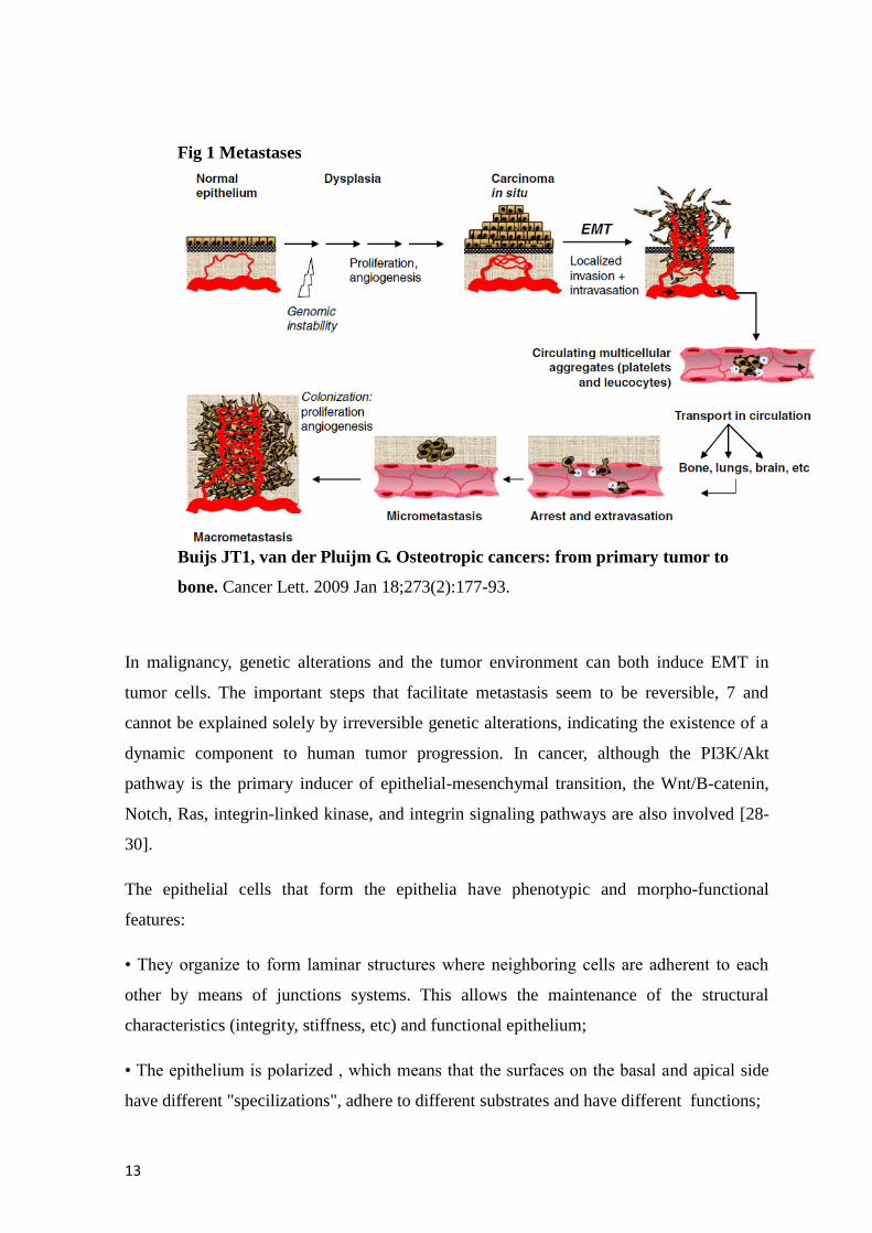
Fig 1 Metastases
Buijs JT1, van der Pluijm G. Osteotropic cancers: from primary tumor to
bone. Cancer Lett. 2009 Jan 18;273(2):177-93.
In malignancy, genetic alterations and the tumor environment can both induce EMT in
tumor cells. The important steps that facilitate metastasis seem to be reversible, 7 and
cannot be explained solely by irreversible genetic alterations, indicating the existence of a
dynamic component to human tumor progression. In cancer, although the PI3K/Akt
pathway is the primary inducer of epithelial-mesenchymal transition, the Wnt/B-catenin,
Notch, Ras, integrin-linked kinase, and integrin signaling pathways are also involved [28-
30].
The epithelial cells that form the epithelia have phenotypic and morpho-functional
features:
• They organize to form laminar structures where neighboring cells are adherent to each
other by means of junctions systems. This allows the maintenance of the structural
characteristics (integrity, stiffness, etc) and functional epithelium;
• The epithelium is polarized , which means that the surfaces on the basal and apical side
have different "specilizations", adhere to different substrates and have different functions;
Page 15
14
• These cells are poorly mobile, movements are limited only within the epithelium.
Mesenchymal cells instead form structures of different shape and density, unorganized and
among them there are only points of focal adhesion and junctional devices as stable
between the epithelial cells. Mesenchymal cells are also equipped with high mobility that
allows migration or as single cells or as chains of cells. When the epithelial-mesenchymal
transition is completed, the epithelial cells has lost some epithelial markers that are
replaced by mesenchymal markers.The reduction of cadherins expression (proteins
involved in cell-cell adhesion), in particular of E-cadherin, seems to be the key event that
allows the realization of the entire process. The formation and stabilization of the clusters
of E-cadherin, at the level of the junctions of adhesion between cells, require the chains, in
particular the β-catenin, which binds to the cytoplasmic portion of E-cadherin. Furthermore
actin filaments (F-actin) stabilize and immobilize E-cadherin clusters at the level of the
adherent junctions [57-59]. When E-cadherin levels decrease till becoming limiting, there
is a loss of intercellular junctions and of the sequestration of E-cadherin β-catenin-
mediated. This means that the β-catenin accumulates and traslocates in the nucleus where,
by binding to LEF/TCF transcription factors, activates target genes EMT related as
vimentin, and the regulators Twist and Snail [60-62].A part from cadherin, some other
proteins involved in tight junction formation are down regulated as ZO-1 (a protein of the
zonula occludens), that interact with different trans membrane proteins as ocludina and
claudin [63].The reduction of cadherins expression is related to cellular migration increase
and with formation of metastases. In presence of N cadherin, for example, FGF-2 causes
the activation of the microtuble-associated protein kinase-extracellular signal-regulated
kinase (MAPK-ERK) and this pathway, inducing transcription of matrix metalloproteinase
9 (MMP-9), increases dramatically the invasiveness of breast cancer cell. Matrix
metalloproteinases are important EMT markers; they are members of the family of neutral
endopeptidases Zn dependent that selectively degrade the extracellular matrix. They are
expressed in several tumors and they are involved in different phases of metastases
development: expansion and escape of single cancer cell from primary tumor, their passage
through the blood vessels, survival of into the circulation, and exit of tumor cells from the
blood vessels at sites secondary [64]. MMPs are able to degrade the growth factors in an
active form and cleave proteins bound and exposed on the surface of the cell as the E-
caderina. Fibronectin and vimentin are other two important mesenchymal markers and are
respectively, a cytoskeleltal proein, and a protein released in the matrix.
Page 16
15
The consequent acquisition of a mesenchymal invasive phenotype by cancer cells causes
the break of the basal lamina and the invasion of the underlying stromal compartments.
The acquisition of an invasive mesenchymal phenotype do not depend only to somatic
mutations and other epigenetic alterations in the cacner cells, but some changen in stromal
environment are also necessary for the neoplastic progression [65-66].
In the tumor, indeed, both genetic alterations and tumoral microenvironment can induce
EMT in cancer cells [67]. The cancer cells are able to activate the local stromal cells, such
as fibroblasts , smooth muscle cells and adipocytes and recruit progenitors of endothelial,
mesenchymal and inflammatory cells. The activation of stromal cells leads to the secretion
of growth factors and proteases that promote further proliferation and cancer cell invasion
[68]. The EMT also enhances angiogenesis. The production of pro-angiogenic factors ,
including VEGF -A and MMPs , is induced mainly by Snail [69].
Among the factors that cause the most epithelial-mesenchymal transition in cancer are the
TGF- β [70-72], FGF , EGF , HGF and IGF. The EMT is also activated by some
extracellular signals arising from the interaction of cancer cells with extracellular matrix
components such as collagen and hyaluronic acid.
This leads to the activation, at a intracellular level, of different effector proteins such as
Ras , Rho, MAPK , Rac and Src that cause a change in the organization of the cytoskeleton
and disassembly of different junctional complexes .
Two of the main targets of Ras and MAPK are Slug and Snail, two transcription factors
that inhibit the expression of genes that have an E -box in the promoter region such as the
E-cadherin and the proteins that constitute the occluding junctions (occludine and
claudine). Recently it has been discovered that Elf5, a key regulator of cell fate in the
development of alveolar gland mammaria, 64 directly represses the transcription of Snail2 ,
key transcription factor nell'EMT [73-74].In carcinogenesis, TGF-β plays a key role but
with different effects; in the early stages it inhibits cell proliferation but subsequently
promotes the formation of metastasis inducing EMT [74]. The signaling of the TGF-β is
one of those best characterized. It is based on SMAD-dependent mechanisms where
SMAD2 and SMAD3 , once phosphorylated and activated , bind to SMAD4 and are
translocated into the nucleus where they activate the co-repressor SIP -1, cha acts as Snail
and Slug , inhibiting the expression of genes that contain E-box sequences at the level of
the promotor. Furthermore this mechanism induces the autocrine production of TGF- β ,
which further increases the EMT process [75-79]. After epithelial-mesenchymal transition,
Page 17
16
cancer cells must go through a multistep process to metastasize to bone, which involves
dislodgement from a primary site, survival in the circulation, binding to the resident cells
in bone, and survival and proliferation in the bone and bone marrow. The dissemination of
cancer cells may take place early in disease progression with tumor cells preferentially
engaged in the bone marrow, and a subset of cells surviving and evolving into clinically
apparent disease. These cells then enter a period of dormancy in which they either stop
proliferating, or proliferate at a reduced rate before showing evidence of metastasis; a
process that can sometimes exceed 10 years. However, in some situations, there is at least
1 further and crucial event that takes place, the trigger that reactivates tumor cell
dormancy. However, the mechanisms that facilitate this process remain not completely
known. Cancer cells have preferential site where grow and finally form a metastases. This
concept of selective homing of cancer cells in a specific organ happens mainly according
to 3 mechanisms:
Selective growth: cells leave the primary tumor in a ubiquitous manner but they can
grow selectively only in specific organs with the necessary growth factors and
miocroenvironment.
Selective adhesione: Cancer cells can attach only to the surface of endothelial cells
of specific organs
Chemotaxis: cancer cells reach the specific organ by chemoattraction due to the
realse of soluble growth factors secreted by the organ where metastasis will be
formed.
1.2.4 Blood and lymphatic dissemination
After leaving of cells from the primary tumor site, they are released into the blood and
lymphatic circulation, and from there they spread throughout the body.
Despite this, the event that leads to the development of metastases is very rare and many
cancer cells are not able to cross the capillary bed of the pulmonary circulation. In the
blood circulation, tumor cells can interact with platelets and leukocytes with the formation
of aggregates which increase the resistance of cancer cells and inhibits the immuno-
mediated clearance. This process facilitate the cancer cells stop in the capillaries of the
various organs and promotes the extravasation. Once the tumor cells have left circulation,
the activated platelets are a source of factors that are able to induce angiogenesis,
stimulating tumor proliferation, and indirectly increase osteoclast activity in the bone
environment [80-82].Angiogenesis is not only important in the development of metastasis
Page 18
17
and invasiveness of the tumor but also in the early pre-invasive stages where nutrients and
oxygen are supplied to the tumor through the neo formed vessels. These vessels are also a
way for cancer cells to spread in the body. The angiogenic inducing factors are VEGF,
FGF1 and FGF2. Interestingly, the PGF (placental growth factor) has been implicated in
the induction of angiogenesis in the disease state but not in the normal conditions.Two
Other factors, VEGF-C and VEGF-D are the major inducers of lymphangiogenesis and are
overexpressed in colon and breast tumors [83-85].
A vertebral venous system with thin walls and lack of valves that can communicate freely
exists , in which a part of the blood origining from pelvis and from thoracic site is released.
This system would explain the predilection of prostate and breast cancer to metastasize to
the level of the axial portion of the skeleton; the tumor cells from the thorax and the pelvis,
indeed, avoid the polmonar circulation and they can spread freely.
1.2.5 Diffusion and colonization of secondary tissues
The arrival of cells in a secondary organ is therefore not a random process. The first
contact between the "seed " and "soil " consists in the interaction between circulating
tumor cells in the blood and lymph vessels and the endothelium of a specific organ. In
particular, with regard to bone metastases tumor cells must reach, colonize and grow in the
bone marrow. The combination of specific chemoattrattive and adhesion molecules in the
bone marrow endothelium promote migration and retention of circulating tumor cells [86].
This phenomenon also depends on the presence of receptors for cytokines and growth
factors, localized on the surface of cancer cells .
CCR-7 and CXCR4 are the most important receptors and they are the expressed
predominantly by prostate and breast cancer cells , which interact with the Chemokines
like monocyte chemoattractant protein 1 (MCP -1) and stromal cell- derived factor 1 (SDF-
1), that are chemoattrattive cytokines and chemokines expressed constitutively by
endothelial cells , osteoblasts and other stromal cells of the bone marrow. The SDF-
1/CXCR4 axis plays a key role in the development of metastases; in normal tissue levels of
CXCR4 are low, meanwhile in breast cancer they are higher. SDF-1 in the bone marrow
is abundantly produced by osteoblasts in particular during the process of bone remodeling
and its production is increased by factors such as PTH , PDGF , IL-1, VEGF and TNF- α .
SDF-1 also recruits osteoclast precursors by inducing chemotaxis , the activity of MMP -9
and the transmigration of collagen. The activation of the SDF-1/CXCR4 pathway not only
Page 19
18
regulates the homing and migration of tumor cells in the bone but also the adhesion,
invasion, and the rearrangement of of cancer cells cytoskeleton [87-90].
The osteopontin, bone sialoprotein and the type I collagen are the predominant components
of mineralized bone: these proteins mediate the local adhesion , motility , survival and
growth by interacting with integrins and adhesion molecules expressed by different types
of cells. The integrin αvβ3 is the receptor for vitronectin ( another molecule of the
extracellular matrix ) and is an essential component for the adhesion of osteoclasts to bone.
This integrin is expressed at high levels on the surface of cells of breast carcinoma and
seems to cooperate with the bone sialoprotein and the MMP- 2 and -9 in the invasion of
bone. The αvβ1 integrin mediate the binding to the vascular cell adhesion molecule 1
(VCAM-1) or to fibronectin promoting a regulation of the expression of cytokines and
growth factors in the stromal cells of the bone marrow increasing tumor growth and
resistance to chemotherapy. CD44 is an adhesion molecule that does not belong to the
integrin family; it is a receptor of glycosaminoglycans ialuronated and osteopontin . It is
expressed by various cancer cells and has a well-defined role in skeletal metastasis. A
portion of the cells that are spread in the bone marrow stroma may reactivate certain
epithelial properties through a mesenchimal-epithelial transition (MET) and xpressing
some epithelial markers . This indicates that the malignant progression is based on the
dynamic processes that can not be explained only by the onset of irreversible genetic
alterations but rather by temporal transitional states that are affected strongly by the
tumoral microenvironment [91-96]. Recent studies have shown that ADAMTS1 and
MMP1, two metalloproteases, synergistically promote the invasion of breast cancer: the
two metalloproteases cut the ligands of EGF (AREG, TGF-α and HB-EGF) from the
surface of tumor cells and, consequently, the expression of OPG by osteoblasts thus is
reduced. ADAMTS1 and MMP1 also increase the production of RANKL. In addition to
the expression of molecules involved in homing to bone , the tumor cells of breast and
prostate acquire the ability to express bone matrix proteins such as osteonectin ,
osteopontin and bone sialoprotein. The acquisition of the typical properties of bone cells by
tumor cells is a process that is termed "osteomimicry " [97-100] and improves the homing,
adhesion, proliferation and survival in the bone microenvironment. The classical
hypothesis according to which the tumor cells begin to interact with the microenvironment
in which the metastasis develop only when they reach the microenvironment itself appears
to be too semplicistic [101] after the discovery of the "premetastatic niche " [102] . It has
been shown indeed, that the hematopoietic stem cells (HSC) that reside in the bone marrow
at the level of two niches, the osteoblastic and the vascular niceh [103-105], are mobilized
Page 20
19
by factors secreted by the primary tumor. HSCs begin to produce growth factors (eg.
VEGF), chemokines and other molecules that prepare the different metastatic sites before
the arrival of the tumor cells .There are many factors that are primarily derived from the
endocrine system that may affect the functions of osteoclasts and osteoblasts both directly
and indirectly leading to the formation of bone metastases .
1.2.6 Types of bone metastases
Bone metastases can be classified in two different types: osteolytic and osteoblastic lesions
1.2.6.1 Osteolytic metastases
The osteolytic metastasis occurs mainly in patients with solid tumors such as breast,
prostate, lung , kidney and thyroid [106] .
In breast cancer the dominant lesion is lytic and destructive although there is also a local
bone formation that probably represents an attempt to repair the bone loss [107]. This
increase in bone formation in patients with osteolytic bone metastases is reflected in an
increase in serum levels of the enzyme alkaline phosphatase (an enzyme localized at the
surface of osteoblasts involved in bone mineralization ), used as a marker for determination
of osteoblastic activities 8.Both serum alkaline and idrossiprolin in urine are cheap and non
invasive markers of, respectively, bone formation and bone resorption. Recently other
more specific and sensitive markers identified to assess response in bone have been
identified: they include bone-specific alkaline phosphatase . Regarding bone resorption the
evaluation of products of collagen degradation as CTX and NTX were quite used [108] .
These markers may be useful for planning and evaluating the use of a preventive treatment
with inhibitors of bone resorption.
Many in vivo studies have shown that osteolysis is associated with increased osteoclast
activity and a reduction in the activity of osteoblasts with a direct effect of cancer cells on
bone tissue [109].
Osteolytic metastasis occurs following a complex interaction between tumor cells and the
bone microenvironment that gives rise to a "vicious cycle" [110] . Bone homeostasis is
regulated by direct interaction between osteoblasts and osteoclasts, in particular, by the
axis of the RANK/ RANKL/ OPG. RANKL, expressed on the surface of osteoblasts and
bone marrow stromal cells, induces the recruitment, activation, and osteoclasts
differentiation by binding to its receptor RANK localized on the surface of the osteoclasts
precursors [110] .
Page 21
20
The process is controlled by the production, by osteoblasts and other cell types in the bone
microenvironment , of Osteoproteogerin(OPG). OPG is a " decoy receptor " able to bind
RANKL limiting its biological activity and thus inhibiting the osteoclasts differentiation ,
mainly by blocking the stages of fusion and differentiation of osteoclasts and their bone
resorption activity [111]. Once activated, the osteoclasts begin the process of bone
resorption by the secretion of proteases and the formation of an acidic environment
between the plasma membrane and the bone surface. The tumor cells that reach the bone
microenvironment secrete factors that influence the process of bone resorption. The
peptide PTHrP (tumor -produced parathyroid hormone-related protein) is the most
important mediator in the activation of osteoclasts in metastatic breast cancer [112]. It has
a 70 % homology with the first 13 amino acids of the thyroid hormone (PTH), it binds to
the same PTH receptor 114 showing a similar biological activity [113-114]. 50-60% of
breast tumors primitive produce PTHrP but its expression appears to be higher in the bone
microenvironment (90% of bone metastases from breast cancer expresses PTHrP) with
respect to the site of the primary tumor and metastases to other sites (only 17% of bone
metastases in different anatomical sites expresses PTHrP) [113-116]. PTHrP stimulates
RANKL production by osteoblasts and inhibits the OPG production increasing
osteoclastogenesis. The signal activated in osteoclast precursors following the binding of
RANKL to RANK leads to increased expression of some transcription factors such as AP1
(activated by JUN N-terminal kinase) and NF -kB (activated by the inhibitor of kB kinase
IKK) leading to the maturation of the osteoclasts progenitors of [117-118]. The newly
formed osteoclasts, then, begin the process of bone resorption.
The induced osteolysis by osteoclasts is related to the release by the matrix of bone growth
factors such as TGF- β and IGF-1 and to an increase in the concentration of extracellular
calcium. These growth factors , and in particular TGF- β, bind to their receptors on the
surface of tumor cells and induce mechanisms of signal transduction mediated by Smad
proteins and Mapk [119-120]. This leads to an increase in the proliferation of cancer cells
and to an increase of the production of PTHrP which in turn increases the production of
RANKL by osteoblasts closing this vicious cycle [110].
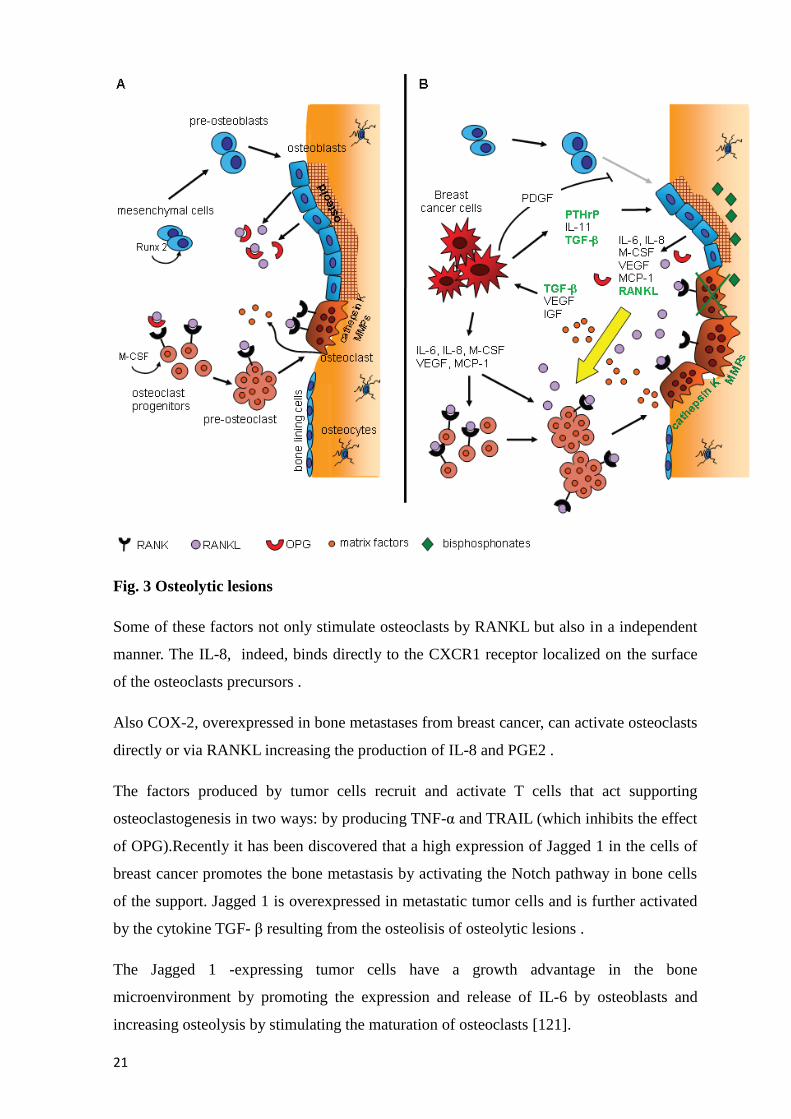
Apart PTHrP, the expression of RANKL on osteoblasts and stromal cells is increased by
other factors produced by tumor cells , such as IL - 1, IL- 6, IL -8 , IL- 11 and PGE2
(Fig.3).
Page 22
21
Fig. 3 Osteolytic lesions
Some of these factors not only stimulate osteoclasts by RANKL but also in a independent
manner. The IL-8, indeed, binds directly to the CXCR1 receptor localized on the surface
of the osteoclasts precursors .
Also COX-2, overexpressed in bone metastases from breast cancer, can activate osteoclasts
directly or via RANKL increasing the production of IL-8 and PGE2 .
The factors produced by tumor cells recruit and activate T cells that act supporting
osteoclastogenesis in two ways: by producing TNF-α and TRAIL (which inhibits the effect
of OPG).Recently it has been discovered that a high expression of Jagged 1 in the cells of
breast cancer promotes the bone metastasis by activating the Notch pathway in bone cells
of the support. Jagged 1 is overexpressed in metastatic tumor cells and is further activated
by the cytokine TGF- β resulting from the osteolisis of osteolytic lesions .
The Jagged 1 -expressing tumor cells have a growth advantage in the bone
microenvironment by promoting the expression and release of IL-6 by osteoblasts and
increasing osteolysis by stimulating the maturation of osteoclasts [121].
Page 23
22
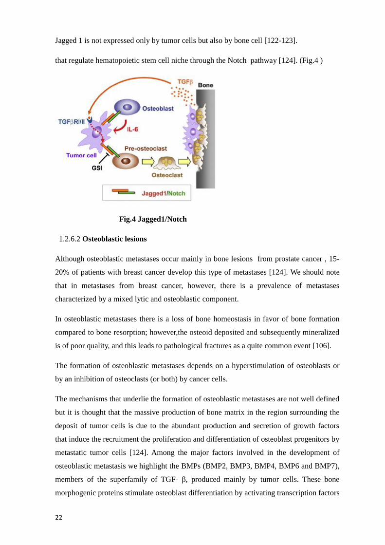
Jagged 1 is not expressed only by tumor cells but also by bone cell [122-123].
that regulate hematopoietic stem cell niche through the Notch pathway [124]. (Fig.4 )
Fig.4 Jagged1/Notch
1.2.6.2 Osteoblastic lesions
Although osteoblastic metastases occur mainly in bone lesions from prostate cancer , 15-
20% of patients with breast cancer develop this type of metastases [124]. We should note
that in metastases from breast cancer, however, there is a prevalence of metastases
characterized by a mixed lytic and osteoblastic component.
In osteoblastic metastases there is a loss of bone homeostasis in favor of bone formation
compared to bone resorption; however,the osteoid deposited and subsequently mineralized
is of poor quality, and this leads to pathological fractures as a quite common event [106].
The formation of osteoblastic metastases depends on a hyperstimulation of osteoblasts or
by an inhibition of osteoclasts (or both) by cancer cells.
The mechanisms that underlie the formation of osteoblastic metastases are not well defined
but it is thought that the massive production of bone matrix in the region surrounding the
deposit of tumor cells is due to the abundant production and secretion of growth factors
that induce the recruitment the proliferation and differentiation of osteoblast progenitors by
metastatic tumor cells [124]. Among the major factors involved in the development of
osteoblastic metastasis we highlight the BMPs (BMP2, BMP3, BMP4, BMP6 and BMP7),
members of the superfamily of TGF- β, produced mainly by tumor cells. These bone
morphogenic proteins stimulate osteoblast differentiation by activating transcription factors
Page 24
23
such as Runx-2 [126-128]. and also indirectly induce angiogenesis. The pattern of
expression of BMPs play an important role in the etiology of osteoblastic metastases
arising mainly from prostate cancer. It was seen that the primary tumor and the metastases
have different patterns of expression of BMPs ; BMP6 is expressed at high levels in both
the primary tumor and in metastases, whereas BMP7 is expressed at high levels only at the
level of bone metastasis. The endothelin-1(ET-1) is another very important factor; it is a
vasoactive peptide of 21 amino acids produced by cancer cells [129-130]. The
pathophysiological role of ET- 1 in the development of metastasis has been demonstrated
in preclinical models for breast and prostate [131-132]. ET-1 binds to its receptor,
Endothelin A receptor (ETA), that is expressed by tumor cells and also by bone cells
(osteoblasts and osteoclasts), suggesting that the activity is paracrine and autocrine [133].
ET-1 increases the activity of cancer cells and enhances the mitogenic effect of other
growth factors such as IGF-1, PDGF and EGF [134]; it also leads to bone formation by
stimulating osteoblasts and inhibiting the resorption mediated by osteoclasts .Also the
Platelet-derived growth factor ( PDGF) and fibroblast growth factors (FGFs ) produced by
many types of tumor cells are implicated in the formation of osteoblastic metastases.
PDGF is a dimeric polypeptide that has 3 isoforms AA, BB and AB . The BB isoform
osteotropic is a powerful factor that contributes to the development of osteoblastic
metastases by promoting the migration and proliferation of osteoblasti [106]. The FGFs ,
both the acid ( FGF1 ) that the basic ( FGF2 ) form stimulate the formation of new bone in
vivo. Both increased osteoblast proliferation while only FGF2 suppresses the formation of
osteoclasts .
VEGF is also involved in bone growth directly by stimulating the differentiation and
activation of osteoblasts, and indirectly promoting angiogenesis.
Some Serine proteases such as protease urockinase (uPA) and the prostate-specific antigen
(PSA), appear to be involved in metastasis formation osteoblastic [135]. uPA , produced by
tumor cells , is synthesized as a precursor (pro-uPA) but subsequently undergoes a
proteolytic cleavage that leads to its activation. The carboxy- terminal proteolytic domain
uPA (ATF) contains 2 domains: a growth factor domain (GDF) , so called because it is
structurally similar to EGF and a Kringle domain. This domain is essential for the
activation and proliferation of osteoblasts . Moreover , uPA cleaves and activates TGF- β ,
which regulates the differentiation of osteoclasts and osteoblasts and promotes the growth
of cancer cells stesse ; hydrolyzes and proteins that bind IGF increasing the concentration
of IGF libero .PSA is a serine protease of the kallikrein family , marker known to be used
Page 25
24
for the assessment of the progression of prostate cancer. PSA cleaves the protein IGFBP -3
that binds IGF-1, IGF-1 making it available to the binding with its receptor and stimulate
the osteoblastic proliferation [136]. PSA can also hydrolyze PTHrP reducing bone
resorption by osteoclasts in order to make the predominant response osteoblastica .
As seen at the beginning of the paragraph, the bone microenvironment more ofter develop
metastases that have mixed characteristics between those osteolytic and osteoblastic.
1.2.7 Complications of Bone Metastases:
About 25% of patients with bone metastases are asymptomatic and the diagnosis is only
made when tests are carried out for other reasons or during primary tumour staging. In the
remaining 75%, bone metastases are responsible for different clinical complications
defined as skeletal-related events (SREs) such as pathologic fractures, spinal cord
compression, hypercalcaemia, bone marrow infiltration and severe bone pain requiring
palliative radiotherapy [137]. Such complications are often devastating for patients and
substantially reduce their functional independence and quality of life, decrease survival
rates and increase healthcare costs [138].
A study evaluated the pattern of metastatic disease in 180 triple-Breast cancer patients
who were compared with other subgroups. The risk of developing bone metastases within
10 years of the diagnosis was 7%-9% for all subgroups[139]. Some clinical trials have
evaluated the bisphosphonates efficacy in decreasing SREs in patients with breast cancer
and bone metastases [140-141]. The median time to the first SRE was 13.9 months among
bisphosphonate-treated women and 7.0 months in the placebo group (P = 0.001) [141]. The
SREs that occurred in the control group were radiation to bone, pathologic fracture,
hypercalcaemia, surgery on bone and spinal cord compression [141].
1.2.8 Bone targeted therapy
While bone metastases contribute significantly to the morbidity associated with breast
cancer, they are rarely the cause of disease related deaths. However, as aready reported,
serious complications are associated with them, including chronic bone pain,
hypercalcemia, SREs, which can lead to a dramatic decrease in the quality of life for breast
cancer patients [142]. The current standard of care for the treatment of bone metastases
includes systemic therapy, such as chemotherapy and bisphosphonates, as well as local
treatments, such as surgery or radiation to bone. Treatment with intravenous
bisphosphonates (IV-BPs) has been the current standard of care for maintaining skeletal
Page 26
25
integrity and preventing skeletal complications. Recently Denosumab (Xgeva ®, Amgen),
a monoclonal antibody against RANKL, has been introduced in the clinical standare of
care [142].
1.2.8.1 Zoledronic Acid
Bisphosphonates are potent antiresorptive drugs in widespread use that are well suited to
the treatment of metabolic bone disease. These drugs bind avidly to hydroxyapatite crystals
at sites of active bone metabolism, achieving therapeutic concentrations. Bisphosphonates
are released during bone resorption and are internalized by osteoclasts, leading to
inhibition of bone resorption itself and induction of osteoclast apoptosis [143].
The use of drug treatments has a positive impact on the quality of life, inducing a reduction
of skeletal related events (SRE) and death risk in patients with bone metastases from breast
cancer [144-146]. Based on the results of large randomised controlled trials conducted 10-
15 years ago, the bisphosphonates have become the standard of care for the treatment and
prevention of skeletal complications associated with bone metastases in patients with
breast cancer. In particular, Zoledronic acid (Zometa ®, Novartis) (ZA) is a potent third-
generation nitrogen-containing bisphosphonate, and, in recent years, it has had widespread
clinical use in patients with breast cancer [147]. Furthermore, many preclinical studies
have demonstrated that ZA has both direct and indirect tumor activity, reducing
proliferation and viability of tumor cell lines in vitro [148]. The direct action occurs in a
dose and time dependent manner to inhibit proliferation and induce apoptosis in breast
cancer cell lines. The indirect action depends on the modification of bone
microenvironment that is less hospitable for cancer cells growth. Furthermore, ZA is
known to inhibit tumor cell adhesion and invasion and its potential antiangiogenic activity
has recently been discovered. In animal models, a reduction in skeletal tumor burden and
slower progression of bone lesions was observed after ZA treatment [149-151].
Zoledronic acid molecular mechanism of action depend on the inhibition of the
mevalonate pathway and in particular the farnesyl diphosphate synthase ( FPP synthase )
[152].. The mevalonate pathway is involved in the production of cholesterol and isoprenoid
lipids such as isopentilinil adenosine diphosphate ( IPP ), the farnesyl diphosphate ( FPP)
and geranylgeranyl diphosphate ( GGPP ) [153-154]. The loss of FPP and GGPP as a result
of the activity of BPs prevents post- translational lipid modification (prenylation) of small
GTPases such as Ras, Rho and Rac. The inhibition of the enzyme farnesyl diphosphate
Page 27
26
synthase is possible because the NBPs act as an analogue of the transition state of
isoprenoidi [155].
The prenylation is important because the lipid groups that are linked to proteins serve to
anchor these on cell membranes where they participate in protein – protein interaction. The
GTPase fail to translocate to the plasma membrane and this leads to the inhibition of the
antiapoptotic regulatory Ras/Raf-1/MEK/ERK1-2 pκB/Akt leading to activation of
caspase-3 leading all'apoptosi. As a result of inhibition of FPP synthase we have the
production and accumulation of Apppl an intracellular ATP analogous is able to induce in
vitro osteoclasts apoptosis by inhibiting the translocase mitocondrial ADP / ATP. It was
recently demonstrated the presence of ApppI in vivo [157-159].
The modified proteins control many cellular functions of osteoclasts , such as traffic
endosomal control, the signaling of integrins, the rippling of the membrane , the control of
cell morphology and the apoptosis [160-163].
Recent clinical data in the adjuvant setting of breast cancer has also shown that ZA also
increases disease free survival [164-165]. However, one of the most important limitations
of this drug, which makes the direct anticancer effects difficult to demonstrate in vivo, is
its pharmacokinetics profile. In fact, after an infusion of 4-mg dose of ZA, the drug
remains in the plasma 1-2 h before localization to bone, with a plasmatic peak of 1µM.
Studies on rats and dogs showed that ZA levels rapidly decreased in plasma and non
calcified tissue, but higher levels persisted in bone and slowly diminished with a half-life
of about 240 days. The results seemed to indicate a portion of ZA is reversibly taken up by
the skeleton, and the disposition in blood and non calcified tissue is governed by extensive
uptake into and slow release from bone; so efforts are required to allow the clinical
translational of in vitro results to reach an increase of anticancer activity of this drugs. A
method to reach this goal is to increase the availability of this drugs in extra-bone tissues
and improve their plasma half life encapsulating them in liposome vehicles. Other
strategies could be change schedule treating patients with low dose protracted
administration of ZA or use synergistic combinations of drugs.
It has been demonstrated that ZA also has direct anti-tumor activity carried out by the
induction of apoptosis and the activation of the immune system through the response of
lymphocytes T [166].
ZA acid also induces the reduction of the expression of the gene COX-2 and then of
prostaglandins in tumor cells , leading to the inhibition of chemioattrattive effect of stromal
cell-derived factor- 1 (SDF-1) and the downregolation of the CXCR-4 receptor for this
factor.
Page 28
27
The recruitment of T cell population Λδ occurs through identification by these cells of the
nitrogenous bisphosphonate that is exposed on cancer cells surface [153]. T cells Λδ then
induce the lysis of neoplastic cells by inhibiting tumor-induced osteolysis. ZA acid shows
anti -tumor activity even outside of the bone microenvironment, particularly when
administered in combination with other anticancer drugs such as taxanes, doxorubicin and
platinum -derived compounds ; showing synergism or addition or in the anti- neoplastic
activity. In particular, it has been shown in some studies that the administration of
chemotherapy and then of ZA acid sensitizes tumor cells to the action of ZA acid thus
inhibiting cancer progression [167].
Several ZA dosing schedules have been proposed for the treatment of osteoporosis and
bone metastases [168]. However, these schedules need to be optimized to maximize its
antitumor effects. The metronomic approach has already been studied, and, in particular,
daily or repeated therapies with bisphosphonates have been reported to inhibit skeletal
tumor growth in mouse models [169]. In cancer patients with bone metastases, repeated
intermittent low-dose therapy with ZA has been shown to induce a decrease in VEGF
levels in cancer patients.
Zoledronic acid reduces the risk of skeletal complications of 30-50% and not only for
breast cancer but for an extensive range of solid cancers [167].
Indeed, it can reduce the production of numerous growth factors and cytokines at the level
of the bone microenvironment (IGF-1 and IGF -2 , FGFs) , making it less attractive as a
site of migration, colonization, adhesion and invasion, proliferation and survival for cancer
cells [170].
1.2.8.2Denosumab
The Denosumab is a monoclonal antibody directed against RANKL that mimics the
effect of endogenous OPG. It binds with high affinity to RANKL , preventing binding to
its receptor RANK, and this leads to inhibition of the processes of recruitment,
maturation and activation of osteoclasts resulting in a reduction of bone resorption [170].
In the United States and Europe Denosumab use was initially permitted only for the
treatment of patients with postmenopausal osteoporosis, while recently it has been
allowed its use for the prevention of SREs in patients with bone metastases from solid
tumors. Denosumab is administered by subcutaneous injection, eliminating the
requirement of ZA for intravenous infusion.
Phase III clinical trials that compared treatment with denosumab and ZA acid have been
conducted on patients with bone metastases from breast cancer and prostate cancer.
Page 29
28
Denosumab treatment appears to be superior to treatment with ZA acid in terms of the risk
of developing SREs . The time for the appearance of the first and subsequent SRE is higher
after treatment with denosumab compared to treatment with ZA acid . Furthermore there is
also a decrease of bone turnover markers (uNTx / Cr) significantly higher in the treatment
with denosumab (uNTx/Cr -80 % versus -68 % with denosumab with ZA acid) [171].
Overall survival, disease progression, and rates of severe and serious adverse events were
similar between both study arms.
In separate analyses evaluating the respective effects of ZA acid and denosumab on pain
and health-related quality of life (HRQoL) in all patients included in the study, a similar
time to pain improvement was observed in both treatment arms. However, patients with a
baseline score of no/mild pain significantly had longer median time to develop
moderate/severe pain when treated with denosumab (295 days) compared with ZA acid
(176 days; HR: 0.78; 95% CI: 0.67e 0.92). Moreover, a greater percentage of patients
treated with denosumab than with ZA acid had a clinically meaningful improvement in
HRQoL, regardless of their pain level at baseline (p < 0.05). These results are in
agreement with those obtained in other phase III trials performed in patients with advanced
cancer such as prostate, other solid tumors or multiple myeloma [172].
This superiority suggests that a greater inhibition of osteoclast-induced bone resorption of
Denosumab compared with ZA acid, as evident by increased suppression of bone turnover
markers, translates into improved clinical outcomes, such as the prevention of SRE.
Safety profile of Denosumab has been generally well tolerated in several clinical trials
conducted in advanced cancer patients. RANKL has been identified as a costimulatory
cytokine for T-cell activation, and this is the reason for expecting a higher risk for
infectious diseases. However, preclinical studies revealed no increased risk of bacterial
infections. Also, in a phase III study comparing denosumab with ZA acid in metastatic
breast cancer, there was no increase in the number of infectious adverse events (48.8%
with ZA acid vs. 46.4% with denosumab) or infectious serious adverse events (8.2% ZA
acid vs. 7.0% denosumab) [173]. In fact, in that trial only hypocalcemia were more
frequently observed with denosumab. In contrast, acute-phase reactions (including pyrexia,
fatigue, bone pain, chills, arthralgia and headache) were 2.7 times more common with ZA
acid than with denosumab (27.3% vs. 10.4%, respectively) as well as adverse events
potentially associated with renal toxicity (8.5% vs. 4.9%, respectively). Renal toxicity
might include increased blood creatinine and blood urea, oliguria, renal impairment,
proteinuria, decreased creatinine clearance, acute renal failure and chronic renal failure.
Thus, denosumab represents a valid therapeutic option for patients with bone metastases
Page 30
29
suffering from chronic renal failure. Lastly, a low incidence of osteonecrosis of the jaw
was anticipated in metastatic cancer patients.
Cancer induced bone loss
Patients with breast cancer often develop bone loss secondary to cancer treatment itself.
Several mechanisms of bone loss due to cancer treatment have been identified [174].
Firstly,there is bone loss as a result of estrogen deprivation. In premenopausal women bone
density loss averages 8% in the first year of treatment with premature ovarian suppression
due to chemotherapy induced amenorrhoea [175]. Secondly, there is bone loss due to
endocrine anticancer therapies. The effects of tamoxifen, a selective estrogen receptor
modulator,on bone are dependent on the actual physiologic estrogen concentration.
Tamoxifen causes bone loss in premenopausal women, but is bone protective in post
menopausal women [176]. Aromatase inhibitors (Ais) in post menopausal women lower
the estrogen level. As a consequence of the estrogen deprivation, on average a 2.6% loss of
bone density in the first year of breast cancer treatment has been found [177]. In contrast,
bone loss during natural menopause is typically 1% per year. Finally, chemotherapies and
adjuvant drugs, such as steroids, affect bone density directly or indirectly. Chemotherapy
treatment causes bone loss by directly damaging bone architecture or inducing early
menopause in premenopausal women. The role of denosumab in preventing aromatase-
inhibitor induced bone loss has been studied in the Hormone Ablation Bone Loss Trial in
Breast Cancer (HALT-BC) study. This trial examined the efficacy of denosumab (60mgs
every 6 months for 2 years) vs. placebo for preventing bone loss among 252
postmenopausal women with early-stage breast cancer who were receiving anaromatase
inhibitor. After 24 months of follow- up, a significant difference of 7.6% in lumbar spine
bone density of patients treated with denosumab compared to placebo was found.
Similarly, a significant difference of 4.7% was detected in total hip bone density in
advantage of the denosumab treated group.
1.3 Tumor markers
In the susequent paragraph a number of possible innovative markers for bone metastases
prediction will be presented.
1.3.1 β2 -microglobulin ( B2M )
The beta 2 -microglobulin is a plasma protein of the family of betaglobuline present mainly
on the surface of immune system cells such as lymphocytes and macrophages. An
identified role of β2 -microglobulin as a growth factor and signaling molecule in cells 186
Page 31
30
-189. The expression of β2- microglobulin increases during the progression of several
types of cancer including also breast cancer [178]. B2M has multiple roles in tumor
development and metastasis because it mediates tumorigenesis, angiogenesis and
osteomimicry. It is also capable of activating bone stromal cells as mesenchymal stem
cells, osteoclasts and osteoblasts [179-180]. The B2M therefore promote the development
of bone metastases in several ways:
- Increases the expression of matrix proteins such as osteocalcin,and bone sialoprotein -
mimicking the bone microenvironment and promoting the growth and survival of tumor
cells ;
- Promotes the growth of osteoclasts, osteoblasts and mesenchymal stem cells in the bone
microenvironment promoting primary and metastatic cancer cells growth ;
- Promotes bone homeostasis and the induction of HIF- 1α in tumor cells promoting the
growth in the skeleton ;
- It acts as a coupling factor between osteoclasts and osteoblasts by increasing the
interactions between the tumor and the bone marrow stroma, triggering a vicious cycle of
metastatic progression to bone ;
- Finally it seems to induce EMT and determines the acquisition of stem-like properties of
the tumor cells .
1.3.2 Connective tissue growth factor (CTGF)
The connective tissue growth factor is a secreted protein, rich in cysteine, which belongs to
the CCN family. This family of proteins interacts with a variety of extracellular molecules
such as adhesion molecules, proteoglycans and growth factors including TGF-β [181].
CTGF also modulates various cellular functions such as chemotaxis, differentiation and
apoptosis.
CTGF is highly expressed in cell lines of breast carcinoma (MDA-MB-231) and, in
combination with other genes such as IL-11, CXCR4 and OPN converts cancer cells with
low metastatic potential in tumor cells with high metastatic potential [182].
Page 32
31
1.3.3 Heparanase (HPSE)
Heparanase (HPSE) is an enzyme whose active form cleaves the glycosidic bonds of the
heparan sulphates glycosidic produce fragments of 10-20 residues that interact with growth
factors but without binding to the extracellular matrix or the cell surface. The cutting of
heparan sulfates promotes the erosion of the basement membrane by facilitating the
invasion and the formation of metastasis. It seems to play an important role in breast
cancer, where its expression is correlated with tumors of large size and high metastatic
power, and it is also implicated in the induction of angiogenesis. The heparanase leads to
the release of osteolytic agents, such as syndecan- 1 [183], which binds and regulates the
activity of effector molecules such as IL- 8, and FGF.
It also increases osteoclastogenesis through synergistic interaction of heparin with IL- 11 ,
and this leads to the activation of STAT3 that promotes the formation of osteoclasts.
Although the IL-8 expressed by tumor cells binds to residues of heparan sulfate by
heparanase produced by the cutting, and this leads to an increase of osteoclastogenesis and
bone resorption and activation of the vicious cycle .
1.3.4 osteonectin (SPARC)
The osteonectin, also called SPARC, is a glycoprotein of 32-46 kDa originally discovered
in bone for its ability to bind collagen type I . Sparc appears to mediate an intermediate
state of adhesion that promotes cell motility. Initially it was thought that the osteonectin
produced in bone serve as a chemo-attractant for cancer cells cancer in prostate cancer, but
the lack of reliable identification of a specific receptor for this protein has modified the
hypothesis of the role of SPARC in bone metastatic process .Later it was demonstrated that
neoplastic cells of breast cancer produce a high levels of osteonectin compared to healthy
breast tissue. It is possible that the cells of breast cancer, overexpressing osteonectin into
the bone microenvironment and overexpressing osteonectin , can promote the process of
invasiveness following the proteolytic cleavage of SPARC by certain proteases such as
cathepsin K. The peptides that are produced have high affinity for collagen and regulate
various growth factors such as VEGF, PDGF and FGF2 promoting tumor associated
angiogenesis. The expression of SPARC is also related to an increased production of
metalol proteinases as 1, 2 , 3 and 9 that regulate the shaping of the matrix and induce a
inflammatory response [184] .
Page 33
32
1.3.5 trefoil factor 1 (TFF1)
TFF1 (formerly pS2) is a small secreted protein rich in cistein 223 , 224. It is constitutively
expressed in the stomach where it has a key role in the normal differentiation of the gastric
glands. In addition, interacting with mucins , TFF1 participates in the proper organization
of gastric mucosa 228. TFF1 is produced and secreted in an autocrine and /or paracrine in
response to inflammation and damage to the gastrointestinal tract. High expression of
TFF1 maintains the integrity of the epithelial cells by inducing the migration of cells and
inhibiting the anoikisi during the migration process. High levels of TFF1 were observed in
a variety of cancers such as prostate and breast [185-187] .
1.3.6 RANK
RANK is a receptor that is expressed primarily on the surface of osteoclasts and is
implicated in the activation of NF -kB . The binding with RANK-L expressed by
osteoblasts and bone marrow stromal cells and secreted by T cells active promotes the
differentiation and maturation of osteoclasts, inhibits apoptosis and leads to a consequent
increase of boneresorption. As already described above, this process can be controlled by
the production of OPG by osteoblasts, and other cell types,as discussed above. RANKL
and OPG are important regulators produced by the bone marrow microenvironment ,
involved in controlling the formation and activation of osteoclasts. The cancer cells that
reach the bone microenvironment secrete factors that influence the process of bone
resorption leading to the establishment of a vicious cycle [110]. It has been demonstrated
that the cells of breast cancer (and also the melanoma cells) express RANK [188-189] and
are attracted by RANKL expressed at the level of the bone microenvironment . It was also
demonstrated in a retrospective study that the combination CXCR4/RANK is a predictive
marker of bone release by increasing the risk of bone metastases by 9.3 -fold in patients
with bone metastases compared to disease-free patients and patients who relapsed to
viscera [190].
1.3.7 Chemokine receptor type 4 ( CXCR4 )
It is known that cytokines and chemokines and their receptors play an important role in the
regulation of the tropism of the organs in metastasis. Among the most important receptors
which are expressed by the cells of breast cancer are the CXCR4 receptor. CXCR4 binds to
its ligand cxcl12/sdf1 constitutively expressed by endothelial cells, osteoblasts and other
stromal cells of the bone marrow. The activation of the SDF-1/CXCR4 pathway not only
regulates the homing and migration of tumor cells in the bone but also the adhesion,
Page 34
33
invasion, and the rearrangement of the cytoskeleton of cancer cells. Confirmation of the
relevance of this pathway in bone metastases were obtained in the murine model [191].
1.3.8 Bone sialoprotein (IBSP)
The bone-sialoprotein is synthesized by bone cells including osteoclasts, osteoblasts and
osteocytes. It facilitates the attachment of cancer cells to the bone and increases the
metastatic power .
The IBSP could act as an adhesion molecule for tumor cells that express it , because its
expression on the surface of tumor cells allows homing and retention of these cells at the
level of the bone microenvironment interacting with integrins that are expressed by
specialized cells bone and hematopoietic stem cells [192].
1.4 Multidisciplinary approach and translational research
Bone Metastases, as already reported, are responsible for high morbidity and reduced
quality of life due to clinical complications defined as SREs. Such complications reduce
functional independence and quality of life, decrease survival rates and increase healthcare
costs.
The current treatment for metastatic breast cancer aims to obtain meaningful clinical
responses, improved quality of life, long-term remissions, prolonged survival and goes so
far as to hope for a complete cure in a small percentage of cases. The treatment of this
malignancy has become progressively complex and currently includes either well-known
anti-tumour agents or bone-targeted molecules aimed at preventing bone complications and
improving patients’ quality of life and the treatment outcome in a multidisciplinary
programme. The importance of a multidisciplinary approach in the management of BMs is
also widely accepted to reduce the frequency bone metastases complications, and to
improve patients’ quality of life and prognosis reducing the high rate of hospitalisation,
with the ensuing social and economic consequences.
Translational research
Translational research is the definition for a type of research in which laboratory scientists
and clinicians work closely so that the waiting time between a laboratory discovery and its
application in clinical practice are as short as possible. The ongoing dialogue between
researchers with different expertise enables the identification of clinical problems for
which you are trying to study a solution in the lab. For example we are in the era of the
tailored therapy in which patients are treated according to their genomic features [193].
Page 35
34
1.5 Aims
Cancer patients mainly do not die for the primary tumor, but rather for the formation of
metastases.Many of the most common cancers such as breast, prostate and lung commonly
metastasize to the bone, indeed more than 50% of patients with prostate cancer or
advanced breast show bone metastases. Bone metastases are responsible for different
clinical complications defined as skeletal-related events (SREs) such as pathologic
fractures, spinal cord compression, hypercalcaemia, bone marrow infiltration and severe
bone pain requiring palliative radiotherapy [137]. Such complications are often devastating
for patients and substantially reduce their functional independence and quality of life,
decrease survival rates and increase healthcare costs [138]. The general aim of these three
years research period was to improve the management of patients with bone metastases
through two different approaches of translational research. Firstly in vitro preclinical tests
were conducted on breast cancer cells and on indirect co-colture of cancer cells and
osteoclasts to evaluate bone targeted therapy singly and in combination with conventional
chemotherapy. The activity of ZA, and Denosumab as bone targeted therapy was evaluated
with the purpose of finding the rationale for improving the available therapeutic strategies.
In order to reach this goal the molecular mechanisms of action of the different drugs were
studied in breast cancer cell lines with different molecular pattern to highlight the
difference in terms of sensitivity to the drugs and in terms of molecular mechanisms of
action. The results obtained could serve as preclinical rationale of possible new clinical
studies on cancer patients with bone metastases.Another important criticism of the
treatment of breast cancer patients, is the selection of patients who will benefit of bone
targeted therapy in the adjuvant setting. In recent years there are a number of studies that
showed a benefit in the use of bishposphonates and Denosumab before the diagnoses of
bone metastases, as a preventive aim only in some subsets of patients. Validated markers
for the prediction of bone metastases has not been found yet, so to fill the gap of
uderstanding who could benefit to be treated in the adjuvant setting, a retrospective case
control study was secondly planned to evaluate the predictive role of bone metastases in
the primary tumors of breast cancer patients. The case series included patients with bone
relapse (cases), patients with visceral metastases (first group of control) and patients with
no evidence of disease (second group of controls), i.e patients operated for a breast cancer,
but without any diagnosed relapse. The markers were chosen according to the literature
about microarray studies aimed at discriminating between patients at higher risk of relapse.
Page 36
35
2 Materials and methods
2 A Preclinical study
Cell culture: cancer cells
The evaluations were performed on four breast cancer cell lines, MCF-7, SKBr3, MDA-
MB-231, obtained from the American Type Culture Collection (Rockville, MD), and BRC-
230, established in our laboratory [194]. Hormone receptor and HER2 status are listed in
Figure 1. The cell lines were maintained as a monolayer at 37°C and subcultured weekly.
Culture medium was composed of 45% HAM F12 and 45% DMEM supplemented with
10% fetal calf serum, 1% insulin and 1% glutamine (Mascia Brunelli s.p.a., Milan, Italy).
Cells in the exponential growth phase were used for all experiments.
Collection of conditioned media
Cells were cultured until they reach 90-100% confluency and then supplied with fresh
media that was collected 24 hours later, aliquoted and stored at – 20°C.
Osteoclasts
Human osteoclasts were obtained from the differentiation of peripheral blood monocytes
(PBMC) of a healthy donor who gave written informed consent to take part in the study.
Monocytes were isolated from whole blood by Ficoll density gradient. Briefly, heparinized
whole blood (30 mL) was diluted 1:1 with phosphate-buffered saline, layered on 15 ml of
lymphocyte separation media (Lymphosep, Biowest, Nuaillé, France) and centrifuged
without brakes at 1,000g for 30 minutes. The PBMC layer was collected and washed twice
with phosphate-buffered saline and resuspended in αMEM (LONZA, Basel, Switzerland)
containing 10% fetal bovine serum and 1% glutamine. Cells were counted and plated at a
concentration of 250,000 cells per 0.32 cm2 well. After 24 hours the media was removed
and differentiation toward osteoclasts was induced by αMEM supplemented with 25 ng/ml
of MCSF and 30 ng/ml RANKL (Peprotech, Rocky Hill, NJ, USA), or by αMEM with
10% MDA-MB-231 conditioned media. The media was changed every 2-3 days and
mature osteoclasts were observed after 7, 14 and 21 days of differentiation.
Drugs
Cisplatin (Bristol-Myers Squibb S.p.A) was stored at room temperature and diluted in
medium before use.
Page 37
36
Zoledronic acid (Zometa®) (ZA), kindly provided by Novartis (Milan, Italy), was
solubilized and stored at a concentration of 50 mM in sterile water at -20°C and diluted in
medium before use.
Denosumab 120 mg/ml (XGEVA®) (Thousand Oaks, CA, USA) was stored at 4°C.
Cancer cell exposure to drugs
Single drug exposure: ZA Acid treatment
Cells were exposed to 12.5, 25 and 50 μM of ZA in chemosensitivity assay, and to 50 μM
of ZA for apoptosis and western blot analysis.
In the chemosensitivity assay, cells were exposed to Repeated (RS) and Non-repeated
schedules (NRS). In NRS experiments, cells were exposed for 168 hrs, while in the RS
experiments, cells were exposed every 48 hrs to the same ZA concentration (Figure 1). All
experiments were done in triplicate and results were reported as the mean inhibition of
50% cell growth (IC50) [195].
Combination drugs exposure: ZA-Cis
The four cell lines were exposed to ZA or platins for 72 h singly and in combination. ZA
was tested at 50 µM for 72 h in continuous, meanwhile Cisplatin and For combination
assays cell lines were exposed first to different concentrations of Cisplatin or Carboplatin
in combination with ZA (50 µM) for 6 hours. After a wash out cells were than exposed to
ZA (50 µM) for 72 hours [196].
Page 38
37
Osteoclasts exposure to drugs
Drug Exposure
After 7 days of differentiation, osteoclasts were exposed to Denosumab (0.5, 1 and 5
µg/ml), ZA (0.1, 1 and 10 µM) and anti-MCSF (25, 75 and 225 ng/ml) for a further 7 days.
The effect of the drugs was evaluated in terms of inhibition of osteoclastogenesis and
osteoclast survival. The same concentrations were tested in MDA-MB-231 for 7 days and
the effect of the drugs was assessed in terms of inhibition of proliferation.
Chemosensitivity assay
Sulforhodamine B (SRB) assay was used according to the method of Skehan et al. [195].
Briefly, cancer cells were trypsinized, counted and plated at a density of 3,000 cells/well
(in 5000/well in the experiments of the association ZA-CIS) in 96-well flat-bottomed
microtiter plates. Experiments were run in octuplicate, and each experiment had 3
biological replicates. The optical density (OD) of cells was determined at a wavelength of
540 nm by a colorimetric plate reader. Growth inhibition and cytocidal effect of drugs were
calculated according to the formula reported by Monks et al. [195]: [(ODtreated -
ODzero)/(ODcontrol - ODzero)] × 100%, when ODtreated is ≥ ODzero. If ODtreated is above ODzero,
treatment has induced a cytostatic effect, whereas if ODtreated is below ODzero, cell killing
has occurred. The ODzero depicts the cell number at the moment of drug addition, the
ODcontrol reflects the cell number in untreated wells and the ODtreated reflects the cell number
in treated wells on the day of the assay.
Treatment of cells for apoptosis, western blot assay and Pull down assays
Cells were plated at a density of 106 cells in a flask (75 cm
2) and were treated after 24 hrs
from the seeding with 50 μM of ZA according to the two different schedules described
above. For apoptosis analysis, cells were detached from the flasks by trypsin treatment at
the end of treatment, washed twice with PBS and stained according to the different
methods specified below. For western-blot analysis, cells were detached from the flasks;
cells were then lysed by shaking for 5 minutes in B-PER Mammalian Protein Extraction
Reagent (Pierce, Rockford, IL). For pull down assays cells after treatment were stimulated
by EGF 100 ng/ml for 10 minutes at 37°C (Miltenyi, Bologna Italy ) for Ras activity
evaluation, and by Rho activator 1 (Cytoskeleton., Denver CO) 1 U/ml for 30 minutes at
37°C for Rho activity evaluation. Then cells were washed once with PBS, lysed by the cell
lysis buffer by Cytoskeleton and detached by scraper. Protein concentration was assessed
using BCA Protein Assay kit (Pierce).
Page 39
38
Wound scratch
Wound scratch assay was used to determine the migration capability of the four cell lines
after ZA and ZA cis treatment. Cells were grown and treated according to the different
chosen schedules. A uniform cell-free area was produced by scratching a confluent
monolayer with a scraper 24 hours befoe the end of the experiment, and then wound
closure was observed to determine if cells could migrate or not [195]. The obtained datum
is qualitative.
Western blot
An equal quantity of proteins was denatured and separated on Criterion-HCL gel 12.5%
Tris (Bio-Rad, Hemel Hempstead, UK) and electroblotted onto Immobilon-P Transfer
Membrane (Millipore). The membrane was stained with Ponceau S (Sigma Aldrich, Milan,
Italy) to verify equal amounts of sample loading and then incubated for 2 hrs at room
temperature with T-PBS 5% non fat dry milk (Bio-Rad). The membrane was probed
overnight at 4°C with the specific primary antibody, after which horseradish peroxidise-
conjugated secondary antibody diluted 1:5,000 (Santa Cruz Biotechnology Inc, Santa Cruz,
CA) was added. Bound antibodies were detected by Immun-Star Western C kit (Bio-Rad),
using Chemidoc XRS Molecular Imager (Bio-Rad). The following primary antibodies were
used: anti-p21 (monoclonal, 1:100) (BioOptica, Milan, Italy), anti-caspase 3 (polyclonal,
1:500), anti-caspase 9 (polyclonal, dilution 1:500), anti-bax (polyclonal, 1:1000), anti-
pMAPK (polyclonal, 1:1000) (Cell Signalling Technology, Inc., Beverly, MA, USA), anti-
caspase 8 (monoclonal, 1:500) (Alexis Biochemicals, San Diego, CA), anti-RAS
(polyclonal, 1:1000) (Stressgen, Exeter,UK), anti-Bcl-2 (monoclonal,1:100) (Dako
Corporation, Glostrup, Denmark), anti MCL-1 (monoclonal 1:100) (BD Pharmingen, San
Jose, CA), anti rap1 (monoclonal 1:1000) (Abcam, Cambridge, UK) and anti-actin
(polyclonal, 1:5000) (Sigma Aldrich), anti p-27 (monoclonal 1:2500) (BD Pharmingen,
San Jose), anti-MAPK (polyclonal 1:1000) (Cell Signaling Technology).
Ras and Rho activity evaluation
For the evaluation of Rho and Ras activity the “Ras/Rho Activation Assay Biochem kit” by
Cytoskeleton was used according to manufacturer’s instructions. Briefly, a pull down of
the RAF-RBD/GTP-Ras complex and GTP-RHO Rhotekin-RBD, respectively, was
performed. Then the amount of activated Ras is determined by a quantitative western blot
using a Ras and Rho pan specific antibody, respectively. The band density was evaluated
by the Quantity one software.
Page 40
39
Apoptosis
Fragmented DNA generated in response to apoptotic signals was detected by terminal
deoxynucleotidyl transferase (TdT)-mediated binding of 3′-OH ends. For TUNEL assay,
cells were fixed for 15 minutes in 1% paraformaldehyde in PBS on ice, suspended in ice
cold ethanol (70%) and stored overnight at -20°C. Cells were then washed twice in PBS
and resuspended for 5 minutes at 4°C in PBS with 0.1% Triton X-100. Then , samples
were incubated in 50 μl of solution containing FITC-conjugated dUTP deoxynucleotides
and TdT 1:1 (Roche Diagnostics GmbH, Mannheim, Germany) in a humidified atmosphere
in the dark at 37°C for 1 hour and 30 minutes, washed in PBS, counterstained with
propidium iodide (2.5 μg/ml, MP Biomedicals, Verona, Italy) and RNAse (10 Kunits/ml,
Sigma Aldrich) at 4°C for 30 minutes in the dark and then evaluated by flow cytometry.
Flow cytometric analysis was performed using a FACSCanto flow cytometer (Becton
Dickinson, San Diego, CA). Data acquisition and analysis were performed using
FACSDiva software (Becton Dickinson). Samples were run in triplicate and 10,000 events
were collected for each replica. Data were the average of three experiments, with errors
under 5% [195].
Cell cycle
After treatment of the evaluated drugs, cells were fixed in ethanol 70%, stained in a
solution containing propidium iodide (10 mg/ml, MP Biomedicals, Verona, Italy), RNAse
(10 kunits/ml, Sigma Aldrich) and NP40 (0.01%, Sigma Aldrich) overnight at 4°C in the
dark, and analyzed by flow cytometry. Data were expressed as fractions of cells in the
different cell cycle phases.
It was performed a pulse and chase experiment to evaluate S Phase. We performed the
pulse and chase experiment on MDA-MB-231 with RT treatment. We decided to perform
analysis on MDA-MB-231, because it is the cell line more sensitive to ZA.Samples were
taken at baseline, after 72h of treatment, at 168 h (end of treatment) and after a 48 h
washout [195].
Bi-parametric BrdU-DNA content determination.
BrdU (20mM, Sigma Aldrich, Milan, Italy) was added to cell medium 15 minutes before
the start of scheduled treatments. Cells were incubated with the reported regimen or in
medium without drugs. At the end of every selected exposure time, cells were fixed in ice-
cold ethanol (70%), stored overnight (O.N.) at -20°C, washed twice in PBS and incubated
in HCl 2N for 25 min at room temperature. Samples were then washed with 4 ml of
Na2B4O7 (0.1M, pH8.5, Sigma Aldrich, Milan, Italy), incubated for 15 min at room
temperature in PBS containing 0.5% Tween 20 (Biorad) and BSA 1% (Sigma Aldrich) and
Page 41
40
subsequently incubated with a anti-BrdU mouse antibody (NeoMarkers) (1/50 dilution in
0.5% Tween 20 and BSA 1%) for 1 h at room temperature in the dark. Cells were washed
in PBS and incubated with a FITC-conjugated anti-mouse immunoglobulin antibody (Dako
Cytomation) (1/50 dilution in 0.5% Tween 20 and BSA 1%) for 1 h at room temperature in
the dark. Before the cytofluorimetric analysis, samples were finally washed with PBS and
stained with propidium iodide 5 mg/ml (MP Biomedicals) and RNAse (MP Biomedicals)
1mg/ml in PBS O.N. at 4°C in the dark.
Osteoclast Quantification
Mature osteoclasts were fixed at the different time points by incubation in 3.7% PBS
buffered formaldehyde (Polyscience, Niles, IL, USA) for 20 minutes at room temperature
and then stained for tartrate resistant acid phosphatase (TRAP kit, Sigma-Aldrich,
Steinheim, Germany). Nuclei were counterstained with hematoxylin (TRAP kit).
Osteoclasts were counted as multinucleated (more that 3 nuclei) TRAP-positive cells.
Images of mature osteoclasts were acquired at different magnifications with Axiovision
software. Each experiment was performed in quadruplicate and repeated at least 3 times.
MSCF and IL-6 Secretion
MCSF and IL-6 secretion were evaluated in MDA-MB-231 and osteoclast media by
ELISA kit (R&D systems, Minneapolis, MN, USA) according to the manufacturer’s
instructions.
Statistical analyses
Differences between dose response, apoptosis and schedules of treatments were determined
using the Student's t test for unpaired observations. Statistical analyses were performed
using the Statistical Package for Social Science (SPSS, version 17.0) and statistical
significance was defined as p < 0.05. All p values were two-sided.
2 B Clinical study
This was a retrospective observational case-control study conducted at the Istituto
Scientifico Romagnolo per lo Studio e la Cura dei Tumori (I.R.S.T.), in Meldola, Italy. Our
primary objective was to evaluate the predictive role of several gene expression markers,
determined by Real Time PCR in fresh frozen primary tissues, in the development of bone
metastases in breast cancer patients.
The study was designed to focus on 3 groups of patients operated on for breast cancer:
the first was composed of 30 patients with radiologically confirmed bone metastases (BM)
Page 42
41
which developed within 5 years of surgery; the second (30 patients) had radiologically
confirmed visceral metastases (VM) which developed within 5 years of surgery; both
groups formed the relapsed patient subgroup. The third group (30 patients) comprised
patients with no evidence of disease (NED) at a minimum follow-up of 5 years. The
protocol was reviewed and approved by the local ethics committee and performed in
accordance with Good Clinical Practice guidelines.
Choice of markers
With regard to choice of marker, we have used microarray results [182, 187] and all studies
found in literature that deal with gene profiling, sites of metastatization, and key molecular
pathways involved in the metastatic process of breast cancer to bone. They were chosen
moreover, on the consideration of metastatic process as a sequential multi-step program.
Tumor cells at the primary site acquire properties allowing them A) to invade surrounding
stroma and supplying vasculature and gain access to the bloodstream: in this phase
epithelial and mesenchymal transition takes place, i.e. the acquisition of a more invasive
phenotype B) to survive in the blood circulation (Beta 2-microglobin, CTGF, TFF1 ;C) to
home to a specific secondary site(s) (as CXCR4 and RANK);D) to survive and colonize
this secondary site (as Beta 2-microglobin, CTGF, CXCR4, RANK, SPP1, SPARC, HPSE,
IBSP) [198-200].
Biomolecular determinations
Surgical tumor specimens were fresh frozen in nitrogen liquid; then tissues were
homogenized and total RNA were extracted by Trizol (Invitrogen, Paisley, UK) following
the manufacturer’s instructions. RNA was purified by a silica–cartridge extraction system
and it was treated with DNAse I (Qiagen).
Five hundred ng of RNA were reverse-transcribed using the iScript cDNA Synthesis Kit
(BioRad, Hercules, CA, USA). The final mixture was incubated at 25°C for 5 min, at
42°C for 20 min, at 47°C for 20 min, at 50°C for 15 min and 5 min at 85°C (mercatali et al
2011).
Real-Time PCR was performed using the 7500 Real-Time PCR System (Applied
Biosystem) using the TaqMan assay custom plate system (Applied Biosystem). After
retrotranscription reactions, amplification was performed in a final volume of 20 µl
containing 2x Gene expression master Mix (Apllied biosystem), 2 µl of CDNA in a total
volume of 20 µl. The reaction mixtures were all subjected to 2 min at 50°C, 10 min at
95°C followed by 40 PCR cycles at at 95°C for 15 sec and 60°C for 1 min for overall
markers.
Page 43
42
The stably expressed endogenous β-actin and HPRT genes were amplified and used as
reference genes. Twelve markers were analyzed: Trefoil factor 1 (TFF1), bone sialoprotein
(ibsp), heparanase (hpse), osteopontin (spp1), agr2, SPARC, CTGF, COMP, delta2-
microglobulin and RANK.
All RT-PCR experiments were run in duplicate. The amount of transcripts was normalized
to the endogenous reference genes and expressed as n-fold mRNA levels relative to a
calibrator using Applied biosystem Software using a comparative threshold cycle (Ct)
value method (delta delta Ct). The calibrator used was a Standard RNA sample extracted
from a normal breast tissue (Ambion).
Statistical analysis
Descriptive statistics were reported as proportions and median values. The relationship
between patient status and markers was analyzed using non-parametric ranking statistics
(Median test). In the absence of internationally accepted cut-off values for overall markers,
the cut-off maximally discriminating between control groups and BM patients was
identified using receiver operating characteristic (ROC) curve analysis. Ninety-five percent
confidence intervals (CI) were calculated for sensitivity and specificity values. Statistical
analyses were carried out with SAS Statistical software (version 9.1, SAS Institute, Cary,
NC, USA).
The diagnostic relevance of the dichotomized markers were evaluated using an univariate
logistic regression model. The significant marker upon univariate analysis were entered
into a multivariate logistic regression model considering marker as continuous variables .
All the analyses were performed using SAS System version 9.3 (SAS Institute, Cary, NC,
USA).
Page 44
43
3 Results
3 A Preclinical study
Single drug exposure: ZA acid
Cytotoxic activity
ZA cytotoxicity was assessed and IC50 value was calculated (Figure 2). The IC50 vaslues
for Repeated (RS) were lower than those for Non-repeated ones (NRS) in all cell lines
analyzed.
Triple negative cell lines: The NRS treatment produced, in MDA-MB-231 cells, a IC50
mean value of 29 μM compared to 23 μM for RS, with a drop of 26% compared to
standard treatment, (p =0.042) (Figure 2). BRC-230 cells were more sensitive to ZA for
both treatment, and more specifically, the IC50 decrease was 14% higher with RS
compared to NRS (p =0.003). Moreover, a cytocidal effect was observed with RS, inducing
a LC50 of 49 μM and 40 μM in MDA-MB-231 and BRC-230, respectively.
MCF-7 and SkBr3 cell lines: NRS treatment induced IC50 values of 23.6 μM and 25.2
μM in MCF-7 and SKBr3, respectively, while the RS schedule resulted in IC50 values of
29.0 μM (MCF-7) and 26.4 μM (SKBr3) (Figure 2). Neither of the two treatment schedules
induced a cytocidal effect. As the highest concentration produced the strongest effect in all
cell lines, this was chosen for all subsequent experiments.
Effect of ZA acid on the mevalonate pathway and proliferation markers
Triple negative cell lines: Both treatments induced a strong decrease in RAS expression in
MDA-MB-231 and BRC-230 cells. There were no changes in MAPK levels after treatment
in BRC-230 cell lines, meanwhile it was observed a strong decrease after both treatment in
MDA-MB-231. Furthermore, a strong reduction of (Figure 3) pMAPK was reported in
BRC-230 and, only slightly, in MDA-MB-231. Although both dosages inhibited the
migration power of both cell lines, this reduction was more evident in BRC-230 (Figure 4).
This result was confirmed by WB analysis of RHO, which decreased after treatment.
Ras activity in MDA-MB-231 was evaluated and a decrease of about a half of its activity
was observed in both schedules, Ras expression levels in the RT decreased of about 10
times in RT and about twice in NRT (Figure 3). Considering the datum as ras
expression/ras activity only with RT treatment a difference pre-post treatment was
observed. About Rho, a difference between Rho activity pre-after treatment (data not
shown) was not observed.
Page 45
44
MCF-7 and SkBr3 cell lines: In these two cell lines, the reduction in RAS and pMAPK
was lower compared to that observed in triple-negative cells, and was more evident in
SKBr3 cells (Figure 3). MAPK levels were not different pre-after treatment. In MCF-7 cell
lines and there was a slight increase of protein after RT treatment in SKBr3. Both treatment
schedules did not modify the migration power of either cell line. This result was also
confirmed by the absence of modulation of RHO expression by WB (Figure 4).
The differences observed in the cytotoxicity data and in the modulation of the mevalonate
pathways cannot be attributed to a different uptake of ZA of the cell lines. In fact, no
difference was detected in the accumulation of unprenylated Rap1A, a surrogate marker of
ZA uptake (Figure 5).
Apoptosis
Triple negative cell lines: ZA induced apoptosis in both the triple negative cell lines
used as experimental models (Figure 6). Both treatment schedules induced a significant
percentage of apoptotic cells compared to the untreated control.
Page 46
45
However, MDA-MB-231 showed a higher percentage of apoptotic cells following RS
compared to NRS treatment, without reaching the statistical significance (44% compared
to 30.6%). Conversely, BRC-230, showed a higher percentage of apoptotic cells after NSR
treatment (48%) compared to RS (40%), without reaching the statistical significance.
Apoptosis was confirmed by western blot by a decrease in the levels of pro-caspase 3,
8 and 9 in both cell lines, without detection of the active forms. In MDA-MB-231, the
levels of Bcl2 expression decreased after both treatments, whereas in BRC-230 the protein
was not appreciably expressed (Figure 6). Furthermore, a decrease of mcl-1 expression was
detected in both cell lines.
MCF-7 and SkBr3 cell lines: No apoptosis was observed in MCF-7, even if the presence
of debris was detected. An almost complete disappearance of Bcl2 expression was also
observed in MCF7 cells treated with RS. In SKBr3, the percentage of apoptotic cells was
higher in treated cells following both treatment schedules compared to untreated control
(not significant) (Figure 6). In addition, a strong reduction of MCL-1 was observed only in
the SKBr3 cell line for both treatments. However, NRS treatment induced a higher
percentage of apoptotic cells (31%) in this cell line compared to the RS treatment (14%).
Page 48
47
Cell cycle analysis
Triple negative cell lines Both treatment schedules induced a significant increase of the
percentage of cells in G0/G1 in all cell lines used (Figure 7) compared to untreated
controls. The percentage of cells accumulated in G0/G1 was 45.5 % higher after RS with
respect to control compared to NRS treatment (16.9%) in the BRC-230 cell line. The
accumulation in G0/G1 was also confirmed by the increase in p21 expression in RS in
MDA-MB-231cells, whereas in BRC-230, the protein was not appreciably expressed. p27
expression can not be evaluated in any of the two lines.
SKBr3 and MCF-7: In SKBr3 cells, RS treatment induced an accumulation of cells in
G0/G1 resulting in an increase of about 9% compared to untreated cells (p =0.005).
Instead, NRS induced a cell accumulation in the S phase with a 50% increase of blocked
cells compared to controls (p =0.01). Cell cycle perturbation was confirmed by an in
increased of p27 in both cell line after ZA treatment.
We performed the pulse and chase experiment on MDA-MB-231 with RT treatment. After
72 and 168 h, all untreated cells were BrdU positive, indicating that every cell had entered
S phase at least once, demonstrating again a regular cell proliferation. On the contrary,
after 72 and 168 h, treated cells showed a fraction of BrdU negative cells, i.e. not
proliferating, confirming ZA has arrested at least a fraction of cells in G0/1 phase.
Moreover, it has to be noted that after 72 h, very few treated cells, BrdU positive or
Page 49
48
negative, entered or leaved the S phase, as showed by the absence of clearly
visible/detectable S and G2/M phase, even from the P.I. fluorescence axis only. 48 h after
the end of treatment almost all cells were dead, in a late apoptosis stage, and therefore no
more analyzable to correctly study the S phase Therefore, these last data were not reported
in fig.
Drug combinations: ZA-cisplatin
Based on the previous results of ZA activity drug combinations were performed using one
dose of ZA -50 µM- for 72 hours. Five doses of Cisplatin -0,001, 0,01, 0,1, 1 and 10 µM
were used. Interesting results were reached with Cisplatin and ZA combination on triple
negative cell lines BRC-230 and MDA-MB-231. MDA-MB-231 cells were very sensitive
to the combined treatment with Cisplatin and ZA. Indeed the IG50 obtained combining the
two drugs in this cell line was lower than 0,001 µM whereas Cisplatin alone did not reach
the IG50, neither when administrated at the highest concentration (Fig 8A). BRC-230 were
more sensitive to Cisplatin alone respect to MDA-MB-231 with an IG50 of 4.6 µM. But the
decrease in survival percentage obtained with drug combination was lower respect to
MDA-MB-231 with an IG50 of 0,005 µM (Fig 8B).
Page 50
49
Fig 8
In MDA-MB-231 the combination of ZA and Cisplatin produced an important synergistic
effect which yielded an R index higher than 1,5 for all Cisplatin concentrations except 10
µM. The synergism was particularly evident at lower concentrations of Cisplatin -0,001
and 0,01 µM- (Fig 8 c). An additive effect was reached combining Cisplatin and ZA in
BRC-230 for all Cisplatin concentrations. Even in this cell line the addictive effect was
higher at lower concentrations of Cisplatin (Fig 8d). No synergistic nor additive effect was
observed combining Cisplatin on the ormonal receptor positive line MCF7 and on the
HER-2 expressing line SKBr3. Based on these results the subsequent experiments were
performed on Cisplatin and ZA combination in triple negative cell lines BRC-230 and
MDA-MB-231.
Effect on proliferation pathways
A strong reduction of p-MAPK level was observed in BRC-230 after combination of
Cisplatin and ZA, respect to untreated control, and especially at the lowest dose of
Cisplatin -0,001 μm-. This reduction was absent in single treatments. Furthermore in
MDA-MB-231 cell line the expression of MCL-1 was down-regulated after combined
treatment but not in single drug exposure. Finally pM-TOR was found dramatically down
regulated in MDA-MB-231 cell line after ZA alone and especially after combination with
all Cisplatin concentrations (Fig 9).
Page 51
50
Fig 9
Figure 9: protein expression levels of p-MAPK and MCL-1 detected by western blot
analysis pre and after treatment with Cisplatin (0,001 μM; 0,01 μM and 0,1 μM) and/or ZA
50 μM.
Apoptosis induction
Assessment of apoptosis by TUNEL assay showed that either single drug exposure either
combination of ZA with Cisplatin induced a small, not statistically relevant, increase in
apoptotic cells percentage respect to controls for both cell lines. In MDA-MB-231 the
apoptotic cell percentage did not exceed 5% in all the tested concentrations of Cisplatin
alone and in combination with ZA (Fig 10A). In BRC-230 the apoptosis percentage
reached 7,7 % and 6,3% after combination of ZA and Cisplatin 0,01 uM and 0,1 uM,
respectively (Fig 10B). These data are in agreement with western blot analysis of caspase
3, 8 and 9. A substantial increase on the clived form of the three caspases neither a
decrease on the pro-caspases levels were observed after all treatments (Fig 10C).
Page 52
51
Fig
10
Figure 10: protein expression levels of pro-caspase 3, pro-caspase 8 and pro-caspase 9
detected by wester blot analysis pre and after single or combined treatments.
3.4 Cell cycle perturbation
Combination of ZA and Cisplatin did not produce a significant block of the cell cycle in
G0-G1 or G2 phases in both triple negative cell lines. We observed a slight increment,
respect to control, on the percentage of cells in G0-G1 after treatment with ZA alone and in
combination with all Cisplatin concentrations in MDA-MB-231(Fig 11A) and with
Cisplatin 0,001 and 0,01 μm in BRC-230 (Fig 11B). This data were in agreement with
western blot analysis of p-21: p-21 levels were found to be up-regulated, respect to control,
after combination of ZA and Cisplatin at all the tested doses in MDA-MB-231, but only at
the lowest doses of Cisplatin for BRC-230 (Fig 11C).
Effect on migration ability
Treatment of cells with combination of ZA and Cisplatin resulted in a decreased migration
rate respect to untreated control cells. The reduction of migration rate was found using the
scratch assay in both triple negative cell lines: untreated cells and cells treated with
Cisplatin alone were able to close the wound scratch by migration, whereas cells treated
Page 53
52
with ZA alone and in combination with Cisplatin at all concentrations did not migrate and
were unable to close the wound scratch.
Fig 11
Fig 12
Culture media OCs Mean
CTRL 11
CM 23
DM 98
CTRL CM DM
B
Nf-kβ
C
CTRL CM DM
A
Page 54
53
Co-colture experiments
Model Validation
The effect of the soluble mediators produced by MDA-MB-231 on the process of the
differentiation of peripheral blood monocytes into osteoclasts were studied. Conditioned
media from MDA-MB-231 (CM) and RANKL-MCSF-supplemented media
(differentiation media, DM) induced in vitro osteoclastogenesis after 14 days of culture.
The number of differentiated osteoclasts, counted as TRAP- positive multinucleated cells,
doubled with breast cancer-conditioned media compared to control media. The nuclear
factor kappa-B was upregulated in CM- and DM- stimulated osteoclasts with respect to
undifferentiated monocytes (Fig. 12A, B, C).
MDA-MB-231 cells were found to secrete high levels of MCSF, a growth factor required
for osteoclast differentiation. MCSF levels in MDA-MB-231 significantly increased when
cells were cultured under confluent conditions (p = 0.009) .
MCSF and IL-6 Profile
The presence of MCSF and IL-6, were evaluated in different samples (CTRL, CM and
DM) during osteoclast culture. Baseline levels of MCSF varied among the 3 culture media:
DM (supplemented with 25 ng/ml of MCSF), showed the highest concentration of this
factor, CTRL showed no concentration, and 10% MDA-MB-231 CM showed a
significantly (p = 0.0003) higher concentration than that of CTRL. Starting IL-6 levels
were not different among the 3 culture media. During culture cytokines leveles changed
with a MCSF decrease after 14 days in DM-cultured osteoclasts but increased in
osteoclasts cultured in CTRL media and CM. Conversely, IL-6 decreased during time with
any of the three different media (Fig. 13A, B).
Effects of drugs on Osteoclastogenesis
It was evaluated if the two different bone-targeted molecules produced an effect
Osteoclasts induced by CM and DM. Treatment with Denosumab blocked osteoclast
differentiation and proliferation: Osteoclasts were numerically and dimensionally reduced
with respect to untreated cells. Treatment with ZA at all of the tested doses induced
apoptosis on osteoclasts. ZA-treated osteoclasts showed a condensed and vacuolated
cytoplasm, respect to undifferentiated control cells (Fig. 14).
Page 55
54
Fig 13
Fig 14
CTRL CM DM
A B
c
Page 56
55
The sensitivity of osteoclasts to the 2 drugs differed on the basis of the osteoclastogenesis
conditions. Breast cancer-induced osteoclasts were less sensitive to ZA than DM-induced
osteoclasts. Only the highest concentration of ZA produced a significant decrease in the
number of osteoclasts when differentiation was stimulated by MDA-MB-231 culture media
(p = 0.0319), whereas all concentrations had a significant effect on DM-induced
osteoclasts (p = 0.004 for ZA 0.1 µM, p = 0.0009 for ZA 1 µM and p = 0.0005 for ZA 10
µM) (Fig. 15A, B).
Breast cancer-induced osteoclasts showed similar sensitivity to Denosumab with respect to
DM-induced osteoclasts. Only the highest concentration of Denosumab induced a
significant decrease in osteoclast numbers when DM was used (p = 0.0031 for Denosumab
1 µg/ml and p = 0.0029 for Den 5 µg/ml), and only the two highest concentrations had a
significant effect on for osteoclasts induced by MDA-MB-231 culture media (p = 0.029)
(Fig.16 A, B).
Drug Effects on Cancer Proliferation
The effect of the 2 drugs on cancer cell proliferation was tested in the MDA-MB-231 cell
line. None of the 2 drugs showed a significant anti-proliferative effect at any of the tested
concentrations. Survival percentages were comparable with those of control cells.
*
* *
*
CTRL DM CM
Dose
uM
Mean Std
Dev
p value Mean Std Dev p value Mean Std Dev p value
NT 1.19 1.91 5.13 0.42 1.73 0.14
Zol 0.1 0.10 0.14 0.5004 1.38 0.65 0.0040 0.85 0.76 0.1215
Zol 1 0 0 0.3927 0.54 0.25 0.0009 1.00 1.34 0.5797
Zol 10 0 0 0.3927 0.03 0.04 0.0005 0.44 0.62 0.0319
A
B
µM
Page 57
56
Fig 16.
The effect of the 2 drugs on MCSF and IL-6 levels in the different culture media (CTRL,
CM and DM) was evaluated after a 14-day culture. IL-6 was only modulated in DM: a
slight increase in the levels of this factor was induced by Denosumab at the highest
concentration and by ZA at the intermediate concentration (Fig. 17A, B).
The highest concentration of Denosumab induced a significant increase in levels of
this factor in DM. No significant changes were detected in CTRL media or CM at any of
the concentrations of drug tested. Finally, ZA did not affect MCSF levels under any of
different media conditions.
*
* *
CTRL DM CM
Dose
ug/ml
Mean Std
Dev
p value Mean Std
Dev
p value Mean Std
Dev
p value
NT 1.19 1.91 5.13 0.42 1.73 0.14
Den 0.5 0.79 1.11 0.7689 4.17 3.54 0.6842 0.92 1.50 0.4452
Den 1 1.41 2.40 0.9069 4.17 3.54 0.8422 0.30 0.36 0.0031
Den 5 0.63 0.57 0.6511 2.30 1.41 0.0294 0.48 0.30 0.0029
A
B
µg/ml
Page 58
57
A
B
Fig 17:Drug Effects on MCSF (a)and IL-6 Profile (b)
4 B Clinical study
Case series
Thirty patients (NED) were disease-free, while 60 had relapsed, 30 to viscera (VM) and 30
to bone (BM) tissue.
Markers analyses as continous variables
The median values of gene expression levels of each marker was evaluated in the 3
subgroups of patients NED, BM and VM: (Tab.1).
µg/ml µM
*
Page 59
58
Different comparisons were performed between median levels of patients divided to
understand if markers can discriminate between NED and metastatic patients first, and
secondly if they have different trend also between patients with bone or visceral
metastases.
Tab1 Median levels
Pazients b2m ctgf Hpse sparc tff1 tnfrsf11a cxcr4 ibsp
BM 0.40 0.71 3.11 4.52 430.64 0.66 2.76 0.69
VM 0.19 0.41 1.76 1.74 99.51 0.28 2.02 0.87
NED 0.22 0.49 2.35 2.41 32.79 0.56 0.78 0.42
In particular we compared
- BM+VM vs NED: to evaluate if the trend of markers are different in metastatic
patients respect those with no relapse
- BM vs NED: to evaluate if the trend of markers are different in BM patients vs
NED patients
- BM vs VM: to evaluate if the trend of markers are different in patients with
different site of relapse
The Wilcoxon test highlighted TFF1 median levels were significantly higher in BM group
in every comparison. Furthermore also CXCR4 expression was significantly different in
metastic patients vs NED patients (P=0.0017 ) and in BM patients respect to NED patients
(p=0.0177). (Tab 2)
Considering the comparison by Kruskal-Wallis test (comparison of medians of 3 groups)
releaved a signifcant difference between TFF1 and CXCR4 (p=0.0043 and p=0.0039,
respectively).In order to reduce variability each level oF gene expression was transformed
on logaritmic scale (log2) and univariate analyses was performed for continuous variables.
Again Tff1 and CXCr4 (Tab 3) showed different levels in the comparisons metastatic
patients vs NED and BM vs NED. In particular TFF1 OR was 1.274 (p di 0.0034) and
CXCR4 OR was 1.636 (p= 0.0221).
Page 60
59
Tab.2 Wilcoxon test for the comaprison of median levels of the different markers in
comparison between two groups. (ns:not significant)
Markers BM+VM vs
NED
BM vs
NED
BM vs VM
B2M ns 0.0261 ns
CTGF ns ns Ns
HPSE ns ns ns
SPARC ns ns ns
TFF1 0.0076 0.0024 0.0546
RANK ns ns ns
CXCR4 0.0017 0.0177 ns
IBSP ns ns ns
Page 61
60
Tab 3 Univariate Analyses
Markers BM vs NED p BM vs VM p BM+VM
vs NED
p
OR IC 95% OR IC 95% OR IC 95%
B2M 1.345 0.953 1.899 0.0916 1.210 0.863 1.696 0.268
3
1.24
4
0.918 1.687 0.159
1
CTGF 1.252 0.992 1.579 0.0583 1.077 0.857 1.354 0.522
4
1.21
8
0.997 1.487 0.053
1
HPSE 1.170 0.910 1.504 0.2198 1.050 0.831 1.327 0.683
5
1.14
7
0.919 1.433 0.225
6
SPARC 1.206 0.948 1.533 0.1267 1.010 0.795 1.283 0.936
5
1.20
5
0.974 1.491 0.085
4
TFF1 1.274 1.084 1.499 0.0034 1.176 0.994 1.391 0.059
2
1.17
2
1.038 1.322 0.010
3
RANK 1.158 0.888 1.509 0.2790 0.967 0.764 1.226 0.783
8
1.17
8
0.938 1.480 0.158
9
CXCR4 1.636 1.073 2.493 0.0221 1.096 0.787 1.526 0.586
5
1.63
5
1.119 2.389 0.011
1
IBSP 1.103 0.971 1.253 0.1316 1.047 0.945 1.160 0.380
9
1.11
0
0.961 1.281 0.155
3
Analyses of markers as dicotomic variables
Cut off values for this markers did not exist, so we chose for each marker the value that
better discriminate between cases and controls according to receiver operating
characteristic (ROC) curves(Fig.18 e Fig.19). Sensitivity and specificity of the prediction
of bone metastases relapse were calculated. TFF1 was the most accurate markers witha
sensitivity of 63% and a specifcity of 79% considering as control group NED patients and
77% considering as control group VM patients. L’Area Under Curve (AUC) was 0.74
considering the comparison BM vs NED and 0.65 considering BM vs VM (Tab.4 e Tab.5).
The association of TFF1, B2M, CTGF e RANK bring to an increase of sensitivity of 79%
without specificity decrease respect to markers considered singly.
Page 62
61
Tab.4 BM vs NED: sensititvity and specificity
Markers Cut-off Sensitivity
IC 95%
(%)
Specificity
IC 95%
(%)
AUC (range)
B2M 1.1 27 94 0.6622 (0.52741 – 0.79701)
CTGF 3 30 97 0.6417 (0.50762 – 0.77580)
HPSE 17 18 97 0.6051 (0.46213 – 0.74804)
SPARC 38 9 100 0.6309 (0.49416 – 0.76755)
TFF1 350 63 79 0.7270 (0.60415 – 0.84990)
RANK 3.2 18 100 0.5788 (0.42643 – 0.73121)
CXCR4 4.5 35 100 0.7484 (0.57911 – 0.91762)
IBSP 10 20 100 0.5533 (0.37823 – 0.72844)
Page 63
62
Tab.5 BM vs VM: sensititvity and specificity
Marcatori Cut-off Sensitivity
IC 95%
(%)
Specificity
IC 95%
(%)
AUC
(range)
B2M 1.1 27 100 0.6085 (0.46236 – 0.75461)
CTGF 3 30 88 0.5501 (0.40070 – 0.69954)
HPSE 17 18 96 0.5148 (0.36032 – 0.66935)
SPARC 38 9 100 0.5344 (0.37551 – 0.69336)
TFF1 350 63 77 0.6514 (0.50871 – 0.79417)
RANK 3.2 18 96 0.4786 (0.31662 – 0.64052)
CXCR4 4.5 35 87 0.5221 (0.30575 – 0.73836)
IBSP 10 20 100 0.4925 (0.30486 – 0.68014)
Page 64
63
Fig.18 TFF1 ROC curves
(BM vs NED e BM vs VM) 0.0
00.2
50.5
00.7
51.0
0
Sen
sitiv
ity
0.00 0.25 0.50 0.75 1.001 - Specificity
Area under ROC curve = 0.7270
0.0
00.2
50.5
00.7
51.0
0
Sen
sitiv
ity
0.00 0.25 0.50 0.75 1.001 - Specificity
Area under ROC curve = 0.6514
A significant datum also in this analyses was observed for CXCR4 in the comparison BM
+ VM patients vs NEDpatients obtaining an OR of 8.425 (p=0.0454) Tab..
Interesting results were also observed in the comparison between BM and NED patients;
the increase of other markers, a part of TFF1 and CXCR4 was associated to a higher risk of
bone relase; significant data were obtained for B2M,CTGF, TFF1 and CXCR4. Only TFF1,
had a significant OR in the comparison BM vs VM.
Page 65
64
Fig.19 CXCR4 ROC curves (BM vs NED e BM vs VM)
0.0
00.2
50.5
00.7
51.0
0
Sen
sitiv
ity
0.00 0.25 0.50 0.75 1.001 - Specificity
Area under ROC curve = 0.7484
0.0
00.2
50.5
00.7
51.0
0
Sen
sitiv
ity
0.00 0.25 0.50 0.75 1.001 - Specificity
Area under ROC curve = 0.5221
Page 66
65
Tab. Univariate analyses of ditotomic variables
Markers BM vs
NED
p BM vs VM p BM+VM
vs NED
p
OR IC 95% OR IC 95% OR IC 95%
B2M 6.00
0
1.18
6
30.3
46
0.0303 Non
stima
bile
2.88
0
0.58
4
14.1
95
0.19
37
CTGF 14.3
48
1.71
6
119.
942
0.0140 3.333 0.81
1
13.704 0.09
51
9.32
5
1.16
2
74.8
29
0.03
56
HPSE 7.32
9
0.83
1
64.6
33
0.0729 5.553 0.62
4
49.392 0.12
42
4.44
0
0.52
2
37.7
24
0.17
22
SPARC Not calculable
TFF1 5.23
5
1.77
7
15.4
23
0.0027 4.524 1.44
1
14.203 0.00
97
2.83
6
1.06
6
7.54
6
0.03
68
RANK 7.32
9
0.83
1
64.6
33
0.0729 1.072 0.28
7
3.998 0.91
80
7.56
2
0.93
1
61.4
19
0.05
83
CXCR4 10.5
60
1.23
9
90.0
08
0.0311 0.568 0.15
0
2.148 0.40
48
8.42
5
1.04
4
67.9
79
0.04
54
IBSP 4.55
2
0.48
1
43.0
71
0.1863 0.290 0.03
0
2.767 0.28
21
3.05
5
0.34
2
27.3
07
0.31
76
With multivariate analyses any association among markers was found; thus, TFF1 was the
most accurate marker for the prediciton of bone metastases in cancer patients.
Page 67
66
4 Discussion
4 A Preclinical study
In the present study, ZA induced cytostatic and cytocidal effects on breast cancer cell lines,
in agreement with results from previous papers [201]. To mimic the bone
microenvironment, concentrations of ZA used in the first sets of experiments (12.5, 25, 50
μM) were higher than the transient circulatory levels detected in patients. However, the
concentrations used were in agreement with previously reported in vitro and in vivo data
[202]. Moreover, it is well known that the pharmacokinetics and pharmakodinamic
properties of ZA result in a rapid drug elimination by renal excretion and rapid uptake and
accumulation within bone. This accumulation has also been supported by a xenograft study
which showed a high bisphosphonate concentration in bone compared to plasma [203-
206]. For the reasons described above, a higher concentration compared to that utilized in
the clinical setting.
As expected, ZA induced dose-dependent effects on cell proliferation in all cell lines
following both treatment exposures. However, the repeated treatment induced a
statistically significant modulation of cell proliferation and cytotoxic effect only in triple
negative breast cancer cell lines. These data support results obtained in a preclinical model
of bone metastasis induced in a triple negative cell line, showing that the antitumor effect
of bisphosphonates increases when the drug is administered at low dose with a daily or
weekly schedule, inducing a reduction of osteolyisis and growth of tumor in the bone.
ZA is known to block enzymes of the mevalonate pathway such as farnesyl
pyrophosphate synthase, and/or geranylgeranyl pyrophosphate synthase [207]. This block
causes a deficiency in isoprenoids which are essential for the post-translation lipid
modification of signalling GTPases such as RHO and RAS [208]. This study on ZA
treatment on triple-negative lines to observe a modulation of RAS and RHO pathways;
indeed, the decrease in RAS and pMAPK expression could explain the observed inhibition
of cell proliferation. Furthermore we have demonstrated also the decrease of RAS activity
after treatment.
There are conflicting literature data on breast cancer sensitivity to ZA, possibly due to the
different HER2 and hormone receptors’ patterns of breast cancers. A study reported that
MCF-7 and MDA-MB-231 cell lines were similarly sensitive to bisphosphonates.
Conversely, another study reported that clodronate reduced cell survival of MDA-MB-231,
but not MCF-7 cells. Hu et al. have characterized genetic alterations and oncogenic
pathway in different breast cancers subtypes, both in tissue and in cell lines, and found that
Page 68
67
all mutations in BRAF, KRAS and HRAS were significantly associated with the triple
negative subtype [209-211]. It has been hypothesized that triple-negative cell lines are
more sensitive to ZA because the mevalonate pathway is blocked and the KRAS pathway
is constitutively active. This hypothesis fits in with the MDA-MB-231 cell line profile,
which harbors mutated KRAS and BRAF, while BRC-230 did not present any BRAF,
KRAS and HRAS alterations (data not shown). However, BRC-230, presented a genetic
amplification of EGFR and concomitant overexpression of the protein as observed in
triple-negative breast cancers. The hormone receptor (MCF-7) and HER2-positive (SKBr3)
cell lines, not presenting any alterations in BRAF, KRAS, NRAS, HRAS or EGFR, appear to
be less sensitive to both ZA schedules. A possible explanation could be the lack of caspase
3 in MCF-7 and the overexpression of HER2 in SKBr3, which are involved in overcoming
inhibition of the RAS pathway.
To evaluate the possible synergic effect of ZA and chemotherapeutic agents, cisplatin was
chosen because conventional chemotherapy for breast cancer often employs DNA
damaging drugs to prevent proliferation and stimulate apoptosis of cancer cells, especially
in triple negatie breast cancer [212].
Cisplatin produced a synergist effect with ZA on the triple negative cell line MDA-MB-
231, whereas an additive effect was reached on BRC-230. No effect was observed on the
other two lines probably due to low drugs sensitivity of these lines. From one side this
finding confirms previous results that highlighted the greater sensitivity of triple negative
cells to ZA. As reported before, this result can be explained by genetic alterations on
oncogenic pathways.
From the other side it has been demonstrated that the two triple negative lines have
different sensitivity to ZA and Cisplatin. Results in the present study are quite interesting
because ZA seems to sensitize MDA-MB-231 to Cisplatin whereas Cisplatin alone did not
produce any effect on proliferation and survival. We do not have information about BRC-
230 subtype because is a cell line isolated in our laboratory. Further molecular
characterization are undergoing.
It is important to highlight that an high inhibition of cell proliferation for MDA-MB-231
was observed at low Cisplatin concentrations in association with ZA. Based on these
results we decided to further evaluate other two lower concentrations -0,001 and 0,01 uM-
of Cisplatin: as a result a greater synergistic effect was obtained..
Finally, the molecular mechanisms involved on the synergistic/additive effects was
investigated: surprisingly, assessment of apoptosis showed that combination of ZA with
Cisplatin induced a small, not statistically relevant, increase in apoptotic cells percentage
Page 69
68
for both cell lines. The main molecular mechanism involved seems to be proliferation
control. Even if we detected only a slight increment of cells percentage in G1 phase, we
observed a relevant decrease of p-MAPK, Mcl-1 and p-mTOR expression levels and an
increased p21. P-MAPK is part of the mevalonate pathway and this result is in agreement
with previous demonstration that ZA produced his effect by modulating this pathway. Mcl-
1, besides his anti apoptotic effect, was found to be involved in cell cycle and proliferation
regulation and it was found modulated by ZA also in prostate cancer cell lines [213].
MTOR is critically involved in the mediation of cell survival and proliferation and some
clinical trials with Everolimus- a new mTOR inhibitor- have already been done in
metastatic breast cancer. In addition the PI3K/Akt/mTOR pathway is involved in
chemotherapeutic drug resistance and response to radiation in breast cancer cells [214]. A
previous study highlighted that inhibitors of mTOR have the potential to overcome drug
resistance from topoisomerase II in solid tumors and it was demonstrated that ZA has the
potential to enhance mTOR inhibition in osteosarcoma cells . Finally we know that MDA-
MB-231 is a mesenchymal stem like subtype cell line that is responsive to mTOR
inhibitors but resistant to Cisplatin [214]. Taken together, our findings can lead to the
hypothesis that inhibition of mTOR proliferation pathway plays an important role in ZA
anticancer activity and ability to overcome MDA-MB-231 resistance to Cisplatin.
All these remarks are essential to identify new molecular targets for the design of new
preclinical and clinical trial investigations, especially in Triple negative breast cancer that
lacks of molecular targeted therapies.
The effects of drugs on osteoclasts were also considered. It is known that cancer in any
steps, from cancerogenis to metastases formation depend on mutations and changes in
cancer cells, but also on the crosstalk between cancer and stromal cells. This deep
communication is even more evident in bone metastases microenvironment, where the
arrival of cancer cells determine the end of bone homeostasis with the develop of a vicious
cycles in which cancer and bone cells help each other. With this in mind an in vitro model
of indirect cocoltures has been developed to reproduce the effect exerted by breast cancer
on human bone cells differentiation and results indicate that it could be used to improve the
efficiency of preclinical trials. In particular, we confirmed that breast cancer cells have the
potential to enhance osteoclastogenesis and to produce one of the soluble mediators needed
for osteoclast differentiation: MCSF. The concentration of this cytokine significantly
increased in breast cancer culture media when cells were cultured to 90%-100%
confluence. After assessing breast cancer-induced osteoclastogenesis, we tested the effect
of two conventional bone-targeted drugs in terms of their ability to interfere with this
Page 70
69
cross-talk. We confirmed the different mechanisms of action of two molecules: ZA induced
osteoclast apoptosis, while Denosumab blocked osteoclast differentiation and survival.
The efficacy of the two drugs differed in relation to the media used for differentiation
induction. Breast cancer-induced osteoclasts proved less sensitive to ZA than osteoclasts
induced by differentiation media containing only RANKL and MCSF.
This observation is in agreement with previous studies that demonstrated that breast cancer
not only induces osteoclastogenesis, but also protects osteoclasts from undergoing
apoptosis [215]. In contrast, sensitivity to Denosumab was similar among the differently
induced osteoclasts, and the most important response was observed in osteoclasts induced
by breast cancer. The superior ability of Denosumab to prevent skeletal-related events
could be the result of these different mechanisms of action, which also resulted in a
different efficacy of the inhibition of breast cancer-induced osteoclastogenesis [216].
Denosumab and ZA did not induce an antiproliferative effect at any of the tested
concentrations, which seems in contrast to results from our previous results on the
antitumor activity of ZA in breast cancer cells. However, the concentrations tested in the
older studies that proved effective in inhibiting cancer cell proliferation were much higher
than those used in the present work. Cytokine modulation in response to osteoclast
differentiation and treatment was an interesting finding. IL-6, a putative cytokine involved
in the direct stimulation of osteoclast maturation by breast cancer, was not produced by
MDA-MB-231cells. IL-6 production by monocytes was not associated with
osteoclastogenesis and progressively decreased during cell culture, independently of
osteoclast stimulation. Furthermore, the modulation of the cytokine was not significantly
involved in drug response, indicating that it was not essential for in vitro
osteoclastogenesis. Conversely, a marked increase in MCSF levels was observed in
osteoclast culture media after administration of the highest dose of Den, which may have
been the result of the monocytes’ response to the RANK ligand blockade. Monocytes,
which produce MCSF in a variety of situations, may overexpress this cytokine when they
are unable to complete differentiation. Our observations highlight a potential indication for
using anti-MCSF antibodies in combination with Den, and further research into this area is
ongoing.
4B Clinical study
The process leading to the development of bone metastases in patients with breast cancer is
complex, and multi-step and requires the expression of specific genes that act together.
Page 71
70
The processes that lead to the development of metastasis are still not fully understood.The
problem of development of bone metastases has been addressed in recent studies, also
considering the gene profile of primary tumors. Several panels of markers appear to
provide important predictive information on distant recurrence of disease using assays with
70 or 21 geni although the results are inconsistent and not reproducible [217-218].One of
the reasons for the lack of homogeneity of the results depends on the type of case series
used, indeed some of these studies have been conducted in experimental animal models;
only a few studies have evaluated the genetic profile of metastasis in biological samples
collected from patients .The data obtained from the microarray analysis are only semi-
quantitative and require confirmation by real -time PCR and studies conducted on samples
of patients have provided for the evaluation of only a few markers. So there are no
validated tumor markers which can predict the development of bone metastases. The
availability of such a tool would provide clinicians with an important aid in the selection of
the most appropriate therapy for each patient. To address this issue, we conducted a
retrospective study of patients with breast cancer to evaluate the predictive role of markers
selected in the development of bone metastasis. Ninety patients operated on for breast
cancer, were selected and divided into three groups ( each consisting of 30 patients): NED
(disease-free patients), BM (patients who developed bone metastases) and VM (patients
who developed visceral metastases). The markers were chosen to be evaluated by
considering the results reported in the scientific literature on the most current gene
profiling , to the sites of metastatization, and the key molecular pathways involved in the
metastasis of breast cancer to bone, using RNA extracted from fresh tissue frozen.Although
current technologies allow to use as starting material paraffin-embedded tissue, the
possibility of exploiting fresh tissue or frozen allows to obtain RNA of better quality (less
degraded) .In our study, the expression of TFF1 also appears to be significantly higher in
the BM group compared to groups NED and VM individually .In univariate analysis with
continuous variables transformed into a logarithmic scale (log2) for TFF1 we observe an
OR of 1.214 with a p of 0.0034 in the comparison between the BM group and the group
NED .
This result emphasizes the role of TFF1 in predicting an increased risk of development of
bone metastases compared to the group of NED patients. The role of TFF1 as a predictor of
bone metastasis was confirmed also considering categorized variables using a cut-off. As
mentioned before the cut-off was chosen considering the ROC curves . The choice of a cut-
off of 350 for TFF1 was made in such a way that the marker can efficiently discriminate
the risk of development of bone metastasis is between groups BM and NED both between
Page 72
71
groups BM and VM maintaining a high specificity and a good feeling. TFF1,indeed, was
the marker showing the highest accuracy with a sensitivity of 63% and a specificity of 79%
whereas the NED as a control group and 77 % considering patients as a control group VM.
In this case, we preferred to select a cut-off which maintains a high specificity, since we
are interested in identifying patients who are most likely to relapse to bone with a
minimum of false positive results. The risk of bone relaspe for TFF1 levels higher repsect
to cut off was 5235 (P = 0.0027 ) timefold than the other.
Taken together, the present data confirm and enhance the results reported by Smid
highlights the important role that TFF1 has as predictive marker for the development of
bone metastases.
Also are the results obtained for the marker CXCR4 . The study we conducted previously
show that , high expression of CXCR4 , allows to identify patients at high risk of relapse to
bone 242. In the current study we have seen, by univariate analysis with continuous
variables transformed into a logarithmic scale ( log2 ) , an OR of 1.636 with a p of 0.0221
in the comparison between the BM group and the group NED for CXCR4 . This indicates a
significantly higher risk, associated with the presence of high levels of CXCR4, of BM
development compared to the group of NED disease free patients
Page 73
72
5 Conclusions
The present work dealt on improve the knowledge of mechanisms of action and potential
of the bone targeted therapies for bone metastases patients. Two types of approaches were
followed. Firstly bone targeted therapy singly or in combination with other drugs in
different in vitro models to highlight new possible unknown mechanisms that could help
clinicians to find new therapeutic strategies for this setting of patients. Secondly, the
research of new markers to introduce in clinical practice to help clinicians to correctly
select patients who could benefit of bone targeted therapy in adjuvant setting as a
prevention for BM relapse.
Preclinical data confirmed the direct antitumor activity of ZA in human cell lines, as
previously reported in in vitro and mouse models. Furthermore, an increase in the efficacy
of ZA with repeated doses was highlighted. In addition, the two triple-negative breast
cancer cell lines were more sensitive to ZA than the other cell lines. These results indicate
that it would be interesting to carry out further trials on animal models and, after successful
completion, on patients. Furthermore, we observed that ZA has a synergistic/additive effect
with Cisplatin on triple negative cell lines; investigating the molecular mechanisms
involved we found that control of proliferation pathways is probably the key of action of
the drug combination. P21, pMAPK and mTOR pathways were find evidently regulated
especially at lower doses of Cisplatin. Even if further evaluations are needed to elucidate
the molecular mechanisms, a lot of new possible targets to be investigated have came out.
Finally it would be very interesting to test these schedules on xenograft models and,
moreover, on patients for several reasons. First of all considering the limited options for
triple-negative breast cancer patients and second because, seeing that the synergist effect
find on MDA-MB-231 was higher at lower doses of Cisplatin, this schedule could consent
to reduce Cisplatin dosage, minimizing side effects of this chemotherapeutic agent. Further
we developed a valid system to test the activity of bone-targeted molecules from a
preclinical standpoint. The experimental model enabled us to investigate the molecular
mechanisms governing the cross-talk between breast cancer and bone cells, and to
understand how these are influenced by bone-targeted treatments.
We considered the results obtained by the translational retrospective study a possible useful
support to clinicians in planning the therapeutic choice given the sometimes conflicting
results reported in the clinic. In fact, some trials have evaluated the potential role in the
prevention of bone metastases of ZA and Denosumab. In clinical practice, the action of
Page 74
73
bisphosphonates on bone resorption mediated by osteoclasts seems to be a useful strategy
to improve the results on adjuvant setting for breast cancer, and also prevents the
development of SREs in patients with bone metastases. Recently it has been used in the
treatment of bone metastases a monoclonal antibody directed against RANKL, the
Densumab, which interferes with the axis RANK/RANKL/OPG. It is clear that the
possibility of use of markers that allow to predict metastasis may help clinicians select the
proper therapy for each patient, delaying the development of bone metastasis. This could
provide a major change in the management of patients and in particular whose with bone
metastases, with the hope of obtaining a reduction in the number and frequency of SREs
with an increase in terms of clinical efficiency and cost.
In addition, patients considered at high risk of relapse in bone might be followed by bone
radiological exam which is not included in the current guidelines for the clinical follow-up
of disease free patients. Instrumental examinations, indeed, are required only when patients
are symptomatic (eg Bone scintigraphy is required when patients have bone pain). Finally,
the present study has identified new molecular players, as TFF1, involved in the natural
history of metastatic process from the primary tumor to secondary anatomical sites, that, in
the future , may be tested as a target for new biological drugs .
Page 75
74
6 References
1 Aebi S, Davidson T, Gruber G et al. Primary breast cancer: ESMO ClinicalPractice
Guidelines for diagnosis, treatment and follow-up. Ann Oncol 2011; 22(Suppl 6): vi12–
vi24.
2. McTiernan A. Behavioral risk factors in breast cancer: can risk be modified?Oncologist
2003; 8: 326–334.
3. Gatta G, Mallone S, van der Zwan JM et al. Cancer prevalence estimates inEurope at the
beginning of 2000. Ann Oncol 2013; 24: 1660–1666.
4. Autier P, Boniol M, La Vecchia C et al. Disparities in breast cancer mortalitytrends
between 30 European countries: retrospective trend analysis of WHOmortality database.
BMJ 2010; 341: c3620.
5. Lakhani SR, Ellis IO, Schnitt SJ et al. WHO Classification of Tumours, 4th edition.
IARC WHO Classification of Tumours, IARC Press, Lyon, 2012.
6. Hammond ME. American Society of Clinical Oncology-College of American
Pathologists guidelines for breast predictive factor testing: an update. Appl
Immunohistochem Mol Morphol 2011; 19: 499–500.
7. Dowsett M, Nielsen TO, A’Hern R et al. Assessment of Ki67 in breast cancer:
recommendations from the International Ki-67 Breast Cancer Working Group. J Natl
Cancer Inst 2011; 103: 1656–1664.
8. Blamey RW, Pinder SE, Ball GR et al. Reading the prognosis of the individual with
breast cancer. Eur J Cancer 2007; 43: 1545–1547.
9. http://www.fda.gov/downloads/Drugs/GuidanceComplianceRegulatory
Information/Guidances/UCM305501.pdf (23 July 2013, date last accessed).
10. Goldhirsch A, Winer EP, Coates AS et al. Personalizing the treatment of women
with early breast cancer: highlights of the St Gallen International Expert Consensus on the
Primary Therapy of Early Breast Cancer 2013. Ann Oncol 2013; 24: 2206–2223.
11. Goldhirsch A, Wood WC, Coates AS et al. Strategies for subtypes—dealing with
the diversity of breast cancer: highlights of the St Gallen International Expert Consensus
on the primary therapy of early breast cancer 2011. Ann Oncol 2011; 22: 1736–1747.
12. International Breast Cancer Study Group, Colleoni M, Gelber S et al. Tamoxifen
after adjuvant chemotherapy for premenopausal women with lymph nodepositive
breast cancer: International Breast Cancer Study Group trial 13–93. J
Page 76
75
Clin Oncol 2006; 24: 1332–1341.
13. Goss PE, Ingle JN, Martino S et al. A randomized trial of letrozole in
postmenopausal women after five years of tamoxifen therapy for early-stage
breast cancer. N Engl J Med 2003; 349: 1793–1802.
14. LHRH-agonists in Early Breast Cancer Overview Group, Cuzick J, Ambroisine L
et al. Use of luteinizing-hormone-releasing hormone agonists as adjuvant treatment in
premenopausal patients with hormone-receptor-positive breast cancer: a meta-analysis of
individual patient data from randomised adjuvant trials. Lancet 2007; 369: 1711–1723
15. Bliss JM, Kilburn LS, Coleman RE et al. Disease-related outcomes with long-term
follow-up: an updated analysis of the intergroup exemestane study. J Clin Oncol
2012; 30: 709–717.
16. Regan MM, Neven P, Giobbie-Hurder A et al. Assessment of letrozole and tamoxifen
alone and in sequence for postmenopausal women with steroid hormone receptor-positive
breast cancer: the BIG 1–98 randomised clinical trial at 8.1 years median follow-up.
Lancet Oncol 2011; 12: 1101–1108.
17. Guiu S, Michiels S, André F et al. Molecular subclasses of breast cancer: how do we
define them? The IMPAKT 2012 working group statement. Ann Oncol 2012; 23: 2997–
3006.
18. Early Breast Cancer Trialists’ Collaborative Group, Clarke M, Coates AS et al.
Adjuvant chemotherapy in oestrogen receptor-poor breast cancer: patient-level meta-
analysis of randomised trials. Lancet 2008; 371: 29–40.
19. Swain SM, Jeong JH, Geyer CE, Jr et al. Longer therapy, iatrogenic amenorrhea, and
survival in early breast cancer. N Engl J Med 2010; 362: 2053–2065.
20. Early Breast Cancer Trialists’ Collaborative Group (EBCTCG), Peto R, Davies C et al.
Comparisons between different polychemotherapy regimens for early breast cancer: meta-
analyses of long-term outcome among 100,000 women in 123 randomised trials. Lancet
2012; 379: 432–444.
21. Jones S, Holmes FA, O’Shaughnessy J et al. Docetaxel with cyclophosphamide is
associated with an overall survival benefit compared with doxorubicin and
cyclophosphamide: 7-year follow-up of US Oncology Research Trial 9735. J Clin
Oncol 2009; 27: 1177–1183
22. Gianni L, Dafni U, Gelber RD et al. Treatment with trastuzumab for 1 year after
adjuvant chemotherapy in patients with HER2-positive early breast cancer: a 4- year
follow-up of a randomised controlled trial. Lancet Oncol 2011; 12:
236–244
Page 77
76
23. Gonzalez-Angulo AM, Litton JK, Broglio KR et al. High risk of recurrence for patients
with breast cancer who have human epidermal growth factor receptor 2- positive, node-
negative tumors 1 cm or smaller. J Clin Oncol 2009; 27: 5700–5706.
24. Perez EA, Romond EH, Suman VJ et al. Four-year follow-up of trastuzumab plus
adjuvant chemotherapy for operable human epidermal growth factor receptor 2- positive
breast cancer: joint analysis of data from NCCTG N9831 and NSABP B- 31. J Clin Oncol
2011; 29: 3366–3373.
25. Eidtmann H, de Boer R, Bundred N et al. Efficacy of zoledronic acid in
postmenopausal women with early breast cancer receiving adjuvant letrozole: 36-month
results of the ZO-FAST study. Ann Oncol 2010; 21: 2188–2194.
26. Reid DM, Doughty J, Eastell R et al. Guidance for the management of breast cancer
treatment-induced bone loss: a consensus position statement from a UK Expert Group.
Cancer Treat Rev 2008; 34(Suppl 1): S3–S18.
27. Pruthi S, Gostout BS, Lindor NM. Identification and management of women with
BRCA mutations or hereditary predisposition for breast and ovarian cancer. Mayo Clin
Proc 2010; 85: 1111–1120.
28. Paget S. The distribution of secondary growths in cancer of the breast. Lancet 1889; 1:
571–572.
29 W.J. Boyle, W.S. Simonet, D.L. Lacey, Osteoclast differentiation and activation, Nature
423 (2003) 337–342.
30 S.L. Teitelbaum, Osteoclasts: what do they do and how do they do it?, Am J. Pathol.
170 (2007) 427–435.
31.C. Marks Jr., P.W. Lane, Osteopetrosis, a new recessive skeletal mutation on
chromosome 12 of the mouse, J. Heredity 67 (1976) 11–18.
32 H. Yoshida, S. Hayashi, T. Kunisada, M. Ogawa, S. Nishikawa, H. Okamura, T. Sudo,
L.D. Shultz, S. Nishikawa, The murine mutation osteopetrosis is in the coding region of
the macrophage colony stimulating factor gene, Nature 345 (1990) 442–444.
33R. Felix, M.G. Cecchini, W. Hofstetter, P.R. Elford, A. Stutzer, H. Fleisch, Impairment of
macrophage colony-stimulating factor production and lack of resident bone marrow
macrophages in the osteopetrotic op/op mouse, J. Bone Miner. Res. 5 (1990) 781–789.
34 H. Hsu, D.L. Lacey, C.R. Dunstan, I. Solovyev, A. Colombero, E. Timms, H.L. Tan, G.
Elliott, M.J. Kelley, I. Sarosi, L. Wang, X.Z. Xia, R. Elliott, L. Chiu, T. Black, S. Scully, C.
Page 78
77
Capparelli, S. Morony, G. Shimamoto, M.B. Bass, W.J. Boyle, Tumor necrosis factor
receptor family member RANK mediates osteoclast differentiation and activation induced
by osteoprotegerin ligand, Proc. Natl. Acad. Sci. USA 96 (1999) 3540–3545.
35 D.L. Lacey, E. Timms, H.L. Tan, M.J. Kelley, C.R. Dunstan, T. Burgess, R. Elliott, A.
Colombero, G. Elliott, S. Scully, H. Hsu, J. Sullivan, N. Hawkins, E. Davy, C. Capparelli,
A. Eli, Y.X. Qian, S. Kaufman, I. Sarosi, V. Shalhoub, G. Senaldi, J. Guo, J. Delaney, W.J.
Boyle, Osteoprotegerin ligand is a cytokine that regulates osteoclast differentiation and
activation, Cell 93 (1998) 165–176.
36 N. Nakagawa, M. Kinosaki, K. Yamaguchi, N. Shima, H. Yasuda, K. Yano, T.
Morinaga, K. Higashio, RANK is the essential signaling receptor for osteoclast
differentiation factor in osteoclastogenesis, Biochem. Biophys. Res. Commun. 253 (1998)
395–400.
37 K. Tsukii, N. Shima, S. Mochizuki, K. Yamaguchi, M. Kinosaki, K. Yano, O. Shibata,
N. Udagawa, H. Yasuda, T. Suda, K. Higashio, Osteoclast differentiation factor mediates
an essential signal for bone resorption induced by 1 alpha,25-dihydroxyvitamin D3,
prostaglandin E2, or parathyroid hormone in the microenvironment of bone, Biochem.
Biophys. Res. Commun. 19 (246) (1998) 337–341.
38 H. Yasuda, N. Shima, N. Nakagawa, K. Yamaguchi, M. Kinosaki, S. Mochizuki, A.
Tomoyasu, K. Yano, M. Goto, A. Murakami, E. Tsuda, T. Morinaga, K. Higashio, N.
Udagawa, N. Takahashi, T. Suda, Osteoclast differentiation factor is a ligand for
osteoprotegerin/osteoclastogenesis-inhibitory factor and is identical to TRANCE/ RANKL,
Proc. Natl. Acad. Sci. USA 95 (1998) 3597–3602).
39. Theoleyre, Y. Wittrant, S.K. Tat, Y. Fortun, F. Redini, D. Heymann, The molecular triad
OPG/RANK/RANKL: involvement in the orchestration of pathophysiological bone
remodeling, Cytokine Growth Factor Rev. 15 (2004) 457–475.
40 S. Khosla, Minireview: the OPG/RANKL/RANK system, Endocrinology 142 (2001)
5050–5055.
41. Korchynskyi, R.L. van Bezooijen, C.W.G.M. Löwik, P. Ten Dijke, Bone morphogenetic
protein receptors and nuclear effectors in bone formation, in: S. Vukicevic, K.T. Sampath
(Eds.), Bone Morphogenetic Proteins: Regeneration of Bone and Beyond, Birkhäuser,
Basel, 2004, pp. 9–44.
Page 79
78
42. van der Horst, R.L. van Bezooijen, M.M. Deckers, J. Hoogendam, A. Visser, C.W.
Lowik, M. Karperien, Differentiation of murine preosteoblastic KS483 cells depends on
autocrine bone morphogenetic protein signaling during all phases of osteoblast formation,
Bone 31 (2002) 661–669.
43. van der Horst, H. Farih-Sips, C.W. Lowik, M. Karperien, Hedgehog stimulates only
osteoblastic differentiation of undifferentiated KS483 cells, Bone 33 (2003) 899–910).
44. Kato, M.S. Patel, R. Levasseur, I. Lobov, B.H. Chang, D.A. Glass, C. Hartmann, L. Li,
T.H. Hwang, C.F. Brayton, R.A. Lang, G. Karsenty, L. Chan, Cbfa1-independent decrease
in osteoblast proliferation, osteopenia, and persistent embryonic eye vascularization in
mice deficient in Lrp5, a Wnt coreceptor, J. Cell Biol. 157 (2002) 303–314.
45 Baron, Anatomy and ultrastructure of bone, in: M.J. Favus (Ed.), Primer on the
Metabolic Bone Diseases an Disorders of Mineral Metabolism, Lippincott Williams &
Wilkins, Philadelphia, 1999, pp. 3–10.
46M.L. Knothe Tate, J.R. Adamson, A.E. Tami, T.W. Bauer, The osteocyte, Int. J.
Biochem. Cell Biol. 36 (2004) 1–8).
47.V. Hauschka, A.E. Mavrakos, M.D. Iafrati, S.E. Doleman, M. Klagsbrun, Growth
factors in bone matrix. Isolation of multiple types by affinity chromatography on heparin–
Sepharose, J. Biol. Chem. 261 (1986) 12665–12674.
48W.S. Pietrzak, J. Woodell-May, N. McDonald, Assay of bone morphogenetic protein-2, -
4, and -7 in human demineralized bone matrix, J. Craniofac. Surg. 17 (2006) 84–90.
49J. Pfeilschifter, G.R. Mundy, Modulation of type beta transforming growth factor activity
in bone cultures by osteotropic hormones, Proc. Natl. Acad. Sci. USA 84 (1987) 2024–
2028
50 Buijs JT1, van der Pluijm G. Osteotropic cancers: from primary tumor to bone. Cancer
Lett. 2009 Jan 18;273(2):177-93.
51 R.D. Rubens, The nature of metastatic bone disease, in: R.D. Rubens, I. Fogelman
(Eds.), Bone Metastases – Diagnosis and Treatment, Springer-Verlag, London, 1991, pp.
1–10.
52 A. Jemal, R. Siegel, E. Ward, T. Murray, J. Xu, M.J. Thun, Cancer statistics, 2007, CA,
Cancer J. Clin. 57 (2007) 43–66.
Page 80
79
53 G.D. Chisholm, A. Rana, G.C. Howard, Management options for painful carcinoma of
the prostate, Semin. Oncol. 20 (1993) 34–37.
54 S. Braun, F.D. Vogl, B. Naume, W. Janni, M.P. Osborne, R.C. Coombes,G. Schlimok,
I.J. Diel, B. Gerber, G. Gebauer, J.Y. Pierga, C. Marth, D. Oruzio, G. Wiedswang, E.F.
Solomayer, G. Kundt, B. Strobl, T. Fehm, G.Y. Wong, J. Bliss, A. Vincent-Salomon, K.
Pantel, A pooled analysis
of bone marrow micrometastasis in breast cancer, N. Engl. J. Med. 353 (2005) 793–802.
55 I.J. Diel, M. Kaufmann, S.D. Costa, R. Holle, G. von Minckwitz, E.F. Solomayer, S.
Kaul, G. Bastert, Micrometastatic breast cancer cells in bone marrow at primary surgery:
prognostic value in
comparison with nodal status, J. Natl. Cancer Inst. 88 (1996) 1652–1658.
56 Thiery, J. P. and J. P. Sleeman (2006). "Complex networks orchestrate epithelial-
mesenchymal transitions." Nat Rev Mol Cell Biol 7(2): 131-42
57Kalluri R, Weinberg RA. The basics of epithelial–mesenchymal transition. J Clin Invest
Jun 2009;119(6):1420–8.
58Kalluri R. EMT: when epithelial cells decide to become mesenchymal-like cells. J Clin
Invest Jun 2009;119(6):1417–9,Review.
59Thiery JP, Acloque H, Huang RY, Nieto MA. Epithelial–mesenchymal transitions in
development and disease. Cell Nov 25 2009;139(5):871–90 Review.
60Kim K, Lu Z, Hay ED. Direct evidence for a role of beta-catenin/LEF-1 signaling
pathway in induction of EMT. Cell Biol Int 2002;26:463–76.
61Gilles C, Polette M, Mestdagt M, Nawrocki-Raby B, Ruggeri P, Birembaut P, Foidart
JM. Transactivation of vimentin by beta-catenin in human breast cancer cells. Cancer Res
May 15 2003;63(10):2658–64.
62Brembeck FH, Rosário M, Birchmeier W. Balancing cell adhesion and Wnt signaling,
the key role of beta-catenin. Curr Opin Genet Dev Feb 2006;16(1):51–9.
63 Ebnet, K., Suzuki, A., Ohno, S. & Vestweber, D. Junctional adhesion molecules
(JAMs): more molecules with dual functions? J. Cell Sci. 117, 19–29 (2004).
64 Kowalski P.J, Rubin M.A, Kleer C.G. E-cadherin expression in primary carcinomas of
the breast and its distant metastases. Breast Cancer Res 2003; 5 (6): 217–222
65Polyak K, Weinberg RA. Transitions between epithelial and mesenchymal states:
acquisition of malignant and stem cell traits. Nat Rev Cancer Apr 2009;9(4):265–73.
Page 81
80
66Chaffer CL, Thompson EW, Williams ED. Mesenchymal to epithelial transition in
development and disease. Cells Tissues Organs 2007;185:7–19.
67R.A. Weinberg, The Biology of Cancer, Garland Science, Taylor & Francis Group, New
York, 2004.
68Mueller MM, Fusenig NE. Friends or foes - bipolar effects of the tumour stroma in
cancer. Nat Rev Cancer 2004;4:839–49.
69Bhowmick NA, Moses HL. Tumor–stroma interactions. Curr Opin Genet Dev
2005;15:97–101.
70M. Oft, J. Peli, C. Rudaz, H. Schwarz, H. Beug, E. Reichmann, TGFbeta1 and Ha-Ras
collaborate in modulating the phenotypic plasticity and invasiveness of epithelial tumor
cells, Genes Dev. 10 (1996) 2462–2477.
71V. Ellenrieder, S.F. Hendler, W. Boeck, T. Seufferlein, A. Menke, C. Ruhland, G. Adler,
T.M. Gress, Transforming growth factor beta1 treatment leads to an epithelial–
mesenchymal transdifferentiation of pancreatic cancer cells requiring extracellular signal-
regulated kinase 2 activation, Cancer Res. 61 (2001) 4222–4228.
72E. Piek, A. Moustakas, A. Kurisaki, C.H. Heldin, P. Ten Dijke, TGF- (beta) type I
receptor/ALK-5 and Smad proteins mediate epithelial to mesenchymal transdifferentiation
in NMuMG breast epithelial cells, J. Cell Sci. 112 (1999) 4557–4568.
73Christiansen, J. J. and A. K. Rajasekaran (2006). "Reassessing epithelial to
mesenchymal transition as a prerequisite for carcinoma invasion and metastasis." Cancer
Res 66(17): 8319-26.
74 Oakes, S. R. et al. The Ets transcription factor Elf5 speci_es mammary alveolar cell
fate. Genes Dev. 22, 581_586 (2008).
75Cano, C. E., Y. Motoo, et al. (2010). "Epithelial-to-mesenchymal transition in pancreatic
adenocarcinoma." Scientific World Journal 10: 1947-57.
76Miyazono, K., ten Dijke, P., and Heldin, C.H. 2000. TGF-beta signaling by Smad
proteins. Adv. Immunol. 75:115–157.
77Derynck, R., Akhurst, R.J., and Balmain, A. 2001. TGF-beta signaling in tumor
suppression and cancer progression. Nat. Genet. 29:117–129
Page 82
81
78Bhowmick, N.A., et al. 2001. Transforming growth factor-beta1 mediates epithelial to
mesenchymal transdifferentiation through a RhoA-dependent mechanism. Mol. Biol. Cell.
12:27–36.
79Saika, S., et al. 2004. Transient adenoviral gene transfer of Smad7 prevents injury-
induced epithelial-mesenchymal transition of lens epithelium in mice. Lab. Invest.
84:1259–1270).
80A. Boucharaba, C.M. Serre, S. Gres, J.S. Saulnier-Blache, J.C. Bordet, J. Guglielmi, P.
Clezardin, O. Peyruchaud, Platelet-derived lysophosphatidic acid supports the progression
of osteolytic bone metastases in breast cancer, J. Clin. Invest. 114 (2004) 1714–1725.
81A. Boucharaba, C.M. Serre, J. Guglielmi, J.C. Bordet, P. Clezardin, O. Peyruchaud, The
type 1 lysophosphatidic acid receptor is a target for therapy in bone metastases, Proc. Natl.
Acad. Sci. USA 20 (103) (2006) 9643–9648.
82B. Nieswandt, M. Hafner, B. Echtenacher, D.N. Mannel, Lysis of tumor cells by natural
killer cells in mice is impeded by platelets, Cancer Res. 59 (1999) 1295–1300.
83 Carmeliet, L. Moons, A. Luttun, V. Vincenti, V. Compernolle, M. De Mol, Y. Wu, F.
Bono, L. Devy, H. Beck, D. Scholz, T. Acker, T. DiPalma, M. Dewerchin, A. Noel, I.
Stalmans, A. Barra, S. Blacher, T. Vandendriessche, A. Ponten, U. Eriksson, K.H. Plate,
J.M. Foidart, W. Schaper, D.S. Charnock-Jones, D.J. Hicklin, J.M. Herbert, D. Collen,
M.G. Persico, Synergism between vascular endothelial growth factor and placental growth
factor contributes to angiogenesis and plasma extravasation in pathological conditions,
Nat. Med. 7 (2001) 575–583.
84C. Fischer, B. Jonckx, M. Mazzone, S. Zacchigna, S. Loges, L. Pattarini, E.
Chorianopoulos, L. Liesenborghs, M. Koch, M. De Mol, M. Autiero, S. Wyns, S.
Plaisance, L. Moons, N. van Rooijen, M. Giacca, J.M. Stassen, M. Dewerchin, D. Collen,
P. Carmeliet, Anti-PlGF inhibits growth of VEGF(R)-inhibitor-resistant tumors without
affecting healthy vessels, Cell 131 (2007) 463–475.
85R.A. Mohammed, A. Green, S. El Shikh, E.C. Paish, I.O. Ellis, S.G. Martin, Prognostic
significance of vascular endothelial cell growth factors-A, -C and -D in breast cancer and
their relationship with angio- and lymphangiogenesis, Br. J. Cancer 96 (2007) 1092–1100.
86T.A. Springer, Traffic signals for lymphocyte recirculation and leukocyte emigration: the
multistep paradigm, Cell 76 (1994) 301–314.
Page 83
82
87D.A. Sipkins, X. Wei, J.W. Wu, J.M. Runnels, D. Cote, T.K. Means, A.D. Luster, D.T.
Scadden, C.P. Lin, In vivo imaging of specialized bone marrow endothelial microdomains
for tumour engraftment, Nature 435 (2005) 969–973.
88A. Muller, B. Homey, H. Soto, N. Ge, D. Catron, M.E. Buchanan, T. McClanahan, E.
Murphy, W. Yuan, S.N. Wagner, J.L. Barrera, A. Mohar, E. Verastegui, A. Zlotnik,
Involvement of chemokine receptors in breast cancer metastasis, Nature 410 (2001) 50–56.
89Y.X. Sun, A. Schneider, Y. Jung, J. Wang, J. Dai, J. Wang, K. Cook, N.I. Osman, A.J.
Koh-Paige, H. Shim, K.J. Pienta, E.T. Keller, L.K. McCauley, R.S. Taichman, Skeletal
localization and neutralization of the SDF-1(CXCL12)/CXCR4 axis blocks prostate cancer
metastasis and growth in osseous sites in vivo, J. Bone Miner. Res. 20 (2005) 318–329.
90R.S. Taichman, C. Cooper, E.T. Keller, K.J. Pienta, N.S. Taichman, L.K. McCauley, Use
of the stromal cell-derived factor-1/CXCR4 pathway in prostate cancer metastasis to bone,
Cancer Res. 62 (2002) 1832–1837.
91M. Rolli, E. Fransvea, J. Pilch, A. Saven, B. Felding-Habermann, Activated integrin
alphavbeta3 cooperates with metalloproteinase MMP-9 in regulating migration of
metastatic breast cancer cells, Proc. Natl. Acad. Sci. USA 100 (2003) 9482–9487.
92Van der Pluijim G, Sijmons B, Vloedgraven H et al. Urokinase-receptor/integrin
complexes are functionally involve in adhesion and progression of human breast cancer in
vivo. Am J Pathol 2001; 159: 971-982.
93Sung V, Stubbs J.T III, Fisher L, AaronThompson E.W. Bone sialoprotein supports
breast cancer cell adhesion proliferation and migration through differential usage of the
alpha(v)beta3 and alpha(v)beta5 integrins. J Cell Physiol 1988; 176: 482-494.
94A. Hill, S. McFarlane, P.G. Johnston, D.J. Waugh, The emerging role of CD44 in
regulating skeletal micrometastasis, Cancer Lett. 237(2006) 1–9.
95Brabletz T, Hlubek F, Spaderna S, Schmalhofer O, Hiendlmeyer E, Jung A, Kirchner T.
Invasion and metastasis in colorectal cancer: epithelial–mesenchymal transition,
mesenchymal–epithelial transition, stem cells and beta-catenin. Cells Tissues Organs
2005;179(1-2):56–65 Review.
96Xin Lu, Qiongqing Wang, Guohong Hu, Catherine Van Poznak, Martin Fleisher,
Michael Reiss, Joan Massague´ and Yibin Kang. ADAMTS1 and MMP1 proteolytically
engage EGF-like ligands in an osteolytic signaling cascade for bone metastasis. Genes
Dev. 2009 Aug 15;23(16):1882-94. Epub 2009 Jul 16.
Page 84
83
97A. Bellahcene, V. Castronovo, Increased expression of osteonectin and osteopontin, two
bone matrix proteins, in human breast cancer, Am. J. Pathol. 146 (1995) 95–100.
98A. Bellahcene, M. Kroll, F. Liebens, V. Castronovo, Bone sialoprotein expression in
primary human breast cancer is associated with bone metastases development, J. Bone
Miner. Res. 11 (1996) 665–670.
99Knerr, K. Ackermann, T. Neidhart, W. Pyerin, Bone metastasis: Osteoblasts affect
growth and adhesion regulons in prostate tumor cells and provoke osteomimicry, Int. J.
Cancer 111 (2004)
100K.S. Koeneman, F. Yeung, L.W. Chung, Osteomimetic properties of prostate cancer
cells: a hypothesis supporting the predilection of prostate cancer metastasis and growth in
the bone environment, Prostate 39 (1999) 246–261).
101Jeroen T. Buijs, Gabri van der Pluijm. Osteotropic cancers: From primary tumor to
bone. Cancer Lett. 2009 Jan 18;273(2):177-93. Epub 2008 Jul 15.
102N. Kaplan, R.D. Riba, S. Zacharoulis, A.H. Bramley, L. Vincent, C. Costa, D.D.
MacDonald, D.K. Jin, K. Shido, S.A. Kerns, Z. Zhu, D. Hicklin, Y. Wu, J.L. Port, N.
Altorki, E.R. Port, D. Ruggero, S.V. Shmelkov, K.K. Jensen, S. Rafii, D. Lyden, VEGFR1-
positive haematopoietic bone marrow progenitors initiate the premetastatic niche, Nature
438 (2005) 820–827.
103A. Rizo, E. Vellenga, G. de Haan, J.J. Schuringa, Signaling pathways in self-renewing
hematopoietic and leukemic stem cells: do all stem cells need a niche?, Hum Mol. Genet.
15 (Spec No 2) (2006) R210–R219.
104R.S. Taichman, Blood and bone: two tissues whose fates are intertwined to create the
hematopoietic stem-cell niche, Blood 105 (2005) 2631–2639.
105T. Yin, L. Li, The stem cell niches in bone, J. Clin. Invest. 116 (2006) 1195–1201.
106Yin J.J, Pollock C.B, Kelly K. Mechanism of cancer metastasis to bone. Cell research
2005; 15(1): 57-62.
107Stewart A.F et al. Quantitative bone histomorphometry in humoral hypercalcemia of
malignancy: uncoupling of bone cell activity. J Clin Endocrinol Metab 1982; 55: 219-227.
108P.Garnero, Markers of bone turnover in prostate cancer, CancerTreat. Rev. 27 (2001)
187–192.
Page 85
84
109 Boyde A, Maconnachie E, Reid S.A, Delling G, Mundy G.R. Scanning electron
microscopy in bone pathology: review of methods, potential and applications. Scan
Electron Microsc 1986; 4: 1537-1554.
110 Guise T.A. Molecular Mechanisms of Osteolytic Bone Metastases. Supplement to
Cancer 2000; 88: 2892-2898.
111.Lacey D.L, Timms E, Tan H.L, Kelley M.J, Dunstan C.R, Burgess T, et al.
Osteoprotegerin ligand is a cytokine that regulates osteoclast differentiation and activation.
Cell 1998; 93: 165–176.
112G.A. Clines, T.A. Guise, Hypercalcaemia of malignancy and basic research on
mechanisms responsible for osteolytic and osteoblastic metastasis to bone, Endocr. Relat.
Cancer 12 (2005) 549–583.
113Horiuchi N, Caulfield M, Fisher J.E, Goldman M, Mckee R, Reagan J, et al. Similarity
of synthetic peptide from human tumor to parathyroid hormone in vivo and in vitro.
Science 1987; 238: 1566–1568.
114Kemp B.E, Moseley J.M, Rodda C.P, Ebeling P.R, Wettenhall R.E.H, Stapleton D, et al.
Parathyroid hormone-related protein of malignancy: active synthetic fragments. Science
1987; 238: 1568–1570.
115 Southby, M.W. Kissin, J.A. Danks, J.A. Hayman, J.M. Moseley, M.A. Henderson,
R.C. Bennett, T.J. Martin, Immunohistochemical localization of parathyroid hormone-
related protein in human breast cancer, Cancer Res. 50 (1990) 7710–7716.
116G.J. Powell, J. Southby, J.A. Danks, R.G. Stillwell, J.A. Hayman, M.A. Henderson,
R.C. Bennett, T.J. Martin, Localization of parathyroid hormone-related protein in breast
cancer metastases: increate incidence in bone compared with other sites, Cancer Res. 51
(1991) 3059–3061.
117Massague´ J. TGFb signal transduction. Annu Rev Biochem 1998; 67: 753–791.
118Zhang Y, Derynck R. Regulation of Smad signalling by protein associations and
signaling crosstalk. Trends Cell Biol 1999; 9: 274 –279.
119Oft M, Heider K.H, Beug H. TGF-b signaling is necessary for carcinoma cell
invasiveness and metastasis. Curr Biol 1998; 8(23): 1243–1252.
120Cui W.E, Fowlis D.J, Bryson S, Akhurst R. TGFb1 inhibits the formation of benign
skin tumors, but enhances progression to invasive spindle carcinomas in transgenic mice.
Cell 1996; 86: 531–542.
Page 86
85
121.Nilay Sethi, Xudong Dai, Christopher G. Winter, and Yibin Kang. Tumor-Derived
Jagged1 Promotes Osteolytic Bone Metastasis of Breast Cancer by Engaging Notch
Signaling in Bone Cells. Cancer Cell. 2011 Feb 15;19(2):192-205. Epub 2011 Feb 3.
122Weber, J.M., and Calvi, L.M. (2010). Notch signaling and the bone marrow
hematopoietic stem cell niche. Bone 46, 281–285.
123Weber, J.M., Forsythe, S.R., Christianson, C.A., Frisch, B.J., Gigliotti, B.J., Jordan,
C.T., Milner, L.A., Guzman, M.L., and Calvi, L.M. (2006). Parathyroid hormone
stimulates expression of the Notch ligand Jagged1 in osteoblastic cells. Bone 39, 485–493.
124Calvi, L.M., Adams, G.B., Weibrecht, K.W., Weber, J.M., Olson, D.P., Knight, M.C.,
Martin, R.P., Schipani, E., Divieti, P., Bringhurst, F.R., et al. (2003). Osteoblastic cells
regulate the haematopoietic stem cell niche. Nature 425,841–846.
125Yin J.J et al. Osteoblastic bone metastases: tumor-produced endothelin-1 mediates new
bone formation via the endothelin A receptor. J Back Musculoskeletal 1999; 14(Suppl.1):
F440
126T.A. Guise, G.R. Mundy, Cancer and bone, Endocr. Rev. 19 (1998)18–54.
127G.D. Roodman, Mechanisms of bone metastasis, N. Engl. J. Med. 350 (2004) 1655–
1664.
128M.G. Cecchini, A. Wetterwald, G. van der Pluijm, G.N. Thalmann, Molecular and
biological mechanisms of bone metastasis, EAU Upd. Ser. 3 (2005) 214–226.
129McCarthy T.L, Ji C, Chen Y, et al. Runt domain factor (Runx)-dependent effects on
CCAAT/ enhancer-binding protein delta expression and activity in osteoblasts. J Cell
Biochem 2000; 275: 21746-21753.
130Mohammad K.S, Guise T.A. Mechanisms of osteoblastic metastases: role of
endothelin-1. Clin Orthop 2003; 415 Suppl: S67-74.
131J.J. Yin, K.S. Mohammad, S.M. Kakonen, S. Harris, J.R. Wu-Wong, J.L. Wessale, R.J.
Padley, I.R. Garrett, J.M. Chirgwin, T.A. Guise, A causal role for endothelin-1 in the
pathogenesis of osteoblastic bone metastases, Proc. Natl. Acad. Sci. USA 100 (2003)
10954–10959.
132J.B. Nelson, S.H. Nguyen, J.R. Wu-Wong, T.J. Opgenorth, D.B. Dixon, L.W. Chung,
N. Inoue, New bone formation in an osteoblastic tumor model is increased by endothelin-1
overexpression and decreased by endothelin A receptor blockade, Urology 53 (1999)
1063–1069.
133Guise T.A, Yin J.J, Mohammad K.S. Role of Endothelin-1 in Osteoblastic Bone
Metastases. Cancer 2003; 97(3 Suppl): 779–784.
Page 87
86
134 Nelson J.B, Chan-Tack K, Hedican S.P, et al. Edothelin-1 production and decreased
endothelin B receptor expression in advanced prostate cancer. Cancer Res 1996; 56: 663-
668.
135 Dunstan C.R. et al. Systemic administration of acidic fibroblast growth factor (FGF-1)
prevents bone loss and increases new bone formation in ovariectomized rats. J Bone Min
Res 1999; 14: 953-959.
136P. Cohen, D.M. Peehl, H.C. Graves, R.G. Rosenfeld, Biological effects of prostate
specific antigen as an insulin-like growth factor binding protein-3 protease, J. Endocrinol.
142 (1994) 407–415
137Ibrahim T, Mercatali L, Casadei R and Sabbatini R: Clinical manifestation. In:
Osteoncology textbook. Amadori D, Cascinu S, Conte P, Ibrahim T, (eds). Poletto editore,
Milan, pp258-276, 2010.
138Delea T, McKiernan J, Brandman J, et al: Retrospective study of the effect of skeletal
complications on total medical care costs in patients with bone metastases of breast cancer
seen in
typical clinical practice. J Support Oncol 4: 341‑347, 2006.
139 Dent R, Hanna WM, Trudeau M, Rawlinson E, Sun P and Narod SA: Pattern of
metastatic spread in triple-negative breast cancer. Breast Cancer Res Treat 115: 423‑428,
2009.
140 Hortobagyi GN, Theriault RL, Lipton A, et al: Long-term prevention of skeletal
complications of metastatic breast cancer with pamidronate. Protocol 19 Aredia Breast
Cancer Study Group. J Clin Oncol 16: 2038‑2044, 1998.
141 Hortobagyi GN, Theriault RL, Porter L, et al: Efficacy of pami- dronate in reducing
skeletal complications in patients with breast cancer and lytic bone metastases. Protocol 19
Aredia Breast
Cancer Study Group. N Engl J Med 335: 1785‑1791, 1996
142 Ana Casas a, Antonio Llombart b,d, Miguel Martín c,eDenosumab for the treatment of
bone metastases in advanced breast Cancer The Breast 22 (2013) 585e592
143 Rogers MJ, Gordon S, Benford HL, Coxon FP, Luckman SP, Monkkonen J, Frith JC:
Cellular and molecular mechanisms of action of bisphosphonates. Cancer 2000, 88(12
Suppl):2961-2978.
144Lipton A, Cook R, Saad F, Major P, Garnero P, Terpos E, Brown JE, Coleman RE:
Normalization of bone markers is associated with improved survival in patients with bone
metastases from solid tumors and elevated bone resorption receiving ZA acid.
Cancer 2008, 113(1):193-201.
Page 88
87
145Doggrell SA: Clinical efficacy and safety of ZA acid in prostate and breast cancer.
Expert Rev Anticancer Ther 2009, 9(9):1211-1218.
146Costa L, Lipton A, Coleman RE: Role of bisphosphonates for the management of
skeletal complications and bone pain from skeletal metastases. Support Cancer Ther
2006, 3(3):143-153.
147 Santini D, Virzi V, Fratto ME, Bertoldo F, Sabbatini R, Berardi R, Calipari N,
Ottaviani D, Ibrahim T: Can we consider ZA acid a new antitumor agent? Recent
evidence in clinical setting. Curr Cancer Drug Targets 2010, 10(1):46-54.
148 Guise TA: Antitumor effects of bisphosphonates: promising preclinical evidence.
Cancer Treat Rev 2008, 34 Suppl 1:S19-24.
149 Boissier S, Ferreras M, Peyruchaud O, Magnetto S, Ebetino FH, Colombel M, Delmas
P, Delaisse JM, Clezardin P: Bisphosphonates inhibit breast and prostate carcinoma
cell invasion, an early event in the formation of bone metastases. Cancer Res 2000,
60(11):2949-2954.
150 Wood J, Bonjean K, Ruetz S, Bellahcene A, Devy L, Foidart JM, Castronovo V, Green
JR: Novel antiangiogenic effects of the bisphosphonate compound ZA acid. J
Pharmacol Exp Ther 2002, 302(3):1055-1061.
151 Santini D, Vincenzi B, Dicuonzo G, Avvisati G, Massacesi C, Battistoni F, Gavasci M,
Rocci L, Tirindelli MC, Altomare V, Tocchini M, Bonsignori M, Tonini G: ZA acid
induces significant and long-lasting modifications of circulating angiogenic factors in
cancer patients. Clin Cancer Res 2003, 9(8):2893-2897.
152 Dunford J.E, Thompson K, Coxon F.P, et al. Structure-activity relationships
for inhibition of farnesyl diphosphate synthase in vitro and inhibition of bone
resorption in vivo by nitrogen containing bisphosphonates. J Pharmacol Exp Ther
2001; 296: 235-242.
153 Winter M.C, Holen I, Coleman R.E. Exploring the anti-tumour activity of
bisphosphonates in early breast cancer. Cancer Treatment Reviews 2008; 34: 453-
475.
154 Lipton A, Cook R, Saad F, et al. Normalization of bone markers in
associated with improved survival in patients with bone metastases from solid
tumors and elevated bone resorption received zoledronic acid. Cancer 2008;
113(1): 193-201.
155 Martin M.B, Arnold W, Heath H.T, Urbina J.A, Oldfield E. Nitrogen-
containing bisphosphonates as carbocation transition state analogs for isoprenoid
biosynthesis. Biochem Biophys Res Commun 1999; 263: 754–758.
Page 89
88
156 Benford H.L, Frith J.C, Auriola S, Monkkonen J, Rogers M.J. Farnesol and
geranylgeraniol prevent activation of caspasesby aminobisphosphonates:
biochemical evidence for two distinct pharmacological classes of bisphosphonate
drugs. Mol Pharmacol 1999; 56: 131–140.
157 Clezardin P, Ebetino F.H, Fournier P.G. Bisphosphonates and cancer-
induced bone disease: beyond their antiresorptive activity. Cancer Res 2005;
65(12): 4971–4974.
158 Monkkonen H., Auriola S., Lehenkari P., et al. A new endogenous ATP
analog (ApppI) inhibits the mitochondrial adenine nucleotide traslocase (ANT) and
is responsible for the apoptosis induced by nitrogen-containing bisphosphonates. Br
J Pharmacol 2006; 147: 437-445.
159 Monkkonen H., Ottewell P.D., Kuokkanen J., et al. Zoledronic acid-induced
IPP/ApppI production in vivo. Life Sci 2007; 81: 1066-1070.
160 Clark E.A, King W.G, Brugge J.S, Symons M, Hynes R.O. Integrin-
mediated signals regulated by members of the rho family of GTPases. J Cell Biol
1998; 142: 573–586.
161 Ridley A.J, Hall A. The small GTP-binding protein rho regulates the
assembly of focal adhesions and actin stress fibers in response to growth factors.
Cell 1992; 70: 389–399.
162 Ridley A.J, Paterson H.F, Johnston C.L, Diekmann D, Hall A. The small
GTP-binding protein rac regulates growth factor-induced membrane ruffling. Cell
1992; 70: 401–410.
163 Zhang D, Udagawa N, Nakamura I, Murakami H, Saito S, Yamasaki K, et
al. The small GTP-binding protein, rho p21, is involved in bone resorption by
regulating cytoskeletal organization in osteoclasts. J Cell Sci 1995; 108: 2285–
2292.
164 Gnant M, Mlineritsch B, Schippinger W, Luschin-Ebengreuth G, Postlberger
S, Menzel C, Jakesz R, Seifert M, Hubalek M, Bjelic-Radisic V, Samonigg H,
Tausch C, Eidtmann H, Steger G, Kwasny W, Dubsky P, Fridrik M, Fitzal F, Stierer
M, Rucklinger E, Greil R, Marth C: Endocrine therapy plus ZA acid in
premenopausal breast cancer. N Engl J Med 2009, 360(7):679-691.
165 Coleman RE, Lipton A, Roodman GD, Guise TA, Boyce BF, Brufsky AM,
Clezardin P, Croucher PI, Gralow JR, Hadji P, Holen I, Mundy GR, Smith MR,
Suva LJ: Metastasis and bone loss: advancing treatment and prevention. Cancer
Treat Rev 2010, 36(8):615-620.
Page 90
89
166 Neville-Webbe H.L, Coleman R.E. Bisphosphonates and RANK ligand
inhibitors for the treatment and prevention of metastatic bone disease. European
Journal of Cancer 2010; 46: 1211-1222.
167 Neville-Webbe H.L, Coleman R.E. Bisphosphonates and RANK ligand
inhibitors for the treatment and prevention of metastatic bone disease. European
Journal of Cancer 2010; 46: 1211-1222.
168 Hershman DL, McMahon DJ, Crew KD, Cremers S, Irani D, Cucchiara G,
Brafman L, Shane E: ZA acid prevents bone loss in premenopausal women
undergoing adjuvant chemotherapy for early-stage breast cancer. J Clin Oncol
2008, 26(29):4739-4745.
169 Daubine F, Le Gall C, Gasser J, Green J, Clezardin P: Antitumor effects of
clinical dosing regimens of bisphosphonates in experimental breast cancer bone
metastasis. J Natl Cancer Inst 2007, 99(4):322-330.
170 Neville-Webb H.L, Evans C.A, Coleman R.E, Holen I. Mechanisms of the
synergistic interaction between the bisphosphonate zoledronic acid and the
chemotherapy agent paclitaxel in breast cancer cells in vitro. Tumor Biol 2006; 27:
92-103
171 Denosumab Denosumab for the treatment of bone metastases in
advanced breast cancer.Casas A, Llombart A, Martín M. Breast. 2013
Oct;22(5):585-92.
172 Coleman RE. Clinical features of metastatic bone disease and risk of
skeletal morbidity. Clin Cancer Res 2006;12:6243se9s.
173 Stopeck AT, Lipton A, Body JJ, Steger GG, Tonkin K, de Boer RH, et al.
Denosumab compared with zoledronic acid for the treatment of bone metastases in
patients with advanced breast cancer: a randomized, double-blind study. J Clin
Oncol 2010;28:5132e9
174 Drooger JC1, van der Padt A, Sleijfer S, Jager A.Eur J Pharmacol.
Denosumab in breast cancer treatment.
2013 Oct 5;717(1-3):12-9.
175 Shapiro,C.L.,Manola,J.,Leboff,M.,2001.Ovarianfailureafteradjuvantchemot
her- apyisassociatedwithrapidbonelossinwomenwithearly-stagebreastcancer. J.
Clin.Oncol.19,3306–3311.
176 Lonning, P.E.,2008.Endocrinetherapyandbonelossinbreastcancer:timetoclose
in theRANK(L)?J.Clin.Oncol.26,4859–4861.
Page 91
90
177 Hadji, P.,2009.Aromataseinhibitor-
associatedbonelossinbreastcancerpatientsis distinct
frompostmenopausalosteoporosis.Crit.Rev.Oncol./Hematol.69, 73–82.
178 Teasdale C, Mander AM, Fifield R, Keyser JW, Newcombe RG, Hughes
LE. Serum beta2-microglobulin in controls and cancer patients. Clin Chim Acta.
1977; 78(1):135–143.).
179 Evans DB, Thavarajah M, Kanis JA. Immunoreactivity and proliferative
actions of beta 2 microglobulin on human bone-derived cells in vitro. Biochem
Biophys Res Commun. 1991; 175(3):795–803.)
180 Menaa C, Esser E, Sprague SM. Beta2-microglobulin stimulates osteoclast
formation. Kidney Int. 2008; 73(11):1275–1281.
181 Grotendorst GR 1997 Connective tissue growth factor: A mediator of TGF-
beta action on fibroblasts. Cytokine Growth Factor Rev 8:171–179.
182 Kang Y, Siegel PM, Shu W, Drobnjak M, Kakonen SM, Cordón-Cardo C,
Guise TA, Massagué J.A multigenic program mediating breast
cancer metastasis to bone. Cancer Cell. 2003 Jun;3(6):537-49.
183 Nakajima M, Irimura T, Di Ferrante D, Di Ferrante N, Nicolson GL.
Heparan sulfate degradation: relation to tumor invasive and metastatic properties of
mouse B16 melanoma sublines. Science 1983;220:611–3.
184 Motamed K: SPARC (osteonectin/BM-40). Int J Biochem Cell Biol 1999,
31:1363–1366.
185 Emami S, Rodrigues S, Rodrigue CM, Le Floch N, Rivat C, Attoub S,
Bruyneel E, Gespach C 2004 Trefoil factor family (TFF) peptides and cancer
progression. Peptides 25:885–898.
186 Vestergaard EM, Borre M, Poulsen SS, Nexø E, Tørring N 2006 Plasma
levels of trefoil factors are increased in patients with advanced prostate cancer. Clin
Cancer Res 12:807–812.
187 Smid M, Wang Y, Klijn JG, Sieuwerts AM, Zhang Y, Atkins D et al. (2006).
Genes associated with breast cancer metastatic to bone. J Clin Oncol 24: 2261–
2267.
188 Santini D, PLoS One. 2011 Apr 29;6(4):e19234.
189 Jones DH, Nature. 2006 Mar 30;440(7084):692-6.
190 Sacanna E Oncology. 2011;80(3-4):225-31. Epub 2011 Jul 5.
191 Gershengorn, M. C., et al. Epithelial-to-mesenchymal transition generates
proliferative human islet precursor cells. Science 306, 2261–2264 (2004).
Page 92
91
192 Zhang L, Lung Cancer. 2010 Jan;67(1):114-9.
193 Expert Opin Ther Targets. 2012 Mar;16 Suppl 1:S7-16Gasparini G1, Longo
R.
194 Amadori D, Bertoni L, Flamigni A, Savini S, De Giovanni C, Casanova S,
De Paola F, Amadori A, Giulotto E, ZAi W: Establishment and characterization of a
new cell line from primary human breast carcinoma. Breast Cancer Res Treat 1993,
28(3):251-260.
195 Ibrahim T, Mercatali L, Sacanna E, Tesei A, Carloni S, Ulivi P, Liverani C,
Fabbri F, Zanoni M, Zoli W, Amadori D. Inhibition of breast cancer cell
proliferation in repeated and non-repeated treatment with zoledronic acid. Cancer
Cell Int. 2012 Nov 22;12(1):48.
196 Ibrahim T, Liverani C, Mercatali L, Sacanna E, Zanoni M, Fabbri F, Zoli W,
Amadori D.Cisplatin in combination with zoledronic acid: a synergistic effect in
triple-negative breast cancer cell lines. Int J Oncol. 2013
197 Role of RANK, RANKL, OPG, and CXCR4 tissue markers in predicting
bone metastases in breast cancer patients.
198 Ibrahim T, Sacanna E, Gaudio M, Mercatali L, Scarpi E, Zoli W, Serra P,
Ricci R, Serra L, Kang Y, Amadori D.The role of CXCR4 in the prediction of bone
metastases from breast cancer: a pilot study. Clin Breast Cancer. 2011
Dec;11(6):369-75. doi: 10.1016/j.clbc.2011.05.001.
199 Sacanna E, Ibrahim T, Gaudio M, Mercatali L, Scarpi E, Zoli W, Serra P,
Bravaccini S, Ricci R, Serra L, Amadori D. Bone metastases detection by
circulating biomarkers: OPG and RANK-L. Oncology. 2011;80(3-4):225-31.
200 Mercatali L, Ibrahim T, Sacanna E, Flamini E, Scarpi E, Calistri D, Ricci M,
Serra P, Ricci R, Zoli W, Kang Y, Amadori D. Int J Oncol. 2011 Jul;39(1):255-61.
201 Hatoum HT, Lin SJ, Smith MR, Barghout V, Lipton A: ZA acid and skeletal
complications in patients with solid tumors and bone metastases: analysis of a
national medical claims database. Cancer 2008, 113(6):1438-1445.
202 Guise TA: Antitumor effects of bisphosphonates: promising preclinical
evidence. Cancer Treat Rev 2008, 34 Suppl 1:S19-24.
203 Chen T, Berenson J, Vescio R, Swift R, Gilchick A, Goodin S, LoRusso P,
Ma P, Ravera C, Deckert F, Schran H, Seaman J, Skerjanec A: Pharmacokinetics
and pharmacodynamics of ZA acid in cancer patients with bone metastases. J Clin
Pharmacol 2002, 42(11):1228-1236.
Page 93
92
204 40. Caraglia M, Santini D, Marra M, Vincenzi B, Tonini G, Budillon A:
Emerging anti-cancer molecular mechanisms of aminobisphosphonates. Endocr
Relat Cancer 2006, 13(1):7-26.
205 41. Sato M, Grasser W, Endo N, Akins R, Simmons H, Thompson DD,
Golub E, Rodan GA: Bisphosphonate action. Alendronate localization in rat bone
and effects on osteoclast ultrastructure. J Clin Invest 1991, 88(6):2095-2105.
206 Salzano G, Marra M, Porru M, Zappavigna S, Abbruzzese A, La Rotonda
MI, Leonetti C, Caraglia M, De Rosa G. Self-assembly nanoparticles for the
delivery of bisphosphonates into tumors. Int J Pharm. 2011 Jan 17;403(1-2):292-7.
207 Amin D, Cornell SA, Gustafson SK, Needle SJ, Ullrich JW, Bilder GE,
Perrone MH: Bisphosphonates used for the treatment of bone disorders inhibit
squalene synthase and cholesterol biosynthesis. J Lipid Res 1992, 33(11):1657-
1663..
208 Coxon FP, Helfrich MH, Van't Hof R, Sebti S, Ralston SH, Hamilton A, Rogers MJ:
Protein geranylgeranylation is required for osteoclast formation, function, and survival:
inhibition by bisphosphonates and GGTI-298. J Bone Miner Res 2000, 15(8):1467-1476
209 Senaratne SG, Pirianov G, Mansi JL, Arnett TR, Colston KW: Bisphosphonates induce
apoptosis in human breast cancer cell lines. Br J Cancer 2000, 82(8):1459-1468.
210 Busch M, Rave-Frank M, Hille A, Duhmke E: Influence of clodronate on breast cancer
cells in vitro. Eur J Med Res 1998, 3(9):427-431.
211 Hu X, Stern HM, Ge L, O'Brien C, Haydu L, Honchell CD, Haverty PM, Peters
BA, Wu TD, Amler LC, Chant J, Stokoe D, Lackner MR, Cavet G: Genetic alterations and
oncogenic pathways associated with breast cancer subtypes. Mol Cancer Res 2009,
7(4):511-522
212 Rachner TD, Singh SK, Schoppet M, Benad P, Bornhauser M, Ellenrieder V, Ebert R,
Jakob F, Hofbauer LC: ZA acid induces apoptosis and changes the TRAIL/OPG ratio in
breast cancer cells. Cancer Lett 2010, 287(1):109-116.
213 Fujise, K.; Zhang, D.; Liu, J.; Yeh, E.T. Regulation of apoptosis and cell cycle progression by
MCL1. Differential role of proliferating cell nuclear antigen. Biol. Chem. 2000 Dec
15;275(50):39458-65.
214 Steelman, L.S.; Navolanic, P.; Chappell, W.H.; Abrams, S.L.; Wong, E.W.; Martelli,
A.M.; Cocco, L.; Stivala, F.; Libra, M.; Nicoletti, F.; Drobot, L.B.; Franklin, R.A.;
McCubrey, J.A. Involvement of Akt and mTOR in chemotherapeutic- and hormonal-
Page 94
93
based drug resistance and response to radiation in breast cancer cells. Cell Cycle.
2011 Sep 1;10(17):3003-15.
215Aharinejad S, Paulus P, Sioud M, Hofmann M, Zins K, Schäfer R, Stanley
ER, Abraham D. Colony-stimulating factor-1 blockade by antisense oligonucleotides
and small interfering RNAs suppresses growth of human mammary tumor xenografts
in mice. Cancer Res. 2004;64(15):5378-84.
216 Hussein O, Tiedemann K, Komarova SV. Komarova. Breast cancer cells inhibit
spontaneous and bisphosphonate induced osteoclast apoptosis. Bone.
2011;48(2):202-11.
217Vestergaard EM, Borre M, Poulsen SS, Nexø E, Tørring N 2006 Plasma levels of
trefoil factors are increased in patients with advanced prostate cancer. Clin Cancer
Res 12:807–812.
218 Henry JA, PiggottNH,Mallick UK, Nicholson S, Farndon JR, Westley BR, May FEB
1991 pNR-2/pS2 immunohistochemical staining in breast cancer: correlation with
prognostic factors and endocrine response. Br J Cancer 63:615–622).
Page 95
94
Publications in the three years of PhD
1. Ibrahim T, Mercatali L, Amadori D.A new emergency in oncology: Bone
metastases in breast cancer patients (Review). Oncol Lett. 2013 Aug;6(2):306-310.
Epub 2013 Jun 4.
2. Ell B, Mercatali L, Ibrahim T, Campbell N, Schwarzenbach H, Pantel K, Amadori
D, Kang Y.Tumor-induced osteoclast miRNA changes as regulators and biomarkers
of osteolytic bone metastasis. Cancer Cell. 2013 Oct 14;24(4):542-56. doi:
10.1016/j.ccr.2013.09.008.
3. Ibrahim T, Mercatali L, Amadori D. Bone and cancer: the osteoncology. Clin Cases
Miner Bone Metab. 2013 May;10(2):121-123. Review.
4. Amadori D, Mercatali L, Nanni O, Aglietta M, Alessi B, Gianni L, Farina G, Gaion
F, Bertoldo F, Santini D, Rondena R, Bogani P, Ripamonti CI, Ibrahim T. What can
we learn from the ZOOM trial?--Authors' reply. Lancet Oncol. 2013
Sep;14(10):e388-90.
5. Mercatali L, Ricci M, Scarpi E, Serra P, Fabbri F, Ricci R, Liverani C, Zanoni M,
Zoli W, Maltoni R, Gunelli E, Amadori D, Ibrahim T. RANK/RANK-L/OPG in
Patients with Bone Metastases Treated with Anticancer Agents and Zoledronic
Acid: A Prospective Study. Int J Mol Sci. 2013 May 23;14(6):10683-93.
6. Ibrahim T, Farolfi A, Mercatali L, Ricci M, Amadori D.Metastatic bone disease in
the era of bone-targeted therapy: clinical impact. Tumori. 2013 Jan-Feb;99(1):1-9.
doi: 10.1700/1248.13780. Review.
7. Ulivi P, Mercatali L, Casoni GL, Scarpi E, Bucchi L, Silvestrini R, Sanna S,
Monteverde M, Amadori D, Poletti V, Zoli W. Multiple marker detection in
peripheral blood for NSCLC diagnosis.PLoS One. 2013;8(2):e57401.
8. Ibrahim T, Liverani C, Mercatali L, Sacanna E, Zanoni M, Fabbri F, Zoli W,
Amadori D.
Cisplatin in combination with zoledronic acid: a synergistic effect in triple-negative
breast cancer cell lines. Int J Oncol. 2013 Apr;42(4):1263-70.
9. Zhang Y, Yang P, Sun T, Li D, Xu X, Rui Y, Li C, Chong M, Ibrahim T, Mercatali
L, Amadori D, Lu X, Xie D, Li QJ, Wang XF. miR-126 and miR-126* repress
recruitment of mesenchymal stem cells and inflammatory monocytes to inhibit
breast cancer metastasis. Nat Cell Biol. 2013 Mar;15(3):284-94.
10. Ibrahim T, Farolfi A, Scarpi E, Mercatali L, Medri L, Ricci M, Nanni O, Serra L,
Amadori D. Hormonal receptor, human epidermal growth factor receptor-2, and
Page 96
95
Ki67 discordance between primary breast cancer and paired metastases: clinical
impact. Oncology. 2013;84(3):150-7.
11. Ibrahim T, Mercatali L, Sacanna E, Tesei A, Carloni S, Ulivi P, Liverani C, Fabbri
F, Zanoni M, Zoli W, Amadori D. Inhibition of breast cancer cell proliferation in
repeated and non-repeated treatment with zoledronic acid. Cancer Cell Int. 2012
Nov 22;12(1):48
12. Chakrabarti R, Hwang J, Andres Blanco M, Wei Y, Lukačišin M, Romano RA,
Smalley K, Liu S, Yang Q, Ibrahim T, Mercatali L, Amadori D, Haffty BG, Sinha S,
Kang Y.
Elf5 inhibits the epithelial-mesenchymal transition in mammary gland development
and breast cancer metastasis by transcriptionally repressing Snail2. Nat Cell Biol.
2012 Nov;14(11):1212-22.
13. Korpal M, Ell BJ, Buffa FM, Ibrahim T, Blanco MA, Celià-Terrassa T, Mercatali L,
Khan Z, Goodarzi H, Hua Y, Wei Y, Hu G, Garcia BA, Ragoussis J, Amadori D,
Harris AL, Kang Y.Direct targeting of Sec23a by miR-200s influences cancer cell
secretome and promotes metastatic colonization. Nat Med. 2011 Aug 7;17(9):1101-
8. doi: 10.1038/nm.2401.
14. Ibrahim T, Sacanna E, Gaudio M, Mercatali L, Scarpi E, Zoli W, Serra P, Ricci R,
Serra L, Kang Y, Amadori D. Role of RANK, RANKL, OPG, and CXCR4 tissue
markers in predicting bone metastases in breast cancer patients. Clin Breast Cancer.
2011 Dec;11(6):369-75.
15. Sacanna E, Ibrahim T, Gaudio M, Mercatali L, Scarpi E, Zoli W, Serra P,
Bravaccini S, Ricci R, Serra L, Amadori D The role of CXCR4 in the prediction of
bone metastases from breast cancer: a pilot study. Oncology. 2011;80(3-4):225-31
16. Mercatali L, Ibrahim T, Sacanna E, Flamini E, Scarpi E, Calistri D, Ricci M, Serra
P, Ricci R, Zoli W, Kang Y, Amadori D. Bone metastases detection by circulating
biomarkers: OPG and RANK-L.Int J Oncol. 2011 Jul;39(1):255-61.
17. Ibrahim T, Di Paolo A, Amatori F, Mercatali L, Ravaioli E, Flamini E, Sacanna E,
Del Tacca M, Danesi R, Amadori D. Time-dependent pharmacokinetics of 5-
fluorouracil and association with treatment tolerability in the adjuvant setting of
colorectal cancer.J Clin Pharmacol. 2012 Mar;52(3):361-9.
Page 97
96
Aknowledgment
I would like to thank:
- Prof. Giorgio Cantelli Forti and Prof Patrizia Hrelia for their professional
competence and his personal qualities.
- Dr. Wainer Zoli, for his support in the three years of PhD.
- Dr Toni Ibrahim to have always believed in my potential and to have driven me
to translational research
- the entire team of CDO-TR Lab, Chiara Liverani, Alessandro de Vita, and
Federico la Manna, for the professional competence and their friendship.
- Prof. Dino Amadori, Scientific Director of I.R.S.T, for making these researches
possible
- My husband and my son because they trust and follow me in all my
professional and personal adventures