- 0 - Università degli Studi di Cagliari DOTTORATO DI RICERCA TOSSICOLOGIA Ciclo XXIV TITOLO TESI RESPONSE TO TREATMENT WITH CHEMICAL AND BIOLOGICAL INHIBITORS OF c-MET MUTATED FORMS Settore/i scientifico disciplinari di afferenza MED/04 PATOLOGIA GENERALE Presentata da: Dr. MARIA MADDALENA ANGIONI Coordinatore Dottorato Prof. GAETANO DI CHIARA Tutor/Relatore Prof. AMEDEO COLUMBANO Prof. SILVIA GIORDANO Esame finale anno accademico 2010 - 2011
Transcript
- 0 -
Università degli Studi di Cagliari
DOTTORATO DI RICERCA
TOSSICOLOGIA
Ciclo XXIV
TITOLO TESI
RESPONSE TO TREATMENT WITH CHEMICAL AND
BIOLOGICAL INHIBITORS OF c-MET MUTATED FORMS
Settore/i scientifico disciplinari di afferenza
MED/04 PATOLOGIA GENERALE
Presentata da: Dr. MARIA MADDALENA ANGIONI Coordinatore Dottorato Prof. GAETANO DI CHIARA
Tutor/Relatore Prof. AMEDEO COLUMBANO Prof. SILVIA GIORDANO
Esame finale anno accademico 2010 - 2011
- 1 -
INDEX
ABSTRACT…………………………………………………………………………..3
1) INTRODUCTION……………………………………………………………………5
The MET (HGF receptor) tyrosine kinase………………………………………….6
The MET ligand: Hepatocyte Growth Factor (HGF)……………………………...8
MET and the Invasive Growth program…………………………………………...8
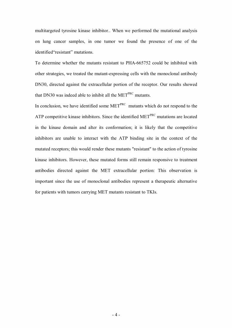
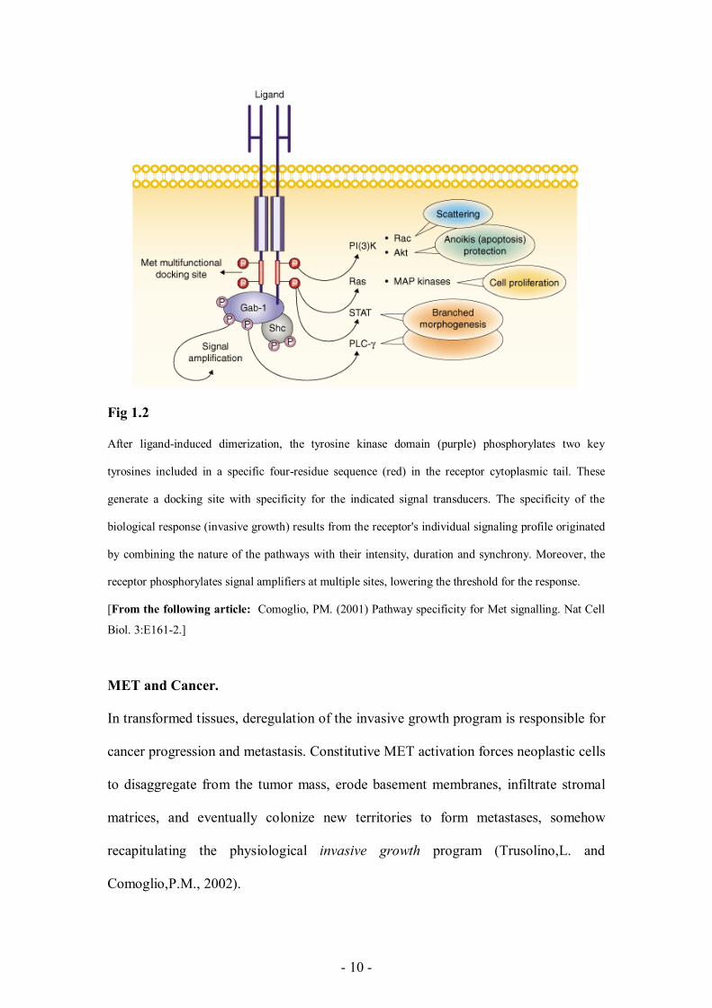
MET and cancer…………………………………………………………………….10
Mechanisms of MET activation in cancer…………………………………………15
1. MET overexpression
2. MET structural alterations
3. HGF-dependent paracrine/autocrine activation
4. HGF-independent mechanisms
Targeting MET……………………………………………………………………...17
1. MET/HGF competitors
2. Monoclonal antibodies
3. Small molecules
4. Clinical trials
Mechanisms of resistance to tyrosine kinase inhibitors…………………………..25
2) THE SCIENTIFIC PROBLEM AND THE AIM OF THE WORK……………..27
3) RESULTS……………………………………………………………………………30
Evaluation of sensitivity of METPRC mutants to the MET TKI inhibitor PHA-
665752………………………………………………………………………………..31
Generation of stably transduced cell lines expressing the METPRC mutants……38
Biological properties of stably transduced cell lines expressing METPRC
nM Also active against ALK: 2-fold less potent Phase III (ALK-altered NSCLC), Phase I–II (lung,
- 24 -
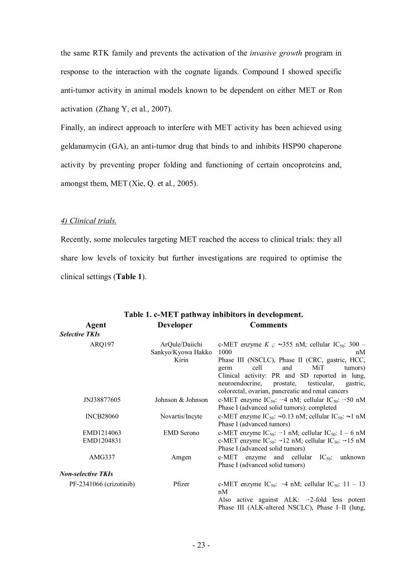
Table 1. c-MET pathway inhibitors in development. Agent Developer Comments
ALCL and other tumors) Clinical activity: PR and SD reported in NSCLC and inflammatory myofibroblastic sarcoma patients with ALK rearrangement
XL184 (cabozantinib) Exelixis c-MET enzyme IC50: 1.8 nM; cellular IC50: 8 nM Also active against VEGFR, RET, KIT, FLT3 and TIE2 Phase III (medullary thyroid cancer), Phase II (glioblastoma/astrocytoma, NSCLC and RDT design in nine tumor types: breast, gastric, NSCLC, ovarian, pancreatic, prostate, SCLC, liver and melanoma) Clinical activity: PR and SD reported in various tumors including thyroid, carcinoid, neuroendocrine, parotid, appendiceal, liver, colorectal, renal, melanoma, mesothelioma, renal, liver, lung, glioblastoma and cutaneous T-cell lymphoma
GSK1363089/ XL880 (foretinib)
GlaxoSmithKline c-MET enzyme IC50: 0.4 nM; cellular IC50: 23 nM Also active against VEGFR, AXL, PDGFR, KIT, FLT3 and TIE2 Phase II (breast, NSCLC, papillary renal, gastric and head and neck), Phase I (liver) Clinical activity: PR and SD reported in thyroid, renal, colorectal, carcinoid, melanoma, nasopharyngeal, urethral, ovarian, mesothelioma and gastric cancers
MGCD265 Methylgene c-MET enzyme IC50: 24 nM; cellular IC50: 40 nM Also active against RON, VEGFR1/2/3 and TIE2 Phase II (NSCLC), Phase I (advanced tumors)
E7050 Eisai c-MET enzyme IC50: 14 nM; cellular IC50: 6 – 37 nM Also active against VEGFR2 Phase II (HCC), Phase I (advanced solid tumors)
AMG208 Amgen c-MET enzyme IC50: 4 nM; cellular IC50: 10 – 100 nM Also active against RON Phase I (various tumors)
MP470 SuperGen c-MET enzyme and cellular IC50: unknown Also active against KIT, PDGFR, FLT3, RET and RAD51 Phase I (various tumors): completed Clinical activity: PR and SD reported in lung cancer
BMS-777607 Bristol-Myers Squibb c-MET enzyme IC50: 3.9 nM; cellular IC50: 20 – 160 nM Also active against Ron, AXL, TYRO3 and MER Phase I–II (advanced solid tumors): completed
MK-2461 Merck c-MET enzyme IC50: 2.5 nM; cellular IC50: 26 – 900 nM Also active against RON, FLT1, 3 and 4, and FGFR1, 2 and 3 Phase I–II (various tumors): completed
Phase II (NSCLC and triple negative breast cancer) Clinical activity: CR and SD reported in lung, gastric and melanoma cancers
- 25 -
Table 1. c-MET pathway inhibitors in development. Agent Developer Comments
AMG102/(rilotumumab) Amgen Humanized anti-human HGF IgG2 Phase II (SCLC, NSCLC, CRC, prostate, glioma, RCC, gastric or esophagogastric junction adenocarcinoma, mesothelioma and gynecologic tumors) Clinical activity: PR and SD reported in glioblastoma and other tumors
AV-299 Aveo Humanized anti-human HGF antibody Phase II (lung), Phase I (advanced solid tumors, lymphomas and MM)
Mechanisms of resistance to tyrosine kinase inhibitors.
In the field of acquired resistance to kinase inhibitors, three major mechanisms of
resistance have begun to emerge: (i) genetic alterations of the target, such as gene
amplifications that leads to receptor overexpression and thus render the amount of
available drug not sufficient to block the target; (ii) mutations in the target kinase that
abrogate the inhibitory action of the drug [e.g., T790M in epidermal growth factor
receptor (EGFR) and T315I in ABL]; (iii) activation of signaling pathways that
bypass the continued requirement for the original target; (iv) constitutive activation of
downstream transducers.
Among the most common mechanisms of resistance, genetic modifications include -
but are not limited- to: point mutations, deletion and amplification of genomic areas.
As previously reported, unequivocal evidence that implicates MET in human cancer is
provided by the activating mutations that have been discovered in both sporadic and
inherited forms of human renal papillary carcinomas (Schmidt, L. et al., 1997).
Activating mutations have also been described in sporadic tumors such as childhood
hepatocellular carcinomas (Park, W. S. et al., 1999), sporadic papillary renal
carcinomas (Schmidt, L. et al., 1997), gastric carcinomas (Lee, J. H. et al., 2000), lung
carcinomas (Kong-Beltran, M. et al., 2006) and head and neck squamous cell
- 26 -
carcinomas (Di Renzo, M. F. et al., 2000). The table 2 recapitulate that such
mutations, which alter sequences within the kinase domain, have also been found in a
large types of cancer and metastatic lesions.
Tab 2 Hepatocyte growth factor/scatter factor, MET and cancer references. [From the following article: Birchmeier, C. et al., (2003) Met, metastasis, motility and more. Nature
Reviews Molecular Cell Biology 4, 915-925]
- 27 -
THE SCIENTIFIC PROBLEM
AND THE AIM OF THE WORK
- 28 -
Targeted cancer therapies are based on the use of drugs that block the growth and
spread of cancer by interfering with specific molecules involved in tumor growth and
progression. By focusing on molecular and cellular changes that are specific to
cancer, targeted cancer therapies may be more effective than other types of treatment,
including chemotherapy and radiotherapy, and less harmful to normal cells. The
development of targeted therapies, therefore, requires the identification of good
targets: in other words, targets that are known to play a key role in cancer cell growth
and survival. In cancers driven by a dominant oncogene, targeted therapies have led to
remarkable improvements in response and survival, whereas in others the outcome
has been more modest.
Once a target has been identified, a therapy must be developed; most targeted
therapies are either small-molecule drugs or monoclonal antibodies. Small-molecule
drugs are typically able to diffuse into cells and can act on targets that are found
inside the cell. Most monoclonal antibodies cannot penetrate the cell plasma
membrane and are directed against targets that are outside the cell or on the cell
surface.
Many targeted cancer therapies have been approved for the treatment of specific types
of cancer, others are being studied in clinical trials, and many more are in preclinical
testing.
Unfortunately, many patient’s tumor types are refractory to targeted therapies
(intrinsic resistance). Moreover, even if an initial response to targeted therapies is
obtained, the vast majority of tumors subsequently become refractory (i.e., acquired
resistance) and patients eventually progress. In the majority of cases this is caused by
expansion of clones containing mutated forms of the target, which confer insensitivity
to the drug.
- 29 -
In addition, multiple factors including pharmacokinetics issues, such as suboptimal
drug delivery, further contribute to resistance formation. Loss of target dependence
due to the activation of parallel signaling pathways has also been reported as cause for
acquired drug insensitivity.
Taking together, recent basic and clinical research is trying to improve the efficacy of
targeted therapies by developing new generations of rationally designed targeted
agents, and translating this information to the clinic to select patients for appropriate
therapy.
However, one key aspect to improve the potential of targeted therapies is, first of all,
a better understanding the intrinsic or acquired resistance mechanisms that limit their
efficacy.
In this scenario, the aim of my PhD project was to evaluate the activity of some
available anti-MET therapies (small molecules and monoclonal antibody) targeting
the MET receptor harboring mutations in the kinase domain and, if some mutants
were insensitive to the inhibitor, to investigate the mechanisms responsible for
resistance. Then, I aimed to evaluate if the mutants resistant to small kinase
inhibitors are still sensitive to other chemicals inhibitors or monoclonal antibodies
against MET. Finally, since the anti-MET therapies are ongoing in NSCLC patients, I
also screened surgically resected lung cancers to identify activating point mutations
- 30 -
RESULTS
- 31 -
Evaluation of sensitivity of MET PRC mutants to the MET TKI inhibitor PHA-
665752
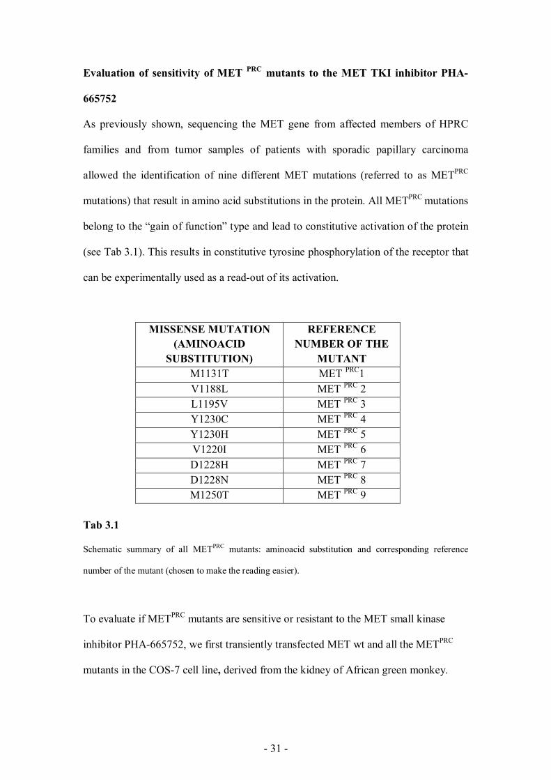
As previously shown, sequencing the MET gene from affected members of HPRC
families and from tumor samples of patients with sporadic papillary carcinoma
allowed the identification of nine different MET mutations (referred to as METPRC
mutations) that result in amino acid substitutions in the protein. All METPRC mutations
belong to the “gain of function” type and lead to constitutive activation of the protein
(see Tab 3.1). This results in constitutive tyrosine phosphorylation of the receptor that
can be experimentally used as a read-out of its activation.
MISSENSE MUTATION (AMINOACID
SUBSTITUTION)
REFERENCE NUMBER OF THE
MUTANT M1131T MET PRC1 V1188L MET PRC 2 L1195V MET PRC 3 Y1230C MET PRC 4 Y1230H MET PRC 5 V1220I MET PRC 6 D1228H MET PRC 7 D1228N MET PRC 8 M1250T MET PRC 9
Tab 3.1
Schematic summary of all METPRC mutants: aminoacid substitution and corresponding reference
number of the mutant (chosen to make the reading easier).
To evaluate if METPRC mutants are sensitive or resistant to the MET small kinase
inhibitor PHA-665752, we first transiently transfected MET wt and all the METPRC
mutants in the COS-7 cell line, derived from the kidney of African green monkey.
- 32 -
This cell line is a fibroblast-like cell line established from CV-1 simian cells which
were transformed by an origin-defective mutant of SV40 encoding for wild-type T-
antigen. This cell line is suitable for high efficient transient transfection (Gluzman, Y.,
1981 ). All the mutants cloned in the plasmidic mammalian expression vector pCEV
29.1 were available in the laboratory (Giordano, S., et al. 2000).
A
B
Fig 3.1 (A) The plasmidic construct pCEV29.1. Wild-type and mutant MET cDNAs were cloned into the
pCEV29.1 expression vector (Giordano, S., et al. 2000) and the quality of the plasmidic DNA was
validated by agarose gel electrophoresis (B).
pCEV 29.1 (10 kb)
MET (4224 bp)
WT 1 2 3 4 5 6 7 8 9
M-MLV LTR
amp
SV-40 polyA
neo
- 33 -
Transfections were performed by the DEAE-dextran technique. Briefly, DEAE-
dextran transfection is one of the oldest chemical, nonviral methods developed to
transfer RNA or DNA to cultured mammalian cells. The standard transfection
protocol involves pretreatment of cells with chloroquine, followed by exposure of the
cells to a DEAE-dextran and DNA solution. Sixteen hours after transfection, cells
were treated with the small molecule tyrosine kinase inhibitor PHA-665752 [250] nM.
Twenty four hours later, cells were lysed with boiling Laemmli buffer (Laemmli,
UK., 1970), proteins were quantified by Pierce BCA (bicinchoninic acid) Protein
Assays and analyzed by western blot (WB). As shown in Fig 3.2, we observed that,
while phoshorylation of MET wt and of some METPRC mutants (i.e. M1130T,
V1188L, V1220I, M1250T) was inhibited in presence of PHA-665752, other mutants
(namely L1195V, Y1230C, Y1230H, D1228H, D1228N) were still phosphorylated.
- 34 -
A
B
Fig 3.2 Equal amounts of pCEV 29.1 containing the cDNA of MET wt or of the different MET PRC mutants
were transfected in COS-7 cells with DEAE-dextran procedure. After transfection, cells were untreated
(A) or treated (B) with the ATP-competitive tyrosine kinase inhibitor PHA-665752 [250] nM. After 24
hours of treatment, cells were washed with phosphate-buffer saline (PBS) and lysed with Laemmli
buffer. Proteins were quantified using the BCA Protein Assay Kit (Pierce, Rockford, IL) and analysed
by Western Blots. As shown, blots probed with anti-phospho MET antibodies (directed against the
phosphorylated tyrosines 1349/1356) showed that some METPRC mutants (the red ones namely
L1195V, Y1230C, Y1230H, D1228H, D1228N, operatively defined as mutants 3, 4, 5, 7 and 8
respectively) remained phosphorylated, and thus active, also in presence of the inhibitor. GTL16 cells,
derived from a gastric carcinoma, over-expressing a constitutively phosphorylated receptor, were used
as positive control for the TKI and antibody functionality.
WT 1 2 3 4 5 6 7 8 9
WT 1 2 3 4 5 6 7 8 9
Anti MET
Anti p-MET 1349-1356
Anti MET
Anti p-MET 1349-1356
PHA-665752 [250]nM
- 35 -
In order to assess whether the lack of receptor inhibition was a dose-dependent effect
or a real inability to respond to the drug treatment, we repeated the same experiments
using a dose of PHA-665752 ten times higher than the IC50. As shown in Fig 3.3,
tyrosine phosphorylation of the MET mutants 4, 5, 7 and 8 was not inhibited even at
these high doses. MET mutant 3 was only partially inhibited, suggesting the existence
of a different mechanism of drug resistance.
A PHA-665752 [250] nM
B PHA-665752 [500] nM
Fig 3.3
COS-7 cells transiently transfected with equal amounts of pCEV 29.1 containing the cDNA of MET wt
or the different METPRC mutants were treated with the PHA-665752 TKI at two different
concentrations: [250] nM (A) and [500] nM (B). After 24 hrs of drug treatment, cells where lysed in
boiling Laemmli buffer. WB analysis revealed that MET phosphorylation of the resistant mutants (the
red ones numbers 3, 4, 5, 7, 8 respectively) was persistent also at higher doses of TKI.
PHA-665752 was identified as a small ATP-competitive molecule, inhibitor of the
catalytic activity of c-MET kinase (Ki of 4 nM, IC50 of 9 nM). PHA-665752 also
exhibited >50-fold selectivity for c-MET compared with a panel of diverse tyrosine
Anti p-MET 1349-1356
Anti p-MET 1349-1356
- 36 -
and serine-threonine kinases. In cellular studies, PHA-665752 potently inhibited
HGF-stimulated and constitutive c-MET phosphorylation, as well as HGF and c-
MET-driven phenotypes such as cell growth (proliferation and survival), cell motility,
invasion, and/or morphology of a variety of tumor cells (Christensen, JG., 2003).
To evaluate if the inability to respond to PHA-665752 was shared also by other ATP-
competitive MET inhibitors, we treated COS-7 cells expressing the different mutants
with another small molecule tyrosine kinase inhibitor, the JNJ-38877605.
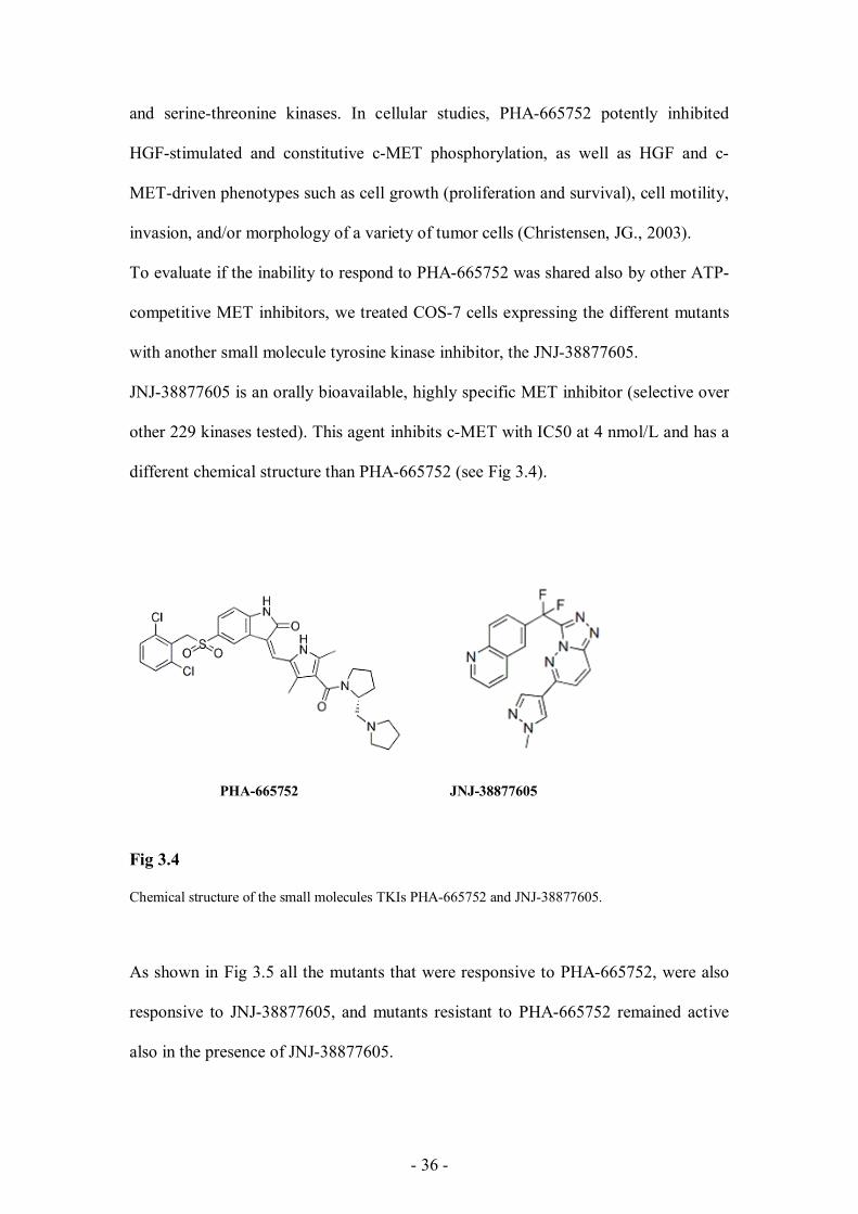
JNJ-38877605 is an orally bioavailable, highly specific MET inhibitor (selective over
other 229 kinases tested). This agent inhibits c-MET with IC50 at 4 nmol/L and has a
different chemical structure than PHA-665752 (see Fig 3.4).
PHA-665752 JNJ-38877605
Fig 3.4
Chemical structure of the small molecules TKIs PHA-665752 and JNJ-38877605.
As shown in Fig 3.5 all the mutants that were responsive to PHA-665752, were also
responsive to JNJ-38877605, and mutants resistant to PHA-665752 remained active
also in the presence of JNJ-38877605.
- 37 -
A
B
Fig 3.5
COS-7 cells transiently transfected with MET cDNAs (WT or mutated), were treated with the small
molecule TKI JNJ-38877605. WB analysis confirmed that mutants responsive to PHA-665752 were
sensitive also to JNJ-38877605 (A) while mutants resistant to PHA-665752 were resistant also to JNJ-
38877605. GTL16 cells (A) were used as a positive control for the TKI inhibition and antibody
detection.
WT
0 250 500
1
0 250 500
2
0 ......250 500
6
0 250 500
9
0 250 500
3
0 250 500
4
0 250 500
5
0 250 500
7
0 250 500
8
0 250 500
Anti p-MET 1349-1356
Anti MET
Anti MET
JNJ nM
JNJ nM
GTL-16
0 250 500
Anti p-MET 1349-1356
- 38 -
Generation of stably transfected cell lines expressing the METPRC mutants
As shown by Jeffers et al., MET receptors containing the different PRC point
mutations display different abilities to induce transformation in NIH 3T3 fibroblasts
(Jeffers M, et al., 1997). In fact, only mutations that affect residues located in the
Left part: Map of MET mutations found in Hereditary Papillary renal carcinomas. Schematic
representation of functional domains of MET tyrosine kinase. The black box depicts the tyrosine kinase
domain (KD), which can be subdivided into amino- and carboxyl-terminal lobes (N-L and C-L,
respectively), separated by a large cleft referred to as the activation loop (AL). YY represents the
receptor multifunctional docking site. Mutations found in PRCs are listed and the homology with
residues mutated in RET and KIT receptors is indicated.
Right part: Transforming ability of MET PRC mutants evaluated using the focus formation assay. Values
reported represent the average of three independent experiments.
[Data and pictures adapted from the following article: Giordano S. et al., Different point mutations
in the met oncogene elicit distinct biological properties. FASEB J. 2000 Feb;14(2):399-406.]
- 39 -
Furthermore, the METPRC mutant endowed with the highest transforming ability
(namely MET M1250T) also displayed the highest catalytic activity (Giordano, S. et
al., 2000).
In order to evaluate if the cells expressing the METPRC mutants display a different
biological behavior in the presence or in the absence of PHA-665752, we aimed at
generating stably transduced NIH 3T3 cells (that express very low levels of
endogenous MET). We thus chose two representative METPRC mutants: METPRC 8
(MET D1228N), resistant to PHA-665752, and METPRC 9 (MET M1250T),
responsive to the drug treatment.
To optimize the transduction efficiency, we decided to express the MET mutants in
lentiviral vectors. We thus mutagenized the MET cDNA cloned in the pRLL2
lentiviral vector, already available in the lab. The two PRC mutants were thus
obtained performing an in vitro site-directed mutagenesis (Strategene’s QuickChange
II XL Site-Directed Mutagenesis Kit) that allows to introduce site-specific mutations
in the double-stranded plasmid pRLL2 containing the MET wild type cDNA. The
obtained mutagenized cDNAs were validated by direct sequencing (Fig 3.7).
- 40 -
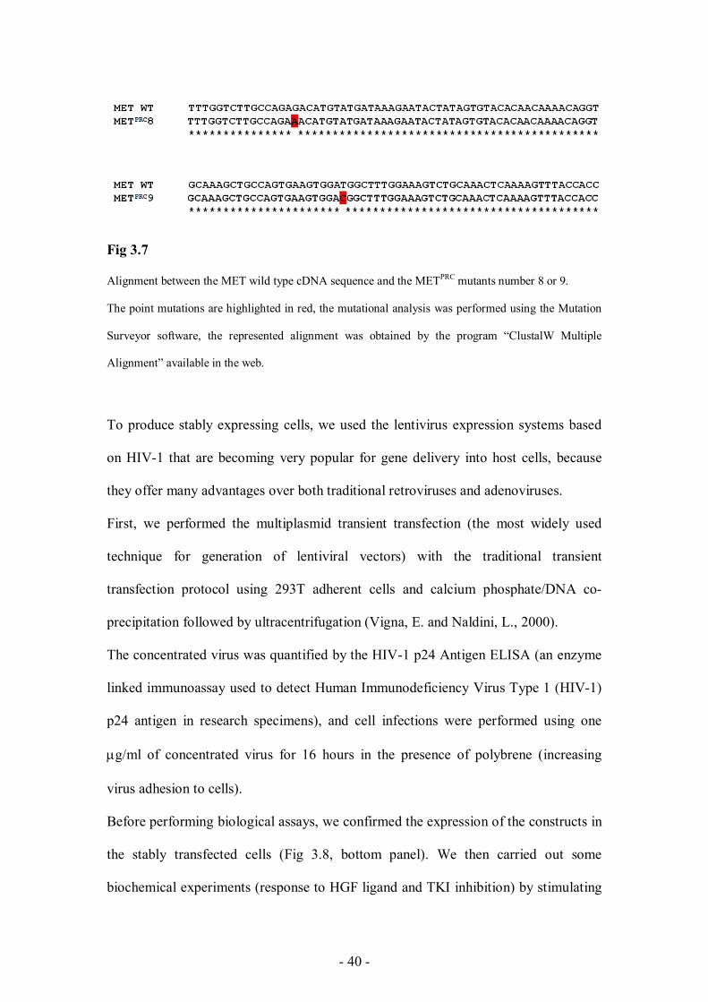
Fig 3.7
Alignment between the MET wild type cDNA sequence and the METPRC mutants number 8 or 9.
The point mutations are highlighted in red, the mutational analysis was performed using the Mutation
Surveyor software, the represented alignment was obtained by the program “ClustalW Multiple
Alignment” available in the web.
To produce stably expressing cells, we used the lentivirus expression systems based
on HIV-1 that are becoming very popular for gene delivery into host cells, because
they offer many advantages over both traditional retroviruses and adenoviruses.
First, we performed the multiplasmid transient transfection (the most widely used
technique for generation of lentiviral vectors) with the traditional transient
transfection protocol using 293T adherent cells and calcium phosphate/DNA co-
precipitation followed by ultracentrifugation (Vigna, E. and Naldini, L., 2000).
The concentrated virus was quantified by the HIV-1 p24 Antigen ELISA (an enzyme
linked immunoassay used to detect Human Immunodeficiency Virus Type 1 (HIV-1)
p24 antigen in research specimens), and cell infections were performed using one
g/ml of concentrated virus for 16 hours in the presence of polybrene (increasing
virus adhesion to cells).
Before performing biological assays, we confirmed the expression of the constructs in
the stably transfected cells (Fig 3.8, bottom panel). We then carried out some
biochemical experiments (response to HGF ligand and TKI inhibition) by stimulating
- 41 -
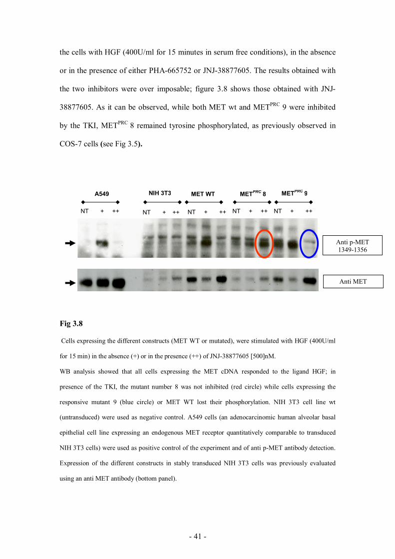
the cells with HGF (400U/ml for 15 minutes in serum free conditions), in the absence
or in the presence of either PHA-665752 or JNJ-38877605. The results obtained with
the two inhibitors were over imposable; figure 3.8 shows those obtained with JNJ-
38877605. As it can be observed, while both MET wt and METPRC 9 were inhibited
by the TKI, METPRC 8 remained tyrosine phosphorylated, as previously observed in
COS-7 cells (see Fig 3.5).
Fig 3.8
Cells expressing the different constructs (MET WT or mutated), were stimulated with HGF (400U/ml
for 15 min) in the absence (+) or in the presence (++) of JNJ-38877605 [500]nM.
WB analysis showed that all cells expressing the MET cDNA responded to the ligand HGF; in
presence of the TKI, the mutant number 8 was not inhibited (red circle) while cells expressing the
responsive mutant 9 (blue circle) or MET WT lost their phosphorylation. NIH 3T3 cell line wt
(untransduced) were used as negative control. A549 cells (an adenocarcinomic human alveolar basal
epithelial cell line expressing an endogenous MET receptor quantitatively comparable to transduced
NIH 3T3 cells) were used as positive control of the experiment and of anti p-MET antibody detection.
Expression of the different constructs in stably transduced NIH 3T3 cells was previously evaluated
using an anti MET antibody (bottom panel).
A549 NIH 3T3 MET WT
NT + ++ NT + ++ NT + ++ NT + ++ NT + ++
METPRC 8 METPRC 9
Anti p-MET 1349-1356
Anti MET
- 42 -
Biological properties of stably transfected cell lines expressing METPRC mutants
To evaluate if the different behavior of the METPRC mutants could impact also the
biological properties of the expressing cells, we decided to analyze some MET-
dependent activities such as cell migration and anchorage-independent growth.
Concerning the biological responses, treatment with the TKI did not modify either
receptor phosphorylation or MET-induced biological activities in cells expressing the
resistant mutant D1228N (mutant 8): this was evident both in the soft agar assay
(which evaluates the ability to grow in anchorage-independent conditions) and in
migration/invasion assays, such as wound healing and transwell assays (see Fig 3.9).
In details, colony formation and viability (both measured by Alamar Blue
quantification) were not impaired in the presence of JNJ-38877605 in cells expressing
the mutant number 8, while they were strongly decreased in those expressing MET wt
or the mutant 9. Untransduced NIH 3T3 cells were used as negative control: as
shown, these cells were not able to form colonies in soft agar.
- 43 -
Fig 3.9
Anchorage-independent growth assays in soft agar and Alamar Blue quantification of cell viability. In
the presence of the MET inhibitor, cells expressing either MET wt or the sensitive mutant number 9
(blue lines) were severely impaired in their ability to grow in anchorage independent conditions, while
cells expressing the resistant mutant 8 (red line) were unaffected. NIH 3T3 cell line wt (not expressing
the MET gene) were used as negative control. The experiment was performed in presence of HGF (20
ng/ml) and in presence or absence of JNJ-38877605.
NIH 3T3 METPRC 9
JNJ
NT
MET WT METPRC 8
0
50
100
150
200
250
300
350
NIH 3T3 MET WT METPRC 8 METPRC 9
NT JNJ
% viability (Alamar Blue)
- 44 -
We also performed in vitro biological assays to evaluate cell motility. In the invasion
assay, cells were seeded in Transwell chamber, on the upper side of a porous
polycarbonate membrane. The medium in both chambers was supplemented with low
percentage of serum; the lower chamber was supplemented with HGF (20 ng/ml) in
presence or absence of JNJ-38877605. After 16 h, cells attached on the upper side of the
membrane were mechanically removed. Cells that migrated to the lower side were fixed
with gluataraldehyde and stained with crystal violet. Stained cells were photographed (see
Fig. 3.10 A).
To evaluate the ability to migrate and repair wounds, we performed a Wound Healing
assay. This method mimics cell migration during wound healing in vivo. The basic
steps involve creating manually a "wound" in a cell monolayer, capturing the images
at the beginning and at regular intervals during cell migration and comparing the
images to quantify the migration rate of the cells. As shown in Fig. 3.10 B, all cells
increased their ability to migrate in presence of HGF (20 ng/ml) compared to the
counterpart not treated (NT), but cells transfected with MET wt or with the responsive
mutant 9 were not able to close the wound in presence of JNJ-38877605. Once again, the
resistant mutant 8, showed an opposite response and its ability to migrate and close the
wound was not impaired by the inhibitor.
- 45 -
A
B
Fig 3.10 Cells expressing MET WT or the mutants 8 and 9 were used to evaluate the ability to invade and migrate.
The upper panel shows the results of a representative invasion assay in Transwells, in the lower part is
represented the ability to migrate and repair the wound by Wound Healing Assay. Both assays were
performed using low percentage of serum, plus (experimental point named HGF) or minus (named NT) HGF
(20 ng/ml), or in presence of HGF (20 ng/ml) plus JNJ-38877605 [500] nM.
In both assays these two mutants showed an opposite response: while cells expressing MET wild type or the
responsive mutant 9 (green rectangles) were inhibited, the resistant mutant 8 (yellow rectangles) was able to
migrate also in the presence of inhibitor in both assays.
HGF+ JNJ-38877605
NT
HGF
MET WT METPRC 8 METPRC 9
HGF+ JNJ-38877605
NT
HGF
MET WT METPRC 8 METPRC 9
- 46 -
Analysis of lung tumors for the presence of MET mutations
Nowadays pivotal studies in NSCLC (Non-Small Cell Lung Cancer) are ongoing,
using specific chemical and biological anti-MET inhibitors; three of them (Met MAb,
Crizotinib, ArQule-197) are in phase III clinical trials. The used drugs fall in two
different categories: small kinase inhibitors (TKIs) and monoclonal antibodies
(mAbs). They act with different mechanisms, since small TKIs interact with the
receptor intracellular portion while mAbs bind to the extracellular domain. It is thus
very likely that mutations present in diverse parts of the receptor can differentially
impact on the ability to respond to either of the drugs. For these reasons and since
METPRC mutations have been found not only in the germline of patients but also in
sporadic tumors, we decided to analyze the sequence of the MET tyrosine kinase in
human surgically resected lung cancers.
As shown in the Tab 3.2, we collected resected lung tumors in collaboration with the
Oncologic Hospital “A. Businco” of Cagliari (Dr. R. Versace) and Hospital
“S.Giovanni Battista” of Turin (Prof. E. Ruffini), and we gathered the follow up of all
patients (unfortunately not complete in all cases). The Classification of Malignant
Tumors (TNM) is one of the most widely used staging systems. The TNM system is
based on the extent of the tumor (T), the extent of spread to the lymph nodes (N), and
the presence of distant metastases (M). A number is added to each letter to indicate
the size or extent of the primary tumor and the extent of cancer spread. Tumor grade
is a system used to classify cancer cells in terms of how abnormal they look under a
microscope and how quickly the tumor is likely to grow and spread. Tumor grade
should not be confused with the stage of a cancer. Cancer stage refers to the extent or
severity of the cancer, based on factors such as the location of the primary tumor,
- 47 -
tumor size, number of tumors, and lymph node involvement (spread of cancer into
lymph nodes).
NUMBER OF
SAMPLE
ISTOLOGY
STAGE
GRADING
T
N
M
3118 Ca pavimentoso G2
pT1aN0
I A G2 T1 N0
2279 Mts polmonari di ca retto
G2
IV G2
119 adenocarcinoma con
estese aree di necrosi
IB G2 2 0 0
280 carcinoma non a piccole
cellule
IIA 1 1 0
285 carcinoma a grandi cellule
con attività
neuroendocrina
IB 2 0 0
286 Adenocarcinoma
288 carcinoma epidermoide 1 0 0
290 carcinoma bronchiolo
alveolare mucinoso
Tab 3.2
List of samples used for mutational analysis of the MET gene and corresponding follow up.
Classification is based on the Classification of Malignant Tumors (TNM), a cancer staging system that
describes the extent of cancer in a patient’s body: T stands for tumor size and invasiveness. The T
number can range from T1 to T4. T1 and T2 are differentiated primarily on size (<3 cm = T1, >3 cm =
T2) and if the tumor is visible within a lobar bronchus (T2). T3 tumors involve the chest wall, but may
- 48 -
be resectable (operable). T4 tumors are not surgically resectable because they have invaded the
mediastinum (the area and organs between the lungs) and involve the heart, great vessels, trachea or
esophagus, or because they involve the pleura (lining of the lung) with a malignant pleural effusion
(accumulation of fluid around the lining of the lung). N stands for Nodal involvement (lymph nodes)
and is staged from N1 to N3. M stands for the presence (1) or absence (0) of metastases (spread to a
distant site). Grading (1–4) refers to the differentiation of the cancer cells (i.e. they are "low grade" if
they appear similar to normal cells, and "high grade" if they appear poorly differentiated). About
staging, Non-small cell lung carcinoma is usually staged from IA (best prognosis) to IV (worst
prognosis).
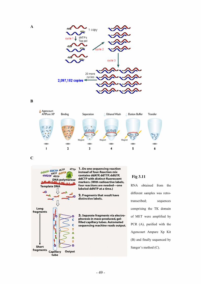
We purified total RNA from 16 samples (8 tumors and the corresponding peritumoral
normal tissues). Upon RNA retrotranscription, we PCR amplified overlapping
portions of the MET intracellular domain; amplified segments were then purified
using the chemical method named Solid Phase Reversible Immobilization (SPRI)
based on speed beads (Agencourt Ampure Xp Kit), and finally sequenced. (see Fig
3.11).
In one tumoral sample (but not in the corresponding peritumoral normal tissue) we
found the presence of a missense mutation (Y1230H) resulting in aminoacid
substitution (see Fig 3.12 A). The mutated aminoacid corresponds to Y1230H, which
belongs to the METPRC mutants resistant to treatment with small kinase inhibitors.
(Fig 3.12 B). Moreover, a very recent study (Qi J., et al., 2011) has shown that this
mutation destabilizes the conformation of the MET TK domain and contributes to the
development of acquired resistance to MET inhibitors.
- 49 -
A
B
C
Fig 3.11
RNA obtained from the
different samples was retro-
transcribed; sequences
comprising the TK domain
of MET were amplified by
PCR (A), purified with the
Agencourt Ampure Xp Kit
(B) and finally sequenced by
Sanger’s method (C).
- 50 -
A
B
Fig 3.12
The sequence analysis of MET tyrosine kinase domain in human resected lung cancer samples, was
performed on RNA derived from both the tumors and corresponding normal tissues. In one sample
(sample number 119 listed in the previous tab) the tumoral counterpart revealed the presence of the
METPRC mutation Y1230H (the number 5) which was not present in the corresponding peritumoral
tissue (A). This mutation, as previously reported, induces resistance to PHA-665752 . As shown the
mutated receptor maintains a persistent phosphorylation also in presence of inhibitor, as highlighted by
the blue circle in the lower part of the panel (B).
Tumoral tissue
Peritumoral tissue
Y1230H
PHA-665752 [[550000]] nnMM
Anti p-MET 1349-1356
- 51 -
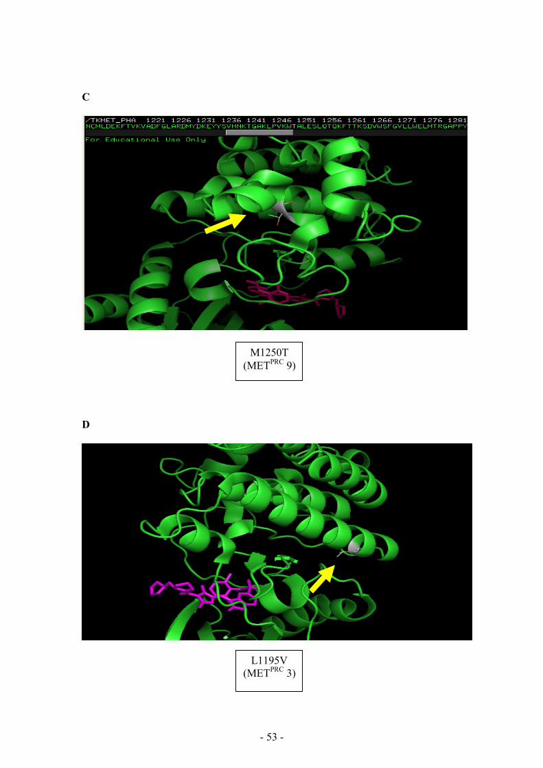
In silico structural analysis of the MET tyrosine kinase domain
Since pharmacological resistance can be due to impaired interaction between the drug
and the RTK, we performed an in silico analysis of the MET tyrosine kinase domain
Structures were mutagenized with the PyMol Software starting from the crystal
structure of the MET TK domain available in the NCBI structure site (NCBI
STRUCTURE_ PDB 2WKM “X-Ray Structure Of PHA-00665752 Bound To The
Kinase Domain Of c-MET
http://www.rcsb.org/pdb/explore/explore.do?pdbId=2WKM). From this analysis it
was evident that the mutations D1228N (METPRC 8) and Y1230H (METPRC 5, found
in one lung cancer sample) conferring resistance to TKIs, were located very close to
the ATP-binding site (Fig 3.13 A, B), while M1250T (METPRC 9), which did not
induce resistance, was placed away from this site (Fig 3.13 C). For this reason, it is
likely that -for steric hindrance- the ATP-competitive inhibitors (PHA-665752 and
JNJ- 38877605) are unable to interact with their binding sites in the context of these
mutated receptors.
Analyzing also all the other PRC mutations (Fig 3.13 E), we observed that all the
mutations conferring resistance were located in the proximity of the PHA-665752
binding site, while mutated aminoacidic residues of the responsive mutants were far
from this region. Concerning the METPRC mutant 3, that was only partially inhibited
in presence of PHA-665752, its localization is outside the ATP binding site (see Fig
3.13 D), suggesting the existence of a different mechanism of drug resistance.
- 52 -
A
B
D1228N (METPRC 8)
Y1230H (METPRC 5)
- 53 -
C
D
L1195V (METPRC 3)
M1250T (METPRC 9)
- 54 -
E
Fig 3.13
In silico analysis of MET TK domain in presence of the ATP-competitive inhibitor PHA-665752.
In all the pictures the secondary structure of MET TK domain is green. In A, B, C, D the mutated
aminoacidic residue is indicated by the yellow arrow and the PHA-665752 molecule is magenta. In A
and B, red balls indicate the steric hindrance. Panel E shows the TK domain with all the PRC mutants:
the red ones are the resistant, while the blue ones are those responsive to small molecules PHA-665752
(the yellow structure, represented by bubbles); the partially resistant PRC 3 is orange.
ALL METPRC
MUTANTS
- 55 -
Bypassing resistance: use of an anti-MET specific monoclonal antibody
As previously mentioned, while the presence of mutations in the intracellular TK
domain can impair the ability to respond to small kinase inhibitors, it is likely that the
mutated receptors are still able to respond to the inhibitory activity of monoclonal
antibodies directed against the extracellular portion.
One of such mAbs, named DN30, was previously produced and studied in the
laboratory (Pietronave S, et al., 2010). This monoclonal antibody is directed against
the extracellular portion of the receptor and behaves as a partial agonist (Prat, M. et
al., 1998). In fact, it induces MET activation, although at low levels, followed by
promotion of MET down-regulation through a molecular mechanism which involves
cleavage of the extracellular portion (also called “shedding”) of the receptor and
proteasomal degradation of the intracellular portion (Petrelli, A. et al., 2006), (fig 3.14
A). To eliminate the partial agonistic activity of this mAb, some colleagues at the
IRCC have ingegnerized the antibody in order to produce a monovalent form (DN30
FAb), which maintains only the antagonistic activity (Pacchiana G., et al., 2010).
To evaluate the inhibitory activity of the DN30 FAb, we grew the transfected COS7
cells for 72 hours in the presence of this molecule. We then examined the supernatant
of the cells to look for the presence of the released MET extracellular portion. As we
observed that FAb DN30 treatment caused “shedding” of extracellular domain in all
METPRC mutants (fig 3.14 B) we could conclude that the DN30 FAb was indeed
active on all the mutants. When we analyzed the activation status of the different
mutants, we found indeed that FAb DN30 treatment resulted in their inhibition,
independently from their sensitivity to TKIs. An example is shown in figure 3.15
where all the TKI-resistant mutants were inhibited (and thus lost their tyrosine
phosphorylation) and the total amount of MET decreased upon DN30 FAb treatment.
- 56 -
A
B
Fig 3.14 Mechanism of action of the monoclonal antibody DN30 (A). This monoclonal antibody (DN-30) is directed against the extracellular portion of MET and binds to MET at subnanomolar affinity, inducing proteolytic cleavage of the extracellular portion close to the cell membrane and release of a soluble receptor in the extracellular space (Petrelli, A. et al., 2006). Following ectodomain shedding, operated by a metalloprotease of the ADAM family, the remaining transmembrane fragment becomes substrate of a second protease (γ-secretase) that detaches the kinase-containing portion from the membrane and rapidly addresses it toward the proteasome degradation pathway (Foveau B, et al. 2009). Therefore, the net result of DN-30 binding to MET is (a) the generation of a soluble “decoy” MET that neutralizes HGF and forms heterodimers with bona fide MET (Michieli, P. et al., 2004) and (b) the proteolytic degradation of the MET kinase domain. This translates into neutralization of HGF/MET-mediated biological activities. In picture 3.10 B is reported the ectodomain shedding of all PRC mutants and MET wt after Fab DN30 treatment. Upon 72h of Fab DN30 treatment (24g/ml), cells were starved for 16hrs, then the medium collected and loaded for the western blot analysis. As shown, FAb DN30 treatment caused the ectodomain shedding in all mutants, including the TKI-resistant group. In these experiments, GTL16 cell line was used as positive control.
MET ECD supernatant
FAb - + - + - + - + - + - + 1 2 3 4 WT GTL
- + - + - + - + - + 6 5 9 8 7
MET
MET
1 2
- 57 -
Fig 3.15
WB analysis on COS-7 cells expressing MET WT and the METPRC mutants.
Red rectangles highlight that FAb DN30 (28 g/ml) treatment significantly decreased the
phosphorylation all the resistant mutants (reference numbers 3, 4, 5, 7, 8) . Also the total amount of
MET, as a direct consequence of ectodomain shedding, was significantly decreased in cells treated with
the antibody respect the counterpart not treated (NT) or inhibited by PHA-665752. GTL16 cells were
used as positive control of the experiment.
Anti p-MET 1349-1356
Anti MET
Anti p-MET 1349-1356
Anti MET
- 58 -
DISCUSSION
- 59 -
Selective inhibition of protein tyrosine kinases is gaining importance as an effective
therapeutic approach for the treatment of a wide range of human cancers.
The fact that the inhibition of a single oncogene can cause the death of cancer cells
(referred as oncogene addiction) supported the idea of using highly specific inhibitors
directed against the oncogenic proteins. The paradigm of the clinical success of
targeted therapies based on oncogenic addiction is represented by the use of Imatinib
(a small kinase inhibitor directed against the cytoplasmic tyrosine kinase ABL) for the
treatment of CML (Chronic Myeloid Leukemia) in patients bearing BCR-ABL
translocation. The oncogenic addiction has been proven also in different biological
contests and on different targets: Trastuzmab in HER2 over-expressing breast cancers,
Cetuximab (anti-HER1) in CRC (Colorectal Cancer) and HNSCC (Head and Neck
Squamous Cell Carcinoma) and Gefitinib and Erlotinib (HER1 inhibitors) in NSCLC
(Non-Small Cell Lung Cancer) (Petrelli, A. & Giordano, S., 2008).
However, as extensively documented, initially successful therapy is often hampered
by acquired resistance to the drug and subsequent relapse and this could be caused by
different mechanisms. Nowadays, given that many patients are starting to benefit from
the discovery of monoclonal antibodies and of small molecules targeting tyrosine
kinases, the investigators are now trying to understand and unveil the mechanisms
through which neoplastic cells lose their ability to respond to these drugs (also
named secondary resistance or acquired resistance). Luckily, it appears that the
majority of the resistance models developed in vitro are predictive of what is observed
in vivo and can thus help researchers in identifying and studying this crucial clinical
problem.
- 60 -
Many different mechanisms have been demonstrated to sustain resistance to targeted
therapies. The most common mechanism of resistance, in terms of genetic alterations
of the target, is the presence or appearance of point mutations impairing or preventing
the interaction between the target and the drug. The most frequent types of mutations
are those decreasing the affinity of the drug for the target kinase domain, while
maintaining the catalytic activity. Mutations that alter the aminoacids surrounding the
binding site of the drug decrease the availability of the target region towards the
inhibitor, without interfering with the ATP binding (Zhang et al., 2009). Other
reported mutations increase the affinity of the kinase for the ATP, decreasing the
effectiveness of the ATP-competitive inhibitors (Tanaka R, Kimura S., 2008).
Some reports support the idea that the appearance of mutations in tumors after
treatment with a specific TKI is the result of a process of selection of a pre-existing
cell population. Such theory supports the idea that a small population of the tumor
bulk a priori contains the mutation, which confers a primary resistance to these cells,
therefore giving them a selective advantage in the presence of the inhibitor. The bulk
tumor mass is thus killed by the drug, allowing a short period of response, lasting until
the cells resistant to the TKI become the majority. This theory is supported by the fact
that some of these “resistance-related mutations” can be found in a small percentage
of tumor cells in patients that have not undergone targeted therapy (Bachleitner-
Hofmann T., et al., 2008; Kreuzer KA, et al., 2003; Roche-Lestienne C, et al., 2002).
On the other hand, other investigators believe that the high dependence of a cell on a
specific oncogenic survival pathway forces genomic instability, allowing the
induction of mutations that confer resistance to the inhibitor. This genomic instability
can induce mutations either in the drug target or in other signal transducers that
activate alternative pathways able to sustain cell viability (Ricci C, et al., 2002).
- 61 -
About the MET gene, as previously mentioned, activating mutations have been
described in sporadic tumors such as childhood hepatocellular carcinomas, sporadic
papillary renal carcinomas, gastric carcinomas, lung carcinomas and head and neck
squamous cell carcinomas. The main proof of the direct involvement of MET in
tumorigenesis was given by the identification of germ-line activating mutations in
patients with hereditary renal papillary carcinoma (HPRC).
Nowadays pivotal studies in NSCLC (Non-Small Cell Lung Cancer) are ongoing
using specific chemical and biological anti-MET inhibitors; three of them (MET
MAb, Crizotinib, ArQule-197) are in phase III clinical trials. They act with different
mechanisms and are directed against different portions of the MET receptor: small
TKIs (crizotinib and ArQule-197) interact with the intracellular portion, while mAbs
bind to the extracellular domain. It is thus very likely that mutations present in
different parts of the receptor can differentially impact on the ability to respond to
either of the drugs.
Recently, MET mutations have been identified within the sema domain,
juxtamembrane domain, and intrcellular regions in small cell and non-small cell lung
cancers, lung adenocarcinomas, gastric cancer, renal carcinomas, and mesotheliomas
(Ma PC, et al., 2003; Kong-Beltran M, et al. 2006; Ma PC, et al., 2008;
Jagadeeswaran R, et al., 2006; Lee JH, et al., 2000). Thus, mutational activation of
MET is not restricted to renal cancer and may be a more common mechanism by
which MET is aberrantly activated during tumorigenesis. A few studies have shown
that some of these mutations induce resistance to MET kinase inhibitors (Timofeevski
SL, et al., 2009; Berthou S, et al., 2004; Bellon SF, et al., 2008). Therefore,
additional studies are required to understand the effect of MET mutations in tumor
progression and resistance to therapy.
- 62 -
From this scenario it is clear the need of choosing the most suitable therapeutic
approaches in order to avoid the phenomenon of pharmacologic resistance. This
requires tailoring of the therapy using drugs able to act also in the presence of
mutations, which could determine pharmacologic resistance to the treatment. On these
bases, my PhD work was aimed at evaluating the activity of some available anti-
MET therapies targeting the MET receptor mutated in the kinase domain.
First of all, our results demonstrated that it is possible to categorize the METPRC
mutants in two different groups: the “responsive” group whose phosphorylation was
inhibited in presence of PHA-665752, and the “resistant” group in which receptor
phosphorylation and activation are not affected by the inhibitor.
Second, the lack of receptor inhibition was not a dose-dependent effect, but a real
inability to respond to the drug treatment. In fact, using PHA-665752 at two different
concentrations: [250] nM and [500] nM (ten times higher than the IC50), tyrosine
phosphorylation of some METPRC mutants was not inhibited even at these high doses.
One mutant (METPCR mutant 3) was only partially inhibited, suggesting the existence
of a different mechanism of drug resistance.
We then demonstrated that the phenomenon of resistance is not restricted to PHA-
665752, but is shared also by other ATP-competitive MET inhibitors, such as the JNJ-
38877605.
To evaluate the biological meaning of these observation, we engineered NIH 3T3
cells (that express very low levels of endogenous MET) to express METPRC mutated
forms. We found that while -induced biological activities (such as migration, invasion
and growth) were impaired by TKIs in cells expressing wt MET or METPRC
“responsive mutants”, they were not affected in cells MET expressing the “resistant
- 63 -
mutants”. These results show that loss of biochemical inhibition was paralleled by
loss of MET-dependent biological activities.
We then asked how these mutations can impair or prevent the response to TKIs. Our
results, obtained by in silico analysis, demonstrated that all the mutations belonging to
the “resistant” group are located very close to ATP binding site. It is likely that, for
the steric hindrance due to the conformational change in the kinase domain (as a result
of the aminoacidic change), the ATP-competitive inhibitors -such as PHA-665752 and
JNJ-38877605- are unable to interact with their binding sites. One other possible
mechanism of resistance in a mutated receptor can be due to the fact that the mutation
alters the domain conformation and leads to a decrease in the affinity for the drug: this
is likely to happen in the case of the mutant number 3, (which showed dose-
dependent resistance) in which the mutation is localized outside the drug binding site.
In the era of targeted therapies, the phenomenon of resistance related to target’s
genetic mutations is extremely important in order to better treat tumors containing
mutations. In the few lung tumors we examined, we found indeed a case presenting a
METPRC mutation. Most importantly, the identified mutation (Y1230H) belongs to
the “resistant” group. It is thus likely that treatment with a TKI of a patient bearing
such a mutant receptor will not end with a favorable outcome.
At this point, we wondered if we could figure a therapeutic approach which could
represent an alternative treatment for patients with tumors carrying MET mutants
resistant to TKIs. Such an approach could be the use of a monoclonal antibody
directed again the extracellular portion of the receptor, which is in the wild type
conformation also in the METPRC mutants.
We thus treated cells expressing the METPRC mutants with the monomeric form of an
anti-MET monoclonal antibody (FAb DN30). Indeed, we found that the antibody was
- 64 -
able to inhibit all TKI mutants, independently from their sensitivity to small TKIs.
These results indicate that despite some mutated forms are unable to respond to small
molecule TKIs, they still remain responsive to treatment with antibodies directed
against the MET extracellular portion, likely because the extracellular domain is in the
wild type conformation. In clinical terms, these data show that we can “bypass”
resistance to TKIs by use of mAbs directed against the MET extracellular portion. In
conclusion, our results indicate that in tumors harboring MET tyrosine kinase
mutations that prevent or impair the interaction between the ATP-competitive TKIs
and the receptor, the use of small molecule TKI could be inappropriate.
We propose that the use of specific anti-MET monoclonal antibodies (such as FAb
DN30) can represent, a therapeutic alternative to treat TKIs-resistant tumors harboring
mutations in the MET tyrosine kinase domain, Our data could thus help in better
tailoring the anti-MET targeted therapies, thus contributing to increasing their
effectiveness.
- 65 -
MATERIALS AND METHODS
- 66 -
Plasmid constructs and mutagenesis
pCEV29.1 expression vector was available in the lab wt MET cDNA was cloned in
the vector as described (Giordano et al., 2000) and PRC mutations were introduced
in by polymerase chain reaction (Bardelli, A. et al., 1998). Human MET residues are
numbered according to Gene Bank# X54559 (Ponzetto, C. et al., 1991).
The human MET cDNA cloned in the plasmidic construct pRLL2 was available in the
lab, then mutagenized by using the QuikChange II XL Site-Directed Mutagenesis Kit
(Agilent Thechnologies). The QuikChange II site-directed mutagenesis kit is used to
make point mutations, replace amino acids, and delete or insert single or multiple
adjacent amino acids. This mutagenesis’ method was performed using PfuUltra high-
fidelity (HF) DNA polymerase for mutagenic primer-directed replication of both
plasmid strands with the highest fidelity. The basic procedure utilizes a supercoiled
double-stranded DNA (dsDNA) vector with an insert of interest and two synthetic
oligonucleotide primers, both containing the desired mutation. The oligonucleotide
primers, each complementary to opposite strands of the vector, are extended during
temperature cycling by PfuUltra HF DNA polymerase, without primer displacement.
Extension of the oligonucleotide primers generates a mutated plasmid containing
staggered nicks. Following temperature cycling, the product is treated with Dpn I. The
Dpn I endonuclease (target sequence: 5´-Gm6ATC-3´) is specific for methylated and
hemimethylated DNA and is used to digest the parental DNA template and to select
for mutation-containing synthesized DNA. The nicked vector DNA containing the
desired mutations is then transformed into competent cells.
- 67 -
Transfection, infection
COS-7 cells were transfected by the DEAE-dextran method. Briefly, plasmid DNA (2
g/ml) was resuspended in DMEM containing DEAE-dextran and added to a 100-mm
dish of subconfluent COS 7 cells. Dimethyl sulfoxide shock was performed after 4 h
of incubation at 37°C.
Lentiviruses were produced by transient transfection of 293T cells whit the calcium-
phosphate procedure containing the DNA to be transfected, as described elsewhere
(Vigna and Naldini, 2000). Cell infection was performed over-night and in the
presence of polybrene (hexadimethrine bromide). Polybrene is a relatively non-toxic
polymer, that was shown to enhance the adsorption of virus complex onto cells in
culture (Coelen et al., 1983).
Cell culture
293T, GTL16, A549, COS-7 and NIH 3T3 cell lines from ATCC were cultivated in
DMEM (293T, NIH 3T3) or RPMI (GTL16 and A549) supplemented with 1% Q,
0.1% penicillin (5000U/ml, Faber), 0.1% streptomycin (5mg/ml, Squibb) and with
10% FBS or Calf Serum deactivated by heating (NIH 3T3), at 37°C in 5% CO2.
Protein extraction and Western blot
For Western blot analysis, cells were lysed in boiling LB buffer [2% SDS, 0.5 mol/L
Tris-HCl (pH 6.8)]. Protein concentration of whole-cell lysates was evaluated with the
BCA Protein Assay kit (Pierce) and equal amounts of total proteins were analyzed by
SDS-PAGE and Western blotting. Western blots were performed according to
standard methods. The antibodies used were as follows: anti-MET antibody DL21
(Prat, M. et al. 1998) and anti-MET Zymed (Invitrogen), anti-phospho MET
- 68 -
Tyr1349/1356 (Cell Signaling Technology). Final detection was done with enhanced
chemiluminescence (ECL) system (Amersham).
Biological assays
For invasion assays, cells were seeded in Transwell chamber, on the upper side of a
porous polycarbonate membrane. The medium in both chambers was supplemented
with low percentage serum; the lower chamber was supplemented with HGF (20
ng/ml) alone or in presence of JNJ-38877605 (Johnson and Johnson) [500] nM. After
16 h, cells attached on the upper side of the membrane were mechanically removed.
Cells that migrated to the lower side were fixed with gluataraldehyde and stained with
crystal violet. Stained cells were photographed.
For analysis of colony formation in soft agar, cells were diluted to a concentration of
7x10³
cells/ml in DMEM containing 10% FBS and 0.5% Seaplaque agar, with HGF
(20 ng/ml), in presence or absence of JNJ-38877605. Cells were seeded in 12-well
plates (1 ml per well) containing a 1% agar underlay and supplemented twice a week
with DMEM containing 10% FBS and, where indicated, HGF and JNJ-38877605.
Colonies were stained with tetrazolium salts three weeks after seeding and the
viability was evaluated using the alamarBlue® cell viability reagent (Invitrogen). This
reagent is used to assess cell viability by adding the 10X, ready-to-use solution to
cells in culture media, followed by a 1–4 hours incubation at 37ºC (to allow cells to
convert resazurin to resorufin). The resulting fluorescence was read on a 96-well plate
reader.
For the wound healing assay, NIH 3T3 cells were plated to create a confluent
monolayer. After scraping the cell monolayer with a p200 pipet tip, cells were washed
- 69 -
and the media replaced with DMEM 10% serum alone, or HGF (20 ng/ml), or HGF
(20 ng/ml) in presence of JNJ-38877605. Cells dishes were incubated for 24-48 hrs.
The migration assay was stopped when the wound was repaired, the cells were fixed
in glutheraldeide then stained with crystal violet and photographed.
Tumor samples collection
Tumor samples were obtained in accordance with consent procedures approved by the
Ethic Committee of Hospital San Giovanni Battista (Turin) and the University Of
Cagliari. The follow-up reports, was adapted to the new guidelines in the 7th Edition
of TNM in Lung Cancer of the International Association for the Study of Lung
Cancer (IASLC).
RNA extraction, RT-PCR , PCR and sequencing
Total RNA was extracted from lung tumors using Trizol reagent (Invitrogen)
according to the manufacturer's instructions. RNA (500 ng) was retrotranscribed into
cDNA using the High Capacity cDNA Reverse Transcription Kit containing the
Bachleitner-Hofmann T., et al. (2008) HER kinase activation confers resistance to MET tyrosine kinase inhibition in MET oncogene-addicted gastric cancer cells. Mol Cancer Ther 7:3499-508. Bardelli, A., et al. (1998) Uncoupling signal transducers from oncogenic MET mutants abrogates cell transformation and inhibits invasive growth. Proc. Natl. Acad. Sci. USA 95, 14379–14383 Barni, S., et al. (2007) From the trastuzumab era to new target therapies: beyond revolution. Ann. Oncol. 18 Suppl 6, vi1-vi4. Bean, J., et al. (2007) MET amplification occurs with or without T790M mutations in EGFR mutant lung tumors with acquired resistance to gefitinib or erlotinib. Proc Natl Acad Sci U S A. 104:20932-7. Beghini, A. et al, (1998) c-kit activating mutations and mast cell proliferation in human leukemia [letter]. Blood 92,701-702 Bellon SF, et al. (2008) c-Met inhibitors with novel binding mode show activity against several hereditary papillary renal cell carcinoma-related mutations. J Biol Chem 283: 2675–2683 Berthou S, et al. (2004) The Met kinase inhibitor SU11274 exhibits a selective inhibition pattern toward different receptor mutated variants. Oncogene 23: 5387–5393. Birchmeier,C. et al. (2003) Met, metastasis, motility and more. Nat. Rev. Mol. Cell Biol. 4, 915-925 Burgess, T. et al. (2006) Fully human monoclonal antibodies to hepatocyte growth factor with therapeutic potential against hepatocyte growth factor/c-Met-dependent human tumors. Cancer Res. 66, 1721-1729. Christensen, J. G., et al. (2005) c-Met as a target for human cancer and characterization of inhibitors for therapeutic intervention. Cancer Lett. 225, 1-26. Christensen, JG. (2003) A selective small molecule inhibitor of c-Met kinase inhibits c-Met-dependent phenotypes in vitro and exhibits cytoreductive antitumor activity in vivo. Cancer Res. 63:7345-55. Coelen RJ, et al. (1983) The effect of hexadimethrine bromide (polybrene) on the infection of the primate retroviruses SSV 1/SSAV 1 and BaEV. Arch Virol.75:307-11 Comoglio, P.M. and Trusolino, L. (2002) Series Introduction: Invasive growth: from development to metastasis. J Clin Invest. 109: 857–862. Comoglio, PM. (2001) Pathway specificity for Met signalling. Nat Cell Biol. 3:E161-2. Comoglio, PM., Giordano, S. Trusolino, L. (2008) Drug development of MET inhibitors: targeting oncogene addiction and expedience. Nat Rev Drug Discov. 7:504-16. Cooper,C.S. et al. (1984) Molecular cloning of a new transforming gene from a chemically transformed human cell line. Nature 311, 29-33 Corso, S. et al. (2008) Silencing the MET oncogene leads to regression of experimental tumors and metastases. Oncogene. 27:684-93. Date, K., et al. (1997) HGF/NK4 is a specific antagonist for pleiotrophic actions of hepatocyte growth factor. FEBS Lett. 420, 1-6.
- 73 -
Desiderio, M. A. (2007) Hepatocyte growth factor in invasive growth of carcinomas. Cell Mol. Life Sci. 64, 1341-1354. Di Renzo, M. F. et al. (2000) Somatic mutations of the MET oncogene are selected during metastatic spread of human HNSC carcinomas. Oncogene 19, 1547-1555 Foveau B, et al. (2009) Down-regulation of the met receptor tyrosine kinase by presenilin-dependent regulated intramembrane proteolysis. Mol Biol Cell. 20:2495-507. Galimi, F. et al. (2001) Hepatocyte growth factor is a regulator of monocyte-macrophage function. J. Immunol. 166, 1241-1247 Gandino,L. et al. (1994) Phosphorylation of serine 985 negatively regulates the hepatocyte growth factor receptor kinase. J. Biol. Chem. 269, 1815-1820 Gille, J., et al. (1998) Hepatocyte growth factor/scatter factor (HGF/SF) induces vascular permeability factor (VPF/VEGF) expression by cultured keratinocytes. J. Invest Dermatol. 111, 1160-1165. Giordano, S. et al. (1993). Transfer of motogenic and invasive response to scatter factor/hepatocyte growth factor by transfection of human MET protooncogene. Proc. Natl. Acad. Sci. U. S. A 90, 649-653 Giordano, S. et al. (2002) The semaphorin 4D receptor controls invasive growth by coupling with Met. Nat. Cell Biol. 4, 720-724 Giordano, S., et al. (2000) Different point mutations in the met oncogene elicit distinct biological properties. FASEB J. 14:399-406. Gluzman, Y. (1981) SV40-transformed simian cells support the replication of early SV40 mutants. Cell. 23(1):175-82. Hofstra, R. M. et al (1994) A mutation in the RET proto-oncogene associated with multiple endocrine neoplasia type 2B and sporadic medullary thyroid carcinoma [see comments]. Nature (London) 367,375-376 Huh, C. G. et al. (2004) Hepatocyte growth factor/c-met signaling pathway is required for efficient liver regeneration and repair. Proc. Natl. Acad. Sci. U. S. A 101, 4477-4482 Jagadeeswaran R, et al. (2006) Functional analysis of c-Met/hepatocyte growth factor pathway in malignant pleural mesothelioma. Cancer Res 66: 352–361. Jeffers M, et al. (1997) Activating mutations for the met tyrosine kinase receptor in human cancer.Proc Natl Acad Sci U S A. 94:11445-50. Kim, K. J. et al. (2006) Systemic anti-hepatocyte growth factor monoclonal antibody therapy induces the regression of intracranial glioma xenografts. Clin. Cancer Res. 12, 1292-1298. Kong-Beltran M, et al. (2006) Somatic mutations lead to an oncogenic deletion of met in lung cancer. Cancer Res 66: 283–289
- 74 -
Kong-Beltran, M. et al. (2006) Somatic mutations lead to an oncogenic deletion of met in lung cancer. Cancer Res 66, 283-289 . Korbie, D.J. & Mattick, J.S. (2008) Touchdown PCR for increased specificity and sensitivity in PCR amplification. Nature Protocols 3, 1452 - 1456 Kreuzer KA, et al. (2003) Preexistence and evolution of imatinib mesylate-resistant clones in chronic myelogenous leukemia detected by a PNA-based PCR clamping technique. Ann Hematol 82:284-9 Kruger, R. P., et al (2005). Semaphorins command cells to move. Nat. Rev. Mol. Cell Biol 6, 789-800 Kuniyasu, H. et al. (1992) Frequent amplification of the c-met gene in scirrhous type stomach cancer. Biochem Biophys Res Commun. 189:227-32. Laemmli, UK. (1970) Cleavage of structural proteins during the assembly of the head of bacteriophage T4. Nature 15;227(5259):680-5. Lee JH, et al. (2000) A novel germ line juxtamembrane Met mutation in human gastric cancer. Oncogene 19: 4947–4953.
Lutterbach, B. et al., (2007) Lung cancer cell lines harboring MET gene amplification are dependent on Met for growth and survival. Cancer Res. 67:2081-8.
Ma PC, et al. (2003) c-MET mutational analysis in small cell lung cancer: novel juxtamembrane domain mutations regulating cytoskeletal functions. Cancer Res 63: 6272–6281. Ma PC, et al. (2008) Expression and mutational analysis of MET in human solid cancers. Genes Chromosomes Cancer. 47:1025-37. Ma, P. C., et al. (2005) A selective small molecule c-MET Inhibitor, PHA665752, cooperates with rapamycin. Clin Cancer Res. 11, 2312-2319. Martens, T. et al. (2006) A novel one-armed anti-c-Met antibody inhibits glioblastoma growth in vivo. Clin. Cancer Res. 12, 6144-6152. Mazzone, M. et al. (2004) An uncleavable form of pro-scatter factor suppresses tumor growth and dissemination in mice. J. Clin. Invest 114, 1418-1432 Michieli, P. et al. (2002) An HGF-MSP chimera disassociates the trophic properties of scatter factors from their pro-invasive activity. Nat. Biotechnol. 20, 488-495 Michieli, P. et al. (2004) Targeting the tumor and its microenvironment by a dual-function decoy Met receptor. Cancer Cell 6, 61-73 Migliore C, et al. (2008) MicroRNAs impair MET-mediated invasive growth. Cancer Res. 68, 10128-36. Morotti, A.,et al. (2002) K252a inhibits the oncogenic properties of Met, the HGF receptor. Oncogene 21, 4885-4893. Naldini,L. et al. (1991) The tyrosine kinase encoded by the MET proto-oncogene is activated by autophosphorylation. Mol. Cell Biol. 11, 1793-1803
- 75 -
Nilkovitch-Miagkova, A. and Zbar, B. (2002) Dysregulation of Met receptor tyrosine kinase activity in invasive tumors. J. Clin. Invest 109, 863-867 Pacchiana G., et al. (2010) Monovalency unleashes the full therapeutic potential of the DN-30 anti-Met antibody. J Biol Chem. 285:36149-57. Park, W. S. et al. (1999) Somatic mutations in the kinase domain of the Met/hepatocyte growth factor receptor gene in childhood hepatocellular carcinomas. Cancer Res. 59, 307-310. Pennacchietti,S. et al. (2003) Hypoxia promotes invasive growth by transcriptional activation of the met protooncogene. Cancer Cell 3, 347-361 Petrelli, A. & Giordano, S. (2008) From single- to multi-target drugs in cancer therapy: when aspecificity becomes an advantage. Curr. Med. Chem 15, 422-432 Petrelli, A. et al. (2006) Ab-induced ectodomain shedding mediates hepatocyte growth factor receptor down-regulation and hampers biological activity. Proc Natl Acad Sci U S A 103:5090-5. Piao, X., Bernstein, A. (1996) A point mutation in the catalytic domain of c-kit induces growth factor independence, tumorigenicity, and differentiation of mast cells. Blood 87,3117-3123 Pietronave S, et al., (2010) Agonist monoclonal antibodies against HGF receptor protect cardiac muscle cells from apoptosis. Am J Physiol Heart Circ Physiol. 298:H1155-65. Prat, M. et al. (1998) Agonistic monoclonal antibodies against the Met receptor dissect the biological responses to HGF. J. Cell Sci. 111 ( Pt 2), 237-247. Qi J, et al. (2011) Multiple mutations and bypass mechanisms can contribute to development of acquired resistance to MET inhibitors. Cancer Res. 711081-91. Ricci C, et al. (2002) Mutation in the ATP-binding pocket of the ABL kinase domain in an STI571-resistant BCR/ABL-positive cell line. Cancer Res.62:5995-8 Rocha-Lima, C. Met al. (2007) EGFR targeting of solid tumors. Cancer Control 14, 295-304. Roche-Lestienne C, et al. (2002) Several types of mutations of the Abl gene can be found in chronic myeloid leukemia patients resistant to STI571, and they can pre-exist to the onset of treatment. Blood. 100:1014-8. Rosen, E. M. et al. (1993) Scatter factor (hepatocyte growth factor) is a potent angiogenesis factor in vivo. Symp. Soc. Exp. Biol. 47, 227-234 Sakakura, C., et al. (1999) Gains, losses, and amplifications of genomic materials in primary gastric cancers analyzed by comparative genomic hybridization. Genes Chromosomes Cancer. 24:299-305. Sanger F, Coulson AR (1975) A rapid method for determining sequences in DNA by primed synthesis with DNA polymerase. J. Mol. Biol. 94:441–8 Schmidt, C. et al. (1995) Scatter factor/hepatocyte growth factor is essential for liver development. Nature 373, 699-702
- 76 -
Schmidt, L. et al. (1997) Germline and somatic mutations in the tyrosine kinase domain of the MET proto-oncogene in papillary renal carcinomas. Nat. Genet. 16, 68-73 Skibinski, G. (2003)The role of hepatocyte growth factor/c-met interactions in the immune system. Arch. Immunol. Ther. Exp. (Warsz. ) 51, 277-282 Smolen, GA. et al., (2006) Amplification of MET may identify a subset of cancers with extreme sensitivity to the selective tyrosine kinase inhibitor PHA-665752. Proc Natl Acad Sci U S A. 103:2316-21. Soman, N.R. et al. (1991) The TPR-MET oncogenic rearrangement is present and expressed in human gastric carcinoma and precursor lesions. Proc. Natl. Acad. Sci. U. S. A 88, 4892-4896 Sonnenberg,E. et al. (1993) Scatter factor/hepatocyte growth factor and its receptor, the c-met tyrosine kinase, can mediate a signal exchange between mesenchyme and epithelia during mouse development. J. Cell Biol. 123, 223-235 Stein, U. et al. (2009) MACC1, a newly identified key regulator of HGF-MET signaling, predicts colon cancer metastasis. Nat Med. ;15:59-67 Takayama,H. et al. (1997) Diverse tumorigenesis associated with aberrant development in mice overexpressing hepatocyte growth factor/scatter factor. Proc. Natl. Acad. Sci. U. S. A 94, 701-706 Tanaka R, Kimura S. (2008) Abl tyrosine kinase inhibitors for overriding Bcr-Abl/T315I: from the second to third generation. Expert Rev Anticancer Ther. 8:1387-98. Timofeevski SL, et al. (2009) Enzymatic characterization of c-Met receptor tyrosine kinase oncogenic mutants and kinetic studies with aminopyridine and triazolopyrazine inhibitors. Biochemistry 48: 5339–5349. Trusolino, L., et al. (2001) A signaling adapter function for alpha6beta4 integrin in the control of HGF-dependent invasive growth. Cell 107, 643-654 Trusolino,L. and Comoglio,P.M. (2002) Scatter-factor and semaphorin receptors: cell signalling for invasive growth. Nat. Rev. Cancer 2, 289-300 Uehara, Y. et al. (1995) Placental defect and embryonic lethality in mice lacking hepatocyte growth factor/scatter factor. Nature 373, 702-705 Van, de Wetering et al. (1999) Heparan sulfate-modified CD44 promotes hepatocyte growth factor/scatter factor-induced signal transduction through the receptor tyrosine kinase c-Met. J Biol Chem 274, 6499-6506 Vigna,E. and Naldini,L. (2000) Lentiviral vectors: excellent tools for experimental gene transfer and promising candidates for gene therapy. J. Gene Med. 2, 308-316 Wang MH, et al. (2010) Potential therapeutics specific to c-MET/RON receptor tyrosine kinases for molecular targeting in cancer therapy. Acta Pharmacol Sin. 31:1181-8. Wang,R. et al. (2001) Activation of the Met receptor by cell attachment induces and sustains hepatocellular carcinomas in transgenic mice. J. Cell Biol. 153, 1023-1034
- 77 -
Xie, Q. et al., (2005) Geldanamycins exquisitely inhibit HGF/SF-mediated tumor cell invasion. Oncogene 24, 3697-3707 Zhang J, et al. (2009) Ligand-binding by catalytically inactive mutants of the cblB complementation group defective in human ATP:cob(I)alamin adenosyltransferase. Mol Genet Metab 98:278-84 Zhang Y, et al. (2007) Identification and characterization of a novel RON/c-Met small molecule inhibitor. AACR-NCI-EORTC- International Conference- Molecular Targets and Cancer Therapeutics . Ref Type: Abstract Zhang, Y. W., et al. (2003) Hepatocyte growth factor/scatter factor mediates angiogenesis through positive VEGF and negative thrombospondin 1 regulation. Proc. Natl. Acad. Sci. U. S. A 100, 12718-12723 Zou, H. Y. et al. (2007) An orally available small-molecule inhibitor of c-Met, PF-2341066, exhibits cytoreductive antitumor efficacy through antiproliferative and antiangiogenic mechanisms. Cancer Res. 67, 4408-4417.
- 78 -
ACKNOWLEDGEMENTS First of all, I am indebted to my tutor Prof. Amedeo Columbano who believed in me and strongly encouraged me to spend a great part of my PhD program outside of his lab to make new experiences, get in touch with different research environments and learn new technologies. I also thank him for giving me the opportunity to come back to his lab to apply what I learnt. I am enormously thankful to Professor Silvia Giordano who has been much more than just a supervisor. She was for me a reference point -inside and outside the laboratory- as she always followed my work, step by step, with an enormous patient, professionalism and humanity. She believed in me and transmitted her positivity to me. I would also like to thank my friend, rather than colleague, Simona Corso, who shared with me not only bench and ideas, but also the good and bad moments of our work. Furthermore, many thanks to all my colleagues, past and present, “the Lilla group”, for sharing with me scientific (and not) advice every day. So, thanks to Annalisa Petrelli, Elena Ghiso, Cristina Migliore, Marilisa Cargnelutti, Margherita Pergolizzi, Valentina Martin, Maria Apicella, Francesca Natale and Professor Luca Tamagnone, Junia Penachioni,, Claudia Muratori, Gabriella Cagnoni, Sabrina Rizzolio, Andrea Casazza and Massimo Accardo. Thanks also to all the people in the “Institute for Cancer Research and Treatment” who spent time helping me: among the others, Laura Palmas, Lara Fontani, Roberta Porporato, Barbara Martinoglio, Michela Buscarino, Luca Lazzari and Francesco Sassi. I am sincerely grateful to to Simona Lamba for her precious suggestions: her friendly help was very important for my work. A special thanks also to Vera Leoni, who has spent her time for me in a free and friendly way. Thanks to my best friends Stefania and Leonardo, Maria and Claudio, Giulia and Giovanni, who shared these years with me: they have been precious friends despite the distance. In the end, the most important thanks to God for the gift of my life, Paolo and my parents who encouraged me and believed in me every single day: without their support, I would have never obtained any result.


![HOME PAGE - RIMAC SERVICE SRL - UNICA 100 · 2015. 10. 13. · UNICA PRINCIPAL] utile Rerrdirnento Autanowa max, SPECIRCHE Irnowruota/diff/idrasl:-l a iå UNICA '100 710 1440 UNICA](https://static.documents.pub/doc/80x56/60d14a5ccbe1ea271c484ac5/home-page-rimac-service-srl-unica-2015-10-13-unica-principal-utile-rerrdirnento.jpg)


