Douglas Mental Health University Institute & Department of Psychiatry, McGill University 2014 Student Research Day Friday, June 6th, 2014 9:00 AM – 7:00 PM Douglas Hall Douglas Mental Health University Institute 6875 Lasalle Boulevard Verdun, QC ______________________________________________________ 9:00 WELCOME, OPENING REMARKS Session 1 9:15 Implication of the melatonin MT1 receptor in the treatment of major depression Ada Posner, Stefano Comai, Ali Nasr, Mohamed Ettaoussi, Martin Van, Nicolas Nuñez, Gabriella Gobbi 9:30 The relative contribution of cognition and symptomatic remission to functional outcome following treatment of a first episode of psychosis Gerald Jordan, Danyael Lutgens, Ridha Joober, Martin Lepage, Srividya Iyer, Ashok Malla 9:45 Neurogenic and affective regulation by hippocampal neuregulin-1/ErbB signalling Ian Mahar, Benoit Labonte, Stephanie Tan, Maria Antonietta Davoli, Sergio Dominguez-Lopez, Calvin Qiang, Adeline Rachalski, Gustavo Turecki, Naguib Mechawar 10:00 Manipulating the sense of agency with magic Jay Olson, Alym Amlani, Amir Raz, Ronald Rensink 10:15 Adolescent amphetamine exposure disrupts the development of medial prefrontal cortex dopamine connectivity Lauren M. Reynolds, Carolina S. Makowski, Sandra V. Yogendran, Cecilia Flores 10:30 Prenatal maternal stress from a natural disaster predicts hippocampus volumes in males at age 11: Project Ice Storm Romane Dufoix, A. Charil, D.P. Laplante, T. Paus, J. Pruessner, S. King 10:45 – 11:15 COFFEE BREAK Session 2 11:15 Effect of TNF-alpha inhibition in a prodromal model of Alzheimer’s disease Chelsea Cavanagh, Remi Quirion, Tak Pan Wong
Transcript
Douglas Mental Health University Institute &
Department of Psychiatry, McGill University
2014 Student Research Day Friday, June 6th, 2014
9:00 AM – 7:00 PM Douglas Hall
Douglas Mental Health University Institute 6875 Lasalle Boulevard
9:15 Implication of the melatonin MT1 receptor in the treatment of major depression Ada Posner, Stefano Comai, Ali Nasr, Mohamed Ettaoussi, Martin Van, Nicolas Nuñez, Gabriella Gobbi
9:30 The relative contribution of cognition and symptomatic remission to functional outcome
following treatment of a first episode of psychosis Gerald Jordan, Danyael Lutgens, Ridha Joober, Martin Lepage, Srividya Iyer, Ashok Malla
9:45 Neurogenic and affective regulation by hippocampal neuregulin-1/ErbB signalling
Ian Mahar, Benoit Labonte, Stephanie Tan, Maria Antonietta Davoli, Sergio Dominguez-Lopez, Calvin Qiang, Adeline Rachalski, Gustavo Turecki, Naguib Mechawar
10:00 Manipulating the sense of agency with magic
Jay Olson, Alym Amlani, Amir Raz, Ronald Rensink
10:15 Adolescent amphetamine exposure disrupts the development of medial prefrontal cortex dopamine connectivity Lauren M. Reynolds, Carolina S. Makowski, Sandra V. Yogendran, Cecilia Flores
10:30 Prenatal maternal stress from a natural disaster predicts hippocampus volumes in males
at age 11: Project Ice Storm Romane Dufoix, A. Charil, D.P. Laplante, T. Paus, J. Pruessner, S. King
10:45 – 11:15 COFFEE BREAK
Session 2
11:15 Effect of TNF-alpha inhibition in a prodromal model of Alzheimer’s disease Chelsea Cavanagh, Remi Quirion, Tak Pan Wong
11:30 The longitudinal effects of neighborhood social and material deprivation change on psychological distress in urban, community-dwelling Canadian adults
Alexandra Blair, Geneviève Gariepy, Norbert Schmitz 11:45 Mesocortical dopamine depletion reverses blunted responses to amphetamine in dcc
haploinsufficient mice Matthew Pokinko, Luc Moquin, Alain Gratton, Cecilia Flores
12:00 Brain markers of suicidal vulnerability in mood disorders: a model-based structural
neuroimaging study with a translational perspective. Yang Ding, Natalia Lawrence, Emilie Olié, Fabienne Cyprien, Emmanuelle le Bars, Alain Bonafé, Mary L. Phillips, Philippe Courtet, Fabrice Jollant
12:15 Melatonin, selective and non-selective MT1/MT2 receptors agonists: Differential effects
on the 24-h vigilance states Rafael Ochoa-Sanchez, Stefano Comai, Gilberto Spadoni, Annalida Bedini, Giorgio Tarzia, Gabriella Gobbi
12:30 Memory reconsolidation blockade to treat substance dependence: A feasibility study
Michelle Lonergan
12:45-1:45 LUNCH
Session 3
1:45 Cognitive endophenotypes of suicide: A study in first-degree relatives of suicide completers
Alexandra Hoehne, Yang Ding, Martin Lepage, Gustavo Turecki, Fabrice Jollant
2:00 Developmental responses of the lateral hypothalamus to leptin and ghrelin in neontatal rats
Eva Gjerde, Hong Long, Claire-Dominique Walker
2:15 Illicit and prescription opiate abuse: Understanding treatment failure and improving outcomes
Kevin Hamdullahpur, Kathryn Gill
2:30 The role of medial septal glutamatergic neurons within the septum and their influence on theta oscillations in the hippocampus
Jennifer Robinson, Frédéric Manseau, Sylvain Williams
2:45 Olfactory sensory input processing as a sign of AD-related pathology-physiology Marie-Elyse Lafaille-Magnan, L. Collins, V. Fonov, D. Fontaine, P. Rosa-Neto, A. LaBonté, J. Poirier, J. Breitner
3:00 Early changes in the offspring mesolimbic dopaminergic system induced by perinatal
maternal high-fat diet MinGi Cho, Greg Dal-Bo, Hong Long, Claire-Dominique Walker
3:15 – 3:45 COFFEE BREAK
Session 4
3:45 Optogenetic dissection of the MCH system: implications for sleep-state modulation
Sonia Jego, Stephen D. Glasgow, Carolina Gutierrez Herrera, Richard Boyce, Antoine R. Adamantidis 4:00 Unit costs of the At Home/Chez Soi Project: Estimating the societal costs of homelessness
and mental illness Guido Powell, Angela Ly, Daniel Rabouin, Eric Latimer
4:15 Analysis of novel genetic factors in the late-onset form of Alzheimer’s disease
Justin Miron, D. Dea, L. Théroux-Lamarre, J. Frappier, C. Picard, J. Poirier
4:30 Peer support, depressive symptoms, and suicidality among young adults Emily Yung, Jennifer O’Loughlin, Erika Dugas, Nancy C.P. Low
4:45 A novel neurological mechanism driving rhythms of dopamine release
Ian D. Blum, Lei Zhu, Luc Moquin, Maia V. Kokoeva, Alain Gratton, Bruno Giros, Kai-Florian Storch
5:00–7:00 Wine and Cheese, Prize presentations
Douglas Mental Health University Institute &
Department of Psychiatry, McGill University
2014 Student Research Day
Abstracts
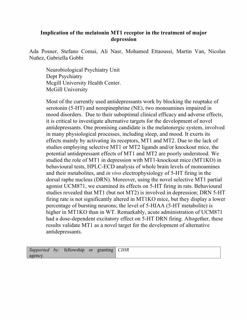
Implication of the melatonin MT1 receptor in the treatment of major depression
Ada Posner, Stefano Comai, Ali Nasr, Mohamed Ettaoussi, Martin Van, Nicolas Nuñez, Gabriella Gobbi
Neurobiological Psychiatry Unit Dept Psychiatry Mcgill University Health Center. McGill University
Most of the currently used antidepressants work by blocking the reuptake of serotonin (5-HT) and norepinephrine (NE), two monoamines impaired in mood disorders. Due to their suboptimal clinical efficacy and adverse effects, it is critical to investigate alternative targets for the development of novel antidepressants. One promising candidate is the melatonergic system, involved in many physiological processes, including sleep, and mood. It exerts its effects mainly by activating its receptors, MT1 and MT2. Due to the lack of studies employing selective MT1 or MT2 ligands and/or knockout mice, the potential antidepressant effects of MT1 and MT2 are poorly understood. We studied the role of MT1 in depression with MT1-knockout mice (MT1KO) in behavioural tests, HPLC-ECD analysis of whole brain levels of monoamines and their metabolites, and in vivo electrophysiology of 5-HT firing in the dorsal raphe nucleus (DRN). Moreover, using the novel selective MT1 partial agonist UCM871, we examined its effects on 5-HT firing in rats. Behavioural studies revealed that MT1 (but not MT2) is involved in depression; DRN 5-HT firing rate is not significantly altered in MT1KO mice, but they display a lower percentage of bursting neurons; the level of 5-HIAA (5-HT metabolite) is higher in MT1KO than in WT. Remarkably, acute administration of UCM871 had a dose-dependent excitatory effect on 5-HT DRN firing. Altogether, these results validate MT1 as a novel target for the development of alternative antidepressants.
Supported by: fellowship or granting agency.
CIHR
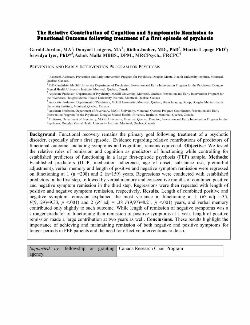
The Relative Contribution of Cognition and Symptomatic Remission to Functional Outcome following treatment of a first episode of psychosis
Gerald Jordan, MA1; Danyael Lutgens, MA2; Ridha Joober, MD., PhD3, Martin Lepage PhD4; Srividya Iyer, PhD*5;Ashok Malla MBBS., DPM., MRCPsych., FRCPC6 PREVENTION AND EARLY INTERVENTION PROGRAM FOR PSYCHOSIS
1 Research Assistant, Prevention and Early Intervention Program for Psychosis, Douglas Mental Health University Institute, Montreal, Quebec, Canada. 2 PhD Candidate, McGill University Department of Psychiatry; Prevention and Early Intervention Program for the Psychoses, Douglas Mental Health University Institute, Montreal, Quebec, Canada 3 Associate Professor, Department of Psychiatry, McGill University, Montreal, Quebec; Prevention and Early Intervention Program for the Psychoses, Douglas Mental Health University Institute, Montreal, Quebec, Canada 4 Associate Professor, Department of Psychiatry, McGill University, Montreal, Quebec; Brain Imaging Group, Douglas Mental Health University Institute, Montreal, Quebec, Canada 5 Assistant Professor, Department of Psychiatry, McGill University, Montreal, Quebec; Program Coordinator, Prevention and Early Intervention Program for the Psychoses, Douglas Mental Health University Institute, Montreal, Quebec, Canada 6 Professor, Department of Psychiatry, McGill University, Montreal, Quebec; Director, Prevention and Early Intervention Program for the Psychoses, Douglas Mental Health University Institute, Montreal, Quebec, Canada
Background: Functional recovery remains the primary goal following treatment of a psychotic disorder, especially after a first episode. Evidence regarding relative contributions of predictors of functional outcome, including symptoms and cognition, remains equivocal. Objective: We tested the relative roles of remission and cognition as predictors of functioning while controlling for established predictors of functioning in a large first-episode psychosis (FEP) sample. Methods: Established predictors (DUP, medication adherence, age of onset, substance use, premorbid adjustment), verbal memory and length of positive and negative symptom remission were regressed on functioning at 1 (n =208) and 2 (n=159) years. Regressions were conducted with established predictors in the first step, followed by verbal memory and consecutive months of combined positive and negative symptom remission in the third step. Regressions were then repeated with length of positive and negative symptom remission, respectively. Results: Length of combined positive and negative symptom remission explained the most variance in functioning at 1 (R² adj =.35, F(9,129)=9.33, p <.001) and 2 (R² adj = .38 F(9,97)=8.21, p <.001) years, and verbal memory contributed only slightly to such outcome. While length of remission of negative symptoms was a stronger predictor of functioning than remission of positive symptoms at 1 year, length of positive remission made a large contribution at two years as well. Conclusions: These results highlight the importance of achieving and maintaining remission of both negative and positive symptoms for longer periods in FEP patients and the need for effective interventions to do so. Supported by: fellowship or granting agency.
Canada Research Chair Program
Neurogenic and affective regulation by hippocampal neuregulin-1/ErbB signalling Ian Mahar1,2, Benoit Labonte1,2, Stephanie Tan1, Maria Antonietta Davoli1, Sergio Dominguez-Lopez2, Calvin Qiang1, Adeline Rachalski1, Gustavo Turecki1,2,3,4, Naguib Mechawar1,2,4,
1McGill Group for Suicide Studies, Douglas Mental Health University Institute, McGill University, Montreal, Quebec, Canada. 2Department of Neurology & Neurosurgery, McGill University, Montreal, Quebec, Canada. 3Department of Human Genetics, McGill University, Montreal, Quebec, Canada. 4Department of Psychiatry, McGill University, Montreal, Quebec, Canada.
Background Hippocampal neurogenesis has been implicated in the mechanism of antidepressant action, and neurotrophic factors can mediate neurogenic changes underlying these effects. The neurotrophic factor neuregulin-1 (NRG1) is involved in many aspects of brain development. However, little is known about the influence of NRG1 on neurodevelopmental processes in the mature hippocampus. Objective These experiments examine the potential role of NRG1-ErbB signaling in hippocampal neurogenesis, affective behaviour, and psychopathology. Methods Adult mice were given subcutaneous NRG1 or saline to assess dentate gyrus (DG) proliferation, survival, differentiation, morphology, and neurogenesis, and underwent behavioral testing. Expression of NRG1 receptors in newborn DG cells was assessed at various time points. ErbB-expressing progenitor cell phenotype was characterized. DG dissections were used for ELISA. We extended these findings into psychiatrically-relevant human subjects, examining hippocampal NRG1/ErbB gene expression and methylation in suicide completers and controls. Results Subchronic peripheral NRG1β administration selectively increased cell proliferation and neurogenesis (but not survival, differentiation, or immature neuronal morphology) in the ventral DG. These effects may have been mediated by ErbB3 receptors, expressed by newborn cells from division to maturity and colocalized with SOX2 in the subgranular zone. NRG1 increased ventral DG ErbB3 phosphorylation. Four weeks (but not acutely) after treatment, animals displayed antidepressant-like behavior. In humans, hippocampal ErbB3 expression was reduced in suicides, which was not due to ErbB3 methylation, although antidepressants induced methylation changes. Conclusions NRG1β has pro-proliferative, neurogenic, and antidepressant properties, potentially through NRG1-ErbB3 signaling, and human psychiatrically-relevant subjects show reduced hippocampal ErbB3 expression. This highlights the importance of peripheral neurotrophic factors in neurogenesis and mood, supports hippocampal neurogenesis as a mediator of antidepressant effects, and suggests a potential role of hippocampal NRG1-ErbB3 signaling in suicidality.
Supported by: fellowship or granting agency.
FRSQ, CIHR, NSERC
Manipulating the Sense of Agency with Magic Jay Olson, Alym Amlani, Amir Raz, Ronald Rensink RAZ LAB, DEPARTMENT OF PSYCHIATRY, MCGILL (J.O. & A.R.); VISUAL COGNITION LAB, DEPARTMENT OF PSYCHOLOGY, UNIVERSITY OF BRITISH COLUMBIA (A.A. & R. R.) Background. A common symptom of schizophrenia is alien control or a distorted sense of agency: thoughts and actions appear to originate from a source other than one’s own will. Scientists do not understand how these distortions occur. Studying similar distortions in the non-clinical population could help shed light on the mechanism underlying the sense of agency and suggest new options for treatment. Objective. Magicians often distort the sense of agency in their audiences. Specifically, forcing occurs when a magician influences the audience’s decisions while they retain a full sense of agency. For example, an audience member may feel as if she is freely choosing a playing card, though the magician is controlling the choice. We investigated the rate and predictors of forcing in order to distort the sense of agency and probe its mechanism. Methods and Results. In Study 1, a magician approached 108 people on the street and flipped through a deck of playing cards while each person chose a card. By making one card appear more salient, the magician could influence almost everyone (98%) to choose that card yet few (9%) noticed the influence. In Study 2, 43 participants observed rapid series of 26 cards shown on a computer. In each of the 28 trials, one target card was made more salient by showing it slightly longer than the rest. Participants chose the target card on 30% of the trials yet many (40%) were unaware of the influence. Among this unaware group, locus of control weakly predicted choice; people who believed they had more control over their lives were harder to influence. Among the aware group, stimulus factors such as the memorability of the card predicted choice; people chose more memorable cards. Studying this paradigm of forcing has demonstrated a robust method of distorting the sense of agency during simple decisions. Future research can explore how such distortions relate to disorders of mental control in order to better understand their symptoms and treatment. Supported by: fellowship or granting agency. NSERC
Adolescent amphetamine exposure disrupts the development of medial prefrontal cortex dopamine connectivity
Lauren M. Reynolds1,3, Carolina S. Makowski3, Sandra V. Yogendran1,3, Cecilia Flores2,3 1 Integrated Program in Neuroscience, McGill University; 2 Department of Psychiatry and Department of Neurology and Neurosurgery, McGill University; 3 Douglas Mental Health University Institute Initiation of drug use during adolescence is a strong predictor of both the incidence and severity of lifetime drug abuse. Intriguingly, adolescence is also a period of dynamic refinement to the organization of neuronal connectivity, in particular dopamine circuitry. The enduring behavioral effects of adolescent drug exposure may therefore result from disruption in dopamine development. In this study we investigated (a) the effects of repeated exposure to amphetamine during adolescence on behavioral sensitization, conditioned locomotor activity, and organization of dopamine circuitry in adulthood; (b) we compare these effects to those induced by an identical amphetamine regimen administered in adulthood. We treated early adolescent (PND 21±1) and adult (PND 75±15) male C57/BL6 mice with i.p. injections of amphetamine (4 mg/kg) or saline. Mice received one injection every other day for a total of 5 days. Behavioral sensitization and conditioned locomotor activity were evaluated 6 weeks after the last pretreatment dose for both adolescent and adult pretreated groups. This delay allowed adolescent-pretreated mice to reach adulthood; adult-pretreated mice were matched to the same interval. We then assessed dopamine connectivity in the medial prefrontal cortex and nucleus accumbens. We found that mice that received amphetamine treatment either in adolescence or in adulthood show conditioned locomotor activity when re-exposed to the previously drug paired chamber. Importantly, this conditioned response was exaggerated in adult mice that received drug treatment during adolescence. Stereological analysis of the expanse and number of dopamine varicosities revealed that amphetamine exposure during adolescence, but not in adulthood, leads to an increase in the volume that dopamine axons occupy in the medial prefrontal cortex, but that these axons are “denuded” from presynaptic sites. This effect appears to be selective to the medial prefrontal cortex because we did not find changes in dopamine innervation in the nucleus accumbens. These findings show that amphetamine in adolescence alters the development of the dopamine input to the medial prefrontal cortex, reducing dramatically dopamine synaptic connectivity, and alters conditioned locomotor response. Remarkably, our most recent results show that the effects of adolescent amphetamine exposure on medial prefrontal cortex development are mediated by DCC signaling within dopamine neurons.
Supported by: fellowship or granting agency.
CIHR, NSERC, FRSQ, FQRNT, Fulbright Canada.
Prenatal Maternal Stress from a natural disaster predicts hippocampus volumes in males at age 11: Project Ice Storm
R. Dufoix1,2, A. Charil3,4, D. P. Laplante2, T. Paus.,5,6, J. Pruessner1,2, S. King1,2
1McGill University, 2Douglas Hospital Research Center, 3Australian Nuclear Science and Technology Organisation, 4University of Sydney, 5Rotman Research Institute and 6University of Toronto Introduction: The hippocampus develops primarily during the fetal period and plays a pivotal role in learning and memory. Non-human primate studies have demonstrated that early and late in utero exposure to maternal stress result in reduced hippocampal volumes and a 32% inhibition of postnatal neurogenesis in the dentate gyrus, that remain noticeable two years later. However, no human prospective studies of the effects of prenatal maternal stress (PNMS) on the hippocampal development have been conducted. Objective: To determine whether in utero exposure to disaster-related PNMS is associated with altered hippocampal volumes in 11½ year-old children. We hypothesized that higher maternal objective or subjective PNMS levels would be related to smaller hippocampal volumes. Methods: Measures of maternal objective (i.e., events the women experienced) and subjective (i.e., the women’s psychological response to the crisis) PNMS were obtained after the 1998 Quebec Ice Storm. We obtained T1-weighted images of 33 males and 32 female 11½ year olds. Hippocampal segmentations from native MRI scans were performed using the automatic SACHA method. The number of obstetric complications was recorded using hospital records. Results: Overall, more obstetric complications were related to smaller hippocampal volumes in both male and females. Higher levels of maternal objective stress were related to smaller right hippocampal volumes (RHCV) in males only. Objective stress and obstetric complications and their interaction term accounted for 34.6% of the variance of the male’s RHCV. Inspection of the objective stress × obstetric complication interaction revealed that males exposed to high levels of objective stress or obstetric complication or both had smaller RHCV compared to males exposed to low levels of objective stress and obstetric complications. Conclusions: Our results suggest that higher levels of disaster-related objective stress, but not subjective distress are related to smaller RHCV in male but not female children. It remains to be determined whether this alteration in RHCV of 11.5 years old males is related to observable phenotypes. Supported by: fellowship or granting agency.
March of Dimes foundation and the Canadian Institutes of Health Research.
Effect of TNF-alpha inhibition in a prodromal model of Alzheimer’s disease Chelsea Cavanagh, Remi Quirion & Tak Pan Wong WONG LAB, DOUGLAS HOSPITAL RESEARCH CENTRE Background: Alzheimer’s disease (AD) develops decades before clinical symptoms arise and can remain undiagnosed for years. For this reason, there is a need to intervene before the development of cognitive symptoms. One possible culprit in the development of AD is tumor necrosis factor-alpha (TNF-alpha), a pro-inflammatory cytokine. TNF-alpha is upregulated in both humans with AD and transgenic models of the disease. TNF-alpha is a well-known immune mediator and is recognized as a modulator of learning, memory, and synaptic function. The role of TNF-alpha in synaptic function is thought to be regulatory at physiological levels, but neurotoxic when found in excess. Since proper synaptic function underlies healthy cognitive processing, it is critical to understand the effect of TNF-alpha on synaptic function in AD. Consequently, the early intervention of TNF-alpha signaling may help prevent the detrimental effects of excess TNF-alpha on synaptic function. We hypothesize that increased levels of TNF-alpha at early stages of AD-like pathology may become pathogenic and lead to deficiencies in synaptic function. Objective: To advance the current understanding of TNF-alpha signaling as it relates to synaptic pathology in the early stages of AD. Methods: We used 1-month-old TgCRND8 mice, which harbor a mutated form of the amyloid precursor protein (APP), the precursor to amyloid-beta, to study synaptic function in acute hippocampal slices using electrophysiological field recordings. Levels of TNF-alpha and amyloid-beta were measured by enzyme-linked immunosorbent assays. Results: We found that TNF-alpha is significantly increased in hippocampal tissue from 1-month-old TgCRND8 mice. These mice also displayed an increased post-synaptic response relative to a given pre-synaptic input compared to control mice. This increase in synaptic output could be attenuated using a TNF-alpha inhibitor. Notably, these alterations were present before the overproduction of amyloid-beta. Conclusion: Our findings provide insight on TNF-alpha as a potential therapeutic target in the prodromal stages of AD and help elucidate the effects of TNF-alpha on synaptic function. Inhibiting TNF-alpha at early stages of the disease could be a novel therapeutic approach to prevent synaptic pathology and consequent memory deficits in AD. Supported by: fellowship or granting agency.
StoP-AD Centre & IPN Returning Student Award
The longitudinal effects of neighborhood social and material deprivation change on psychological distress in urban, community-dwelling Canadian adults
Alexandra Blair (1,2), Geneviève Gariepy (2,3), Norbert Schmitz (1,2,3) (1) DR. NORBERT SCHMITZ PSYCHIATRIC EPIDEMIOLOGY LAB, DOUGLAS MENTAL HEALTH UNIVERSITY INSTITUTE, (2) DEPARTMENT OF PSYCHIATRY, MCGILL UNIVERSITY, (3) DEPARTMENT OF EPIDEMIOLOGY, BIOSTATISTICS, AND OCCUPATIONAL HEALTH, MCGILL UNIVERSITY. Background: Neighborhood characteristics are known to be associated with affective health outcomes. However, neighborhoods have rarely been examined as time-varying spaces of ecological exposure. Objective: To examine how changes in neighborhood material and social deprivation affect distress outcomes in adult Canadians. Methods: This study examines 2745 participants from Canada's National Population Health Survey, all of whom are urban- and community-dwelling, non-institutionalized adult Canadians (aged 18 and above at baseline in 2000). Participants did not change neighborhood of residence between baseline and follow-up in 2006. Psychological distress was measured at baseline and follow-up using the K6 screening tool. Changes in social and material deprivation were measured using the Pampalon Social and Material Deprivation Index. Associations were analyzed using multivariate linear regressions, controlling for key demographic characteristics, and stratified by baseline deprivation exposure. Results: At baseline, most participants lived in neighborhoods with low levels of social deprivation, and nearly half of the sample lived in highly materially deprived neighborhoods. Most neighborhoods maintained the same level of social and material deprivation through time. In fully adjusted models, an improvement of social and material settings was associated with a significant increase in distress scores at follow-up. This association remained positive after controlling for baseline distress scores, but the confidence interval shifted slightly to cross the null. Conclusions: This study suggests that an improvement in neighborhood deprivation, when unplanned and caused by systematically occurring shifts in economic, political, and social environments (i.e. urban gentrification) is associated with worsening psychological distress outcomes. This finding is relevant for urban municipalities, as well as future public health policy. The psychological effects of gentrification should be explored in more detail in future research. Supported by: fellowship or granting agency.
FRSQ, CIHR
Mesocortical dopamine depletion reverses blunted responses to amphetamine in dcc haploinsufficient mice
Matthew Pokinko1,2, Luc Moquin2, Dr. Alain Gratton2,3, Dr. Cecilia Flores2,3,4 1 Integrated Program in Neuroscience, McGill University 2 Douglas Mental Health University Institute 3 Department of Psychiatry, McGill University 4 Department of Neurology and Neurosurgery, McGill University The netrin-1 receptor deleted in colorectal cancer (DCC) determines a) the extent of dopamine (DA) innervation to the medial prefrontal cortex (mPFC), b) the organization of local circuitry, and c) individual vulnerability to the effects of drugs of abuse. Adult dcc haploinsufficient mice have a selective increase in mPFC DA fiber innervation and DA release in comparison to wild-type littermates. Furthermore, adult dcc haploinsufficient mice show blunted DA release in the nucleus accumbens (NAcc) and reduced locomotor activity when challenged with drugs of abuse such as amphetamine (AMPH). Because mPFC DA transmission in the mPFC can negatively regulate DA release in the NAcc in response to drugs of abuse or stressors, we hypothesized that the blunted effects of AMPH in dcc haploinsufficient mice result from increased mPFC DA innervation. We therefore examined the effects of selective mPFC DA depletion on AMPH-induced locomotion in dcc haploinsufficient and wild-type mice. Adult mice received bilateral mPFC injections of 6-hydroxydopamine (lesion) or vehicle (sham) 10 days before an i.p. AMPH challenge. In wild type mice, AMPH-induced locomotion was similar between lesion and sham groups. Remarkably, mPFC DA lesions in dcc haploinsufficient mice reversed their blunted response to AMPH. These findings demonstrate that the protective phenotype of adult dcc haploinsufficient mice results from the effects of DCC on the organization of mPFC DA connectivity. Supported by: fellowship or granting agency.
NSERC, CIHR, FRSQ, FQRNT, IPN returning student award
Brain markers of suicidal vulnerability in mood disorders: a model-based structural neuroimaging study with a translational perspective.
Yang Dinga; Natalia Lawrenceb; Emilie Oliéc; Fabienne Cyprienc; Emmanuelle le Barsd; Alain Bonaféd; Mary L. Phillipse; Philippe Courtetc; Fabrice Jollanta
a McGill University and Douglas Mental Health University Institute, McGill Group for Suicide Studies, Montreal (Québec) Canada. b Mood Disorders Centre, School of Psychology, College of Life and Environmental Sciences, University of Exeter, Exeter, UK. c Université Montpellier I & CHU Montpellier, department of psychiatry & Inserm, U1061, Montpellier, France. d Université Montpellier I & CHU Montpellier, department of radiology, Montpellier, France. e Clinical and Translational Affective Neuroscience Program, University of Pittsburgh School of Medicine, USA.
Background: The vulnerability to suicidal behavior has been modeled in deficits in both valuation processes, mediated by ventral prefrontal cortex, and cognitive control processes, associated with dorsal prefrontal cortex. Objective: To uncover potential markers of suicidality based on this model, we compared several brain morphometric parameters using magnetic resonance imaging in a large sample and tested their classificatory properties. Method: Three groups were recruited: euthymic suicide attempters with a past history of mood disorders and suicidal behaviour (N=67); patient controls with a past history of mood disorders but not suicidal behaviour (N=82); healthy controls without any history of mental disorder (N=82). A theory-driven region-of-interest approach was applied targeting the orbitofrontal cortex (OFC), the anterior cingulate cortex (ACC) and the dorsolateral prefrontal cortex (DLPFC). Both voxel-based (SPM8) and surface-based morphometry (Freesurfer) analyses were used to comprehensively evaluate cortical gray matter signal, volume, surface area and thickness. Results: Suicide attempters showed lower signal than controls in bilateral DLPFC (p<0.003) with a very large effect size (Cohen’s d=4.8). This measure was correlated with the number of previous suicidal acts and impulsivity trait. Moreover, it improved specificity in detecting suicide attempters when combined with mood disorder status. Contrasts between attempters and patient controls additionally showed large effect sizes (>1) for OFC area, ACC volume, and DLPFC area and thickness. Overall, individuals with histories of suicide attempts show structural deficits independently of mood disorders, which may play a significant role in their vulnerability to suicidal acts. A potential future clinical application of these biomarkers is suggested. Supported by: fellowship or granting agency.
FRSQ PhD Grant
Melatonin, selective and non-selective MT1/MT2 receptors agonists: Differential effects on the 24-h vigilance states
1 Neurobiological Psychiatry Unit, Department of Psychiatry, McGill University, Montreal, QC, Canada. 2 Department of Biomolecular Sciences, University of Urbino “Carlo Bo”, Urbino, PU, Italy.
The neurohormone melatonin (MLT) activates two G-protein coupled receptors, MT1 and MT2. MLT is implicated in sleep regulation, however, its role as hypnotic agent still controversial, and the neurobiological mechanism through which it controls the vigilance states has not yet been elucidated. Therefore, the aim was to study the effects of MLT (40 mg/kg) on the 24-h vigilant states by using electroencephalogram and electromyogram in Sprague-Dawley male rats and then to compare these effects with those produced by a non-selective MT1/MT2 receptor agonist (UCM793, 40 mg/kg) and a selective MT2 receptor partial agonist (UCM924, 40 mg/kg). MLT reduced the latency (+33%) to the first episode of non-rapid eye movement sleep (NREMS) but does not affect the duration of wakefulness, NREMS and rapid eye movement sleep (REMS) during the 24-h sleep–wake cycle. Differently, UCM793 altered the architecture of the awake state increasing its number of episodes (+52%) and decreasing the length of the episodes (-38%) but did not alter the 24-h duration of the three vigilance states. These results corroborate our recent findings in MT1/MT2 receptors knockout mice showing that the modulation of both MT1 and MT2 receptors yields no effects on sleep stages because of the selective and opposite role of each MLT receptor subtype on NREMS. Then, UCM924 instead reduced the latency (-56%) and increased (+31%) the duration of NREMS. UCM765 also increased the number of REMS episodes (+57%) without affecting REMS duration. Taken together, these findings show that MLT and non-selective MT1/MT2 receptor agonists do not increase the quantity of sleep but differently influence the three vigilance states. In addition, they support the evidence that selective MT2 receptor agonists increase NREMS duration compared to MLT and non-selective MT1/MT2 agonists. Supported by: fellowship or granting agency.
Memory reconsolidation blockade to treat substance dependence: A feasibility study.
Michelle Lonergan, MSc. DOULGAS MENTAL HEALTH UNIVERSITY INTITUTE, PSYCHOSOCIAL DIVISION; MCGILL UNIVERSITY, DEPARTMENT OF PSYCHIATRY Background: Conditioned drug-related memories formed during repeated drug using episodes are proposed to underlie craving and the long-term propensity for addicted individuals to relapse, posing a formidable barrier to sustained recovery. Reconsolidation theory suggests that the act of retrieval transiently destabilizes previously consolidate memories, during which time they can be pharmacologically manipulated prior to re-stabilizing back to long-term storage. Previous literature has revealed that the noradrenergic beta-blocker propranolol can reduce drug-seeking behaviour in rodents and drug craving in humans when administered in conjunction with the retrieval of drug-related memories, opening the door to a novel treatment for addiction. Objective: In a feasibility study, we examined whether memory reconsolidation blockade using propranolol can be successfully implemented in a sample of treatment-seeking individuals with substance dependence. We further explored preliminary treatment effects. Methods: Eligible participants (18-65 years old) were randomized to receive six treatments of memory reconsolidation blockade under propranolol or placebo. Memory reactivation was achieved by having participants read aloud to the investigator a personal drug-using narrative. Outcome measures included evaluating recruitment and retention, the eligibility criteria, protocol adherence, preliminary treatment effects as measured by difference scores on self-report craving severity between the baseline and post-treatment assessments, and rates of relapse. Data Analysis: Feasibility outcomes are reported as counts and percentages. For analysis of treatment effects, missing data was imputed using multiple imputation procedures, and two-tailed independent t-tests were used to examine between-group differences on craving change between baseline and 1-week follow-up. Relapse occurrence was dichotomized and compared between groups. Results: Although comparable to literature examining attrition in clinical trials and addiction treatment programs, we observed a 45% attrition rate. The eligibility criteria and protocol adherence were considered appropriate and adequate. Results from preliminary analyses of treatment effects revealed no significant between-group differences on change in subjective craving or relapse during the trial, despite propranolol treated participants tending to demonstrate slightly greater improvement. Conclusion: Despite finding no significant effect of treatment, large multi-center trials of disrupting memory reconsolidation to treat substance dependence are warranted, provided several procedural changes are implemented. The authors discuss ways to address potential methodological pitfalls in future studies. Supported by: fellowship or granting agency.
Partially supported by FRSQ (2013-2014) and CIHR Master’s (2012-2013) awards
Cognitive endophenotypes of suicide: a study in first-degree relatives of suicide completers. Alexandra Hoehne1,2, Yang Ding1,2, Martin Lepage1, Gustavo Turecki1,2, Fabrice Jollant1,2
1Psychiatry, McGill University, Montreal, Quebec, Canada; 2McGill Group for Suicide Studies, Douglas Mental Health University Institute, Montreal, Quebec, Canada. Background Heritability of suicide has been well established in family, twin and adoption studies. Transmission of risk appears to follow vulnerability traits more than co-morbid disorders like depression. Suicide attempters have been found to show cognitive deficits including impaired decision-making and cognitive inhibition. Objective In the present project, we aimed at investigating potential cognitive deficits in relatives of suicide completers. We hypothesized that the same cognitive deficits observed in suicide attempters would be found in first-degree relatives of suicide completers. Method 17 first-degree relatives of suicide completers without any personal history of suicidal act (RSC) were compared to 18 first-degree relatives of individuals with Major Depressive Disorder but no family history of suicidal act (RMDD), and 19 first-degree relatives of healthy controls (RHC). A large battery of neuropsychological tests was used (National Adult Reading Test, Iowa Gambling Task, Wechsler Adult Intelligence Scale, Trail-Making Test, Stroop, Hayling Sentence Completion Test) evaluating decision-making, cognitive inhibition, selective attention, mental flexibility and processing speed. Results Preliminary analyses show a trend for poorer decision-making performance in RSC vs. RMDD and RHC. Previous findings have associated this decision-making deficit to an abnormal functioning of the orbito-frontal cortex in suicide attempters compared to non-attempters. Present findings suggest that a similar dysfunction may be found in first-degree RSC. Additional analyses will be conducted soon. These preliminary findings suggest that cognitive deficits, notably disadvantageous decision-making, may represent endophenotypes of suicide. Impairment in cognitive functioning may be a relevant marker of suicidality and the target of future preventative interventions. Supported by: fellowship or granting agency.
Faculty of Medicine Internal Studentship
Developmental Responses of the Lateral Hypothalamus to Leptin and Ghrelin in Neontatal Rats
Eva Gjerde1,2, Hong Long2, Claire-Dominique Walker2 1Integrated Program in Neuroscience, McGill University, 2Douglas Mental Health University Institute, Montreal, Canada
Food intake is regulated by a close communication between the homeostatic system in the hypothalamus and the hedonic system (including the mesocorticolimbic pathway). A key region for this communication is the lateral hypothalamus (LH), also known as the “feeding center” of the brain. Both the homeostatic and hedonic systems are still developing during the late fetal and early post-natal periods in the rat, and are known targets for peripheral metabolic hormones such as leptin and ghrelin. Here we examined the onset of leptin and ghrelin responsiveness in the LH, and the activation of cellular signaling molecules in identified neurons on postnatal day (PND) 10 and 16. Leptin significantly activated secondary messengers pERK and pSTAT3 in the LH on PND16, while on PND10, only sparse pSTAT3 cells were identified and the activation of pERK did not reach significance. Double-immunofluorescence staining showed that the majority of pERK-activated neurons are orexin-A (ORX-A) positive neurons. However, leptin-induced pSTAT3 was clearly observed in another population of neurons, potentially neurotensin (NT) neurons. It is unknown whether pERK activation in ORX-A neurons is a direct effect of leptin on ORX-A cells via specific leptin receptors (ObRb), or an indirect activation via leptin-sensitive projections to the ORX-A neurons. Follow-up experiments involving in-situ hybridization of ObRb’s with immunohistochemical detection of ORX-A and NT neurons will determine whether ObRb’s can be identified on these neurons in the LH during neonatal development. In contrast to leptin responses, ghrelin administration significantly increased Fos activity in ORX-A neurons of the LH on PND10, but the increase over saline injection was not significant on PND16. Our results suggest that metabolic hormone responsiveness in the LH is only partially developed prior to the onset of independent feeding on PND16, and that responses to ghrelin might precede those to leptin in this area of the hypothalamus. Furthermore, the maturation of functional connections between the LH and the ventral tegmental area (VTA – a major center of the hedonic system) may play a crucial role in the onset of responsiveness to metabolic hormones and initiation of independent feeding in rat neonates. Supported by CIHR to CDW.
Illicit and Prescription Opiate Abuse: Understanding Treatment Failure And Improving Outcomes
Kevin Hamdullahpur, Dr. Kathryn Gill MUHC ADDICTIONS UNIT Background: Dependence on opiates is a major health issue in North America. The recent increases in both prescription and illicit opiate abuse have exacted enormous tolls in terms of health care, mental illness, quality of life, unemployment, and crime, while the difficulty in treating opioid dependent patients with standard abstinence-based therapies is not well understood. Objectives: The objective of this study was to provide a novel approach to understanding the poor outcomes of opiate dependent patients by focusing on identifying predictors of treatment failure, and by performing qualitative interviews focused on patient treatment experiences. Methods: This study was conducted at the Addictions Unit of the McGill University Health Center in Montreal. Patients were prospectively monitored during inpatient detoxification for opiate dependence or sedative-hypnotic dependence in terms of craving, mood, withdrawal symptoms, vital signs, subjective experiences of pain, and objective measures of hyperalgesia and allodynia. Patient psychiatric comorbidity (Axis I and Axis II disorders), chronic medical conditions (pain syndromes), and severity of substance dependence were also considered. Qualitative interviews were performed following treatment completion to compliment quantitative results and to provide a richer, more comprehensive understanding of the difficulties experienced by patients undergoing detoxification. Results indicated that during treatment patients with cluster B personality disorders reported more negative mood symptoms (anger, anxiety, depression) and greater scores on objective measures of withdrawal. Opiate dependent patients were more likely to have chronic pain conditions, and demonstrate increased physical sensitivity and lower pain thresholds. Together these findings suggest that hyperalgesic, highly sensitive opiate-dependent patients with cluster B personality disorders may have substantial difficulties tolerating both the physical and emotion symptoms of withdrawal, and may benefit from the development of targeted interventions. Supported by: fellowship or granting agency.
CIHR
The Role of Medial Septal Glutamatergic Neurons within the Septum and their Influence on Theta Oscillations in the Hippocampus
Robinson, J., Manseau, F., Williams, S. WILLIAMS LAB, MCGILL UNIVERSITY Background: Neurons in the medial septum diagonal band of Broca (MS-DBB) are well known to provide important connections to the hippocampus, and are critical for spatial learning and memory. Three main neuronal populations have been identified in this region: cholinergic, GABAergic and glutamatergic. Glutamate neurons were shown (by Williams’s lab) to project to the hippocampus, release glutamate on interneurons and principal cells and to discharge rhythmically at theta frequency. To further explore the role of MS-DBB glutamatergic cells we have used optogenetics to specifically target these neurons. Objectives: The first aim of our study has been to determine at the cellular level how glutamate release from MS-DBB neurons can modulate post-synaptic targets within the septum as well as interneurons and principal neurons across the hippocampus. The second aim of the current study has been to determine whether increasing the firing rate of glutamate neurons is sufficient to modulate hippocampal theta oscillations. Methods: To explore the role of MS-DBB glutamatergic neurons we have used optogenetics to specifically target these neurons. We have targeted glutamatergic VGLUT2 neurons of the MS-DBB with the light-sensitive protein channelrhodopsin-2 variant, ChETA. The VGLUT2 CRE-mouse line are injected with a ChETA Cre-recombinase adeno-associated virus to specifically target glutamate neurons of the MS-DBB. Recording techniques used to achieve the specified goals of this study include both patch-clamp and field recordings to either in slice or the entire septo-hippocampal preparation in vitro. Results: By selectively activating this neuronal population, we have obtained large postsynaptic excitatory potentials onto both neurons within the septum as well as to interneurons in CA3 of the hippocampus. In addition, glutamatergic MS-DBB neurons can modulate hippocampal theta rhythm in the whole septo-hippocampal network in vitro, modulating both the power and frequency of theta rhythms. These experiments will provide for the first time an explanation of how medial septum glutamate neurons contribute to hippocampal theta activity. Finally, these results will also provide the background necessary to reinvestigate why medial septum glutamatergic input is critical for learning and memory in freely behaving mice. Supported by: fellowship or granting agency.
CIHR to SW lab
Olfactory Sensory Input Processing as a sign of AD-related pathology-physiology Lafaille-Magnan, M.-E.1,2 , Collins, L.1,3, Fonov 1,3, V., Fontaine, D. 2, Rosa-Neto, P. 1,2 LaBonté, A. 2, Poirier, J. 1,2, Breitner, J.1,2 1) McGill University 2) Douglas Mental Health Institute 3) Montreal Neurological Institute 1) MCGILL UNIVERSITY 2) DOUGLAS MENTAL HEALTH INSTITUTE 3) MONTREAL NEUROLOGICAL INSTITUTE Background: Olfactory sensory deficit is as a sign of neurodegeneration in the pre-motor phase of Parkinson's disease, and dysfunction is a likely early symptoms of Alzheimer's disease (AD). The olfactory bulb and entorhinal cortex are among the first brain structures affected by AD pathology. Progression of sensory deficit, especially olfaction, is often overlooked in longitudinal studies of Alzheimer's disease pathogenesis. Methods: We are investigating the potential of diminished olfactory identification as a quantitative marker for the progress of pre-symptomatic AD in a longitudinal study of cognitively normal people with a parental history of AD. We have evaluated > 100 people, a majority having at least one follow-up examination. 42% of active participants are enrolled in a protocol of four serial lumbar punctures, and almost all undergo serial MRI scanning, genetic testing, a smell identification testing, and neurocognitive assessment. Results: Preliminary findings show significant association of deficits in cognition (RBANS total index score) and smell identification (UPSIT) (Spearman’s r=0.262, p=0.001, n=155). We also note significant correlations between smell identification and bilateral hippocampal volume as well as total ventricular (r=0.238, p=033, n=80; r=-0.299, p=0.007, n=80). Remarkably in a subset of participants, olfactory deficit contribute to a model to predict CSF levels of total tau, 181P-tau and Aβ1-42. Discussion: We are the first to measure an association between CSF biomarkers of AD in living human beings and smell identification. It will be important to assess olfactory sensory processing (and possibly interpretation of other sensory modalities) may serve as a convenient and non-invasive metric of evolving neurodegenerative disease. Supported by: fellowship or granting agency.
Pfizer McGill Chair, McGill, FRSQ, CFI
Early changes in the offspring mesolimbic dopaminergic system induced by perinatal maternal high-fat diet
MinGi Cho 1,2, Greg Dal-Bo 2, Hong Long 2, Claire-Dominique Walker 2 1Integrated Program in Neuroscience, McGill University and 2Douglas Mental Health University Institute, Montreal, Canada Recent evidence demonstrates that maternal high-fat consumption during pregnancy and lactation increases the risk of metabolic disorders and obesity in the offspring; however, the underlying mechanisms remain unknown. We previously showed that adult offspring of mothers exposed to a high-fat (HF) diet during the last week of gestation and lactation have altered mesolimbic dopamine (DA) activity, a system that regulates the hedonic value of food. In particular, adult HF offspring display increased tyrosine hydroxylase (TH) activity and reduced D2 receptor expression in the VTA, but the time of onset of these changes is unknown. Here we examined when these changes first appear and whether they are dependent on the post-weaning diet. Contrary to the adult offspring, D2 receptor mRNA expression in the VTA of HF offspring was significantly increased at PND10 and PND16, suggesting that early exposure to high fat could modify the expression of presynaptic D2R in VTA target areas. On PND25, no differences were observed between HF and CD pups. We are currently investigating whether the dynamic regulation of D2R expression in the VTA is dependent upon constant exposure to the HF diet after weaning and are testing offspring during puberty (PND 45) maintained on either a HF or CD diet. In contrast to the increased TH levels observed in the adult HF offspring, pre-weaning HF pups on PND 10 or 16 did not exhibit significant differences in the concentration of TH in the VTA compared to its CD counterpart. To further examine potential early changes in DA synthesis, we are also examining whether the expression of Nurr-1, a transcription factor required for the development of DA cells and upregulation of DA synthesis, is increased in HF rats. Our studies demonstrate that expression of D2R mRNA levels in the VTA is a first and sensitive index of changes occurring in the DA system following exposure to HF diet in the early postnatal period. Increased production of DA in VTA neurons might be upregulated after weaning to a control diet, due to a mismatch between pre- and postweaning diets and compensatory changes induced to maintain adequate dopaminergic function. Supported by: fellowship or granting agency.
Supported by CIHR to CDW
Optogenetic dissection of the MCH system: implications for sleep-state modulation
Sonia Jego1, Stephen D. Glasgow1, Carolina Gutierrez Herrera1,2, Richard Boyce & Antoine R. Adamantidis1,2*
1 Douglas Institute, Department of Psychiatry, McGill University, Montreal 2 Department of Neurology, Inselspital, Bern University, Switzerland
The hypothalamus consists of intermingled inhibitory and excitatory neural circuits. The activity of these circuits correlates with vigilance states, including wakefulness, non-Rapid Eye Movement (REM) sleep and REM sleep. Recent evidence suggests that neurons expressing Melanin-Concentrating Hormone (MCH) are potentially sleep-promoting; however, the extent of their ability to selectively modulate sleep states remains unclear. To investigate the specific role of MCH neurons in the modulation of sleep states, we first genetically targeted the expression of excitatory (ChETA, SSFO) or inhibitory (eNpHR3.0) opsins to MCH neurons using an engineered mouse model. We then showed that we could optically activate (ChETA, SSFO) or inhibit (eNpHR3.0) MCH neurons with high reliability. Using real-time detection of vigilance state changes with EEG/EMG recordings, we next found that bilateral optogenetic activation of MCH neurons during NREM sleep increased the probability of NREM-to-REM sleep transitions, while MCH neuron activation during REM sleep extended REM sleep duration. These results were confirmed through the use of a step function opsin (SSFO) which increases excitability of targeted cells through sustained depolarization. In contrast, we showed that optogenetic silencing of MCH neurons during REM sleep reduced the amplitude of the cortical theta rhythm concomitant to an increase of oscillation strength in the slow theta range (3 to 5 Hz). Using an unbiased automatic detection of the slow theta events, we found that their occurrence was significantly increased in NpHR3.0 transfected animals compared to EYFP-expressing controls suggesting that these events are physiological, but rare, in natural sleep. Finally, we demonstrated that optical activation of MCH terminals induced fast GABAA-mediated inhibitory currents in local wake-promoting histaminergic (HA) neurons. This inhibitory tone was enhanced by optogenetically-induced MCH peptide release. Collectively, these results support a causal role for MCH neurons in the onset and maintenance of cortical REM sleep in the mammalian brain. Supported by: fellowship or granting agency.
Granting agency: CIHR, NSERC, CFI
Unit costs of the At Home/Chez Soi Project: estimating the societal costs of homelessness and mental illness Guido Powell1*, Angela Ly2, Daniel Rabouin3, & Eric Latimer11, 2, 3 1Department of Epidemiology, Biostatistics, and Occupation Health, McGill University, 1020 Pine Ave W., Montreal, Qc, H3A 1A2 2Université de Montréal, 2900 boul Edouard-Montpetit, Montreal, Qc H3T 1J4 3Douglas Mental Health University Institute, 6875 boul LaSalle, Montreal, Qc H4H 1R3 Objective: As part of the economic analysis of a Housing First intervention across 5 Canadian cities (Vancouver, Winnipeg, Toronto, Montreal and Moncton), we estimated the unit costs of service use (health, social, and justice) and residential stays from a societal perspective (combining public funds and private donations) Methods: The 5-year At Home/Chez Soi project 2148 homeless individuals with mental illness who were randomly assigned to private-market rent subsidies with recovery-oriented clinical services under the Housing First model (HF) or treatment as usual (TAU). Data on participants' service use and residential stays were obtained from questionnaires every 6 months or every 3 months, respectively. A top-down approach to estimating costs based on annual reports, financial statements, and personal communications was used whenever possible. In other instances, unit costs were estimated using a bottom-up approach or were adapted from previous literature, Statistics Canada and other reports. Unit costs included overhead costs and indirect costs while the opportunity cost of the building and land were estimated and used instead of reported amortization costs. Capitalization rates and municipal property assessments, validated using Google Street View, were used to estimate the market value of the building usage costs. Values in the cost estimates are from the 2009-2010 fiscal year, with values from previous years adjusted for inflation. Results: A total of 289 service use costs and 103 residential costs were estimated. Averaging across cities, service use costs include $263/outpatient hospital visit, $392/ER visit, $612/ambulance transportation, $116-$231/case manager visit (at office or in community), $29/visit to a drop-in center, $660/police arrest, and $3071/court appearance. Average residential costs include $68/bednight in an emergency shelter, $819/bednight in a psychiatric hospital, $170/stay in provincial jails, and $65/day in non-market housing. These unit costs were used in combination with the recorded costs of the intervention to calculate the cost-effectiveness of the At Home/Chez Soi project. Discussion: Both the methodology and the estimates obtained may be helpful for other economic analyses in the fields of health, social, and justice services, as well as homelessness and housing policy. Supported by: fellowship or granting agency.
Analysis of novel genetic factors in the late-onset form of Alzheimer’s disease J. Miron, D. Dea, L. Théroux-Lamarre, J. Frappier, C. Picard and J. Poirier Douglas Mental Health University Institute Research Centre, Departments of Neuroscience and Psychiatry, McGill University, Montreal, PQ, Canada, H4H 1R3
Recently, a genome-wide association study (GWAS) was performed by our team on patients with the late-onset form of Alzheimer’s disease (AD). These patients are from the Quebec Founding Population (QFP). This population originates from the 3000 people that emigrated from France 400 years ago to colonize Eastern Canada. After the 550 000 single nucleotide polymorphisms (SNPs) of our patients were measured, all of the significant SNPs (-Log (p) > 3.5) were validated in a second independent cohort, namely the population from France used in a GWAS performed by Dr Philippe Amouyel’s team. Among the SNPs that were found significant in both the QFP and the French population, one SNP (rs10984186) was located between CDK5RAP2 and TLR4 genes, while another SNP (rs10406151) was located close to PPP2R1A gene. CDK5RAP2 binds to the activator of CDK5 (p35/p25). CDK5 is the main kinase that phosphorylates Tau. Hyperphosphorylated Tau proteins are involved in the formation of neurofibrillary tangles, a key hallmark in the pathophysiology of AD. It is thus quite conceivable that CDK5RAP2 is directly implicated in the pathophysiology of Alzheimer’s disease. As for TLR4, the binding of amyloid plaques to this receptor activates the NF-κB transcription factor, which in turn increases the expression of inflammatory genes, such as cytokines and chemokines. These inflammatory products contribute in turn to the neuronal damage reported in AD. Finally, the PPP2R1A gene encodes for the α isoform of the regulatory subunit A of the PP2A complex, the main Tau phosphatase found in the mature adult brain. In this presentation, we will present our most recent findings on the gene expression of these risk-related genes in AD and discuss novel genetic variants found to be associated with pathophysiological markers typically associated with the AD brain. Supported by: fellowship or granting agency.
CIHR, Prevent-AD Centre, FRSQ and NSERC (JP)
***
Peer support, depressive symptoms, and suicidality among young adults Emily Yung, MSc1, Jennifer O’Loughlin, PhD2,3, Erika Dugas, MSc3, Nancy CP Low, MD, MSc1 1 LOW LAB OF ALLAN MEMORIAL INSTITUTE, DEPARTMENT OF PSYCHIATRY, MCGILL UNIVERSITY 2 DEPARTMENT OF SOCIAL AND PREVENTATIVE MEDICINE, UNIVERSITY OF MONTREAL 3 CENTRE HOSPITALIER DE L`UNIVERSITÉ DE MONTRÉAL RESEARCH CENTER Background: With the onset of most mental disorders occurring by age 24 years, seeking appropriate help is imperative to prevent or reduce impairment. However, less than 25% of young adults with a mental illness seek the help they need. Social support has been examined as a protective factor against depressive symptoms and suicidality in young adults. Young adults who are suicidal tend to not only show warning signs that others can recognize but also turn to a friend for help. Thus, peers who are supportive may have the potential to play a significant role in alleviating distress of another peer. Objectives: To test if peer support is associated with (1) depressive symptoms and (2) suicidal ideation in a population-based sample of young adults. Methods: Data were from the Nicotine Dependence in Teens (NDIT) Study, a prospective school-based cohort in Montreal. In 2011-12, data were collected from 858 participants in a mailed self-report questionnaire. 781 were included in this current analysis as 77 participants who had been diagnosed with a mood disorder were excluded. The self-report questionnaire measured the number of close friends (“people you feel at ease with and can talk to about what is on your mind”), depressive symptoms in the past two weeks by the Major Depressive Inventory (MDI), and suicidality in the past year (“felt suicidal or seriously considered committing suicide or taking your own life”). Linear and logistic regression models were conducted, controlling for age, sex, and work, marital and living status. Results: In this sample of participants who were aged 24 (0.7) years, the number of friends ranged from 0-20 with the median of 5 friends (IQR = 3). The MDI score ranged from 0-49 with the median of 6 (IQR = 8). Suicidal thoughts in the past year were reported by 8% of the sample. Young women had more depressive symptoms than young men (p = 0.000). More peer support was associated with fewer depressive symptoms (p = 0.003) and lesser suicidal thoughts (p = 0.022). Thus, peer support should be considered in the development of interventions that promote mental health among young adults. Supported by: fellowship or granting agency.
McGill University Faculty of Medicine Internal Studentship
A novel neurological mechanism driving rhythms of dopamine release IAN D. BLUM1, LEI ZHU1, LUC MOQUIN1, MAIA V. KOKOEVA2, ALAIN GRATTON1, BRUNO GIROS1, KAI-FLORIAN STORCH1
1Department of Psychiatry, McGill University, Douglas Mental Health University Institute, Montreal, QC, CANADA 2Department of Medicine, McGill University, Royal Victoria Hospital, Montreal, QC, CANADA Ultradian (~4hr) rhythms of locomotor activity have been repeatedly observed across mammalian species, including humans, yet not a single study has attempted to identify the neural substrate underlying them. We observed that distinct alterations of the dopaminergic system – either by genetic ablation of the dopamine transporter (DAT) or pharmacological manipulation with psychostimulants – leads to ultradian rhythm lengthening from approximately 4hrs up to 110hrs in extreme cases. Importantly the antipsychotic haloperidol had the opposite effect; shortening periodicity in naïve, methamphetamine treated, and DAT-/- animals. As period altering effects strongly suggest that dopamine resides at the core of a putative oscillator controlling locomotion we performed in vivo microdialysis in freely moving animals and demonstrate that dopamine fluctuates in synchrony with the ultradian oscillations of locomotor activity in naïve mice. Amazingly, the mean dopamine tone during sampling, calculated by high pressure liquid chromatography, is highly correlated with the endogenous period of locomotor activity of the individual mice (R2 = 0.91, p < 0.0001) thereby providing a measure with enormous predictive potential. Furthermore, total dopamine measured from brain punches taken from methamphetamine treated mice provide corroborating evidence and pinpoint the Caudate Putamen (rather than the Nucleus Accumbens) as the output site of this oscillator. Pilot studies currently underway using viral targeted expression of designed receptors activated by designer drugs (DREADDs) in dopaminergic neurons (DAT-Cre) are just now allowing us to remotely excite these cells and observe ultradian period lengthening, narrowing the list of candidate nuclei to the midbrain. Our data support the existence of a dopaminergic ultradian oscillator (DUO) operating in the mammalian brain that controls arousal in concert with the circadian clock. The striking similarity between the aberrant sleep/wake patterns observed here and in human subjects afflicted with either bipolar disorder or schizophrenia suggests that this clock is similarly perturbed in psychiatric conditions associated with hyperdopaminergia and that locomotor activity patterns may provide a new biomarker for altered dopaminergic tone and associated mental illnesses. Supported by: fellowship or granting agency.
NSERC discovery grant (K.F.S.), CIHR operating grants (K.F.S. and A.G.) and by the Graham Boeckh Foundation (B.G.). K.F.S. holds a CIHR New Investigator Award and B.G. a Canada Research Chair.


![Financial Decentralization in Malaysian [1] Faculty of Education, … · 2016. 1. 25. · Shahril@Charil Marzuki, Rahimah Ahmad, & Hussein Ahmad, 2010). This was in congruence with](https://static.documents.pub/doc/80x56/61180012c7612f5ab449a4ba/financial-decentralization-in-malaysian-1-faculty-of-education-2016-1-25.jpg)
