Clinical Roundtable Monograph Discussants Stephanie A. Gregory, MD Head, Section of Hematology Elodia Kehm Professor of Medicine Professor, Department of Internal Medicine Rush University Medical Center/Rush University Chicago, Illinois Ruben A. Mesa, MD Professor of Medicine Chair, Division of Hematology and Medical Oncology Mayo Clinic Scottsdale, Arizona Ronald Hoffman, MD Professor of Medicine, Hematology and Medical Oncology Professor of Gene and Cell Medicine The Mount Sinai Medical Center New York, New York Jamile M. Shammo, MD Associate Professor of Medicine and Pathology Rush University Medical Center Chicago, Illinois Clinical Advances in Hematology & Oncology September 2011 Clinical and Laboratory Features of Myelofibrosis and Limitations of Current Therapies Abstract: Myelofibrosis (MF) is a life-threatening clonal stem cell malignancy characterized by progressive bone marrow fibrosis and ineffective hematopoiesis. The term “MF” encompasses primary myelofibrosis (PMF) as well as 2 other phenotypically similar malignancies: post-polycythemia vera (PV) MF (PPV-MF) and post-essential thrombocythemia (ET) MF (PET-MF). The World Health Organization classification system for myeloid malignancies recognizes PMF, PV, ET, and chronic myeloid leukemia (CML) as the “classic” myeloproliferative neoplasms (MPNs). Patients with low- or intermediate-1-risk disease have a median survival of 6–15 years, in contrast to those with intermediate-2- or high- risk disease, which is associated with a considerably worse prognosis. Following transformation into (secondary) acute myeloid leukemia (AML), the prognosis of MF is even worse, with a median survival of 3 months or less. Due to the heterogeneous nature of MF, the diagnosis and treatment of this malignancy can be challenging. At present, the only treatment that can be applied with curative intent is allogeneic stem cell transplantation (SCT), whereas no other specific therapies exist that are approved by the US Food and Drug Administration (FDA) for MF. Since most patients with MF appear not to be eligible for allogeneic SCT, patients are often treated by conventional “older” drugs such as androgens and hydroxyurea (HU; hydroxycarbamide), with the principal objective being palliation. Following the establishment of a causal role of a specific mutation in the Janus kinase type 2 (JAK2) gene, namely JAK 2V617F , in the molecular pathogenesis of MPNs in 2005, many efforts have been directed towards the development of novel JAK2 (including JAK1/JAK2) inhibitors. Other investigative approaches include immunomodulatory agents, histone deacetylase inhibitors, hedgehog inhibitors, and others. Recently, the positive results of the first in class of the JAK1/JAK2 inhibitors, ruxolitinib (formerly INCB18242), from 2 large phase III studies were presented and are discussed herein. Supported through an educational grant from Incyte Corporation A CME Activity Approved for 1.25 AMA PRA Category 1 Credit(s) TM Release Date: September 2011 Expiration Date: September 20, 2012 Estimated time to complete activity: 1.25 hours Project ID: 8153 Sponsored by the Postgraduate Institute for Medicine
Transcript
Clinical Roundtable Monograph
Discussants
Stephanie A. Gregory, MDHead, Section of HematologyElodia Kehm Professor of MedicineProfessor, Department of Internal MedicineRush University Medical Center/Rush UniversityChicago, Illinois
Ruben A. Mesa, MDProfessor of MedicineChair, Division of Hematology and Medical OncologyMayo ClinicScottsdale, Arizona
Ronald Hoffman, MDProfessor of Medicine, Hematology and Medical OncologyProfessor of Gene and Cell MedicineThe Mount Sinai Medical CenterNew York, New York
Jamile M. Shammo, MDAssociate Professor of Medicine and PathologyRush University Medical CenterChicago, Illinois
C l i n i c a l A d v a n c e s i n H e m a t o l o g y & O n c o l o g y S e p t e m b e r 2 0 1 1
Clinical and Laboratory Features of Myelofibrosis and Limitations of Current Therapies
Abstract: Myelofibrosis (MF) is a life-threatening clonal stem cell malignancy characterized by progressive bone marrow fibrosis and ineffective hematopoiesis. The term “MF” encompasses primary myelofibrosis (PMF) as well as 2 other phenotypically similar malignancies: post-polycythemia vera (PV) MF (PPV-MF) and post-essential thrombocythemia (ET) MF (PET-MF). The World Health Organization classification system for myeloid malignancies recognizes PMF, PV, ET, and chronic myeloid leukemia (CML) as the “classic” myeloproliferative neoplasms (MPNs). Patients with low- or intermediate-1-risk disease have a median survival of 6–15 years, in contrast to those with intermediate-2- or high-risk disease, which is associated with a considerably worse prognosis. Following transformation into (secondary) acute myeloid leukemia (AML), the prognosis of MF is even worse, with a median survival of 3 months or less. Due to the heterogeneous nature of MF, the diagnosis and treatment of this malignancy can be challenging. At present, the only treatment that can be applied with curative intent is allogeneic stem cell transplantation (SCT), whereas no other specific therapies exist that are approved by the US Food and Drug Administration (FDA) for MF. Since most patients with MF appear not to be eligible for allogeneic SCT, patients are often treated by conventional “older” drugs such as androgens and hydroxyurea (HU; hydroxycarbamide), with the principal objective being palliation. Following the establishment of a causal role of a specific mutation in the Janus kinase type 2 (JAK2) gene, namely JAK2V617F, in the molecular pathogenesis of MPNs in 2005, many efforts have been directed towards the development of novel JAK2 (including JAK1/JAK2) inhibitors. Other investigative approaches include immunomodulatory agents, histone deacetylase inhibitors, hedgehog inhibitors, and others. Recently, the positive results of the first in class of the JAK1/JAK2 inhibitors, ruxolitinib (formerly INCB18242), from 2 large phase III studies were presented and are discussed herein.
S u p p o r t e d t h r o u g h a n e d u c a t i o n a l g r a n t f r o m I n c y t e C o r p o r a t i o n
A CME Activity
Approved for 1.25
AMA PRA
Category 1 Credit(s) TM
Release Date: September 2011Expiration Date: September 20, 2012
Estimated time to complete activity: 1.25 hoursProject ID: 8153
Sponsored by the Postgraduate Institute for Medicine
Target AudienceThis activity has been designed to meet the edu cational needs of oncologists, hematologists, and other health care professionals involved in the management of patients with myelofibrosis.
Statement of Need/Program OverviewMyelofibrosis is a rare, clonal, hematologic neoplastic condition with a median survival ranging from 27 months to 5.7 years. It is characterized by splenomegaly, bone marrow fibrosis, anemia, and debilitating constitutional symptoms, which are thought to be related to high levels of circulating inflammatory cytokines. Splenomegaly can have a severe effect on quality of life, due to symptoms such as decreased activity, early satiety, abdominal pain or discomfort, and cough. The prognosis of myelofibrosis patients varies greatly according to disease characteristics. Stem cell transplantation may be curative in a small subset of patients, but this approach is associated with significant risk. Pharmacotherapy is directed toward palliation of symptoms. With the recent discovery that most myeloma patients have JAK2, MPL, and TET2 mutations, research is focusing on novel agents that target these pathways. Clinical trials have demonstrated benefit with such agents in terms of shrinkage of splenomegaly and improvement in constitutional symptoms.
Educational ObjectivesAfter completing this activity, the participant should be better able to:• Describe the importance of new study findings in the form of selected
abstracts/poster summaries in the natural history of myelofibrosis• Explain the therapeutic limitations of current therapies in the management
of myelofibrosis• Integrate into clinical practice the latest knowledge and methods for
diagnosing and treating patients with myelofibrosis in an effort to improve current prognosis statistics
• Identify future research directions for all therapies in myelofibrosis
Accreditation StatementThis activity has been planned and implemented in accordance with the Essential Areas and policies of the Accreditation Council for Continuing Medical Education (ACCME) through the joint sponsorship of Postgraduate Institute for Medicine (PIM) and Millennium Medical Publishing. PIM is accredited by the ACCME to provide continuing medical education for physicians.
Credit DesignationThe Postgraduate Institute for Medicine designates this journal-based CME activity for a maximum of 1.25/AMA PRA Category 1 Credit(s)TM/. Physicians should claim only the credit commensurate with the extent of their participation in the activity.
Disclosure of Conflicts of Interest Postgraduate Institute for Medicine (PIM) assesses conflict of interest with its instructors, plan ners, managers, and other individuals who are in a position to control the content of CME activities. All relevant conflicts of interest that are identified are thoroughly vetted by PIM for fair balance, scientific objectivity of studies utilized in this activity, and patient care rec ommendations. PIM is committed to providing its learners with high-quality CME activities and related materials that promote improvements or quality in health care and not a specific proprietary business interest or a commercial interest.
The faculty reported the following financial relationships or relationships to products or devices they or their spouse/life partner have with commercial in-terests related to the content of this CME activity:
DisclosuresStephanie A. Gregory, MD—No real or apparent conflicts of interest to report.Ruben A. Mesa, MD—No real or apparent conflicts of interest to report.Ronald Hoffman, MD—No real or apparent conflicts of interest to report.Jamile M. Shammo, MD—Contracted research: Incyte Corporation.
The following PIM planners and managers, Jan Hixon, RN, BSN, MA, Trace Hutchison, PharmD, Julia Kimball, RN, BSN, Samantha Mattiucci, PharmD, Jan Schultz, RN, MSN, CCMEP, and Patricia Staples, MSN, NP-C, CCRN, hereby state that they or their spouse/life partner do not have any financial relationships or relationships to products or devices with any commercial interest related to the content of this activity of any amount during the past 12 months. Jacquelyn Matos: No real or apparent conflicts of interest to report.
Method of ParticipationThere are no fees for participating in and receiving CME credit for this activity. During the period September 2011 through September 30, 2012, participants must read the learning objectives and faculty disclosures and study the educational activity. PIM supports Green CE by offering your Request for Credit online. If you wish to receive acknowledgment for completing this activity, please complete the post-test and evaluation on www.cmeuniversity.com. On the navigation menu, click on “Find Post-test/Evaluation by Course” and search by course ID 8153. Upon registering and successfully completing the post-test with a score of 70% or better and the activity evaluation, your certificate will be made available immediately. Processing credit re-quests online will reduce the amount of paper used by nearly 100,000 sheets per year.
MediaMonograph
Disclosure of Unlabeled UseThis educational activity may contain discussion of published and/or investigational uses of agents that are not indicated by the FDA. PIM, Millennium Medical Publishing, and Incyte Corporation do not recommend the use of any agent outside of the labeled indications.
The opinions expressed in the educational activity are those of the faculty and do not necessarily represent the views of PIM, Millennium Medical Publishing, and Incyte Corporation. Please refer to the official prescrib-ing information for each product for discussion of approved indications, contraindications, and warnings.
DisclaimerParticipants have an implied responsibility to use the newly acquired information to enhance patient outcomes and their own professional development. The information presented in this activity is not meant to serve as a guideline for patient management. Any procedures, medications, or other courses of diagnosis or treatment discussed or suggested in this activity should not be used by clinicians without evaluation of their patient’s conditions and possible contraindications or dangers in use, review of any applicable manufacturer’s product information, and comparison with recommendations of other authorities.
DisclaimerFunding for this clinical roundtable monograph has been provided through an educational grant from Incyte Corporation. Support of this monograph does not imply the supporter’s agreement with the views expressed herein. Every effort has been made to ensure that drug usage and other information are presented accurately; however, the ultimate responsibility rests with the prescribing physician. Millennium Medical Publishing, Inc., the supporter, and the participants shall not be held responsible for errors or for any consequences arising from the use of information contained herein. Readers are strongly urged to consult any relevant primary literature. No claims or endorsements are made for any drug or compound at present under clinical investigation.
Clinical Advances in Hematology & Oncology Volume 9, Issue 9, Supplement 22 September 2011 3
C L I n I C A L r O u n d T A b L e M O n O g r A p H
In 1951, Dr. William Dameshek first described a group of disorders—including polycythemia vera (PV), essential thrombocythemia (ET), and myelofibrosis
(MF)—that had overlapping clinical and laboratory find-ings.1 Dameshek argued that, given the difficulties in dis-tinguishing among PV, PMF, and ET, it might be easiest to consider them “closely interrelated.” As these disorders were all characterized by bone marrow proliferation, Dameshek coined the term myeloproliferative disorders (MPDs).
I was a Fellow in Hematology from 1970–1972, dur-ing which time, therapy for myelofibrosis (MF) was purely palliative, and we depended on transfusions for the treat-ment of symptomatic anemia, along with folic acid and iron (if iron-deficiency anemia was present). Hydroxyurea (HU; also known as hydroxycarbamide) was prescribed if a patient developed leukocytosis or symptomatic splenomeg-aly. When a patient’s spleen continued to increase in size, and the white count and platelet counts dropped to danger-ously low levels, we promptly called in our “best surgeon” to remove the patient’s spleen, stating that we had a 50% chance that the cytopenias would improve. The counts did improve in most patients, but often the liver would become massively enlarged, and most patients eventually developed ascites with signs of extramedullary hematopoiesis (EMH). Within 2 years, most of our patients had died.
The progress that has taken place in our understand-ing of the biology of these disorders in the past 50 years is astonishing. To discover a mutation that accounts for the disorder is something we only dreamed about many years ago. It all started in 1961, when Nowell and Hungerford at the University of Pennsylvania described the presence of a minute chromosome, later named the Philadelphia chromosome (Ph), in patients with CML.2 This was the first time a human cancer was found to have a consistent genetic abnormality. The groundbreaking work done by Dr. Janet Rowley at the University of Chicago culminated in the recognition of the mechanism of emergence of the Ph chromosome. She described the reciprocal transloca-tion occurring between chromosomes 9 and 22 [t(9;22)]. Landmark molecular anatomy work in 1984 established the presence of the hybrid gene, BCR-ABL1, on the Ph chromosome, which in 1990 was established as the princi-pal pathogenetic event resulting in the chronic phase (CP) of chronic myeloid leukemia (CML).3 This paved the way for the seminal work by Dr. Brian Druker, in Portland,
Oregon, and colleagues at Ciba-Geigy (now Novartis), in Basel, Switzerland, to develop a tyrosine kinase inhibitor (TKI), imatinib mesylate, which is selective for the BCR-ABL1 protein. This agent revolutionized the treatment of CML and probably ushered in the targeted era in cancer medicine.4,5 This work prompted investigators to search for other activating tyrosine kinase mutations in related diseases. In 2005, it was observed that in most—but not all—patients, PV, ET, and PMF were associated with a specific mutation within Janus kinase (JAK)-2.6,7
The description of the initial JAK2V617F mutation in myeloproliferative neoplasm (MPNs) in 2005 led to many efforts to develop targeted agents for patients with MPNs. Several other mutants of JAK2 have now been described.6 The various JAK2 mutations have now been confirmed to play a central role in the emergence of the cardinal pathophysiologic features of these neoplasms and lend impetus to the development of JAK2 inhibitors. Sev-eral such novel agents are currently in clinical trials. It is noteworthy that, excluding anagrelide, there are currently no agents approved by the US Food and Drug Adminis-tration (FDA) for the treatment of MPNs, and there is, therefore, a great need for new—and importantly, effica-cious—therapies for patients with MPNs.
This “roundtable” monograph reviews the current and emerging management of patients with MF. The discussants are established experts in this field, and I hope you enjoy the discussion.
AcknowledgmentDr. Gregory has no real or apparent conflicts of interest to report.
References
1. Dameshek W. Some speculations on the myeloproliferative syndromes. Blood. 1951;6:372-375.2. Nowell PC, Hungerford DA. Chromosome studies on normal and leukemic human leukocytes. J Natl Cancer Inst. 1960;25:85-109.3. Rowley J. A new consistent chromosomal abnormality in chronic myelogenous leukaemia identified by quinacrine fluorescence and giemsa staining. Nature. 1973;243:290-293.4. Druker BJ, Lydon NB. Lessons learned from the development of an Abl tyrosine kinase inhibitor for chronic myelogenous leukemia. J Clin Invest. 2000;105:3-7.5. Druker BJ, Guilhot F, O’Brien SG, et al. Five-year follow-up of patients receiv-ing imatinib for chronic myeloid leukemia. N Engl J Med. 2006;355:2408-2417.6. Levine RL, Gilliland DG. Myeloproliferative disorders. Blood. 2008;112:2190-2198.7. Vardiman JW, Thiele J, Arber DA, et al. The 2008 revision of the World Health Organization (WHO) classification of myeloid neoplasms and acute leukemia: rationale and important changes. Blood. 2009;114:937-951.
PrefaceStephanie A. Gregory, MD
4 Clinical Advances in Hematology & Oncology Volume 9, Issue 9, Supplement 22 September 2011
C L I n I C A L r O u n d T A b L e M O n O g r A p H
MF is a life-threatening clonal stem cell malignancy characterized by progressive bone marrow fibrosis and ineffective hematopoiesis. MF is a highly heterogeneous disorder with regard to age of onset, phenotypic mani-festations, presenting features, and prognosis. Thus, optimal management of this disorder can be quite challenging and requires an understanding of the indi-vidual patient’s prognosis and ability to tolerate different therapies. Current diagnosis of MF is based on recently updated World Health Organization (WHO) criteria and includes morphologic, cytogenetic, clinical, and molecular assessments.1
Among the MPNs, MF induces the most morbid-ity and is associated with the poorest life expectancy.2 The estimated incidence of MF in the United States is 0.4–0.7/100,000 person/years.3 The disease predomi-nantly affects elderly patients, with a median age of 65 years at onset, although up to 20% of patients are younger than 55 years when diagnosed.3 The median survival in primary myelofibrosis (PMF) is estimated at 6 years, and the causes of MF-related death include leukemic transformation, bone marrow failure, and complications from thrombosis and bleeding. After the disease transforms into secondary AML, the median survival is often less than 3 months.3,4 Several adverse prognostic factors for survival have been identified at diagnosis, including advanced age, anemia, leukocyto-sis, and an abnormal karyotype.5
Although the pathogenetic origins of MF can vary from patient to patient, the disease occurs both in indi-viduals with apparently de novo MF and in those who developed MF from a clear antecedent MPN—either PV or ET (post-ET/PV MF). The precise disease-orig-inating molecular event(s) leading to an abnormal clone in MF remain(s) currently unknown. Nonetheless, MF is associated with genetic mutations that induce abnor-mal cytokine expression, clonal myeloproliferation, and dysregulation of kinase signaling, and these mutations
“drive” the clinicopathologic and laboratory features of this disease.6 The discovery of the JAK2V617F mutation in a significant majority of patients with MPNs led to the development of a number of novel JAK2 inhibitor compounds, which are now in clinical trials.
As of August 2011, there are no FDA-approved agents specifically for patients with MF, and, impor-tantly, no agents have clearly demonstrated an ability to change the natural history of the disease. Histori-cally, management of MF has included allogeneic SCT for a highly selected subgroup of severely afflicted patients, or palliative interventions in efforts to relieve constitutional symptoms related to splenomegaly (eg, hydroxyurea, splenic radiation, or splenectomy) or anemia (eg, androgens or erythropoietin).2 Currently available non-SCT therapies have led to neither sig-nificant nor sustained benefit with regard to control of splenomegaly and symptoms in MF patients; further, none of these therapies have been shown to result in prolonged survival. Allogeneic stem cell transplanta-tion (SCT) remains the only therapy with curative intent in MF, but it is associated with substantial mor-bidity and mortality.3
References
1. Vardiman JW, Thiele J, Arber DA, et al. The 2008 revision of the World Health Organization (WHO) classification of myeloid neoplasms and acute leukemia: rationale and important changes. Blood. 2009;114:937-951.2. Mesa RA. Assessing new therapies and their overall impact in myelofibrosis. Haematology Am Soc Hematol Educ Program. 2010;2010:115-121.3. Lissandre S, Bay JO, Cahn JY, et al. Retrospective study of allogeneic haema-topoietic stem-cell transplantation for myelofibrosis. Bone Marrow Transplantation. 2011;46:557-561.4. Gangat N, Caramazza D, Vaidya R, et al. DIPSS Plus: a refined dynamic inter-national prognostic scoring system for primary myelofibrosis that incorporates prognostic information from karyotype, platelet count, and transfusion status. J Clin Oncol. 2011;29:392-397.5. Cervantes F, Dupriez B, Pereira A, et al. New prognostic scoring system for primary myelofibrosis based on a study of the International Working Group for Myelofibrosis Research and Treatment. Blood. 2009;113:2895-2901.6. Tefferi A. How I treat myelofibrosis. Blood. 2011;117:3494-3504.
Introduction
Clinical Advances in Hematology & Oncology Volume 9, Issue 9, Supplement 22 September 2011 5
C L I n I C A L r O u n d T A b L e M O n O g r A p H
The development of reticulin and/or collagen fibro-sis in the bone marrow space in myelofibrosis (MF) contributes to insufficient hematopoiesis followed
by worsening cytopenias, resulting in significant morbidity and mortality.1,2 Historically, the term MF is sometimes—erroneously—used interchangeably with a variety of condi-tions, such as other malignancies and infections that can also result in bone marrow fibrosis. Myeloproliferative neo-plasm-associated myelofibrosis (MPN-MF) is a non-clonal bone marrow reaction to clonal proliferation, and it occurs primarily in 1 of 3 settings, according to the nomenclature established by the International Working Group for Myelo-fibrosis Research and Treatment (IWG-MRT).3 Patients with primary myelofibrosis (PMF) are generally diagnosed in the fibrotic stage, as defined by several major and minor WHO criteria.4 Although PMF seemingly arises de novo, from a histopathologic standpoint, the bone marrow fibrosis component of the disease represents a polyclonal response to an existing myeloproliferative process; indeed, PMF patients can undergo an initial phase of granulocyte and megakaryocyte proliferation prior to the advent of bone marrow fibrosis. MF can also follow clinically overt PV or ET; these settings are termed “post-PV MF” and “post-ET MF,” respectively; nevertheless, the basis for the develop-ment of bone marrow fibrosis in these 2 latter forms of MF seems to be identical to that of PMF (see above). All 3 types of MF (ie, PMF, post-PV MF, and post-ET MF) share com-mon features of an advanced MPN, including cytogenetic abnormalities and an increased risk of transformation to a blastic phase.2 Since these conditions are clinically very similar and have not shown differences in response rates in therapeutic trials, the term “MPN-MF” has been recently proposed to encompass all 3 disorders.
Pathogenesis and Natural History
The precise molecular mechanisms underlying clonal myeloproliferation in MPN and the subsequent develop-ment of MF remain enigmatic. The principal pathoge-netic event “driving” the clinical, pathologic, imaging, and laboratory features of MF appears to be a JAK2 muta-tion, but other events might also be important. The JAK family members, which consist of JAK1, JAK2, JAK3, and TYK2, are intimately associated with cytokine and other hematopoietic growth factor receptors. For JAK-dependent signaling to occur, ligand binding to a cognate transmembrane receptor attracts cytoplasmic JAKs to a
specific intracellular protein-interacting domain of the receptor (which itself lacks a kinase domain). Immedi-ately after that molecular aggregation occurs, JAKs are activated by autophosphorylation of tyrosine residues, triggering a cascade of signaling events, including the phosphorylation of signal transducers and activators of transcription (STATs). The most common mutation in the JAK2 allele in MPN patients, JAK2V617F, occurs within the autoinhibitory JH2 domain of the JAK2 enzyme. The valine (V) to phenylalanine (F) switch pre-vents the autoinhibitory actions of that domain, leading to constitutive (activating) phosphorylation and aberrant downstream signaling. In addition to the dominant JAK2V617F mutation, other JAK2-activating mutations—such as JAK2T875N in the kinase domain, JAK2DIREED in the JH2 pseudokinase domain, and various JAK2 exon 12 mutations—have been found in a subset of MPN patients lacking JAK2V617F.5 Less common mutations have been found in MPL, LNK, CBL, TET2, ASXL1, IDH, IKZF1, and EZH2 genes. The individual frequency of the above-mentioned mutations, except for JAK2V617F, is too low for their consideration as therapeutic targets.6
As noted above, clonal myeloproliferation in MF is accompanied by bone marrow fibrosis, from which the name of MF is derived historically. Although fibrosis is recognized as a secondary phenomenon, it remains pathognomonic for MF. In the prefibrotic stage, the bone marrow displays marked hypercellularity with several classes of atypical megakaryocytes and granulocytes, fol-lowed by reticulin collagen fibrosis or osteosclerosis in the fibrotic stage. The fibrotic stage is typically associated with leukoblastosis, hepatic splenomegaly, and extramedullary hematopoiesis (EMH), particularly in the spleen but also at other sites. Cellular abnormalities in MF are detected in a peripheral blood smear, which typically shows nucle-ated red blood cells and immature granulocytes.
The clinical phenotype of MF includes massive sple-nomegaly, profound constitutional symptoms, progres-sive anemia, and cachexia.7 The development of anemia due to inadequate production of cells in the bone marrow can occur in all 3 types of MF, and is most pronounced in PMF. Anemia is least common in post-PV MF patients, because they sometimes retain the prior erythropoietic “drive” that existed during the PV phase of the illness. Bone marrow fibrosis results in leukocytosis and abnormal release of immature cells, cytokines, and chemokines into the peripheral blood. Immature cells—including myelo-
The Natural History of MyelofibrosisRuben A. Mesa, MD
6 Clinical Advances in Hematology & Oncology Volume 9, Issue 9, Supplement 22 September 2011
C L I n I C A L r O u n d T A b L e M O n O g r A p H
myeloblasts in the bone marrow that they literally “spill over,” causing an increase in peripheral myeloblast circula-tion. Now, we recognize that the presence of myeloblasts in the blood is a negative prognostic factor. We recognize that MF patients can have 1–10% of myeloblasts without a clear change in natural history. In contrast, as we have observed in research from the Mayo Clinic and from M.D. Anderson, MF patients with greater than 10% of myeloblasts in their peripheral blood clearly develop a natural history that is much more aggressive and accelerated. These patients tend to have poorer survival and tend to progress towards AML. Although the relationship is not necessarily the one-to-one relationship we might experience in other illnesses, a sus-tained myeloblast count at or above 20% in the peripheral blood can result in a prognosis as poor as that of AML. Frequently, one can also demonstrate the 20% threshold in myeloblast count in the bone marrow as well.
H&O What are some issues with diagnosing MF, par-ticularly in a community oncology setting?Ruben A. Mesa, MD Earlier cases of MF that overlap with other myeloid disorders, such as myelodysplasia (MDS), can be difficult to diagnose. However, I do think that the diagnosis of MF has become easier in the current era, particularly if patients have the cardinal features of a big spleen and a fibrotic bone marrow. Major mimicking diseases that are important to distinguish are other malig-nancies, including chronic myelomonocytic leukemia (CMML) and hairy cell leukemia. In the latter disease, patients often present with fibrotic bone marrow and a large spleen, like in MF. It is clearly critical to distinguish between these malignancies, since the management is diverse. One must make sure that the disease is not CML with fibrosis, so excluding the presence of the BCR-ABL1 translocation is important. But that being said, diagnosis is relatively accurate for most overt MF cases.
H&O Clinically, is it better to address MF before it becomes secondary acute myeloid leukemia?Ruben A. Mesa, MD Absolutely. At this point, our ther-apeutic interventions for patients who have progressed from MF to AML are relatively ineffectual. Currently, we do not necessarily have therapies that will prevent the onset of acute leukemia, but this is certainly a key goal of therapy. A patient being considered for an aggressive therapy such as allogeneic SCT should clearly be treated prior to the onset of acute leukemia.
H&O Were there any reports on the treatment of myelo-fibrosis at the ASCO meeting this year?Ruben A. Mesa, MD Yes. There are multiple JAK2 inhibitors, including ruxolitinib, cyt387, and SB1518, that are currently being tested. Other novel investiga-
cytes, metamyelocytes, lymphoblasts, and other early myeloid precursors—are predisposed to sequestration in the spleen, resulting in ineffective hematopoiesis. Aber-rancy in cell-cell interactions involving megakaryocytes, monocytes, and neutrophils contributes to abnormal peripheralization of CD34+ endothelial cells and myeloid progenitors. Elevated plasma levels of proinflammatory cytokines may also be linked to disease-associated consti-tutional symptoms and cachexia.6 A recent study showed that increases in interleukin-8 (IL-8), IL-10, IL-15, or IL-2 receptor (IL2R) were associated with inferior overall survival and leukemia-free survival in PMF, suggesting that inflammatory cytokines might affect survival in MF.8
MF can cause a tremendous burden of symptoms, as shown in a prospective study of 128 MF patients that was presented at the 2011 American Society of Clini-cal Oncology (ASCO) Annual Meeting.9 Worsening fatigue, abdominal discomfort, insomnia, decreased mental concentration, early satiety, intimacy problems, sad mood, night sweats, dizziness, cough, and bone pain are present in over 50% of patients with MF. Weight loss and fever occur in 30–50% of patients.
The natural history of MF is heterogeneous, and patients vary widely in the presence of symptoms, transformation to AML, and cytopenias. Early or prefibrotic MF can often behave like ET and PV, with an increased risk of vascular events, such as bleeding and thrombosis. In some patients, the prefibrotic phase can persist for more than 10 years before MF occurs. Development of overt MF is accompanied by constitutional symptoms, organomegaly, extramedullary hematopoiesis (EMH), and cytopenia. Progression to AML occurs in 10% or more of patients, particularly in younger patients. The natural history of MF patients who progress to AML is exceedingly poor. One study showed that the median survival of patients with PMF who transformed to acute leukemia had a median survival of less than 6 months. Patients who received only supportive care had a median survival of only 2.1 months, and those who were treated with induction chemotherapy or other interventions had a median survival of only 3.3 months.10
Discussion
H&O Acute myeloid leukemia (AML) is usually defined as more than 20% of myeloblasts circulating in the peripheral blood, but in the case of MF, the bone mar-row is so fibrotic that the myeloblasts would not be able to fit into such an environment. How do you feel about diagnosing AML in that setting?Ruben A. Mesa, MD We know that an increase in peripheral blood myeloblasts in MF is not the same as having increased myeloblast counts in myelodysplastic syndromes (MDS) or de novo AML. In these latter malignancies, there are so many
Clinical Advances in Hematology & Oncology Volume 9, Issue 9, Supplement 22 September 2011 7
C L I n I C A L r O u n d T A b L e M O n O g r A p H
Therapeutic decisions regarding MF are primarily determined by the patient’s disease severity. Indi-viduals with early forms of MF are often asymp-
tomatic and may require only observation (“watchful waiting”). Patients with advanced forms of the disease, characterized by worsening symptomatic splenomegaly, high peripheral blood myeloblast counts, and anemia, are seriously considered for treatment options with available agents. Currently, there is no firm consensus on the treatment of patients with MF. Physicians often select allogeneic SCT, pharmacologic drug therapies, red blood cell transfusions, or splenectomy, based on indi-vidual patient indications and eligibility.
Allogeneic SCT
Of the available MF therapies, at present allogeneic SCT remains the only curative treatment. Allogeneic SCT is preceded by the administration of either myeloablative or dose-reduced conditioning (non-myeloablative or reduced-intensity regimens) and followed by immuno-suppressive therapy to prevent graft rejection and the
development of graft-versus-host disease (GvHD). Since the risk of allogeneic SCT far outweighs the benefits in patients with earlier stages of the disease, patients with advanced MF are the most likely candidates for allogeneic SCT. Overall survival rates between 40% and 80% have been reported with allogeneic SCT,1 and the long-term survival of patients who are younger than 65 years of age with an HLA-matched sibling donor is between 50% and 60%. The survival rate of patients with unrelated donors is likely substantially lower due to the higher incidence of graft failure and GvHD. In a study of the use of targeted busulfan plus cyclophosphamide, the following factors were statistically significant for improved survival among allogeneic or syngeneic transplant patients with MF: high platelet count at transplantation (P=.01 for PV/ET; P=.39 for other diagnoses), younger patient age (P=.04), and decreased comorbidity score (P=.03).2 Given the high risk of surgery-related complications and delayed engraftment, splenectomy is not recommended prior to transplant. An analysis of splenectomized and non-splenectomized MF patients did not show a significant difference in the 3-year probability of survival between the 2 groups.3
2. Mesa RA, Green A, Barosi G, et al. MPN-associated myelofibrosis (MPN-MF). Leuk Res. 2011;35:12-13. 3. Mesa RA, Verstovsek S, Cervantes F, et al. Primary myelofibrosis (PMF), post polycythemia vera myelofibrosis (post-PV MF), post essential thrombocythemia myelofibrosis (post-ET MF), blast phase PMF (PMF-BP): consensus on terminol-ogy by the international working group for myelofibrosis research and treatment (IWG-MRT). Leuk Res. 2007;31:737-740.4. Vardiman JW, Thiele J, Arber DA, et al. The 2008 revision of the World Health Organization (WHO) classification of myeloid neoplasms and acute leukemia: rationale and important changes. Blood. 2009;114:937-951.5. Chan D, Koren-Michowitz M. Update on JAK2 inhibitors in myeloproliferative neoplasm. Ther Adv Hematol. 2011;2:61-71.6. Tefferi A. How I treat myelofibrosis. Blood 2011;117:3494-3504.7. Tefferi A. Pathogenesis of myelofibrosis with myeloid metaplasia. J Clin Oncol. 2005;23:8520-8530.8. Vaidya R, Caramazza D, Finke C, Lasho T, Pardanani A, Tefferi A. Circulat-ing IL-2R, IL-8, IL-15 and CXCL 10 levels are independently prognostic in primary myelofibrosis: a comprehensive cytokine profiling study [abstract]. Blood. 2010;116:Abstract 3068.9. Scherber RM, Dueck AC, Johansson P, et al. Symptomatic burden in myelofi-brosis (MF): prospective international assessment in 128 MF patients. J Clin Oncol (ASCO Annual Meeting Proceedings). 2011;29:(suppl);Abstract 6610.10. Mesa RA, Li CY, Ketterling RP, Schroeder GS, Knudson RA, Tefferi A. Leuke-mic transformation in myelofibrosis with myeloid metaplasia: a single-institution experience with 91 cases. Blood. 2005;105:973-977.
tional therapies being tested include pomalidomide, anti-TGF-α antibodies, and hedgehog inhibitors.
Certain combinations of these agents remain an area of interest. Combinations of novel therapies that come to mind could be, for example, a JAK2 (or JAK1/JAK2) inhibitor in combination with an immunomodu-latory drug, such as pomalidomide or lenalidomide. Other potential combinations could include pegylated interferon-α, histone deacetylating agents, and mamma-lian target of rapamycin (mTOR) inhibitors.
AcknowledgmentDr. Mesa has no real or apparent conflicts of interest to report.
References
1. Kreft A, Büsche G, Ghalibafian M, et al. The incidence of myelofibrosis in essen-tial thrombocythaemia, polycythaemia vera and chronic idiopathic myelofibrosis: a retrospective evaluation of sequential bone marrow biopsies. Acta Haematol. 2005;113:137-143.
Current Treatment OptionsRonald Hoffman, MD
8 Clinical Advances in Hematology & Oncology Volume 9, Issue 9, Supplement 22 September 2011
C L I n I C A L r O u n d T A b L e M O n O g r A p H
Red Blood Cell Transfusions
MF patients frequently experience anemia and require red cell transfusions, which can result in iron overload. The use of either parenteral or oral iron-chelating agents can sometimes prevent this. Iron-chelating agents are usually administered after a patient has received 20 or more red blood cell trans-fusions, and these agents—in conjunction with transfusion therapy—can prevent organ damage and other iron overload syndromes. One study showed that iron-chelation therapy significantly improved the overall survival of red blood cell transfusion–dependent PMF patients.4
Pharmacologic Drug Therapies
The currently available drug therapies for MF are pal-liative rather than curative, as they have not been clearly proven to prolong survival or alter the natural history of the disorder. Further, even the palliative effect observed with the currently available agents is often unsatisfactory and short-lived. In clinical practice, the benefit of utilizing these drugs must be weighed against their known toxici-ties. Commercially available MF drugs typically attempt to target cytopenias, myeloproliferation, or both (Table 1).5
Therapy with interferon (IFN)-α has been utilized in MF patients based on its cytoreductive properties in vitro and in vivo. Histopathologically, IFN-α treatment has been shown to reverse cytopenias and bone marrow abnormalities in patients with earlier forms of MF, prior to the advent of extensive fibrosis.5 However, inconvenient dosing schedules and excessive toxicity have prevented the generalized use of recombinant human IFN-α. For example, in a phase II study of 11 treatment-naïve PMF patients, no clinically relevant improvement was observed in any patients, and 6 patients experienced unacceptable
drug toxicity.6 There has been a recent resurgence of interest in the use of IFN-α following the development of pegylated IFN-α2a (PEG-IFN-α), which has a bet-ter toxicity and pharmacokinetic profile compared with conventional IFN-α. One study showed that the major-ity of patients experienced complete remission or major responses following treatment with PEG-IFN-α.7
Assessment of vitamin and iron deficiencies in ane-mic MF patients is also important, as such deficiencies can contribute to cytopenias. If appropriate supple-mentation does not resolve the anemia, erythropoiesis-stimulating agents (ESAs) or androgenic steroids can be used. In a small study by Cervantes and colleagues, 9 out of 20 (45%) PMF patients showed a favorable response rate after treatment with 30,000 units of recombinant human erythropoietin (rHuEPO).8 A serum erythropoietin (EPO) level of less than 125 U/L was associated with a significantly higher likelihood of response. In a meta-analysis of PMF patients with anemia, Rodriguez and coworkers reported a response rate of 33% with rHuEPO doses of up to 600 units per kg per week, and patients with endogenous EPO levels of less than 125 U/L again had the highest likelihood of response.9 Thus, erythropoietin treatment should focus on patients with anemia and inadequate EPO levels. Parenthetically, in the United States, the Cen-ters for Medicare & Medicaid Services (CMS) do not reimburse the use of ESAs for MF patients, so access to erythropoietin or erythropoietin derivatives can be challenging for relatively large patient populations.
Danazol, a nonvirilizing androgenic steroid, is some-times useful to treat anemia in MF patients. One study showed that 4 out of 7 (57%) PMF patients treated with danazol 600–800 mg/day achieved a complete or partial response, and 3 responders also showed a significant
Table 1. Commercially Available Pharmacotherapy for Palliation of Myelofibrosis5
AgentMyelofibrosis (Disease Component) Intended Target Class
Clinical Advances in Hematology & Oncology Volume 9, Issue 9, Supplement 22 September 2011 9
C L I n I C A L r O u n d T A b L e M O n O g r A p H
genesis has tempered any remaining enthusiasm for the long-term use of alkylators in MF.
In a small subset of patients with persistent symp-tomatic splenomegaly, strong consideration should be given to surgical splenectomy, either laparoscopically or through a full surgical approach. Patients should receive appropriate immunizations prior to splenectomy, and the surgery must be performed by an experienced abdominal surgeon who is intimately familiar with spleen removal in MF patients. Careful selection of patients eligible for splenectomy is important, as the surgery is associated with significant morbidity of 5–10% due to infection, throm-bohemorrhagic complications, or both.
Discussion
H&O Which patients are optimal candidates for trans-plantation in MF?Ronald Hoffman, MD I often consider patients who have advanced disease for an allogeneic SCT, provided, of course, that the patient supports this option and is suitable for the procedure, particularly with regard to potential comorbid conditions and identification of an appropriate HLA-identical donor.
H&O What is the age range for transplanting patients with advanced myelofibrosis?Ronald Hoffman, MD We transplant patients up to age 70 years. For those patients who do not have a sibling donor but do have an unrelated HLA matched donor, we transplant up to age 65 years.
H&O Are there patients who are clearly not candidates for a splenectomy?Ronald Hoffman, MD An evaluation by an anesthesi-ologist and a surgeon is required to determine whether a patient is a reasonable surgical candidate. Patients with disseminated intravascular coagulopathy (DIC) or ongo-ing thrombosis are at an extremely high risk of developing complications. But if the patient has a performance status that would allow for general anesthesia and abdominal surgery, one could proceed with spleen removal.
AcknowledgmentDr. Hoffman has no real or apparent conflicts of interest to report.
References
1. Lissandre S, Bay JO, Cahn JY, et al. Retrospective study of allogeneic haemato-poietic stem-cell transplantation for myelofibrosis. Bone Marrow Transplantation. 2011;46:557-561.2. Kerbauy DM, Gooley TA, Sale GE, et al. Hematopoietic cell transplan-tation as curative therapy for idiopathic myelofibrosis, advanced polycythe-mia vera, and essential thrombocythemia. Biol Blood Marrow Transplant. 2007;13:355-365.
increase in platelet counts.10 A 37% response rate was achieved in a study of 30 PMF patients who were given danazol 600 mg/day, with progressive tapering to the minimum effective dose in responders after 6 months.11
The anti-inflammatory and antiangiogenic proper-ties of immunomodulatory drugs (IMiDs) may aid in the treatment of MF, which is characterized by increased angiogenesis and the production of cytokines. Lenalido-mide and thalidomide are 2 immunomodulatory agents used to treat MF-associated complications, including anemia, thrombocytopenia, and splenomegaly. A phase II trial of MF patients found that lenalidomide plus pred-nisone therapy resulted in significantly longer response duration (median: 34 months) than single-agent lenalido-mide or thalidomide (median: 7 and 13 months, respec-tively; P=.042), and fewer patients (P=.001) discontinued lenalidomide plus prednisone therapy (13%) because of side effects than patients receiving single-agent therapies (32–39%).12 These results suggest that the combination of lenalidomide plus prednisone is safer and more effective than single-agent thalidomide or lenalidomide, although lenalidomide is a myelosuppressive agent and should be administered with caution. Neuropathy, constipation, cytopenias, and depression are among the side effects of these drugs. Pomalidomide, another immunomodulatory agent, was shown to improve anemia in MF patients in a phase II trial.13 Following low-dose (0.5 mg) adminis-tration of pomalidomide alone, 14 of 24 patients (58%) with platelet counts at or less than 100 x 109 cells/L experienced a greater than 50% increase in platelet counts. There were no spleen responses, and grade 3/4 thrombocytopenia occurred in 2% of patients. There were no reports of grade 3/4 neutropenia.
The development of splenomegaly and the resulting splenic infarcts in MF patients can lead to premature sati-ety and weight loss. The reduction in spleen size is there-fore a paramount therapeutic target for the treatment of MF patients. The chemotherapeutic agent hydroxyurea (HU) can be administered with acceptable toxicity for the management of splenomegaly, although the doses are sometimes limited by the patient’s degree of cytopenia, and the effect has been described only in the context of consecutive patients treated in tertiary referral centers (rather than that of randomized clinical trials). In the event that HU is not effective for control of splenomegaly, the use of low doses of the alkylating agents busulfan or melphalan intermittently can result in satisfactory responses. In a study by Petti and associates,14 patients with PMF were treated with 2.5 mg of oral melphalan 3 times a week during a 7-year period. After a median of 7 months of therapy, 66% of patients achieved a response, although blastic transformation occurred in 26% of the cohort. The latter effect of enhanced secondary leukemo-
10 Clinical Advances in Hematology & Oncology Volume 9, Issue 9, Supplement 22 September 2011
C L I n I C A L r O u n d T A b L e M O n O g r A p H
3. Tefferi A, Mesa RA, Nagorney DM, Schroeder G, Silverstein MN. Splenectomy in myelofibrosis with myeloid metaplasia: a single-institution experience with 223 patients. Blood. 2000;95:2226-2233.4. Leitch HA, Chase JM, Goodman TA, et al. Improved survival in red blood cell transfusion dependent patients with primary myelofibrosis (PMF) receiving iron chelation therapy. Hematol Oncol. 2010;28:40-48.5. Kroger N, Mesa RA. Choosing between stem cell therapy and drugs in myelofi-brosis. Leukemia. 2008;22:474-486.6. Tefferi A, Elliott MA, Yoon SY, et al. Clinical and bone marrow effects of interferon alfa therapy in myelofibrosis with myeloid metaplasia. Blood. 2001;97:1896-1897.7. Ianotto JC, Kiladjian JJ, Demory JL, et al. PEG-IFN-alpha-2a therapy in patients with myelofibrosis: a study of the French Groupe d’Etudes des Myelofi-broses (GEM) and France Intergroupe des syndromes Myéloprolifératifs (FIM). Br J Haematol. 2009;146:223-225.8. Cervantes F, Alvarez-Larrán A, Hernández-Boluda JC, Sureda A, Torrebadell M, Monst-serrat E. Erythropoietin treatment of the anaemia of myelofibrosis with myeloid metapla-sia: results in 20 patients and review of the literature. Br J Haematol. 2004;127:399-403.
9. Rodriguez JN, Martino ML, Dieguez JC, Prados D. rHuEpo for the treatment of anemia in myelofibrosis with myeloid metaplasia. Experience in 6 patients and meta-analytical approach. Haematologica. 1998;83:616-621.10. Cervantes F, Hernández-Boluda JC, Alvarez A, Nadal E, Montserrat E. Dan-azol treatment of idiopathic myelofibrosis with severe anemia. Haematologica. 2000;85:595-599.11. Cervantes F, Alvarez-Larrán A, Domingo A, Eduardo AR, Monsterrat E. Efficacy and tolerability of danazol as a treatment for the anaemia of myelofi-brosis with myeloid metaplasia: long-term results in 30 patients. Br J Haematol. 2005;129:771-775.12. Jabbour E, Thomas D, Kantarjian H, et al. Comparison of thalidomide and lenalidomide as therapy for myelofibrosis. Blood. 2011; doi:10.1182/blood-2010-12-325589 [Epub ahead of print].13. Begna KH, Mesa RA, Pardanani A, et al. A phase-2 trial of low-dose pomalido-mide in myelofibrosis. Leukemia. 2011;25:301-304.14. Petti MC, Latagliata R, Spadea T, et al. Melphalan treatment in patients with myelofibrosis with myeloid metaplasia. Br J Haematol. 2002;116:576-581.
The diagnosis and classification of MPNs have continued to evolve since its first nosologic descriptions in 1951. MF can develop as either
de novo (PMF) or in the setting of antecedent polycy-themia vera (post-PV MF) or essential thrombocythemia (post-ET MF). Current diagnosis of PMF is based on the 2008 WHO classification, which was recently updated following the discovery of the JAK2V617F mutation in the majority of PV patients and in about 50–60% of patients with ET and MF. The WHO guidelines for the diagnosis of MF require that 3 major criteria and 2 out of 4 minor criteria be met to diagnose a patient with this entity.1,2 The diagnosis of post-PV or post-ET MF is made according to IWG-MRT criteria, which requires the documentation of prior PV and ET along with the presence of bone marrow fibrosis, a grade of 2–3 on a standard scale, and 2 minor criteria (Table 1).1-3
In all 3 MF variants, typical diagnostic indicators include anemia, peripheral blood leukoerythroblasto-sis, bone marrow fibrosis, osteosclerosis, and increased degree of angiogenesis. Notably, the distinction between ET-associated bone marrow fibrosis and a prefibrotic PMF is clinically relevant, since leukemia-free survival and overall survival are significantly lower in the latter.1
Prognosis in MF
The prognosis of advanced MF patients remains poor.4 Determining an accurate prognosis for MF patients is crucial for treatment decisions, but it has been challenging due to the heterogeneous nature of MF. Retrospective trial analyses have led to the development of several prognostic scoring systems for MF. A study of 1,024 PMF patients by the IWG-MRT showed that age over 65 years, presence of constitutional symptoms, Hb levels less than 10 g/dL, leukocyte count greater than 25 x 109/L, and circulating myeloblast cells ≥1% were associated with decreased sur-vival. These variables were assigned a score, the sum of which identified 4 groups: low risk (0 variables), interme-diate risk-1 (1 variable), intermediate risk-2 (2 variables), or high risk (3 or more variables); with median survival of 11.3, 7.9, 4.0, and 2.3 years, respectively (P<.001).5
This study, which led to the creation of the International Prognosis Scoring System (IPSS) for MF, displayed higher predictive accuracy, replicability, and discriminatory power compared to previous models.
Although the IPSS model remains a landmark in the prognostication of MF, it can be used only to stratify patients at the time of diagnosis. Because the acquisition of additional risk factors during the disease course might
New Approaches to Diagnosing and Treating MyelofibrosisJamile M. Shammo, MD
Clinical Advances in Hematology & Oncology Volume 9, Issue 9, Supplement 22 September 2011 11
C L I n I C A L r O u n d T A b L e M O n O g r A p H
affect patient outcome, a dynamic prognostic model was subsequently developed to account for modifications of the risk profile after diagnosis. The Dynamic International Prognostic Scoring System (DIPSS) analyzed the 5 IPSS variables as time-dependent covariates in a multivariate Cox proportional hazard model, allowing for the prognos-tic assessment of PMF patients at any time during their clinical course.6 The more recently published “DIPSS Plus” model further refined the MF scoring system by combining prognostic information from DIPSS with karyotype, platelet count, and transfusion status to predict overall survival in MF. Unfavorable karyotypes included +8, -7/7q-, i(17q), -5/5q-, 12p-, inv(3), or 11p23 rear-rangement. Thrombocytopenia (platelets <100 x 109/L) and red cell transfusion dependence were additional prog-nostic factors for MF survival.7 The DIPSS Plus model has the potential to identify seemingly low-risk patients using both the original IPSS factors along with the added factors that are independent of IPSS; this newer system needs to be independently validated, and further research is necessary before its wider acceptance. Notably, the IPPS, DIPPS, and DIPPS Plus scoring systems do not include assessment of molecular markers, although future prog-nostic scoring systems might incorporate known molecu-lar markers, such as JAK2V617F.
New MF Therapeutic Strategies
The landmark discovery of the JAK2V617F mutant allele in a high percentage of PV, ET, and PMF patients ignited interest in the use of JAK2 inhibitors for the treatment of MF (Table 2).8 Overall, some JAK2 inhibitors have shown significant effects in the reduction of splenomegaly, which is often evident within the first 1–2 months of treatment. All JAK2 inhibitors currently in clinical trials inhibit the JAK-signal transducer and activator of transcription (STAT) pathway, but they appear not to be specific for the JAK2V617F mutant protein only. Efficacy has been noted in patients regardless of JAK2 mutation status.9
The agent in this class that is furthest in development is ruxolitinib (formerly INCB18424), an equipotent JAK1 and JAK2 inhibitor, which has undergone phase III trials in the United States, Canada, and Australia, as well as Europe. In a phase I/II trial of 153 patients with JAK2V617F-positive or JAK2V617F-negative PMF, post-ET MF, or post-PV MF, ruxolitinib was associated with marked and durable clinical benefits.10 At a 15-mg twice-daily starting dose followed by individualized dose titration, 17 of 33 patients (52%) had a rapid objective response (>50% reduction of splenomegaly, as assessed by palpation). Patients with debilitating symptoms, includ-
Table 1. Diagnostic Criteria for PMF, Post-PV, and Post-ET Myelofibrosis1-3
WHO diagnostic criteria for PMF requires meeting all 3 major criteria and 2 minor criteria Major criteria
• Megakaryocyte proliferation and atypia, usually accompanied by either reticulin or collagen fibrosis. In the absence of significant reticulin fibrosis, megakaryocyte changes must be accompanied by increased bone marrow cellularity
• Not meeting WHO criteria for CML, PV, MDS, or other myeloid neoplasms• Demonstration of JAK2V617F or another clonal marker, or no evidence of reactive marrow fibrosis
Minor criteria • Leukoerythroblastosis• Increased serum LDH• Anemia• Palpable splenomegaly
IWG-MRT criteria for post-PV/ET requires meeting both major criteria and 2 minor criteria Major criteria
• Previous PV or ET diagnosis as defined by the WHO criteria• Bone marrow fibrosis grade 2–3 (on 0–3 scale, European classification) or 3–4 (on 0–4 scale, standard classification)
Minor criteria• Leukoerythroblastic peripheral blood picture (for both PV and ET)• Increasing splenomegaly (for both PV and ET)• Development of at least 1 of 3 constitutional symptoms: >10% body weight loss in 6 months, night sweats, unexplained
fever (for both PV and ET)• Anemia or sustained loss of requirement for phlebotomy in the absence of cytoreductive therapy (for PV)• Anemia and decreased Hb level >2 g/dL from baseline (for ET)• Increased serum LDH (for ET)
CML=chronic myelogenous leukemia; ET=essential thrombocythemia; Hb=hemoglobin; IWG-MRT= International Working Group for Myelofibrosis Research and Treatment; LDH=lactate dehydrogenase; MDS=myelodysplastic syndromes; PMF=primary myelofibrosis; PV=polycythemia vera; WHO=World Health Organization.
12 Clinical Advances in Hematology & Oncology Volume 9, Issue 9, Supplement 22 September 2011
C L I n I C A L r O u n d T A b L e M O n O g r A p H
or greater reduction in spleen volume at 24 weeks as mea-sured by magnetic resonance imaging (MRI) or computed tomography (CT), compared with 0.7% of patients in the placebo arm (P<.0001). The vast majority of ruxolitinib-treated patients had some reduction in spleen volume, with a median reduction of 33%. In addition, the COM-FORT-I study showed statistically significant, clinically meaningful improvements of symptoms, a key secondary efficacy endpoint.12 The COMFORT-II study, which was an open-label, placebo-controlled trial with a 2:1 randomization, demonstrated that ruxolitinib produced a volumetric spleen size reduction of 35% or greater in 28.5% of MF patients compared to 0% of patients in the best available therapy (BAT) arm at 48 weeks (P<.0001). This trial also met its key secondary endpoint, with 31.9% of ruxolitinib-treated patients demonstrating a 35% or greater volumetric spleen size reduction compared to 0% in the BAT arm at week 24 (P<.0001). Data based on European Organisation for Research and Treatment of Cancer Quality of Life Questionnaire (QLQ-C30) scores showed a marked improvement in overall quality of life measures, functioning, and symptoms relative to the BAT arm.13 Ruxolitinib was well-tolerated by MF patients, with minimal nonhematologic adverse events, as well as transi-tory and predictable thrombocytopenia and anemia; the thrombocytopenia was managed via dose reductions.12,13
ing fatigue, night sweats, weight loss, and pruritus, also showed improvement. Ruxolitinib therapy was associated with grade 3 or grade 4 adverse events (mainly myelo-suppression) in less than 10% of patients. This agent was similarly effective in patients both with and without the JAK2V617F mutation, suggesting that some of the effects of this drug might be due to other alterations in the JAK-STAT pathway.8 Serial administration of the MF Symptom Assessment Form (MF-SAF) as a tool for symp-tom assessment during this trial also showed significant improvement in MF-associated symptoms, and responses were equivalent regardless of MF subtype or JAK2V617F mutation status.11
Currently, one global phase III clinical trial program of ruxolitinib in MF (the COMFORT [Controlled Myelo-fibrosis Study with Oral JAK Inhibitor Treatment] I and II trials) has been completed, while another large, global, phase III clinical study (RESPONSE [Randomized, Open Label, Multicenter Phase III Study of Efficacy and Safety in Polycythemia Vera Subjects Who Are Resistant to or Intolerant of Hydroxyurea: JAK Inhibitor INC424 Tablets Versus Best Available Care Trial in Patients With PV]) is under way. The COMFORT-I study was a double-blind, placebo-controlled trial with a 1:1 randomization. The study met its primary efficacy endpoint, showing that 41.9% of ruxolitinib-treated patients experienced a 35%
Table 2. JAK2-Inhibiting Agents in Clinical Trials8
DrugJAK Inhibitory Activity Selectivity Stage of Development
Ruxolitinib (INCB18424)
JAK1JAK2
Phase III (for myelofibrosis): COMFORT-I (placebo controlled)/COMFORT-II (best available oral/parenteral therapy–controlled)Phase III (for PV): RESPONSE (best available care-controlled)
TG101348/SAR302503 JAK2 Phase I/IICYT387 JAK1
JAK2JAK3
Phase I/II
SB1518 JAK2 Phase I/IILY2784544 Uncertain (reported to be JAK2V617F
inhibition; this compound is not a direct JAK inhibitor)
Phase II
AT9283 JAK2 Phase I/IICOMFORT=Controlled Myelofibrosis Study with Oral JAK Inhibitor Treatment; JAK=Janus kinase; PV=polycythemia vera; RESPONSE=Randomized, Open Label, Multicenter Phase III Study of Efficacy and Safety in Polycythemia Vera Subjects Who Are Resistant to or Intolerant of Hydroxyurea: JAK Inhibitor INC424 Tablets Versus Best Available Care Trial in Patients With PV.
Clinical Advances in Hematology & Oncology Volume 9, Issue 9, Supplement 22 September 2011 13
C L I n I C A L r O u n d T A b L e M O n O g r A p H
Several other JAK inhibitors are in earlier stages of investigation. A phase I trial investigated the safety and efficacy of the oral JAK2-selective inhibitor TG101348 (also known as SAR302503) in 59 patients with high-risk or intermediate-risk PMF, post-PV MF, or post-ET MF.14 By 6 and 12 cycles of treatment, 39% and 47% of patients, respectively, showed a spleen response according to IWG-MRT criteria. Over half the patients achieved rapid and durable improvement in early satiety, fatigue, night sweats, pruritus, and cough following TG101348 treatment. TG101348 treatment also led to a significant decrease in the JAK2V617F allele burden at 6 months in mutant-positive patients (P=.04). TG101348 had a mod-est effect on cytokine levels, and adverse effects included nausea, diarrhea, and vomiting, as well as anemia and thrombocytopenia (which were predictable due to the agent’s mechanism of action).14
SB1518, a selective JAK2 inhibitor, has shown similar activity for MF-associated splenomegaly and symptom bur-den in phase I/II trials. In a report presented at the 2010 American Society of Hematology (ASH) meeting, signifi-cant reductions in spleen size and a trend for reduction in MF-associated symptoms were noted.15 SB1518 also does not seem to cause myelosuppression, although it does cause gastrointestinal disturbances in some patients.11 CYT387 (a JAK1 and JAK2 inhibitor) is in phase II testing. In a murine model, treatment with CYT387 normalized white blood cell counts, hematocrit, and spleen size, and it restored physiologic levels of inflammatory cytokines.16 Early clinical (late phase I) data have shown that CYT387 can reduce sple-nomegaly, control constitutional symptoms, and potentially improve anemia in PMF patients.8 LY2784544, an agent targeting mutant JAK2V617F, is also being developed follow-ing encouraging in vitro and in vivo data; recruitment for a phase I trial of LY2784544 is ongoing.
Lestaurtinib (CEP-701), a mixed JAK2/fetal liver tyrosine kinase-3 (FLT3) inhibitor, was found to reduce splenomegaly in an open-label, phase II trial involving 40 advanced-phase PV and ET patients.17 In another phase II study, JAK2V617F-positive MF patients were treated with 80 mg oral lestaurtinib twice daily. Only 6 out of 22 (27%) patients responded by IWG-MRT criteria, and no improvement was seen in bone marrow fibrosis or JAK2V617F allele burden. Mild but frequent gastroin-testinal toxicity was reported.18 Thus, lestaurtinib so far has shown only modest effects in PMF and post-PV/ET MF. Following encouraging preclinical results in various MPN preclinical models, 1 additional JAK2 inhibitor—AZD1480—is currently in phase I clinical trials.
Several JAK2 inhibitors are undergoing preclinical testing in vitro and in vivo. LS104, a novel non-ATP mimetic JAK2 inhibitor, strongly inhibited the growth of cytokine-independent endogenous erythroid colonies
from JAK2V617F-positive MPN patients. LS104 did not have a significant effect on the growth of myeloid colo-nies from normal control subjects.19 TG101209, a potent JAK2 inhibitor, effectively treated JAK2V617F-induced hematopoietic disease in mice and suppressed the pro-liferation of human erythroleukemia cells expressing the JAK2V617F mutation. Preclinical data regarding 2 addi-tional novel JAK2 inhibitors, NS-018 and BMS-911543, were presented at the 2010 ASH meeting.8
Although the development of JAK inhibitors has yielded positive clinical results, complete remission follow-ing their use in MF is still very rare. Off-target and non-JAK2 inhibitors, such as inhibitors of histone deacetylase (HDAC), are currently under investigation for the treat-ment of MF. HDAC inhibitors represent a novel class of chemotherapeutic drugs that can alter the acetylation status of both histone and non-histone proteins, thereby affecting a range of cellular functions in neoplastic cells.20 In vitro data have shown that the HDAC inhibitor ITF2357 pref-erentially inhibits proliferation of cells with the JAK2V617F mutation.21 In a phase IIa study involving patients with PMF and post-PV/ET MF (n=13), PV (n=12), and ET (n=1), ITF2357 treatment was well-tolerated overall. Of the 13 MF patients, 2 patients had major responses and 2 patients had moderate responses.22 Another HDAC inhibi-tor, panobinostat (also known as LBH589), has demon-strated an ability to improve anemia and splenomegaly, and is being tested in a phase II, multicenter trial.11
Inhibitors of farnesyl transferases and DNA hyper-methylation are also of interest as potential non–JAK2 pathway targeting drugs. Administration of 600 mg/day of tipifarnib (R115777), a non-peptidomimetic farnesyl-transferase inhibitor (FTI), to 34 PMF patients achieved a clinically relevant decrease in organomegaly in 11 patients (33%), but resulted in little improvement in anemia. Patient responses did not correlate with reductions in bone marrow fibrosis, neoangiogenesis, osteosclerosis, or resolution of baseline karyotypic abnormalities.23 Azacitidine and decitabine, 2 DNA methyltransferase inhibitors that induce reactivation of methylated genes, are FDA-approved for the treatment of patients with myelodysplastic syndromes (MDS), and are currently under investigation for PMF treatment in phase II trials.8
Given the established response in a subset of patients with MF to immunomodulatory agents such as thalido-mide and lenalidomide, there is continued interest in devel-oping novel agents belonging to this class of drugs, such as pomalidomide, which has been evaluated at various dose levels in MF. Pomalidomide appears to have fewer myelo-suppressive properties, which appear to be dose-dependent. Patients treated on clinical trials with pomalidomide dem-onstrated an improvement in their anemia, but the drug had a limited ability to control splenomegaly.1,24
14 Clinical Advances in Hematology & Oncology Volume 9, Issue 9, Supplement 22 September 2011
C L I n I C A L r O u n d T A b L e M O n O g r A p H
Finally, the use of interferon in this patient population continues to be under investigation, with a recent report demonstrating clinical benefit or stability in 80% of early MF patients with only grade 1 or 2 marrow fibrosis.25
Future Directions in MF Management
Future directions for MF therapies are likely to focus on additional molecular targets in MF disease pathways.11 Continued investigation into newly identified molecular aberrations, such as ASXL1 and LNK mutations, might yield additional novel therapeutic targets. Although most JAK inhibitors have been helpful in alleviating MF symptoms and controlling progressive splenomegaly, additional studies regarding long-term side effects, util-ity in early (prefibrotic) disease stages, activity in PV and ET, amenability to combination with other agents, and optimal dosage/duration are necessary.
Discussion
H&O Transplant is usually reserved for high-risk patients, but are there any situations in which you would consider transplant in intermediate-risk patients?Jamile M. Shammo, MD The problem with MF patients in the intermediate-1 disease category (per IWG-MRT) is that there is a great deal of heterogeneity within this prognostic stratum, so it would be useful to apply a time-dependent scoring system to sort this specific group of patients and identify those who have a rapidly progressive disease and may benefit from an allogeneic transplantation. The decision to offer a stem cell transplant option to patients with MF in this category should be made on an individual basis.
H&O Do you think the development of a JAK2 inhibitor will influence the decision of the timing of transplant?Jamile M. Shammo, MD I am not sure that the develop-ment or even the commercial availability of JAK2 inhibi-tors will necessarily impact the decision to recommend or consider allogeneic transplantation for patients with high-risk MF, as this approach continues to represent the only curative treatment option for this subset. Their availability, however, might influence who we consider for transplantation; for example, if a JAK2 (or JAK1/JAK2) inhibitor were to be incorporated into the management of patients debilitated by their disease, it might improve their chances of undergoing a stem cell transplant by improving their constitutional symptoms and performance status.
AcknowledgmentDr. Shammo has performed contracted research for Incyte Corporation.
References
1. Tefferi A. How I treat myelofibrosis. Blood. 2011;117:3494-3504.2. Vardiman JW, Thiele J, Arber DA, et al. The 2008 revision of the World Health Organization (WHO) classification of myeloid neoplasms and acute leukemia: rationale and important changes. Blood. 2009;114:937-951.3. Barosi G, Mesa RA, Thiele J, et al. Proposed criteria for the diagnosis of post-polycythemia vera and post-essential thrombocythemia myelofibrosis: a consensus statement from the International Working Group for Myelofibrosis Research and Treatment. Leukemia. 2008;22:437-438.4. Lissandre S, Bay JO, Cahn JY, et al. Retrospective study of allogeneic haematopoietic stem-cell transplantation for myelofibrosis. Bone Marrow Transplantation. 2011;46:557-561.5. Cervantes F, Dupriez B, Pereira A, et al. New prognostic scoring system for primary myelofibrosis based on a study of the International Working Group for Myelofibrosis Research and Treatment. Blood. 2009;113:2895-2901.6. Passamonti F, Cervantes F, Vannucchi AM, et al. A dynamic prognostic model to predict survival in primary myelofibrosis: a study by the IWG-MRT (Inter-national Working Group for Myeloproliferative Neoplasms Research and Treat-ment). Blood. 2010;115:1703-1708.7. Gangat N, Caramazza D, Vaidya R, et al. DIPSS Plus: a refined dynamic inter-national prognostic scoring system for primary myelofibrosis that incorporates prognostic information from karyotype, platelet count, and transfusion status. J Clin Oncol. 2011;29:392-397.8. Chan D, Koren-Michowitz M. Update on JAK2 inhibitors in myeloproliferative neoplasm. Ther Adv Hematol. 2011;2:61-71.9. Quintás-Cardama A, Verstovsek S. New JAK2 inhibitors for myeloproliferative neoplasms. Expert Opin Investig Drugs. 2011;20:961-972.10. Verstovsek S, Kantarjian H, Mesa RA, et al. Safety and efficacy of INCB018424, a JAK1 and JAK2 inhibitor, in myelofibrosis. N Engl J Med. 2010;363:1117-1127.11. Mesa RA. Assessing new therapies and their overall impact in myelofibrosis. Haematology Am Soc Hematol Educ Program. 2010;2010:115-121.12. Verstovsek S, et al. Results of COMFORT- I, a randomized double-blind phase III trial of JAK 1/2 inhibitor INCB18424 (424) vs placebo (PB) for patients with myelofibrosis (MF). J Clin Oncol (ASCO Annual Meeting Abstracts). 2011;22(suppl);Abstract 6500.13. Harrison C, et al. Results of a randomized study of the JAK inhibitor ruxoli-tinib (INC424) vs best available therapy (BAT) in primary myelofibrosis (PMF), post-polycythemia vera-myelofibrosis (PPV-MF) or post-essential thrombocythe-mia-myelofibrosis (PET-MF). J Clin Oncol (ASCO Annual Meeting Abstracts). 2011;22(suppl);Abstract LBA6501.14. Pardanani A, Gotlib JR, Jamieson C, et al. Safety and efficacy of TG101348, a selective JAK2 inhibitor, in myelofibrosis. J Clin Oncol. 2011;29:789-796.15. Verstovsek S, Deeg H, Odenike O. Phase 1/2 study of SB1518, a novel JAK2/FLT3 inhibitor, in the treatment of primary myelofibrosis. Blood. 2010;116:2830.16. Tyner JW, Bumm TG, Deininger J, et al. CYT387, a novel JAK2 inhibitor, induces hematologic responses and normalizes inflammatory cytokines in murine myeloproliferative neoplasms. Blood. 2010;115:5232-5240.17. Moliterno A, Roboz G, Carroll M. An open-label study of CEP-701 in patients with JAK2 V617F-positive polycythemia vera and essential thrombocytosis. Blood. 2011;112:99.18. Santos FP, Kantarjian HM, Jain N, et al. Phase 2 study of CEP-701, an orally available JAK2 inhibitor, in patients with primary or post-polycythemia vera/essential thrombocythemia myelofibrosis. Blood. 2010;115:1131-1136.19. Lipka DB, Hoffmann LS, Heidel F, et al. LS104, a non-ATP-competitive small-molecule inhibitor of JAK2, is potently inducing apoptosis in JAK2V617F-positive cells. Mol Cancer Ther. 2008;7:1176-1184.20. Mithraprabhu S, Grigoriadis G, Khong T, Spencer A. Deactylase inhibition in myeloproliferative neoplasms. Invest New Drugs. 2010;28:S50-S57.21. Guerini V, Barbui V, Spinelli O, et al. The histone deacetylase inhibitor ITF2357 selectively targets cells bearing mutated JAK2(V671F). Leukemia. 2011;22:740-747.22. Rambaldi A, Dellacasa CM, Finazzi G, et al. A pilot study of the histone-deacetylase inhibitor givinostat in patients with JAK2V617F positive chronic myeloproliferative neoplasms. Br J Haematol. 2010;150:446-455.23. Mesa RA, Camoriano JK, Geyer SM, et al. A phase II trial of tipifarnib in myelofibrosis: primary, post-polycythemia vera and post-essential thrombocythe-mia. Leukemia. 2007;21:1964-1970.24. Lacy MQ, Tefferi A. Pomalidomide therapy for multiple myeloma and myelo-fibrosis: an update. Leuk Lymph. 2011;52:560-566. 25. Silver RT, Vandris K, Goldman JJ. Recombinant interferon-α may retard progres-sion of early primary myelofibrosis: a preliminary report. Blood. 2011;117:6669-6672. Epub 2011 Apr 25.
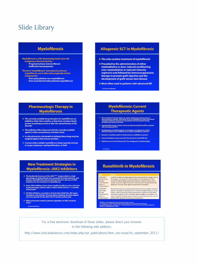
Slide Library
For a free electronic download of these slides, please direct your browser to the following web address: