Age- and sleep-related changes in brain plasticity and brain architecture in relation with cognition Julie Carrier, Ph.D. Département de psychologie, Université de Montréal Research Center, Institut universitaire de gériatrie de Montréal Center for Advanced Research in Sleep Medicine Hôpital du Sacré-Cœur de Montréal ESRS Sleep Science School 2019
Transcript
Age- and sleep-related changes in brain plasticity and brain architecture in relation with cognition
Julie Carrier, Ph.D.Département de psychologie, Université de Montréal
Research Center, Institut universitaire de gériatrie de MontréalCenter for Advanced Research in Sleep Medicine
Hôpital du Sacré-Cœur de MontréalESRS Sleep Science School 2019
• Targeting cerebral mechanisms underlying age-related changes in sleep and circadian rhythms is crucial to develop preventive and therapeutic interventions adapted for older individuals.
• Sleeping brain is a proxy of cognitive integrity in older individuals and may be used as a biomarker of neurodegeneration.
• Improving sleep in the older population may slow down the aging process and neurodegeneration (cognition, oxidative stress, inflammation, amyloid).
Demystifying sleep in aging: towards new diagnostic, preventive, and
therapeutic targets
ESRS Sleep Science School 2019
During adulthood, increasing age is associated with…
Less time asleep More wakefulness, lower sleep efficiency (especially in
second half of the night Less slow-wave sleep (min and %) More stage 1 and stage 2 sleep Shorter REM latency; less REM sleep
Carrier et al. 1997. Journal of Sleep Research; Carrier et al. 2001. Psychophysiology. Carrier et al. et al. 2009. Sleep Medicine. Gaudreau et al. 2001. Neurobiology Aging ; Lafortune et al. 2012. PlosOne; Robillard et a;.2016)
Earlier bedtimes and waketimes Less time in bed
ESRS Sleep Science School 2019
Slow waves
- N2+ N3
< 4 Hz
> 75 µV
NREM sleep EEG Two electrophysiological markers
N3
N2
N1
Spindles
Prevalence+ N2- N3
Frequency 11-15 Hz
Duration > 500 ms
Amplitude NA
NREM sleep oscillations functions • Sleep and synaptic homeostasis • Sleep protection against external stimulations • Learning, memory, cognition
ESRS Sleep Science School 2019
Carrier, Viens et al. 2011. European Journal of NeuroscienceMartin al. (2013). Neurobiology of Aging
Important age-related modifications in NREM sleep EEG oscillations
SW densitySW amplitudeSW slope
Spindle density Spindle amplitudeSpindle duration
ESRS Sleep Science School 2019
Age-related change in NREM sleep is a proxy of cerebral and cognitive health in
aging
• May underlie the difficulty that older people have in recuperating and/or maintaining sleep under challenging conditions
• Predict the development of dementia in neurodegenerative aging
Latreille et al. 2019, Dube et al., 2015; Fogel et al., 2012; Mander et al., 2016; Mander et al., 2017; Lafortune et al., 2014; Latreille et al., 2015; Fogel et al., 2012; Pace-Schott and Spencer, 2015; Mander et al., 2015; Carrier et al., 2009; Fogel et al., 2012
ESRS Sleep Science School 2019
Sleep become more vulnerable to disturbance with increasing age:
Need to take extra care
;Carrier et al. 1997. Journal of Sleep Research; Carrier et al. 2001. Psychophysiology. Carrier et al. et al. 2009. Sleep Medicine. Gaudreau et al. 2001. Neurobiology Aging ; Lafortune et al. 2012. PlosOne; Robillard et a;.2016 Munch et al. 2014
Lower ability to enhance slow-wave sleep after sleep deprivation
Lower ability to maintain sleep at an abnormal circadian phase (jet lag, shift work)
Higher sensitivity to caffeine
ESRS Sleep Science School 2019
Munch et al. 2004
Lower ability to increase sleep intensity after sleep loss
Gaudreau et al. 2001. Neurobiology Aging ;Munch et al. 2014; Lafortune et al. 2012. PlosOne
de sensibilité à une de pression homéostatique
ESRS Sleep Science School 2019
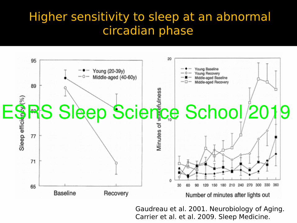
Higher sensitivity to sleep at an abnormal circadian phase
Gaudreau et al. 2001. Neurobiology of Aging. Carrier et al. et al. 2009. Sleep Medicine.
ESRS Sleep Science School 2019
NREM/REM EEG, cognition, normal aging
ESRS Sleep Science School 2019
What is the link between NREM sleep oscillations and general cognitive ability in
healthy older subjects ?
Lafortune et al. (2013). Journal of Sleep Research.
In healthy older subjects (between 50-91 y.o):
Spindle density is associated to performance:
• Declarative memory (Rey verbal learning task) • Attention (Bell test and Continuous Performance tasks)• Verbal fluency
SW density is associated to performance :
• Verbal fluency
ESRS Sleep Science School 2019
NREM/REM EEG : Proxy of neurodegenrescence
ESRS Sleep Science School 2019
Do spindles in Parkinson Disease predict the development of dementia?
Latreille et al. Neurobiology of Aging (2015). V.Latreille J-F Gagnon
Spindles at baseline are associated with likelihood of developing dementia at follow-up (4.5 later)
At follow-up (4.5 years) 18 PD patients developed dementia 50 remained dementia free
Spindles in patients with PD (Christensen et al. 2014)
ESRS Sleep Science School 2019
REM sleep: a window on cholinergic transmission in Parkinson
Cholinergic degeneration may be especially key to early cognitive impairment in PD (Kehagia et al., 2010; Gratwicke et al., 2015; Bohnen and Albin, 2011; Hall et al., 2014; Liu et al., 2015).
N-REM sleep: cholinergic activity is nearly absent
REM sleep differs from wakefulness: sustain activity mainly cholinergic, with very little input from other neurotransmitter systems (i.e., noradrenaline, serotonin, and dopamine).
REM sleep is the ideal state to investigate cholinergic transmission integrity in PD.
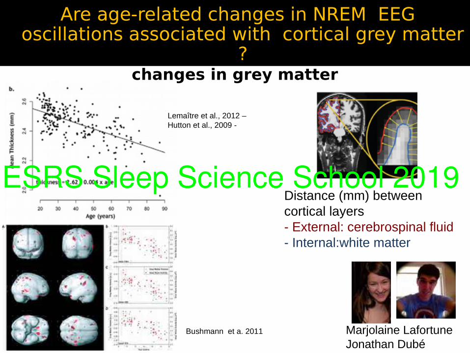
Thinning in a network of cortical regions involved in SW generation and propagation, but also in cognitive functions, explained the age-related decrease in SW density and amplitude.
Dubé et al. (2015) Journal of Neuroscience
ESRS Sleep Science School 2019
Age-related changes in relative spectral power in REM and NREM sleep
Latreille et al. (2019). Neurobiology of Aging
ESRS Sleep Science School 2019
After controlling for age, spectal power is associated with cortical thickness
in both REM and non-REM sleep
Latreille et al. (2019). Neurobiology of Aging
REM sleepDelta: Central Left superior frontal gyrus Left lateral orbitofrontal cortex Right rostral anterior cingulate cortexDelta: Temporal Left superior frontal gyrusDelta: Parietal Left/right superior frontal gyrus Right superior temporal gyrusDelta: Occipital Left superior frontal gyrus
NREM sleepDelta: Central Left medial orbitofrontal cortexTheta: Occipital Right lateral occipital cortex
ESRS Sleep Science School 2019
Cortical thickness explains age-related changes in REM and NREM sleep
Latreille et al. (2019). Neurobiology of Aging)
Sup. frontal gyrus
Sup. temporal gyrus
Medial orbitalfrontal cortex
ESRS Sleep Science School 2019
Are age-related changes in REM sleep associated to grey matter?
Latreille et al. (2019). Neurobiology of Aging.
Thinning of the left superior frontal gyrus (spreading to the anterior cingulate and medial orbitofrontal cortices) drives EEG desynchrony during both REM and NREM sleep with aging
Common mechanisms for both REM and NREM
Medial frontal and cingulate cortices: major hubs of the human brain, in synchronizing neuronal assemblies during sleep
ESRS Sleep Science School 2019
Are age-related changes in NREM EEG oscillations associated with white matter
integrity ?
Samuel Deslaurier-Gauthier, Ph.D.Pierre-Olivier Gaudreault
In the young: sigma spectral power and spindle density is associated with higher AD (Piantoni et al 2013)
White Matter older adults moderates the benefit of sleep spindles on motor memory consolidation (Mander et al. 2018)
Diffusion tensor imaging (DTI): water diffusion in each voxel (tensor)
Anisotropy of the diffusion=one preferential direction
Moderation analyses: the association between white matter and sleep spindles differs in young and older individuals
Gaudreault et al. SLEEP (2018)
Young
Older
Diffusion metrics explained between 14% and 39% of SS amplitude and sigma power variance in the young subjects only
ESRS Sleep Science School 2019
To connect or not connect during sleep: this is the question!
MPFCCognitive and mental functions emerges from the brain’s complex networks.
Most conclusions on functional connectivity during human sleep come from studies in young individuals.
Age-related change in functional connectivity during sleep may very well underlying cerebral and cognitive health in aging
ESRS Sleep Science School 2019
fMRI functional connectivity (FC) during NREM
MPFC
EEG/fMRI studies in the young: NREM sleep induces important modifications in cortical and sub-cortical networks underlying sensory awareness, information transfer, memory consolidation and executive control.
Fading of consciousness: greater local cortical FC, but a breakdown of long range cortico-cortical FC during NREM sleep with the descent from wakefulness to SWS sleep.
Horovitz et al. 2008; Samann et al. 2011; Wu et al. 2012; Spoormaker et al .2010,2011,2012; Tagliazucchi et al. 2013; Boly et al. 2012; Lee et al. 2012
ESRS Sleep Science School 2019
Our fMRI study
NREM sleep in older individuals: Important modifications including lighter sleep, lower slow-waves and spindle density/amplitude.
Objective: to investigate changes in fMRI FC that occur during NREM sleep in aging.
Hypothesis: Older individuals will show a lower breakdown of FC during NREM sleep than young participants
ESRS Sleep Science School 2019
Participants:
Young (20-30 y) = 16(8F); mean= 23.3±3.3
Older (52-69 y) = 14(9F); mean = 59.5±5.9
Data acquisition (90 minutes) after a 26-hour sleep deprivation
Ballistocardiographic artifacts: Constrained cICA (Leclercq et al., 2009).
Sleep scoring
fMRI (BASC-GLM)
Bootstrap analysis of stable clusters (BASC): (Bellec et al., 2010): identify brain regions that consistently exhibited similar spontaneous activity in individual subjects, and were spatially stable across both young and older participants.
Cerebral functional connectivity analyses: Standard correlation measures based on Bold signal fluctuations
Nicolas Martin, Ph.D. Pierre Orban, Ph.D. Pierre Bellec, Ph.D Véronique Daneault, Ph.D.
ESRS Sleep Science School 2019
Data-driven parcellization20 regions
20 homogeneous regions in terms of fluctuations of the BOLD signal
ESRS Sleep Science School 2019
NREM2 vs WakeSimilar effects in young and older
Lower FC in NREM2 Higher FC in NREM2
ESRS Sleep Science School 2019
NREM2 vs NREM1Similar effects in young and older
Lower FC in NREM2 Higher FC in NREM2
ESRS Sleep Science School 2019
NREM2vsNREM1 Sronger FC in young and
FC in olderLess FC in NREM2 More FC in NREM2
Age
-rel
ated Inf. mpfc
ESRS Sleep Science School 2019
Conclusions (1)Young and older individuals: very similar decreases in cortico-cortical fMRI FC between wakefulness and NREM2 and globally, between NREM1 and NREM2.
Age-related difference in FC: between NREM2 and NREM1 in specific brain networks (16%)
Older subjects: shallower fMRI FC or FC between NREM2 and NREM1 between networks underlying sensory awareness, information transfer, memory consolidation , executive controlESRS Sleep Science School 2019
Age related changes in EEG coherence during sleep
How two EEG sensors (brain regions) show similar neuronal oscillatory activity (frequency specific).
Complete night study
All sleep stages
More ecological environment Jean-Marc Lina, Ph.D., ETS, CÉAMS
Maude Bouchard,B.Sc., Ph.D. student
Bouchard et al. (2019). SLEEP.
ESRS Sleep Science School 2019
Imaginary EEG coherence
• The “strength” of functional
interactions between two cortical
areas in different frequency bands
(delta, theta, sigma, alpha, beta).
• Removal of 0 lag contribution
Castro et al., 2014
ESRS Sleep Science School 2019
EEG coherence differences between older and younger individuals
= Older < Younger= Older > Younger
Bouchard et al. (2019) SLEEP
ESRS Sleep Science School 2019
Changes in EEG coherence (2-4 Hz) across the night
Bouchard et al. 2019 (SLEEP)
Global connectivity index: the sum of the differences in connectivity between two conditions, across all the significant pairs of electrodes
ESRS Sleep Science School 2019
Conclusion (2)
Older individuals showed lower EEG connectivity in N2 than the young (linked to spindles density).
Older individuals showed higher EEG connectivity than younger ones in both REM and N3 sleep
Older individuals do not show reduced EEG connectivity in N3 as compared to N2 during the first NREM sleep cycle
EEG connectivity during REM sleep is lower than in N3 (data not shown)
EEG connectivity in N2 and REM sleep linked to cognition (data not shown)
ESRS Sleep Science School 2019
PLI during the transition between SW hyperpolarization and
depolarization(contant delay)
Global connectivity index: the sum of the differences in connectivity between two conditions, across all the significant pairs of electrodes
ESRS Sleep Science School 2019
To connect or not connect during sleep: this is the question!
MPFC
Brain imaging (EEG, MEG, MRI, fMIR): new insights on brain areas involved in the functions of human sleep.
Next step: to understand how brain areas interacts to give rise to human sleep functions.
Functional connectivity is estimated by several metrics at different time scale (e.g. during all night vs SW) : unique information and processes to understand the complexity of the sleeping brain.
Age-related changes in fMRI functional connectivity, NREM EEG coherence, synchronization during slow-waves.
Age-related change in functional connectivity during sleep may very well underlying cerebral and cognitive health in aging
ESRS Sleep Science School 2019
NREM/REM sleep: Age-related changes in sleep dependent consolidation
ESRS Sleep Science School 2019
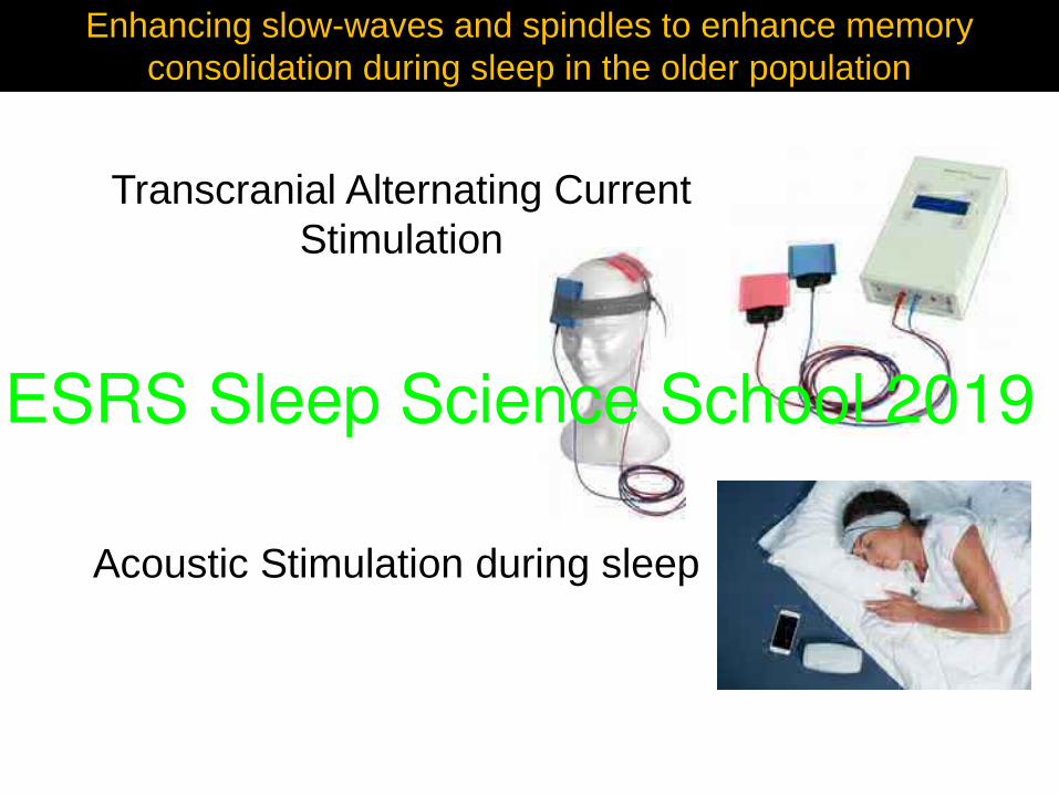
Transcranial Alternating Current Stimulation
Enhancing slow-waves and spindles to enhance memory consolidation during sleep in the older population
Acoustic Stimulation during sleep
ESRS Sleep Science School 2019
Enhancing slow-waves and spindlesTranscranial stimulation
ESRS Sleep Science School 2019
Enhancing slow-waves and spindlesAcoustic stimulations
ESRS Sleep Science School 2019
Precious collaborators
Undergraduate, graduate, post-graduate students, research, research associate
Marjolaine LafortuneVéronique Latreille
Nicolas MartinJonathan Dubé
Maude BouchardPierre-Olivier Gaudreault
Véronique DaneaultJustin Bouvier
Thomas LehouxTarek Lajnek
Samuel Deslaurier-Gauthier
Center for advanced research in sleep medicine (CÉAMS)
Jean-Marc-Lina, École de Technologie SupérieureJean-François Gagnon, UQAM
Nadia Gosselin, UdeMJY Montplaisir, UdeM
Sonia FrenetteJean Paquet
Gaétan PoirierChristophe Bedetti
Julien Doyon, BIC, MNI, McGill Pierre Maquet, Université de LiègeCélyne Bastien, Université Laval
Alan Evans, McGill University Pierre Bellec, CRIUGM, UdeM
Pierre Jolicoeur, CERNEC, UdeMKarim Jerbi, CERNEC, UdeM