64
DR: ABIR MOHIEDIN SAID
| Date post: | 17-Dec-2015 |
| Category: |
Documents |
| Upload: | verity-hancock |
| View: | 214 times |
| Download: | 0 times |

DR: ABIR MOHIEDIN SAID

Involuntary loss of urine Social and hygienic problem It affects individuals physical, psychological
and social which is associated with a significant reduction in quality of life
Urinary incontinence

The prevalence increases with age 5% of women between 15-44 years of age
being affected Increases to 10% between 45-64 Increases to 20% > 65 years Higher in women in residential nursing
homes about 40%

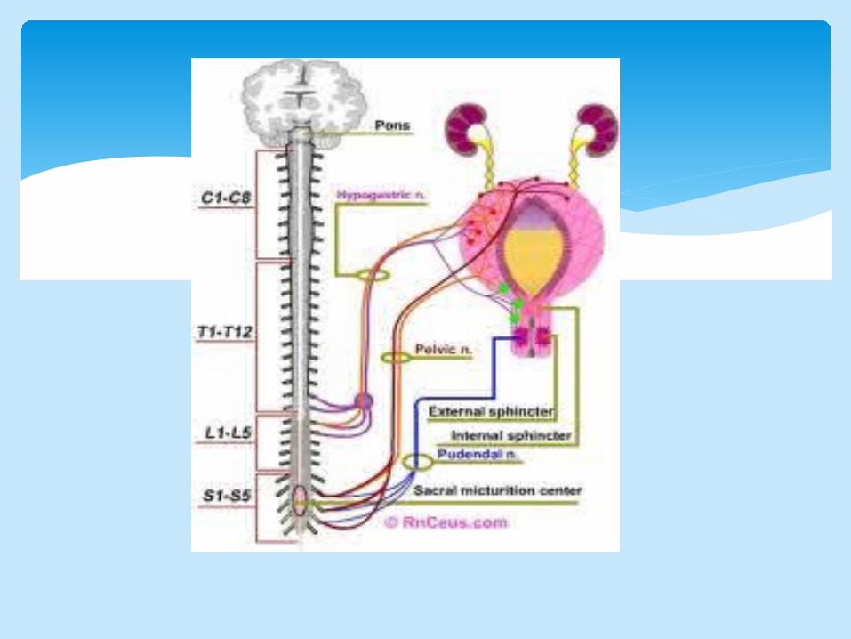
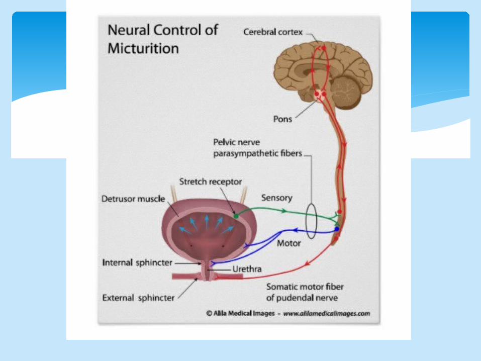
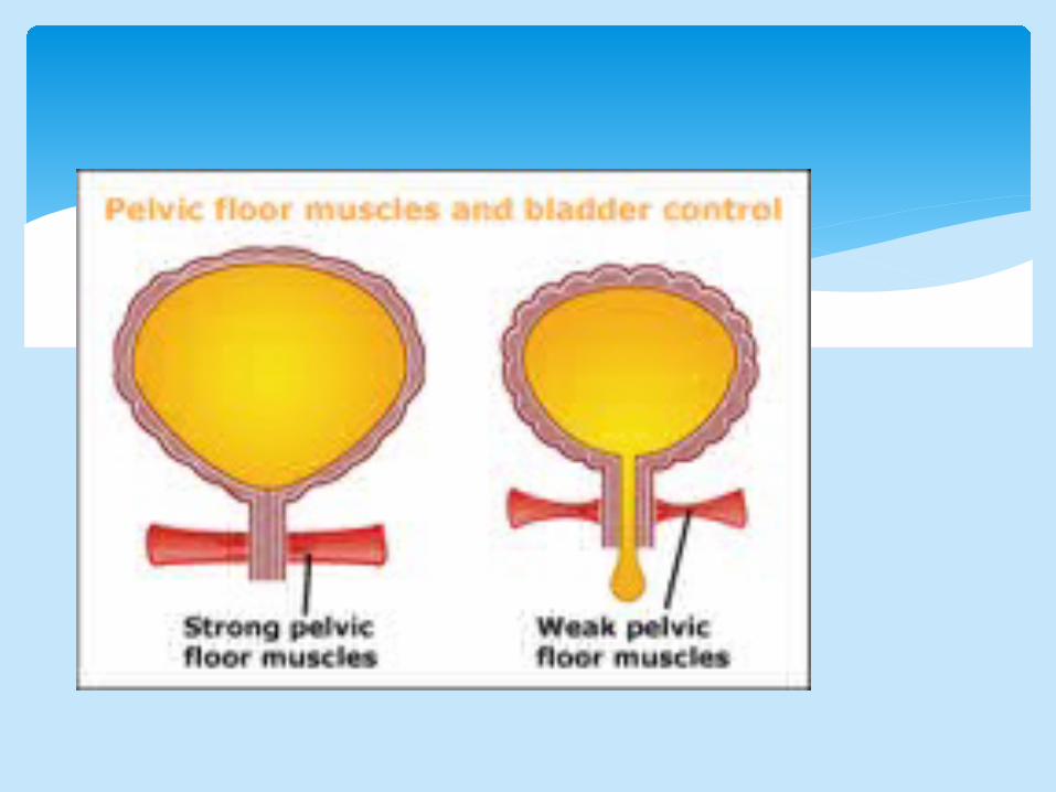
To hold urine and control urination, the lower urinary tract and nervous system need to be working normally
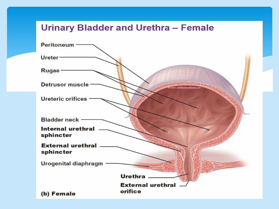
The average adult bladder can hold over 2 cups (350 ml - 550 ml) of urine. Two muscles are involved in controlling urine flow:
-The sphincter, which is a circle-shaped muscle around the urethra. You must be able to squeeze this muscle to prevent urine from leaking out.
-The detrusor, which is the muscle of the bladder wall, this must stay relaxed so that the bladder can expand
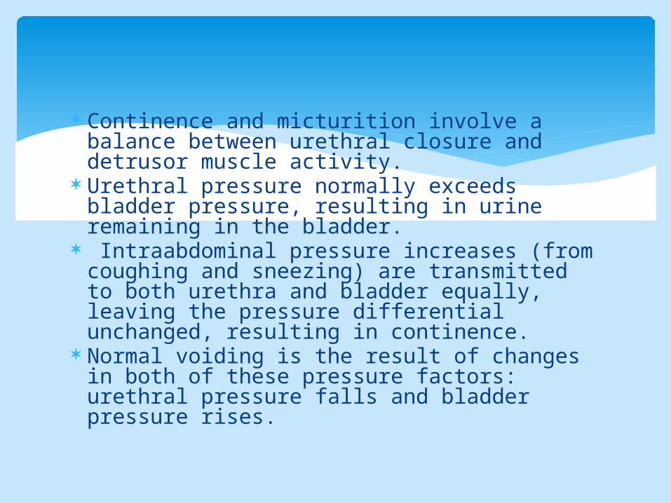
Continence and micturition involve a balance between urethral closure and detrusor muscle activity.
Urethral pressure normally exceeds bladder pressure, resulting in urine remaining in the bladder.
Intraabdominal pressure increases (from coughing and sneezing) are transmitted to both urethra and bladder equally, leaving the pressure differential unchanged, resulting in continence.
Normal voiding is the result of changes in both of these pressure factors: urethral pressure falls and bladder pressure rises.

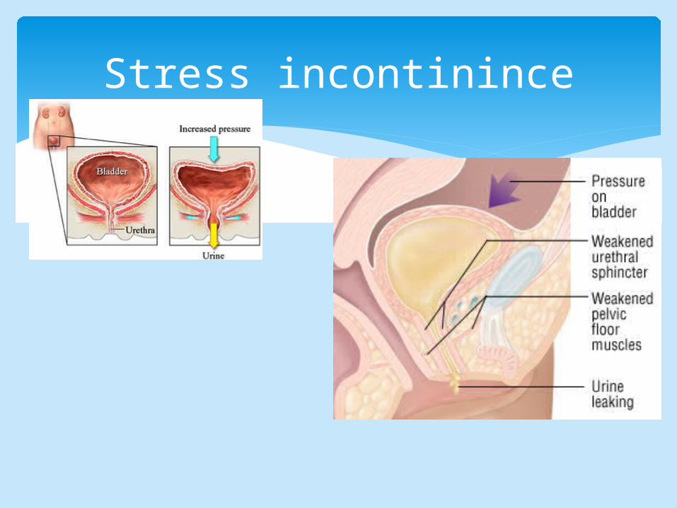
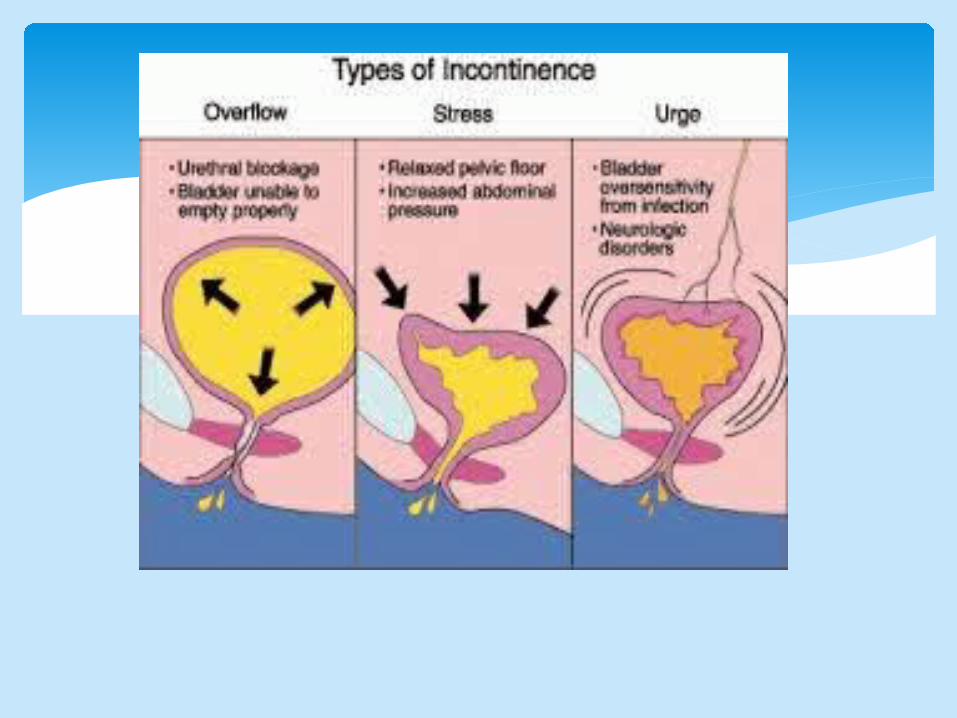
Stess incontinence:Urethral causes , involuntary leakage of urin during increased abdominal pressure in the absence of a detrusor contraction
CLASSIFICATION
Stress incontinince
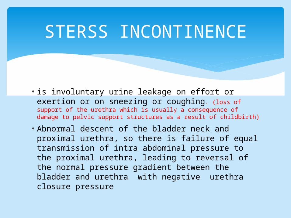
• is involuntary urine leakage on effort or exertion or on sneezing or coughing. (loss of support of the urethra which is usually a consequence of damage to pelvic support structures as a result of childbirth)
• Abnormal descent of the bladder neck and proximal urethra, so there is failure of equal transmission of intra abdominal pressure to the proximal urethra, leading to reversal of the normal pressure gradient between the bladder and urethra with negative urethra closure pressure
STERSS INCONTINENCE
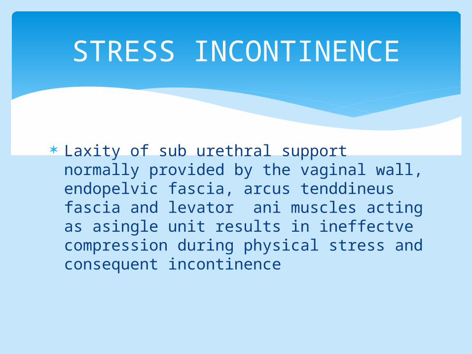
Laxity of sub urethral support normally provided by the vaginal wall, endopelvic fascia, arcus tenddineus fascia and levator ani muscles acting as asingle unit results in ineffectve compression during physical stress and consequent incontinence
STRESS INCONTINENCE
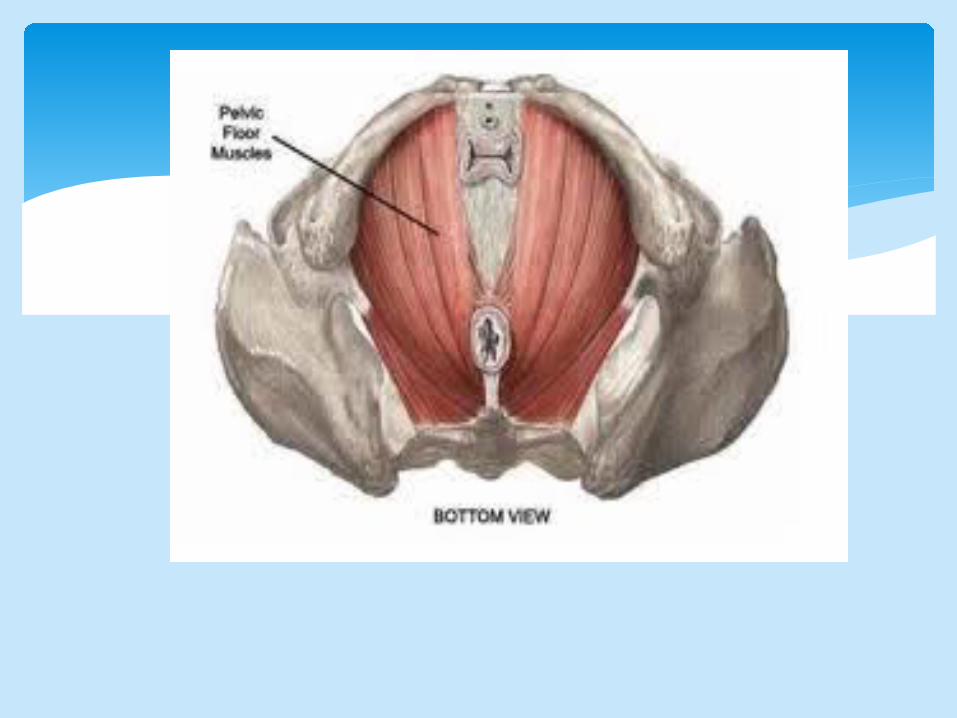
Damage to the nerve supply of the pelvic floor and urethral sphincter caused by childbirth
Mechanical trauma to the pelvic floor muscles and endopelvic fascia and ligamentsduring vaginal delivary
Prolong second stage, large babies and instrumental deliveries
Aetiolgy of USI
Menopause and associated tissue atrophy Chronic disease (obesity, chronic obstructive
pulmonary disaese, constipation) Conginital causes( connective tissue and collagen)
Causes
suddenly feeling the need or urge to urinate, A common cause of urge incontinence is inappropriate bladder contractions.
Urge incontinence can mean that the bladder empties during sleep, after drinking a small amount of water, or touch water or hear it running .
Certain fluids and medications such as diuretics or emotional states such as anxiety can worsen this condition. Some medical conditions, such as hyperthyroidism and uncontrolled diabetes, can also lead to or worsen urge incontinence.
Urge incontinence

Involuntary actions of bladder muscles can occur because of damage to the nerves of the bladder, to the nervous system (spinal cord and brain), or to the muscles themselves. Multiple sclerosis, Parkinson's disease, Alzheimer's disease, stroke, and injury—including injury that occurs during surgery—all can harm bladder nerves or muscles.
Urge incontinence

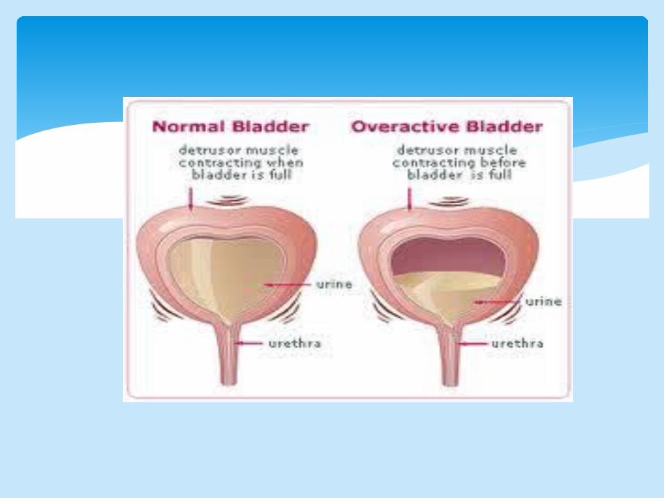
Involuntary detrusor contractions Overactive bladder occurs when abnormal
nerves send signals to the bladder at the wrong time, causing its muscles to squeeze without warning. Voiding up to seven times a day is normal for many women, but women with overactive bladder may find that they must urinate even more frequently.
Detrusor overactivity

the symptoms of overactive bladder include urinary frequency—bothersome urination
eight or more times a day or two or more times at night
urinary urgency—the sudden, strong need to urinate immediately
urge incontinence—leakage or gushing of urine that follows a sudden, strong urge
nocturia—awaking at night to urinate
OverActiveBladder
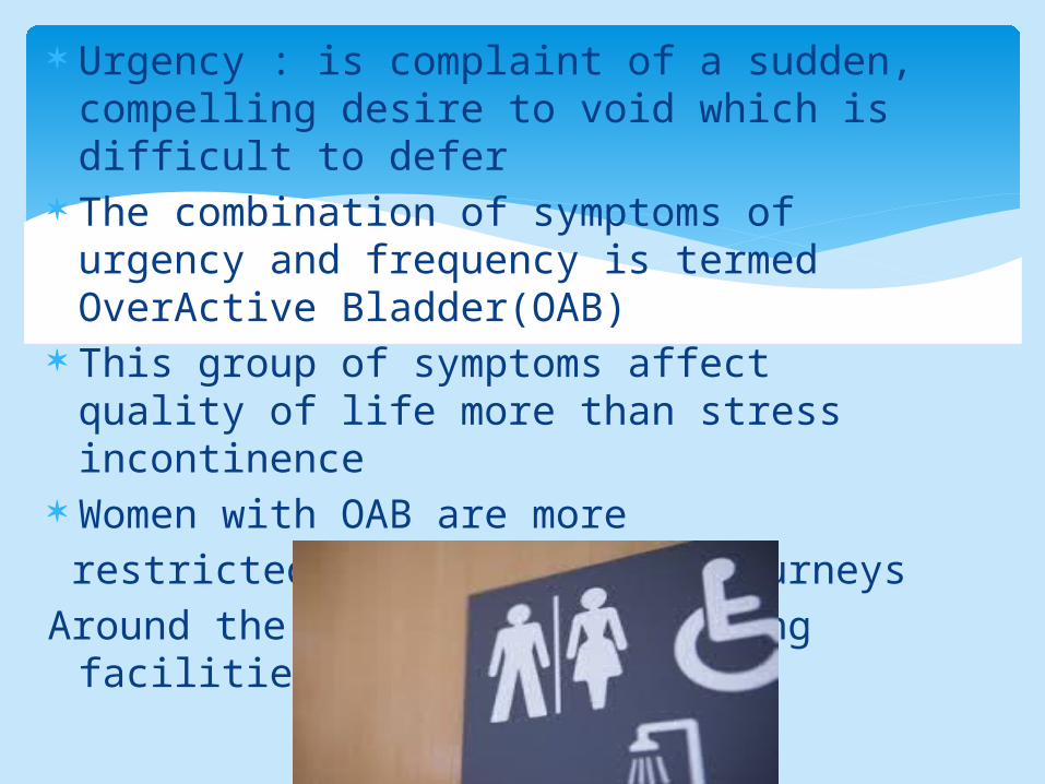
Urgency : is complaint of a sudden, compelling desire to void which is difficult to defer
The combination of symptoms of urgency and frequency is termed OverActive Bladder(OAB)
This group of symptoms affect quality of life more than stress incontinence
Women with OAB are more restricted and often there journeysAround the location of toileting facilities
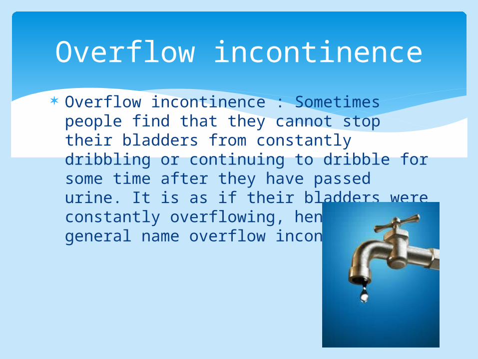
Overflow incontinence : Sometimes people find that they cannot stop their bladders from constantly dribbling or continuing to dribble for some time after they have passed urine. It is as if their bladders were constantly overflowing, hence the general name overflow incontinence.
Overflow incontinence
Failure of bladder emptying may lead to chronic retention and overflow incontinence
causes:-Lower motor neurone or upper motor neurone lesions-Urethral obstruction-pharmacological
Retention with overflow
Poor stream Incomplete bladder emptying Overflow stress incontinence Recurrent urinary tract infection Cystometry to make diagnosis Ultrasonography Intravenous urogram for any upper urinary tract
reflux CT may be necessary
Symptoms and diagnosis
Any mass that cause compression of the bladder must be excluded, prolapse, vaginal atrophy
Observation of involuntary loss of urin with coughing may be suggest stress incontinence
Observation of urin leakage through channels other than urethra from urethra ( conginital anomaly,fistula)
Examination
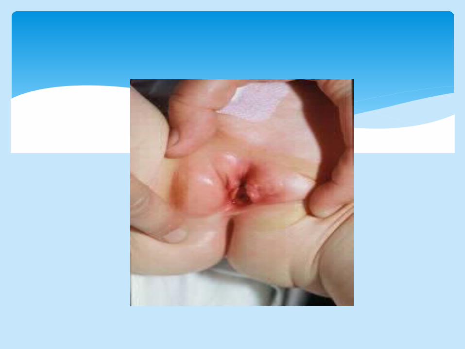
Epispadias( widened bladder neck,shortened uretha,separation of symphysis pubis and imperfect sphincter)
the patient complains of stress incontinence which may not be apparent when lying down but noticeable when standing up
X-ray of pelvis will show symphsial separation
Suprapubic operation to elevate the bladder neck
Congenital anomalies
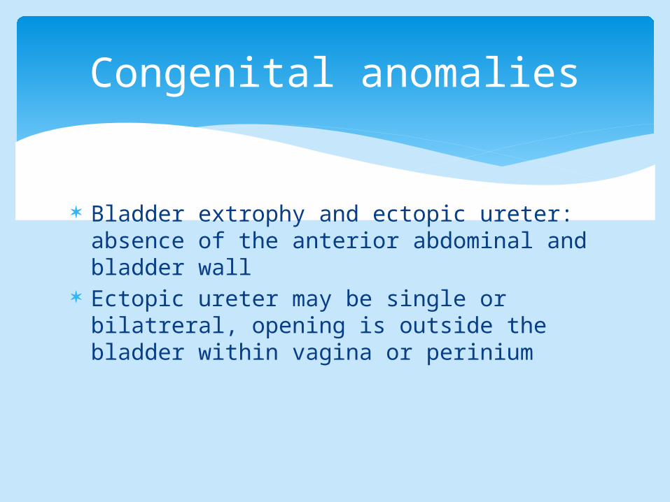
Bladder extrophy and ectopic ureter: absence of the anterior abdominal and bladder wall
Ectopic ureter may be single or bilatreral, opening is outside the bladder within vagina or perinium
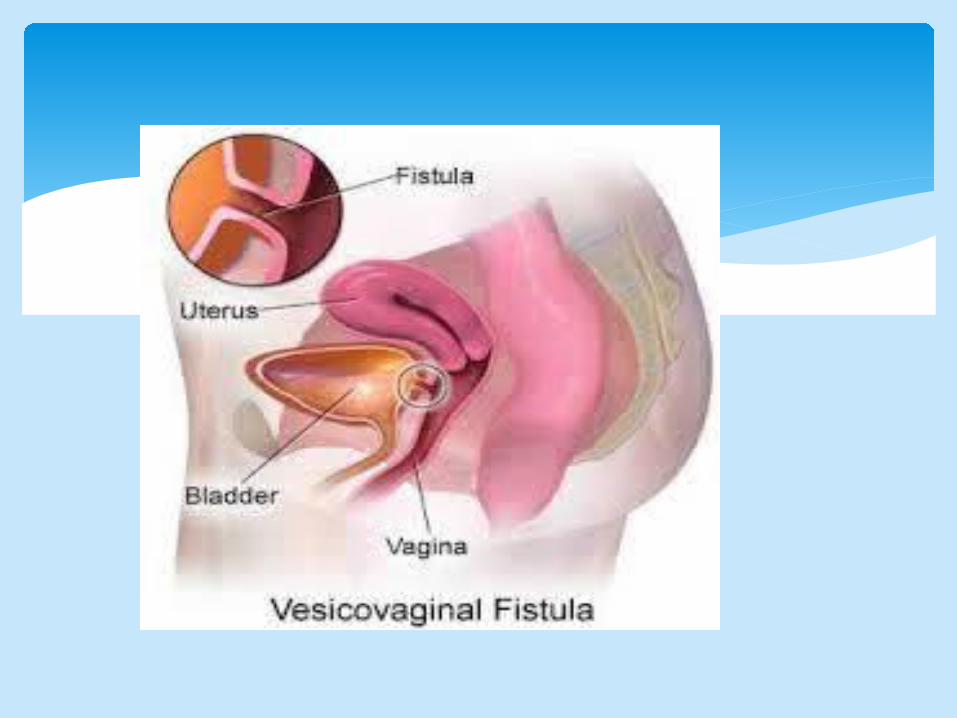
Congenital anomalies

Abnormal opening between the urinary tract and the outside
Causes (obstetric or gynaecological) Obstructive labour Pelvic surgery Pelvic malignancy Radiotherapy It can treated by surgery ( isolation and removal of fistula
tract, suture and closure of each layer separately without tension)
fistula

The women have short urethra which is prone to entry of bacteria during intercourse, poor perineal hygiene
Unefficient voiding ability Unnecessory catheterization Postmenopausal atrophy and change in
vaginal PH The common organisms : E-Coli, Proteus mirabilis,
Klebsiella aerogenes, Pseudomonas aeruginosa and Streptococcus faecalis
Urinary tract infection

Symtoms: dysuria, frequecy,hematuria→ loin pain, fever and riger (acut pyelonephritis has
developd)
Urin stick test , a nitrate can suggest infection
Infection counts ↑ A culture and sensitivity C/S of midstream
specimen of urin is requierd IV or CT urography or renal U/S may be
required in ptatient with recurrent infection
Urinary tract infection

With acute infection we should send urin for C/S and start antimicrobial therapy, the regimen can be changed later according to the result of the urin C/S
Trimethoprim 200mg x2 commonly used or Nitrofurantoin 100mg x4 or Cephalosporin With recurrent infection which an identifiable
source has not been found may be managed by long –term low dose antimicrobial therapy such as trimethoprim
Recurrent infection,vaginal oestrogen in postmenopausal women
Urinary tract infection
Failure of bladder emptying this leads to acute or chronic urinary retension, poor stream
Causes: failure of detrusor contraction Sphincteric relaxation Urethral obstruction Bladder overdistension
Voiding difficulties
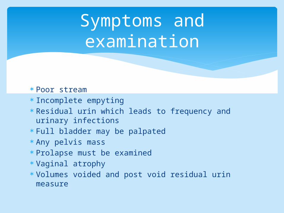
Poor stream Incomplete empyting Residual urin which leads to frequency and urinary
infections Full bladder may be palpated Any pelvis mass Prolapse must be examined Vaginal atrophy Volumes voided and post void residual urin measure
Symptoms and examination

History-taking and physical examination Assessment of pelvic floor muscles Assessment of prolapse Urine testing Assessment of residual urine Referral Symptom scoring and quality-of-life assessment Bladder diaries Pad testing Urodynamic testing Cystoscopy Imaging
Assessment and investigation
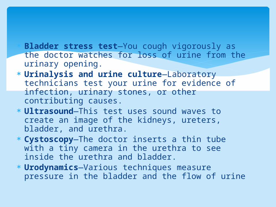
Bladder stress test—You cough vigorously as the doctor watches for loss of urine from the urinary opening.
Urinalysis and urine culture—Laboratory technicians test your urine for evidence of infection, urinary stones, or other contributing causes.
Ultrasound—This test uses sound waves to create an image of the kidneys, ureters, bladder, and urethra.
Cystoscopy—The doctor inserts a thin tube with a tiny camera in the urethra to see inside the urethra and bladder.
Urodynamics—Various techniques measure pressure in the bladder and the flow of urine
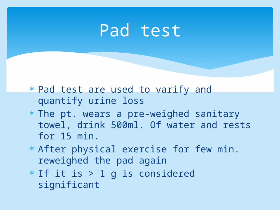
Pad test are used to varify and quantify urine loss
The pt. wears a pre-weighed sanitary towel, drink 500ml. Of water and rests for 15 min.
After physical exercise for few min. reweighed the pad again
If it is > 1 g is considered significant
Pad test
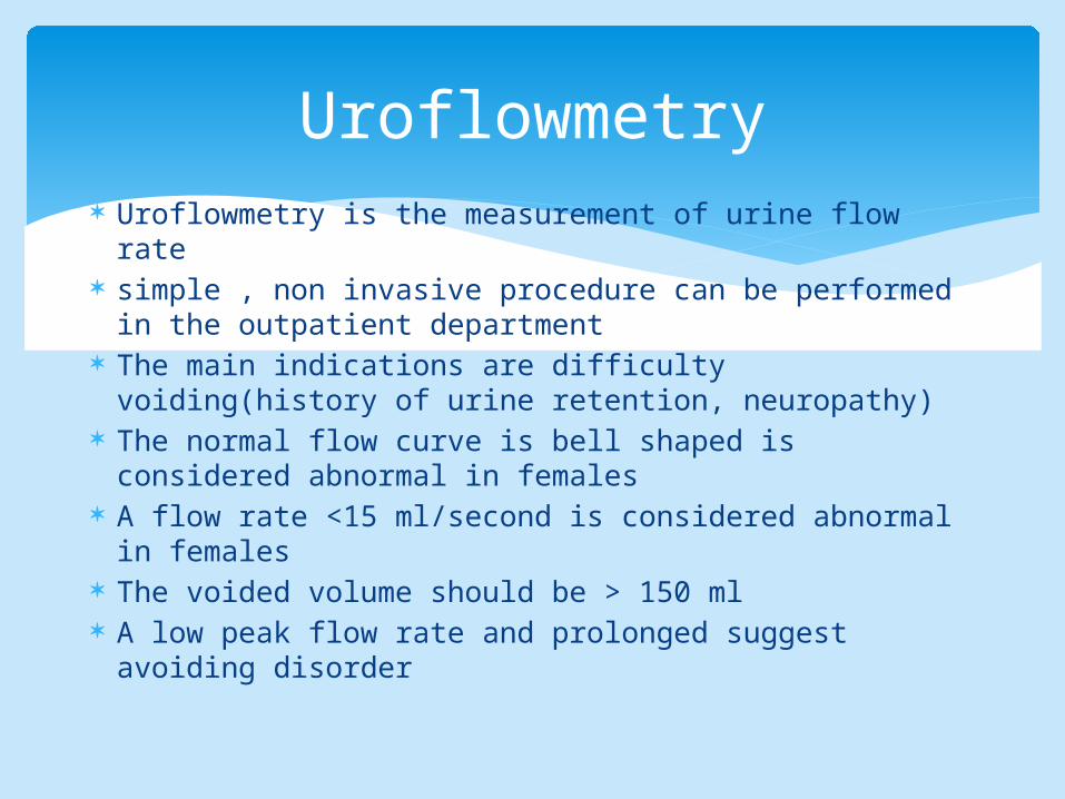
Uroflowmetry is the measurement of urine flow rate simple , non invasive procedure can be performed
in the outpatient department The main indications are difficulty voiding(history of
urine retention, neuropathy) The normal flow curve is bell shaped is considered
abnormal in females A flow rate <15 ml/second is considered abnormal
in females The voided volume should be > 150 ml A low peak flow rate and prolonged suggest
avoiding disorder
Uroflowmetry

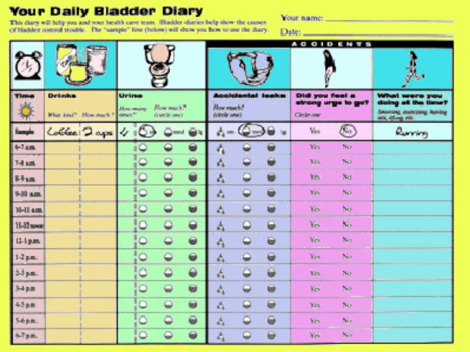
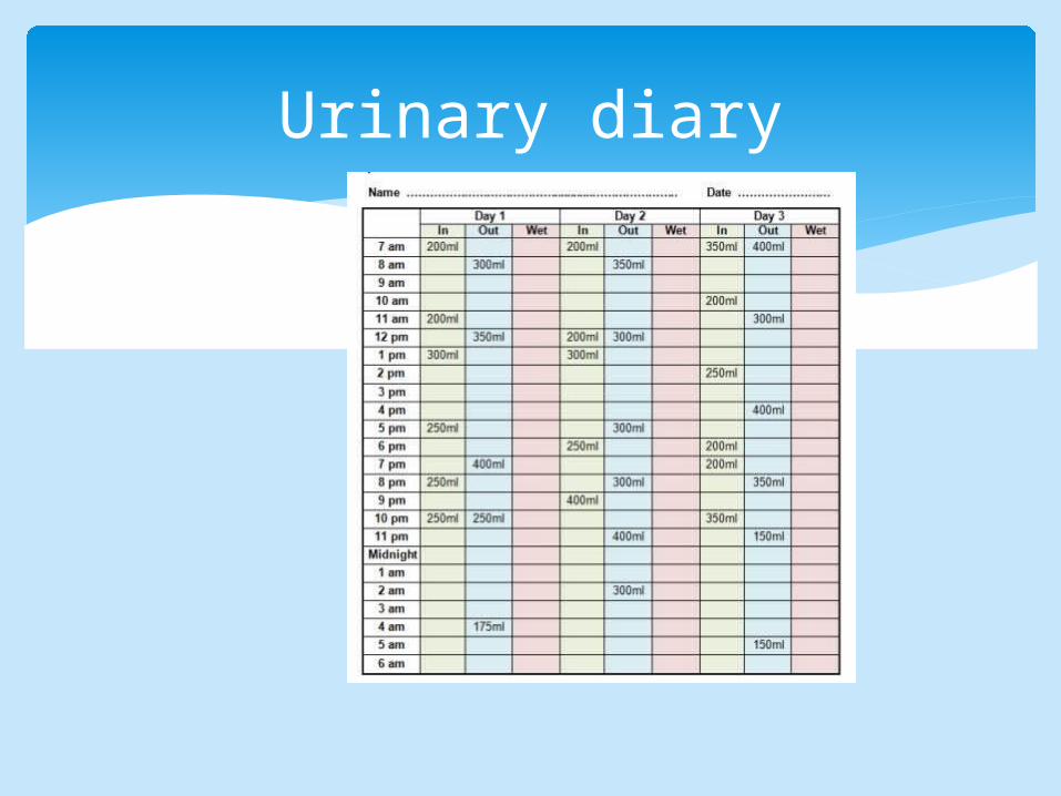
Urinary diary

Measurment of the pressure -volume relationship of the bladder
It involves abdominal pressure recording in addition to intravesical abdominal pressure monitoring during bladder filling and voiding
Indication for cystometry: Previous unsuccessful continence surgery Mixed incontinence both stress and urge Voiding disorder Neurogenic bladder Prior to primary continence operation
Cystometry
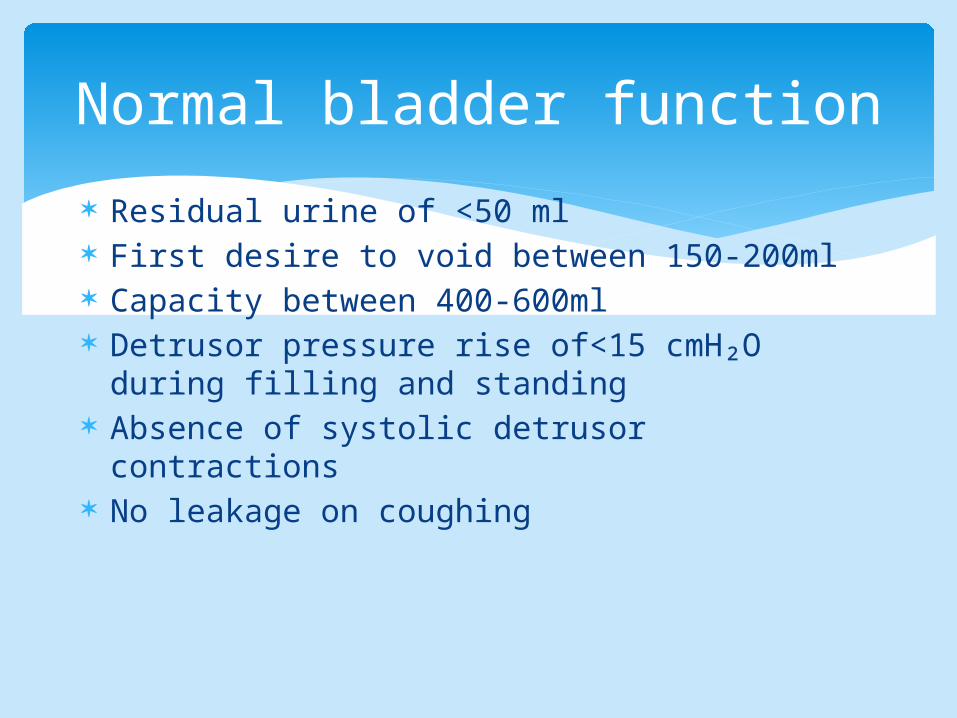
Residual urine of <50 ml First desire to void between 150-200ml Capacity between 400-600ml Detrusor pressure rise of<15 cmH₂O during
filling and standing Absence of systolic detrusor contractions No leakage on coughing
Normal bladder function

Videocystourethrography:(Aradio-opaque filling medium is used during cystometry)
Intravenous urography(indicated in cases of haematuria,uretrovaginal fistula)
MRI magnatic resonance imaging(anatomatical pictures of the pelvic floor and urinary
tract Cystourethroscopy
(in cases of hematuria,persistent UTI,reduced bladder capacity) Urethral pressure profilometry
Other investigation
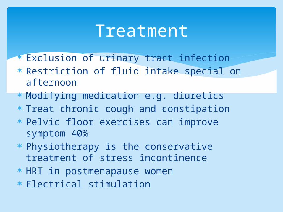
Exclusion of urinary tract infection Restriction of fluid intake special on afternoon Modifying medication e.g. diuretics Treat chronic cough and constipation Pelvic floor exercises can improve symptom
40% Physiotherapy is the conservative treatment of
stress incontinence HRT in postmenapause women Electrical stimulation
Treatment
Pelvic floor muscle training should be offered to women in their first pregnancy as a preventive strategy for UI
There is evidence that pelvic floor muscle training used during a first pregnancy reduces the likelihood of postnatal UI
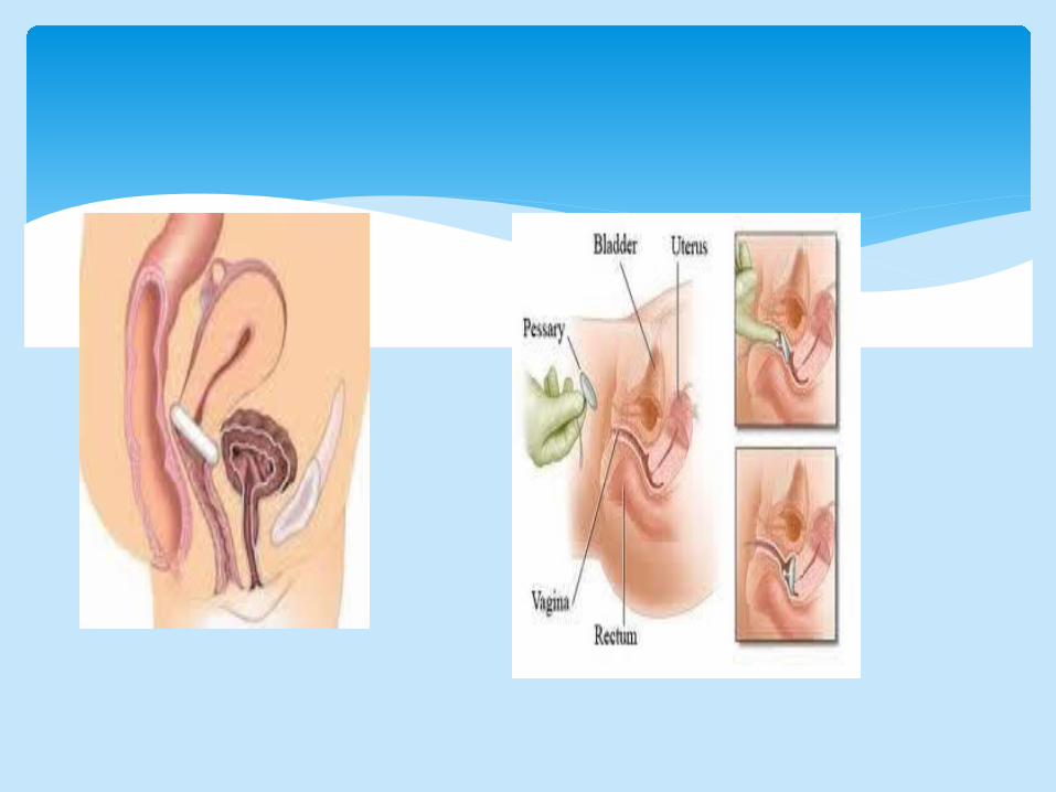
Intravaginal devices are not recommended for the routine management of UI in women for example during physical exercise.
Conservative managment
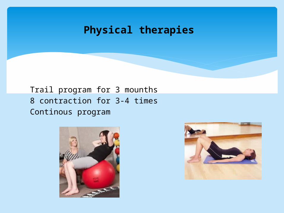
Trail program for 3 mounths
8 contraction for 3-4 times
Continous program
Physical therapies

Lifestyle interventions Coffein Daily fluid BMI
.
Conservative management
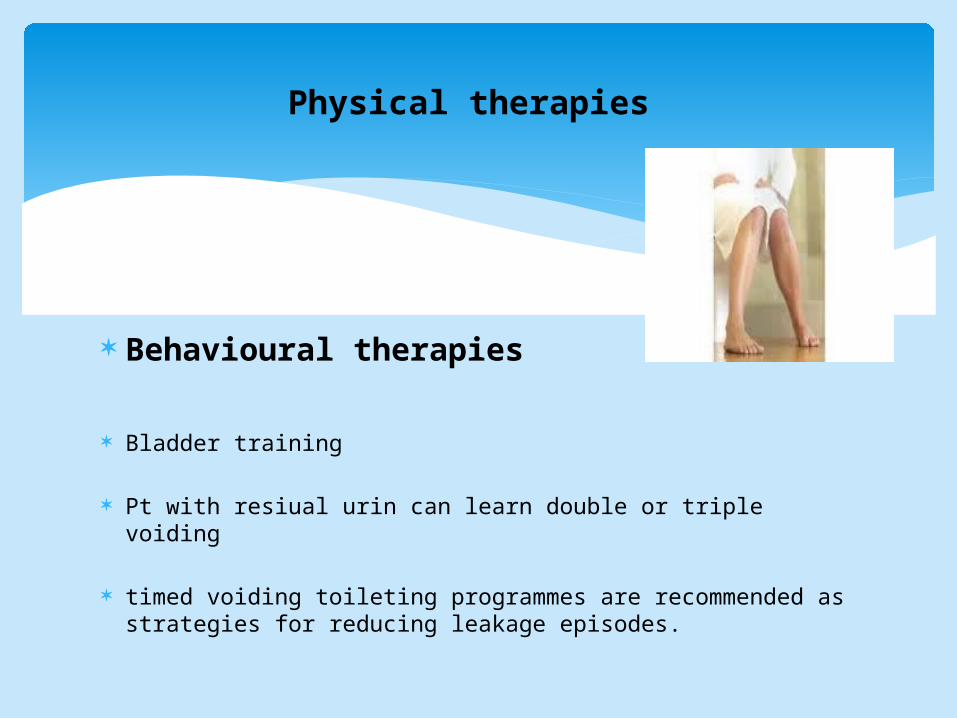
Behavioural therapies
Bladder training
Pt with resiual urin can learn double or triple voiding
timed voiding toileting programmes are recommended as strategies for reducing leakage episodes.
Physical therapies
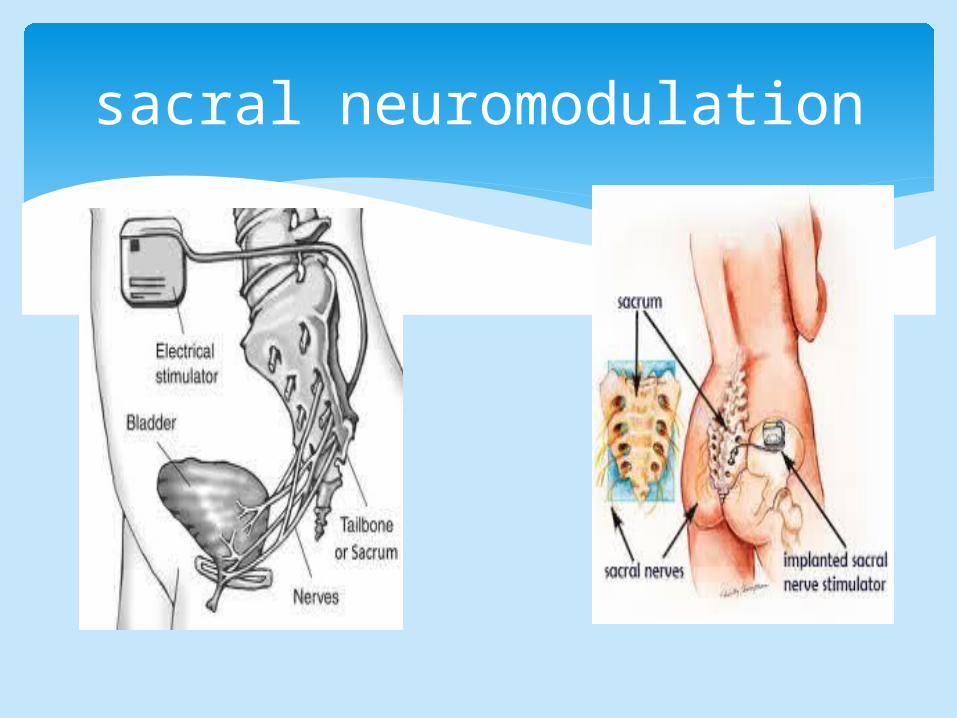
sacral neuromodulation
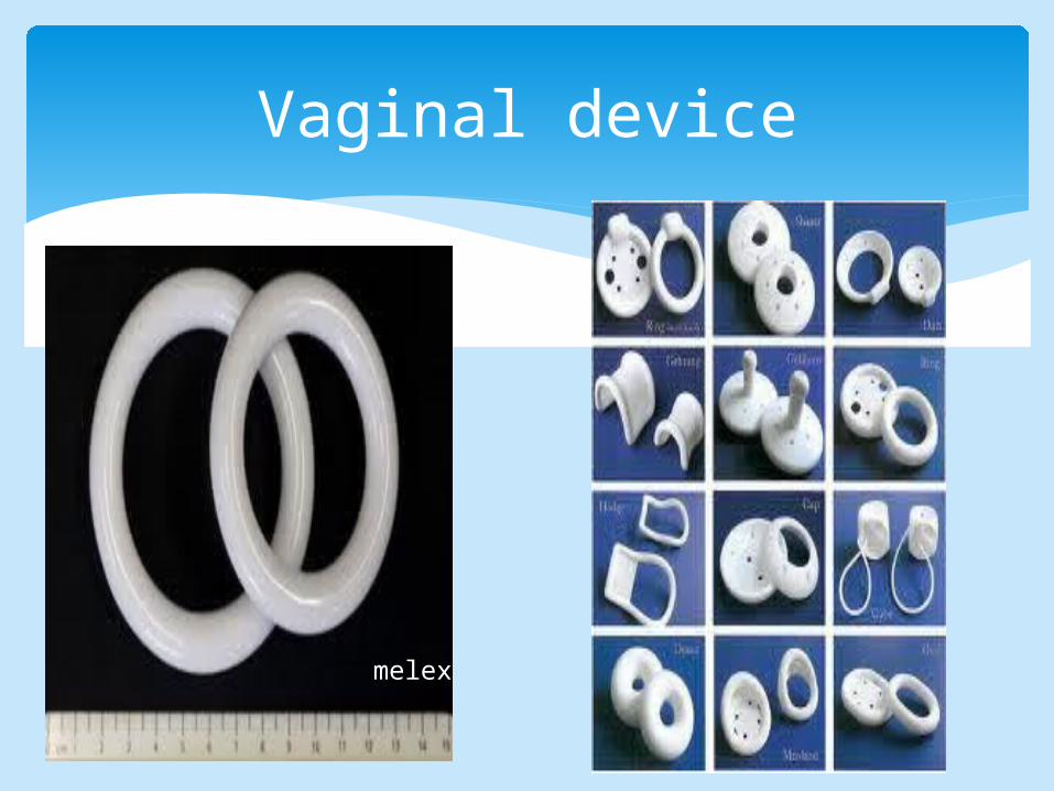
Vaginal device
melex
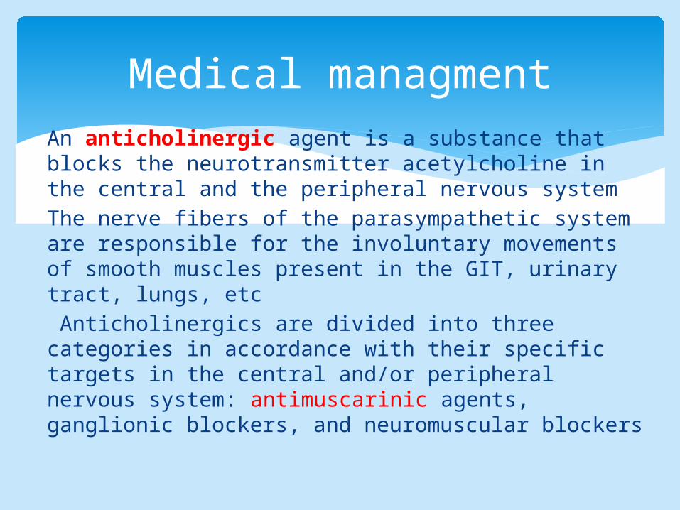
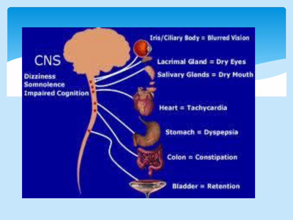
An anticholinergic agent is a substance that blocks the neurotransmitter acetylcholine in the central and the peripheral nervous system The nerve fibers of the parasympathetic system are responsible for the involuntary movements of smooth muscles present in the GIT, urinary tract, lungs, etc Anticholinergics are divided into three categories in accordance with their specific targets in the central and/or peripheral nervous system: antimuscarinic agents, ganglionic blockers, and neuromuscular blockers
Medical managment
The tension-free transvaginal (TVT) sling (86-95%) The transobturator tape (TOT) sling (82%) The mini-sling procedure also known as TVT-
Secure(67-83%) Open colposuspention
Marshall-Marchetti-Krantz(retropubic suspension or bladder neck suspension surgery)
Burch Periurethral bulking agents
Procedures for stress UI
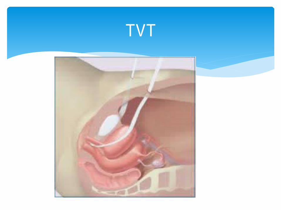
TVT
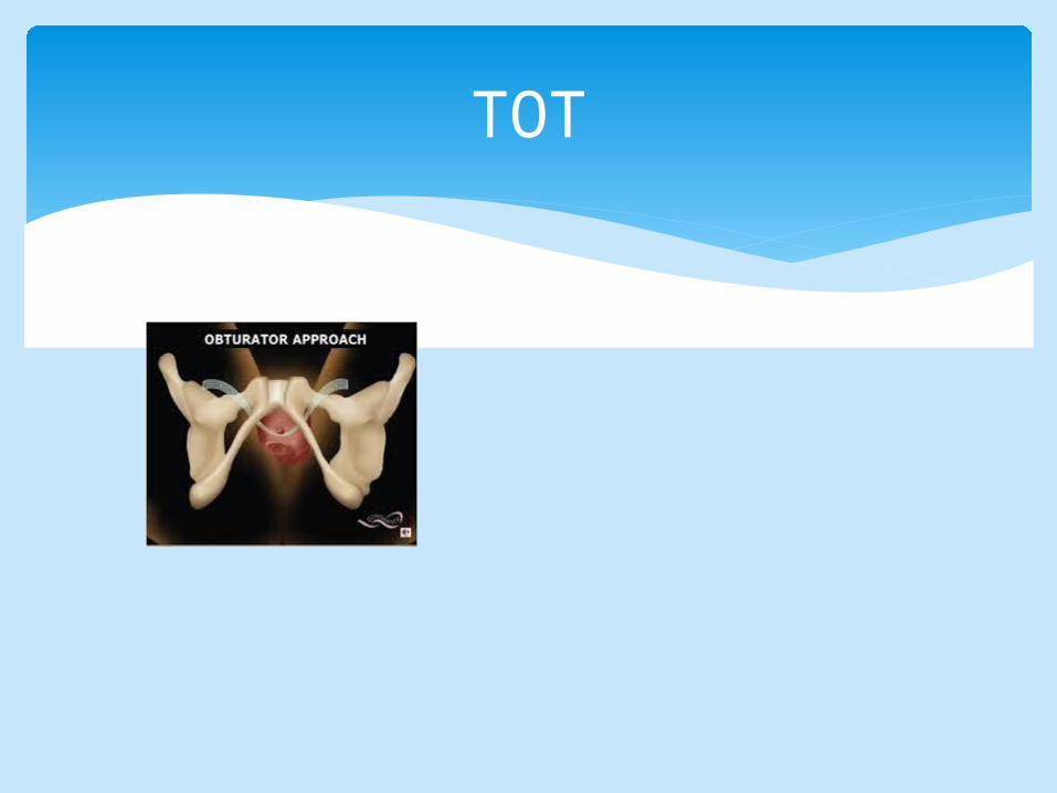
TOT

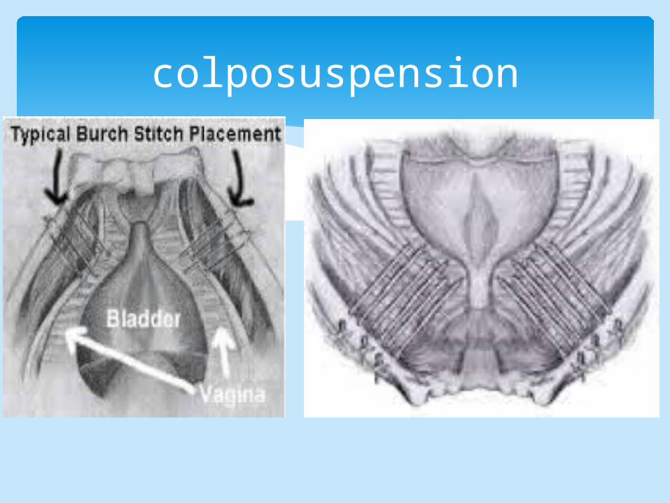
colposuspension
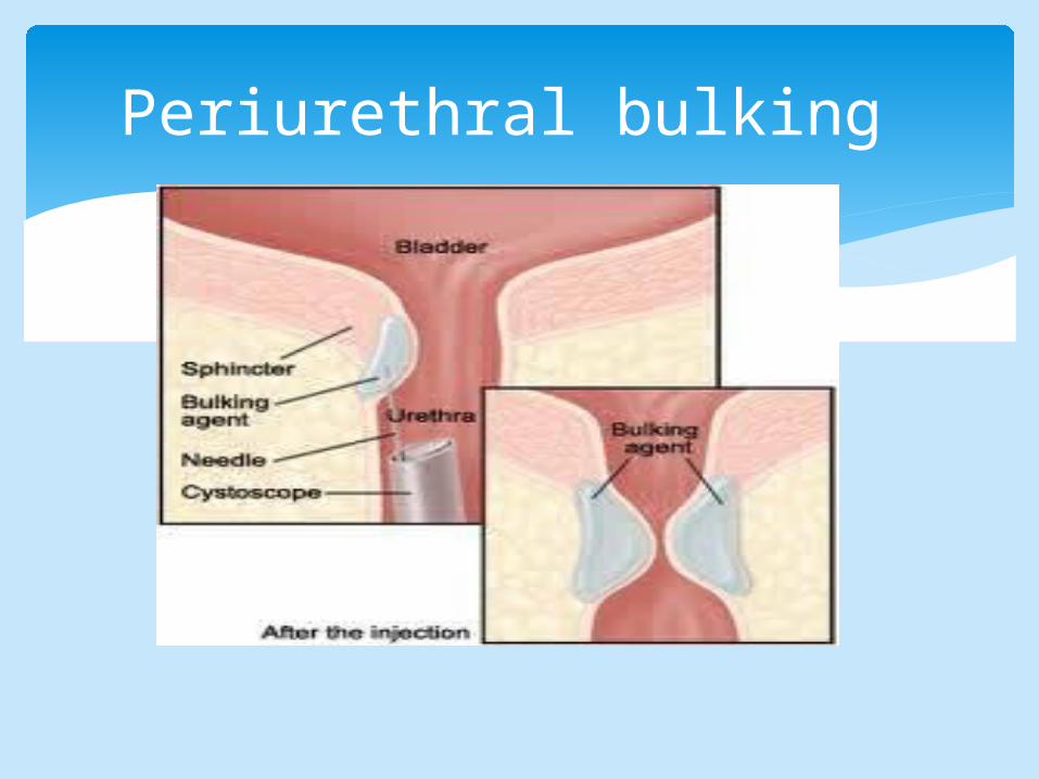
Periurethral bulking

THANKS


