57
Dr. Bryan Hawley PART 2 CST OVERVIEW
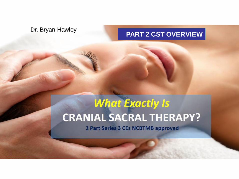
Dr. Bryan HawleyPART 2 CST OVERVIEW

• 2 Part series on CST
• Overall time
• Session 1 agenda Energy
• Session 2 agenda CST Intro.
• 3 CEs NCBTMB approved provider #485
Housekeeping

Origins of Cranial Sacral Therapy
The first written reference to the movement of the spinal nerves and its importance in
life, clarity, and "bringing quiet to the heart" is found in a 4,000-year-old text from China.
Craniosacral work was referred to as "the art of listening." Bone setters in the Middle Ages also sensed the subtle movements of the body. They used these movements to
help reset fractures and dislocations and to treat headaches.
In the early 1900s, the research of Dr. William Sutherland, an American osteopathic
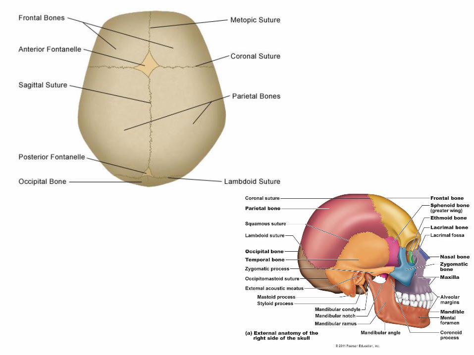
physician, detailed the movement of the cranium and pelvis. Before his research it was believed that the cranium was a solid immovable mass. Sutherland reported that the
skull is actually made up of 22 separate and movable bones that are connected by
layers of tissue. He called his work Cranial Osteopathy. Nephi Cotton, an American
chiropractor and contemporary of Sutherland, called this approach Craniology. The
graduates of these two disciplines have refined and enhanced these original approaches and renamed their work as Sacro-Occipital technique, Cranial Movement
Therapy, or Craniosacral Therapy.
Dr. John Upledger, an osteopathic physician, and others at the Department of
Biomechanics at Michigan State University, College of Osteopathic Medicine learned of Sutherland's research and developed it further. He researched the clinical observations
of various osteopathic physicians. This research provided the basis for Upledger's work
that he named Craniosacral Therapy.

Craniosacral therapists can most easily feel the CSR in the body by lightly touching the
base of the skull or the sacrum. During a session, they feel for disturbances in the rate,
amplitude, symmetry, and quality of flow of the CSR. A therapist uses very gentle touch to
balance the flow of the CSR. Once the cerebrospinal fluid moves freely, the body's natural
healing responses can function.
A craniosacral session generally lasts 30-90 minutes. The client remains fully clothed and
lays down on a massage table while the therapist gently assesses the flow of the CSR.
Although Upledger describes several techniques which may be used in a craniosacral
therapy session, usually the first technique used is energy cyst release. "This technique is a hands-on method of releasing foreign or disruptive energies from the patient's body.
Energy cysts may cause the disruption of the tissues and organs were they are located."
The therapist feels these cysts in the client's body and gently releases the blockage of
energy.

• ADD/ADHD
• Autism Spectrum Disorder
• Birth Trauma
• Cerebral Palsy
• CNS Disorders of Unknown
Etiology
• Constipation
• Difficulty Chewing/Swallowing
• Difficulty with eye tracking
• Ear Infection
• Headaches
• Immune Disorders
• Learning Differences
• Plagiocephly
• Poor Motor Planning and/or
Execution
• Stress
• Reflux
• Seizure Disorder
• Sensory Processing Issues
• Speech Issues
• Strabismus
• Torticollis
• Traumatic Impact
CranioSacral Therapy has been shown to aid correction of:
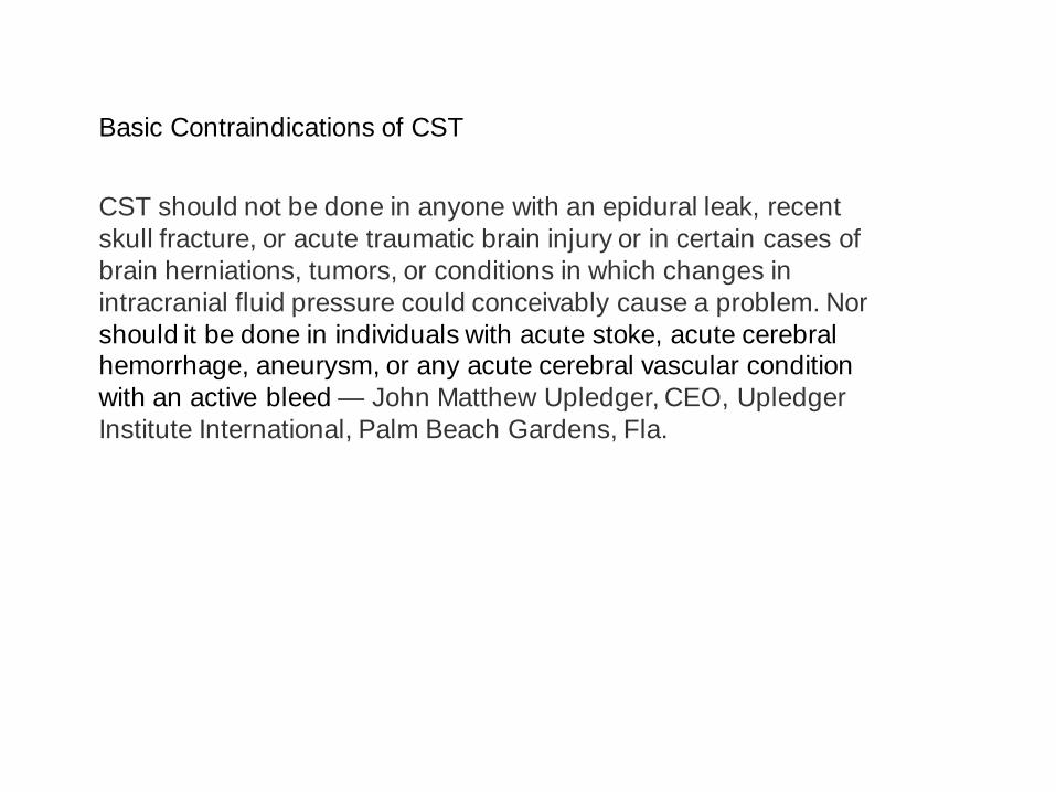
CST should not be done in anyone with an epidural leak, recent
skull fracture, or acute traumatic brain injury or in certain cases of
brain herniations, tumors, or conditions in which changes in
intracranial fluid pressure could conceivably cause a problem. Nor
should it be done in individuals with acute stoke, acute cerebral hemorrhage, aneurysm, or any acute cerebral vascular condition
with an active bleed — John Matthew Upledger, CEO, Upledger
Institute International, Palm Beach Gardens, Fla.
Basic Contraindications of CST

This focus on the central nervous system is what also helps
distinguish CranioSacral Therapy from many other forms of
bodywork.
The central nervous
system consists of the
brain and spinal cord. It
is referred to as
"central" because it combines information
from the entire body and
coordinates activity
across the whole
organism.

Mechanical Basis ofHow CST Works
• In CST, bones in the cranium and the spine are used
as “handles” to release the restrictions in the
membrane system to allow the fluid to flow properly.
• Functionally, the cranial system is related to the
central nervous system, the autonomic nervous
system, the neuromusculoskeletal system and the
endocrine system.
• The system’s fluid intake is via the choroid plexus
which allows passage of fluid from the vascular
system into the ventricular system of the brain.
8

The craniosacral system is the cranium,
spine, and sacrum that are connected by a
continuous membrane of connective tissue
deep inside the body, called the dura
mater. The dura mater also encompenses the brain and the central nervous system.
Sutherland noticed that cerebral spinal fluid
rises and falls within the compartment of
the dura mata. He called this movement
the primary respiratory impulse; today it is known as the Craniosacral Rhythm (CSR)
or the Cranial Wave.

You may be wondering, "What rhythm,
what system?" The craniosacral system is
the cerebrospinal fluid and membranes
that bathe the brain and spinal cord. The
system runs from your head – including your skull, face, and mouth – to your
tailbone. The fluid and membranes of the
craniosacral system move in slow,
rhythmical waves, at about 6-12 cycles per
minute. Imagine your brain and spinal cord sitting in a protective fluid that slowly
undulates, and you have a good picture of
how a healthy craniosacral system works.
A disruption of this rhythm can cause dysfunction in the brain and spinal cord,
which in turn, can cause problems
throughout the body. Examples include
chronic pain, lowered vitality, recurrent
infections, and the build-up of stress.

Therapeutic Pulse The Therapeutic Pulse
is a phenomenon which we have observed
on many occasions when the subject’s
body is in the process of self-correction. It
may occur anywhere on or in the body under treatment. The amplitude of the
Therapeutic Pulse seems to increase from
near zero until it comes into the conscious
awareness of the therapist. It is not the
cardiac pulse, although it seems almost the same when you first experience it. The
high-amplitude therapeutic pulse may last
seconds or minutes. Its presence seems to
indicate that something good is occurring.
After the self-correction is complete, the Therapeutic Pulse diminishes in amplitude
until it becomes imperceptible. It is my
policy not to change whatever I am doing
while the Therapeutic Pulse is perceptible.

Can we physically feel these pulses?
There are 2 ways
1. Physical 2. Intuitive

Personal method of
“Patterns of awareness”
Outward
eyestouch
smells
Inward
energysystems connection
outer energy bodies

Early exploration of cranial manipulation was performed primarily by
osteopaths and chiropractors who formed societies to investigate and teach
cranial methods. These pioneers were at odds with the larger scientific
community, and often with their own peers, over one central aspect of the
cranial system: the movement of the cranial bones.
Conventional anatomical wisdom taught that cranial bones were movable
only in young infants and were solidly fused in adulthood. The
controversy raged until quite recently.

In the early 1970s, the College of Osteopathic Medicine at Michigan State University
sought to resolve this controversy. It brought together a team of researchers with the
objective of proving or disproving the basic tenets of cranial manipulative techniques.
Of course, the major premise involved the movement of cranial bones.
Optical and electron microscopy showed the existence of blood vessels, nerve
fibers, collagen and elastic fibers within cranial sutures. There was little evidence of
sutural ossification, which would prevent movement of cranial bones in relation to
each other.
With the existence of cranial bone motion established, elucidating the mechanisms
behind this motion became the next task of the Michigan State University team. It
was here that the role of the craniosacral dura mater and cerebrospinal fluid were
integrated into a comprehensive model of the craniosacral system. They called it the
Pressurestat Model.
It was now known that the dura mater plays a key role in cranial bone movement.
Techniques for evaluating and treating the dural membranes were developed largely
by Dr. John E. Upledger, a member of the Michigan State University team.

Simplified craniosacral system used to explain the Pressurestat
Model.

A closer view of the craniosacral system

Components of the CSS: container, fluid, production and inflow
system, outflow system, and regulatory system.

Skull bonesSuture
s
Cerebrospinal fluid
Dural membrane (spinal region)
Semi-Closed Hydraulic System of the Cerebrospinal Fluid and Dural
Membrane
Duralmembrane
20

Anterior-Posterior and Superior-Inferior Axes of the Dural Membrane System
ANTERIOR
Sacrococcygeal complex
Sagittal suture
21
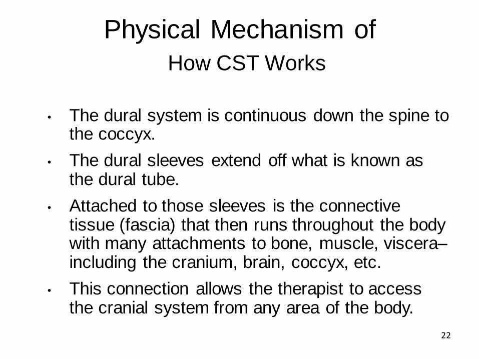
Physical Mechanism of
How CST Works
• The dural system is continuous down the spine to the coccyx.
• The dural sleeves extend off what is known as the dural tube.
• Attached to those sleeves is the connective tissue (fascia) that then runs throughout the body with many attachments to bone, muscle, viscera–including the cranium, brain, coccyx, etc.
• This connection allows the therapist to access the cranial system from any area of the body.
22

The cerebral spinal fluid serves four purposes: buoyancy allowing the brain to
maintain its density, protecting the brain tissue from injury when jolted or hit,
provides chemical stability rinsing metabolic waste from the central nervous
system through the blood brain barrier, and prevention of brain ischemia,
sufficient blood flow to the brain

Fascia is the “webbing”
that is a continuous net-
like sheet of connective
tissue that binds,
connects, glides,
supports and envelops
all structures of the
body, Including the dural
system.
Sagittal section

The craniosacral system
The craniosacral
system surrounds,
protects, nourishes
and cleanses the
brain and spinal
cord. It also
houses the
Cerebral spinal
fluid.
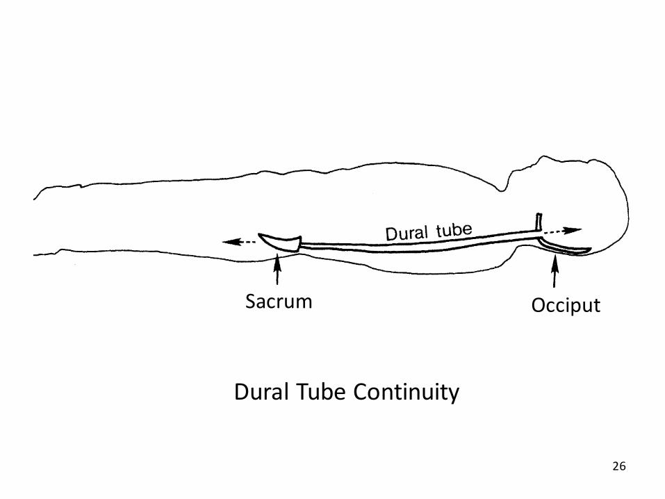
Dural Tube Continuity
Sacrum Occiput
26

A balloon to represent the craniosacral system and the
Pressurestat Model.

Water (CSF) flows into
the balloon in “on and
off” cycles creating an
increase and decrease
of volume and
pressure within the
balloon.

Pressurestat motion causes the
body to move via the fascial
system.

This brings us to Palpation When we think of palpation we normally
think of getting a pulse or feeling a tight
muscle. However there are other forms of
palpation.
But palpation can be light, heavy, physical,
or energetic even.
It can assess the taughtness of a muscle,
body fluids, mobility of a joint, bone articulations, tendons, or even
electromagnetic fields.
At one end of this continuum is intrusive or
invasive palpation, which uses firm, heavy force to probe beneath the skin’s surface.
At the other end of this continuum is nonintrusive palpation, which permits
examination without evoking resistance. It is this method of palpation which is
most useful to the CranioSacral Therapy practitioner.

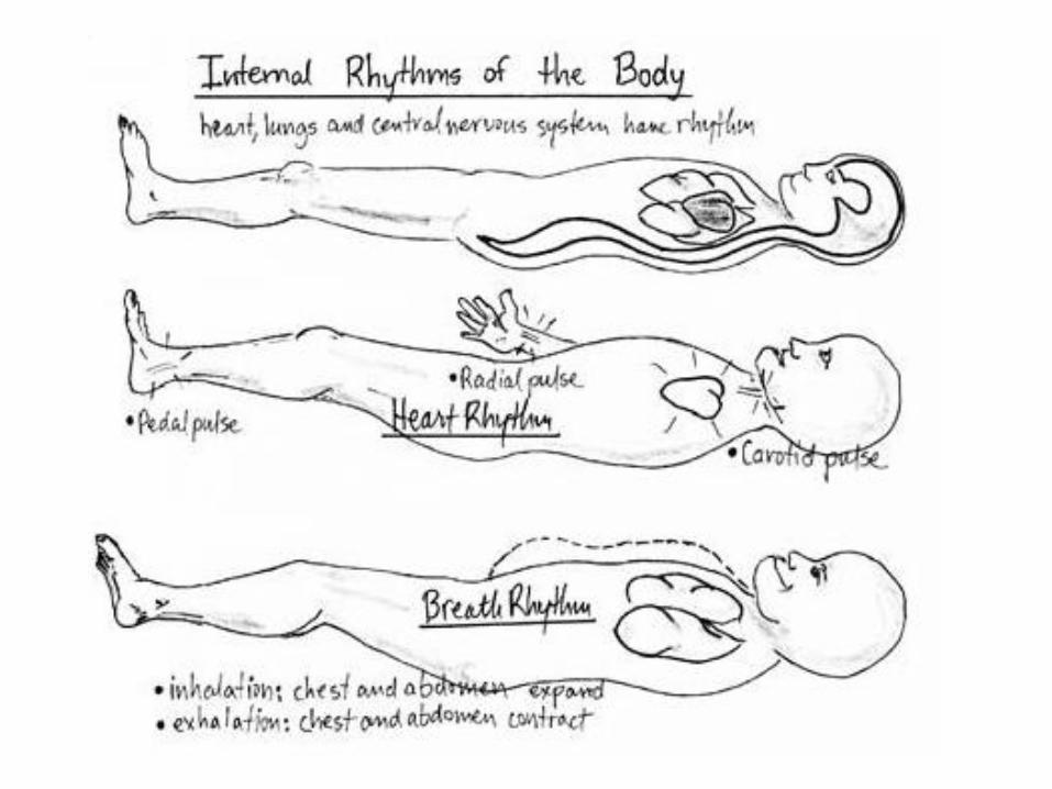
Palpating the Craniosacral Rhythm The craniosacral rhythm, like the cardiac and
respiratory pulse, can be felt throughout the body. Also, like the other pulses, the
craniosacral rhythm has a distinctive character at different locations in the body.
The expansion phase of the craniosacral system is termed flexion, while the
contraction phase is termed extension. Thus it is said that the cranium expands
during flexion and contracts during extension
Since you are familiar with the cardiac and respiratory pulses, palpate them first.
You can actually visualize these. Then remove them from your awareness and feel
the craniosacral rhythm, which is slower and more subtle than either the cardiac or
respiratory pulse. The craniosacral rhythm occurs with a frequency of about six to
twelve cycles per minute. This means that flexion takes place to a slow count of 1-2-3. There is a slight pause between flexion and extension, then extension occurs
at a slow count of 1-2-3.

Some of the areas to feel and coordinate these pulses.

The craniosacral rhythm occurs with a frequency of about six to twelve cycles per
minute. This means that flexion takes place to a slow count of 1-2-3. There is a slight
pause between flexion and extension, then extension occurs at a slow count of 1-2-3.
FLEXION
EXTENSION

To evaluate the dural tube, the therapist sits at the side of the supine client and places one hand under the client’s occiput, and the other hand beneath the client’s sacrum. This can be done side lying or supine.This is done passively at first just observing the connection and rhythm at both ends.If differences or restrictions are observed the therapist may induce a traction like effect by simply turning both hands in relation to the pulses.

Dural Tube Rock: With the client supine, place one hand under the occiput and the
other hand under the sacrum. Encourage a gentle rocking between the two ends with
the craniosacral rhythm. In doing so, you will help to release restrictions of the
transverse rings of fascia in the dural tube. The more you rock, the better the dural
tube will like it. Dural Tube Glide: With the client and your hands in the same position, “tune-in” to
the longitudinal motion at the occiput and sacrum. (This motion is happening
simultaneously with the rocking/rotational motion.) By enhancing this longitudinal
motion by slight tractioning at each end during flexion and extension, you address the
nerve roots as well as any remaining restrictions of the dural tube within the vertebral canal. Restrictions are freed by moving the dural tube. Be patient and move it through
several cycles. You can also use prolonged traction on a restricted dural tube. Simply
hold and await the release.

Fascia runs like a continuous web of tissue throughout the body and remains somewhat mobile under normal circumstances. Gentle traction applied on the fascia in arbitrary directions from various positions helps localize restricted areas.
An example of how the palpation of fascial motion is observed is at the feet, when the therapist takes the client’s heels into their palms, and obtains subtle physiological information via the entire posterior fascial train. The mechanism is a slight even traction and controlled inward and outward rotation of the legs feeling for tightness.

Arcs or Arcing
Active lesions/problems are differentiated from inactive residual effects by a
technique known as "arcing" By placing hands side by side the therapist can glide over adjacent regions and triangulate the spot of interference. This is done on a more intuitive level.
Arcs can also be palpated “off” the client’s body, in the same way radiant heat or an energy aura is sensed because while dysfunctions have physical manifestations which inhibit or create physically palpable waves on the body, the underlying energy also presents radiating arcs.
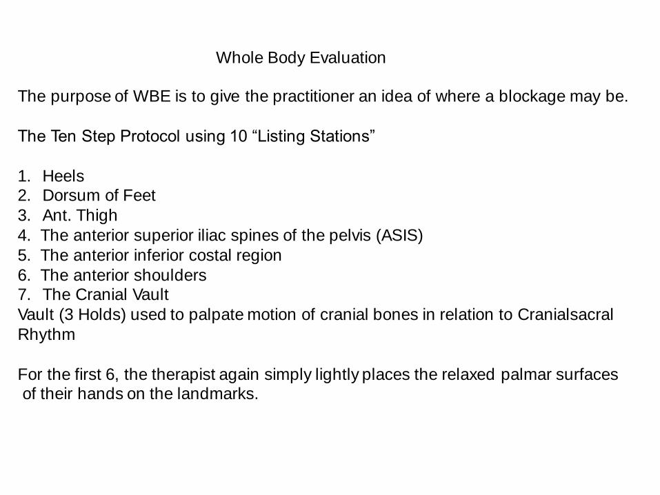
Whole Body Evaluation
The purpose of WBE is to give the practitioner an idea of where a blockage may be.
The Ten Step Protocol using 10 “Listing Stations”
1. Heels2. Dorsum of Feet
3. Ant. Thigh
4. The anterior superior iliac spines of the pelvis (ASIS)
5. The anterior inferior costal region
6. The anterior shoulders7. The Cranial Vault
Vault (3 Holds) used to palpate motion of cranial bones in relation to Cranialsacral
Rhythm
For the first 6, the therapist again simply lightly places the relaxed palmar surfacesof their hands on the landmarks.
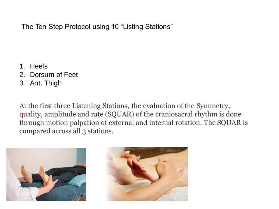
At the first three Listening Stations, the evaluation of the Symmetry, quality, amplitude and rate (SQUAR) of the craniosacral rhythm is done through motion palpation of external and internal rotation. The SQUAR is compared across all 3 stations.
1. Heels
2. Dorsum of Feet
3. Ant. Thigh
The Ten Step Protocol using 10 “Listing Stations”

4. The anterior superior iliac spines of the pelvis (ASIS)5. The anterior inferior costal region6. The anterior shoulders
The Ten Step Protocol using 10 “Listing Stations”
With these next three Listening Stations, the therapist continues to evaluate the craniosacral rhythm through the motion palpation of external and internal rotation, and compares their evaluation with that of the distal Listening Stations.For example, if the amplitude and rate are greater above the pelvis than below it, there is a restriction. If the symmetry is equal above the pelvis but unequal at the feet, there is a restriction. However, if the SQUAR is equal above and below the pelvis, there is no palpable restriction to craniosacral motion at the pelvis.
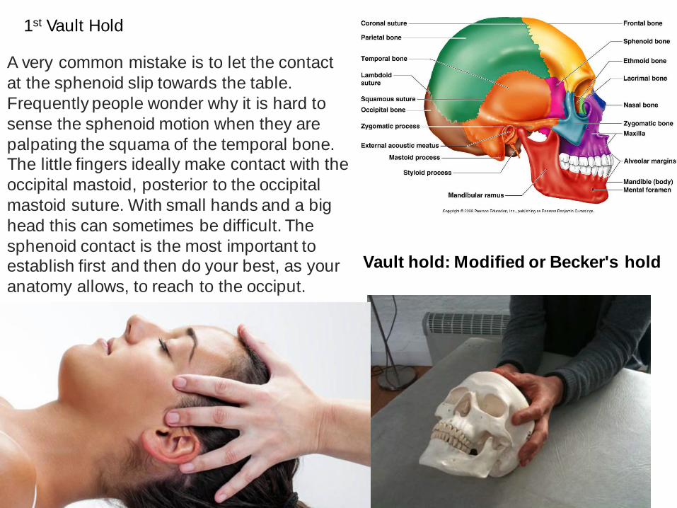
1st Vault Hold
A very common mistake is to let the contact
at the sphenoid slip towards the table.
Frequently people wonder why it is hard to
sense the sphenoid motion when they are
palpating the squama of the temporal bone. The little fingers ideally make contact with the
occipital mastoid, posterior to the occipital
mastoid suture. With small hands and a big
head this can sometimes be difficult. The
sphenoid contact is the most important to establish first and then do your best, as your
anatomy allows, to reach to the occiput.
Vault hold: Modified or Becker's hold
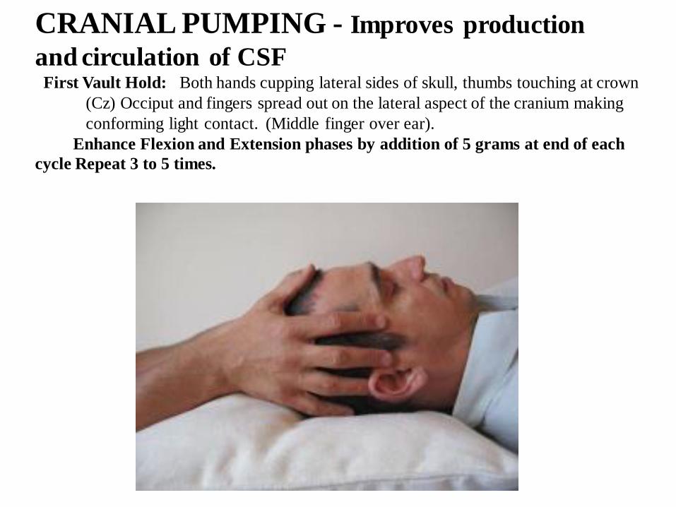
CRANIAL PUMPING - Improves production
and circulation of CSFFirst Vault Hold: Both hands cupping lateral sides of skull, thumbs touching at crown
(Cz) Occiput and fingers spread out on the lateral aspect of the cranium making
conforming light contact. (Middle finger over ear).
Enhance Flexion and Extension phases by addition of 5 grams at end of each
cycle Repeat 3 to 5 times.

2nd Vault Hold
Second Vault Hold: (Vulcan Mind Meld “ Live Long and Prosper”)
Vulcan spread Palms on forehead at eyebrow line, thumb and 5th finger at
greater wings of sphenoid
Posterior Hand – Parallel to body, palm cupping Occiput with thumb below
ear
The Second Vault hold facilitates perception of the flexion and extension between the sphenoid through the thumb and fifth fingers of one hand, while the occiput is palpated through the other hand, in which it is cupped. In this hold, the superior hand can gently traction and palpate the cranial membrane system easily also by lifting the sphenoid and evaluating the freedom of occipital movement.
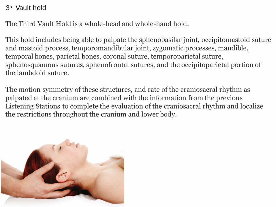
3rd Vault hold
The Third Vault Hold is a whole-head and whole-hand hold.
This hold includes being able to palpate the sphenobasilar joint, occipitomastoid suture and mastoid process, temporomandibular joint, zygomatic processes, mandible, temporal bones, parietal bones, coronal suture, temporoparietal suture, sphenosquamous sutures, sphenofrontal sutures, and the occipitoparietal portion of the lambdoid suture.
The motion symmetry of these structures, and rate of the craniosacral rhythm as palpated at the cranium are combined with the information from the previous Listening Stations to complete the evaluation of the craniosacral rhythm and localize the restrictions throughout the cranium and lower body.

Sutherland also wrote about a second practice called Direction of Energy.
In this technique the therapist uses his hands to pass energy from one of his
hands, through the patient, into the other hand. With this technique, the
redirected energy can flow more freely in a productive pattern throughout the
body.
Sutherland first wrote about the concept in the 1930s. He was using it to release the
joints (sutures) between cranial bones that were "stuck" for one reason or another.
He would use his hands to direct energy from one side of the skull to the other
through the suture. He believed the energy was somehow recruited from the
patient's cerebrospinal fluid and directed into the suture by his hand positions
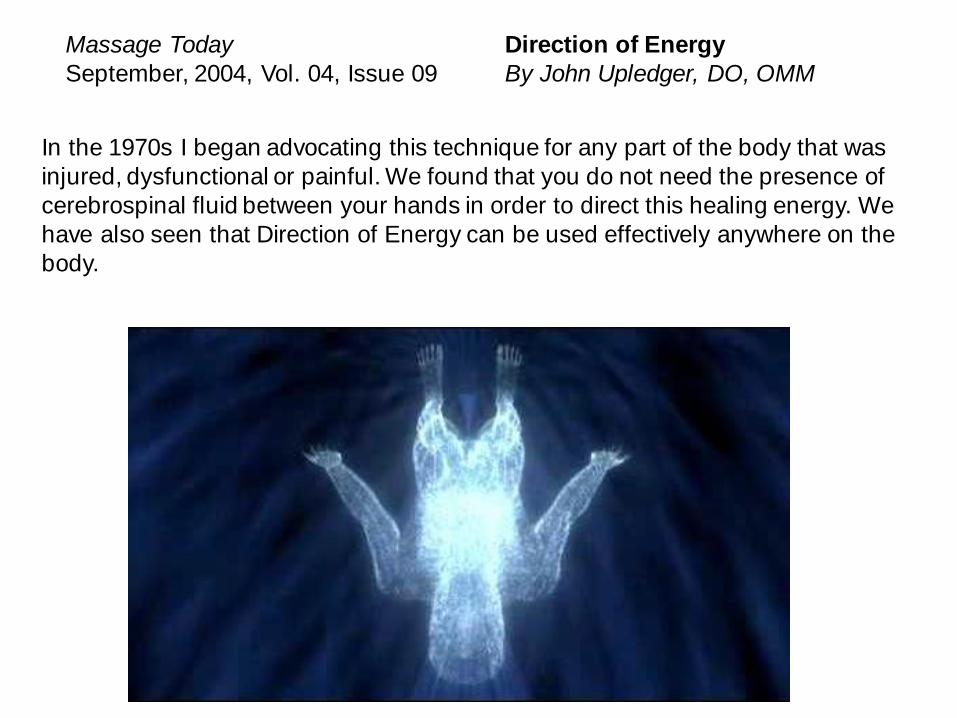
In the 1970s I began advocating this technique for any part of the body that was
injured, dysfunctional or painful. We found that you do not need the presence of
cerebrospinal fluid between your hands in order to direct this healing energy. We
have also seen that Direction of Energy can be used effectively anywhere on the
body.
Massage Today
September, 2004, Vol. 04, Issue 09
Direction of Energy
By John Upledger, DO, OMM

Albert Einstein visualized himself riding on a beam of light and imagined
what he would experience in order to discover the Theory of Relativity.
Thomas Edison placed himself in a trance-like state called hypnagosis to
bring forth his most important inventions. Crick and Watson played with
Tinker Toys in their discovery of the structure of DNA. Imagination came first, analysis later.
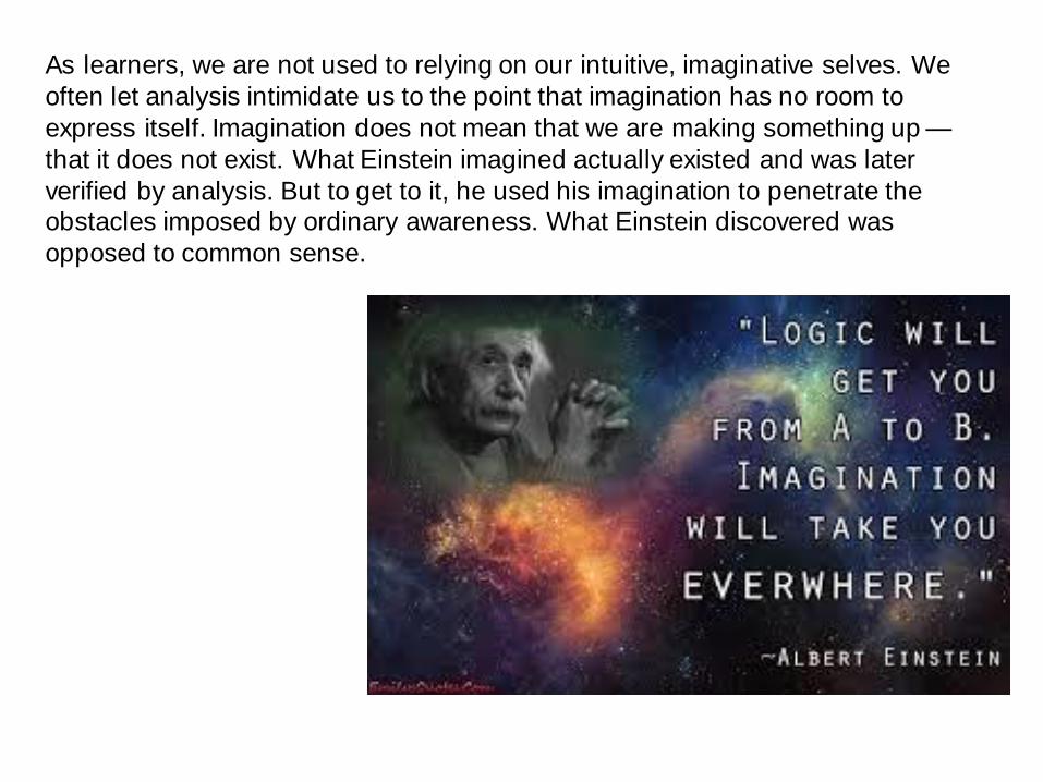
As learners, we are not used to relying on our intuitive, imaginative selves. We
often let analysis intimidate us to the point that imagination has no room to
express itself. Imagination does not mean that we are making something up —
that it does not exist. What Einstein imagined actually existed and was later
verified by analysis. But to get to it, he used his imagination to penetrate the obstacles imposed by ordinary awareness. What Einstein discovered was
opposed to common sense.

V-Spread (Reference CranioSacral Therapy pp. 74, 139-40, 164-66
and 263)
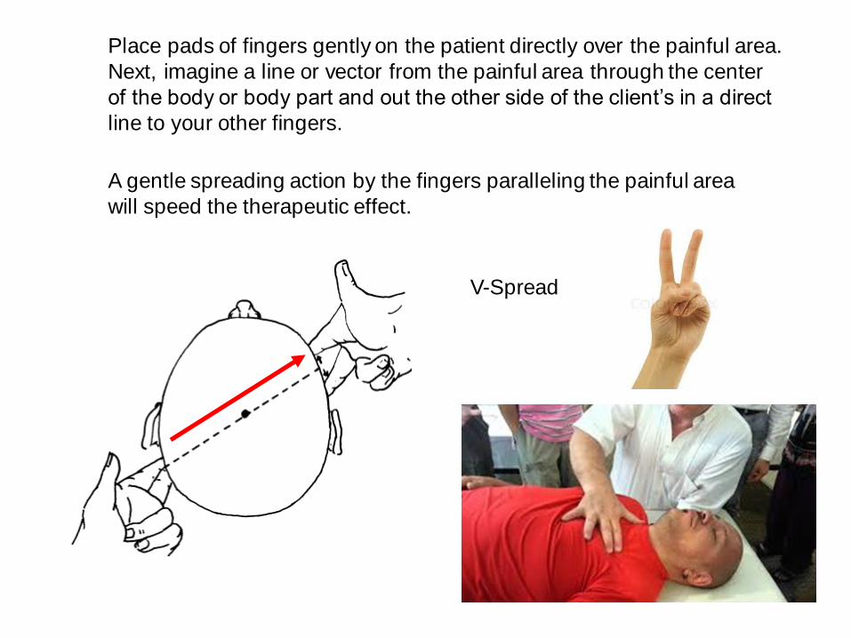
Place pads of fingers gently on the patient directly over the painful area.
Next, imagine a line or vector from the painful area through the center
of the body or body part and out the other side of the client’s in a direct
line to your other fingers.
A gentle spreading action by the fingers paralleling the painful area
will speed the therapeutic effect.
V-Spread
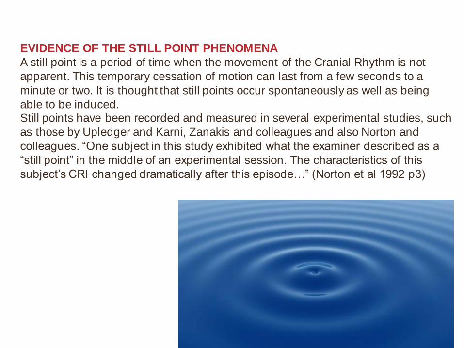
EVIDENCE OF THE STILL POINT PHENOMENA
A still point is a period of time when the movement of the Cranial Rhythm is not
apparent. This temporary cessation of motion can last from a few seconds to a
minute or two. It is thought that still points occur spontaneously as well as being
able to be induced.Still points have been recorded and measured in several experimental studies, such
as those by Upledger and Karni, Zanakis and colleagues and also Norton and
colleagues. “One subject in this study exhibited what the examiner described as a
“still point” in the middle of an experimental session. The characteristics of this
subject’s CRI changed dramatically after this episode…” (Norton et al 1992 p3)
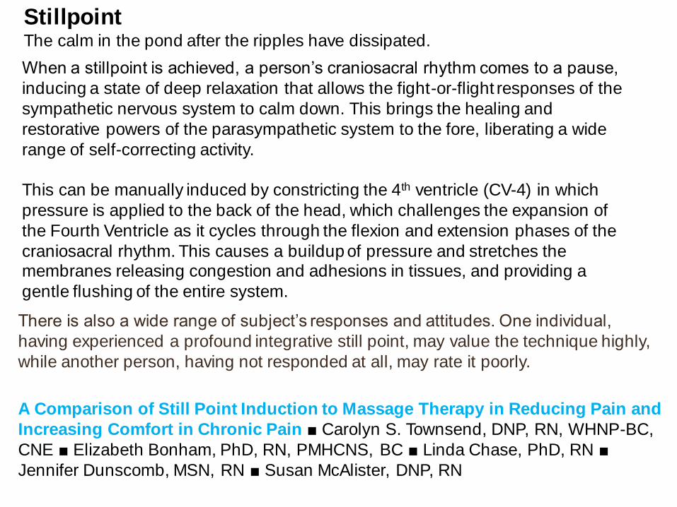
When a stillpoint is achieved, a person’s craniosacral rhythm comes to a pause,
inducing a state of deep relaxation that allows the fight-or-flight responses of the
sympathetic nervous system to calm down. This brings the healing and
restorative powers of the parasympathetic system to the fore, liberating a wide
range of self-correcting activity.
This can be manually induced by constricting the 4th ventricle (CV-4) in which
pressure is applied to the back of the head, which challenges the expansion of
the Fourth Ventricle as it cycles through the flexion and extension phases of the
craniosacral rhythm. This causes a buildup of pressure and stretches the membranes releasing congestion and adhesions in tissues, and providing a
gentle flushing of the entire system.
StillpointThe calm in the pond after the ripples have dissipated.
A Comparison of Still Point Induction to Massage Therapy in Reducing Pain and
Increasing Comfort in Chronic Pain ■ Carolyn S. Townsend, DNP, RN, WHNP-BC,
CNE ■ Elizabeth Bonham, PhD, RN, PMHCNS, BC ■ Linda Chase, PhD, RN ■
Jennifer Dunscomb, MSN, RN ■ Susan McAlister, DNP, RN
There is also a wide range of subject’s responses and attitudes. One individual,
having experienced a profound integrative still point, may value the technique highly,
while another person, having not responded at all, may rate it poorly.

• CranioSacral Therapy I
• CranioSacral Therapy II
• Applying Acupuncture Principles to
CranioSacral Therapy
• Clinical Application of CranioSacral Therapy
• CranioSacral Dissection
• Therapeutic Imagery &Dialoguesm I
• SomatoEmotional Release® I
• Clinical Application of SomatoEmotional Release
• CranioSacral Therapy for Pediatricssm
• SomatoEmotional Release ® II
• CranioSacral Therapy and the Immune
Response • The Brain Speaks
• Advanced I CranioSacral Therapy
• Clinical Application of Advanced
CranioSacral Therapy
• BioAquatic Explorations
• Advanced II CranioSacral Therapy
• Advanced Preceptorship • Advanced II Preceptorship
• CranioSacral Techniques for Estheticians
• ShareCare®
• Equine CranioSacral Techniques I
• Clinical Application of CranioSacral and SomatoEmotional Release for Pediatrics
• Clinical Application of Advanced
CranioSacral Therapy for Pediatrics
• CranioSacral Therapy Symposium
Coursework continues with:

www.Upledger.com

• Tests
• Session 1 agenda Energy
• Session 2 agenda CST Intro.
• 3 CEs NCBTMB approved provider #485
Housekeeping
Bryan


