46
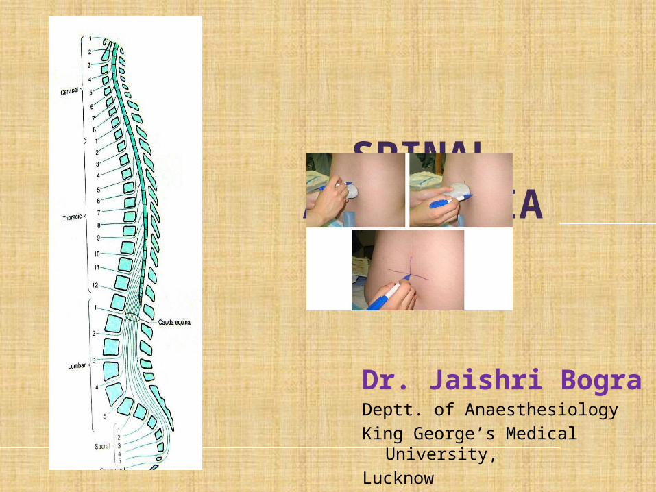
SPINAL ANESTHESIA Dr. Jaishri Bogra Deptt. of Anaesthesiology King George’s Medical University, Lucknow
| Date post: | 27-Dec-2015 |
| Category: |
Documents |
| Upload: | johnathan-reynolds |
| View: | 217 times |
| Download: | 0 times |
SPINAL ANESTHESIA
Dr. Jaishri BograDeptt. of Anaesthesiology
King George’s Medical University,
Lucknow
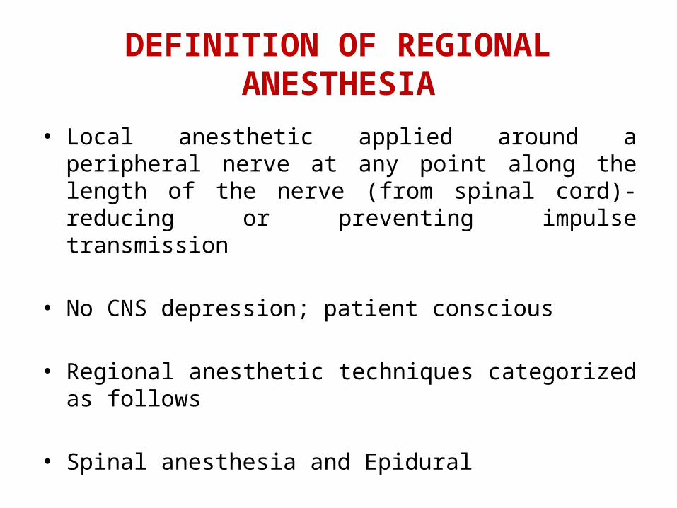
DEFINITION OF REGIONAL ANESTHESIA
• Local anesthetic applied around a peripheral nerve at any point along the length of the nerve (from spinal cord)- reducing or preventing impulse transmission
• No CNS depression; patient conscious
• Regional anesthetic techniques categorized as follows
• Spinal anesthesia and Epidural
• Peripheral nerve blockades

The Advantages of Spinal Anaesthesia
1.Cost
2.Patient satisfaction
3.Respiratory disease
4.Patent airway
5.Diabetic patients
6.Elderly Patients
7.Muscle relaxation
8.Blood loss during operation is less
9. Post operative pain relief
Contd…
• Full and complete anaesthesia• Prolonged block: Pain free postoperatively• Alternative to GA for certain poor risk patients
esp.: Difficult airway Respiratory disease
• Contracted bowel• Suitable for certain surgical procedures• Blunt the stress response to surgery

Indication of SA
Subarachnoid block can be used to provide surgical anesthesia for all procedures carried out on the lower half of the body.
Indications include surgery on the lower limb, pelvis, genitals, and perineum, and most urological procedures.
Can be used for analgesia (Intrathecal opoid)
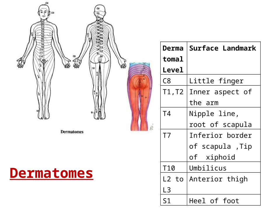
Dermatomal Level
Surface Landmark
C8 Little fingerT1,T2 Inner aspect of the armT4 Nipple line, root of
scapulaT7 Inferior border of
scapula ,Tip of xiphoidT10 UmbilicusL2 to L3
Anterior thigh
S1 Heel of foot
Dermatomes
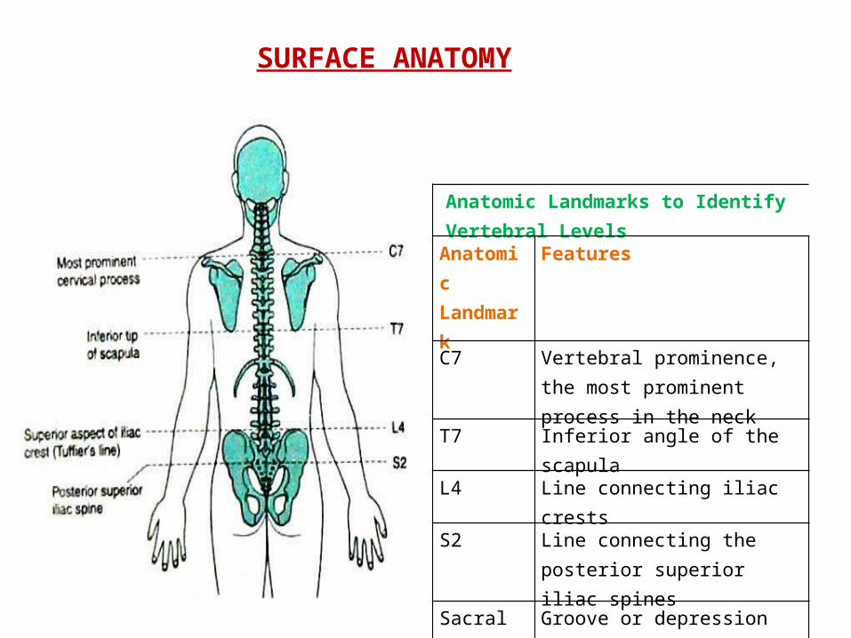
SURFACE ANATOMY
Anatomic Landmarks to Identify Vertebral Levels
Anatomic Landmark
Features
C7 Vertebral prominence, the most prominent process in the neck
T7 Inferior angle of the scapula
L4 Line connecting iliac crests
S2 Line connecting the posterior superior iliac spines
Sacral hiatus
Groove or depression just above or between the gluteal clefts above the coccyx

Spinal Cord
Extends from foramen magnum to Adult : lower border of L1 in /upper border of L2 Infants/children : L3It is about 45 cm longDuramater, Subarachnoid space & subdural space: S2 in adults( S3 in children)S. C gives 31 pairs of spinal nerveAn extension of piamater , the FILUM TERMINALE penetrate the dura and attach the terminal end of spinal cord [conus medullaris]to the periosteum of the coccyx

Vertebrae Anatomy

Important Facts
Cardiac accelerator fibre: T1-T4(Bradycardia & ↓ contractility)
Vasomotor fibre : T5-L1( Determine vasomotor tone)(vasodilation on blockade)
Sympathetic outflow arise from T5-L1(Block ↑vagal tone, small contacted gut with active peristalsis)
Most dependent part in supine position is T4-T8 (imp. For hyperbaric solution)

Spinal Anesthesia/Analgesia

SITE
Adult : L3-L4 or L4-L5 ( or even L2-L3)Infant : L4-L5A line drawn b/w the highest pt. of iliac crests (Tuffier’s line) usually cross either body of L4 or the L4-L5 interspace
Position Sitting lateralProne(anorectal procedure, hypobaric solution, jackknife position)
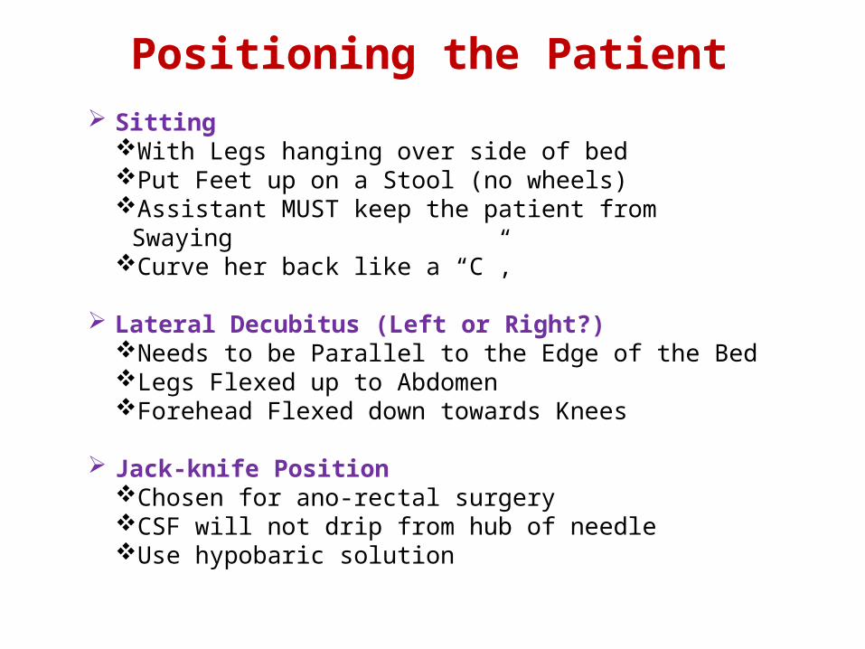
Positioning the Patient Sitting
With Legs hanging over side of bedPut Feet up on a Stool (no wheels)Assistant MUST keep the patient from SwayingCurve her back like a “C”,
Lateral Decubitus (Left or Right?)Needs to be Parallel to the Edge of the BedLegs Flexed up to AbdomenForehead Flexed down towards Knees
Jack-knife PositionChosen for ano-rectal surgeryCSF will not drip from hub of needleUse hypobaric solution

Surface landmarks

.
The patient and operating table should then be placed in the position appropriate for the surgical procedure and drugs chosen.
Lateral decubitus positioning for a neuraxial block. The assistant can help the patient assume the ideal position of “forehead to knees.”
Anesthetic dose is injected at a rate of approximately 0.2 mL/sec

Spinal Anesthesia
A single injection of a local anesthetic solution into the subarachnoid space usually at the lumbar level
Intrathecal Narcotics
Commonly at L3-L4
Largest Interspace
L5-S1

Important Factors Affecting Block Height - SAB
Baricity of anesthetic solution Position of the patient During injection Immediately after injection Drug Dosage (mg) Concentration times volume Addition of Opioids Site of Injection

Additional Factors to Consider with SAB Height
Patient Age Elderly patients > 80 yrs Patient Height Intra-abdominal Pressure Pregnancy & Obesity Drug Volume
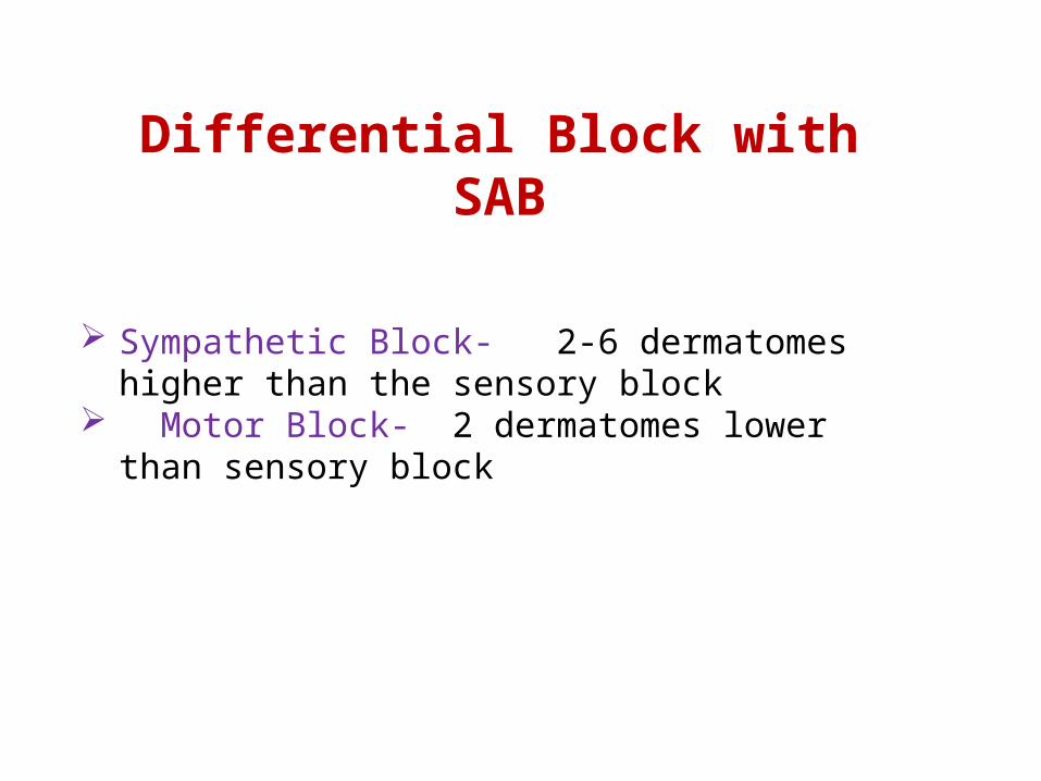
Differential Block with SAB Sympathetic Block- 2-6 dermatomes higher than the
sensory block Motor Block- 2 dermatomes lower than sensory block

When performing a spinal anesthetic, appropriate monitors should be placed, and airway and resuscitation equipment should be readily available.
All equipment for the spinal blockade should be ready for use, and all necessary medications should be drawn up prior to positioning the patient for spinal anesthesia.
Adequate preparation for the spinal reduces the amount of time needed to perform the block and assists with making the patient comfortable.
Proper positioning is the key to making the spinal anesthetic quick and successful.
Technique of Lumbar Puncture
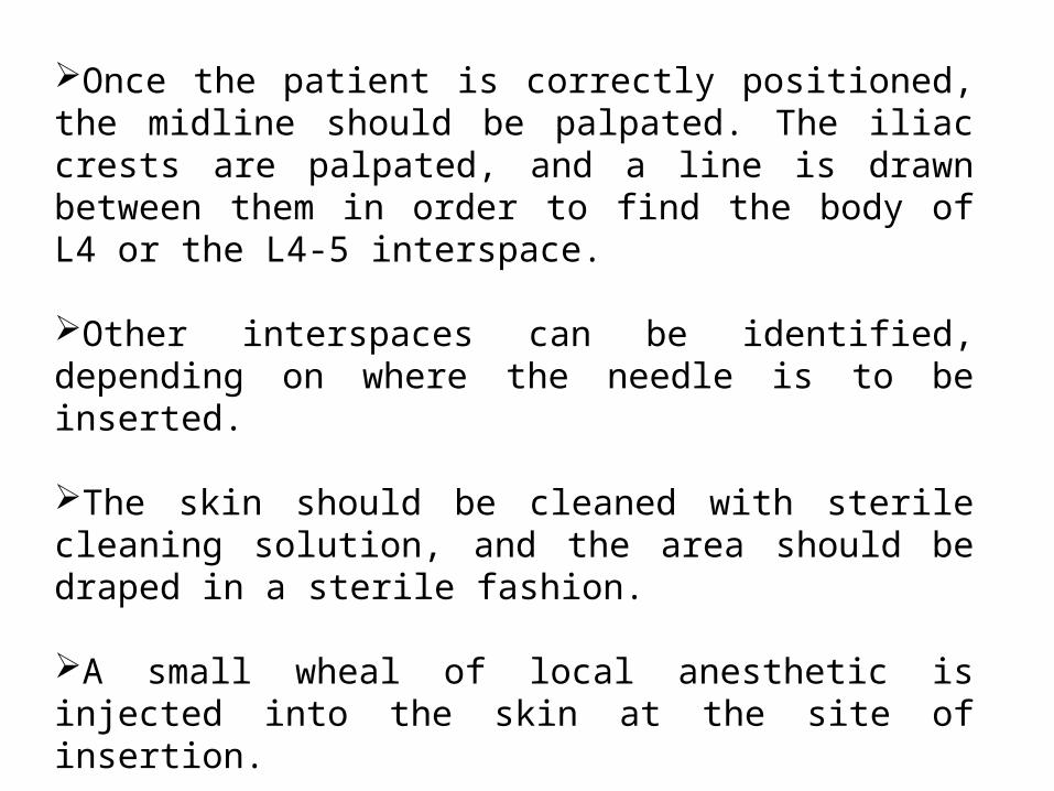
Once the patient is correctly positioned, the midline should be palpated. The iliac crests are palpated, and a line is drawn between them in order to find the body of L4 or the L4-5 interspace.
Other interspaces can be identified, depending on where the needle is to be inserted.
The skin should be cleaned with sterile cleaning solution, and the area should be draped in a sterile fashion.
A small wheal of local anesthetic is injected into the skin at the site of insertion.
More local anesthetic is then administered along the intended path of the spinal needle insertion to a depth of 1 to 2 in.
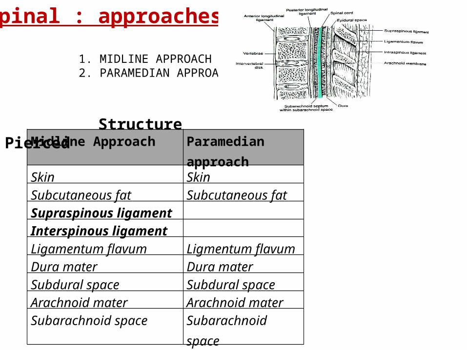
1. MIDLINE APPROACH2. PARAMEDIAN APPROACH
Midline Approach Paramedian approach
Skin SkinSubcutaneous fat Subcutaneous fat Supraspinous ligamentInterspinous ligamentLigamentum flavum Ligmentum flavumDura mater Dura materSubdural space Subdural spaceArachnoid mater Arachnoid materSubarachnoid space Subarachnoid space
Spinal : approaches
Structure Pierced
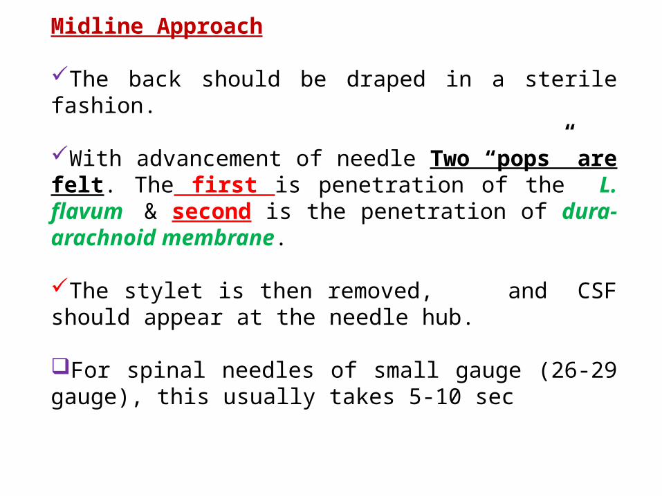
Midline Approach
The back should be draped in a sterile fashion.
With advancement of needle Two “pops” are felt. The first is penetration of the L. flavum & second is the penetration of dura-arachnoid membrane.
The stylet is then removed, and CSF should appear at the needle hub.
For spinal needles of small gauge (26-29 gauge), this usually takes 5-10 sec
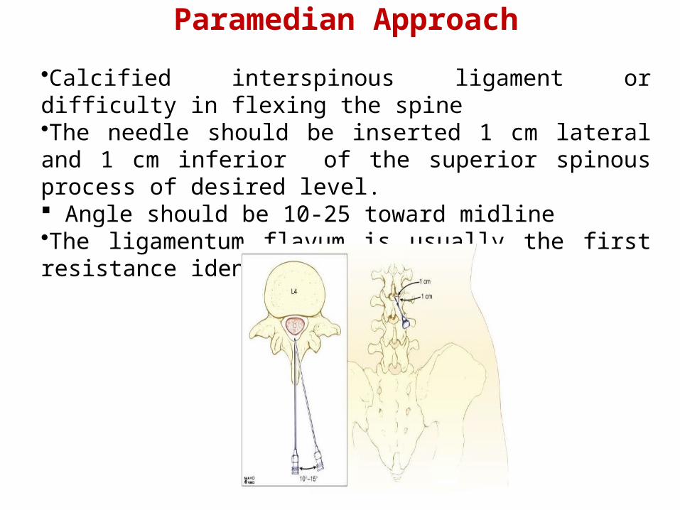
Paramedian Approach
•Calcified interspinous ligament or difficulty in flexing the spine•The needle should be inserted 1 cm lateral and 1 cm inferior of the superior spinous process of desired level. Angle should be 10-25 toward midline•The ligamentum flavum is usually the first resistance identified.
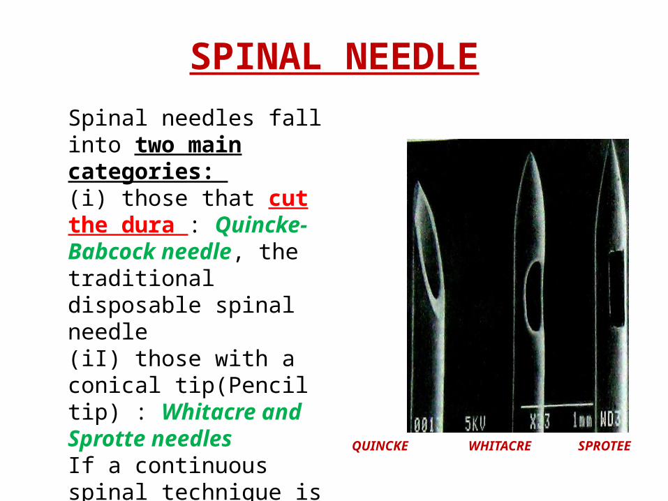
SPINAL NEEDLE
QUINCKE WHITACRE SPROTEE
Spinal needles fall into two main categories: (i) those that cut the dura : Quincke- Babcock needle, the traditional disposable spinal needle(iI) those with a conical tip(Pencil tip) : Whitacre and Sprotte needles If a continuous spinal technique is chosen, use of a Tuohy or Hustead needle can facilitate passage of the catheter
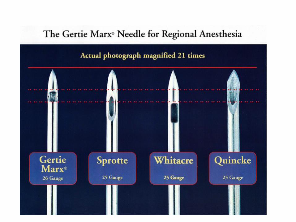

Blunt tip (pencil-point) needle decreased the incidence of PDPH
Sprotte is a side-injection needle with a long opening. It has the advantage of more vigorous CSF flow compared with similar gauge needles.
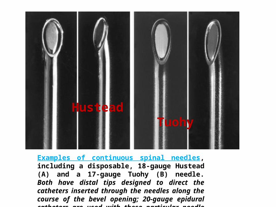
Examples of continuous spinal needles, including a disposable, 18-gauge Hustead (A) and a 17-gauge Tuohy (B) needle. Both have distal tips designed to direct the catheters inserted through the needles along the course of the bevel opening; 20-gauge epidural catheters are used with these particular needle sizes.
Hustead Tuohy
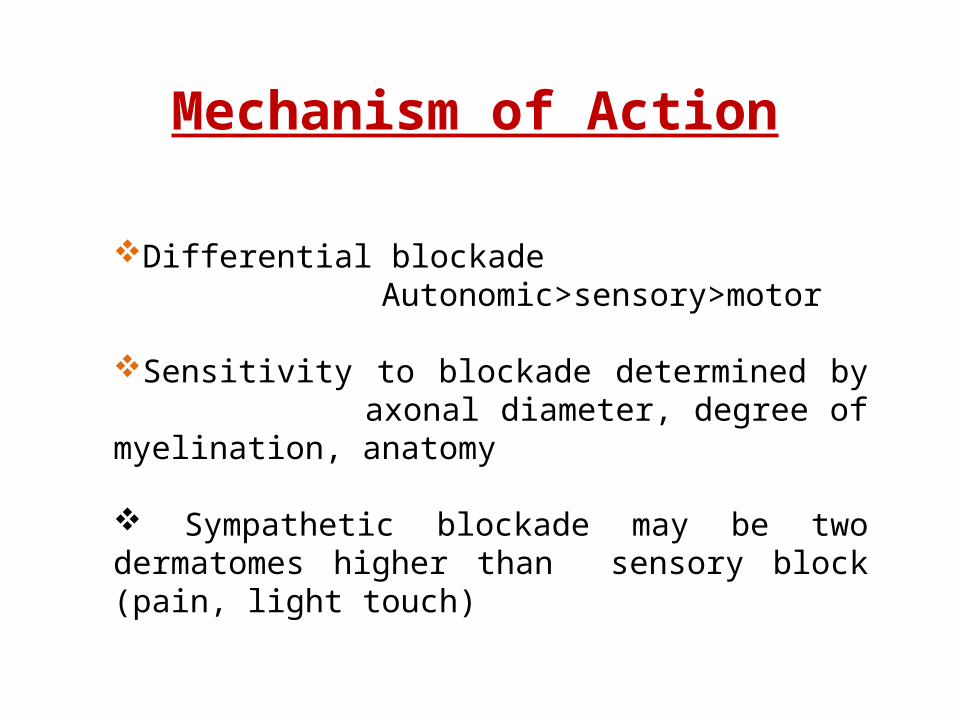
Differential blockade Autonomic>sensory>motor�
Sensitivity to blockade determined by axonal diameter, degree of myelination, anatomy
Sympathetic blockade may be two dermatomes �higher than sensory block (pain, light touch)
Mechanism of Action

Baricity of Local Anesthetics
Isobaric – Stays where you put it LA has the same density or specific gravity as CSF
(1.003-1.008) – Normal Saline
Hypobaric – “Floats” up – Lighter than CSF LA has a density or specific gravity that is less than
CSF (<1.003) – Sterile Water
Hyperbaric – Settles to Dependent aspect of the subarachnoid space – Heavier than CSF
LA has a density or specific gravity that is greater than CSF (>1.008) - Dextrose

Hypobaric and Isobaric Spinal Anesthesia
To make a drug hypobaric to CSF, it must be less dense than CSF, with a baricity appreciably less than 1.0000 or a specific gravity appreciably less than 1.0069 (the mean value of the specific gravity of CSF).
A common method of formulating a hypobaric solution is to mix solution with sterile water & for hyperbaric mix with dextrose

Local Anesthetic Mixture
Dose (mg) * Duration (min)
To T10 To T4 Plain Epinephrine, 0.2 mg
Lidocaine (5% in 7.5% dextrose)
50-60 75-100 60 75-100
Tetracaine (0.5% in 5% dextrose)
6-8 10-16 70-90 100-150
Bupivacaine (0.75% in 8.5% dextrose)
8-10 12-20 90-120 100-150
Ropivacaine (0.5% in dextrose)
12-18 18-25 80-110 —
Levobupivacaine 8-10 12-20 90-120 100-150
* Doses are for use in a 70-kg adult male of average height.
Drug Selection for Hyperbaric Spinal Anesthesia(Miller)
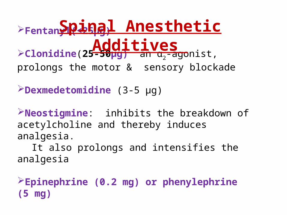
Fentanyl(<25µg)
Clonidine(25-50µg) an α2-agonist, prolongs the motor &
sensory blockade
Dexmedetomidine (3-5 µg)
Neostigmine: inhibits the breakdown of acetylcholine and thereby induces analgesia.
It also prolongs and intensifies the analgesia
Epinephrine (0.2 mg) or phenylephrine (5 mg)
Spinal Anesthetic Additives

In patients should be allowed to leave the recovery room after spinal anesthesia as soon as it can be demonstrated that their block is receding appropriately (at least four dermatomes’ regression or a spinal level of less than T10), they are hemodynamically stable, and they are comfortable.
Outpatients should be able to ambulate without orthostatic changes and void before discharge if they are in a high-risk group for urinary retention

Contraindications of Spinal
ABSOLUTE
Infection at the site of injectionPatient refusalCoagulopathy and other bleeding disordersSevere hypovolemiaIncreased intracranial pressureSevere MS & AS

Cont…RelativeSepsisUncoperative patientPreexisting neurological deficitsSevere spinal deformity
ControversialPrior surgery at the site of injectionComplicated surgeryProlonged operationMajor blood loss
BRADYCARDIA•Defined as HR < 50 beats/ min.•T1-4 involvement leads to unopposed vagal tone and decreased venous return which leads to bradycardia and asystole
NAUSEA AND VOMITING Causes(Hypotension, Increased peristalsis, Opioid analgesia)Nausea and vomiting may be associated with neuraxial block in up to 20% of patients, atropine is almost universally effective in treating the nausea associated with high (T5) neuraxial anesthesia.
Complications

CRANIAL NERVE PALSY
TRANSIENT NEUROLOGICAL SYMPTOM (More common with lidocaine)
CAUDA EQUINA SYNDROME (Bowel-bladder dysfunction)
HIGH NEURAL BLOCKADE : Excessive dose, failure to reduce standard
dose[elderly, pregnant, obese, very short stature]Unconsciousness, hypotension, apnea is
referred to as high spinal or total spinal

HYPOTENSIONPrevented by: Volume loading with 10-20 mL/kg of
intravenous fluidPredictors of hypotension
low intravascular volume in case of hypovolemia due external loss by trauma, dehydration, internal loss
sensory block ≥ T5age > 40 yearssystolic BP < 120 mm Hgcombined spinal and general anesthesiadural puncture between L2-3 and aboveemergency surgerypt with h/o uncontrolled hypertensionunderlying autonomic dysfunction

Treatment of hypotension
100% O2Elevation of leg .Head down position FLUIDS-
crystalloidColloid [500-1000ml] preferred due to increased
intravascular time, maintaining CO, uteroplacental circulation.

Contd…
SYMPATHOMIMETICS:
– Epinephrine: increases HR, CO, SBP, decrease DBP.
– Phenylephrine: Increase in SVR, SBP, DBP. Causes reflex bradycardia, coronary blood flow increased.
– Ephedrine; increase myocardial contractility and rate.
- Mephentermin
Total Spinal
Management of total spinal•Airway - secure airway and administer 100% oxygen •Breathing - ventilate by facemask and intubate. •Circulation - treat with i/v fluids and vasopressor e.g. ephedrine 3-6mg or metaraminol 2mg increments or 0.5-1ml adrenaline 1:10 000 as required •Continue to ventilate until the block wears off (2 - 4 hours) •As the block recedes the patient will begin recovering consciousness followed by breathing and then movement of the arms and finally legs.

Post Dural Puncture Headache:
Due to leak of CSF from dural defect leads to traction in supporting structure especially in dura and tentorium & vasodialatation of cerebral blood vessels.
Usually bifrontal and or occipital, usually worse in upright , coughing , straining
Causes nausea, photophobia, tinnitus, diplopia[6th nerve], cranial nerve palsy
Treatment plan include keeping patient supine, adequate hydration, NSAIDS with without caffeine [increases production of csf and causes vasoconstriction of intracranial vessels], if not relieved within 12-24 hr then epidural blood patch.
Epidural blood patch consists of giving 20 ml

Factors that May Increase the Incidence of Post–spinal Puncture Headache
Age Younger more frequent
Gender Females > males
Needle size Larger > smaller
Needle bevel Less when the needle bevel is placed in the long axis of the neuraxis
Pregnancy More when pregnant
Dural punctures (no.) More with multiple punctures
Factors Not Increasing the Incidence of Post–spinal Puncture Headache
Continuous spinals
Timing of ambulation
Relationships Among Variables and Post–spinal Puncture Headache
Onset of headache :Usually 12-72 h following the procedure
References
• Miller’s Anesthesia, 6th edition. • Morgan Anesthesia 4th edition. • Textbook of regional Anesthesia & Pain MX; By
Prithviraj• Baras Clinical Anesthesia • Neuraxial Anesthesia by D.E. Longnecker et al New
York: McGraw-Hill Medical.• Wylie Anesthesia• Internet Google Scholar•
Thank You


