50
Thyroid Diseases Dr Rodney Itaki Lecturer Anatomical Pathology Discipline University of Papua New Guinea School of Medicine & Health Sciences Division of Pathology
Thyroid DiseasesDr Rodney Itaki
LecturerAnatomical Pathology Discipline
University of Papua New GuineaSchool of Medicine & Health SciencesDivision of Pathology
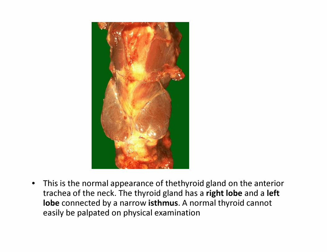
• This is the normal appearance of thethyroid gland on the anterior trachea of the neck. The thyroid gland has a right lobe and a left lobe connected by a narrow isthmus. A normal thyroid cannot easily be palpated on physical examination
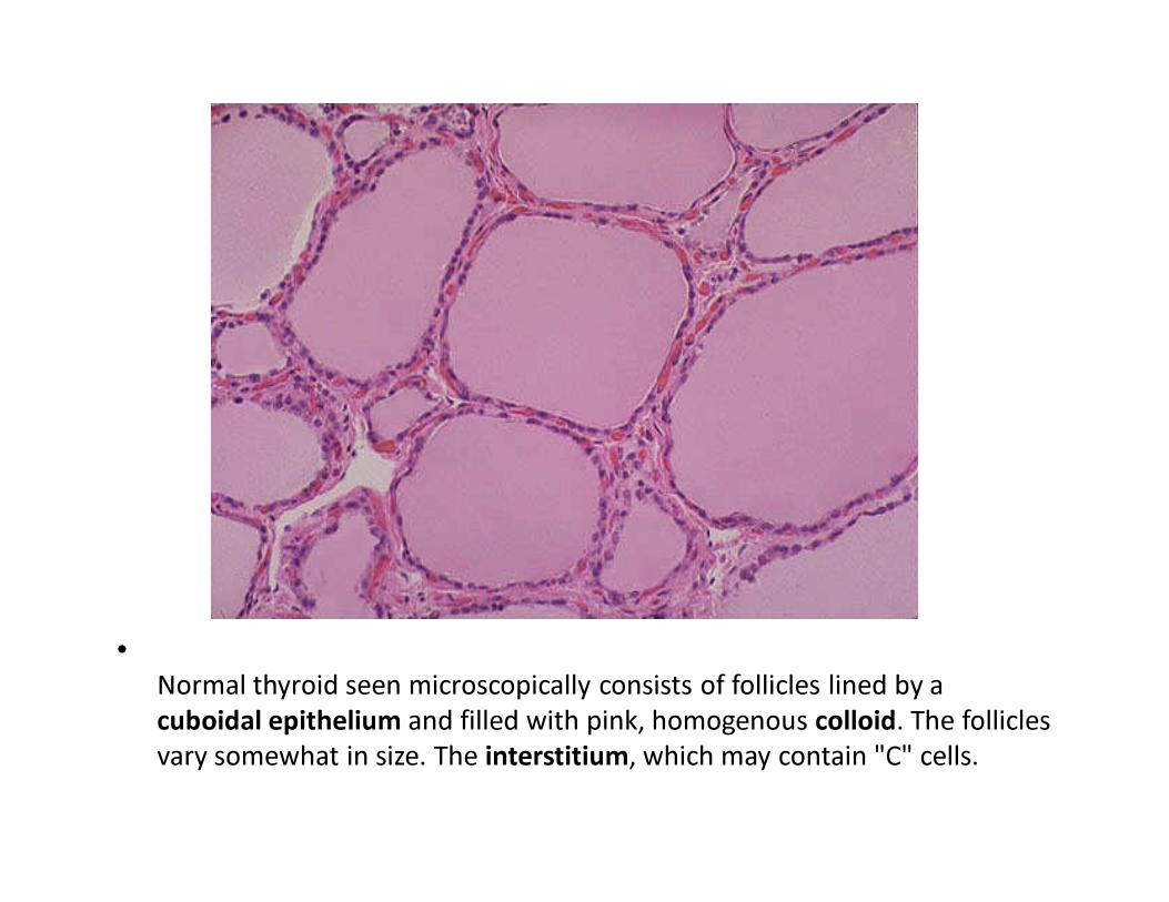
•Normal thyroid seen microscopically consists of follicles lined by a cuboidal epithelium and filled with pink, homogenous colloid. The follicles vary somewhat in size. The interstitium, which may contain "C" cells.
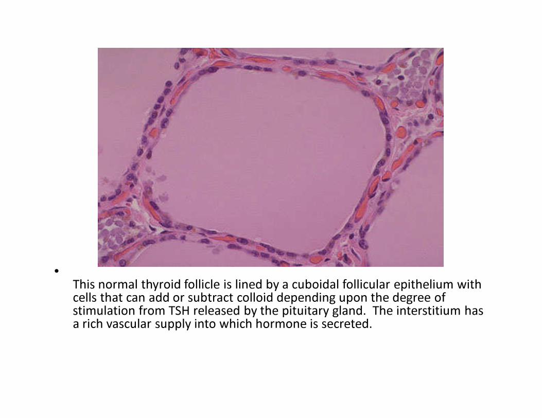
•This normal thyroid follicle is lined by a cuboidal follicular epithelium with cells that can add or subtract colloid depending upon the degree of stimulation from TSH released by the pituitary gland. The interstitium has a rich vascular supply into which hormone is secreted.
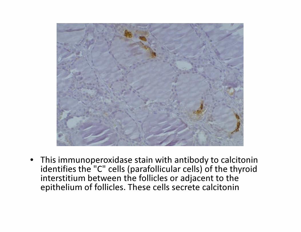
• This immunoperoxidase stain with antibody to calcitoninidentifies the "C" cells (parafollicular cells) of the thyroid interstitium between the follicles or adjacent to the epithelium of follicles. These cells secrete calcitonin
Thyroid Gland• Hormones include thyroxine (T4) or triiodothyronine
(T3)• Stored as thyroglobulin• Synthesis depends on sufficient quantities of iodine
from dietary causes• Rate of extraction of iodine from blood stream & rate
at which T3 & T4 are synthesized & released from thyroglobuline and secreted into blood stream is regulation from pituitary TSH
• Feed back mechanisms regulate pituitary prodxn of TSH
• Serum T3 & T4 are bound to thyroid-binding-globulin (TBG)
Congenital anomalies• Thyroglossal duct cyst – most common
congenital thyroid anomaly• It is a remnant of thyroglossal duct• Does not lead to functional disturbances• Ectopic thyroid tissue may be found anywhere
along course of thyroglossal duct (e.g. In the mouth or mediasternum)
Goiter• Goiter: General term given for enlargement of thyroid• Causes of goiter:
– Physiological – puberty & pregnancy– Iodine deficiency – geographical areas in which diet
deficient in iodine– Hashimoto thyroiditis– Goitrogens – foods & drugs that suppress thyroid
hormones synthesis– Dyshormonogenesis – partial or complete failure of thyroid
hormone synthesis due to various enzyme deficiencies
Goiter - terminology• Simple (nontoxic goiter) goiter: goiter without
thyroid hormone dysfunction• Toxic goiter: associated with hyperthyroidism; if
pt is euthyroid or hypothyroid, term nontoxic goiter is appropriate
• Endemic goiter: goiter occurring with high frequency in iodine-deficient areas.
• Sporadic goiter: goiter due to iodine deficiency but occurring in non-iodine-deficient areas
Nodular goiter• Refers to irregular enlargement of the thyroid
resulting in nodule formation• Nodular colloid goiter refers to late stage of
simple goiter in which goiter is often nodular• Nodules maybe single or multiple (multinodular
goiter)• Most nodules are hypoplastic & do not take up
radioactive iodine (“cold” nodules).• Occasionally nodules are hyperplastic & actively
produce thyroid hormones & take up radioactive iodine (“hot” nodules)
Thyroid Disorders• Functional thyroid disorders cause metabolic
disorders related to an ↑ or ↓ of thyroid hormones
HYPERTHYROIDISMThyrotoxicosis
Hyperthyroidism• Clinically presents with signs of hypermetabolism• CNS – anxiety, restlessness, hyperactivity, fatigue• Eye – exopthalmos. In Graves disease maybe
autoimmune mechanisms involve & independent of hyperfunction
• Neck – diffusely enlarged soft warm thyroid gland• CVS – tachycardia, palpitations & arrhythmia
Hyperthyroidsim - Presentation• GIT – muscle wasting & weight loss despite
good appetite, diarrhoea• Limbs – fine hand tremor, palm sweating,
pretibial oedema• Skin – itching• GUS – oligomenorrhoea or amenorrhoea
Hyperthyroidism – Causes• Graves disease• Most common cause of hyperthyroidism• Diffuse toxic goiter• Autoimmune disorder
– increased incidence in HLA-DR3 & HLA-B8 +ve individuals
• Mediated by Ab to TSH receptors.• Ab bound to TSH receptors stimulate thyroid cells to
synthesise throxine (T4)– Ab is thyroid-stimulating immunoglobin (TSI) an IgG & reacts
with TSH receptors & stimulates thyroid hormone prodxn– Other Ab present: antimicrosomal and other autoantibodies
characteristic
Graves Disease• Women are 5x more affected than men• Peak incidence: 20-40 years of age• Pathological findings:
– Hyperplastic follicular epithelium– “Scalloping” where colloid meets the follicular
epithelium (due to increased resorption of colloid)– Lymphoid infiltrates
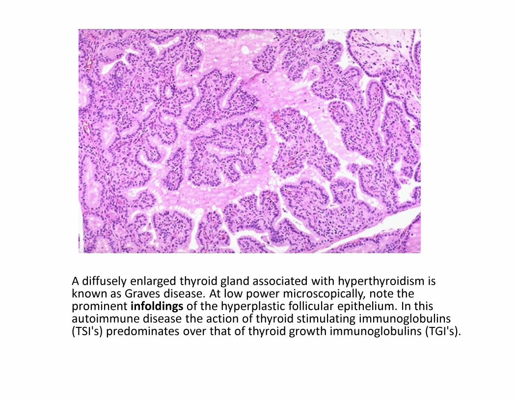
A diffusely enlarged thyroid gland associated with hyperthyroidism is known as Graves disease. At low power microscopically, note the prominent infoldings of the hyperplastic follicular epithelium. In this autoimmune disease the action of thyroid stimulating immunoglobulins(TSI's) predominates over that of thyroid growth immunoglobulins (TGI's).
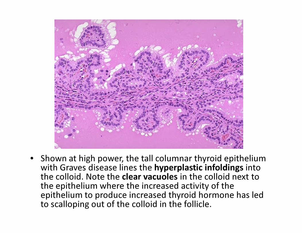
• Shown at high power, the tall columnar thyroid epithelium with Graves disease lines the hyperplastic infoldings into the colloid. Note the clear vacuoles in the colloid next to the epithelium where the increased activity of the epithelium to produce increased thyroid hormone has led to scalloping out of the colloid in the follicle.
Hyperthyroidism - causes• Toxic thyroid adenoma
– This is a solitary hyperfunctioning benign tumor of the follicular epithelium
• Toxic multinodular goiter (Plummer disease)– this is a hormonally active form of thyroid hyperplasia– Combination of hyperthyroidism, nodular goiter & absence of
exopthalmos– “hot” nodules are seen & can be adenomas or non-neoplastic
areas of nodular hyperplasia• Stuma ovarii: ovarian teratoma made up of thyroid tissue. • Exogenous administration of thyroid hormone• TSH-secreting pituitary adenoma
– Rare cause of hyperthyroidism
HYPOTHYROIDISM
Hypothyroidism • Presents with signs of hypometabolism• Adults: manifest as myxoedema• Children: manifest as cretinism
Hypothyroidism – presentation • CNS/Head – coarse brittle hair, loss of lateral eye
brow, puffy face, periorbital oedema, large tongue, hoarseness of voice, slow speech
• CVS – cardiomegaly, bradycardia• Limbs – muscle weakness, myxoedema• GIT – constipation• Skin – cold intolerance, dry skin, scanty axillary &
pubic hair• GUS – menstrual abnormalities
Hypothyroidims - causes• Congenital hypothyroidism
– Results from agenesis of thyroid or a deficiency of the enzymes involved in T4 synthesis
• Clinical presentation:– Dwarfism – retarded bone growth– Mental retardation – Myxoedema – dry, waxy swelling of skin of the extremities &
face• Lab investigation: low serum triiodothyronine (T3 &
thyroxine (total & freeT4) and compensatory high serum TSH
• Decreased T3 resin uptake: inversely proportional to number of unbound thyroid hormone binding sites on TBG. It is measured by competitive uptake of radioactive T3 by resin which competes for unbound sites on TBG
Hypothyroidism - causes• Endemic iodine deficiency
– Absent in developed countries– Present in less developed nations, rural areas
especially– Children particularly vulnerable – severe mental
retardation, short stature & coarse facial features, a protruding tongue and umbilical hernia
– Adults – nodular goiter (enlargment of the thyroid gland)
Hypothyroidism - causes• Thyroiditis – inflammation of the gland• Hashimoto thyroiditis most common cause hypothyroidism
– An autoimmune disorder. Abnormal T cell function stimulating B cells to produce auto antibodies or direct cytotoxic destruction
– Women 10x more affected than men– May also associated with other autoimmune disorders such as pernicious
anaemia, diabetes mellitus, Sjogren syndrome– Increased incidence HLA-DR5 & HLA-B5 positive individuals
• Pathological findings: – infiltration of thyroid with lymphocytes leads to destruction of normal follicles.– Remaining normal follicles show oncocytic change (i.e their cytoplasm is pink
owing to increased number of mitochondria)– Thyroid follicles atrophied– Prominent Hurthle cells (epithelial cells with eosinophilic granular cytoplasm)
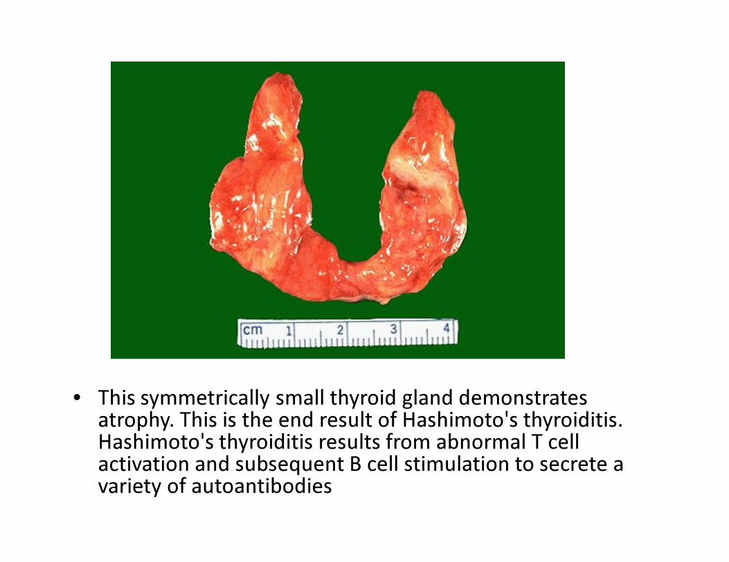
• This symmetrically small thyroid gland demonstrates atrophy. This is the end result of Hashimoto's thyroiditis. Hashimoto's thyroiditis results from abnormal T cell activation and subsequent B cell stimulation to secrete a variety of autoantibodies
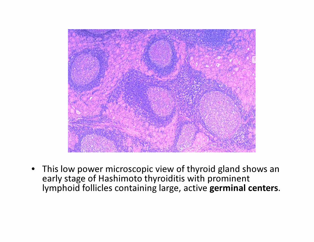
• This low power microscopic view of thyroid gland shows an early stage of Hashimoto thyroiditis with prominent lymphoid follicles containing large, active germinal centers.
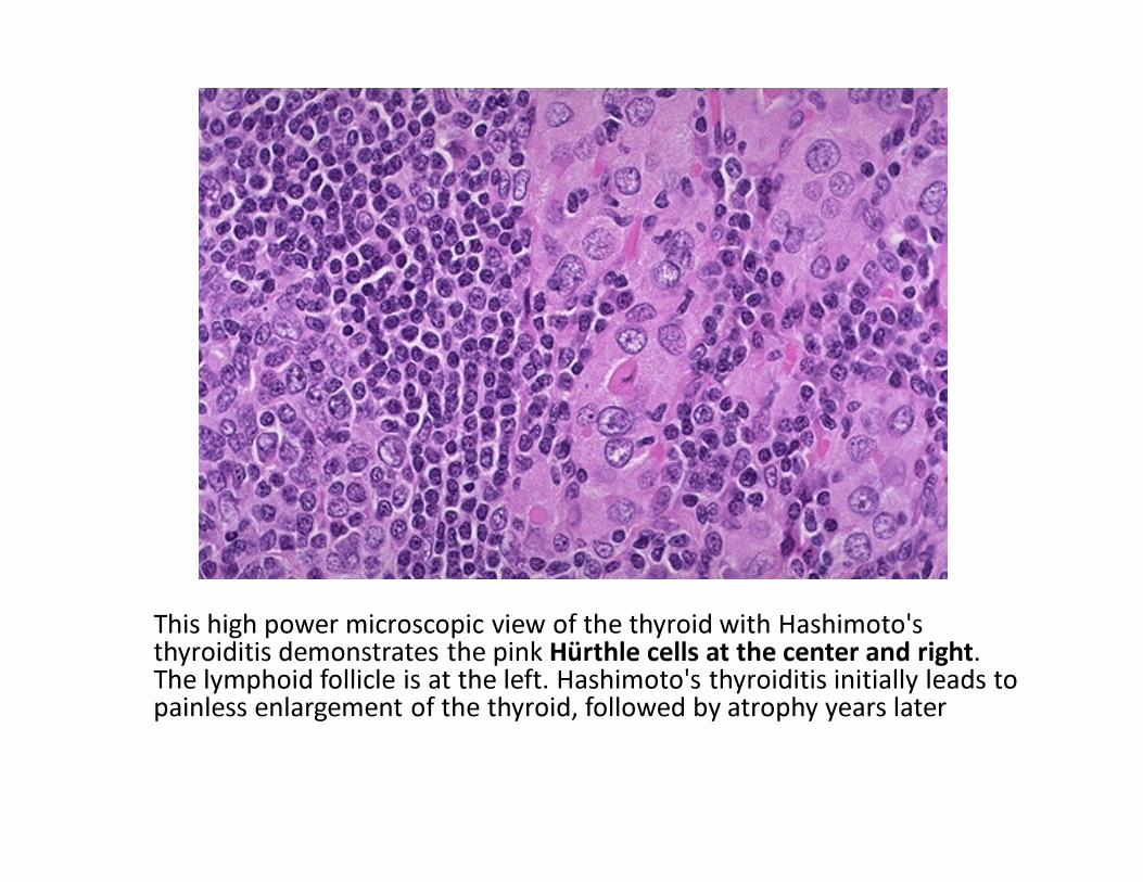
This high power microscopic view of the thyroid with Hashimoto's thyroiditis demonstrates the pink Hürthle cells at the center and right. The lymphoid follicle is at the left. Hashimoto's thyroiditis initially leads to painless enlargement of the thyroid, followed by atrophy years later
Hashimoto Thyroiditis• Clinical presentation: goiter if early and
hypothyroidism in advance cases– Slow course. Pt euthyroid at first, transient hyper
thyroidism may occur and hypothyroidism when gland shrunken & scarred
• Lab findings: ↑ TSH, ↓ T3 & T 4; Ab to thyroglobulinpresent but not diagnostic. Other Abs such as anti-thyroid peroxidase, anti-TSH receptor & anti-iodine receptor are also present
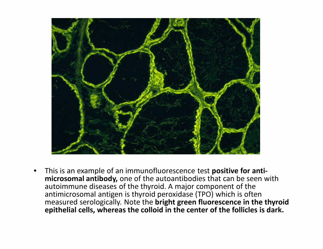
• This is an example of an immunofluorescence test positive for anti-microsomal antibody, one of the autoantibodies that can be seen with autoimmune diseases of the thyroid. A major component of the antimicrosomal antigen is thyroid peroxidase (TPO) which is often measured serologically. Note the bright green fluorescence in the thyroid epithelial cells, whereas the colloid in the center of the follicles is dark.
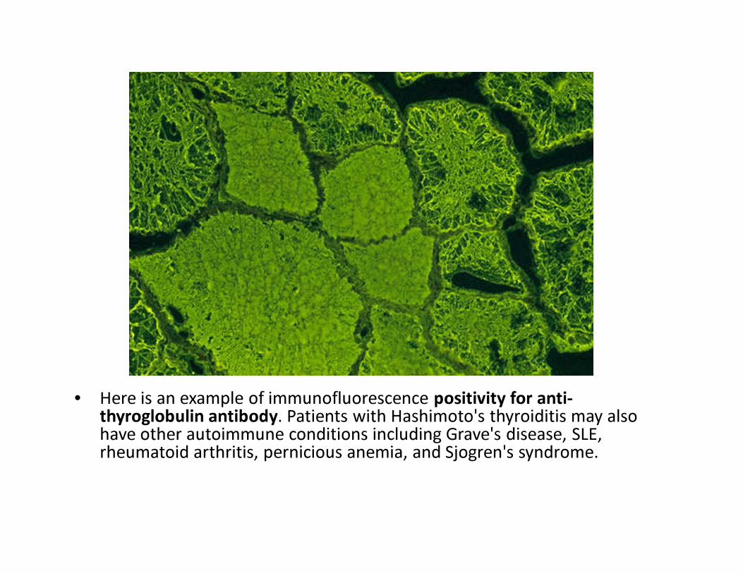
• Here is an example of immunofluorescence positivity for anti-thyroglobulin antibody. Patients with Hashimoto's thyroiditis may also have other autoimmune conditions including Grave's disease, SLE, rheumatoid arthritis, pernicious anemia, and Sjogren's syndrome.
Hypothyroidism - cause• Rare forms of thyroiditis include:
– Subacute thyroiditis (granulomatous thyroiditis or deQuervainthyroiditis)
• Caused by viral infection such as mumps or coxsackievirus• Focal destruction of thyroid tissue & granulomatous inflammation
characteristic (Post viral infection)• Self limited course of several weeks duration, sometimes with transient
hyperthyroidism.• Flu-like illness with pain and tender thyroid gland
– Riedel thyroiditis – fibrosing disease of unknown aetiology. Thyroid tissue replaced with fibrosis. Can mimic carcinoma
– Lymphocytic thyroiditis – most common form of thyroid inflammation of unknown etiology
• Does not cause functional disturbances (i.e. Pts are euthyroid)• Post-surgery for hyperthyroidism, irradiation or drugs• Unknown cause: primary idiopathic myxoedema.
– Poorly defined form of myxoedema– ?autoimmune in nature as TSH receptor-blocking Abs have been
identified.
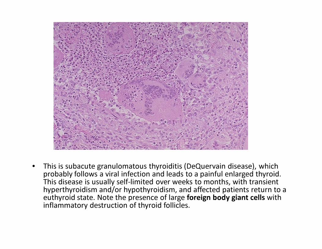
• This is subacute granulomatous thyroiditis (DeQuervain disease), which probably follows a viral infection and leads to a painful enlarged thyroid. This disease is usually self-limited over weeks to months, with transient hyperthyroidism and/or hypothyroidism, and affected patients return to a euthyroid state. Note the presence of large foreign body giant cells with inflammatory destruction of thyroid follicles.
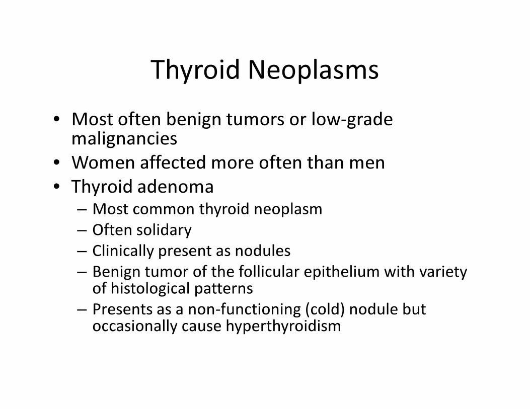
Thyroid Neoplasms• Most often benign tumors or low-grade
malignancies• Women affected more often than men• Thyroid adenoma
– Most common thyroid neoplasm– Often solidary– Clinically present as nodules– Benign tumor of the follicular epithelium with variety
of histological patterns– Presents as a non-functioning (cold) nodule but
occasionally cause hyperthyroidism
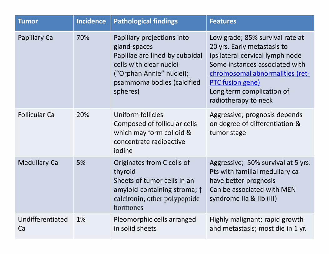
Tumor Incidence Pathological findings FeaturesPapillary Ca 70% Papillary projections into
gland-spacesPapillae are lined by cuboidalcells with clear nuclei (“Orphan Annie” nuclei); psammoma bodies (calcified spheres)
Low grade; 85% survival rate at 20 yrs. Early metastasis to ipsilateral cervical lymph nodeSome instances associated withchromosomal abnormalities (ret-PTC fusion gene)Long term complication of radiotherapy to neck
Follicular Ca 20% Uniform folliclesComposed of follicular cells which may form colloid & concentrate radioactive iodine
Aggressive; prognosis depends on degree of differentiation & tumor stage
Medullary Ca 5% Originates from C cells of thyroidSheets of tumor cells in an amyloid-containing stroma; ↑ calcitonin, other polypeptide hormones
Aggressive; 50% survival at 5 yrs. Pts with familial medullary ca have better prognosisCan be associated with MEN syndrome IIa & IIb (III)
Undifferentiated Ca
1% Pleomorphic cells arranged in solid sheets
Highly malignant; rapid growth and metastasis; most die in 1 yr.
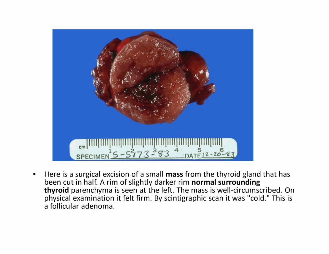
• Here is a surgical excision of a small mass from the thyroid gland that has been cut in half. A rim of slightly darker rim normal surrounding thyroid parenchyma is seen at the left. The mass is well-circumscribed. On physical examination it felt firm. By scintigraphic scan it was "cold." This is a follicular adenoma.
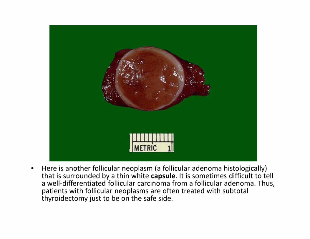
• Here is another follicular neoplasm (a follicular adenoma histologically) that is surrounded by a thin white capsule. It is sometimes difficult to tell a well-differentiated follicular carcinoma from a follicular adenoma. Thus, patients with follicular neoplasms are often treated with subtotal thyroidectomy just to be on the safe side.
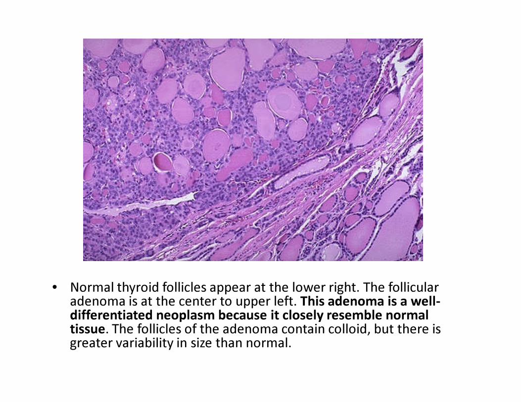
• Normal thyroid follicles appear at the lower right. The follicular adenoma is at the center to upper left. This adenoma is a well-differentiated neoplasm because it closely resemble normal tissue. The follicles of the adenoma contain colloid, but there is greater variability in size than normal.
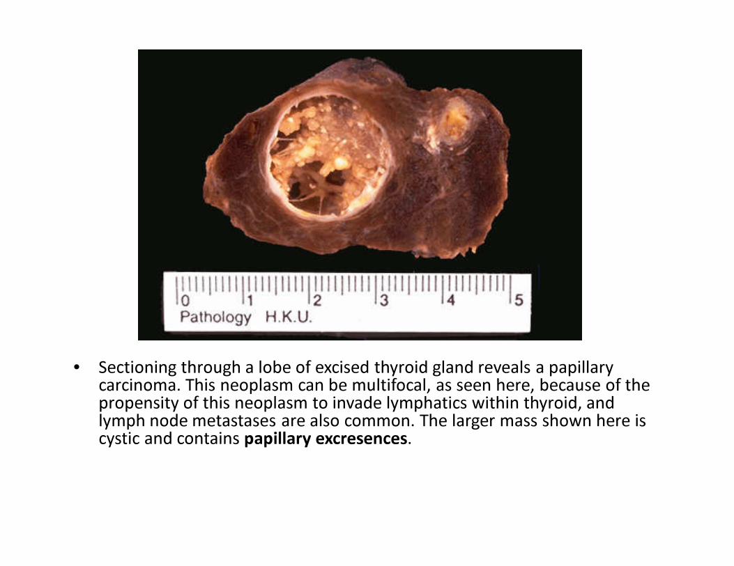
• Sectioning through a lobe of excised thyroid gland reveals a papillary carcinoma. This neoplasm can be multifocal, as seen here, because of the propensity of this neoplasm to invade lymphatics within thyroid, and lymph node metastases are also common. The larger mass shown here is cystic and contains papillary excresences.
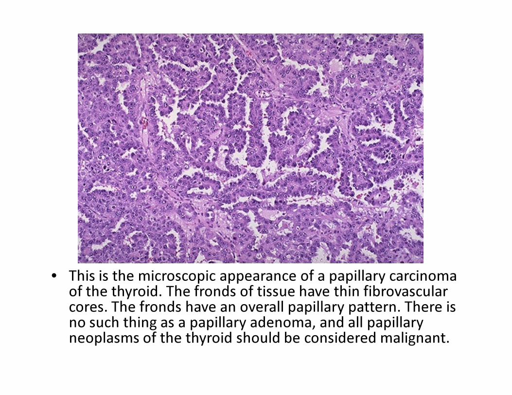
• This is the microscopic appearance of a papillary carcinoma of the thyroid. The fronds of tissue have thin fibrovascularcores. The fronds have an overall papillary pattern. There is no such thing as a papillary adenoma, and all papillary neoplasms of the thyroid should be considered malignant.
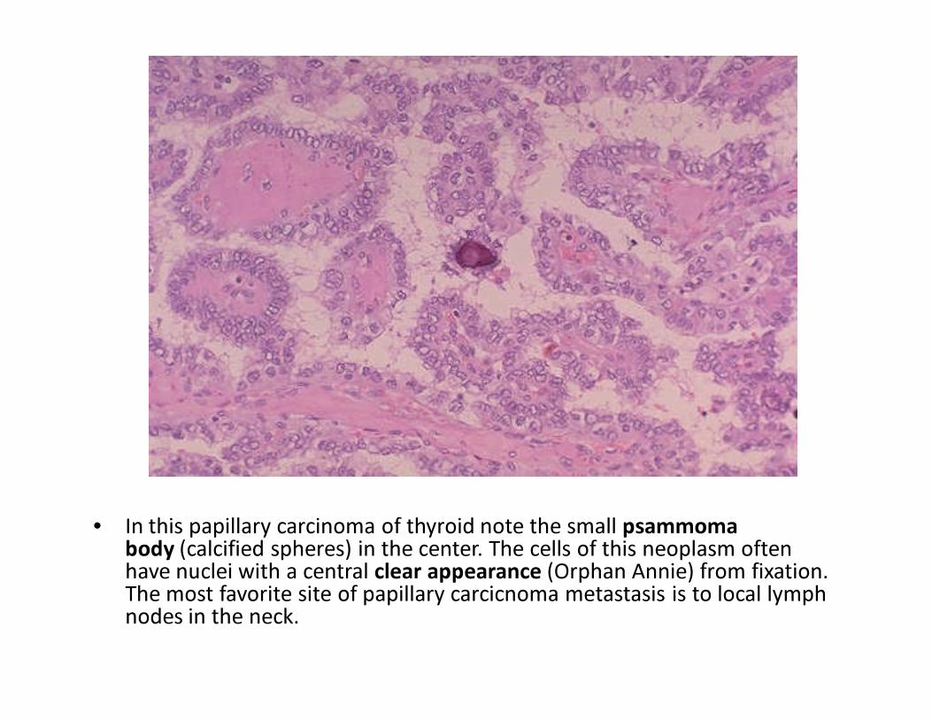
• In this papillary carcinoma of thyroid note the small psammomabody (calcified spheres) in the center. The cells of this neoplasm often have nuclei with a central clear appearance (Orphan Annie) from fixation. The most favorite site of papillary carcicnoma metastasis is to local lymph nodes in the neck.
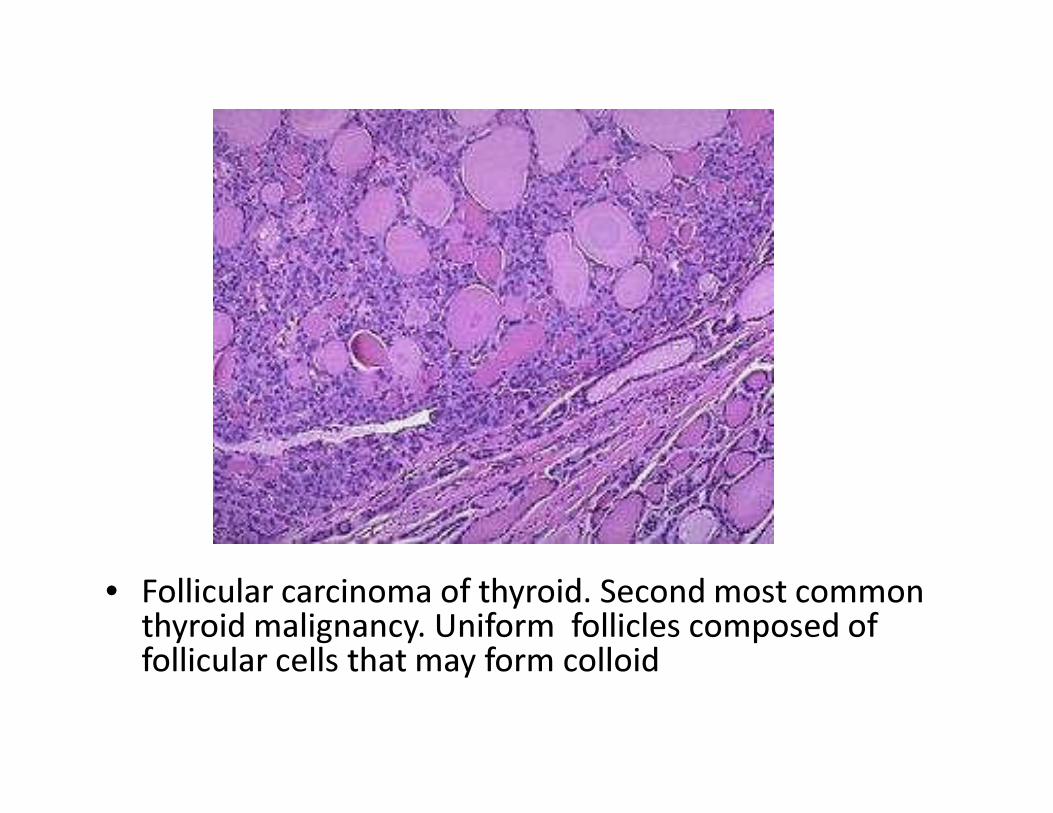
• Follicular carcinoma of thyroid. Second most common thyroid malignancy. Uniform follicles composed of follicular cells that may form colloid
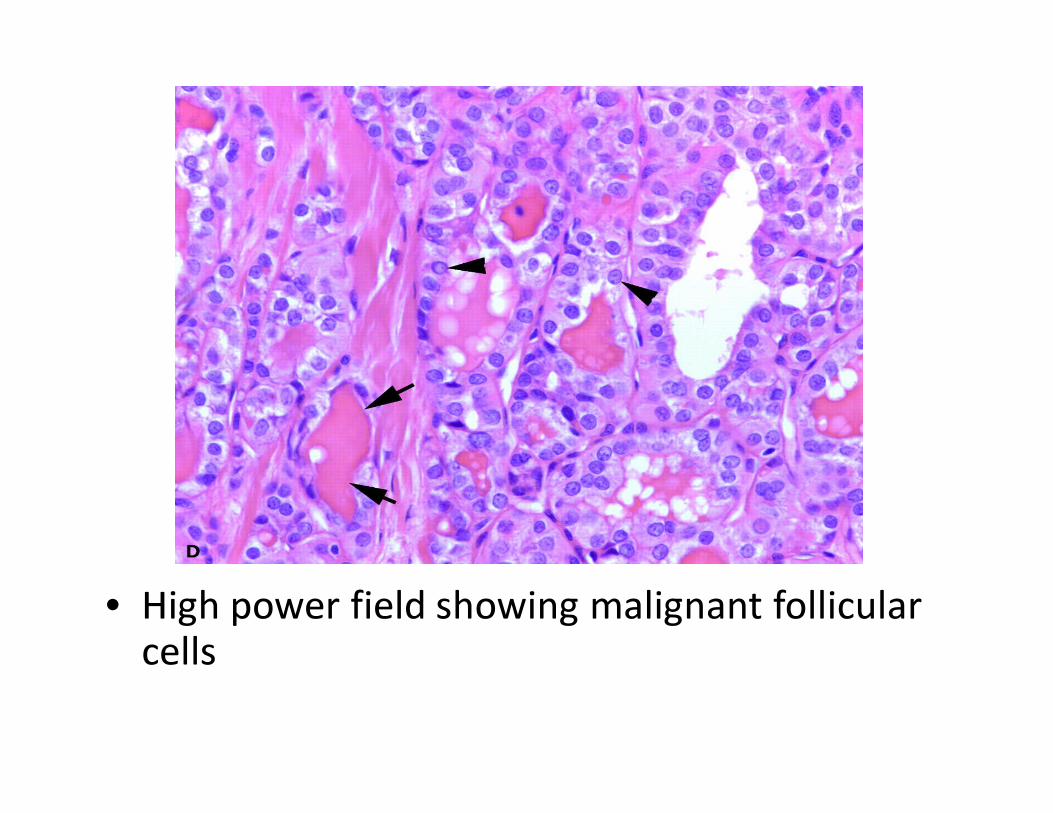
• High power field showing malignant follicular cells
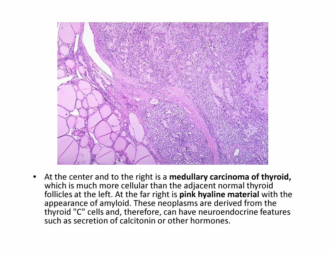
• At the center and to the right is a medullary carcinoma of thyroid, which is much more cellular than the adjacent normal thyroid follicles at the left. At the far right is pink hyaline material with the appearance of amyloid. These neoplasms are derived from the thyroid "C" cells and, therefore, can have neuroendocrine features such as secretion of calcitonin or other hormones.
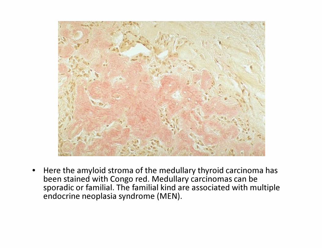
• Here the amyloid stroma of the medullary thyroid carcinoma has been stained with Congo red. Medullary carcinomas can be sporadic or familial. The familial kind are associated with multiple endocrine neoplasia syndrome (MEN).
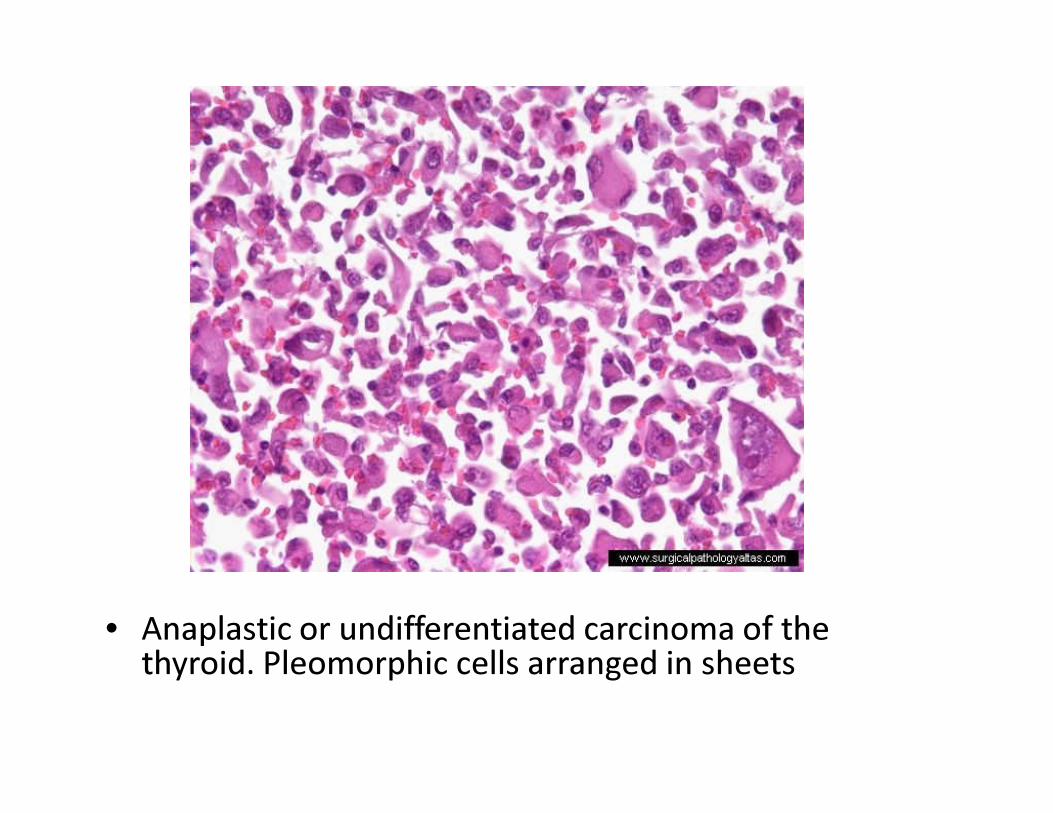
• Anaplastic or undifferentiated carcinoma of the thyroid. Pleomorphic cells arranged in sheets
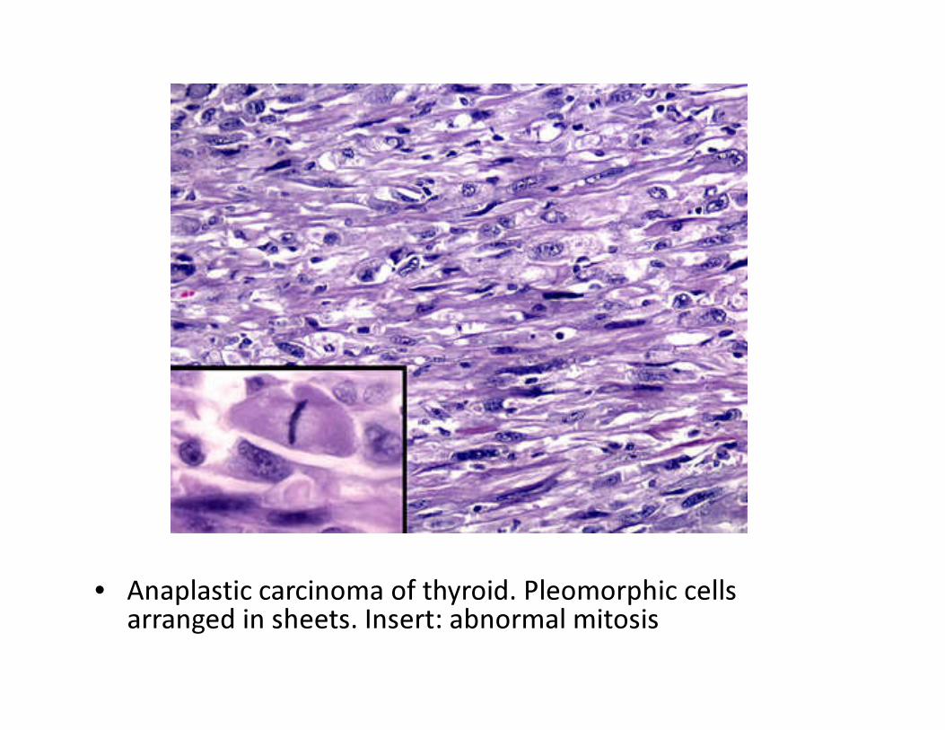
• Anaplastic carcinoma of thyroid. Pleomorphic cells arranged in sheets. Insert: abnormal mitosis
Thyroid Disorders• Tumors of thyroid must be distinguished from
nodular goiter.• Common cause of thyroid enlargement –
nodular goiter.• Most times cause of nodular goiter is not
known
Laboratory Diagnosis• TFT• FNA• Tissue Biopsy• USS• Radioactive iodine uptake study – thyroid scan• MRI – pituitary lesions
ENDReference: Robins Pathological Basis of Diseases
www.pathologyatsmhs.wordpress.com


