35
Antigen-Antibody System Dr Sanjesh G Rathi Associate Professor SIPS

Antigen-Antibody System
Dr Sanjesh G Rathi
Associate
Professor
SIPS

Antigen
• An antigen is a substance which when introduced into a
body evokes an immune response to produce a specific
antibody with which it reacts specifically.
• Most of antigens are proteins but some are
carbohydrates,lipids or nucleic acids.
• It can be classified as-
Complete antigen
Incomplete antigen (Haptens)

• Epitope is the smallest unit of antigenicity.
• The combining site on the antibody molecule,
corresponding to the epitope is called the Paratope.


Antibody • These are substances which are formed in the serum
and tissue fluids in response to an antigen and react
with that antigen specifically and in some observable
manner.
• Chemically they are globulins, hence they are named
immunoglobulins.
• They constitute about 20 – 25% of the total serum
proteins and are mainly synthesized by plasma cells.

Structure

Antigen Antibody Reactions
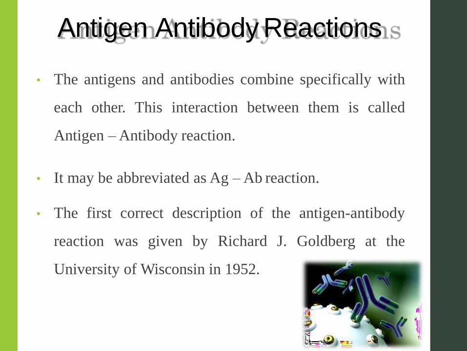
• The antigens and antibodies combine specifically with
each other. This interaction between them is called
Antigen – Antibody reaction.
• It may be abbreviated as Ag – Ab reaction.
• The first correct description of the antigen-antibody
reaction was given by Richard J. Goldberg at the
University of Wisconsin in 1952.

• These reactions form the basis for detection of
infectious disease causing agents and also some non
specific antigens like enzymes.
• The reactions between Ag and Ab occur in three stages.
In first or primary stage the reaction involves
formation of Ag-Ab complex.
The secondary stage leads to visible events like
precipitation, agglutination etc.
The tertiary stage includes destruction of Ag or its
neutralization.

Its USES are
1. In vivo
• Forms basis of immunity against infectious diseases.
2. In vitro
• For diagnosis of infections
• Helpful in epidemiological studies
• For identification of enzymes
• Detection and quantification of antigens or antibodies.

Characteristics
• Reaction is specific, an antigen combines only with its
homologous antibody and vice-versa. However, cross
reactions may occur due to antigenic similarity.
• Entire molecules of antigen and antibody react and not
the fragments.
• Only the surface antigens participate in the antigen
antibody reaction.
• The reaction is firm but reversible.

Types
Precipitation reactions
Agglutination
Neutralization test
Immunofluorescence
Radioimmunoassay
Enzyme linked immunosorbent assay
Immunoelectronmicroscopic test

Precipitation reactions
When a soluble antigen reacts with its antibody in the
presence of electrolytes at an optimal temperature and
pH, antigen antibody complex forms an insoluble
precipitate that usually sediments at the bottom of the
tube and it is called precipitation.
Precipitation may occur in liquid media or in gels such
as agar, agarose etc.
Applications
• Identification of bacteria. E.g., Lancefield grouping of
streptococcus.
• Detection of antibody for diagnostic purposes. E.g.,
VDRL in syphilis
• Forensic application in identification of human blood
and seminal stains
• To standardize toxins and antitoxins.

Types of precipitation reactions
• Ring test
e.g. C- reactive protein test
Streptococcal grouping by Lancefield
technique
• Flocculation test
- Slide test. E.g., VDRL in syphilis
- Tube test. E,g., Kahn’s test in syphilis
• Immunodiffusion test


Agglutination
It is an antigen antibody reaction in which a particulate
antigen combines with its antibody in the presence of
electrolytes at an optimal temperature and pH resulting
in visible clumping of particles.
It is more sensitive than precipitation for detection of
antibodies.
The reactions take place better with IgM antibody.

Types
Slide Agglutination test
Routine procedure to identify
bacterial stains. E.g., Salmonella species
Also used for blood grouping.
Tube Agglutination test
Standard quantitative method for determination of antibodies.
Routinely employed in diagnosis of typhoid, brucellosis and typhus fever

Coombs Test
• Originally devised by Coombs, Mourant and Race (1945)
for detection of incomplete Rh antibodies.
• When sera containing incomplete anti-Rh antibodies are
mixed with corresponding Rh-positive erythrocytes but no
agglutination occurs.
• When such erythrocytes are treated with antiglobulin or
COOMBS serum (rabbit antiserum with human gamma
globulin), the cells are
agglutinated.

Neutralization Test
• Bacterial exotoxins are capable of producing neutralizing
antibodies (antitoxins) which play protective role in
diseases such as diphtheria and tetanus.
• Toxin – antitoxin neutralization can be measured in vivo
and in vitro.

In vivo tests:
Toxigenicity test. E.g., C. diphtheriae
Shick test (similar test in human)
In vitro test:
Antistreptolysin ‘O’ (ASO) test. E.g., Strep pyogenes
Virus neutralization tests.

Immunofluorescence • Fluorescence is the property of certain dyes which absorb rays of
one particular wavelength (ultraviolet light) and emit rays with a
different wavelength (visible light)
• Most commonly used dyes are:
1. Fluorescin isothiocyanate – blue green
2. Lissamine rhodamine – orange red
They are of two types:
1. Direct immunofluorescence
2. Indirect immunofluorescence


Direct immunofluorescence
Uses:
• Commonly employed for detection of bacteria, viruses or other
antigens in blood, urine, tissues and other specimens.
• Sensitive method to diagnosis Rabies.
Disadvantage: Separate fluorescent labelled
prepared for each antigen to be tested.
antibody has to be
Indirect immunofluorescence
• A single antihuman globulin fluorescent conjugate
employed for detection of antibody to any antigen
can be
• This has overcome the disadvantage of direct immunofluorescence

ELISA
• Enzyme linked immunosorbent assay is a simple and a
sensitive test.
• Requires only microlitre quantities of test reagents.
• The principle of ELISA is almost same as that of
immunofluorescence, the only difference being, an
enzyme is used instead of fluorescent dye.
• It can be used for detection of Antigen or Antibody.
• Types: Sandwich, Indirect, Competitive ELISA


Uses:
Detection of HIV antibodies in serum
Detection of mycobacterial antibodies in TB
Detection of Hepatitis B markers in serum
Detection of enterotoxin of E.coli in feces.

Immuno electron microscopic tests
1. Immunoelectron microscopy
2. Immuno Ferritin test
3. Immuno enzyme test

• Immuno electron microscopy: Viral particles are mixed
with specific antisera and are observed under electron
microscope. These are seen as clumps.
Used in detection of Hepatitis A virus.
• Immuno ferritin test: Ferritin (electron dense substance)
conjugated antibody is used to react with an antigen.
Used in identification of Legionella species.
• Immuno enzyme test: Tissue sections are treated with
peroxidase labelled antisera to detect the corresponding
antigen and in viewed under electron microscope.

Conclusion • Therefore we see the application of antigen antibody
reactions in the diagnosis of diseases which can help
in developments of varieties of diagnostic tests.
• In clinical practice, they help in:
Preventing destructive diseases.
Preventing progression of the diseases.
Identifying high risk patients
Target treatment of specific diseases
Monitor the effects of the treatment.

Reference
s • Textbook of Microbiology for dental students. Dir. Prof.
C. P Baveja. 4th edition
• Textbook of Microbiology for dental students.
Ananthnarayan and Paniker. 8th edition
• Essential Microbiology for Dentistry, 4th Edition.
Lakshman Samaranayake.
• https://en.wikipedia.org/wiki/Antigen
antibody_interaction

Previous year questions
• Antigen and Antibody system.
Degree Examination May 2009).
(10marks) (MDS


