Draft Variation in stiffness regulates cardiac myocyte hypertrophy via signaling pathways Journal: Canadian Journal of Physiology and Pharmacology Manuscript ID cjpp-2015-0578.R2 Manuscript Type: Article Date Submitted by the Author: 09-May-2016 Complete List of Authors: Li, Jieli; University of Illinois at Chicago College of Medicine, Physiology and Biophysics Mkrtschjan, Michael; University of Illinois at Chicago, Bioengineering Lin, Ying-Hsi; University of Illinois at Chicago College of Medicine, Physiology and Biophysics Russell, Brenda; University of Illinois at Chicago, Physiology and Biophysics Keyword: Mechano-transduction, focal adhesion kinase, lipid signaling, actin assembly, substrate stiffness https://mc06.manuscriptcentral.com/cjpp-pubs Canadian Journal of Physiology and Pharmacology
Transcript
Draft
Variation in stiffness regulates cardiac myocyte hypertrophy
via signaling pathways
Journal: Canadian Journal of Physiology and Pharmacology
Manuscript ID cjpp-2015-0578.R2
Manuscript Type: Article
Date Submitted by the Author: 09-May-2016
Complete List of Authors: Li, Jieli; University of Illinois at Chicago College of Medicine, Physiology and Biophysics Mkrtschjan, Michael; University of Illinois at Chicago, Bioengineering Lin, Ying-Hsi; University of Illinois at Chicago College of Medicine, Physiology and Biophysics Russell, Brenda; University of Illinois at Chicago, Physiology and Biophysics
Tse JR, Engler AJ. Preparation of hydrogel substrates with tunable mechanical properties. 2010.
Curr Protoc Cell Biol. Chapter 10:Unit 10.16.
Torsoni AS, Constancio SS, Nadruz W Jr, Hanks SK, Franchini KG. Focal adhesion kinase is
activated and mediates the early hypertrophic response to stretch in cardiac myocytes. 2003.
Circ Res. 93:140-7. PMID: 12805241.
Page 19 of 28
https://mc06.manuscriptcentral.com/cjpp-pubs
Canadian Journal of Physiology and Pharmacology
Draft
20
Wang HB, Dembo M, Hanks SK, Wang Y. Focal adhesion kinase is involved in
mechanosensing during fibroblast migration. 2001. Proc Natl Acad Sci U S A. 98:11295-300.
PMID: 11572981.
Wei SC, Fattet L, Tsai JH, Guo Y, Pai VH, Majeski HE, Chen AC, Sah RL, Taylor SS, Engler AJ,
Yang J. Matrix stiffness drives epithelial-mesenchymal transition and tumour metastasis through
a TWIST1-G3BP2 mechanotransduction pathway. 2015. Nat Cell Biol. 17:678-88. PMID:
25893917.
Xu JX, Si M, Zhang HR, Chen XJ, Zhang XD, Wang C, Du XN, Zhang HL. Phosphoinositide
kinases play key roles in norepinephrine- and angiotensin II-induced increase in
phosphatidylinositol 4,5-bisphosphate and modulation of cardiac function. 2014. J Biol Chem.
289:6941-8. PMID: 24448808.
Yang C, Tibbitt MW, Basta L, Anseth KS. Mechanical memory and dosing influence stem cell
fate. 2014. Nat Mater. 13:645-52. PMID: 24633344.
Figure legends
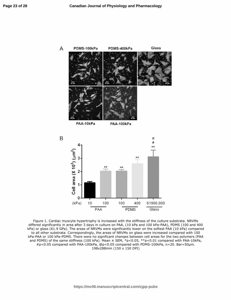
Figure 1. Cardiac myocyte hypertrophy is increased with the stiffness of the culture substrata.
NRVMs differed significantly in area after 3 days in culture on PAA, (10 kPa and 100 kPa-PAA),
PDMS (100 and 400 kPa) or glass (61.9 GPa). The areas of NRVMs were significantly lower on
the softest PAA (10 kPa) compared to all other substrata. Correspondingly, the areas of NRVMs
on glass were increased compared with 100 kPa-PAA or 100 kPa-PDMS. There were no
significant changes between cell areas for the two polymers (PAA and PDMS) of the same
stiffness (100 kPa). Mean ± SEM, *p<0.05, **p<0.01 compared with PAA-10kPa, #p<0.05
compared with PAA-100kPa, @p<0.05 compared with PDMS-100kPa, n=20. Bar=50µm.
Page 20 of 28
https://mc06.manuscriptcentral.com/cjpp-pubs
Canadian Journal of Physiology and Pharmacology
Draft
21
Figure 2. FAK phosphorylation in cultured NRVMs is increased with higher stiffness of the
substrata. The ratio of p-FAK (Y397) to total FAK was quantified by Western blotting. The level
of total FAK was not significantly changed when normalized to H2B intensity. P-FAK
significantly increased in NRVM cultured for 3 days on the 400 kPa PDMS or 61.9 GPa glass,
compared with 100 kPa PDMS. Mean ± SEM. *p<0.05, n=3.
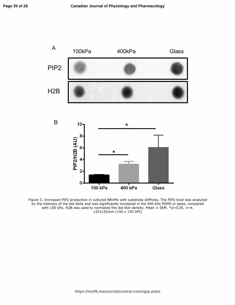
Figure 3. Increased PIP2 production in cultured NRVMs with substrata stiffness. The PIP2 level
was analyzed by the intensity of the dot blots and was significantly increased in the 400 kPa
PDMS or glass, compared with 100 kPa. H2B was used to normalize the dot blot density. Mean
± SEM. *p<0.05, n=4.
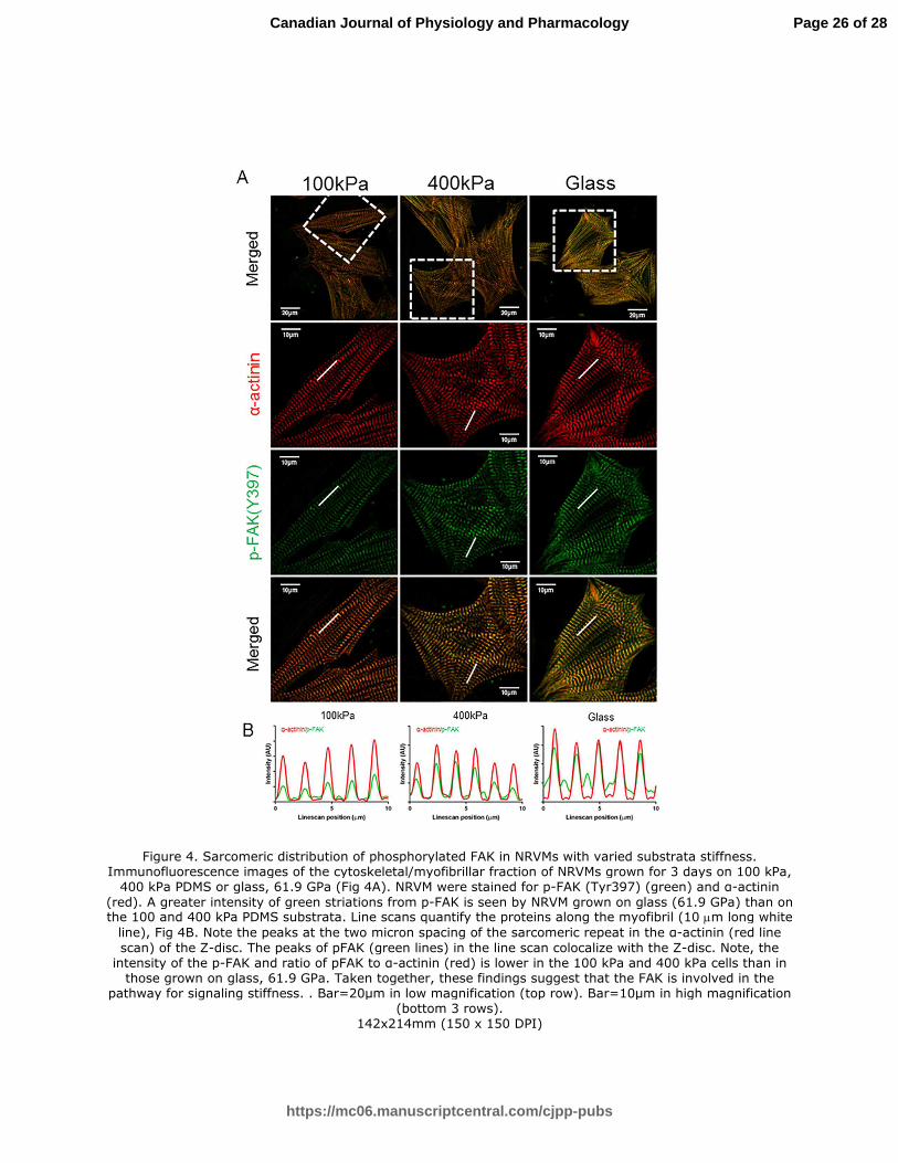
Figure 4. Sarcomeric distribution of phosphorylated FAK in NRVMs with varied substrata
stiffness. Immunofluorescence images of the cytoskeletal/myofibrillar fraction of NRVMs grown
for 3 days on 100 kPa, 400 kPa PDMS or glass, 61.9 GPa (Fig 4A). NRVM were stained for p-
FAK (Tyr397) (green) and α-actinin (red). A greater intensity of green striations from p-FAK is
seen by NRVM grown on glass (61.9 GPa) than on the 100 and 400 kPa PDMS substrata. Line
scans quantify the proteins along the myofibril (10 µm long white line), Fig 4B. Note the peaks at
the two micron spacing of the sarcomeric repeat in the α-actinin (red line scan) of the Z-disc.
The peaks of pFAK (green lines) in the line scan colocalize with the Z-disc. Note, the intensity of
the p-FAK and ratio of pFAK to α-actinin (red) is lower in the 100 kPa and 400 kPa cells than in
those grown on glass, 61.9 GPa. Taken together, these findings suggest that the FAK is
involved in the pathway for signaling stiffness. . Bar=20µm in low magnification (top row).
Bar=10µm in high magnification (bottom 3 rows).
Page 21 of 28
https://mc06.manuscriptcentral.com/cjpp-pubs
Canadian Journal of Physiology and Pharmacology
Draft
22
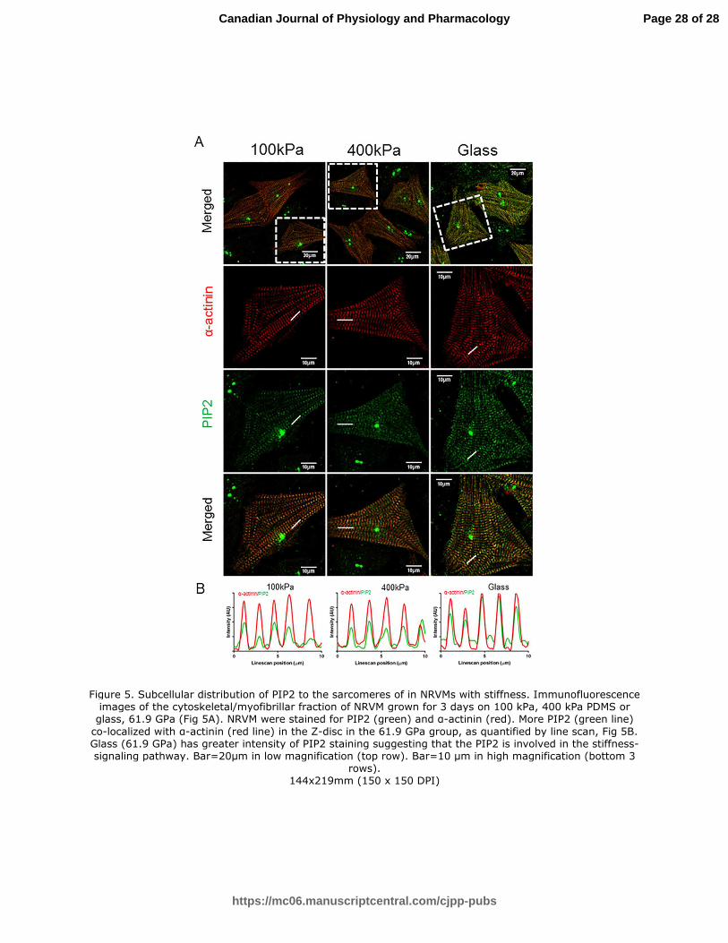
Figure 5. Subcellular distribution of PIP2 to the sarcomeres of in NRVMs with stiffness.
Immunofluorescence images of the cytoskeletal/myofibrillar fraction of NRVM grown for 3 days
on 100 kPa, 400 kPa PDMS or glass, 61.9 GPa (Fig 5A). NRVM were stained for PIP2 (green)
and α-actinin (red). More PIP2 (green line) co-localized with α-actinin (red line) in the Z-disc in
the 61.9 GPa group, as quantified by line scan, Fig 5B. Glass (61.9 GPa) has greater intensity
of PIP2 staining suggesting that the PIP2 is involved in the stiffness-signaling pathway.
Bar=20µm in low magnification (top row). Bar=10 µm in high magnification (bottom 3 rows).
Figure 6. Actin and CapZβ1 dynamics measured by FRAP in myocytes cultured on different
stiffnesses. NRVMs were plated on PDMS (100 kPa, 400 kPa) and glass (61.9 Gpa) substrates
for 3 days. (A) The increased dynamics of actin was measured by FRAP. Microscopic images of
whole living NRVMs infected with actin-GFP in myocytes on 100 kPa, 400 kPa and 61.9 GPa.
The enlarged inset below shows FRAP of the region of interest (10µmX10µm box of dashed
white lines) for actin-GFP before, immediately after, and 8 min after photobleaching. Bar=10µm.
(B) Kfrap of actin-GFP on glass had increased kinetic rates compared with 100 kPa PDMS. The
FAK inhibitor abolished the high Kfrap of increased actin-GFP on the glass surface. Mean ±
SEM *p<0.05, **p<0.01 n=15. (C) The inset below shows FRAP of the region of interest
(10µmX10µm box of dashed white lines) for GFP-CapZβ1 before, immediately after, and 15 min
after photobleaching. (D) Kfrap of CapZB1-GFP on glass had increased kinetic rates compared
with 100 kPa PDMS. Mean ± SEM *p<0.05, n=15.
Page 22 of 28
https://mc06.manuscriptcentral.com/cjpp-pubs
Canadian Journal of Physiology and Pharmacology
Draft
Figure 1. Cardiac myocyte hypertrophy is increased with the stiffness of the culture substrata. NRVMs differed significantly in area after 3 days in culture on PAA, (10 kPa and 100 kPa-PAA), PDMS (100 and 400 kPa) or glass (61.9 GPa). The areas of NRVMs were significantly lower on the softest PAA (10 kPa) compared
to all other substrata. Correspondingly, the areas of NRVMs on glass were increased compared with 100 kPa-PAA or 100 kPa-PDMS. There were no significant changes between cell areas for the two polymers (PAA and PDMS) of the same stiffness (100 kPa). Mean ± SEM, *p<0.05, **p<0.01 compared with PAA-10kPa,
#p<0.05 compared with PAA-100kPa, @p<0.05 compared with PDMS-100kPa, n=20. Bar=50µm. 198x288mm (150 x 150 DPI)
Page 23 of 28
https://mc06.manuscriptcentral.com/cjpp-pubs
Canadian Journal of Physiology and Pharmacology
Draft
Figure 2. FAK phosphorylation in cultured NRVMs is increased with higher stiffness of the substrata. The ratio of p-FAK (Y397) to total FAK was quantified by Western blotting. The level of total FAK was not
significantly changed when normalized to H2B intensity. P-FAK significantly increased in NRVM cultured for 3
days on the 400 kPa PDMS or 61.9 GPa glass, compared with 100 kPa PDMS. Mean ± SEM. *p<0.05, n=3. 131x83mm (150 x 150 DPI)
Page 24 of 28
https://mc06.manuscriptcentral.com/cjpp-pubs
Canadian Journal of Physiology and Pharmacology
Draft
Figure 3. Increased PIP2 production in cultured NRVMs with substrata stiffness. The PIP2 level was analyzed by the intensity of the dot blots and was significantly increased in the 400 kPa PDMS or glass, compared
with 100 kPa. H2B was used to normalize the dot blot density. Mean ± SEM. *p<0.05, n=4. 147x152mm (150 x 150 DPI)
Page 25 of 28
https://mc06.manuscriptcentral.com/cjpp-pubs
Canadian Journal of Physiology and Pharmacology
Draft
Figure 4. Sarcomeric distribution of phosphorylated FAK in NRVMs with varied substrata stiffness. Immunofluorescence images of the cytoskeletal/myofibrillar fraction of NRVMs grown for 3 days on 100 kPa,
400 kPa PDMS or glass, 61.9 GPa (Fig 4A). NRVM were stained for p-FAK (Tyr397) (green) and α-actinin
(red). A greater intensity of green striations from p-FAK is seen by NRVM grown on glass (61.9 GPa) than on the 100 and 400 kPa PDMS substrata. Line scans quantify the proteins along the myofibril (10 µm long white
line), Fig 4B. Note the peaks at the two micron spacing of the sarcomeric repeat in the α-actinin (red line
scan) of the Z-disc. The peaks of pFAK (green lines) in the line scan colocalize with the Z-disc. Note, the intensity of the p-FAK and ratio of pFAK to α-actinin (red) is lower in the 100 kPa and 400 kPa cells than in
those grown on glass, 61.9 GPa. Taken together, these findings suggest that the FAK is involved in the pathway for signaling stiffness. . Bar=20µm in low magnification (top row). Bar=10µm in high magnification
(bottom 3 rows). 142x214mm (150 x 150 DPI)
Page 26 of 28
https://mc06.manuscriptcentral.com/cjpp-pubs
Canadian Journal of Physiology and Pharmacology
Draft
Page 27 of 28
https://mc06.manuscriptcentral.com/cjpp-pubs
Canadian Journal of Physiology and Pharmacology
Draft
Figure 5. Subcellular distribution of PIP2 to the sarcomeres of in NRVMs with stiffness. Immunofluorescence images of the cytoskeletal/myofibrillar fraction of NRVM grown for 3 days on 100 kPa, 400 kPa PDMS or
glass, 61.9 GPa (Fig 5A). NRVM were stained for PIP2 (green) and α-actinin (red). More PIP2 (green line)
co-localized with α-actinin (red line) in the Z-disc in the 61.9 GPa group, as quantified by line scan, Fig 5B. Glass (61.9 GPa) has greater intensity of PIP2 staining suggesting that the PIP2 is involved in the stiffness-signaling pathway. Bar=20µm in low magnification (top row). Bar=10 µm in high magnification (bottom 3
rows). 144x219mm (150 x 150 DPI)
Page 28 of 28
https://mc06.manuscriptcentral.com/cjpp-pubs
Canadian Journal of Physiology and Pharmacology
Draft
Figure 6. Actin and CapZβ1 dynamics measured by FRAP in myocytes cultured on different stiffnesses. NRVMs were plated on PDMS (100 kPa, 400 kPa) and glass (61.9 Gpa) substrates for 3 days. (A) The
increased dynamics of actin was measured by FRAP. Microscopic images of whole living NRVMs infected with
actin-GFP in myocytes on 100 kPa, 400 kPa and 61.9 GPa. The enlarged inset below shows FRAP of the region of interest (10µmX10µm box of dashed white lines) for actin-GFP before, immediately after, and 8
min after photobleaching. Bar=10µm. (B) Kfrap of actin-GFP on glass had increased kinetic rates compared with 100 kPa PDMS. The FAK inhibitor abolished the high Kfrap of increased actin-GFP on the glass surface.
Mean ± SEM *p<0.05, **p<0.01 n=15. (C) The inset below shows FRAP of the region of interest (10µmX10µm box of dashed white lines) for GFP-CapZβ1 before, immediately after, and 15 min after photobleaching. (D) Kfrap of CapZB1-GFP on glass had increased kinetic rates compared with 100 kPa
PDMS. Mean ± SEM *p<0.05, n=15. 151x273mm (150 x 150 DPI)