Select populations may be more susceptible to toxicities associated with specifi c agents.
Primary treatment is discontinuation of the off ending agent and supportive care.
The manifestations of drug-induced pulmonary diseases span the entire spectrum of pathophysiologic conditions of the respiratory tract. As with most drug-induced diseases, the pathological changes are nonspecifi c. Therefore, the diagnosis is often diffi cult and, in most cases, is based on exclusion of all other possible causes. In addition, the true incidence of drug-induced pulmonary disease is diffi cult to assess as a result of the pathological nonspecifi city and the interaction between the underlying disease state and the drugs.
Considering the physiologic and metabolic capacity of the lung, it is surprising that drug-induced pulmonary disease is not more common. The lung is the only organ of the body that receives the entire circulation. In addition, the lung contains a heteroge-neous population of cells capable of various metabolic functions, including N -alkylation, N -dealkylation, N -oxidation, reduction of N -oxides, and C -hydroxylation.
Evaluation of epidemiologic studies on adverse drug reactions provides a perspective on the importance of drug-induced pulmo-nary disease. In a 2-year prospective survey of a community-based general practice, 41% of 817 patients experienced adverse drug reac-tions. 1 Four patients, or 0.5% of the total respondents, experienced adverse respiratory symptoms. Respiratory symptoms occurred in 1.2% of patients experiencing adverse drug reactions. In a recent retrospective analysis of clinical case series in France, 898 patients had reported drug allergy, with a bronchospasm incidence of 6.9%. When these patients were rechallenged with the suspected drug, only 241 (17.6%) tested positive. The incidence of bronchospasm in patients with positive provocation test was 7.9%. 2
Adverse pulmonary reactions are uncommon in the general population but are among the most serious reactions, often requiring intervention. In a study of 270 adverse reactions leading to hospital-ization from two populations, 3.0% were respiratory in nature. 3 Of the reactions considered to be life threatening, 12.3% were respira-tory. An early report on death caused by drug reactions from the Boston Collaborative Drug Surveillance Program indicated that 7 of 27 drug-induced deaths were respiratory in nature. 4 This was con-fi rmed in a follow-up study in which 6 of 24 drug-induced deaths were respiratory in nature. 5
DRUG-INDUCED APNEA Apnea may be induced by central nervous system depression or respi-ratory neuromuscular blockade ( eTable 15-1 ). Patients with chronic obstructive airway disease, alveolar hypoventilation, and chronic
1
2
carbon dioxide retention have an exaggerated respiratory depressant response to narcotic analgesics and sedatives. In addition, the injudi-cious administration of oxygen in patients with carbon dioxide reten-tion can worsen ventilation-perfusion mismatching, further elevating partial pressure of carbon dioxide (pCO2) and thus producing apnea. 6 Although the benzodiazepines are touted as causing less respiratory depression than barbiturates, they may produce a profound additive or synergistic effect when taken in combination with other respira-tory depressants. Combining IV diazepam with phenobarbital to stop seizures in an emergency department frequently results in admissions to an intensive care unit for a short period of assisted mechanical ventilation, regardless of the drug administration rate. Too rapid IV administration of any of the benzodiazepines, even without coadmin-istration of other respiratory depressants, will result in apnea. The risk appears to be the same for the various available agents (diaze-pam, lorazepam, and midazolam). Respiratory depression and arrests resulting in death and hypoxic encephalopathy have occurred fol-lowing rapid IV administration of midazolam for conscious sedation prior to medical procedures. 1 This has been reported more com-monly in the elderly and the chronically debilitated or in combination with opioid analgesics. Concurrent use of inhibitors of cytochrome P450 3A4 with benzodiazepines is likely to lead to greater risk of respiratory depression.
1 Prolonged apnea may follow administration of any of the neuromuscular blocking agents used for surgery, particularly in patients with hepatic or renal dysfunction. In addition, persistent neuromuscular blockade and muscle weakness have been reported in critically ill patients who are receiving neuromuscular blockers continuously for more than 2 days to facilitate mechanical venti-lation. 7 , 8 This has resulted in delayed weaning from mechanical ventilation and prolonged intensive care unit stays. The prolonged neuromuscular blockade has been confi ned principally to pan-curonium and vecuronium in patients with renal disease. Both agents have pharmacologic active metabolites that are excreted renally. The persistent muscular weakness is less well defi ned but appears to represent an acute myopathy. 7 , 9 – 11 High-dose corticoste-roids appear to produce a synergistic effect, supported by animal studies showing that corticosteroids at dosages ≥2 mg/kg per day of prednisone produce atrophy in denervated muscle. 12 The fl uorinated corticosteroids (e.g., triamcinolone) appear to be more myopathic. 13
Dose-dependent respiratory muscle weakness has been reported in chronic obstructive pulmonary disease (COPD) and asthma patients receiving repeated short courses of oral prednisone in the previous 6 months, 14 , 15 as well as patients with steroid-dependent asthma.
Respiratory failure has been known to occur following local spinal anesthesia. Apnea from respiratory paralysis and rapid respi-ratory muscle fatigue has followed the administration of polymyxin and aminoglycoside antibiotics. 6 The mechanism appears to be related to the complexation of calcium and its depletion at the myo-neural junction. IV calcium chloride has been variably effective in
e|CHAPTER 15 Drug-Induced Pulmonary Diseases Hengameh H. Raissy and Michelle Harkins
resulting in oxidant damage in the lung.16 However, these findings should be interpreted with caution since confounding by indication may play a part in this association.
DRUG-INDUCED BRONCHOSPASMBronchoconstriction is the most common drug-induced respira-tory problem. Bronchospasm can be induced by a wide variety of drugs through a number of disparate pathophysiologic mechanisms (eTable 15-2). Regardless of the pathophysiologic mechanism,
reversing the paralysis.6 The aminoglycosides competitively block neuromuscular junctions. This has resulted in life-threatening apnea when neomycin, gentamicin, streptomycin, or bacitracin has been 1 administered into the peritoneal and pleural cavities.6 The aminoglycosides will produce an additive blockade and ventila-tory paralysis with curare or succinylcholine and in patients with myasthenia gravis or myasthenic syndromes.6 IV administration of aminoglycosides has resulted in respiratory failure in babies with infantile 2 botulism. Treatment consists of ventilatory support and administration of an anticholinesterase agent (neostigmine or edrophonium).6
DRUG-INDUCED ASTHMAEpidemiologic studies demonstrate an increase in the prevalence of asthma and COPD with increased use of acetaminophen. The use of aspirin or ibuprofen is not associated with asthma or COPD.16 A relationship with acetaminophen in the etiology of asthma, COPD or allergic diseases has been reported from in utero, infant, child-hood, and adulthood exposures. A weak association between use of acetaminophen during pregnancy and asthma in children 28 months and 7 years of age was reported using the Danish National Birth Cohort.17–19 The association between use of acetaminophen in infancy and childhood and risk of childhood asthma was reported by the International Study of Asthma and Allergies in Childhood, including data from 31 countries.20 Administration of acetamino-phen in the first year of life was associated with a 46% increase in risk of asthma symptoms at the age of 6 to 7 years.20,21 Furthermore, The European Respiratory Health Survey reported an increase in prevalence of wheezing in adults by 0.26% for each gram increase in per capita acetaminophen sales.22 In a prospective birth cohort study, the association between frequency of acetaminophen use and risk of childhood asthma at age 7 disappeared when adjusted for frequency of respiratory infections.23 The acetaminophen–asthma/COPD associations may be explained by reduction of glutathi-one, an endogenous antioxidant enzyme in the airway epithelium
eTABLE 15-1 Drugs That Induce ApneaRelative Frequency of Reactions
Central nervous system depressionNarcotic analgesicsBarbituratesBenzodiazepinesOther sedatives and hypnoticsTricyclic antidepressantsPhenothiazinesKetaminePromazineAnestheticsAntihistaminesAlcoholFenfluraminel-dopaOxygen
dissimilar NSAIDs do produce reactions.25,26 eTable 15-3 lists the analgesics that do and do not cross-react with aspirin.
The currently accepted hypothesis of aspirin-induced asthma is that aspirin intolerance is integrally related to inhibition of cycloox-ygenase (COX). There are at least two COX enzymes coded by dif-ferent genes and only COX-I is sensitive to inhibition by NSAID.25,26 This is supported by the following evidence: (a) All NSAIDs that inhibit COX produce reactions; (b) the degree of cross-reactivity is proportional to the potency of cyclooxygenase inhibition; and (c) each patient with aspirin sensitivity has a threshold dose for precipi-tating bronchospasm that is specific for the degree of cyclooxygen-ase inhibition produced, and once established, the dose of another cyclooxygenase inhibitor needed to induce bronchospasm can be estimated.26,32
The mechanism by which cyclooxygenase inhibition produces bronchospasm in susceptible individuals is unknown. Arachidonic acid metabolism through the 5-lipoxygenase pathway may lead to the excess production of leukotrienes C
4 and D
4.27,29 Leukotrienes
C4, D
4, and E
4 produce bronchospasm and promote histamine release
from mast cells.25,26 The precise mechanism by which augmented leukotriene production occurs is unknown, and available hypotheses do not explain why only a small number of asthmatic patients react to aspirin and NSAIDs.
DesensitizationPatients with aspirin sensitivity can be desensitized. The ease of desensitization correlates with the sensitivity of the patient.32 Highly sensitive patients who react initially to less than 100 mg aspirin require multiple rechallenges to produce desensitization.32 Desensiti-zation usually persists for 2 to 5 days following discontinuance, with full sensitivity reestablished within 7 days.32 Cross-desensitization has been established between aspirin and all NSAIDs tested to date. Because patients may experience life-threatening reactions, desen-sitization should be attempted only in a controlled environment by personnel with expertise in handling these patients. In addition, there are reports of patients who have failed to maintain a desensitized state despite continued aspirin administration.27,32 In one open follow-up trial in 172 aspirin-sensitive asthmatics who had undergone desen-sitization and continued daily aspirin treatment (1,300 mg/day) an improvement in nasal-sinus and asthma symptoms occurred after 6 months of treatment, which persisted up to 5 years.33
eTABLE 15-3 Tolerance of Antiinflammatory and Analgesic Drugs in Aspirin-Induced Asthma
Cross-Reactive Drugs Drugs with No Cross-ReactivityDiclofenac Acetaminophena
aA very small percentage (5%) of aspirin-sensitive patients react to acetaminophen and phenacetin.
drug-induced bronchospasm is almost exclusively a problem of patients with preexisting bronchial hyperreactivity (e.g., asthma, COPD).24 By definition, all patients with nonspecific bronchial hyperreactivity will experience bronchospasm if given sufficiently high doses of cholinergic or anticholinesterase agents. Severe asth-matics with a high degree of bronchial reactivity may wheeze fol-lowing the inhalation of a number of particulate substances, such as the lactose in dry-powder inhalers and inhaled corticosteroids, presumably through direct stimulation of the central airway irritant receptors. Other pharmacologic mechanisms for inducing broncho-spasm include β
2-receptor blockade and nonimmunologic histamine
release from mast cells and basophils.24 A large number of agents are capable of producing bronchospasm through immunoglobulin (Ig)E-mediated reactions.24 These drugs can become a significant occupational hazard for pharmacists, nurses, and pharmaceutical industry workers.24
ASPIRIN-INDUCED BRONCHOSPASMOverall prevalence of aspirin sensitivity or intolerance in the gen-eral population ranges from 0.6% to 2.5% which increases up to 11% in adult patients with asthma.25 The frequency of aspirin-induced bronchospasm increases with age, on average at 30 years of age. Women predominate over men, and there is no evidence for a genetic or familial predisposition.26,27 The classic description of the aspirin-intolerant asthmatic includes the triad of severe asthma, nasal polyps, and aspirin intolerance. The typical patient experi-ences rhinorrhea and nasal congestion as early symptoms followed by nasal polyps. Asthma and aspirin hypersensitivity will develop over the next 2 to 15 years. Bronchospasm typically begins within minutes to hours following ingestion of aspirin and is associated with rhinorrhea, flushing of the head and neck, and conjunctivitis. The reactions are severe and often life-threatening, and once devel-oped, aspirin hypersensitivity remains throughout life.25,28
All aspirin-sensitive asthmatics do not fit the classic “aspirin triad” picture, and not all patients with asthma and nasal polyps develop sensitivity to aspirin.27 In most cases, aspirin-sensitive asth-matics are clinically indistinguishable from the general population of asthmatics except for their intolerance to aspirin and other non-steroidal antiinflammatory drugs (NSAIDs). Aspirin-induced asth-matics are not at higher risk of having fatal asthma if aspirin and other NSAIDs are avoided.29
Diagnosis of aspirin-induced asthma requires a detailed medi-cal history. The definitive diagnosis is made by aspirin provocation tests, which may be done via different routes.25–27,30 An oral provoca-tion test is used commonly where threshold doses of aspirin induce a positive reaction measured by a drop in forced expiratory volume in the first second of expiration (FEV
1) and/or the presence of symp-
toms.30,31 Both nasal and bronchial provocation tests are done by the application of one dose of lysine-aspirin, and aspirin sensitivity is manifested with clinical symptoms of watery discharge and a sig-nificant fall in inspiratory nasal flow or mild bronchospasm.25,26,30,31 The oral provocation test remains the most sensitive in comparison with other routes.25,26
PathogenesisAspirin-induced asthma is correctly classified as an idiosyncratic reaction in that the pathogenesis is still unknown. Patients with aspi-rin intolerance have increased plasma histamine concentrations after ingestion of aspirin and elevated peripheral eosinophil counts.25,26 All attempts to define an immunologic mechanism have been unsuc-cessful. Chemically similar drugs such as salicylamide and choline salicylate do not cross-react, whereas a large number of chemically
with aspirin sensitivity does not differ from that for other asthmat-ics. There is no evidence to support that aspirin-sensitive asthmatics respond better to leukotriene modifiers. In a double-blind, random-ized, placebo-controlled study, aspirin-sensitive asthmatic patients on montelukast showed a 10% improvement in FEV
1 compared with
the placebo group.56 Similar results were reported when montelukast was compared with placebo in patients with intermittent or persis-tent asthma.57
β-BLOCKERS1 β-Adrenergic receptor blockers comprise the other large class of
drugs that can be hazardous to a person with asthma. Even the more cardioselective agents such as acebutolol, atenolol, and metoprolol have been reported to cause asthma attacks.24 Patients with asthma may take nonselective and β
1-selective blockers without incidence
for long periods; however, the occasional report of fatal asthma attacks resistant to therapy with β-agonists should provide ample warning of the dangers inherent in β-blocker therapy.24
If a patient with bronchial hyperreactivity requires β-blocker therapy, one of the selective β
1-blockers (e.g., acebutolol, atenolol,
or metoprolol) should be used at the lowest possible dose. In a meta-analysis of 29 clinical trials in patients with mild-to- moderate airway obstruction, cardioselective β-blockers did not produce any clinically significant respiratory effects in short term.58 Similar results were reported in patients with COPD.59 In a large cohort study in the United Kingdom, more than 53,000 patients with asthma were identified who were issued oral β-blocker therapy and followed for at least 84 days. The authors did not find a significant difference in asthma exacerbation, defined as use of oral cortico-steroid, after prescribing a new oral β-blocker therapy compared to baseline. There was no difference in stratification for β-blocker selectivity in the cohort.60 Celiprolol and betaxolol appear to pos-sess greater cardioselectivity than currently marketed drugs.61,62 Fatal status asthmaticus has occurred with the topical administra-tion of the nonselective timolol maleate ophthalmic solution for the treatment of open-angle glaucoma.63 Although ophthalmic betaxolol suggest that it is well tolerated even in timolol-sensi-tive asthmatics, long-term betaxolol therapy in glaucoma patients with history of pulmonary diseases have been associated with pulmonary obstruction.64–66 Airway obstruction following topical β-blockers for glaucoma has also been reported in patients with no previous history of airway obstruction and close monitoring is warranted.67
SULFITESSevere, life-threatening asthmatic reactions following consumption of restaurant meals and wine have occurred secondary to ingestion of the food preservative potassium metabisulfite.68,69 Sulfites have been used for centuries as preservatives in wine and food. As antiox-idants, they prevent fermentation of wine and discoloration of fruits and vegetables caused by contaminating bacteria.70 Previously, sul-fites had been given “generally recognized as safe” status by the FDA. Sensitive patients react to concentrations ranging from 5 to 100 mg, amounts that are consumed routinely by anyone eating in restaurants. Consumption of sulfites in U.S. diets is estimated to be 2 to 3 mg/day in the home with 5 to 10 mg per 30 mL of beer or wine consumed.69 Anaphylactic or anaphylactoid reactions to sulfites in nonasthmatics are extremely rare. In the general asthmatic popula-tion, the overall presence of reactions to sulfites are 1 about 3.9% with more persistent asthma patients at a higher rate.71 Approxi-mately 5% of steroid-dependent asthmatics demonstrate sensitivity to sulfiting agents.70
Cross-Sensitivity with Food and Drug Additives
1 The yellow azo dye tartrazine (FD&C Yellow No. 5), which is used widely for coloring foods, drinks, drugs, and cosmet-ics, has occasionally been reported to trigger asthma.34 However, adequately controlled trials have not confirmed a cause-and-effect role for tartrazine and asthma even among aspirin-sensitive asthma patients who were thought to be at higher risk.35 Reactions to other azo dyes, monosodium glutamate, parabens, and nonazo dyes have been reported much less frequently than reactions to tartrazine and have been equally difficult to confirm with controlled challenges. Acetaminophen is a weak inhibitor of cyclooxygenase and may be used as an alternative for analgesia in patients with aspirin sensi-tive asthma; however, acetaminophen at high doses (1000 mg) will produce-reactions in sensitive patients.36 Studies have shown that less than 2% of patients with asthma are sensitive to both aspirin and acetaminophen. Well-designed studies have shown that selec-tive COX 2 inhibitors are well tolerated at therapeutic doses and may be used safely in aspirin-sensitive patients,27,37–41 At this point, the package inserts of these agents state that they are contraindicated for aspirin-sensitive asthmatics27,38–41 because there are reports of cross-reactivity in extremely sensitive patients.42–46 Sporadic cases of worsening bronchospasm and anaphylaxis have been reported in aspirin-sensitive asthmatics receiving IV hydrocortisone succinate, but such reactions have not been reported with use of other corti-costeroids.32 It is not known whether it is the hydrocortisone or the succinate that is the problem.
TREATMENT
Aspirin-Sensitive Asthma2 Therapy of aspirin-sensitive asthmatics takes one of two general
approaches: desensitization or avoidance. Avoidance of triggering substances seldom alters the clinical course of patients’ asthma. The therapy of asthma has been nonspecific; however, in theory, 5-lipox-ygenase inhibitors such as zileuton or leukotriene antagonists should provide specific therapy. A few studies have investigated use of leukotriene modifiers to prevent aspirin-induced bronchospasm in aspirin-sensitive asthmatic patients.47–51 Pretreatment with zileu-ton in eight aspirin-sensitive asthmatic patients protected them from the same threshold-provoking doses of aspirin.47 However, larger, escalating doses of aspirin above the threshold challenge doses were not examined in this study. Furthermore, when doses of aspirin were escalated above the threshold provocative doses, zileuton did not prevent formation of leukotrienes.48 In a similar study, pretreatment with montelukast 10 mg/day did not protect patients when aspirin doses were increased above their threshold doses.42 In another study, the mean provoking dose of aspirin did not differ in the asthmat-ics who were taking leukotriene modifiers and the control group (60.4 mg vs. 70.3 mg, respectively).52 Although initial studies sug-gested that leukotriene modifiers blocked aspirin-induced reac-tions, it is now apparent that they merely shift the dose–response curve to the right, leaving the patient at risk at higher doses.53 Thus even patients who might benefit from leukotriene modifiers should avoid aspirin and all NSAIDs. A case of ibuprofen 400-mg–induced asthma was reported in an asthmatic patient on zafirlukast 20 mg twice daily.54 Furthermore, most of the challenge studies are based on incremental doses of aspirin or NSAIDs, and exposure of patients to full clinical doses of aspirin or NSAIDs can overcome the antag-onistic effect of leukotriene modifiers. The respiratory symptoms can be decreased but not prevented by pretreatment with antihis-tamines and cromolyn.55 The long-term asthma control of patients
NATURAL RUBBER LATEX ALLERGYAllergy to natural rubber latex, first reported in 1989 in the United States, is a common cause of occupational allergy for healthcare workers.79 Natural rubber is a processed plant product from the com-mercial rubber tree, Hevea brasiliensis.80 Latex allergens are pro-teins found in both raw latex and the extracts used in finished rubber products. Latex gloves are the largest single source of exposure to the protein allergens.80
1 The reported prevalence of latex allergy depends on the sample population. In the general population, latex allergy is between 5% and 10%; however, the prevalence increases in health-care workers to 0.5% to 17%.80,81 Risk factors for latex allergy include frequent exposure to rubber gloves, history of atopic dis-ease, and presence or history of hand dermatitis. Patients with spina bifida are at an increased risk of latex allergy, with an incidence of 18% to 64% as a result of early and repeated exposure to rubber devices during the surgical procedures.80,82,83
Clinical manifestations of latex allergy range from contact dermatitis and urticaria, rhinitis and asthma, and reported cases of anaphylaxis.79,80 The early manifestation of rubber allergy is con-tact urticaria, which is an IgE-mediated reaction to rubber proteins following direct contact with the medical devices: mainly rubber gloves.80 Contact dermatitis may occur within 1 to 2 days. Contact dermatitis is a cell-mediated delayed-type hypersensitivity reaction to the additive chemical component of rubber products.80 Rhinitis and asthma may follow inhalation of allergens carried by cornstarch powder used to coat the latex gloves. Asthma caused by occupa-tional exposure is seen mostly in atopic patients with histories of seasonal and perennial allergies and asthma.80 Isolated cases of wheezing secondary to latex exposure in patients without a history of asthma have also been reported.80
The diagnosis of latex allergy is based on the presence of latex-specific IgE, as well as symptoms consistent with IgE-medi-ated 2 reactions.84 The mainstay of therapy for latex allergy is avoidance. Substitution of powdered latex gloves with low protein natural rubber latex has reduced the rate of latex allergy and sensi-tivity in healthcare workers.85 The FDA requires appropriate label-ing for all medical devices containing natural rubber latex to ensure avoidance and a latex-free environment. The role of pretreatment with antihistamines, corticosteroids, and allergen immunotherapy remains to be determined.80,84 Specific immunotherapy for latex allergy (either subcutaneous or sublingual immunotherapy) has been evaluated and sublingual immunotherapy seems more toler-able than the subcutaneous injection; however, systemic reactions have been reported during the buildup phase of immunotherapy86 and it may not be the best option for patients with moderate-to-severe asthma.81,87
1 Cough has become a well-recognized side effect of angiotensin-converting enzyme (ACE) inhibitor therapy. According to spontane-ous reporting by patients, cough occurs in 1% to 10% of patients receiving ACE inhibitors, with a preponderance of females. In a retrospective analysis, 14.6% of women had cough compared with 6.0% of the men on ACE inhibitors. It is suggested that women have a lower cough threshold, resulting in their reporting this adverse effect more commonly than men.88 Studies specifically evaluat-ing cough caused by ACE inhibitors report a prevalence of 19% to 25%.88,89 Patients receiving ACE inhibitors had a 2.3 times greater likelihood of developing cough than a similar group of patients
MechanismThree different mechanisms have been proposed to explain the reac-tion to sulfites in asthmatic patients.70,72 The first is explained by the inhalation of sulfur dioxide, which produces bronchoconstriction in all asthmatics through direct stimulation of afferent parasympa-thetic irritant receptors. Furthermore, inhalation of atropine or the ingestion of doxepin protects sulfite-sensitive patients from reacting to the ingestion of sulfites. The second theory, IgE-mediated reac-tion, is supported by reported cases of sulfite-sensitive anaphylaxis reaction in patients with positive sulfite skin test. Finally, a reduced concentration of sulfite oxidase enzyme (the enzyme that catalyzes oxidation of sulfites to sulfates) compared with normal individuals has been demonstrated in a group of sulfite-sensitive asthmatics.
A number of pharmacologic agents contain sulfites as preser-vatives and antioxidants. The FDA now requires warning labels on drugs containing sulfites. Most manufacturers of drugs for the treatment of asthma have discontinued the use of sulfites. In addi-tion, labeling is required on packaged foods that contain sulfites at 10 parts per million or more, and sulfiting agents are no lon-ger allowed on fresh fruits and vegetables (excluding potatoes) intended for sale.
Pretreatment with cromolyn, anticholinergics, and cyanoco-balamin have protected sulfite-sensitive patients.70,73 Presumably, pharmacologic doses of vitamin B
12 catalyze the nonenzymatic
oxidation of sulfite to sulfate.
OTHER PRESERVATIVESBoth ethylenediamine tetraacetic acid (EDTA) and benzalkonium chloride, used as stabilizing and bacteriostatic agents, respectively, can produce bronchoconstriction.47 In addition to producing bron-choconstriction, EDTA potentiates the bronchial responsiveness to histamine.74 These effects presumably are mediated through cal-cium chelation by EDTA. Benzalkonium chloride is more potent than EDTA, and its mechanism appears to be a result of mast cell degranulation and stimulation of irritant C fibers in the airways.74
The bronchoconstriction from benzalkonium chloride can be blocked by cromolyn but not the anticholinergic ipratropium bromide.75 Benzalkonium chloride is found in the commercial multiple-dose nebulizer preparations of ipratropium bromide and beclomethasone dipropionate marketed in the United Kingdom and Europe and is presumed to be in part responsible for para-doxical wheezing following administration of these agents.74–76 Benzalkonium chloride is also found in albuterol nebulizer solu-tions marketed in the United States and has been implicated as a possible cause of paradoxical wheezing in infants receiving this preparation.74 The effect of these agents on FEV
1 when used in the
amount administered for treatment of acute asthma was evaluated in subjects with stable asthma.77 Patients were assigned randomly to inhale up to four 600-mcg nebulized doses of EDTA and ben-zalkonium chloride and normal saline. The change in FEV
1 was
not different between EDTA and the placebo group; however, ben-zalkonium chloride was associated with a statistically significant decrease in FEV
1 compared with placebo. It is important to con-
sider that these agents are always used in combination with bron-chodilators and β
2-agonists, which are potent mast cell stabilizers,
and the anecdotal reports have not yet been confirmed with con-trolled investigations.64,75
CONTRAST MEDIAIodinated radiocontrast materials are the most common cause of anaphylactoid reactions producing bronchospasm.78 Chapter 97 dis-cusses this topic.
The clinical presentation of pulmonary edema includes persis-tent cough, tachypnea, dyspnea, tachycardia, rales on auscultation, hypoxemia from ventilation–perfusion imbalance and intrapulmo-nary shunting, widespread fluffy infiltrates on chest roentgenogram, and decreased lung compliance (stiff lungs). Noncardiogenic pul-monary edema may progress to hemorrhage; cellular debris collects in the alveoli, followed by hyperplasia and fibrosis with a residual restrictive mechanical defect.6,98
NARCOTIC-INDUCED PULMONARY EDEMAThe most common drug-induced noncardiogenic pulmonary edema is produced by the narcotic analgesics (eTable 15-4).6 Narcotic-induced pulmonary edema is associated most commonly with IV heroin use but also has occurred with morphine, methadone, meperidine, and propoxyphene use.98,99 There have also been a few reported cases associated with the use of the opiate antagonist nal-oxone and nalmefene, a long-acting opioid antagonist.98,100,101 The mechanism is unknown but may be related to hypoxemia simi-lar to the neurogenic pulmonary edema associated with cerebral tumors or trauma or a direct toxic effect on the alveolar capillary membrane.99 Initially thought to occur only with overdoses, most evidence now supports the theory that narcotic-induced pulmonary edema is an idiosyncratic reaction to moderate as well as high nar-cotic doses.98,99
Patients with pulmonary edema may be comatose with depressed respirations or dyspnea and tachypnea. They may or may
receiving diuretics.88 Patients with hyperreactive airways do not appear to be at greater risk.88,90 African Americans and Chinese have a higher incidence of cough.91 When different disease states were compared, 26% of patients with heart failure had ACE inhibitor–induced cough compared with 14% of those with hypertension.91 Cough can occur with all ACE inhibitors.92
The cough is typically dry and nonproductive, persistent, and not paroxysmal.92 The severity of cough varies from a “tickle” to a debilitating cough with insomnia and vomiting. The cough can begin within 3 days or have a delayed onset of up to 12 months following initiation of ACE inhibitor therapy.92 The cough remits within 1 to 4 days of discontinuing therapy but (rarely) can last up to 4 weeks and recur with rechallenge.92 Patients should be given a 4-day withdrawal to determine if the cough is induced by ACE inhibitors. The chest radiograph is normal, as are pulmonary func-tion tests (spirometry and diffusing capacity). Bronchial hyperre-activity, as measured by histamine and methacholine provocation, may be worsened in patients with underlying bronchial hyperre-activity such as asthma and chronic bronchitis. However, bron-chial hyperreactivity is not induced in others.92,93 The cough reflex to capsaicin is enhanced but not to nebulized distilled water or citric acid.92
The mechanism of ACE inhibitor–induced cough is still unknown. ACE is a nonspecific enzyme that also catalyzes the hydro-lysis of bradykinin and substance P (see Chap. 3) that produce or facilitate inflammation and stimulate lung irritant receptors.92 ACE inhibitors may also induce cyclooxygenase to cause the produc-tion of prostaglandins. NSAIDs, benzonatate, inhaled bupivacaine, theophylline, baclofen, thromboxane A
2 synthase inhibitor,91,94 and
cromolyn sodium all have been used to suppress or inhibit ACE inhibitor–induced cough.92,95 The cough is generally unresponsive to cough suppressants or bronchodilator therapy. No long-term tri-als evaluating different treatment options for ACE inhibitor–induced cough exist. Cromolyn sodium may be considered first because it is the 2 most studied agent and has minimal toxicity.91 The preferred therapy is withdrawal of the ACE inhibitor and replacement with an alternative antihypertensive agent. Owing to their decrease in ACE inhibitor–induced side effects, angiotensin II receptor antagonists are often recommended in place of an ACE inhibitor; however, there are rare reports of this agent inducing bronchospasm.90,96 The clini-cal trials suggest that angiotensin II receptor antagonists have the same incidence of cough as placebo. Furthermore, when angiotensin II receptor antagonists were compared with ACE inhibitors, cough occurred much less frequently. Reduction in the incidence of cough with angiotensin II receptor antagonists is likely caused by the lack of effect on clearance of bradykinin and substance P.97 The use of alternative therapies to treat ACE inhibitor–induced cough is gener-ally not recommended.97
PULMONARY EDEMAPulmonary edema may result from the failure of any of a number of homeostatic mechanisms. The most common cause of pulmonary edema is an increase in capillary hydrostatic pressure because of left ventricular failure. Excessive fluid administration in compensated and decompensated heart failure patients is the most frequent cause of iatrogenic pulmonary edema. Besides hydrostatic forces, other homeostatic mechanisms that may be disrupted include the osmotic and oncotic pressures in the vasculature, the integrity of the alveolar epithelium, the interstitial pulmonary pressure, and the interstitial lymph flow.6 The edema fluid in cardiogenic pulmonary edema con-tains a low amount of protein, whereas noncardiogenic pulmonary edema fluid has a high protein concentration.6 This indicates that noncardiogenic pulmonary edema results primarily from disruption of the alveolar epithelium.
eTABLE 15-4 Drugs That Induce Pulmonary EdemaRelative Frequency of Reactions
Cardiogenic pulmonary edemaExcessive IV fluids FBlood and plasma transfusions FCorticosteroids FPhenylbutazone RSodium diatrizoate RHypertonic intrathecal saline Rβ
bilateral pulmonary infiltrates, and eosinophilia in the blood.6 Lung biopsy has revealed perivasculitis with infiltration of eosinophils, macrophages, and proteinaceous edema fluid in the alveoli. The symptoms and eosinophilia generally respond rapidly to withdrawal of the offending drug.
Sulfonamides were first reported as causative agents in users of sulfanilamide vaginal cream.6 para-Aminosalicylic acid fre-quently produced the syndrome in tuberculosis patients being treated with this agent.6 There are nine reported cases associated with sulfasalazine use in inflammatory bowel disease.105 The drug associated most frequently with this syndrome is nitrofurantoin.6,99 Nitrofurantoin-induced lung disorders appear to be more common in postmenopausal women.99 Lung reactions made up 43% of 921 adverse reactions to nitrofurantoin reported to the Swedish Adverse Drug Reaction Committee between 1966 and 1976.105 No apparent correlation exists between duration of drug exposure and severity or reversibility of the reaction.105 Most cases occur within 1 month of therapy. Typical symptoms include fever, tachypnea, dyspnea, dry cough, and, less commonly, pleuritic chest pain. Radiographic find-ings include bilateral interstitial infiltrates, predominant in the bases and pleural effusions 25% of the time. Although there are anecdotal reports that steroids are beneficial, the usual rapid improvement fol-lowing discontinuation of the drugs brings the usefulness of steroids into question. Complete recovery usually occurs within 15 days of withdrawal.
A few cases of pulmonary eosinophilia have been reported in asthmatics treated with cromolyn.6,105 The significance of this is unknown in light of the occasional spontaneous occurrence of pul-monary eosinophilia in asthmatic patients. Cases of acute pneumo-nitis and eosinophilia have been reported to occur with phenytoin and carbamazepine therapy.105 Patients have had other symptoms of hypersensitivity, including fever and rashes. The symptoms of dyspnea and cough subside following discontinuation of the drug.
not have other signs of narcotic overdose. Symptomatology varies from cough and mild crepitations on auscultation with characteris-tic radiologic findings to severe cyanosis and hypoxemia, even with supplemental oxygen. Symptoms may appear within minutes of IV administration but may take up to 2 hours to occur, particularly fol-lowing oral methadone.99 Hemodynamic studies in the first 24 hours have demonstrated normal pulmonary capillary wedge pressures in the presence of pulmonary edema.
Clinical symptoms generally improve within 24 to 48 hours, and radiologic clearing occurs in 2 to 5 days, but abnormalities in pulmonary function tests may persist for 10 to 12 weeks. Therapy consists of naloxone administration, supplemental oxygen, and ven-tilatory support if required. Mortality is less than 1%.99
Cough has been reported with IV administration of fentanyl in adult and pediatric population.102,103 A cohort of 1,311 adult patients undergoing elective surgery had 120 patients with vigorous cough within 20 seconds after administration of fentanyl. The cough was associated with young age and absence of cigarette smoking.102 Among anesthetic factors, it was associated with the absence of epidurally administered lidocaine and the absence of a priming dose of vecuronium. A history of asthma or COPD had no predic-tive effect.102 Further clinical trials are required to understand the mechanism of paradoxical cough with fentanyl and to identify the means to prevent it.
OTHER DRUGS THAT CAUSE PULMONARY EDEMAA paradoxical pulmonary edema has been reported in a few patients following hydrochlorothiazide ingestion but not any other thiazide diuretic.6,104 Acute pulmonary edema rarely has followed the injec-tion of high concentrations of contrast medium into the pulmonary circulation during angiocardiography.6,104 Rare occurrences of pul-monary edema have followed the IV administration of bleomycin, cyclophosphamide, and vinblastine.6
The selective β2-adrenergic agonists terbutaline and ritodrine
have been reported to induce pulmonary edema when used as toco-lytics.6,104 This disorder commonly occurs 48 to 72 hours after toco-lytic therapy.101 This has never occurred with their use in asthma patients, even in inadvertent overdosage. This reaction may result from excess fluid administration used to prevent the hypotension from β
2-mediated vasodilation or the particular hemodynamics of
pregnancy. In a review of 330 patients who received tocolytic ther-apy and were monitored closely for their fluid status, no episode of pulmonary edema was reported.101
Interleukin-2, a cytokine used alone or in combination with cytotoxic drugs, has been reported to induce pulmonary edema. Although other cytokines have been associated with pulmo-nary edema, the problem is most significant with interleukin-2. A weight gain of 2 kg has been reported after treatment with interleukin-2.101
Pulmonary edema has occurred occasionally with salicylate overdoses. The serum salicylate concentrations are often greater than 45 mg/dL, and the patients have other signs of toxicity, although some cases have been associated with concentrations in the usual therapeutic range.98,99
PULMONARY EOSINOPHILIAPulmonary infiltrates with eosinophilia (Löffler’s syndrome) are associated with nitrofurantoin, para-aminosalicylic acid, metho-trexate, sulfonamides, tetracycline, chlorpropamide, phenytoin, NSAIDs, and imipramine (eTable 15-5).6,104–106 The disorder is characterized by fever, nonproductive cough, dyspnea, cyanosis,
eTABLE 15-5 Drugs That Induce Pulmonary Infiltrates with Eosinophilia (Löeffler’s Syndrome)
are normally produced in small quantities during cellular respi-ration and include the superoxide anion, hydrogen peroxide, the hydroxyl radical, singlet oxygen, and hypochlorous acid.108 Oxy-gen free radicals are normally formed in phagocytic cells to kill invading microorganisms, but they are also toxic to normal cell components. The oxidants produce toxicity through destructive redox reactions with protein sulfhydryl groups, membrane lipids, and nucleic acids.108
The oxidants are products of normal cellular respiration that are normally counterbalanced by an antioxidant defense system that prevents tissue destruction. The antioxidants include super-oxide dismutase, catalase, glutathione peroxidase, ceruloplasmin, and α-tocopherol (vitamin E).111 Antioxidants are ubiquitous in the body. Hyperoxia produces toxicity by overwhelming the antioxidant system. There is experimental evidence that a number of drugs and chemicals produce lung toxicity through increasing production of oxidants (e.g., bleomycin, cyclophosphamide, nitrofurantoin, and paraquat) and/or by inhibiting the antioxidant system (e.g., carmus-tine, cyclophosphamide, and nitrofurantoin).112,113
PULMONARY FIBROSISA large number of drugs are associated with chronic pulmonary fibrosis with or without a preceding acute pneumonitis (eTable 15-6). The cancer chemotherapeutic agents and hematopoietic stem cell transplantation make up the largest group and have been the subject
OXYGEN TOXICITYBecause of the similarity to pulmonary fibrosis, oxygen-induced lung toxicity is reviewed briefly. More extensive reviews on this topic have been published.107,108
The earliest manifestation of oxygen toxicity is substernal pleu-ritic pain from tracheobronchitis.108 The onset of toxicity follows an asymptomatic period and presents as cough, chest pain, and dys-pnea. Early symptoms are usually masked in ventilator-dependent patients. The first noted physiologic change is a decrease in pulmo-nary compliance caused by reversible atelectasis. Then decreases in vital capacity occur, followed by progressive abnormalities in carbon monoxide diffusing capacity.108 Decreased inspiratory flow rates, reflected in the need for high inspiratory pressures in ventilator-dependent patients, occur as the fractional concentration of inspired oxygen requirement increases. The lungs become progressively stiffer as the ability to oxygenate becomes more compromised.
The fraction of inspired oxygen and duration of exposure are both important determinants of the severity of lung damage. Normal human volunteers can tolerate 100% oxygen at sea level for 24 to 48 hours with minimal to no damage.107 Oxygen concentrations of less than 50% are well tolerated even for extended periods. Inspired oxygen concentrations between 50% and 100% carry a substantial risk of lung damage, and the duration required is inversely propor-tional to the fraction of inspired oxygen.107 Underlying disease states may alter this relationship. Lung damage may not be lasting and may improve months to years after the exposure.109,110
Oxygen-induced lung damage is generally separated into the acute exudative phase and the subacute or chronic prolifera-tive phase. The acute phase consists of perivascular, peribronchio-lar, interstitial, and alveolar edema with alveolar hemorrhage and necrosis of pulmonary endothelium and type I epithelial cells.107 The proliferative phase consists of resorption of the exudates and hyper-plasia of interstitial and type II alveolar lining cells. Collagen and elastin deposition in the interstitium of alveolar walls then leads to thickening of the gas-exchange area and the fibrosis.107
The biochemical mechanism of the tissue damage during hyperoxia is the increased production of highly reactive, par-tially reduced oxygen metabolites (eFig. 15-1).108 These oxidants
eTABLE 15-6 Drugs That Induce Pneumonitis and/or Fibrosis
eFIGURE 15-1 Schematic of the interaction of oxygen radicals and the antioxidant system. (GSH, glutathione; G6PD, glucose-6-phosphate dehydrogenase; NADP, nicotinamide-adenine dinucleotide phosphate; NADPH, reduced NADP.)
DRUGS ASSOCIATED WITH PULMONARY FIBROSISAntineoplasticsA number of cancer chemotherapeutic agents produce pulmonary fibrosis.118 In an excellent review,112 six predisposing factors for the development of cytotoxic drug—induced pulmonary disease were described: (a) cumulative dose, (b) increased age, (c) concurrent or previous radiotherapy, (d) oxygen therapy, (e) other cytotoxic drug therapy, and (f) preexisting pulmonary disease. Drugs that are directly toxic to the lung would be expected to show a dose–response relationship. Dose–response relationships have been established for bleomycin, busulfan, and carmustine (BCNU).112 Bleomycin and busulfan exhibit threshold cumulative doses below which a very small percentage of patients exhibit toxicity, but BCNU shows a more linear relationship.113 Older patients appear to be more susceptible, possibly as a result of a decrease in the antioxidant defense system. The Childhood Cancer Survivor Study (CCSS), a retrospective cohort of more than 14,000 survivors of cancer over 5 years reported a cumulative incidence of pulmonary fibrosis, chronic cough and shortness of breath with cyclophos-phamide, bleomycin, busulfan, BCNU, and lomustine (CCNU). The incidence will continue to rise up to 25 years from the time of diagnosis.119
Excessive irradiation produces a pneumonitis and fibrosis thought to be caused by oxygen-free radical formation.112,114 Evi-dence for synergistic toxicity with radiation exists for bleomycin, busulfan, and mitomycin. Hyperoxia has shown synergistic toxic-ity with bleomycin, cyclophosphamide, and mitomycin.112 BCNU, mitomycin, cyclophosphamide, bleomycin, and methotrexate all appear to show increased lung toxicity when they are part of multi-ple-drug regimens.
NitrosoureasBCNU is associated with the highest incidence of pulmonary toxic-ity (20% to 30%).112 The lung pathology generally resembles that produced by bleomycin and busulfan. Unique to BCNU is the find-ing of fibrosis in the absence of inflammatory infiltrates. BCNU preferentially inhibits glutathione reductase, the enzyme required to regenerate glutathione, thus reducing glutathione tissue stores.112,113 The patients present with dyspnea, tachypnea, and nonproductive cough that may begin within a month of initiation of therapy but may not develop for as long as 3 years.112 Most patients receiving BCNU develop fibrosis that may remain asymptomatic or become symptomatic any time up to 17 years after therapy.120 The cumulative dose has ranged from 580 to 2,100 mg/m2.113 The disease is usually slowly progressive with a mortality rate from 15% to greater than 90% depending on the study and period of follow-up. In a retrospec-tive study, the risk factors for development of IPS and prognostic factors for outcomes were evaluated in 94 patients with relapsed Hodgkin disease treated with BCNU containing high-dose chemo-therapy and hematopoietic support. The risk factors for pulmonary fibrosis and mortality were female sex and dose of BCNU, with all deaths reported in those who received BCNU at doses of more than 475 mg/m2.121 Rapid progression and death within a few days occur in a small percentage of patients.112 Corticosteroids do not appear to be effective in reducing damage.112 Other nitrosoureas, CCNU, and semustine have also been reported to produce lung damage in patients receiving unusually high doses.112
BleomycinBleomycin is the best-studied cytotoxic pulmonary toxin. Because of its lack of bone marrow suppression, pulmonary toxicity is the
of numerous reviews.112–114 Although the mechanisms by which all the drugs produce pneumonitis and fibrosis are not known, the clinical syndrome, pulmonary function abnormalities, and histo-pathology present a relatively homogeneous pattern.112 The histo-pathological picture closely resembles oxidant lung damage, and in some experimental cases, oxygen enhances the pulmonary injury.99 Although the terms pulmonary fibrosis or interstitial pneumonitis have been used widely to describe pneumonia after bone marrow transplantation, in 1991, a National Institutes of Health workshop recommended that the term idiopathic pneumonia syndrome (IPS) should be used to avoid histopathological terms and to define the inherent heterogeneity of this disorder.115 IPS accounts for more than 40% of deaths related to bone marrow transplantation.79 Sug-gested causes of IPS include radiation or chemotherapy regimens prior to transplantation, graft-versus-host disease, unrecognized infections, and other inflammation-related lung injuries.114,116,117 IPS is characterized by dyspnea, hypoxemia, nonproductive cough, dif-fuse alveolar damage, and interstitial pneumonitis in the absence of lower respiratory infection. IPS has been reported early and late, up to 24 months after bone marrow transplantation.114,117
The lung damage following ingestion of the contact herbicide paraquat classically resembles hyperoxic lung damage. Hyperoxia accelerates the lung damage induced by paraquat. Lung toxicity from paraquat occurs following oral administration in humans and aerosol administration and inhalation in experimental animals.113 The pulmonary specificity of paraquat results in part from its active uptake into lung tissue. Paraquat readily accepts an electron from reduced nicotinamide-adenine dinucleotide phosphate and then is reoxidized rapidly, forming superoxide and other oxygen radicals.113 The toxicity may be a result of nicotinamide-adenine dinucleotide phosphate depletion (see Fig. 35-1) and/or excess oxy-gen free radical generation with lipid peroxidation. Treatment with exogenous superoxide dismutase has had limited and conflicting results.113
A number of furans have been shown to produce oxidant injury to lungs.113 Occasionally, patients with acute nitrofurantoin lung toxicity will progress to a chronic reaction leading to fibrosis, and rarely, a patient may develop chronic toxicity without an anteced-ent acute reaction. Like paraquat, nitrofurantoin undergoes cyclic reduction and reoxidation that may produce superoxide radicals or deplete nicotinamide-adenine dinucleotide phosphate. In addition, nitrofurantoin inhibits glutathione reductase, an enzyme involved in the glutathione antioxidant system (see eFig. 15-1). eTable 15-7 lists possible nondrug causes of pulmonary fibrosis.
however, epithelial cell damage that triggers the arachidonic acid inflammatory cascade may be the initiating event.112 The clinical presentation is insidious, with 4 years being the average dura-tion of therapy before the onset of symptoms. Patients present with low-grade fever, weight loss, weakness, dyspnea, cough, and rales.112 Pulmonary function tests initially show abnormal diffu-sion capacity followed by a restrictive pattern (low vital capac-ity). The histopathologic findings are nonspecific. The prognosis is one of slow progression with a mean survival of 5 months fol-lowing diagnosis.112 Although there is no direct dose-dependent correlation, patients receiving less than 500 mg of busulfan do not develop the syndrome without concomitant radiation or use of other pulmonary toxic chemotherapeutic agents.112 There are anecdotal reports of beneficial responses to corticosteroids, but no controlled studies have been done.
Cyclophosphamide infrequently produces pulmonary toxicity. More than 20 well-documented cases have been reported to date. In animal models, cyclophosphamide produces reactive oxygen radicals. High oxygen concentrations produce synergistic toxicity with cyclophosphamide. The duration of therapy before the onset of symptoms is highly variable, and there may be a delay of several months between the onset of symptoms and discontinuation of the drug.112 Cyclophosphamide may potentiate carmustine lung toxic-ity.112 Clinical symptoms usually consist of dyspnea on exertion, cough, and fever. Inspiratory crackles and the bibasilar reticular pattern typical of cytotoxic drug-induced radiographic changes are present. Histopathological changes are also nonspecific. Approxi-mately 60% of patients recover. Corticosteroid therapy has been reported to be beneficial; however, death despite corticosteroid administration has also been reported.
Chlorambucil, melphalan, and uracil mustard are also associ-ated with pulmonary fibrosis. Of the alkylating agents, only nitrogen mustard and thiotepa have not been reported to cause fibrotic pul-monary toxicity.112
AntimetabolitesMethotrexate was first reported to induce pulmonary toxicity in 1969.112 The pulmonary toxicity to methotrexate is unique in that discontinuation is not always necessary, and reinstitution of the drug may not produce recurrence of symptoms.6 Methotrexate pulmonary toxicity most commonly appears to result from hypersensitivity,105 and it can occur 3 or more years following methotrexate therapy.125 Age, sex, underlying pulmonary disease, duration of therapy, or smoking is not associated with an increased risk of pneumonitis with methotrexate.125 Serial pulmonary function tests did not help to identify pneumonitis in patients receiving methotrexate before the onset of clinical symptoms.125 Reductions in diffusing capacity of carbon monoxide and lung volumes are the most common mani-festations of methotrexate lung toxicity.101 Pulmonary edema and eosinophilia are common, and fibrosis occurs in only 10% of the patients who develop acute pneumonitis.112 Systemic symptoms of chills, fever, and malaise are common before the onset of dyspnea, cough, and acute pleuritic chest pain. Methotrexate is also associ-ated with granuloma formation.112
The prognosis of methotrexate-induced pulmonary toxicity is good, with a 1% or less mortality rate.105 Pulmonary toxicity has followed intrathecal as well as oral administration and has occurred after single doses as well as long-term daily and intermittent admin-istration. Pneumonitis has been reported to occur up to 4 weeks fol-lowing discontinuation of therapy.112 Numerous anecdotal reports have claimed dramatic benefit from corticosteroid therapy. It is unknown whether intermittent (weekly) dosing, as is done for rheu-matoid arthritis, decreases the risk of methotrexate-induced pulmo-nary toxicity because pneumonitis has occurred with this form of dosing.
dose-limiting toxicity of bleomycin therapy. The incidence of bleo-mycin lung toxicity is approximately 4%, which may be affected by the following risk factors: bleomycin cumulative dose, age, high concentration of inspired oxygen, radiation therapy, and multidrug regimens, particularly those with cyclophosphamide.101 Age at the time of treatment with bleomycin may also be a risk factor; patients younger than 7 years of age at the time of receiving bleomycin ther-apy are more likely to develop pulmonary toxicity compared with older subjects.101 The cumulative dose above which the incidence of toxicity significantly increases is 450 to 500 units.112 However, rapidly fatal pulmonary toxicity has occurred with doses as low as 100 units.112
Experimentally, bleomycin generates superoxide anions, and the lung toxicity is increased by radiation and hyperoxia.112 Pre-treatment with superoxide dismutase and catalase reduces toxicity in experimental animals.112 Bleomycin also oxidizes arachidonic acid, which may account for the marked inflammation. Bleomycin may also affect collagen deposition by its stimulation of fibroblast growth.112 Combination of bleomycin with other cytotoxic agents, particularly regimens containing cyclophosphamide, may predis-pose patients to pulmonary damage.
There are two distinct clinical patterns of bleomycin pulmo-nary toxicity. Chronic progressive fibrosis is the most common; acute hypersensitivity reactions occur infrequently. Patients present with cough and dyspnea. The first physiologic abnormality seen is a decreased diffusing capacity of carbon monoxide.112 Chest radio-graphs show a bibasilar reticular pattern, and gallium scans show marked uptake in the involved lung.112 Chest radiographic changes lag behind pulmonary function abnormalities. Spirometry tests before each bleomycin dose are not predictive of toxicity. The sin-gle-breath diffusing capacity of carbon monoxide is the most sen-sitive indicator of bleomycin-induced lung disease. Although it is not absolutely predictive, a drop of 20% or greater in the diffusing capacity of carbon monoxide is an indication for using alternative therapies.112 The prognosis of bleomycin lung toxicity has improved as a consequence of early detection, but the mortality rate is approxi-mately 25%. Mild cases respond to discontinuation of bleomycin therapy.101 Corticosteroid therapy appears to be helpful in patients with acute pneumonitis, although there have been no controlled tri-als. Patients with chronic fibrosis are less likely to respond. Although corticosteroids have been used for a number of drug-induced pulmo-nary problems, a study in mice showing a potential for worsening of lung damage when administered early during the repair stage should sound a word of caution against their indiscriminate use.122 Current clinical trials do not support use of glucocorticoids in pre-vention, early, or late phases of acute lung injury or acute respiratory distress.123
MitomycinMitomycin is an alkylating antibiotic that produces pulmonary fibro-sis at a frequency of 3% to 12%.112 The mechanism is unknown, but oxygen and radiation therapy appear to enhance the development of toxicity.112 The clinical presentation and symptoms are the same as for bleomycin. The mortality rate is approximately 50%. Early withdrawal of the drug and administration of corticosteroids appear to improve the outcome significantly. In a prospective trial, routine pulmonary function test monitoring did not appear to be predictive of pulmonary toxicity.124
Alkylating AgentsA number of alkylating agents are associated with pulmonary fibrosis (see eTable 15-5). The incidence of clinical toxicity is around 4%, although subclinical damage is apparent in up to 46% of patients at autopsy. The mechanism of toxicity is unknown;
bilateral interstitial changes consistent with a pneumonitis. Pulmo-nary function abnormalities include hypoxia, restrictive changes, and diffusion abnormalities.
The mechanism of amiodarone-induced pulmonary toxicity is multifactorial. Amiodarone and its metabolite can damage lung tis-sue directly by a cytotoxic process or indirectly by immunologic reactions.130,131 Amiodarone is an amphiphilic molecule that contains both a highly apolar aromatic ring system and a polar side chain with a positively charged nitrogen atom.128 Amphiphilic drugs char-acteristically produce a phospholipid storage disorder in the lungs of experimental animals and humans.113 Chlorphentermine, an anorec-tic, is the prototype amphiphilic compound. The mechanism is cur-rently believed to be the inhibition of lysosomal phospholipases.113 The inflammation and fibrosis are thought to be a late finding result-ing from nonspecific inflammation following the breakdown of phospholipid-laden macrophages.128
In a review of 39 cases, 9 patients died, and the remaining 30 patients had resolution of abnormalities after withdrawal of the drug.128 Some patients have had resolution with lowering of the dosage, and therapy has been reinstituted at lower doses without problems in others. Of the patients who died, one half had received corticosteroids. There are reports of a protective effect with pro-phylactic corticosteroids and other reports of patients developing amiodarone lung toxicity while on corticosteroids.128 The use of corticosteroids for months to one year after stopping amiodarone is recommended, despite the lack of controlled trials.134
MISCELLANEOUS PULMONARY TOXICITYDrugs may produce serious pulmonary toxicity as part of a more generalized disorder. The pleural thickening, effusions, and fibrosis that occur as an extension of the retroperitoneal fibrotic reactions of methysergide and practolol or as part of a drug-induced lupus syn-drome are the most common examples (eTable 15-8).
Pleural and pulmonary fibrosis has been reported in one patient taking pindolol, a β-blocker structurally similar to practolol, an agent
Rarely, azathioprine and its major metabolite 6-mercaptopurine have been reported to produce an acute restrictive lung disease. Procarbazine, a methylhydrazine associated more commonly with Löffler syndrome, rarely has been associated with pulmonary fibrosis.105 The vinca alkaloids vinblastine and vindesine have been reported to produce severe respiratory toxicity in association with mitomycin. The incidence with the combination is 39% and may represent a true synergistic effect between these agents.112 The safety profile of gemcitabine was reviewed in 22 completed clinical trials with more than 900 patients and pulmonary toxicity was rare at a rate of 1.4%.126 Gemcitabine has been reported to cause noncar-diogenic pulmonary edema and use of corticosteroids and diuretics should be considered early on to prevent mortality.127
Noncytotoxic DrugsPulmonary fibrosis associated with the ganglionic-blocking agent hexamethonium was first reported in 1954 (see eTable 15-6).6 Patients developed extreme dyspnea after several months on the drug. Patho-logical findings were consistent with bronchiectasis, bronchiolec-tasis, and fibrosis.6 This phenomenon has occurred occasionally with use of the other ganglionic blockers (i.e., mecamylamine and pentolinium).6
In 1959, radiographic changes characteristic of diffuse pulmo-nary fibrosis were reported in 27 (87%) of 31 patients who had taken phenytoin for 2 years or more.99 Since then, studies have been con-flicting. If phenytoin does produce chronic fibrosis, it would appear to be a relatively rare event.
Gold salts (sodium aurothiomalate) used in the treatment of rheumatoid arthritis have produced pulmonary fibrosis with cough, dyspnea, and pleuritic pain 5 to 16 weeks following institution of therapy.99 Pulmonary function tests show a restrictive defect, and patients generally have an eosinophilia. The reactions improve on discontinuation of the gold therapy and recur promptly on reexpo-sure. The pulmonary deficit may not resolve completely.
AmiodaroneAmiodarone, a benzofuran derivative, produces pulmonary fibro-sis when used for supraventricular and ventricular arrhythmias (see eTable 15-6).128 The duration of amiodarone therapy before the onset of symptoms has ranged from 4 weeks to 6 years.99,128,129 The estimated incidence is 1 in 1,000 to 2,000 treated patients per year. The clinical course is variable, ranging from acute onset of dyspnea with rapid progression into severe respiratory failure and death caused by slowly developing exertional dyspnea over a few months. Patients generally improve on discontinuation of the drug.128,129 The majority of patients develop reactions while tak-ing maintenance doses greater than 400 mg daily for more than 2 months or smaller doses for more than 2 years. The risk of amio-darone pulmonary toxicity is higher during the first 12 months of therapy even at a low dosage.130 Other risk factors include car-diopulmonary surgery combined with the administration of high concentrations of oxygen,130 maintenance dose, cumulative dose of amiodarone, and age.131 The prevalence of lung toxicity increases from 4.2% to 10.6% from the first to the fifth year of amiodarone use. Patients 60 years or older have a threefold increase in risk of toxicity for each subsequent decade compared to those younger than 60 years of age.131 Pulmonary function including the diffus-ing capacity of the lung for carbon monoxide (DLCO) at baseline and routinely or for unexpected pulmonary symptoms is recom-mended. A reduction in DLCO of 15% has a sensitivity of 68% to 100% and a specificity of 69% to 95% to diagnose pulmonary toxicity.132,133 Clinical findings include exertional dyspnea, nonpro-ductive cough, weight loss, and occasionally low-grade fever.99,129 Radiographic changes are nondiagnostic and consist of diffuse
eTABLE 15-8 Drugs That May Induce Pleural Effusions and Fibrosis
Relative Frequency of Reactions
IdiopathicMethysergide FPractolol FPindolol RMethotrexate RNitrofurantoin R
REFERENCES 1. Martys CR. Adverse reactions to drugs in general practice.
BMJ 1979;2:1194–1197. 2. Messaad D, Sahla H, Benahmed S, Godard P, et al. Drug
provocation tests in patients with a history suggesting an immediate drug hypersensitivity reaction. Ann Intern Med 2004;140:1001–1006.
3. Levy M, Kewitz H, Altwein W, et al. Hospital admissions due to adverse drug reactions: A comparative study from Jerusalem and Berlin. Eur J Clin Pharmacol 1980;17: 25–31.
4. Shapiro S, Slone D, Lewis GP, et al. Fatal drug reactions among medical inpatients. JAMA 1971;216:467–472.
5. Porter J, Jick H. Drug-related deaths among medical inpatients. JAMA 1977;237:879–881.
6. Keaney NP. Respiratory disorders. In: Davies DM, Ferner RE, Glanville Hde, ed. Textbook of Adverse Drug Reactions, 5th. New York: Chapman & Hall Medical, 1998:202–233.
7. Hansen-Flaschen J, Cowen J, Raps EC. Neuromuscular blockade in the intensive care unit: More than we bargained for. Am Rev Respir Dis 1993;147:234–236.
8. Prielipp RC. Pharmacology, selection and complications associated with neuromuscular blocking drugs in ICU patients. Yale J Biol Med 1998;71(6):469–484.
9. Danon MJ, Carpenter S. Myopathy with thick filament (myosin) loss following prolonged paralysis with vecuronium during steroid treatment. Muscle Nerve 1991;14(11):1131–1139.
10. Khan J, Harrison TB, Rich MM. Mechanisms of neuromuscular dysfunction in critical illness. Crit Care Clin 2008;24(1):165–177.
11. Partridge BL, Abrams JH, Bazemore C, Rubin R. Prolonged neuromuscular blockade after long-term infusion of vecuronium bromide in the intensive care unit. Crit Care Med 1990;18(10):1177–1779.
12. Lieu F, Powers SK, Herb RA, et al. Exercise and glucocorticoid-induced diaphragmatic myopathy. J Appl Physiol 1993;75:763–771.
13. Dekhuijzen PNR, Gayan-Ramirez G, de Bock V, et al. Triamcinolone and prednisolone affect contractile properties and histopathology of rat diaphragm differently. J Clin Invest 1993;92:1534–1542.
14. Decramer M, Lacquet LM, Fagard R, et al. Corticosteroids contribute to muscle weakness in chronic airflow obstruction. Am J Respir Crit Care Med 1994;150:11–16.
15. Perez T, Becquart LA, Stach B, Wallaert B, Tonnel AB. Inspiratory muscle strength and endurance in steroid-dependent asthma. Am J Respir Crit Care Med 1996;153(2):610–615.
16. McKeever TM, Lewis S, Smit HA, et al. The association of acetaminophen, aspirin, and ibuprofen with respiratory disease and lung function. Am J Respir Crit Care Med 2005:171;966–971.
17. Rebordosa C, Kogevinas M, Sørensen HT, Olsen J. Pre-natal exposure to paracetamol and risk of wheezing and asthma in children: a birth cohort study. Int J Epidemiol 2008;37(3):583–590.
18. Shaheen SO, Newson RB, Smith GD, Henderson AJ. Prenatal paracetamol exposure and asthma: Further evidence against confounding. Int J Epidemiol 2010;39(3):790–794.
19. Shaheen SO, Newson RB, Henderson AJ, et al.; ALSPAC Study Team. Prenatal paracetamol exposure and risk of asthma and elevated immunoglobulin E in childhood. Clin Exp Allergy 2005;35(1):18–25
20. Beasley R, Clayton T, Crane J, et al. Association between paracetamol use in infancy and childhood, and risk of asthma, rhinoconjunctivitis, and eczema in children aged
known to produce fibrosis.67 Acute pleuritis with pleural effusions and fibrosis is a prominent manifestation of drug-induced lupus syndrome. Procainamide is associated with the largest number of pulmonary reactions, with 46% of patients with the lupus syndrome developing pulmonary complications.6 Symptoms include pleuritic pain and fever with muscle and joint pain. Chest radiographs show bilateral pleural effusions and linear atelectasis. Patients have a positive antinuclear antibody test. Symptoms usually resolve within 6 weeks of drug withdrawal.6
Hydralazine is the next most common cause of lupus syn-drome. Most patients who develop pleuropulmonary manifestations have antecedent symptoms of generalized lupus.6 Other drugs that produce the lupus syndrome include isoniazid and phenytoin. Phe-nytoin can also produce hilar lymphadenopathy as part of a general-ized pseudolymphoma or lymphadenopathy syndrome.6
MONITORING THERAPEUTIC OUTCOMESMonitoring for drug-induced pulmonary diseases consists primarily of having a high index of suspicion that a particular syndrome may be drug induced. Most hypersensitivity or allergic reactions (bron-chospasm) occur rapidly, within the first 2 weeks of therapy with the offending agent, and reverse rapidly with appropriate therapy (e.g., withdrawal of the offending agent and administration of corticoste-roids and bronchodilators). Dyspnea associated with Löffler syn-drome and acute pulmonary edema syndromes also improve rapidly in 1 to 2 days. However, some residual defect in diffusion capacity and the roentgenogram may persist for a few weeks. It is probably unnecessary to do follow-up spirometry or diffusion capacity deter-minations in these patients unless there is some concern that the syndrome will progress to pulmonary fibrosis (through the use of bleomycin or nitrofurantoin).
The routine monitoring of patients receiving known pulmonary toxins with dose-dependent toxicity such as amiodarone, bleomy-cin, or BCNU is still controversial. For chronic fibrosis, the diffus-ing capacity of carbon monoxide is the most sensitive test and may be useful in patients receiving bleomycin for detecting and prevent-ing further deterioration of lung function with continued administra-tion. BCNU lung toxicity may be delayed up to 10 years following administration, and routine monitoring has not proved preventive. Monitoring patients receiving amiodarone in doses greater than 400 mg/day every 4 to 6 months may prove useful in detecting early disease that requires lowering the amiodarone or stopping the drug. Because there is no evidence of a cumulative dose effect once it has been established that the patient can tolerate the elevated dose, con-tinued routine monitoring past the first year is unnecessary.
ABBREVIATIONSACE angiotensin-converting enzymeBCNU carmustineCCNU lomustineCCSS Childhood Cancer Survivor StudyCOPD chronic obstructive pulmonary diseaseCOX cyclooxygenaseDLCO diffusing capacity of lung for carbon monoxideEDTA ethylenediamine tetraacetic acidFDA Food and Drug AdministrationFEV
1 forced expiratory volume in the first second of expiration
42. Mastalerz L, Sanak M, Gawlewicz A, et al. Different eicosanoid profile of the hypersensitivity reactions triggered by aspirin and celecoxib in a patient with sinusitis, asthma, and urticaria. Allergy Clin Immunol 2006;118(4):957–958.
43. Levy MB, Fink JN. Anaphylaxis to celecoxib. Ann Allergy Asthma Immunol 2001;87(1):72–73.
44. Passero M, Chowdhry S. Cyclooxygenase-2 inhibitors in aspirin-sensitive asthma. Chest 2003;123(6):2155–2156.
45. Woessner KM, Simon RA, Stevenson DD. Safety of high-dose rofecoxib in patients with aspirin-exacerbated respiratory disease. Ann Allergy Asthma Immunol 2004;93(4):339–344.
46. Baldassarre S, Schandene L, Choufani G, Michils A. Asthma attacks induced by low doses of celecoxib, aspirin, and acetaminophen. J Allergy Clin Immunol 2006;117(1): 215–217.
47. Israel E, Fischer A, Rosenberg M, et al. The pivotal role of 5-lipoxygenase products in the reaction of aspirin-sensitive asthmatics to aspirin. Am Rev Respir Dis 1993;148: 1447–1451.
48. Paul JD, Simon RA, Daffern PJ, et al. Lack of effect of the 5-lipoxygenase inhibitor zileuton in blocking oral aspirin challenges in aspirin-sensitive asthmatics. Ann Allergy Asthma Immunol 2000;85:40–45.
49. Stevenson DD, Simon RA, Mathison DA, Christiansen SC. Montelukast is only partially effective in inhibiting aspirin response in aspirin-sensitive asthmatics. Ann Allergy Asthma Immunol 2000;85:477–482.
50. Dahlén B, Nizankowska E, Szczeklik A, et al. Benefits from adding the 5-lipoxygenase inhibitor zileuton to conventional therapy in aspirin-intolerant asthmatics. Am J Respir Crit Care Med 1998;157:1187–1194.
51. Dahlén SE, Malmström K, Nizankowska E, et al. Improvement of aspirin-intolerant asthma by montelukast, a leukotriene antagonist: A randomized, double-blind, placebo-controlled trial. Am J Respir Crit Care Med 2002;165:9–14.
52. Berges-Gimeno MP, Simon RA, Stevenson DD. The effect of leukotriene-modifier drugs on aspirin-induced asthma and rhinitis reactions. Clin Exp Allergy 2002;32:1491–1496.
53. Park JS, Jang AS, Park SW, et al. Protection of leukotriene receptor antagonist against aspirin-induced bronchospasm in asthmatics. Allergy Asthma Immunol Res 2010;2(1):48–54.
54. Menendez R, Venzor J, Ortiz G. Failure of zafirlukast to prevent ibuprofen-induced anaphylaxis. Ann Allergy Asthma Immunol 1998;80:225–226.
55. Robuschi M, Gambaro G, Setini P, et al. Attenuation of aspirin-induced bronchoconstriction by sodium cromoglycate and nedocromil sodium. Am J Respir Crit Care Med 1997;155:1461–1464.
56. Dahlen S, Malmstrom K, Nizankowska E, et al. Improvement of aspirin-intolerant asthma by montelukast, a leukotriene antagonist: A randomized, double-blind, placebo-controlled trial. Am J Respir Crit Care Med 2002;165:9–14.
57. Reiss TF, Chervinsky P, Dockhorn RJ, et al. Montelukast, a once-daily leukotriene receptor antagonist, in the treatment of chronic asthma: A multicenter, randomized, double-blind trial. Arch Intern Med 1998;158:1213–1220.
58. Salpeter S, Ormiston T, Salpeter E. Cardioselective beta-blockers for reversible airway disease. Cochrane Database Syst Rev 2002;(4):CD002992.
59. Salpeter S, Ormiston T, Salpeter E. Cardioselective beta-blockers for chronic obstructive pulmonary disease. Cochrane Database Syst Rev 2005;19(4):CD003566.
60. Morales DR, Guthrie B, Lipworth BJ, et al. Prescribing of β-adrenoceptor antagonists in asthma: an observational study. Thorax 2011;66(6):502–507.
6–7 years: Analysis from Phase Three of the ISAAC programme. Lancet 2008;372:1039–1048.
21. Perzanowski MS, Miller RL, Tang D, et al. Prenatal acetaminophen exposure and risk of wheeze at age 5 years in an urban low-income cohort. Thorax 2010;65(2):118–123.
22. Newson RB, Shaheen SO, Chinn S, Burney PG. Paracetamol sales and atopic disease in children and adults: an ecological analysis. Eur Respir J 2000;16(5):817–823.
23. Lowe AJ, Carlin JB, Bennett CM, et al. Paracetamol use in early life and asthma: prospective birth cohort study. BMJ 2010;341:c4616.
24. Fisher HK. Drug-induced asthma syndromes. In: Weiss EB, Segal MS, Stein M, eds. Bronchial Asthma: Mechanisms and Therapeutics, 3d ed. Boston: Little, Brown, 1993:938–949.
25. Pfaar O, Klimek L. Eicosanoids, aspirin-intolerance and the upper airways—current standards and recent improvements of the desensitization therapy. J Physiol Pharmacol 2006; 57(suppl 12):5–13.
26. Szczeklik A, Sanak M. The broken balance in aspirin hypersensitivity. Eur J Pharmacol 2006 8;533(1-3): 145–155.
27. Stevenson DD, Szczeklik A. Clinical and pathologic perspectives on aspirin sensitivity and asthma. J Allergy Clin Immunol 2006;118:773–786.
28. Szczeklik A, Stevenson DD. Aspirin-induced asthma: Advances in pathogenesis, diagnosis, and management J Allergy Clin Immunol 2003;111(5):913–921.
29. Matsuse H, Shimoda T, Matsua N, et al. Aspirin-induced asthma as a risk factor for asthma mortality. J Asthma 1997; 34:314–317.
30. Szcezklik A, Nizankowaska E. Clinical features and diagnosis of aspirin induced asthma. Thorax 2000; 55(suppl 2):S42–S44.
31. Dahlen B, Melillo G. Inhalation challenge in ASA-induced asthma. Respir Med 1998;92:378–384.
32. Szcekik A, Eizankowska-Mogilnicka E, Sanak M. Hypersensitivity to aspirin and non-steroidal anti-inflammatory drugs. In: Adkinson NF JR, Bochner BS, Busse WW, et al., eds. Middleton’s Allergy Principles and Practice, 7th ed: Mosby Elsevier, 2009;1227–1243.
33. Berges-Gimeno MP, Simon RA, Stevenson DD. Long term treatment with aspirin desensitization in asthmatic patients with aspirin-exacerbated respiratory disease. J Allergy Clin Immunol 2003;111:180–186.
34. Beausoleil JL, Fiedler J, Spergel JM. Food Intolerance and childhood asthma: What is the link? Paediatr Drugs 2007;9(3):157–163.
35. Stevenson DD, Simon RA, Lumry WR, et al. Pulmonary reactions to tartrazine. Pediatr Allergy Immunol 1992;3:222.
36. Namazy JA, Simon RA. Sensitivity to nonsteroidal anti-inflammatory drugs. Ann Allergy Asthma Immunol 2002 Dec;89(6):542–550.
37. West PM, Fernández C. Safety of COX-2 inhibitors in asthma patients with aspirin hypersensitivity. Ann Pharmacother 2003;37(10):1497–1501.
38. Yoshida S, Ishizaki Y, Onuma K, et al. Selective cyclooxygenase 2 inhibitor in patients with aspirin-induced asthma. J Allergy Clin Immunol 2000;106:1201–1202.
39. Szczeklik A, Niankowska E, Bochenek G, et al. Safety of a specific COX-2 inhibitor in aspirin-induced asthma. Clin Exp Allergy 2001;31:219–225.
40. Dahlen B, Szczeklik A, Murray JJ. Celecoxib in patients with asthma and aspirin intolerance. N Engl J Med 2001;344:142.
41. Stevenson DD, Simon RA. Lack of cross-reactivity between rofecoxib and aspirin-sensitive patients with asthma. J Allergy Clin Immunol 2001;108:47–51.
85. LaMontagne AD, Radi S, Elder DS, Abramson MJ, Sim M. Primary prevention of latex related sensitisation and occupational asthma: a systematic review. Occup Environ Med 2006;63(5):359–364.
86. Quirce S, Sastre J. Recent advances in the management of occupational asthma. Expert Rev Clin Immunol 2008;4(6): 757–765.
87. Calamita Z, Saconato H, Pelá AB, Atallah AN. Efficacy of sublingual immunotherapy in asthma: Systematic review of randomized-clinical trials using the Cochrane Collaboration method. Allergy 2006;61:1162–1172.
88. Sebastian JL, McKinney WP, Kaufman J, et al. Angiotensin-converting enzyme inhibitors and cough: Prevalence in an outpatient medical clinic population. Chest 1991;99:36–39.
89. Simon SR, Black HR, Moser M, Berland WE. Cough and ACE inhibitors. Arch Intern Med 1992;152:1698–1700.
90. Packard KA, Wurdeman RL, Arouni AJ. ACE inhibitor-induced bronchial reactivity in patients with respiratory dysfunction. Ann Pharmacother 2002;36(6):1058–1067.
91. Luque CA, Ortiz MV. Treatment of ACE inhibitor–induced cough. Pharmacotherapy 1999;19:804–810.
92. Israili ZH, Hall WD. Cough and angioneurotic edema associated with angiotensin-converting enzyme inhibitor therapy: A review of the literature and pathophysiology. Ann Intern Med 1992;117:234–242.
93. Kaufman J, Casanova JE, Riendl P, et al. Bronchial hyperreactivity and cough due to angiotensin-converting enzyme inhibitors. Chest 1989;95:544–548.
94. Irwin RS, Baumann MH, Bolser DC, et al; American College of Chest Physicians (ACCP). Diagnosis and management of cough executive summary: ACCP evidence-based clinical practice guidelines. Chest 2006 Jan;129(1 suppl):1S–23S.
95. Allen TL, Gora-Harper ML. Cromolyn sodium for ACE inhibitor-induced cough. Ann Pharmacother 1997;31:773–775.
96. Dicpinigaitis PV, Thomas SA, Sherman MB, et al. Losartan-induced bronchospasm. J Allergy Clin Immunol 1996;98: 1128–1130.
97. Pylypchuk GB. ACE inhibitor-versus angiotensin II blocker—induced cough and angioedema. Ann Pharmacother 1998;32:1060–1066.
98. Lee-Chiong T Jr, Matthay RA. Drug-induced pulmonary edema and acute respiratory distress syndrome. Clin Chest Med 2004;25:95–104.
99. Cooper JAD, White DA, Matthay RA. Drug-induced pulmonary disease, II: Noncytotoxic drugs. Am Rev Respir Dis 1986;133:488–505.
100. Henderson CA, Reynolds JE. Acute pulmonary edema in a young male after intravenous nalmefene. Anesth Analg 1997;84:218–219.
101. Copper JA Jr. Drug-induced lung disease. Adv Intern Med 1997;42:231–268.
102. Oshima T, Kasuya Y, Okumura Y, et al. Identification of independent risk factors for fentanyl-induced cough. Can J Anaesth 2006:53;753–758.
103. Han JI, Lee H, Kim CH, Lee GY. The frequency of fentanyl-induced cough in children and its effects on tracheal intubation. J Clin Anesth 2010 Feb;22(1):3–6.
104. Flieder DB, Travis WD. Pathologic characteristics of drug-induced lung disease. Clin Chest Med 2004;25(1):37–45.
105. Obermiller T, Lakshminarayan S. Drug-induced hypersensitivity reactions in the lung. Immunol Allergy Clin North Am 1991;11:575–594.
106. Allen JN. Drug-induced eosinophilic lung disease. Clin Chest Med 2004;25:77–88.
61. Riddell JG, Shanks RG. Effects of betaxolol, propranolol, and atenolol on isoproterenol-induced β-adrenoceptor responses. Clin Pharmacol Ther 1985;38:554–559.
62. Hauck RW, Schulz CH, Emslander HP, Bohm M. Pharmacological actions of the selective and non-selective β-adrenoreceptor antagonists celiprolol, bisoprolol and propranolol on human bronchi. Br J Pharmacol 1994;113: 1043–1049.
63. Fraunfeder FT, Barker AF. Respiratory effects of timolol. N Engl J Med 1984;311:1441.
64. Dunn TL, Gerber MJ, Shen AS, et al. The effect of topical ophthalmic instillation of timolol and betaxolol on lung function in asthmatic subjects. Am Rev Respir Dis 1986;133:264–268.
65. Anonymous. Systemic adverse reactions with betaxolol eye drops. Med J Aust 1995;162:84.
66. Weinreb RN, van Buskirk EM, Cherniack R, Drake MM. Long-term betaxolol therapy in glaucoma patients with pulmonary disease. Am J Ophthalmol 1988;106(2):162–167.
67. Kirwan JF, Nightingale JA, Bunce C, Wormald R. Do selective topical beta antagonists for glaucoma have respiratory side effects? Br J Ophthalmol 2004;88(2):196–198.
68. Vally H, Misso NL, Madan V. Clinical effects of sulphite additives. Clin Exp Allergy 2009;39(11):1643–1651.
69. Mathison DA, Stevenson DD, Simon RA. Precipitating factors in asthma: Aspirin, sulfites, and other drugs and chemicals. Chest 1985;87(suppl):50–54.
70. Simon RA. Update on sulfite sensitivity. Allergy 1998;53:78–79. 71. Bush RK, Taylor SL, Holden K, Nordlee JA, Busse WW.
Prevalence of sensitivity to sulfiting agents in asthmatic patients. Am J Med 1986 81(5):816–820.
72. Middleton 66 Bush RK, Taylor S. Adverse reaction to drug and food additives. In: Adkinson NF JR, Bochner BS, Busse WW, et al., eds. Middleton’s Allergy Principles and Practice, 7th ed: Mosby Elsevier, 2009:1169–1188.
73. Anibarro B, Caballero T, Garcia-Ara C, et al. Asthma with sulfite intolerance in children: A blocking study with cyanocobalamin. J Allergy Clin Immunol 1992;90:103–109.
74. Beasley R, Rafferty P, Holgate ST. Adverse reactions to the nondrug constituents of nebulizer solutions. Br J Clin Pharmacol 1988;25:283–287.
75. Zhang YG, Wright WJ, Tam WK, et al. Effect of inhaled preservatives on asthmatic subjects, II: Benzalkonium chloride. Am Rev Respir Dis 1990;141:1405–1408.
76. Beasley R, Fishwick, D, Miles JF, et al. Preservatives in nebulizer solutions: Risks without benefit. Pharmacotherapy 1998;18:130–139.
77. Asmus MJ, Barros MD, Liang J, et al. Pulmonary function response to EDTA, an additive in nebulized bronchodilators. J Allergy Clin Immunol 2001;107:68–72.
121. Rubio C, Hill ME, Milan S, et al. Idiopathic pneumonia syndrome after high-dose chemotherapy for relapsed Hodgkin’s disease. Br J Cancer 1997;75:1044–1048.
122. Jantz MA, Sahn SA. Corticosteroids in acute respiratory failure. Am J Respir Crit Care Med 1999;160:1079–1100.
123. Cepkova M, Matthay MA. Pharmacotherapy of acute lung injury and the acute respiratory distress syndrome. J Intensive Care Med 2006;21(3):119–143.
124. Castro M, Veeder MH, Mailliard JA, et al. A prospective study of pulmonary function in patients receiving mitomycin. Chest 1996;109(4):939–944.
125. Lynch JP, McCune WJ. Immunosuppressive and cytotoxic pharmacotherapy for pulmonary disorders. Am J Crit Care 1997;155:395–420.
126. Aapro MS, Martin C, Hatty S. Gemcitabine—a safety review. Anticancer Drugs 1998 Mar;9(3):191–201.
127. Pavlakis N, Bell DR, Millward MJ, Levi JA. Fatal pulmonary toxicity resulting from treatment with gemcitabine. Cancer 1997;80(2):286–291.
128. Rakita L, Sobol SM, Mostow N, et al. Amiodarone pulmonary toxicity. Am Heart J 1983;106:906–914.
129. Camus P, Martin WJ 2nd, Rosenow EC 3rd. Amiodarone pulmonary toxicity. Clin Chest Med 2004;25:65–75.
130. Jessurun GAJ, Boersma WG, Crijns HJGM. Amiodarone-induced pulmonary toxicity: Predisposing factors, clinical symptoms and treatment. Drug Saf 1998;18:339–344.
131. Papiris SA, Triantafillidou C, Kolilekas L, Markoulaki D, Manali ED. Amiodarone: Review of pulmonary effects and toxicity. Drug Saf 2010 Jul 1;33(7):539–558.
132. Nikaido A, Tada T, Nakamura K, et al. Clinical features of and effects of angiotensin system antagonists on amiodarone-induced pulmonary toxicity. Int J Cardiol 2010 30;140(3):328–335.
133. Kosseifi SG, Halawa A, Bailey B, et al. Reduction of amiodarone pulmonary toxicity in patients treated with angiotensin-converting enzyme inhibitors and angiotensin receptor blockers. Ther Adv Respir Dis 2009;3(6):289–294.
134. Goldschlager N, Epstein AE, Naccarelli GV, et al.; Practice Guidelines Sub-committee, North American Society of Pacing and Electrophysiology (HRS). A practical guide for clinicians who treat patients with amiodarone: 2007. Heart Rhythm 2007 Sep;4(9):1250–1259.
107. Frank L, Massaro D. Oxygen toxicity. Am J Med 1980;69: 117–126.
108. Jackson RM. Pulmonary oxygen toxicity. Chest 1985;88: 900–905.
109. Elliott CG, Rasmusson BY, Crapo RO, et al. Prediction of pulmonary function abnormalities after adult respiratory distress syndrome (ARDS). Am Rev Respir Dis 1987;135:634–638.
110. Neff TA, Stocker R, Frey HR, et al. Long-term assessment of lung function in survivors of severe ARDS. Chest 2003;123:845–853.
111. Crimi E, Sica V, Williams-Ignarro S, et al. The role of oxidative stress in adult critical care. Free Radic Biol Med 2006;40(3):398–406.
112. Cooper JAD, White DA, Matthay RA. State of the art: Drug-induced pulmonary disease, I: Cytotoxic drugs. Am Rev Respir Dis 1986;133:321–340.
113. Kehrer JP, Kacew S. Systematically applied chemicals that damage lung tissue. Toxicology 1985;35:251–293.
114. Panoskaltsis-Mortari A, Griese M, Madtes DK, et al.; American Thoracic Society Committee on Idiopathic Pneumonia Syndrome. An official American Thoracic Society research statement: Noninfectious lung injury after hematopoietic stem cell transplantation: Idiopathic pneumonia syndrome. Am J Respir Crit Care Med 2011; 183(9):1262–1279.
115. Clark JG, Hansen JA, Hertz MI, et al. Idiopathic pneumonia syndrome after bone marrow transplantation. Am Rev Respir Dis 1993;147:1601–1606.
116. Wiedemann HP. Toward an understanding of idiopathic pneumonia syndrome after bone marrow transplantation. Crit Care Med 1999;27:2040–2041.
117. Quabeck K. The lung as a critical organ in marrow transplantation. Bone Marrow Transplant 1994;14:S19–S28.
118. Matsuno O. Drug-induced interstitial lung disease: mechanisms and best diagnostic approaches. Respir Res 2012;13:39.
119. Armstrong GT, Stovall M, Robison LL. Long-term effects of radiation exposure among adult survivors of childhood cancer: results from the childhood cancer survivor study. Radiat Res 2010;174(6):840–850.
120. O’Driscoll BR, Hasleton PS, Taylor PM, et al. Active lung fibrosis up to 17 years after chemotherapy with carmustine (BCNU) in childhood. N Engl J Med 1990;323:378–382.
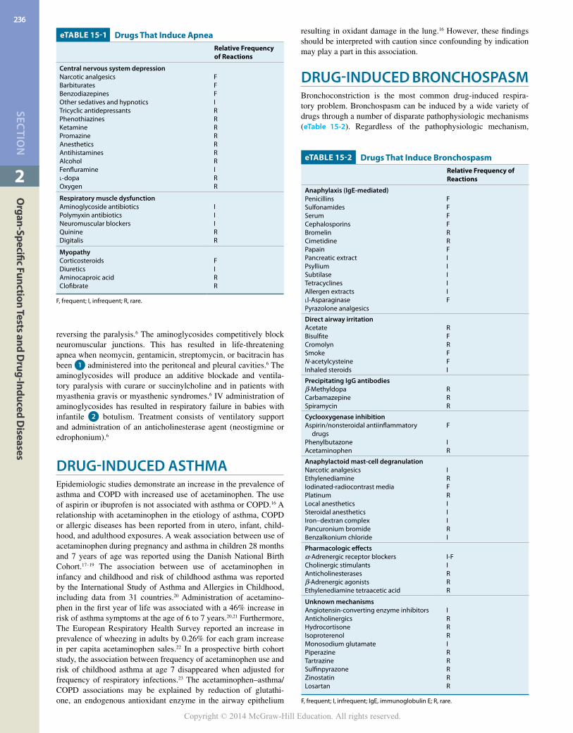
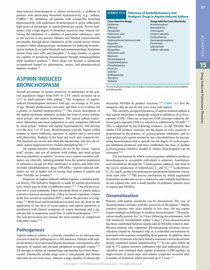
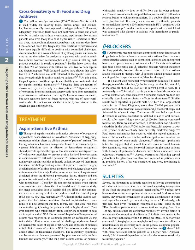
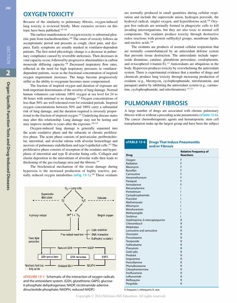


![(9/21) Thambi Lecture: Drug-Induced Renal Disease ... · (9/21) Thambi Lecture: Drug-Induced Renal Disease Identifying Drug-Induced AKI [Drug-Induced] to be shorted to DI - Sx: Decreased](https://static.documents.pub/doc/80x56/5ee17160ad6a402d666c535c/921-thambi-lecture-drug-induced-renal-disease-921-thambi-lecture-drug-induced.jpg)


