Calvin Biddle, Charles Kim 1 Drug interactions • What kind of local anesthetics are available at UBC? o Lidocaine 2%, 1:100,000 epinephrine o Prilocaine 4% plain – do not use for IAN block due to risk of paresthesia o Articaine 4%, 1:100,000 epinephrine – good bone penetration, so may be useful in the mandible. However, do not use for IAN block due to risk of paresthesia o Bupivacaine 0.5%, 1:100,000 epinephrine – 5~7 hours of profound anesthesia, good for wisdom teeth exo’s o Mepivacaine 4% plain – lowest pKa, so useful in infected tissues • Drug interactions are uncommon in dentistry, because: o Prescribed duration is usually only 5~7 days o Most dental drugs have a large margin of safety o Oral route most prescribed, IV rarely used o Number of drugs prescribed is small o Large number of pre-clinical trials good knowledge of drug properties • Classifying the severity of drug interactions o No universally accepted rating system, but Lexicomp has 5 categories A -No known interaction B -No action needed -There may be interactions, but little/no clinical evidence of concomitant use C -Monitor therapy -Clinically significant interaction, but benefits usually outweigh risks. Appropriate plan should be implemented to identify potential negative effects D -Consider therapy modification -Clinically significant interaction with risks possibly outweighing benefits (depends on patient specific assessment) -Tx modifications: aggressive monitoring, dosage changes, alternative drugs X -Avoid combination -Risks associated with interaction usually outweigh the benefits -Agents are usually considered contraindicated • Drug interactions in dentistry o Pharmacodynamic interaction: one drug affects another drug’s action on a target or system o Pharmacokinetic interaction: one drug affects another drug’s absorption/distribution/metabolism/excretion ▪ Most common interaction would be metabolism related ▪ Many drugs need to be metabolized into its active form, this usually happens in the liver ▪ Most common enzyme responsible would be the cytochrome P450 family of isozymes • As such, they are implicated in many drug interactions • CYP interactions o CYP3A4 and CYP2D6 are the most relevant for dental practice o A drug may inhibit or induce CYP activity if another drug was a CYP substrate, it would be affected o CYP induction takes 7~10 days to manifest clinically o CYP inhibition can happen in a single dose, and is more relevant to dental practice • Antibiotic spectrums
Transcript
Calvin Biddle, Charles Kim
1
Drug interactions
• What kind of local anesthetics are available at UBC?
o Lidocaine 2%, 1:100,000 epinephrine
o Prilocaine 4% plain – do not use for IAN block due to risk of paresthesia
o Articaine 4%, 1:100,000 epinephrine – good bone penetration, so may be useful in the mandible. However, do not
use for IAN block due to risk of paresthesia
o Bupivacaine 0.5%, 1:100,000 epinephrine – 5~7 hours of profound anesthesia, good for wisdom teeth exo’s
o Mepivacaine 4% plain – lowest pKa, so useful in infected tissues
• Drug interactions are uncommon in dentistry, because:
o Prescribed duration is usually only 5~7 days
o Most dental drugs have a large margin of safety
o Oral route most prescribed, IV rarely used
o Number of drugs prescribed is small
o Large number of pre-clinical trials good knowledge of drug properties
• Classifying the severity of drug interactions
o No universally accepted rating system, but Lexicomp has 5 categories
A -No known interaction
B -No action needed -There may be interactions, but little/no clinical evidence of concomitant use
C -Monitor therapy -Clinically significant interaction, but benefits usually outweigh risks. Appropriate plan should be implemented to identify potential negative effects
D -Consider therapy modification
-Clinically significant interaction with risks possibly outweighing benefits (depends on patient specific assessment) -Tx modifications: aggressive monitoring, dosage changes, alternative drugs
X -Avoid combination -Risks associated with interaction usually outweigh the benefits -Agents are usually considered contraindicated
• Drug interactions in dentistry
o Pharmacodynamic interaction: one drug affects another drug’s action on a target or system
o Pharmacokinetic interaction: one drug affects another drug’s absorption/distribution/metabolism/excretion
▪ Most common interaction would be metabolism related
▪ Many drugs need to be metabolized into its active form, this usually happens in the liver
▪ Most common enzyme responsible would be the cytochrome P450 family of isozymes
• As such, they are implicated in many drug interactions
• CYP interactions
o CYP3A4 and CYP2D6 are the most relevant for dental practice
o A drug may inhibit or induce CYP activity if another drug was a CYP substrate, it would be affected
o CYP induction takes 7~10 days to manifest clinically
o CYP inhibition can happen in a single dose, and is more relevant to dental practice
• Antibiotic spectrums
Calvin Biddle, Charles Kim
2
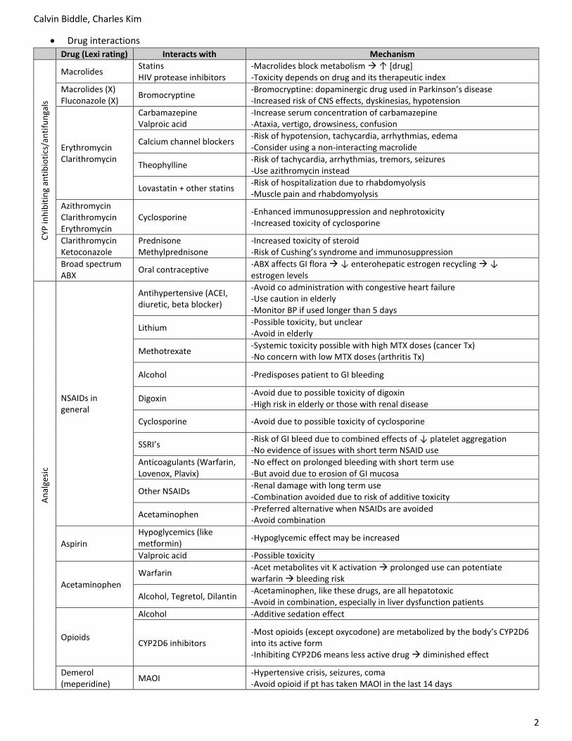
• Drug interactions
Drug (Lexi rating) Interacts with Mechanism C
YP in
hib
itin
g an
tib
ioti
cs/a
nti
fun
gals
Macrolides Statins HIV protease inhibitors
-Macrolides block metabolism ↑ [drug] -Toxicity depends on drug and its therapeutic index
Macrolides (X) Fluconazole (X)
Bromocryptine -Bromocryptine: dopaminergic drug used in Parkinson’s disease -Increased risk of CNS effects, dyskinesias, hypotension
Erythromycin Clarithromycin
Carbamazepine Valproic acid
-Increase serum concentration of carbamazepine -Ataxia, vertigo, drowsiness, confusion
Calcium channel blockers -Risk of hypotension, tachycardia, arrhythmias, edema -Consider using a non-interacting macrolide
Theophylline -Risk of tachycardia, arrhythmias, tremors, seizures -Use azithromycin instead
Lovastatin + other statins -Risk of hospitalization due to rhabdomyolysis -Muscle pain and rhabdomyolysis
Azithromycin Clarithromycin Erythromycin
Cyclosporine -Enhanced immunosuppression and nephrotoxicity -Increased toxicity of cyclosporine
Clarithromycin Ketoconazole
Prednisone Methylprednisone
-Increased toxicity of steroid -Risk of Cushing’s syndrome and immunosuppression
-Avoid co administration with congestive heart failure -Use caution in elderly -Monitor BP if used longer than 5 days
Lithium -Possible toxicity, but unclear -Avoid in elderly
Methotrexate -Systemic toxicity possible with high MTX doses (cancer Tx) -No concern with low MTX doses (arthritis Tx)
Alcohol -Predisposes patient to GI bleeding
Digoxin -Avoid due to possible toxicity of digoxin -High risk in elderly or those with renal disease
Cyclosporine -Avoid due to possible toxicity of cyclosporine
SSRI’s -Risk of GI bleed due to combined effects of ↓ platelet aggregation -No evidence of issues with short term NSAID use
Anticoagulants (Warfarin, Lovenox, Plavix)
-No effect on prolonged bleeding with short term use -But avoid due to erosion of GI mucosa
Other NSAIDs -Renal damage with long term use -Combination avoided due to risk of additive toxicity
Acetaminophen -Preferred alternative when NSAIDs are avoided -Avoid combination
Aspirin
Hypoglycemics (like metformin)
-Hypoglycemic effect may be increased
Valproic acid -Possible toxicity
Acetaminophen
Warfarin -Acet metabolites vit K activation prolonged use can potentiate warfarin bleeding risk
Alcohol, Tegretol, Dilantin -Acetaminophen, like these drugs, are all hepatotoxic -Avoid in combination, especially in liver dysfunction patients
Opioids
Alcohol -Additive sedation effect
CYP2D6 inhibitors -Most opioids (except oxycodone) are metabolized by the body’s CYP2D6 into its active form -Inhibiting CYP2D6 means less active drug diminished effect
Demerol (meperidine)
MAOI -Hypertensive crisis, seizures, coma -Avoid opioid if pt has taken MAOI in the last 14 days
Calvin Biddle, Charles Kim
3
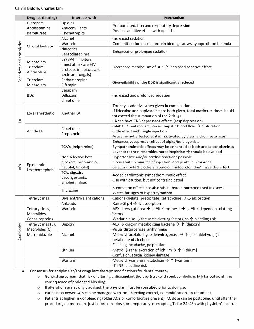
Drug (Lexi rating) Interacts with Mechanism Se
dat
ives
an
d a
nxi
oly
tics
Diazepam, Antihistamine, Barbiturate
Opioids Anticonvulants Psychotropics
-Profound sedation and respiratory depression -Possible additive effect with opioids
Chloral hydrate
Alcohol -Increased sedation
Warfarin -Competition for plasma protein binding causes hypoprothrombinemia
Narcotics Benzodiazepines
-Enhanced or prolonged sedation
Midazolam Triazolam Alprazolam
CYP3A4 inhibitors (most at risk are HIV protease inhibitors and azole antifungals)
-Decreased metabolism of BDZ increased sedative effect
Triazolam Midazolam
Carbamazepine Rifampin
-Bioavailability of the BDZ is significantly reduced
BDZ Verapamil Diltiazem Cimetidine
-Increased and prolonged sedation
LA
Local anesthetic Another LA
-Toxicity is additive when given in combination -If lidocaine and bupivacaine are both given, total maximum dose should not exceed the summation of the 2 drugs -LA can have CNS depressant effects (resp depression)
Amide LA Cimetidine Propranolol
-Inhibit LA metabolism, lowers hepatic blood flow ↑ duration -Little effect with single injection -Articaine not affected as it is inactivated by plasma cholinesterases
VC
s Epinephrine Levenordephrin
TCA’s (imipramine) -Enhances vasopressor effect of alpha/beta agonists -Sympathomimetic effects may be enhanced as both are catecholamines -Levenordephrin resembles norepinephrine should be avoided
Non selective beta blockers (propranolol, nadolol, timolol)
-Hypertensive and/or cardiac reactions possible -Occurs within minutes of injection, and peaks in 5 minutes -Selective beta 1 blockers (atenolol, metoprolol) don’t have this effect
TCA, digoxin, decongestants, amphetamines
-Added cardiotonic sympathomimetic effect -Use with caution, but not contraindicated
Thyroxine -Summation effects possible when thyroid hormone used in excess -Watch for signs of hyperthyroidism
• Consensus for antiplatelet/anticoagulant therapy modifications for dental therapy
o General agreement that risk of altering anticoagulant therapy (stroke, thromboembolism, MI) far outweigh the
consequence of prolonged bleeding
o If alterations are strongly advised, the physician must be consulted prior to doing so
o Patients on newer AC’s can be managed with local bleeding control, no modifications to treatment
o Patients at higher risk of bleeding (older AC’s or comorbidities present), AC dose can be postponed until after the
procedure, do procedure just before next dose, or temporarily interrupting Tx for 24~48h with physician’s consult
Calvin Biddle, Charles Kim
4
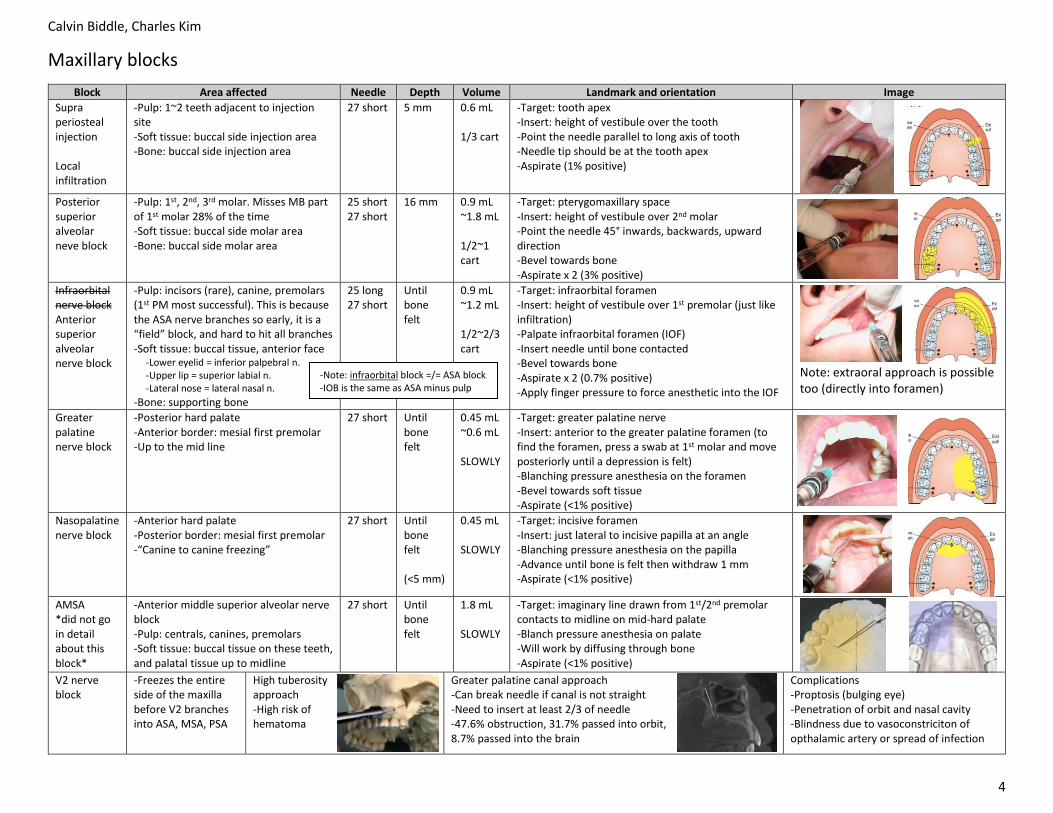
Maxillary blocks
Block Area affected Needle Depth Volume Landmark and orientation Image
Supra periosteal injection Local infiltration
-Pulp: 1~2 teeth adjacent to injection site -Soft tissue: buccal side injection area -Bone: buccal side injection area
27 short 5 mm 0.6 mL 1/3 cart
-Target: tooth apex -Insert: height of vestibule over the tooth -Point the needle parallel to long axis of tooth -Needle tip should be at the tooth apex -Aspirate (1% positive)
Posterior superior alveolar neve block
-Pulp: 1st, 2nd, 3rd molar. Misses MB part of 1st molar 28% of the time -Soft tissue: buccal side molar area -Bone: buccal side molar area
25 short 27 short
16 mm 0.9 mL ~1.8 mL 1/2~1 cart
-Target: pterygomaxillary space -Insert: height of vestibule over 2nd molar -Point the needle 45° inwards, backwards, upward direction -Bevel towards bone -Aspirate x 2 (3% positive)
Infraorbital nerve block Anterior superior alveolar nerve block
-Pulp: incisors (rare), canine, premolars (1st PM most successful). This is because the ASA nerve branches so early, it is a “field” block, and hard to hit all branches -Soft tissue: buccal tissue, anterior face -Lower eyelid = inferior palpebral n. -Upper lip = superior labial n. -Lateral nose = lateral nasal n.
-Bone: supporting bone
25 long 27 short
Until bone felt
0.9 mL ~1.2 mL 1/2~2/3 cart
-Target: infraorbital foramen -Insert: height of vestibule over 1st premolar (just like infiltration) -Palpate infraorbital foramen (IOF) -Insert needle until bone contacted -Bevel towards bone -Aspirate x 2 (0.7% positive) -Apply finger pressure to force anesthetic into the IOF
Note: extraoral approach is possible too (directly into foramen)
Greater palatine nerve block
-Posterior hard palate -Anterior border: mesial first premolar -Up to the mid line
27 short Until bone felt
0.45 mL ~0.6 mL SLOWLY
-Target: greater palatine nerve -Insert: anterior to the greater palatine foramen (to find the foramen, press a swab at 1st molar and move posteriorly until a depression is felt) -Blanching pressure anesthesia on the foramen -Bevel towards soft tissue -Aspirate (<1% positive)
Nasopalatine nerve block
-Anterior hard palate -Posterior border: mesial first premolar -“Canine to canine freezing”
27 short Until bone felt (<5 mm)
0.45 mL SLOWLY
-Target: incisive foramen -Insert: just lateral to incisive papilla at an angle -Blanching pressure anesthesia on the papilla -Advance until bone is felt then withdraw 1 mm -Aspirate (<1% positive)
AMSA *did not go in detail about this block*
-Anterior middle superior alveolar nerve block -Pulp: centrals, canines, premolars -Soft tissue: buccal tissue on these teeth, and palatal tissue up to midline
27 short Until bone felt
1.8 mL SLOWLY
-Target: imaginary line drawn from 1st/2nd premolar contacts to midline on mid-hard palate -Blanch pressure anesthesia on palate -Will work by diffusing through bone -Aspirate (<1% positive)
V2 nerve block
-Freezes the entire side of the maxilla before V2 branches into ASA, MSA, PSA
High tuberosity approach -High risk of hematoma
Greater palatine canal approach -Can break needle if canal is not straight -Need to insert at least 2/3 of needle -47.6% obstruction, 31.7% passed into orbit, 8.7% passed into the brain
Complications -Proptosis (bulging eye) -Penetration of orbit and nasal cavity -Blindness due to vasoconstriciton of opthalamic artery or spread of infection
-Note: infraorbital block =/= ASA block -IOB is the same as ASA minus pulp
Calvin Biddle, Charles Kim
5
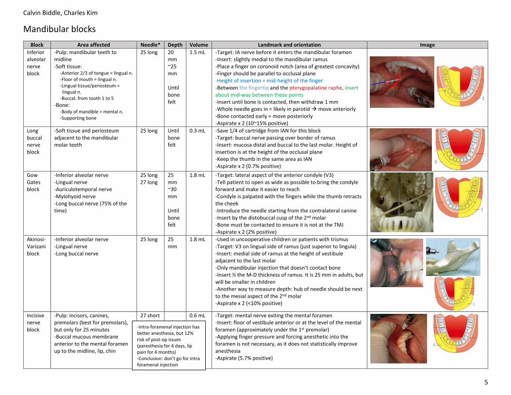
Mandibular blocks
Block Area affected Needle* Depth Volume Landmark and orientation Image
Inferior alveolar nerve block
-Pulp: mandibular teeth to midline -Soft tissue: -Anterior 2/3 of tongue = lingual n. -Floor of mouth = lingual n. -Lingual tissue/periosteum = lingual n. -Buccal: from tooth 1 to 5
-Bone: -Body of mandible = mental n. -Supporting bone
25 long 20 mm ~25 mm Until bone felt
1.5 mL -Target: IA nerve before it enters the mandibular foramen -Insert: slightly medial to the mandibular ramus -Place a finger on coronoid notch (area of greatest concavity) -Finger should be parallel to occlusal plane -Height of insertion = mid-height of the finger -Between the fingertip and the pterygopalatine raphe, insert about mid-way between these points -Insert until bone is contacted, then withdraw 1 mm -Whole needle goes in = likely in parotid move anteriorly -Bone contacted early = move posteriorly -Aspirate x 2 (10~15% positive)
Long buccal nerve block
-Soft tissue and periosteum adjacent to the mandibular molar teeth
25 long Until bone felt
0.3 mL -Save 1/4 of cartridge from IAN for this block -Target: buccal nerve passing over border of ramus -Insert: mucosa distal and buccal to the last molar. Height of insertion is at the height of the occlusal plane -Keep the thumb in the same area as IAN -Aspirate x 2 (0.7% positive)
Gow Gates block
-Inferior alveolar nerve -Lingual nerve -Auriculotemporal nerve -Mylohyoid nerve -Long buccal nerve (75% of the time)
25 long 27 long
25 mm ~30 mm Until bone felt
1.8 mL -Target: lateral aspect of the anterior condyle (V3) -Tell patient to open as wide as possible to bring the condyle forward and make it easier to reach -Condyle is palpated with the fingers while the thumb retracts the cheek -Introduce the needle starting from the contralateral canine -Insert by the distobuccal cusp of the 2nd molar -Bone must be contacted to ensure it is not at the TMJ -Aspirate x 2 (2% positive)
1.8 mL -Used in uncooperative children or patients with trismus -Target: V3 on lingual side of ramus (just superior to lingula) -Insert: medial side of ramus at the height of vestibule adjacent to the last molar -Only mandibular injection that doesn’t contact bone -Insert ½ the M-D thickness of ramus. It is 25 mm in adults, but will be smaller in children -Another way to measure depth: hub of needle should be next to the mesial aspect of the 2nd molar -Aspirate x 2 (<10% positive)
Incisive nerve block
-Pulp: incisors, canines, premolars (best for premolars), but only for 25 minutes -Buccal mucous membrane anterior to the mental foramen up to the midline, lip, chin
27 short 0.6 mL -Target: mental nerve exiting the mental foramen -Insert: floor of vestibule anterior or at the level of the mental foramen (approximately under the 1st premolar) -Applying finger pressure and forcing anesthetic into the foramen is not necessary, as it does not statistically improve anesthesia -Aspirate (5.7% positive)
-Intra-foramenal injection has better anesthesia, but 12% risk of post-op issues (paresthesia for 4 days, lip pain for 4 months) -Conclusion: don’t go for intra foramenal injection
o Some individuals may be ultra-rapid metabolizers because of a specific CYP2D6 genotype (e.g., gene duplications
denoted as *1/*1×N or *1/*2×N)
o The prevalence of this CYP2D6 phenotype varies widely and has been estimated at 1 to 10% for Whites (European,
North American), 3 to 4% for Blacks (African Americans), 1 to 2% for East Asians (Chinese, Japanese, Korean), and
may be greater than 10% in certain racial/ethnic groups (i.e., Oceanian, Northern African, Middle Eastern,
Ashkenazi Jews, Puerto Rican)
o These individuals convert codeine into its active metabolite, morphine, more rapidly and completely than other
people. This rapid conversion results in higher than expected serum morphine levels.
o Even at labeled dosage regimens, individuals who are ultra-rapid metabolizers may have life-threatening or fatal
respiratory depression or experience signs of overdose (such as extreme sleepiness, confusion, or shallow
breathing) (see OVERDOSAGE). Therefore, individuals who are ultra-rapid metabolizers should not use TYLENOL®
with Codeine tablets.
o Thanks Osama
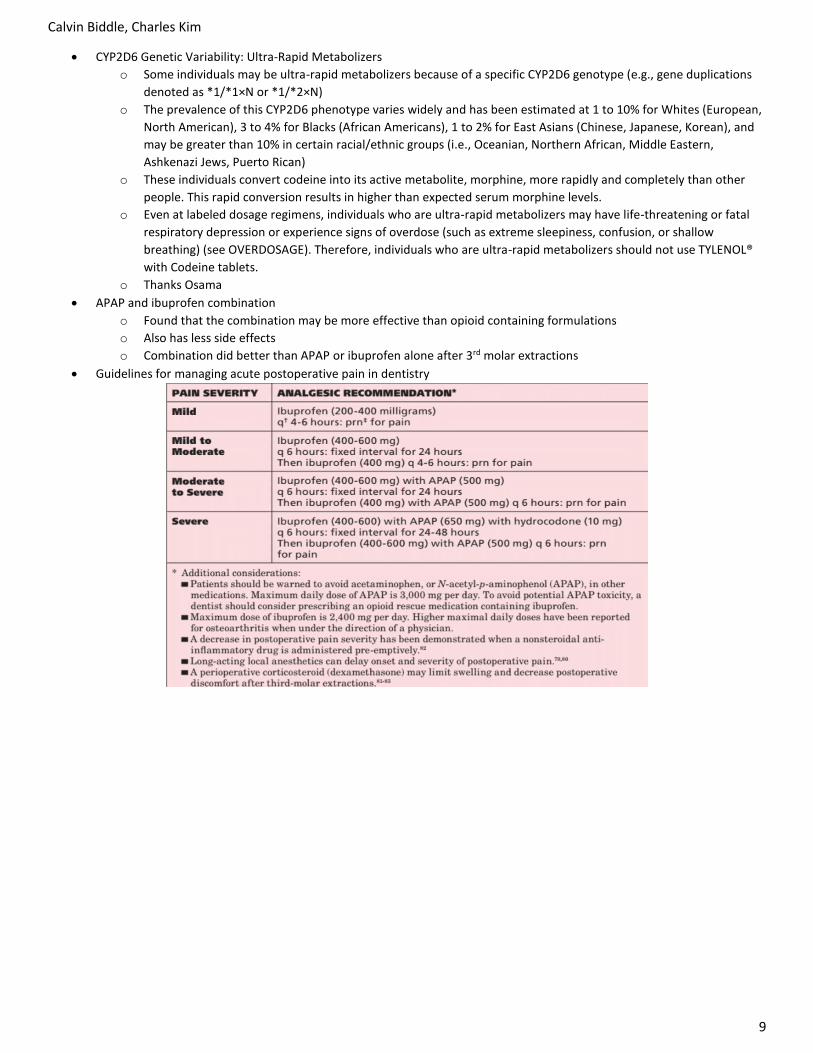
• APAP and ibuprofen combination
o Found that the combination may be more effective than opioid containing formulations
o Also has less side effects
o Combination did better than APAP or ibuprofen alone after 3rd molar extractions
• Guidelines for managing acute postoperative pain in dentistry
Calvin Biddle, Charles Kim
10
Medical emergencies
• Code blue
o Decreased level of responsiveness
o Fainting/collapse
o Chest pain
o Shortness of breath
o Seizure
o Presumed overdose
o Severe allergic reaction
• Emergency kits
o OHC crash cart CSD return window
o AED ends of bays 14 and 15
o Oxygen ends of bays 2, 10, 15
o Kit contents see table
• Who gets the crash cart?
o Student, CDA, or first aid assistant will get the crash card after
they have notified the CSD
o Reception will announce over PA of a code blue
• Follow ABC’s
o Airway
o Breathing
o Circulation
• Emergencies in the dental office
o 74.4% of dentists reported a medical emergency in their
career
o 3% had to perform CPR
Emergency Freq Signs and symptoms Reason Management
Syncope 30% -Brief loss of consciousness and muscle tone -Preceded by presyncope
-↓ blood to the brain -Due to heart failing, loss of vessel tone, lack of blood, or a combination -More serious causes: cardiac failure, subclavian steal syndrome, aortic stenosis
-Trendelenberg -Basic life support + monitor vitals -100% oxygen -Monitor vitals -Apply cold compress -EMS if LOC >5min, or >10 mins of recovery
-Change in body position causing drop in BP -Increased risk with nitrates, Parkinson’s drugs, antipsychotics, neuroleptics, antianxiety, sedatives, hypnotics, TCA’s, antihypertensives
-Lie down immediately -Trendelenberg -Oxygen -EMS if condition worsens or due to steroid use -Reposition slowly -Monitor vitals
Hyperventilation 10% -Breathing >40 bpm -Impaired consciousness -Tightness of chest -Apprehention -Palpitation of heart -Fullness in throat -Tetany if prolonged -Perioral numbness
-Most commonly anxiety -Others: fever, aspirin OD, infection, stroke, diseases of brain or CNS
-Relax patient -Give reassurance (“you are not going to die” “you will be fine”) -Speak softly -Have patient breathe through pursed lips -Cover pts mouth and one nostril legal implications??
Calvin Biddle, Charles Kim
11
Emergency Freq Signs and symptoms Reason Management
Hypoglycemia 5.1% -Blood sugar <2.5 mmol/L -Fatigue -Loss of consciousness
-Supine -Airway + monitor vitals -Treat blood sugar levels <2.8 mmol/L, even if asymptomatic -Conscious: oral glucose -Unconscious: activate EMS, give 1mg glucagon IM. Check [sugar] in 15 mins, and repeat glucagon dose if not normalized
-Emotional stress -Exposure to hot/cold -Heavy meals -Smoking
-100% oxygen -Place patient comfortably -Nitroglycerin SL spray q5min up to 15 mins (3 doses) -After 3rd dose, assume acute MI -Set up AED -Activate EMS if signs of hemodynamic instability + chewable aspirin 325mg
-Angina pectoris can be prevented by consulting physician prior to dental treatment -Pharmacological preventative measures: oral sedation, preoperative nitroglycerin dose -Limit epinephrine to 0.04 mg max
Seizure 4.6% -Brief blackout followed by confusion -Changes in behaviour (picking at clothing) -Drooling or frothing at mouth -Eye movements -Grunting and snorting -Loss of bladder/bowel control -Mood changes -Shaking of body -Sudden falling -Bitter metallic taste -Teeth clenching -Halted breathing
-Supine position -Loosen clothing -Relocate instruments -Establish airway -Continue to observe
Bronchospasm 3% -Narrowing of bronchi -Wheezing -Coughing -Shortness of breath
-Genetic -Environment -Immune system -GERD -Medications
-Prevented by salbutamol before dental treatment -Treatment: upright position + EMS -Monitor vitals + 100% oxygen -Salbutamol 2 puffs every 20 mins -If worsening, 0.3 mg epi IM every 20 mins and prednisone 40~60 mg orally
Emergency Freq Signs and symptoms Reason Management
Anaphylactic shock
1.2% -CV collapse or even arrest (hypotension) -Respiratory compromise (bronchospasm) -Symptoms will show 5~30 min if injected, up to 2 hours if ingested -Flushed face, rash, urticaria, tingling, angioedema -Diaphoresis -Impending doom -Loss of consciousness -Incontinence -Cyanosis/pallor -Dizziness
-Foods -Environment (latex, bee stings) -Medications Penicillin allergy: -1~10% of patients -Accoutns for 75% of anaphylaxis deaths -Fatality rate of 1/60,000, with 96% of deaths happening in 60 minutes
-EMS -Supine -BLS + monitor vitals -Oxygen -Ventilate manually if necessary, using bag valve mask -Epi 0.3~0.5mg SL, SC, or IM -Diphenhydramine 25~50 mg IM/IV
Cardiac arrest 1.1% -No pulse + breaths -Loss of consciousness -Gasping, laboured breathing -Can be preceded by chest pain
-Acute MI -Cardiomyopathy -hypoxia -Medication reaction
-EMS -BLS -Switch on AED
Acute adrenal insufficiency
-Weakness, fatigue -Headache -Nausea, vomiting -Myalgia, joint pain -Abdominal pain -Lethargy -Flank pain -HIS PALMS ARE SWEATY KNEES WEAK ARMS ARE HEAVY
-Exaggerated hyperthyroidism -Seen in pts with mod~severe antecedent Graves’ disease -Precipitated by stress
-EMS -100% oxygen -Place patient in comfortable position -Monitor vitals every 5 mins -Initiate BLS
• American Society of Anesthesiology’s classification system
Class Description Example Treatment BP
1 Normal healthy patient Healthy with good exercise tolerance No special precautions <140/90
2 Mild systemic disease Controlled hypertension, controlled diabetes mellitus without system effects, cigarette smoking without evidence of COPD, anemia, mild obesity, age less than 1 or greater than 70 years, pregnancy
Elective tx OK, consider tx modification
140~159 90~94
3 Severe systemic disease, but not incapacitating
Controlled CHF, stable angina, old MI, poorly controlled hypertension, morbid obesity, bronchospastic disease with intermittent symptoms, chronic renal failure
▪ Sulfites may exist as a preservative for epinephrine in LA cartridges avoid LA with epi in these pts
Calvin Biddle, Charles Kim
18
Tetris time
Calvin Biddle, Charles Kim
19
Calvin Biddle, Charles Kim
20
Calvin Biddle, Charles Kim
21
Calvin Biddle, Charles Kim
22
Calvin Biddle, Charles Kim
23
Principles of surgery
• Oral surgery outcomes are predictable if you abide by the principles of surgery
o Always stick to a standard format: CC, Hx of CC, Med Hx, Social Hx, and special investigations
o Then, formulate a diagnosis. Never make assumptions or cut corners
o We rely on the patient to provide all accurate information, but the physician can be consulted if the information is
not known by the patient, or the reliability of it is questionable
o If the patient has been referred by other practitioners, gain as much information from them as you can
• Causes of swelling near/at the angle of the mandible
o Compensatory hypertrophy due to hypotrophy/hypoplasia on the other side
o Masseter muscle intrinsic myopathy
o Masseter muscle neoplasia
o Salivary gland diseases (sialosis, parotitis)
o Parotid neoplasia (pleomorphic adenoma)
o Parotid inflammatory disease
o Odontogenic problems (chronic dental abscess)
o Neoplasia of soft tissues (lipoma)
o Vascular lesion
• Necessities for surgery
o Adequate visibility: access, light, free of blood/fluids
o Assistance: trained assistant familiar with procedures
o Aseptic technique: minimise wound contamination
• Incisions
o Use a sharp blade (usually #15) for oral surgery
▪ Bone and ligaments dull blades more rapidly than buccal mucosa
▪ Change the blade when the scalpel does not seem to cut with ease
o Firm, continuous strokes when incising
▪ Repeated strokes will impair wound healing and visibility
▪ Long, continuous strokes are preferred to short, interrupted ones
▪ Rotate the wrist to cut, don’t pull the whole arm
o Avoid cutting vital structures
▪ Incise deep enough to get the layer you need, but avoid underlying vessels and nerves
▪ Vessels can be more easily controlled before they are cut
▪ Nerves can be retracted away before incision too
▪ Focus on the blade only, to avoid accidental cutting of lip and other structures
o Blade should be perpendicular to cutting surface
▪ Essential if the tissues are to be re-approximated
▪ Reduces chances of necrosis on the incision borders, and are easier to reorient to suture
o Ensure properly positioned incisions
▪ Incisions over attached gingiva and healthy bone are better than unattached gingiva and unhealthy bone
▪ Incisions should extend a few mm away from damaged bone allows suturing over healthy bone
▪ Incisions near teeth for extractions are made in the gingival sulcus, unless it is necessary to excise the
marginal gingiva or to leave the marginal gingiva untouched
• Flap design
o Apex (coronal of tooth) part of flap is never wider than the base (apical of tooth),
unless a major artery is present in the base
o Flap sides (releasing incisions) should be parallel or convergent towards the apex
o Width (X) should be longer than height (Y), preferably X = 2Y
o Axial blood supply should be included if possible
▪ Example: a flap in the palate should be based toward the greater
palatine artery if possible
o Handle tissue with care (don’t twist, stretch, grasp the base) and don’t expose
it to harmful environments (temperature, dessication, noxious chemicals)
Calvin Biddle, Charles Kim
24
• Preventing flap dehiscence (exposure of underlying bone causing bone loss, pain, and scarring)
o Approximate edges of the flap over healthy bone
o Handle edges gently
o Do not place flap under tension
• Preventing flap tearing
o Producing a clean, long incision will take the same time to heal as a short one
o So, create a long enough flap right when you start, rather than trying to be conservative making a short incision,
and traumatizing the tissue to get adequate access
• Releasing incisions
o 2 sided flap: 1 vertical and 1 horizontal incision
o 3 sided flap: 2 vertical and 1 horizontal incision
o 1 sided flap: also called an envelope flap, there
is only 1 horizontal incision along the necks of
several teeth. This is the staple of dental surgery
• Tissue handling
o Avoid aggressive tissue retraction for improved surgical access
o Be generous with irrigation when drilling bone prevent frictional heat
o Only allow physiologic substances to contact living tissue
▪ If forceps were used to place a specimen into formalin, thoroughly rinse forceps
• Hemostasis
o Why meticulous hemostasis is necessary
▪ Avoid decreased visibility due to uncontrolled bleeding
▪ Could form a hematoma which further increases complications by placing pressure on a wound (↓
vascularity), increased tension on wound edges, act as a culture medium for infection
o Promoting hemostasis
▪ 2x2 gauze and pressure
• 20~30 seconds in small vessels, 5~10 mins in large vessels
• Dab around the wound with gauze afterwards, don’t wipe
▪ Hemostat
• Pinched around bleeding vessel
▪ Cautery
• Heat fuses tissue coagulates ends of vessels
• Electrical current can be applied on wound indirectly through a metal instrument like a hemostat,
or directly with the electrocautery tip
• Caution in nitrous patients: electricity may ignite oxygen in nasal prongs nasal burn
• Patient must be grounded to allow current to enter body
• Current should only be applied around bleeding vessel. Anywhere else risk of electricity
following an undesirable path and causing a burn
• Remove blood or fluid before cautery, as it may inhibit enough heat transfer to cauterize tissue
▪ Suture ligation
• Grasp each end of a cut vessel with a hemostat and tie them together with a non resorbable
suture
• Alternatively, if the vessel can be dissected freely: clamp the vessel with 2 hemostats and dissect
the vessel between the hemostats. Suture each end then release hemostats
▪ Epinephrine
• Soak LA containing epi with a gauze and apply on tissue. Or, inject directly
• Best vasoconstriction can be acquired if this is done 7~10 mins before surgery
• A pro-coagulant (collagen, commercial thrombin) can be used as well
Calvin Biddle, Charles Kim
25
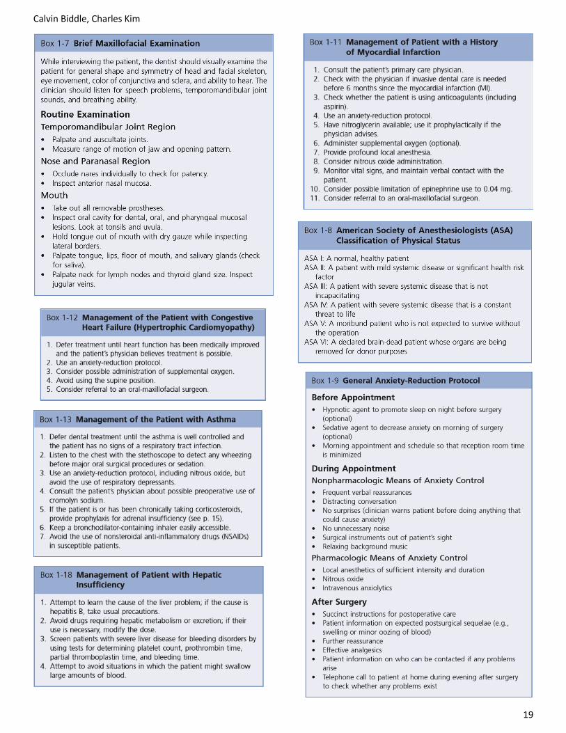
• Dead space
o Dead space: area in a wound devoid of tissue after wound closure (AKA, an air pocket). Usually fills with blood,
which puts it at risk of a hematoma and possibly an infection
▪ In dental surgeries, dead space creation is not a major problem
▪ Can happen if surgeon removes deep tissues in a wound or not reapproximating correctly during closure
o Managing dead space
▪ Deep sutures
• Insert needle deep into the superficial fascia exit needle at dermal-
epidermal junction re-insert needle on other side’s dermal-
epidermal junction exit in the deep superficial fascia lock with
3~4 throws and bury knot
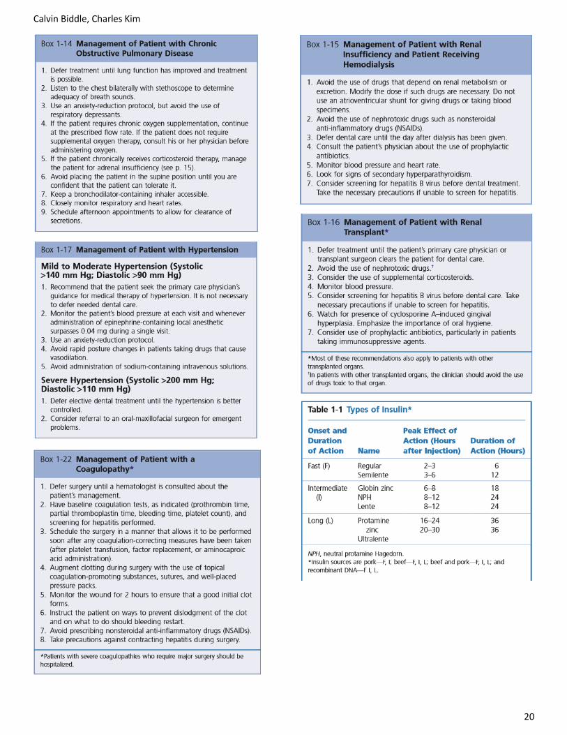
▪ Pressure with a dressing
• Compresses tissue planes together until wound is bound by fibrin or
pressed together from surgical edema (or both)
• Usually takes 12~18 hours
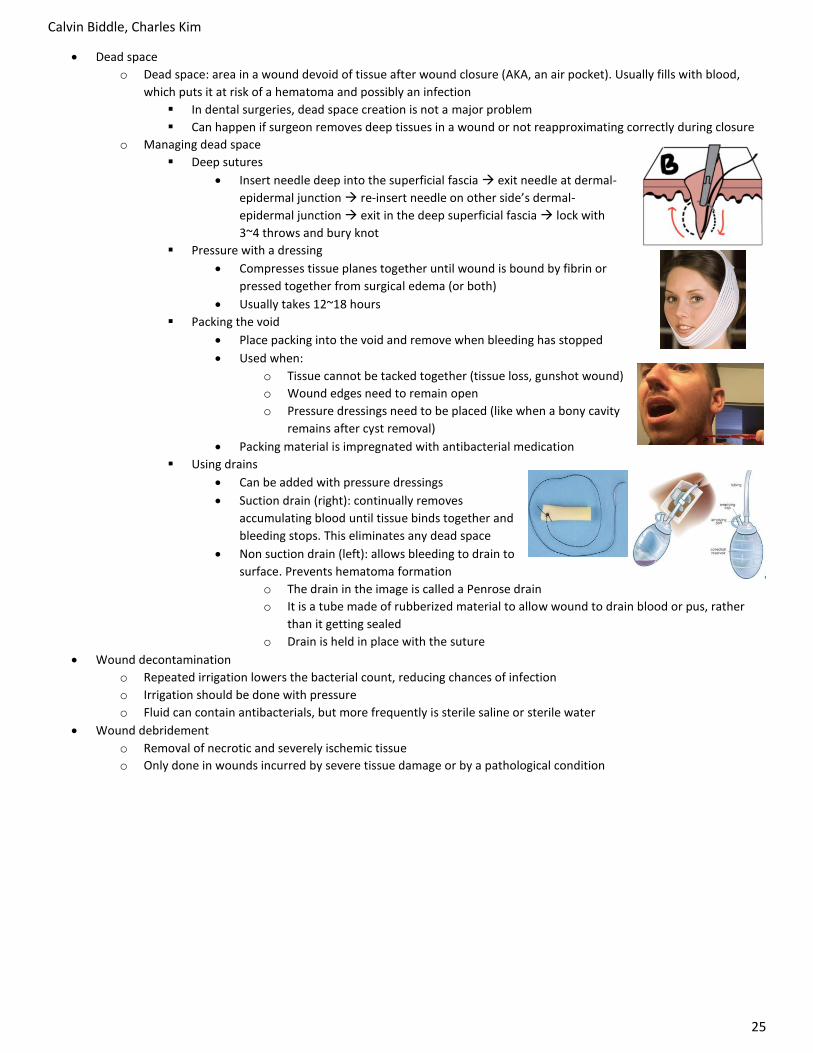
▪ Packing the void
• Place packing into the void and remove when bleeding has stopped
• Used when:
o Tissue cannot be tacked together (tissue loss, gunshot wound)
o Wound edges need to remain open
o Pressure dressings need to be placed (like when a bony cavity
remains after cyst removal)
• Packing material is impregnated with antibacterial medication
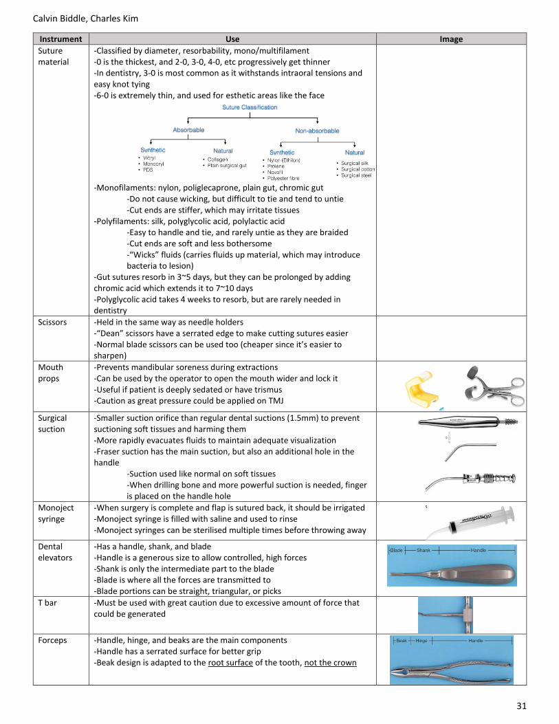
▪ Using drains
• Can be added with pressure dressings
• Suction drain (right): continually removes
accumulating blood until tissue binds together and
bleeding stops. This eliminates any dead space
• Non suction drain (left): allows bleeding to drain to
surface. Prevents hematoma formation
o The drain in the image is called a Penrose drain
o It is a tube made of rubberized material to allow wound to drain blood or pus, rather
than it getting sealed
o Drain is held in place with the suture
• Wound decontamination
o Repeated irrigation lowers the bacterial count, reducing chances of infection
o Irrigation should be done with pressure
o Fluid can contain antibacterials, but more frequently is sterile saline or sterile water
• Wound debridement
o Removal of necrotic and severely ischemic tissue
o Only done in wounds incurred by severe tissue damage or by a pathological condition
Calvin Biddle, Charles Kim
26
Principles of wound repair
• When a wound leaves 2 free ends exposed, the epithelium will start proliferating
until the gap is closed
o Proliferation happens at the free ends, where cells start migrating, until
the gap is closed off
o There needs to be a vascular bed underneath and coagulated (scab)
tissue above for the epithelium to migrate
o When cells come in contact, contact inhibition prevents further
proliferation
• Clinical applications of contact inhibition
o Malignant epithelial cells have lost contact inhibition and continue to grow
o When a maxillary tooth is extracted, the sinus epithelium and oral mucosa may be injured. This causes an opening
from the mouth into the sinus. In some cases, the sinus epithelium will proliferate towards the oral cavity, and stop
proliferating when it contacts oral mucosa. This still leaves an oro-antral communication
• Types of healing
o Primary intention: no tissues are lost, and tissues are stabilized in the same anatomical position
o Secondary intention: gap is left when tissues are approximated, needing cell migration
o Tertiary intention: healing of wounds through tissue grafts to cover large wounds
• Stages of wound healing
Inflammation 3~5 days
-Cardinal signs of inflammation: redness, swelling, warmth, pain, loss of function -Considered the “lag phase” because no significant gain in wound strength happens in this stage Vascular phase -Injured cells release PG’s and TXA’s vasoconstriction -WBC’s release histamine, PGE1, PGE2 makes vessels leaky, so immune cells can easily migrate out -Exudated fluid also dilutes contaminants called inflammatory edema -Fibrin within the fluid will block off lymphatics to allow further fluid accumulation -Begins coagulation cascade (review in FMS BLI) Cellular phase -Triggered by serum complement and tissue trauma -C3a and C5a act as chemotactic factors to recruit PMNs -Cell undergoes margination diapedesis chemotaxis degranulation/phagocytosis Clinical application -Blood can be centrifuged to isolate the fibrin and platelets. It is a yellow gelatinous mass formed after centrifugation -“PRF” can be put into extraction sockets to promote quicker healing and hemostasis
Proliferation 2~3 weeks
Fibroblasts/fibroplastic stage -Fibroblasts migrate into the wound, along with capillary ingrowths when necrotic + foreign cells are dealt with from the inflammatory stage -Fibrin strands form lattices which is the initial lattice work for cells to migrate into -Fibroblasts also secrete fibronectin: stabilizes fibrin, recognizes foreign material, act as a chemotactic factor for other fibroblasts, guides macrophages along fibrin strands -With incoming cells and capillaries, plasmin is also brought in which causes fibrinolysis -Fibroblasts deposit excessive amounts of tropocollagen which crosslinks to collagen -Initially laid in a random pattern which is not as strong as normal tissue, but much stronger than the wound before (70~80% strength of normal tissue) -Wound is stiff due to excess collagen and erythematous due to new capillaries Epithelium -Proliferates to increase thickness and form a normal epithelium under the scab
Remodelling -Randomly laid collagen fibers are destroyed and replaced by new fibers which are oriented better -Less fibers are needed when they are oriented correctly. This leads to a softening of the scar -Epithelium stratification is restored -Erythema resolves as vascularity is decreased -Elastin is not replenished in normal skin and ligaments tissue elasticity decreases -Wound strength never reaches more than 80~85% of normal tissue strength
Calvin Biddle, Charles Kim
27
• Wound contraction
o Edges of a wound contract toward each other, diminishing the size of the wound
▪ Unclear how a wound contracts or the mechanism behind it
o Generally favourable, but can be harmful in injuries like 3rd degree burns where contractions will cause
deformation and debilitate the patient if not covered by a skin graft
o Also harmful in sharply curved lacterations where the concave piece of tissue contracts detrimentally. Epithelium
can be placed on free edges of a wound to reduce contraction
▪ Example: in vestibuloplasties or full thickness burn wounds, skin grafts are placed to avoid contraction
• Factors that impair wound healing
Foreign material -Bacteria can proliferate infection bacterial proteins destroy host tissue -Non bacterial foreign materials harbor and shelter bacteria promotes infection -Foreign materials can also be antigenic and stimulate a chronic inflammatory reaction that decreases fibroplasia
Necrotic tissue -Necrotic cells are dead, and require removal by WBC’s (lysis + phagocytosis), but take a long time -Necrotic tissue acts as a barrier to the ingrowth of reparative cells prolongs inflammation -Necrotic tissue frequently has an associated hematoma, which can harbor bacterial growth
Ischemia -Decreased blood supply can worsen necrosis, lower WBC delivery, increased chance of infection -Less oxygen and nutrients to site necessary for proper healing -Caused by: tight sutures, improper flaps, internal wound pressure (hematoma), hypotension, peripheral vascular disease, or anemia
Wound tension -Placing tissues under tension will cause ischemia -If sutures are removed too early in a wound under tension, it can open back up excessive scarring -If sutures are left in too long under tension, it will still tend to open during remodelling
• Healing of extraction sockets
o Because a gap is formed, it will have to heal by secondary intention
o Tooth sockets involve cortical bone (lamina dura), torn PDL ligaments, and a rim of gingiva
o Socket fills and coagulates with blood seals from oral environment
o Week 1
▪ Inflammatory stage happens to remove bacteria and bone fragments
▪ Fibroplasia also starts fibroblasts and capillaries enter the tissues
▪ Epithelium migrates down socket wall until it meets epithelium on the other side of the socket or
encounters a bed of granulation tissue
▪ Osteoclasts accumulate along the crestal bone
o Week 2
▪ Large amount of granulation tissue fills the socket (immature capillaries + fibroblasts)
▪ In smaller sockets, epithelium may be fully intact by now
o After week 2
▪ Epithelialisation of most sockets are complete
▪ Cortical bone continues to be resorbed from crest + walls, and trabecular bone deposited in socket
▪ It will take 4~6 months for the cortical bone (lamina dura) to disappear completely
▪ As bone fills the socket, epithelium will migrate crestally until it matches adjacent gingiva
o After 1 year
▪ The only visible sign of the socket would be scar tissue remaining on the alveolar ridge
• Bone healing
o Very similar stages to soft tissue healing, but with the addition of osteoclasts and osteoblasts
o Osteoblasts are derived from the periosteum, endosteum, and circulating pluripotent mesenchymal cells
▪ Lay down new bone in areas of sufficient oxygen tension
▪ In areas of low oxygen tension, chondroblasts come in instead and lay down cartilage
o Osteoclasts are derived from monocyte precursor cells
▪ Responsible for clearing necrotic bone
o Primary and secondary intentions of healing also apply to bone
▪ Primary = bone is only partially fractured, or completely fractured but reapproximated with plates/screws
▪ Secondary = bone gap is >1mm, which means a larger callus and fiber deposition needs to happen
▪ A “callus” of bone forms during healing to represent the inorganized fibre matrix that’s laid down quickly
▪ Vascularity and immobility are key factors to allow bone healing. The bone also needs to be placed under
continuous/repeated cycles of tension to stimulate osteoblasts (but not enough to damage healing bone)
Calvin Biddle, Charles Kim
28
• Implant osseointegration
o Implants need both bone and soft tissue to heal around it to integrate successfully
o Bone integration must happen before soft tissue integration, because once soft tissue enters bone will not
attach there. For bone to win this “race”, it depends on 4 factors:
▪ Short distance between bone and implant (good adaptation of implant and drill)
▪ Viable bone at/near surface of bone along implant (low heat when drilling)
▪ No movement of the implant while bone is attaching (no forces either)
▪ Implant surface free of contamination by organic or inorganic particles
o Guided tissue regeneration could be used
▪ Keeps soft tissue out, while being permeable enough to allow oxygen and nutrients to reach bone
▪ Comes in the form of woven membranes
o Heat must be kept to a minimum to prevent bone damage when you drill
▪ Use sharp bone cutting instruments
▪ Limited cutting speeds to minimise frictional heat
▪ Keep bone cool with irrigation
o Implant material must be inert, like titanium
o Aseptic technique is crucial. Systemic or topical antibiotics may be used in rare occasions
o During healing, the implant will have no crown so no forces will be applied on it. Implant can even be covered with
gingiva to further protect it, but not required
o Threaded and tight fitting implants are better protected from soft
tissue migration
o Once initial integration has occurred, limited daily pressure (~1mm
strain) hastens cortical bone deposition
o Abutment
▪ Part of the implant in contact with soft tissues
▪ When oral epithelium reaches the titanium surface, it stops
and secretes ground substance that attaches to the metal
▪ Hemidesmosomal basal lamina system forms, further
strengthening soft tissue attachment
• Facial neuropathology of traumatic origin
o Nerve damage can happen due to trauma, extractions, pathologic conditions, or reconstructive surgery
o Most injured nerves spontaneously recover
o 2 most common branches of the trigeminal nerve that are injured
▪ Inferior alveolar mental nerve
▪ Lingual nerve
o Classification of nerve injury
Neurapraxia
-No loss of continuity of nerve or endoneurium -Trauma, compression, stretching, or inflammamation around a nerve could be the reason -Will recover in a few days~weeks
Axonotmesis -Loss of axonal continuity but preserves endoneurium -Could be due to aggressive retraction of the mental nerve (severe blunt trauma), nerve crushing, or extreme traction of a nerve -Can (but not always) recover in 2~6 months
Neurotmesis -Loss of axonal and endoneurium continuity -Could be due to badly displaced fractures, severance by bullets/knives, or iatrogenic transection -Prognosis is poor, unless the nerve ends have been left in approximation after injury
o Nerve degeneration
Segmental demyelination Wallerian degeneration
-Myelin sheath dissolves into isolated segments -Paresthesia, dysesthesia, hyperesthesia, or hypoesthesia may result -Can happen with neurapraxic injuries or vascular/CT disorders
-Axons and myelin distal to the injury will degrade -Proximal to the injury, there is some degeneration but generally only for a few nodes of Ranvier -Follows nerve transection and other destructive processes that affect peripheral nerves
Calvin Biddle, Charles Kim
29
o Nerve healing
▪ Regeneration happens almost immediately after nerve injury
▪ Normally, proximal stump sends out a group of new fibers (called growth cones) that grow down the
remnant Schwann cell tube
▪ Growth is 1~1.5mm/day until site is reached, or growth is blocked by fibrous CT or bone
• If it is blocked by fibrous CT, things can go wrong
• Ideally, the axon will grow around the blockage
• Or, the axon may grow into a mass of aimless nerve fibers called an axonal neuroma
• Neuromas can cause pain when disturbed
▪ As functional contacts are made, patient will experience altered sensations in the previous numb area
(paresthesias or dysesthesias)
▪ Growth after crushing
• The endoneurial tube contains all the molecular cues, which the growth cone uses to find the
pathway to grow in to
• Axon can grow in very precise pathways to find the same targets eventually
▪ Growth after transection
• The endoneurial tube is destroyed, so molecular cues are not concentrated
• Growth can happen in all directions, until an axon finds any other endoneurial tube
Instrumentation
Instrument Use Image
Scalpels -15 = intraoral surgery -10 = large skin incisions -11 = incise and drain abscesses -12 = mucogingival procedures where incisions are made on posterior teeth or in maxillary tuberosity area
Scalpel handles and Crile-Wood needle holder
-Handle attaches to the blade using a Crile-Wood needle holder -#15 blade goes with #3 handle -Removal of the blade can also be done with the needle holder
Periosteal elevator
-#9 Molt periosteal elevator is most commonly used -Used to reflect flap from cortical bone in one smooth layer -Pointed end = twist and pry soft tissue to begin reflection. Most commonly used to elevate dental papillae -Round end = extends the reflection -Push strokes give clean separation, but pull strokes may tear/shred the periosteum, so perform with caution
Flap retractors
-Big retractors = Austin and Minnesota -Small retractors = Seldin and Molt #9 -Henahan retractor can be used to retract mucoperiosteal flaps (basically a double ended Molt #9)
.
Tongue retractors
-Mouth mirror – good, easy to use, can retract cheek too, and comfortable. Used for most routine exodontias -Weider retractor – use pediatric size, adult is too big. Don’t insert it too deep as it may cause gagging
Grasping soft tissue
-Adson forceps (picture) = Used for plastic surgery on skin -Can be toothed or non toothed -Too short to be useful in dentistry, but we have them at OHC -Use Stillies forceps or Gillies dissecting forceps instead as they are much longer -Niftyinstruments in Toronto sells super cheap good quality instruments – Dr Matthew
.
Calvin Biddle, Charles Kim
30
Instrument Use Image Cotton pliers -Placing dressings or taking them out
-Picking up loose fragments of teeth, amalgam, foreign material -Locking pliers are useful in endodontics, but rarely of value in oral surgery
Allis tissue forceps
-Grasping biopsy tissues -Could also be used to grab the tongue in an emergency -Causes a lot of tissue destruction and too large for oral tissues
Hemostat -Can be curved or straight -Grasps tissue and locks, useful for clamping vessels -Also useful to pick up fragmented debris in the mouth, or granulation tissue in a tooth socket -Not used to hold needles because hatches are parallel
Rongeurs -Removing bone via sharp blades -Rebound mechanism will open back up when you relax the hand, so that multiple trimming actions can be done without having to reopen the instrument -They can be side cutting or end cutting -Never use to pull teeth because it will dull and destroy the instrument and it will not grip teeth well
Surgical handpiece
-High speed, high torque handpieces -Doesn’t spray air at the tip -Useful when large amounts of bone must be removed, like tori -Must be completely sterilisable, should have high speed and torque, and must not exhaust air into the operative field -Air forced under pressure may cause surgical emphysema or even pneumothorax -Burs are acrylic bur shaped
Mallet and chisel
-Used when removing lingual tori -Chisel must be sharp to function properly
Bone file -Smoothing of bone before completing surgery -Doesn’t remove a lot of bone, only for smoothing -Only pull, pushing will burnish and crush bone
Curette -Removes soft tissue from bony defects -Removes granulomas, small cysts, granulation tissue
Towel clips -Penetrates towels and drapes -Useful for placing drapes on patient and keeping it there -Be careful not to pinch skin
Needle holder
-6 inch needle holder is best -Beaks are stronger than a hemostat’s and cross hatched to grip the suture needle better -Held with an underhand grip with thumb and 4th finger in rings -Grips the suture needle 2/3 away from the tip to allow cutting surface to be exposed while needle is held at its strongest point
Sutures -Small half circle or 3/8 circle curved needle to allow passage through limited space where a straight needle cannot reach -Distal 1/3 cross section is a triangle for cutting -Proximal 2/3 cross section is rounded -Can cut through tissue lateral to the track if not used carefully -Tapered suture needles are used for delicate tissues like ocular or vascular surgery
Calvin Biddle, Charles Kim
31
Instrument Use Image Suture material
-Classified by diameter, resorbability, mono/multifilament -0 is the thickest, and 2-0, 3-0, 4-0, etc progressively get thinner -In dentistry, 3-0 is most common as it withstands intraoral tensions and easy knot tying -6-0 is extremely thin, and used for esthetic areas like the face
-Monofilaments: nylon, poliglecaprone, plain gut, chromic gut -Do not cause wicking, but difficult to tie and tend to untie -Cut ends are stiffer, which may irritate tissues -Polyfilaments: silk, polyglycolic acid, polylactic acid -Easy to handle and tie, and rarely untie as they are braided -Cut ends are soft and less bothersome -“Wicks” fluids (carries fluids up material, which may introduce bacteria to lesion) -Gut sutures resorb in 3~5 days, but they can be prolonged by adding chromic acid which extends it to 7~10 days -Polyglycolic acid takes 4 weeks to resorb, but are rarely needed in dentistry
Scissors -Held in the same way as needle holders -“Dean” scissors have a serrated edge to make cutting sutures easier -Normal blade scissors can be used too (cheaper since it’s easier to sharpen)
Mouth props
-Prevents mandibular soreness during extractions -Can be used by the operator to open the mouth wider and lock it -Useful if patient is deeply sedated or have trismus -Caution as great pressure could be applied on TMJ
Surgical suction
-Smaller suction orifice than regular dental suctions (1.5mm) to prevent suctioning soft tissues and harming them -More rapidly evacuates fluids to maintain adequate visualization -Fraser suction has the main suction, but also an additional hole in the handle -Suction used like normal on soft tissues -When drilling bone and more powerful suction is needed, finger is placed on the handle hole
Monoject syringe
-When surgery is complete and flap is sutured back, it should be irrigated -Monoject syringe is filled with saline and used to rinse -Monoject syringes can be sterilised multiple times before throwing away
Dental elevators
-Has a handle, shank, and blade -Handle is a generous size to allow controlled, high forces -Shank is only the intermediate part to the blade -Blade is where all the forces are transmitted to -Blade portions can be straight, triangular, or picks
.
T bar -Must be used with great caution due to excessive amount of force that could be generated
Forceps -Handle, hinge, and beaks are the main components -Handle has a serrated surface for better grip -Beak design is adapted to the root surface of the tooth, not the crown
.
Calvin Biddle, Charles Kim
32
• Elevators in more detail
Straight -The elevator should only be used in a rotation motion -Never is the elevator to be used as a class 1 lever, as this will generate forces that could fracture the mandible
301 -Concave on one side, which is the side placed on the tooth to be elevated -Used for beginning luxation prior to using forceps
34S, 46, 77R -Larger straight elevators -Displaces roots from their sockets and luxate teeth more widely spaced
Miller and Potts elevator
-Angled from the shank, but the blade is still straight -Useful for more posterior parts of the mouth
Triangular -Useful when a broken root remains in the tooth socket and the adjacent socket is empty
Cryer -Most common -Comes in “east” or “west” pairs -Tip of elevator engages cementum -Wheel and axle motion is used to deliver root -Example: mand 6’s distal root is fractured, but crown and mesial root came out. Triangular elevator is inserted into the mesial s ocket and rotated to elevate distal root
Pick -Used to remove roots
Crane pick -Heavy version -Usually, a bur is needed to drill a purchase point approximately 3mm deep into the root just at the bony crest -Tip of pick is inserted into hole and buccal bone is used as a fulcrum
Root tip pick -Tease small roots from sockets -Delicate and not to be used as a wheel/axle or lever type of elevation -Insert pick into PDL space around root and tease it out
• Forceps
o Grip on forceps depends on max or mand
o Suggested forceps for adult teeth (left) and primary teeth (right)
• Periotome
o Extracts teeth while preserving anatomy of tooth’s socket
o Severs PDL ligaments
o Insert 2~3mm into sulcus, take it out, then reinsert at an adjacent site
o Once this is done around the tooth, proceed with elevation and extraction
• Tray setup
Calvin Biddle, Charles Kim
33
• INSTRUMENTATION CHART FROM XXXTRACTION
Calvin Biddle, Charles Kim
34
Professional negligence and informed consent
• Professional negligence
o The professional owes “a duty of care” to the claimant;
o The duty was breached
o The breach caused loss or injury that should be compensated in damages
• Standard of care
o The legal standard of care for dentists, like other professionals, is that they must provide dental services to their
patients in a reasonable and prudent manner
• Types of negligence claims
Poor craftsmanship -Faulty crowns and bridges; cuts to the patient’s lip or tongue; fractured root tips remaining after extraction and root fractures following extraction; chemical burns.
Inattention to the patient and/or patient records
-Extraction of the wrong tooth; failure to diagnose cavities and periodontal disease; problems associated with TMJ disorder; paresthesia due to extrusion of endodontic medicaments and sealers; complications arising from a failure to obtain an adequate medical history; and problems associated with anaesthesia
Communication breakdown -Failure to obtain informed consent; and failure to inform the patient about a problem during a dental procedure or treatment
Injuries consequent to treatment -Infection after tooth removal; and aspiration of foreign objects such as crowns
General dentists practicing out of their scope
-Failure to refer patients to specialists to obtain second opinions, and performing work outside of the general dentist’s expertise
• Management tips when a problem arises
Do’s Don’t’s
-Remain calm -Notify your professional liability program immediately of any legal action or incident that could result in legal action -Instruct staff not to speak with anyone inside or outside the workplace about the incident
-Do not admit liability for the alleged error -Don’t ignore it and assume it will go away -Do not contact a patient who has started a lawsuit against you or retained a lawyer -Don’t talk to the patient’s lawyer. Instead, refer him/her to your insurer or your lawyer -Do not treat the patient after the suit begins, except in an emergency -Do not seek information about the patient from other providers -Do not give away original records -Never alter or add any notes to the patient’s record -Don’t make any chart notations about the legal action, whatsoever!!! Otherwise, the notes may be an admission against interest or you may risk waiving privilege. (Keep them in a separate marked “legal file”
• What is informed consent?
o Patients have the right to make reasoned and informed decisions regarding their health care. “Informed consent”
is a legal concept and is that which is given by a patient to a doctor for treatment with full knowledge of the
possible risks and benefits.
o A patient must be educated by the dentist to make a reasoned choice.
o A patient’s consent to treatment may be vitiated if there is no disclosure or incomplete disclosure of the risks
o Common law: A patient has the right to know the nature of the proposed medical treatment, its risks and benefits,
and any alternatives that may exist, in order to meaningfully consent to medical treatment.
▪ Treatment alternatives include “doing nothing”
o What needs to be disclosed to a patient is not what a reasonable and prudent health care provider would regard as
relevant to disclose, but rather on what a “reasonable person” in the patient’s position would need to know and
understand to provide a valid consent
• Is a signed consent form sufficient on its own?
o The short answer is “no”
o The reason is because judges always look at the substance of the discussion between the doctor and the patient to
determine whether informed consent was given
o In order to the patient’s consent to treatment, a medical provider is required to disclose to his or her patient the
“nature of the proposed operation, its gravity, any material risks and any special or unusual risks attendant upon
the operation
o Where the odds are one in every 100,000 wisdom tooth extractions results in a jaw fracture, the risk is low and no
warning is required
Calvin Biddle, Charles Kim
35
• Did the lack of disclosure cause the plaintiff’s injury?
o If the dentist fails to obtain the patient’s informed consent, the patient’s claim will only succeed if the failure to
disclose the risks would have stopped the patient from selecting the treatment.
o The difficult question the court must answer is whether, a “reasonable person” in the plaintiff’s position would
have proceeded with the treatment anyway, had the dentist provided full disclosure of the material risks?
o Patients can and often do blame their dentist for inadvisable treatment choices they have made.
o The court will ask whether the patient was adequately informed of his or her options, and with this information did
the patient decide to proceed with an option that they were advised against?
o Expert evidence and evidence as to the dentist’s own invariable practices are key to defending these allegations
• Dental record keeping
o Dental records also include e-mails, x-rays, casts, study models, tracings, molds, impressions, and photographs
made of the patient in the course of treatment.
▪ These collectively needed to be provided to counsel in the event of a claim.
o What to include?
▪ The patient’s name, contact information treatment dates and missed appointments
▪ Up-to-date medical and dental history, allergies and medications, reason for service/complaint(s)
▪ Patient expectations
▪ Clinical findings and impressions differential diagnosis
▪ Treatment plan and explanation given to the patient, including discussion of prescribed meds
▪ Informed consent notes and documents
▪ Notes regarding explanation of known or suspected complications and side effects from treatment and
any medications involved
▪ Recommendations or referrals treatment performed and followed up consultation with or referral to
other providers
o Dental records should be
▪ Written in ink, not pencil, legibly-written or typed
▪ Standard templates and records typed from dictation should be checked for accuracy
▪ Diagrams where required to illustrate complex conditions, such as the location and presentation of
lesions, growths, or abnormalities
▪ Dental records should indicate clearly when each record was created, and note the dates on which any
record is updated
o Requirements if information is kept on the computer
▪ Create login and password to protect against unauthorized access
▪ Maintain the capacity to retrieve and print stored information
▪ Keep an audit trail capacity
▪ Provide links between clinical and financial records
▪ Be capable of displaying and printing the information for each patient in chronological and entered order
▪ Prevent entry and alteration of data files from the back-end
▪ Back-up files on a removable medium that allows data recovery or other reasonable protection against
loss, damage, and/or inaccessibility of patient information
• Dentists must maintain patient confidentiality over records. Specifically, physical and electronic records must be secured,
and disclosure must occur pursuant to a consistent office policy, communicated to all staff, and only with the patient’s
consent.
Calvin Biddle, Charles Kim
36
Principles of simple tooth extraction
• Indications for removal of teeth
Caries -Most common reason is due to unrestorable teeth -Even if it could be restored, complexity or cost may be too great
Pulpal necrosis -Tooth needing endodontic treatment but opting not to due to financial concerns, tortuous, or calcified roots -Endo treatment has failed to relieve pain/drainage and does not want retreatment
Periodontal disease -Excessive bone loss and irreversible tooth mobility -May complicate chance of implant placement
Malposed teeth -Malposed teeth may traumatize soft tissue, and cannot be repositioned by ortho
Cracked teeth -Can be painful and unmanageable without extraction
Impacted teeth -Tooth cannot erupt into occlusion there is interference, etc, it should be exo’d
Supernumerary teeth -May interfere with adult teeth, cause resorption and displacement
Teeth associated with pathological lesions
-Example: odontogenic cysts -Sometimes endo can be done, but in complicated cases an extraction is the only option
Financial issues -Inability to afford treatment
• Contraindications for removal of teeth
o Systemic contraindications
▪ Severe uncontrolled metabolic disease: ESRD with severe uremia, brittle diabetes
▪ Uncontrolled leukemia/lymphoma: risk of excessive bleeding
▪ Severe uncontrolled cardiac disease: severe recent MI, unstable angina
▪ Malignant hypertension: at risk of persistent bleeding, acute MI, CVA
▪ Pregnancy: avoid exo’s in 1st and 3rd trimester. 2nd trimester is OK, but only under LA
▪ Severe bleeding risk: hemophilia, platelet disorders
▪ Polypharmacy: patients on many medications
o Local contraindications
▪ Hx of radiation therapy: increases risk of osteoradionecrosis
▪ Teeth located in area of tumor: at risk of disseminating tumor
▪ Severe pericoronitis around tooth: treat infection before extracting
▪ Acute dentoalveolar abscess: if access and anesthesia is possible, then extract ASAP. If not, start on
antibiotic therapy
• Clinical evaluation of teeth for removal
Access -Trismus may limit opening, consider surgical approach -Malposition of teeth may require surgical approach
Mobility -If tooth is more mobile than normal, then extraction will be simple but be careful of soft tissue management afterwards -If tooth is less mobile than normal, consider hypercementosis or ankylosis -Ankylosis is most often seen in retained primary molars or endo treated teeth. They will need surgical approach to get out
Condition of crown
-If the tooth has large resto, caries, or a crown, there is a higher chance of fracture -Endo treated teeth are more brittle -Elevate as much as possible and insert forceps as apically as possible -If crown has excessive calculus, gross removal can be scaled off before extraction
• Radiographic evaluation of teeth for removal
o Periapicals are useful for seeing tooth and surrounding structures
o Panoramics are useful for identifying impacted teeth
o In primary dentition: relationship of roots to underlying permanent teeth is important
o Relationship to vital structures:
▪ Max molars: beware of proximity to the maxillary sinus
▪ Mand molars: beware of proximity to the inferior alveolar canal
▪ Mand premolar: beware of proximity to the mental foramen, especially if a flap is planned
Calvin Biddle, Charles Kim
37
o Configuration of roots
▪ Shape: long roots with curves and hooks are difficult to manage
▪ Size: long and bulbous roots are hard to remove
▪ Caries: weakens roots, increases chances of fracture
▪ Resorption: weakens roots, increases chances of fracture
o Surrounding bone
▪ Less dense bone is easier
▪ Look for pathologies like periapical lesions
• Preparation of extraction
o Operator: PPE
o Patient: sterile drape on body, CHX rinse (PRN), 4x4 gauze loosely placed at back of mouth
• Role of the non-operating hand
o Reflects soft tissues for better visualization
o Protects opposing teeth from forceps if tooth suddenly pops out
o Stabilizes head and jaw
o Gives information about how much the alveolar bone is expanding during luxation
• Role of the assistant
o Reflects soft tissues for better visualization
o Suctioning blood, saliva, and irrigating solutions
o Can also help protect opposing arch
o Support mandible during extraction
o Psychological and emotional support
• Chair positioning
Maxillary extraction Mandibular extraction
-Straight wrists, and don’t lean in to patient -Patient’s mouth below elbow level -Tipped back at 60 degrees to the floor
-Bite block placed to stabilize mandible -Occlusal plane parallel to floor, patient more upright -Patient should be lower so that the operator’s arm forms a 100 degree angle
-Quadrant 1: head slightly turned to operator -Quadrant 2: head substantially turned to operator -Anteriors: patient looking straight ahead
-Quadrant 3: head substantially turned to operator -Quadrant 4: head slightly turned to operator -Anteriors: patient looking straight ahead
• Mechanical principles of oral surgery
Lever Wedge Wheel and axle
-Elevators -Extraction forceps -Elevator pushed into PDL space for root fragments
-Triangular elevators
-Large movement with modest force translates to small movement with much higher force
-Expand, split, and displace substances -With forceps, it expands the bone
-Rotation of an instrument used to pull a fragmented root out (see instruementation)
• Steps in doing a closed extraction
o Closed = no flap = simple, routine
o Step 1 = Loosening of soft tissues from cervical part of tooth using scalpel or periosteal elevator
o Step 2 = luxation of tooth using elevators
o Step 3 = adaptation of forceps and luxation using forceps
o Step 4 = removal of tooth from socket
• Elevator use
o Straight elevator is inserted perpendicular to tooth on the mesiobuccal or distobuccal
line angles
▪ Inferior blade rests on alveolar bone
▪ Superior blade turned towards tooth
o Slow, forceful turning of the handle moves tooth in a posterior direction
o Be cautious of adjacent teeth. Excessive luxation may damage adjacent teeth or restos
o Larger elevators can be used when smaller ones can’t provide enough force
Calvin Biddle, Charles Kim
38
• Forceps use
o Goal is to expand bone and remove tooth from socket
o Forceps must be gripped to the tooth as apically as possible
o 5 forces can be applied
▪ Apical force: center/axis of rotation of tooth moves apically reduces root stress (and hence risk of root
fracture), allows greater bone expansion, reduces movement at apex
▪ Buccal force: expands buccal plate and causes lingual apical pressure. Excessive buccal force may fracture
root or buccal bone
▪ Lingual force: opposite of buccal
• In the maxilla and mandibular molars, the palatal bone is very thick so will not expand easily.
Should focus on more buccal expansion when extracting maxillary teeth
• In the mandibular anteriors and premolars, the buccal bone is thicker so more lingual force
should be applied
• General rule is to apply most force in thinnest part of bone
▪ Rotational force: internal expansion and tearing of PDL. Works with single rooted teeth with straight and
conical roots.
▪ Tractional force: force to pull tooth out of socket. Should be gentle and done as the final step
o Forceps should be parallel to the long axis of the tooth
o As the socket expands, continually push the forceps deeper
• Anatomic specific techniques
o Maxillary incisors
▪ Centrals are conical, laterals have a distal curvature at the apical 1/3 of root
▪ Bone expansion should be focused on buccal side
▪ Rotation should be done in centrals, but avoided in laterals if they are curved
o Maxillary canines
▪ Longest root in the mouth, and has an oblong cross section
▪ Due to all the surface area, there is significant PDL attachment hard to remove
▪ Buccal bone (canine eminence) may fracture, and needs to be managed:
• If it’s a small amount of bone, then continue on
• If a large chunk fractures, try to separate the bone from the tooth using periosteal elevators
while keeping the periosteum intact
• If successful, the bone will survive due to blood supply from the periosteum
• If the bone unattaches from the periosteum, discard as it’s not likely to survive
▪ May need to do open extraction if unable to extract
▪ Small rotational force can be used, but deliver tooth with labial traction forces
o Maxillary first premolar
▪ Starts as a single root, but bifurcates in apical 1/3~1/2 avoid rotation
▪ High risk of fracture due to thin roots
▪ Luxate as much as possible before delivery
▪ Buccal luxation ↑ risk of breaking buccal root and lingual luxation ↑ risk of breaking lingual root
• Buccal root is easier to retrieve due to thinner bone
o Maxillary second premolar
▪ Thick and blunted root that rarely fractures
▪ May be difficult to remove and may need strong buccal, lingual, occlusal, and tractional forces
o Maxillary molars
▪ Buccal roots are often close together, and palatal root diverges widely into the palate
▪ Study root relationship to the sinus. Divergent roots may pull + tear sinus membrane when being pulled
▪ Luxate with strong buccal and palatal force, no rotation
▪ Second molars are generally the same, but less divergent and shorter. Buccal roots are often fused
together as well
▪ Third molars often have conical roots
Calvin Biddle, Charles Kim
39
o Mandibular anteriors
▪ More likely to fracture than maxillary anteriors, so focus on luxation
▪ Incisors have thin buccal and lingual bone, canine has thin buccal bone
▪ Equal lingual and buccal pressure is recommended, with some rotation
o Mandibular premolars
▪ One of the easiest to remove due to conical single roots
▪ Thin buccal bone and thick lingual bone
o Mandibular molars
▪ 2 roots, with the first molar having much longer and more divergent roots
▪ #17 or #23 forceps can be used to grab the tooth at the furcation, which works on first and second
molars. The third molar usually has conical roots
▪ Stronger lingual force as the lingual bone is thinner
o Primary teeth
▪ Primary teeth are long, delicate, and susceptible to fracture
▪ Use 150S and 151S forceps
▪ Slow steady buccal and lingual pressure is recommended
▪ If the primary molar roots engages the underlying permanent premolar’s crown, then section the primary
• Post extraction care
o Debridement
o Curette the socket if necessary
▪ If the tooth had a periapical lesion on the radiograph, look for granulation tissue
▪ Bits of calculus or amalgam may have fallen into the socket
▪ If there is no periapical lesion, no curettage is necessary as it will delay healing
▪ Teeth extracted due to periodontal disease may have granulation tissue around the gingival cuff
o Expanded buccolingual plates should be compressed back to original configuration to prevent bony undercuts
o Bone should be palpated to check for sharp bony projections. Smooth with a bone file or trim with a Rongeur
o Hemostasis is achieved by biting down on a moist 2x2 gauze
Principles of more complex exodontia
• Open extractions
o Creating a flap to extract a tooth
o May be less traumatic than doing a closed extraction and risking removal of healthy bone
• Indications for open extraction
o Excessive force will be required to remove the tooth, risking fractures of bone or root
o Initial attempts at a closed extraction have failed
o Thick/dense bone making it hard to expand the socket (applies to older people)
o Short clinical crowns due to attrition tooth has caused dense bone formation and strong PDL attachments
o Hypercementosis causing a bulbous root
o Widely divergent roots with severe dilacerations or hooks
o Pneumatization of maxillary sinus, especially with divergent first molar roots
o Crowns with extensive caries and a high risk of crown fracture
Calvin Biddle, Charles Kim
40
• Flap design principles
o Flap: Incision made to soft tissues that allows access to underlying tissues while maintaining original blood supply
and can be placed back to its original position with sutures
Base wider than free margin -Prevents necrosis of flap, maintains vasculature
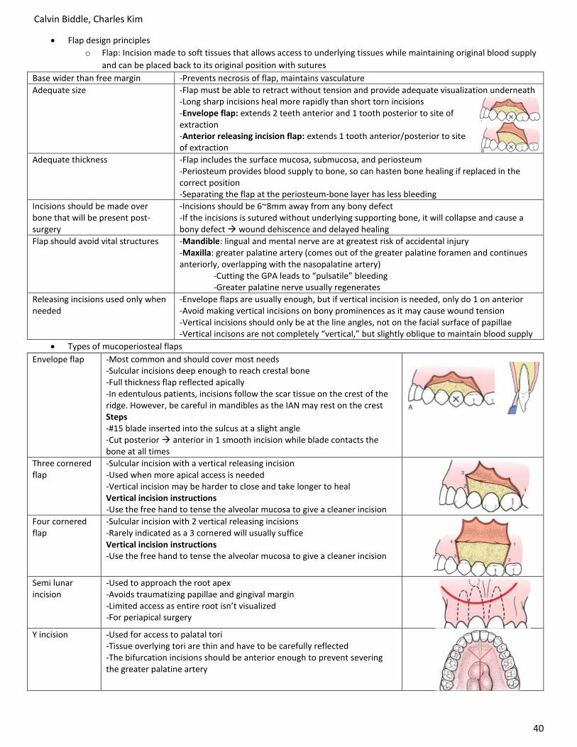
Adequate size -Flap must be able to retract without tension and provide adequate visualization underneath -Long sharp incisions heal more rapidly than short torn incisions -Envelope flap: extends 2 teeth anterior and 1 tooth posterior to site of extraction -Anterior releasing incision flap: extends 1 tooth anterior/posterior to site of extraction
Adequate thickness -Flap includes the surface mucosa, submucosa, and periosteum -Periosteum provides blood supply to bone, so can hasten bone healing if replaced in the correct position -Separating the flap at the periosteum-bone layer has less bleeding
Incisions should be made over bone that will be present post-surgery
-Incisions should be 6~8mm away from any bony defect -If the incisions is sutured without underlying supporting bone, it will collapse and cause a bony defect wound dehiscence and delayed healing
Flap should avoid vital structures -Mandible: lingual and mental nerve are at greatest risk of accidental injury -Maxilla: greater palatine artery (comes out of the greater palatine foramen and continues anteriorly, overlapping with the nasopalatine artery) -Cutting the GPA leads to “pulsatile” bleeding -Greater palatine nerve usually regenerates
Releasing incisions used only when needed
-Envelope flaps are usually enough, but if vertical incision is needed, only do 1 on anterior -Avoid making vertical incisions on bony prominences as it may cause wound tension -Vertical incisions should only be at the line angles, not on the facial surface of papillae -Vertical incisons are not completely “vertical,” but slightly oblique to maintain blood supply
• Types of mucoperiosteal flaps
Envelope flap -Most common and should cover most needs -Sulcular incisions deep enough to reach crestal bone -Full thickness flap reflected apically -In edentulous patients, incisions follow the scar tissue on the crest of the ridge. However, be careful in mandibles as the IAN may rest on the crest Steps -#15 blade inserted into the sulcus at a slight angle -Cut posterior anterior in 1 smooth incision while blade contacts the bone at all times
Three cornered flap
-Sulcular incision with a vertical releasing incision -Used when more apical access is needed -Vertical incision may be harder to close and take longer to heal Vertical incision instructions -Use the free hand to tense the alveolar mucosa to give a cleaner incision
Four cornered flap
-Sulcular incision with 2 vertical releasing incisions -Rarely indicated as a 3 cornered will usually suffice Vertical incision instructions -Use the free hand to tense the alveolar mucosa to give a cleaner incision
Semi lunar incision
-Used to approach the root apex -Avoids traumatizing papillae and gingival margin -Limited access as entire root isn’t visualized -For periapical surgery
Y incision -Used for access to palatal tori -Tissue overlying tori are thin and have to be carefully reflected -The bifurcation incisions should be anterior enough to prevent severing the greater palatine artery
Calvin Biddle, Charles Kim
41
o Once the incisions are made, reflection needs to be done
▪ Insert the sharp end of a #9 periosteal elevator into the papilla and start reflecting
▪ Lateral turning will pry the papilla from underlying bone
▪ If it is difficult to reflect, it means your reflection was inadequate, so go back to incising
▪ Once marginal part of the flap has all been reflected, use the broad end of the #9 to reflect the flap to its
desired length
▪ Flap can be retraced by pressing the retractor on bone, perpendicular to it. Wide flaps can be retracted
using a Seldin, Minnesota, or Austin retractor
▪ There should be no tension in the flap when you retract
• Principles of suturing
o Functions of suturing
▪ Approximates wound edges faster healing and less need for healing by secondary intention
▪ Hemostasis prevents hematoma formation and acts as a tamponade to oozing blood
▪ Holds a soft tissue flap over bone and maintains bone’s vitality
▪ Helps maintain a blood clot in the alveolar socket figure 8 suture is used to accomplish this o Instrumentation
▪ Thumb and ring finger is used to control the needle holder
▪ Suture needle (1/2~3/8 semicircles work) with a reverse cutting edge
▪ Suture material
• Technique to suturing
o General principles
▪ Suture is first passed through the mobile side (usually facial) released
regrasped between tissues passed through the attached side
• #9 periosteal elevator can be used to slightly elevate the tissues on
the more attached side to allow easy needle passage
▪ Needle should enter the mucosa at a perpendicular angle
▪ There should be a minimum of 3mm of tissue around the suture. <3mm risks
tearing of tissue
▪ There should be no blanching of tissues when sutured
▪ Knot should be tied to the side of the incision, not right above it
▪ Length of suture left after tying everything down should be <1cm
▪ If an incision needs multiple sutures, start with the most mobile site. This is
seen in the picture of suturing a vertical releasing flap
o Types of sutures
Simple interrupted Surgeon’s knot
-First knot is a double loop, to give enough friction until the second knot can be applied -Second knot is a single loop the other way. At this stage, it is completed -Most surgeons like to add a third knot with a single loop as well
. Figure of 8 suture -Holds clots in after extractions
Continuous simple suture
-Can efficiently accomplish closure in long incisions -Knots don’t have to be made for each suture -Downside: if one suture comes loose, everything falls apart -Suture the first socket like a simple interrupted, but continue the long end to the next socket
Locking continuous simple suture
-Long end of suture is passed underneath the loop before being pulled through tissue
.
Calvin Biddle, Charles Kim
42
o Removing sutures
▪ Non resorbable sutures should be removed after 5~7 days as it is an infection risk afterwards
▪ Suture should be cut and pulled towards the incision, not away. This is to prevent reopening incision
• Open extraction of single rooted teeth
o Create flap using principles discussed above
o Determine if bone removal is necessary
▪ First option: try to seat forceps as apically as possible, without any bone removal
▪ Second option: grasp a bit of buccal bone, which will fracture off with the tooth
▪ Third option: straight elevator oriented to long axis of tooth pushed into the PDL space and wiggled
▪ Fourth option: removal of bone
o Removal of bone
▪ Use a surgical handpiece with irrigation to remove the buccal bone
▪ Width of buccal bone removed = M-D width of tooth
▪ Height of buccal bone removed = 1/2~2/3 length of root
▪ Once removed, use straight elevator or forceps to remove tooth
▪ If there is still difficulty, drill a 3mm diameter hole into the root as its most apical part
• This is to be used as a purchase point for elevation with a Crane pick
• Open extraction of mandibular molars
o Create flap using principles discussed above
o Conventional method
▪ Section the tooth into a mesial half and distal half with a long #8 straight bur
▪ Some of the inter-radicular crestal bone may be drilled during this pro cess
▪ Luxate and elevate with a small straight elevator
▪ Use forceps to remove the fragments
o Alternative method
▪ With the flap open, remove buccal bone until furcation is
visible
▪ Bur is used to section mesial root from the tooth ▪ Crown + distal root are removed in one piece with #17
forceps ▪ Remaining mesial root is removed with a Cryer elevator
o If crown is missing
▪ Separate roots if they aren’t separated already
▪ Use a straight or triangular elevator to expand/deliver, and a
Cryer to deliver ▪ If this does not work, then remove more bone or create a
purchase point on the root
o Smooth rough areas with a bone file and irrigate
• Open extraction of maxillary molars
o Create flap using principles discussed above o Remove crestal bone to expose trifurcation area
o Section away the MB and DB roots, leaving just the crown attached
to the palatal root
o Bucco-occlusal forces are used to deliver the crown + palatal root
▪ Avoid palatal force as it may break the root
o Small straight elevator + Cryer are used to luxate and deliver roots
o Minimize apical forces due to proximity of the sinus
o If the crown is missing
▪ Same principles, but remove buccal roots before palatal
Calvin Biddle, Charles Kim
43
• Removal of root fragments
o Irrigate to get visualization
o Closed techniques
▪ First, try to remove by suction as root may be loose in its socket
▪ If unsuccessful, use a root tip pick wedged into the PDL and wiggled
▪ Avoid excessive apical (risk of sinus perforation) or lateral forces (risk of damage) o Open techniques
▪ Open flap
▪ Option 1: Remove buccal bone and deliver root through buccal opening
▪ Option 2: Remove buccal bone only at the apex of the root to create a “window.” Small
elevator or root tip pick is used to push root through the window. This may be preferable
as it leaves buccocrestal bone intact
• 3 corner flap may be used in this case as flap needs to be very apically extended
• Leaving root fragments
o When closed approach is unsuccessful and open approach is too traumatic
o Weigh the following factors:
▪ Destruction needed to remove root
▪ Risk of damage to vital structures like the IAN
▪ Risk of displacement to maxillary sinus
o Conditions to leave a root tip
▪ Fragment is <4~5mm
▪ Fragment is deeply embedded in bone, such that it will not expose in the future and impair prostheses
▪ Tooth must not be infected with no radiolucency around the apex
o Patient should be informed why the root was left
o Radiographs should be taken and followup done
• Multiple extractions in an appointment
o Maxillary extracted first
▪ Infiltration LA doesn’t last as long
▪ Debris from maxillary could fall into mandibular, if mandibular was done first
▪ Maxillary teeth are removed mostly by buccal force and less tractional force. Doing this first means
mandibular teeth are at less risk of damage
▪ One disadvantage: if bleeding isn’t controlled, could affect visualization in mandibular
o Most posterior teeth extracted first
o Most difficult (canines) extracted last
o Techniques
▪ All teeth should be luxated before removal because you can use the adjacent tooth can be used as an
anchor for luxation. Damage to the adjacent tooth is no concern as it will be extracted as well
▪ Make sure to remove sharp ledges of bone and granulation tissue
▪ If flaps overlap when suturing (seen in severe periodontitis), trim the gingiva
▪ Don’t try to suture the sockets shut
Calvin Biddle, Charles Kim
44
Management of impacted teeth
• Impacted teeth
o Tooth fails to erupt due to blockage form adjacent teeth, dense overlying bone, excessive soft tissue, or a genetic
abnormality
o Most common reason is inadequate arch length and space
o Usually retained for life
o Most common: 3rd molars > maxillary canines > mandibular premolars
o Removal difficulty increases with age, bone density, and completeness of root formation
• 3rd molar impactions
o Average age for complete eruption is 20, but may happen until 25
o Initially, the 3rd molar erupts horizontally, but orients vertically as the jaw grows and more room is made
o Failure of this conversion can cause impaction, or lack of room can cause impaction
o What if room is made by extracting the 7’s? if this happens after 20 y.o., bone is likely too hardened to allow the
3rd molar to orient to normal
o 3rd molar removals are better if done early as young people can heal better, higher chance of healing after a nerve
injury, and have less dense bone
o Ideal time for impacted 3rd molar extractions is when the roots are 1/3 ~ 2/3 formed (17~20 yo)
• Indications for removal of impacted teeth
o General rule is that all impacted teeth should be removed unless removal is contraindicated
Prevention of periodontal disease
-Teeth adjacent to impacted teeth are predisposed to periodontal disease -Impacted teeth like 3rd molars can decrease amount of bone on the 2nd molar -Attachment on 2nd molar has apical migration and a deep pocket can form -More serious in maxilla as the distal furcation is more easily reached
Prevention of caries -Distal aspect of 2nd molar and impacted 3rd molar can have caries
Prevention of pericoronitis
-Pericoronitis = infection of soft tissue in operculum of a partially impacted tooth -Bacteria responsible = streptococci spp and other anaerobic bacteria -Host defenses can control infection, but can become severe (to the point of hospitalization) in immunocompromised and progress to facial space infections -Can be associated with trismus, fever, facial swelling, pain, malaise -Food can also become trapped under the operculum -Can be managed with irrigation with hydrogen peroxide, CHX, iodophors, saline, or surgical operculectomy, but all options are temporary -Definitive management is to remove the 3rd molar, but should not be removed until all signs of infection have resolved -Having one episode increases risk for recurrence
Prevention of root resorption
-Impacted tooth can press against the roots of an erupted tooth -Roots can resorb -Seen in primary teeth, as they exfoliate
Impacted teeth under a prosthesis
-Teeth need to be removed prior to prosthetic fabrication -If it’s unerupted, keep in mind that alveolar resorption may cause it to be erupted in the future, and cause ulcerations + odontogenic ifnection
Prevention of odontogenic cysts and tumors
-Impacted teeth may still have follicles attached, which can degenerate into a dentigerous cyst or keratocyst -If follicular space > 3mm, preoperative diagnosis of a dentigerous cyst is reasonable -Most common tumor is the ameloblastoma, but overall incidence is not very high
Treatment of unexplained pain
-Rule out myofascial pain dysfunction syndrome and other pain disorders -Delayed removal increases chances of TMD
Prevention of jaw fractures
-Impacted 3rd molars are surrounded by bone which weakens mandible
Orthodontic treatment -In some ortho plans, 1st and 2nd molars need to be retracted -3rd molars may need to be removed to give room for this movement
Optimal periodontal healing
-Optimal healing depends on extent of bone loss on distal of 2nd molar and age of patient -For a patient >30 with hard impaction of 3rd molars, may be better to leave in place as removal will cause more bone loss in the end
Calvin Biddle, Charles Kim
45
• Contraindications of tooth removal
Extremes of age -50+ yr old will have discomfort for 4~5 days, while 18 yr old will have it for 1~2 days -Older patients have denser bone which complicates removal -Leave tooth alone unless pathology develops (monitor every 1~2 years)
Compromised medical status -Cardio or respiratory compromise -Immunosuppressed or has a coagulopathy
Probable damage to adjacent structures
-If there is a bridge or a tooth that will likely be lost due to removal of impacted tooth, then avoid
• Classification of mandibular third molar impactions
o Classification by angulation (least difficult to most difficult)
▪ Distoangular is most difficult because it has a withdrawal pathway that runs into the ramus
▪ Teeth can also be angled buccal/lingual/palatal, but they are less significant and doesn’t alter the
approach to surgery. Just take a CBCT to see how it relates to vital structures and nerves
Mesioangular Horizontal Vertical Distoangular
43% 3% 38% 6%
o Classification by relationship of molar to anterior border of ramus
▪ Called the Pell and Gregory classification, and ranked from 1 to 3
▪ 1 has best accessibility, 3 has the worst
1 2 3
-Crown completely anterior to ramus
-1/2 of crown is embedded in ramus
-Crown is completely embedded in ramus
o Classification by relationship to occlusal plane
▪ Also called the Pell and Gregory classification, and ranked from A to C
A B C
-Crown at level with occlusal plane of second molar
-Crown between occlusal plane and cervical line of second molar
-Crown below cervical line of second molar
o Classification by type of impaction
▪ Soft tissue impaction: superficial crown is only covered by soft tissue
▪ Partial bony impaction: superficial part covered by soft tissue, but HOC of tooth under bone
o Trauma and resulting inflammation may involve muscles of mastication trismus
o Trismus may also result from LA injections puncturing through muscles
o Medial pterygoid muscle is most commonly injured
o Not usually severe and does not hamper patient’s normal activities
o May last for a week
• Ecchymosis
o Blood can ooze submucosally and subcutaneously
o Seen in older patients due to ↓ tissue tone, capillary fragility, weaker intercellular attachments
o Does not increase pain or chances of infection
o Usually 2~4 days after surgery and resolves in 7~10 days
• Post op follow up
o Follow up should be done after 1 week, or whenever patient has any issues
o Sutures removed if needed
o Monitor for signs of infection
o If pain decreases, but comes back on day 3/4 without swelling or any signs of infection, most likely dry socket
▪ Usually confined to lower molar sockets and does not represent an infection
▪ Seen more in smokers and women taking oral contraceptives
▪ No blood clot seen, and smells bad
▪ Treatment:
• Will require many appointments
• Irrigation with monoject syringe
• Apply medicated dressing (contains eugenol to kill nerves, benzocaine to numb area, balsam of
Peru as a carrying agent)
• Change dressing every 1~2 days and remove when pain subsides
Prevention and management of extraction complications
• Soft tissue injuries
o Prevention
▪ Pay strict attention to soft tissue injuries
▪ Develop adequate sized flaps
▪ Use minimal force for retraction
o Management
Injury Due to Management
Tear of mucosal flap
-Most common -Due to inadequate sized flap, and usually occurs on one end of the incision
-Create a releasing incision if flap begins to tear -Reposition after surgery: healing will be delayed, but will adequately heal -If edges are jagged, consider excising edges to leave smooth wound edges
Puncture wound
-Straight elevators or periosteal elevator slippage
-Usually left unsutured and left to heal with secondary intention -If infected, it will conveniently drain
Stretch or abrasion
-Shank of bur or metal retractors on tissue
-Keep area clean with regular mouth rinses -Heals in 4~7 days depending on severity -If abrasion happens on skin, antibiotic ointment should be used to keep area moist and increase comfort -Skin abrasions take 5~10 days to heal -May scar or discolour
Calvin Biddle, Charles Kim
50
• Problems with tooth being extracted
Problem Due to Management
Root fracture -Most common -Long, curved roots in dense bone
- Consider possibility of root fracture -Use open extraction technique if there is a high possibility of fracture -Do not use strong apical force on a broken root
Root displacement
-Commonly the maxillary root into the sinus
-Determine size of root in sinus, if the tooth was infected, and status of sinus (healthy or chronically infected?) If the fragment is small and no infection is present: -Take a radiograph to document and measure size -Irrigate small opening and suction to try to dislodge fragment -If this does not work, no further attempt should be made as the tip will likely fibrose into sinus membrane with no issues -Inform patient and monitor -Close oro-antral communication with figure 8 suture over socket -Give ABX and nasal spray to decrease chance of infection If the fragment is large or an infection is present: -Caldwell Luc approach or endoscopic approach into the sinus via the canine fossa is indicated
Displacement of tooth structure with 3rd molars
-Elevator use forcing tooth posteriorly
Maxillary 3rd molar displacement: -Tooth displaces into infratemporal fossa lateral to the lateral pterygoid plate and inferior to the lateral pterygoid muscle -Try to access with a hemostat and remove -If tooth cannot be retrieved, close incision and stop operation -Patient should be informed it will be removed later -Prescribe ABX to prevent infection -Tooth will fibrose and stabilize in its position -Refer to OMFS Mandibular 3rd molar displacement: -Fractured mandibular roots may displace through the submandibular space -Prevent by avoiding all apical force when removing mandibular roots -Try to remove by pushing lingual aspect of mandible with index finger
Tooth lost into pharynx
-Treating tooth like a rubber dam clamp
-Turn patient towards surgeon with mouth facing floor -Encourage spitting and coughing -If there is no respiratory distress, tooth was most likely swallowed and will pass through GI tract in 2~4 days -If there is respiratory distress, tooth was most likely aspirated. Manage airway and breathing, bring patient to emergency room. Can be removed via bronchoscopy
• Injuries to adjacent teeth
Problem Management
Fracture or dislodgement of an adjacent restoration
-Most common injury to adjacent teeth -Most common would be excessive tractional forces being used on mandibular teeth. When the tooth suddenly detaches, the forceps fly up and injure maxillary teeth -Prevented with more B/L luxation so less tractional forces are needed -Make sure fractured pieces don’t get aspirated/swallowed
Luxation of adjacent tooth
-Common in crowded areas like mandibular incisors -Use thin forceps like #286 to extract these areas and not luxate adjacent teeth -If adjacent tooth was severely luxated, usually it can be left alone to stabilize -Ensure tooth is not in hyperocclusion or traumatic occlusion -Tooth can be stabilized with semi rigid fixation (silk suture crossing occlusal table and sutured to adjacent gingiva) -Tooth can be stabilized with rigid fixation (wires and arch bars), but may cause root resorption or ankylosis, so avoid
Extraction of wrong tooth
-Quickly replaced into socket -If extraction was due to ortho, ask orthodontist if that removed tooth can be a suitable substitute -If not, wait 4~5 weeks for replanted tooth to attach, then proceed with correct tooth extraction
Calvin Biddle, Charles Kim
51
• Injuries to osseous structures
Problem Management
Fracture of alveolar process
-Bone fractures when trying to expand the socket, most likely due to excessive force with forceps -Bone fragments stay attached to tooth -Can be prevented by opening flap + removing some bone before attempting expansion -Most likely sites: max buccal plate > floor of mx sinus > mx tuberosity > buccal of mand anteriors -Do not replace bone, just close up the soft tissue as best as you can -Smooth sharp edges along fracture line -Not ideal for prosthetics -If you feel the buccal bone fracture but it’s still attached to periosteum, it can be saved -Stabilize tooth, use #9 to separate fractured bone from tooth surface -Fractured bone will be attached to soft tissue via periosteum -Reposition and suture will heal in favourable ridge form
Fracture of maxillary tuberosity
-Max tuberosity is important for complete dentures -Removal of tuberosity may cause and oro antral opening -Like mentioned above, try to save the tuberosity by keeping it attached to periosteum -If the tuberosity is too mobile and cannot be dissected from tooth: -Splint tooth and defer extraction for 6~8 weeks reattempt via open extraction -OR, section crown off and leave tuberosity + roots to heal for 6~8 weeks then remove roots -Be cautious if molar was infected prior to extraction -If the tuberosity completely separated -Smooth edges of remaining bone -Reposition and suture remaining soft tissues -Check for oro antral communication
-Area of sensation is small, and reinnervation happens quickly -Not much of a bother to patient
Mental nerve -Damaged when doing mandibular premolar procedures -If planning a 3 corner flap, must be done well anterior to mental nerve area -Recovery in few days ~ weeks
Lingual nerve -Rarely regenerates if severely traumatized -Could innervate retromolar pad -Avoid cutting at lingual aspect of posterior mandible, incline it buccally
IAN -Most commonly injures due to 3rd molar
Injuries to TMJ -Can happen if mandible is not supported during molar extraction -Manage with soft diet, moist heat, resting of jaw -Analgesics: 600~800 mg ibuprofen q4h or 500~1000 mg acetaminophen q4h for several days
• Oro antral communications
o Most common complications: post operative sinusitis or formation chronic oro antral fistula
o Diagnosing communication
▪ Check the extracted molar to see if there is bone attached to it
▪ Ask patient to blow nose air and blood will bubble through communication. Not to be done with
excessive force because it may tear open a closed membrane
o Management
▪ If there is no bone on tooth, communication is <2mm no Tx is necessary
• Inform patient to be careful and prevent dislodgement of clot
▪ Granulation tissue removed from periapical region and adjacent teeth
▪ Clamp bleeding arteries with hemostat and ligate with non resorbable suture
▪ Crush bone to occlude bleeding vessels within bone
▪ Get patient to bite on wet gauze for 30 minutes
▪ If still persistent, then use hemostatic agents like one of the below:
• Gelfoam in socket held in via figure of 8 suture
• Surgicel packed under pressure (better than Gelfoam, but may delay healing of socket)
• Gelatin sponge saturated in topical thrombin
• Collagen materials
o Controlling secondary bleeding
▪ Rinse mouth with chilled water and place damp gauze for 30 minutes
▪ If bleeding persists, repeat cold rinse and bit on damp teabag
▪ If bleeding persists, return to dentist
▪ At the dentist’s office:
• Suck away all fluids and remove large liver clots if present
• Determine source of bleeding and apply damp gauze for 5 minutes
• If still persistent, give LA and curette clot out of socket and clean it all out
• Check for diffuse oozing, arterial bleeding, or bleeding from bone
• Add hemostatic agent to socket, hold it in with figure of 8 suture, and wait 30 mins
• If still no hemostasis, consult hematologist
• Epinephrine may stop bleeding acutely, but may cause rebound bleeding when effects wear off
• Reasons for delayed healing
Infection -Most common cause of delayed healing -Usually associated with surgeries involving flaps and bone removal, rarely due to routine dental extraction -Managed with antibiotics
Wound dehiscence
-Separation of wound edges due to inadequate bone support underneath or wound under tension -Most commonly involves internal oblique ridge showing through the lingual flap
Dry socket (alveolar osteitis)
-Delayed healing not associated with infection -Pain happens on day 3~4, usually with lower molars -Tooth socket appears empty with partially/completely lost clots and bony surfaces exposed -Exposed bone causes dull aching pain that radiates to the ear -May be associated with foul smell or taste -Cause is unclear. Maybe due to high fibrinolysis activity (due to subclinical infection, bone marrow inflammation, etc) breakdown of clot
Prevention Treatment
-Minimize trauma -Small amount of topical tetracycline on gelatin sponge ↓ incidence of dry socket -CHX may help
-Relieve pain during healing period -Irrigate with saline suction dry strip of iodoform gauze soaked/coated in medication is inserted into wound -Preparation is eugenol, benzocaine, and balsam of Peru -Pain should relieve in 5 minutes, dressing changed every 2 days for 3~6 days -Irrigate socket at each appointment -Do not place dressing once pain is resolved
Fractures of mandible
-Associated only with impacted molar removal -Usually due to excessive force, usually with elevators -Fracture must be adequately reduced and stabilized
Calvin Biddle, Charles Kim
53
Management and prevention of odontogenic infections
• Odontogenic infections originate form caries, periodontal disease, or pulpitis
o Most frequently due to normal flora bacteria (aerobic/anaerobic gram + cocci, anaerobic gram – rods)
o As infection progresses deeper, pathogenic bacteria in flora flourish more
o Odontogenic infections involve 5~8 species of bacteria, rarely just 1
o Periapical infection results if there is pulpal necrosis and bacterial invasion (more common)
▪ Especially deep caries causing bacterial infiltration into pulp periapical lesion
▪ Infection will spread in every direction, but follow the path of least resistance
▪ Will break down cancellous bone and then the cortical bone (if thin)
▪ Antibiotics may arrest, but will not cure. Must extract or do root canal therapy
o Periodontal infection results if there is inoculation of bacteria in a deep pocket
• Species of bacteria
o Aerobic (6%)
▪ 65% are S. milleri group (S. anginosus, S. intermedius, S. constellatus)
▪ 75% involve gram – rod (porphyromonas) and 50% involve gram – rod (fusobacterium)
▪ Gram – cocci and gram + rods play a minor role
o Aerobic + anaerobic (44%)
• Pathogenesis
o Initial inoculation by S. milleri group
o S. milleri makes hyaluronidase breaks down CT initiates cellulitis
o Environment turns into lower pH and less O2 which favours growth of
anaerobes
o Bacteria now synthesize collagenases to cause necrosis and abscess
formation
o This is why early infections are associated with cellulitis (aerobic) and
late infections are associated with abscesses (anaerobic) o If abscesses can be drained, they will resolve
• Spread of periapical infections
o Depends on:
▪ Thickness of bone near apex of tooth
• Labial bone is usually thinner, so infection will spread labially
• If the tooth is severely proclined, infection will spread palatally
▪ Relationship of bone to muscles
• Infection may spread to different spaces, depending on muscle positioning
• If infection is superior to buccinator, it will infect the buccal space
• If infection is inferior to buccinator, it will infect the vestibular space
o Site specific spreading patterns
Anterior Posterior
Maxillary -Infection usually goes buccally and below
muscles vestibular abscess most common -Severely proclined incisors or thick labial bone may cause palatal infection -Canine may cause infection to erode superiorly, causing infra-orbital space infection
-Palatal root of molar will infect palatal space -More commonly, infection will go buccally and superior to buccinator buccal space infection
Mandible -Infection usually goes buccally and above muscles vestibular abscess most common
-1st molar: drains buccal or lingually -2nd molar: usually lingually -3rd molar: almost always lingual -May go above mylohyoid muscle (submandibular space infection) or below (sublingual space infection)
Calvin Biddle, Charles Kim
54
• Vestibular space abscesses
o Most common odontogenic fascial space infection
o If the abscess ruptures and drains, infection will not cause symptoms and continue draining. This forms a chronic
sinus tract
o If the opening closes, infection will recur
• Treatment of odontogenic infections
Determine severity -Obtain history of lesion and symptoms (duration, rapidity, pain, swelling, erythema, warmth, loss of function) -Perform a physical exam (temperature and HR may be elevated in infection -Odontogenic infections can cause upper airway obstruction due to spread in deep tissues -Normal vitals + mild temp elevation readily treated -Abnormal vitals + elevated temp/pulse/RR refer to OMFS -Examine areas of swelling, sinus tracts, and look for causes of infection (caries, periodontal abscess, fractures) -Determine if this infection is in the inoculation, cellulitis, or abscess stage (see table above)
Evaluate state of patient’s host defense
-Determine if patient has one of these diseases: -If present, these patients must be referred to OMFS as infection is more urgent
Determine if dentist can treat or must be referred
Criteria for referral to hospital due to airway compromise: 1. Rapidly progressing infection (rapid growth over 1~2 days) 2. Difficulty breathing 3. Dysphagia or drooling (narrowing of oropharynx) Criteria for referral to OMFS/hospital: -Extraoral involvement of swelling -Temperature > 101 F hospital -Trismus (20~30mm = mild, 10~20mm = mod, <10mm = severe) -Mod/severe trismus indicates infection in masticator space, lateral pharyngeal space +/- retropharyngeal space -Toxic facial appearance (glazed eyes, open mouth, dehydration, sick appearance) -Compromised host defenses
Treat infection surgically
-Remove cause of infection and provide drainage for pus and necrotic debris -Typical presentation: carious tooth with periapical lesion and a small vestibular abscess -Pulpectomy will remove infection and limits drainage through apical foramen -If tooth is non restorable, should be extracted -Incision and drainage may be needed to remove infection beyond PA region Incision and drainage -Reduces pressure on the tissues, allowing blood to enter freely and resolve infection -Decreases bacterial load and necrotic debris -Not required for inoculation stage, only for abscess or cellulitis
Patient support -Encourage drinking fluids and frequent urination -High calorie nutritional supplements -Analgesics for pain relief
Prescribe antibiotic if necessary
-See below
Followup with patient frequently
-Reassess 2~3 days after surgery. Swelling should be significantly resolved if successful -If tx was a failure, assess why (see below) -Assess for toxicity related symptoms of drugs -Monitor for recurrent infection
Calvin Biddle, Charles Kim
55
• Steps in doing an incision and drainage
o Regional nerve block or infiltration
▪ Infected tissues may need a block to get
adequate anesthesia
o Consider culture and sensitivity if
▪ Collect specimen with large gauge needle
(aspirate 1~2mL of fluid)
▪ Transfer specimen to anaerobic/aerobic culturettes
o Incision on maximum site of swelling
o Insert hemostat into abscess and open/close in many directions
o Disinfect and dry the surface mucosa
o Aspirate pus into suction
o Small drain can be inserted to maintain opening
▪ Use a ¼ inch Penrose drain or small strip of rubber dam or glove
▪ Needs to reach depth of abscess
▪ Suture drain to edge of incision with non resorbable suture
▪ Keep in drain for 2~5 days
▪ Remove by cutting suture
• Is an antibiotic needed?
o Assess 3 factors: seriousness of infection, if surgical approach may be enough, and state of host defenses
o Use ABX if the host defense cannot contain the infection
o ABX will not hasten wound healing and help with non-bacterial conditions
Calvin Biddle, Charles Kim
56
• Deciding which antibiotic to give
o Empirical therapy is used (following fixed guidelines rather than waiting for a culture test to come back) because
odontogenic infections are highly predictable
▪ C&S testing may be done if needed (see page above for indications)
o Antibiotics we can use: penicillin, amoxicillin, clindamycin, azithromycin, metronidazole, moxifloxacin
▪ All work against aerobic/facultative streptococci and oral anaerobes except metronidazole (metro only
covers obligate anaerobes)
▪ Drug of choice is typically penicillin. If allergic, clindamycin or azithromycin are used
▪ Amoxicillin may be preferable over penicillin due to less frequent dosing
▪ 3~4 day course of penicillin has the highest compliance
▪ Peak plasma level should be 4-5x the minimum inhibitory concentration for the bacteria
o Factors in deciding which drug to give
▪ More frequent dosing = lower compliance from patient
▪ Use narrowest spectrum antibiotic
• Penicillin will kill strep and oral anaerobes, but little effect on staphylococci on skin and GI flora.
This prevents formation of resistant bacteria
Definition Which drug to use
Simple infection
-Swelling limited to alveolar process and vestibular space -First attempt at treatment -Patient not immunocompromised
▪ Use antibiotic with lowest toxicity and side effects
ABX Incidence of toxicity Description
Penicillin Low -Allergy in 2~3%
Clindamycin Low -Severe diarrhea, pseudomembranous colitis in ill and debilitated patients -Due to overgrowth of C.difficile and associated toxins
Azithromycin Low -Best combination of effectiveness + low toxicity
Erythromycin -No longer considered effective on oral pathogens
Moxifloxacin High -Best effectiveness against oral pathogens -Muscle weakness, mental clouding, potentially fatal drug interactions -Contraindicated in <18 and pregnants as it interferes with cartilage growth -Reserved for severe/recalcitrant infections
Cephalosporin Mild -Not effective for oral cavity -Could have cross sensitivity to penicillins
Tetracycline Mild -Not effective for oral cavity (except at high local doses) -Nausea, cramps, diarrhea, photosensitivity -Contraindicated in pregnants as it discolours infant teeth (chelates with calcium incorporated into teeth)
Metronidazole Mild -GI disturbances -Disulfiram effect (severe SE’s with alcohol)
▪ Use bactericidal drugs if possible
• Bactericidal will kill the bacteria. Bacteriostatic antibiotics only hamper with their reproduction
and growth, and relies on immune system to kill off bacteria
▪ Cost varies widely. Consider older antibiotics as they are cheaper
o Other advice to patient regarding antibiotics
▪ Finish the whole course, or throw out if patient decides to stop
▪ Don’t self medicate with leftover pills in the future, can be dangerous
Calvin Biddle, Charles Kim
57
• Reasons for Tx failure
o Inadequate surgery: infection may extend more than previously thought. May need to admit to hospital for airway
security, further surgery, or IV abx
o Depressed immune system
o Foreign body presence: take radiograph to make sure abscess is cleared out
o Antibiotic issues: noncompliance, dose too low, wrong antibiotic used, blood supply didn’t deliver enough
antibiotic to site of infection
• Prevention of infection – antibiotic prophylaxis
o Pros/cons
Pros Cons
-Reduced post op morbidity by avoiding infection -Appropriate ABX prophylaxis may reduce costs -Short term administration, less ABX used overall
-Inappropriate use resistant bacteria formation -Alters host flora to favour resistant bacteria -Risk of infection is so low, not really needed -May encourage poor infection control by dentist -Unnecessary cost and toxicity to patient
o Factors at play when deciding to give antibiotic prophylaxis
Procedure has high risk of infection
-Clean surgery has infection rate of 3%. Use prophylactic ABX if risk is >10% -When prophylaxis is indicated: -Cellulitis or abscess present (shows that inoculum is significant) -Dental operations lasting >4 hours (rare) -Presence of foreign body, implant, or dead space -Immunocompromised* *Chemo/radio patients should have ABX prophylaxis during Tx + 1 year after Tx *Patients receiving kidney dialysis *Diabetic patients with HbA1c >7%
Pick correct antibiotic
-Consider proper coverage, narrow spectrum, least toxic, bactericidal -DOC is amoxicillin or penicillin clindamycin azithromycin
Antibiotic plasma concentration
-Must be higher than when used therapeutically (usually 2x) – must be enough at the tissues where surgery is being performed -Amoxicillin = 2g, clindamycin = 600mg, azithromycin = 250mg
Time antibiotic administration correctly
-<2 hours before surgery begins (1 hour oral, even less if IV) -ABX after surgery has decreased efficacy or no efficacy -During prolonged surgery, therapeutic doses are given more frequently (1/2 dose every 3 hours for penicillin and clindamycin)
▪ Conditions that need to be met for metastatic infection to occur
• There is a susceptible location for infection
• Bacterial seeding must take place
• Impairment of host local defenses
▪ Infective endocarditis
• Viridans group strep commonly found in IE, and associated with bacteremias
• Large number of case reports of patients getting IE after dental procedures
• Morbidity of ABX is low, while morbidity of IE is high
o IE patients will need high doses of ABX in a hospital and possible valve replacement
o Risk of recurrence of IE as well
• Only a small portion of IE cases are due to dental procedures and very few cases would have
been prevented by Abx prophylaxis even if it was 100% effective
Calvin Biddle, Charles Kim
58
• Guidelines for prophylaxis of infective endocarditis
o Which dental procedures require prophylaxis?
▪ Procedures involving manipulation of
gingival tissue or periapical regions of
teeth or perforation of oral mucosa
▪ Not needed for radiographs, placement
of prostheses, placement of orthodontic
appliances/brackets, shedding of primary
teeth, bleeding due to trauma, LA
injections through non infected mucosa
o Drug of choice
▪ 2g amoxicillin taken orally 30min~1h
before procedure
▪ Amoxicillin is well absorbed by GI tract
and provides higher, more sustained
plasma levels
▪ If allergic, give clindamycin 600 mg orally
1h before procedure
▪ If patient is taking penicillin daily for other
reasons (rheumatic fever), use clindamycin,
azithromycin, or clarithromycin
▪ If multiple rounds of prophylaxis are
needed due to multiple appointments,
space it out every 10 days so oral flora can
repopulate
• Evidence shows it may actually
take months to re-establish
normal flora again
o Non drug measures patient should take
▪ Excellent oral hygiene
▪ Incipient dental and periodontal disease
treated
▪ Mouth rinsed preoperatively with CHX
o Note that despite all these regimens and
precautions, it is not 100% effective at preventing
infective endocarditis
• Guidelines for prophylaxis of total joint replacements
o Recent literature states bacteremias from oral
procedures do not cause prosthetic joint infections
o Most dental procedures cause transient
bacteremias, too acute for prosthetic joint infection
o However, chronic septicemias like in odontogenic
infections may cause joint infection
o Up to clinician’s judgement to use when appropriate
• Other conditions that may need prophylaxis
o Coronary artery bypass graft – no need
o Pace makers – no need
o Coronary artery angioplasty +/-stent – no need
o Patient with non valvular cardiovascular devices – needed only when doing incision&drains of abscesses
o Renal dialysis – may be needed, contact nephrologist
o Hydrocephaly with ventriculoatrial shunt – may be needed, contact neurosurgeon
Calvin Biddle, Charles Kim
59
Complex odontogenic infections
• Infections can spread to deep fascial spaces
o “Fascial spaces” don’t exist in healthy people, and are
created with infections
o The tissue surrounding fascial spaces are loose and
cushioning. They can become very edematous and
recruit WBC’s in response to nearby infection
o Liquifactive necrosis of WBC and CT in this area leads
to abscess formation
o Will resolve if drained
• Spreading pattern for typical odontogenic infections
o Any tooth: vestibular, buccal space, subcutaneous
o Maxillary teeth: infraorbital, palatal, orbital, infratemporal spaces, and maxillary sinus
o Mandibular teeth: submandibular, sublingual, submental, and masticator spaces
• Spreading pattern into deep fascial spaces
o Lateral pharyngeal
o Retropharyngeal
o Carotid
o Pretracheal
o Brain or intracranial dural sinuses • These infections are classified on risk to threatening airway or affecting other vital structures
o Low severity – not likely threatening
o Moderate severity – may hinder airway via trismus or elevation of tongue
o High severity – directly compresses of deviates airway or likely damage to brain/lung/heart
• Will go over the main types of infection
Calvin Biddle, Charles Kim
60
Spreads to Infection origin About In
fect
ion
s fr
om
an
y to
oth
Buccal space -Any tooth
-Most commonly posterior maxillary teeth
-Part of the subcutaneous space (space that exists continuously from head to toe) -Bound by skin on lateral site and buccinator on medial side -Presents as swelling between zygomatic arch and inferior border of the mandible -Swelling over the zygomatic arch gives a dimpled appearance -Zygomatic arch and inferior border of mandible should remain palpable -Will drain spontaneously through the skin at its inferior extent near inferior border of mandible -If infection follows the buccal fat pad, it can spread to the superficial temporal space, infratemporal space, and periorbital space
Infe
ctio
ns
of
max
illar
y te
eth
Palatal space -Upper laterals or palatal roots of upper molars
-Perforates through the palatal bone and cause a palatal space infection
Infraorbital space
-Maxillary canine or spread from buccal space
-Thin space between levator anguli oris and levator labii superioris muscles -Canine apex can erode through thin alveolar space and enter this space -Swelling of anterior face obliterates nasolabial food -Drains to medial/lateral canthus (corners) of eye, depending on which side of the levator superioris muscle the infection spreads to
Infratemporal space
-Rarely infected -Usually due to maxillary third molar
-Space that lies posterior to the maxilla, bound medially by the lateral pterygoid plate, bound superiorly by base of skull -Space is continuous with the deep temporal space, and is considered the bottom of the deep temporal space -Space contains internal maxillary artery and pterygoid venous plexus -Infection can spread into cranial space by going from plexus emissary vein cavernous sinus -Veins here do not have valves, so infection can spread in other direction too
Infe
ctio
ns
of
max
illar
y si
nu
s Peri-orbital infection
-Rarely from a tooth infection
-Infection from maxillary sinus ethmoid sinus orbital floor periorbital -Even though 20% of maxillary sinusitis cases are odontogenic, getting a periorbital infection is rarely odontogenic
Cavernous sinus
-From peri-orbital infection
-Periorbital region ophthalmic vein superior orbital fissure cavernous sinus -Inflammation and clotting in cavernous sinus septic cavernous sinus thrombosis -Life threatening and requires aggressive treatment
Infe
ctio
ns
of
man
dib
ula
r te
eth
Space of body of mandible
-Buccal infection of mandibular teeth
-Sub-periosteal space infection (erodes through bone, but not periosteum) -Periosteum peels off the bony surface -Clinically looks like enlargement of the mandible, but regular shape-wise -If the periosteum is penetrated and goes below the buccinator, a buccal space infection will result (see above: buccal space)
Submaxillary space (sub mandibular + sublingual + submental)
Sublingual -Lingual infection of mand premolars or molars
-Infection from tooth goes through lingual bone and above mylohyoid muscle attachment -Intraoral swelling on floor of mouth, and raising of tongue -In the posterior region, the sublingual space communicates freely with the submandibular space can spread
Submandibular -Lingual infection of mand 3rd molars
-Infection causes triangular swelling (base at angle of mandible, apex at hyoid bone) -Infection from tooth goes through lingual bone and below mylohyoid muscle attachment -Infection is between mylohyoid and superficial layer of deep cervival fascia
Submental -Spread from submandibular infection
-Between anterior bellies of right and left digastrics and between mylohyoid and overlying fascia -Most commonly due to spread of infection from submandibular space -Rarely due to lingual infection of mandibular incisors
Calvin Biddle, Charles Kim
61
Infe
ctio
ns
of
man
dib
ula
r te
eth
Spreads to Infection origin About
Masticator space
-Most frequently involved in odontogenic infections requiring hospitalization -Mand 3rd molar most common
-Space is formed between anterior layer of deep cervical fascia and muscles of mastication -Fascia is extensive and is bound by the following boundaries: 1. Inferior border: inferior border of mandible 2. Lateral border: medial side of the masseter and temporalis 3. Medial border: pterygoid plates and sphenoid bone 4. Superior border: follows lateral side of temporalis, up to its attachment site on the cranium -This fascia forms 4 compartments:
Dee
p c
ervi
cal
fasc
ial s
pac
e in
fect
ion
s
Lateral pharyngeal space infection
Submandibular and sublingual space infections -These spaces join together in the posterior mylohyoid area, via a gap called the buccopharyngeal gap -Submandibular + sublingual infections can pass through this gap and enter the lateral pharyngeal space (deep fascial space of neck) -Submandibular infections can go around the posterior belly of the digastric and enter the lateral pharyngeal space directly
-Serious life threatening sequelae: blockage of airway, invasion of vital structures via the mediastinum Anatomy of the lateral pharyngeal space -Extends from sphenoid bone to hyoid bone -Lateral border: medial pterygoid -Medial border: superior pharyngeal constrictor -Anterior border: pterygomandibular raphe -Posterior border: retropharyngeal space -Further separated to anterior compartment (contains loose CT) and posterior compartment (contains carotid sheath, CN9, 10, 11, 12). The styloid process and attached muscles separate these 2 sub-compartments Clinical presentation -Inflammation of medial pterygoid -Lateral swelling of neck (angle and SCM area) -Swelling of lateral pharyngeal wall -Dysphagia -Increased temperature -Most serious complications are due to compression of posterior compartment (thrombosis of internal jugular vein, erosion of carotid artery, interference of CN’s)
Retro-pharyngeal space infection
Spread from lateral pharyngeal space infection
-Behind the posterior pharynx, between C6 and T4 -Anterior border: pharyngeal constrictor mucles, retropharyngeal fascia -Posterior border: alar fascia -Contains only loose CT and lymph nodes (infection spreads easily) -Infection here can rupture alar fascia enters posteriorly to “danger space” -Could also enter the mediastinum
Danger space infection
Spread from retro-pharyngeal space infection
-Anterior border: alar fascia, posterior border: prevertebral fascia -Extends from base of skull to diaphragm -Prevertebral space is rarely involved (usually due to osteomyelitis of vertebrae) -Could also enter the mediastinum
Space Anatomy Due to About
Sub-masseteric space
-Between masseter and lateral surface of ascending ramus
-Spread from buccal space -Pericoronitis of mand 3rd -Mandibular angle fracture
-Masseter will be inflamed and swollen -Moderate~severe trismus
Pterygo-mandibular space
-Between medial pterygoid and medial ascending ramus -Where we aim for IANB
-Mand 3rd molar infection -None~minor facial swelling, but severe trismus -May be swelling/erythema of anterior tonsillar pillar and displacement of uvula -Do CT scan -Risk of airway blockage
Superficial temporal space
-Temporalis and temporalis fascia
-Rarely becomes infected, only in severe cases -Swelling in temporal region (superior to zygomatic arch and posterior to lateral orbital rim), but not very big due to how tight the attachment of the temporalis is
Deep temporal space
-Between temporalis and skull
*When submasseteric space and temporal spaces are infected together, swelling is an hourglass shape
Calvin Biddle, Charles Kim
62
• Ludwig’s angina
o Bilateral perimandibular infection that commonly spreads posteriorly to deep fascial spaces of the neck as well
o Rapidly spreading cellulitis that can obstruct the airway high risk of death
o Most commonly due to odontogenic infection
o Clinical presentation:
▪ Severe swelling (elevation of tongue, induration of submandibular region above hyoidbone
▪ Drooling, dysphagia
▪ Trismus
▪ Dyspnea, anxiety
o Management: secure airway, incision & drain, antibiotic therapy
• Mediastinum infection
o Submandibular/sublingual lateral pharyngeal retropharyngeal +/- danger space mediastinum
o Mortality is high
o Involves heart, phrenic nerve, vagus nerve, trachea, bronchi, esophagus, great vessels
o Mediastinitis compresses heart and lungs
▪ Interferes with neural control of heart and lungs
▪ Could rupture into lung, trachea, or esophagus
o Requires open surgical drainage and follow up with CT scans
• Necrotizing fasciitis
o Flesh eating bacterial infection
o Rapid spread of infection on superficial surface anterior, or investing layer of deep fascia (like just deep to the
platysma muscle)
o Thrombosis and occlusion of arterioles necrosis of platysma, subcutaneous tissue, and skin
o Symptoms
▪ Early course: skin forms vesicles and dusky purple discoloration (ischemia)
▪ Late course: frank necrosis and undermining of skin
o Treatment
▪ Emergency high dose antibiotics
▪ Treat underlying medical condition
▪ Correct fluid and electrolyte imbalances
▪ Surgical debridement of late necrotic tissue
• Management of fascial space infections
o Support of patient
▪ Protect airway, continually monitor
▪ Intubation may be indicated
▪ Give fluids and nutrition
▪ Analgesics prn
o Remove source of infection
▪ Explore fascial space with a hemostat
▪ Even if infection is not yet at abscess stage, incision
and draining a cellulitis stage infection will help it heal faster
▪ Place drain in infection site
o Prescribe antibiotics
o Frequent reevaluation to resolution
Calvin Biddle, Charles Kim
63
• Ostemyelitis
o Inflammation of bone, implying there is an infection
o Cause
▪ Preceding odontogenic infection and fractures of the mandible
▪ Location, size, presence of pain, degree of fixation, texture
▪ Multiple slightly enlarged nodes suggest “shotty nodes” which are benign and usually due to viral cause
o Adjuncts
▪ VELscope
o Radiographic exam
▪ Lesions with defined radiolucencies are
suggestive of cysts
▪ Ill defined lesions suggest
aggressive/malignant lesions
▪ Radiopaque dyes can be used to assist in
diagnosis (in glands) o Other lab tests
▪ Only useful if there is also a systemic
involvement of disease
▪ Not as useful as biopsy
• Biopsy or no biopsy?
o Pre-biopsy
▪ Chart appearance and recall patient in 1~2
weeks
▪ Enlargement/alteration/no change
biopsy
▪ High risk areas are FOM, lateral/ventral
tongue, buccal mucosa, lower lip mucosa
Calvin Biddle, Charles Kim
65
o Followup
▪ Done by dentist, not other staff
▪ Referral appointment should ideally be set up before patient leaves office
▪ If biopsy results are positive, may need to refer to OMFS for management
▪ If biopsy results are negative, formulate a schedule to regularly follow up (1m, 3m, 6m, 12m)
• Types of biopsies
Oral cytology
-Not a substitute for surgical biopsy, but used as a screening or followup measure Exfoliative cytologic exam of mucosal cells -Not as reliable on keratinized tissues -Significant postoperative discomfort Oral brush cytologic examination (superior method) -Handheld rotary wire brush collects epithelial cells fix cells on to glass slide and examine -Good for monitoring leukoplakia, lichen planus, and irradiation damage -Technique: brush placed on lesion rotated 5~10x under pressure brush smeared on to glass slide apply fixative -Need to collect all 3 layers of epithelium
Incisional biopsy
-Removes part of the lesion to biopsy -Used in lesions >1cm, in risky location, or for getting diagnosis before removal -May need multiple sites if lesion has different characteristics -Labial/buccal mucosa: incision goes down to depth of musculature -Incision biopsy principles -Incise the lesion border between regular and irregular tissue -Incision cross section should be wedge shaped -Central areas can be necrotic and have no value -Narrow and deep incisons are more desirable than broad shallow ones
Excisional biopsy
-Removal of lesion entirely + 2~3mm of normal tissue perimeter -An additional 2~3mm border removed if malignancy is suspected/confirmed -Often considered definitive therapy of lesion -For lesions <1cm diameter
Aspiration biopsy
-16~18 gauge needle and syringe penetrating lesion -Biopsy for fluid or aspirating cells for diagnosis -Avoid scar to tissues -Effective diagnostic tool for neck masses -Effective for intraosseous lesions to rule out vascular origin or to determine if the lesion is cystic or solid -Aspiration is performed on any fluid filled lesion except for mucoceles
• Biopsy technique – soft tissue
o Anesthesia
▪ Block is preferred to infiltration, as nearby injections can distort cell architecture of lesion
▪ Inject vasoconstrictor at least 1 cm away from lesion (improves hemostasis and visualization)
o Stabilize tissue
▪ Use hands or place retraction sutures
o Hemostasis
▪ Use suction as little as possible, as it may aspirate specimen
▪ Place gauze over suction to prevent it
▪ Suction can increase bleeding
o Incisions and closure
▪ Use a scalpel to cut an ellipse around the lesion
▪ Ellipse should be wedge shaped in cross section
▪ In flexible mucosa (lips, cheeks, FOM, soft palate): undermine
wound in every direction, so the epithelial layer can be
reapproximated without tension and heal with primary intention
▪ In attached mucosa (gingiva), leave it to heal with secondary
intention. Dressings could be used
▪ Should be parallel to direction of nerves, vessels, and muscles to improve esthetics and secondary injury
Calvin Biddle, Charles Kim
66
• Handling of soft tissue sample
o Should be placed immediately in capable container with 10% formalin 20x volume of specimen
o Don’t let tissue dry or be wrapped in towels
o Sutures can be placed on specimen to tag the margins or identify one specimen from another
o If a negative diagnosis comes in, second biopsy may be considered. It is possible that the area selected for biopsy
did not have pathologic changes
• Biopsy technique – hard tissue
o Types of lesions seen
▪ Usually periapical granulomas and odontogenic cysts
▪ Generally asymptomatic, but found on radiograph
▪ Treatment involves removal of lesion by excisional biopsy (enucleation)
▪ Incisional biopsy if lesion perforates to soft tissue, is large, or is suspected of malignancy
o Prior to biopsy (if in the jaw)
▪ Compare both sides
▪ Normal bone feels smooth and firm
▪ Neoplastic bone feels spongey, compresses, and shows erosion/thinning of cortical plates
o Mucoperiosteal flaps
▪ Create a full thickness flap that extends 4~5mm beyond lesion margin
o Precautionary aspiration
▪ Aspirate before removing the lesion – to determine if the lesion is fluid filled
▪ Needle can be inserted through bone, or small bur hole can be drilled
▪ If no aspirate is obtained is it a solid, soft tissue tumor
▪ Round surgical bur with fluid irrigation to open and enlarge window
▪ Size of window depends on size of lesion, proximity to significant anatomy
▪ Removed bone should be submitted with primary specimen
o Specimen management
▪ If lesion has CT capsule, it can be completely enucleated
from bone, without rupturing it
• Curette can be used to separate (concave on
bone, convex on tissue)
• Note any difficulty detaching from bone
• Fix specimen in formalin
• Irrigate, suction, and examine cavity
▪ If lesion does not have a CT capsule
• Try to remove it following methods above
• If resistant to curettage, remove 1mm of bone
adjacent to osseous tissue after bulk of lesion is removed
• Tooth root surfaces should be thoroughly curetted
• Could do an incisional biopsy as well
▪ Results may take longer as it involves dissolving bone
Calvin Biddle, Charles Kim
67
Surgical management of oral pathological lesions
• Basic principles
o Identify the lesion before removing it, so you know how to approach the lesion
o Allow functional rehab of patient after eradication is achieved
o Functional defects can be mild~severe and may need reconstruction
• Cyst and cyst-like lesions
o Etiology
▪ Proliferating epithelium turns into a cyst due to unknown stimuli, but related to inflammation
▪ Proliferating epithelium comes from 2 sources:
• Arising from odontogenic epithelium during tooth formation
• Arising from oral epithelium during jaw fusion in embryogenesis
o Presentation
▪ Cysts expand in the bone over time and some can be more aggressive than others
▪ Usually asymptomatic unless infected as well
▪ Overlying mucosa appears healthy and no sensory deficits from nerves
▪ Contour and firmness of bone is normal, unless cortical bone has been eroded
▪ Complete epithelium needs to be removed, or else cyst can regrow
▪ Epithelial lining can rarely turn malignant, so always submit for histopathological exam
o Radiographic presentation
▪ Usually unilocular, but sometimes multilocular
▪ Doesn’t cause resorption of roots of teeth (if root resorption present, suspect cancer)
▪ Radiolucent center with reactive bone (condensing osteitis) surrounding it
o Cyst removal techniques
About the procedure How to do it
Enucleation Indications: -Fibrous CT layer present, which allows cyst to be peeled off from bone without popping -If an endo treated tooth has an unresolving radiolucency, likely a cyst -Done in jaw cysts and cysts that can be removed without damaging adjacent structures Pros: -Exam of entire cyst can be done -Treats the lesion as everything is removed Cons: -Could get iatrogenic damage (fracture of jaw, devitalization of teeth, removal of impacted teeth)
-Antibiotic use is unnecessary -If it’s seen after an extraction, curette it out -If lesion is large, will need a flap and bone removal to take out cyst -Use the largest curette that will fit -Concave surface of curette on bone, convex on cyst tissue to separate them -Cysts around tooth roots or inaccessible areas need to be aggressively curettaged to remove fragments of cyst -Be careful of nerves and vessels that may be displaced due to pressure from cyst -Remove cyst -Irrigate and dry for visualization -Smooth bony edges with a file -If teeth devitalize, endo treat it -Close flap clot will fill space and bone will fill in 6~12 months -If flap opens during healing irrigate, pack gauze + ABX ointment into cavity repeat every 2/3 days secondary healing will slowly close the cavity
Enucleation with curettage
-After enucleation, 1~2mm of bone is removed around the cavity to remove any cells that may cause recurrence -Reduces recurrence, but is more damaging/devitalizing Indications -Odontogenic keratocyst (frequent recurrence + thin lining) -Any cyst that’s recurred
-Enucleate cyst -Inspect for proximity to adjacent structures and remove 1~2mm of bone in periphery of cavity
Calvin Biddle, Charles Kim
68
Marsupialization -Window made at wall of cyst, evacuating contents, and leaving open communication between cyst and its outer environment (mouth, nose, sinus) -Decreases pressure in cyst promotes healing Indications: -Enucleation will require too much tissue removal -Surgical access is difficult -Assists eruption in teeth Pros: -Simpler surgery -Spares vital structures Cons: -Leaves pathologic tissue -Can only histologically examine removed tissue, not the whole lesion -Cavity needs to be irrigated several times daily to prevent infection (by pt)
-Circular or elliptical incison and bone removal to create a >1cm window into cyst -Incise cyst lining to also make a window submit this lining tissue for histology exam -If cyst lining have ulcers or other signs of neoplastic changes, then enucleation of incisional biopsy is indicated -Drain and irrigate cyst -If cyst lining is thick enough, its perimeter can be sutured on to oral mucosal lining -If it’s too thin to suture, pack strip gauze with benzoin + ABX ointment into cavity x 10~14 days -Cyst must remain open communication to the oral cavity, nasal cavity, or the maxillary sinus Large cysts that have eroded maxilla + sinus/nasal cavity -Initial access is done through buccal maxilla -Through the first window, create a second window leading into the sinus or nasal cavity -Close up the first window cyst becomes continuous with sinus or nasal cavity -If possible enucleate the cyst as well
Enucleation after marsupialization
-Marsupialization causes quick initial healing, but slows down drastically -Follow up with enucleation to reduce morbidity and accelerate complete healing -Spares vital structures initially, allows some healing, then enucleation is done Indications -Marsupialization happened, and following up with enucleation -Patient may have trouble cleaning area -Clinician may desire
-Marsupialize cyst -Allows osseous healing and cyst will decrease in size a bit -When bone covers vital structures and is thick enough to reduce risk of fracture, do enucleation Enucleation -Cut the margins between cystic epithelium and oral epithelium, which were fused together in marsupialization -Soft tissue flaps may be needed to close defects -Alternatively, can pack strip gauze in cavity until granulation tissue forms (3~4 days) remove gauze epithelium will grow to close wound
• Surgical management of jaw tumors
o Managed by enucleation for very benign lesions (see above), or resection
o 3 types of resections can be done to manage jaw tumors
▪ Muscle attachments near the crest may obscure bony anatomy
o Interarch
▪ Anteroposterior and vertical relationships
▪ Skeletal asymmetries
▪ Supraeruption or malposition
▪ Overclosure may cause a class 1 occlusion to appear as class 3
▪ Cephalometric analysis and panoramic radiograph
▪ Interarch distance
o Soft tissue
▪ Hypermobile and low amount of keratinized tissue is unfavourable
▪ Vestibules should be healthy and undamaged
▪ Muscles and soft tissue can cause loss of peripheral seal during mastication
▪ Lingual flange depth can be assessed by mylohyoid + genioglossus + tongue movements
• Recontouring of alveolar ridges
o Simple alveoloplasty associated with removal of multiple teeth
▪ Extract teeth create envelope flap recontour bone reapproximate flap run finger along ridge
to feel for irregularities reduce irregularities close and suture
▪ Recontouring can be done with a Rongeur, bone file, or a bur
▪ Copious irrigation to prevent bone necrosis from overheating
▪ Resorbable sutures placed as a continuous suture
▪ Soft tissue will overlap as bone was removed will shrink and readapt to new alveolar bone level
o Simple alveoloplasty associated with a knife edged alveolar ridge
▪ LA incision 1cm beyond ridge minimal reflection remove major areas with Rongeur smooth
everything with a bone file close up area
Calvin Biddle, Charles Kim
71
o Intraseptal alveoloplasty
▪ Removal of interseptal bone (bone between sockets)
during extractions, or shortly after extractions
▪ Used in areas with an undercut, but ridge has
adequate contour and height
▪ Advantages:
• Reduces ridge without change in height
• Periosteal attachment maintained so bone heals better
• Muscle attachments undisturbed
▪ Disadvantages:
• Decreased ridge thickness, which is bad for implants
▪ Expose crest Rongeur used to remove intraseptal bone digital pressure to fracture buccal plate
recontour irregularities suture closed
▪ Small vertical cuts on buccal plate can help with repositioning
o Maxillary tuberosity reduction
▪ Removal of vertical or horizontal aspect of
tuberosity
▪ LA (greater palatine + PSA) crestal incision
full thickness flap remove bone in elliptical
fashion smooth with bone file irrigate
readapt flap suture
▪ Allow healing for 4 weeks before denture impressions
▪ If sinus is perforated, give ABX + decongestants for 7~10 days
o Buccal exostoses and mandibular tori
▪ Most common in the maxilla
▪ LA (infiltration, IANB) crestal incision 1~1.5cm beyond area full thickness flap bone removal
readapt soft tissue suture
▪ Allow healing for 4 weeks before denture
impressions
▪ If undercuts are formed while shaving bone
• Crestal incision + vertical releasing
incision place bone graft material
• Alternatively: subperiosteal tunnel is
made with one vertical incision place bone graft material (see picture) o Lateral palatal exostoses
▪ Same as buccal exostoses, but be mindful of neurovascular bundles
o Mylohyoid ridge reduction
▪ Thin covering of mucosa with underlying muscle can dislodge dentures
▪ LA (long buccal, IANB) incision on crest of ridge flap remove muscle attachment from ridge
shave down sharp prominence of mylohyoid ridge
▪ Immediate denture placement is favourable to prevent muscle reattachment
o Genial tubercle reduction
▪ Genioglossus can be come prominent when mandible resorbs
▪ Expose area incise genioglossus muscle attachment remove genial tubercles genioglossus will
reattach in a random fashion
▪ Lowering FOM procedures could be done too
o Palatal tori
▪ Removal indicated in extreme undercuts, irregular shape, or in posterior
palatal seal ▪ Linear incision with bilateral oblique releasing incisions
▪ Mallet + osteotome can help remove pedunculated tori
▪ Larger tori are segmented with a bur
▪ Smooth, readapt, and suture closed
▪ Temporary denture or soft liner can help healing and prevent necrosis + hematoma
Calvin Biddle, Charles Kim
72
• Soft tissue procedures
o Maxillary tuberosity reduction
▪ LA elliptical incision on tuberosity remove tissue thin out lateral and medial flaps to ensure
tension free closure close and suture ▪ If too much tissue was removed, don’t pull flaps under tension. Suture it tension-free then allow to heal
by secondary intention
o Mandibular retromolar pad reduction
▪ Rarely done
▪ Determine if patient is protruding their mandible or overclosing fix this instead of doing reduction
▪ Procedure same as tuberosity reduction
o Lateral palatal soft tissue access
▪ Soft tissue hypertrophy can narrow the palatal vault and create undercuts
▪ Soft tissue excess can be excised
o Maxillary unsupported hypermobile tissue
▪ Due to resorption of bone, ill fitting dentures, or both
▪ If bone is deficient do a bone augmenting procedure
▪ If bone is fine reduce the soft tissue
▪ 2 full thickness incisions on the crest: 1 buccal 1 lingual
▪ Use periosteal elevator to remove tissue
▪ Close wound
▪ Complication: buccal vestibule obliteration
o Mandibular unsupported hypermobile tissue
▪ Cord-like band of mobile tissue
▪ Remove with supraperiosteal incision, and no suturing is necessary
o Inflammatory fibrous hyperplasia (epulis fissuratum)
▪ Due to ill fitting dentures
▪ Early lesion: fix denture fit and monitor
▪ Established lesion (does not go away): excise
superficial to periosteum suture closed
▪ Wide established lesion:
• May obliterate the vestibule with removal
• Epulides are still excised, but healing is
done through secondary intention
• Surgical splint or denture lined with soft
tissue conditioner worn for first 5~7 days
• Remove only for saline rinses
Calvin Biddle, Charles Kim
73
o Labial frenectomy
▪ Frenums are bands of fibrous tissue covered with mucosa extending from lip to alveolar periosteum
▪ Frenums can dislodge and cause discomfort in a denture
▪ Don’t LA directly into frenum as it can obscure anatomy
▪ Can be removed in 4 ways
Simple excision Z plasty Localized vestibuloplasty with 2° epithelialization
Laser assisted frenectomy
-Narrow frenum base -Narrow frenum base -Wide frenum base
-Elliptical incision around frenum, down to periosteum -Undermine wound edges and reapproximate -First suture at depth of vestibule (include periosteum) allows tissues to heal with maximal vestibule height
-Elliptical incision just like simple -After removal of frenum, 2 oblique excisions are made at 60 degrees -Flaps are undermined and rotated -Decreases amount of vestibular ablation then a linear incision
-Wide “V” incision into mucosa and submucosa, but leave periosteum intact -Edge of flap sutured to maximum depth of vestibule -Allow secondary healing -Can splint soft tissues or give a soft relined denture
-Good for local excision and ablation of excessive mucosal tissue + fibrous tissue attachment -$$$$$
o Lingual frenectomy
▪ Can sometimes contain superior fibers of the genioglossus muscle
▪ Extends from tip of tongue to mandibular alveolar ridge
▪ Can affect dislodge denture with tongue movement
▪ Hold tongue back with a traction suture
▪ Transverse incision to release frenum and sutured in linear direction
▪ Hemostat applied for 3 minutes can give a nearly bloodless field
▪ Be careful of salivary glands
• Immediate dentures
o Dentures given right after extractions + bone contouring
o Disadvantage: alteration of dentures post-op, new dentures needed after healing
o Steps ▪ Posterior teeth are extracted first and allowed to heal
▪ Prior to full mouth clearance
• Take impressions and bite record mount on articulator
(proper vertical dimension and esthetics are already set
• Trim the anterior teeth off the cast cast now represents ridge after extraction
• Use this edentulous cast to make the immediate denture and also a splint
▪ After anterior teeth extractions
• Use the clear splint as a guide to see where high spots/low spots are (tissue blanching = high
spot) remove underlying bone with a bur
• Denture is then inserted with a soft liner
• Worn for 24 hours and followed up next day
• For 5~7 days, wear constantly and remove only for saline rinse
• Bupivacaine can help with immediate postoperative pain
o Most conservative method in removing teeth
Calvin Biddle, Charles Kim
74
• Alveolar ridge preservation
o Alveolar ridge will shrink after extractions
o Bone grafting materials can maintain alveolar height and width after extraction
o Extract teeth curette irrigate pack graft material into socket
o Extraction site coverage: reflect some soft tissue buccal and lingual to extraction socket insert collagen
membrane suture socket closed
o Graft site will re-epithelialize in a few weeks
o Implant can be placed in 2~6 months
• Overdenture surgery
o Teeth are shaved down to stumps and denture is placed on top
o Advantages
▪ Alveolar bone is maintained because stresses are placed on root PDL
▪ Improved retention, stability
▪ Gives proprioception of the denture
o Indicated for teeth with good bone support and periodontal health (ideally bilateral canines)
• Advanced pre-prosthetic surgery
o Mandibular ridge extension
▪ Bone resorbs ridge height approaches muscles dentures dislodge easier
▪ These surgeries aim at increasing ridge height
Transpositional flap vestibuloplasty
-Mucosal tissue of the alveolar ridge and buccal mucosa is elevated to a flap -Periosteum is separated from mandible and attached to lip -Mucosal flap is brought into the bare bone, which is the new height of the vestibule -Inner lip heals by secondary intention Indications -Adequate anterior mandible height -Inadequate facial vestibular depth -Presence of adequate vestibular depth on lingual mandible Advantages: no hospitalization, no donor site needed, no prolonged period w/o denture Disadvantages: unpredictable, scarring of vestibule, problems with denture adaptation later
Vestibule and floor of mouth procedures (Obwegeser’s technique)
-Mylohyoid and genioglossus muscles can present problems on lingual side of mandible -Labial vestibuloplasty, floor of mouth lowering, and skin grafting Steps -Crestal incision B-L flaps made with supraperiosteal dissection -Suture passed under inferior border of mandible tethering labial and lingual flaps near the inferior border -Graft held over supraperiosteal dissection with stent-stabilized with circumandibular wires Indications: inadequate ridge height, but >15mm of mandible thickness remains Advantages: broad base of KT, prevents dislodging from muscles Disadvantages: hospitalization, donor site surgery, moderate discomfort + swelling
Calvin Biddle, Charles Kim
75
o Soft tissue surgery for maxillary ridge extension
Submucosal vestibulopathy
-For when maxillary ridge is resorbed, but there is enough for denture support -Underlying submucosal tissue is excised or repositioned to allow direct apposition of labiovestibular mucosa to periosteum of remaining maxilla -Prior to surgery, push a mirror into the vestibule. If the lip doesn’t invert or shorten, then this is a viable procedure
Maxillary vestibulopathy with tissue grafting
-If there is lip shortening when trying to extend the vestibule, tissue grafts can be used to give more mucosa to work with
o Correction of abnormal ridge relationships
▪ Class III skeletal relationships can worsen with resorption
▪ Segmental alveolar in partially edentulous patient
• Supra eruption and bony segments can reduce
interarch space
• Segmental surgery can reposition teeth
▪ Correction of skeletal abnormalities in a totally edentulous patient