61
d.h
d.h
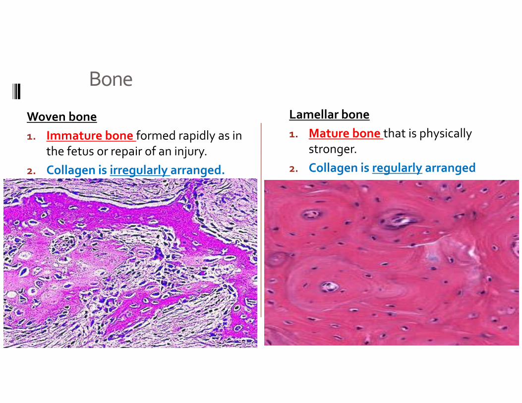
Bone
Woven bone1. Immature bone formed rapidly as in
the fetus or repair of an injury.2. Collagen is irregularly arranged.
Lamellar bone1. Mature bone that is physically
stronger.2. Collagen is regularly arranged
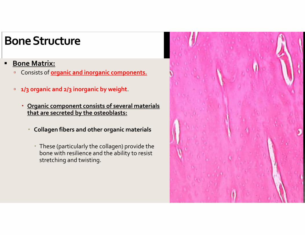
Bone Structure
Bone Matrix: Consists of organic and inorganic components.
1/3 organic and 2/3 inorganic by weight.
Organic component consists of several materialsthat are secreted by the osteoblasts:
Collagen fibers and other organic materials
These (particularly the collagen) provide thebone with resilience and the ability to resiststretching and twisting.
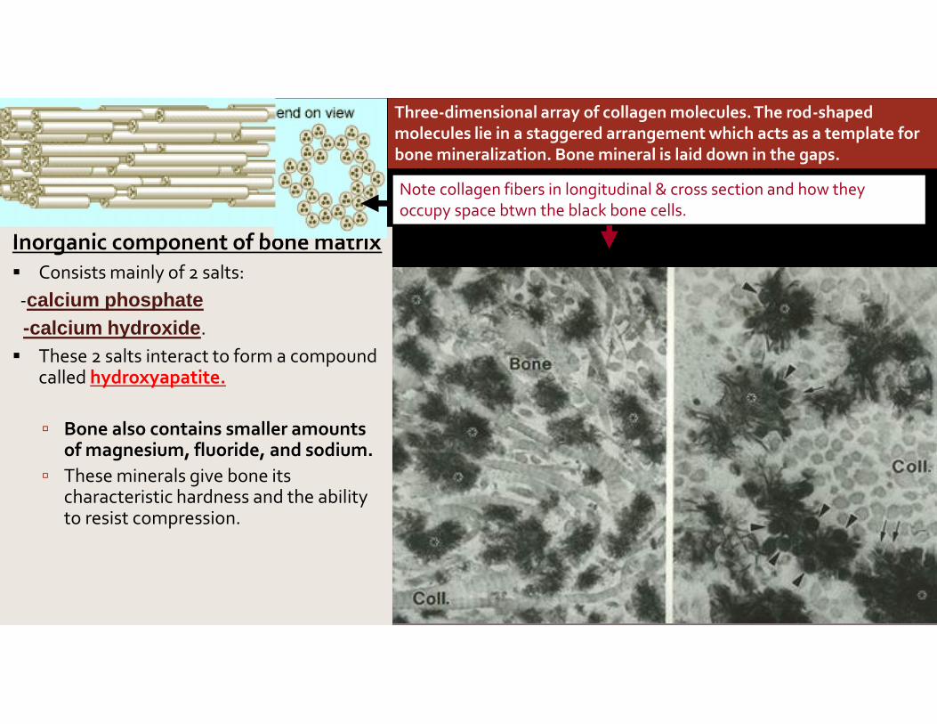
Inorganic component of bone matrix Consists mainly of 2 salts:-calcium phosphate-calcium hydroxide. These 2 salts interact to form a compound
called hydroxyapatite.
Bone also contains smaller amountsof magnesium, fluoride, and sodium.
These minerals give bone itscharacteristic hardness and the abilityto resist compression.
Three-dimensional array of collagen molecules. The rod-shapedmolecules lie in a staggered arrangement which acts as a template forbone mineralization. Bone mineral is laid down in the gaps.
Note collagen fibers in longitudinal & cross section and how theyoccupy space btwn the black bone cells.

This bone:
a. Has been demineralized
b. Has had its organic component removed
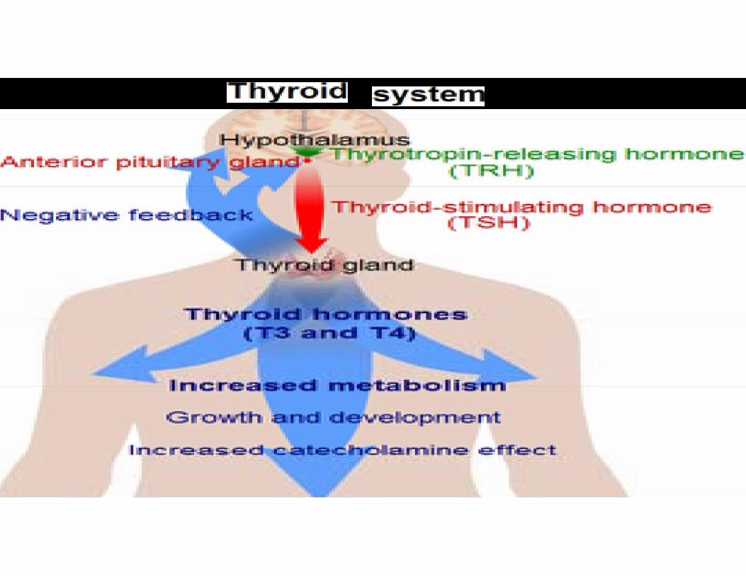
Hormonal control

Hormones important to bone growth andhomeostasis:
•Growth Hormone (GH) –
•The thyroid hormones (e.g. thyroxine) –
•Testosterone -
• Estrogens ‐
•Calcitonin –
•Parathyroid hormones
•Insulin
•Glucocorticoids
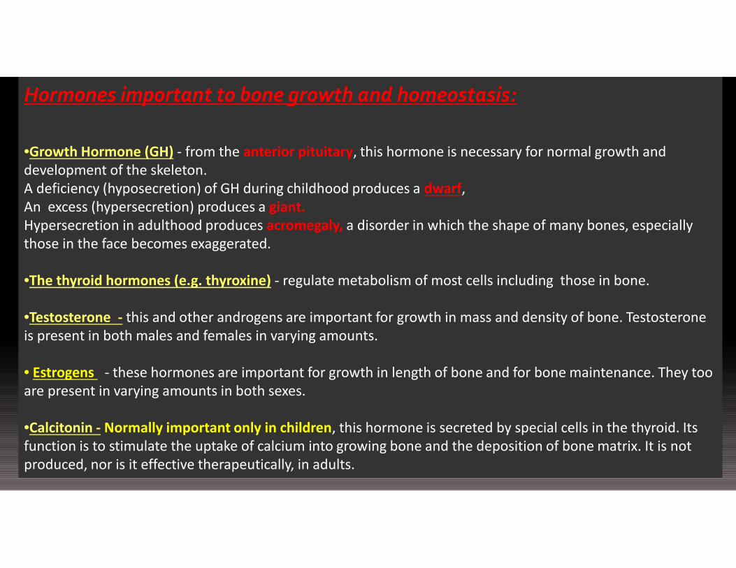
Hormones important to bone growth and homeostasis:
•Growth Hormone (GH) - from the anterior pituitary, this hormone is necessary for normal growth anddevelopment of the skeleton.A deficiency (hyposecretion) of GH during childhood produces a dwarf,An excess (hypersecretion) produces a giant.Hypersecretion in adulthood produces acromegaly, a disorder in which the shape of many bones, especiallythose in the face becomes exaggerated.
•The thyroid hormones (e.g. thyroxine) - regulate metabolism of most cells including those in bone.
•Testosterone - this and other androgens are important for growth in mass and density of bone. Testosteroneis present in both males and females in varying amounts.
• Estrogens - these hormones are important for growth in length of bone and for bone maintenance. They tooare present in varying amounts in both sexes.
•Calcitonin - Normally important only in children, this hormone is secreted by special cells in the thyroid. Itsfunction is to stimulate the uptake of calcium into growing bone and the deposition of bone matrix. It is notproduced, nor is it effective therapeutically, in adults.
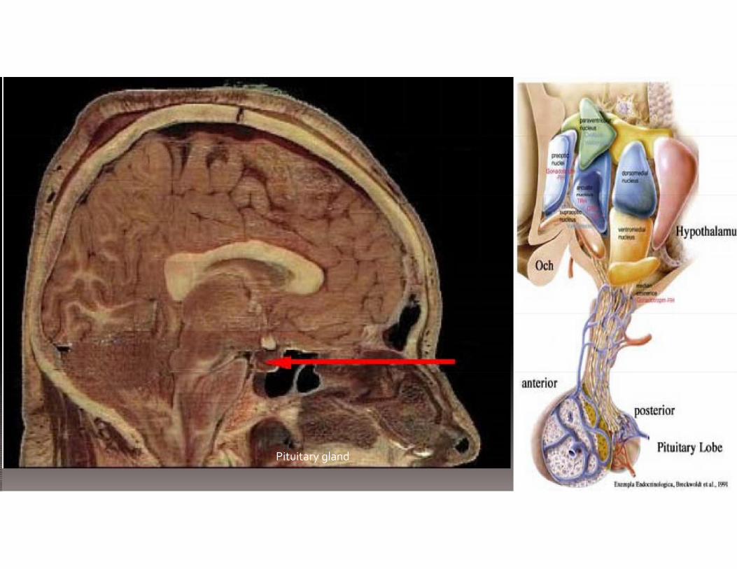
Pituitary gland


l h i h d i h bl d hi i
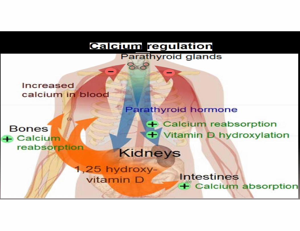
Parathyroid hormone ‐• this hormone exerts the primary control in calcium homeostasis.• Calcium is necessary in the blood for many functions and when its level falls
parathyroid hormone is secreted.
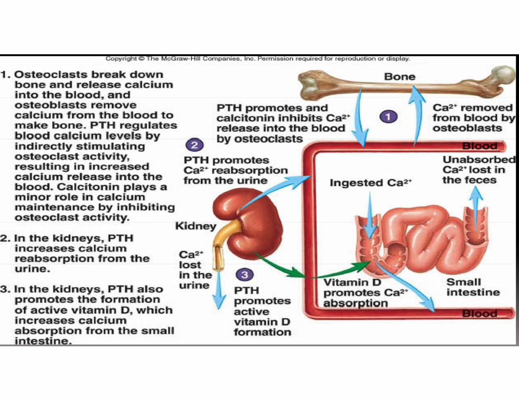
This hormone uses several methods to raise calcium levels in the blood:
1) increased Vitamin D production. Vitamin D is a hormone whose precursor is producedin the skin in response to sunlight and then processed in the liver and kidney tobecome active Vitamin D3. Vitamin D3 increases calcium absorption in the gut.Without this vitamin calcium is not absorbed to any great degree.
2) Increased reabsorption of calcium in the kidney. Much calcium is lost to the urine, sowhen you need more in the blood this is an important source.
3) Resorption of bone. PTH increases osteoclastic activity to release calcium into theblood.
Other hormones that affect bone growth include insulin and theglucocorticoids.
• Insulin stimulates bone formation• Glucocorticoids inhibit osteoclast activity.

Estrogens
• are produced primarily by developing follicles inthe
• ovaries, the corpus luteum, and the placenta.• Follicle‐stimulating hormone (FSH) stimulates
the production of estrogen in the granulosa cellsof
• the ovaries.• Some estrogens are also produced in smaller
amounts by other tissues such as the liver,adrenal glands, and the breasts.
• Fat cells also produce estrogen
Testosteroneis primarily secreted in the testicles of males
and the ovaries of females, althoughsmall amounts are also secreted by the adrenal glands.
In the testes the leydig cells [interstitial cells ofLeydig] produce the testosterone

•At puberty, the rising levels of sex hormones (estrogens in femalesand androgens in males) cause osteoblasts to produce bone fasterthan the epiphyseal cartilage can divide.
•This causes the characteristic growth spurt as well as the ultimateclosure of the epiphyseal plate.
•Estrogens cause faster closure of the epiphyseal growth platethan do androgens.
•Estrogen also acts to stimulate osteoblast activity


Parathyroid Hormone
PTH increases calcitriol synthesiswhich increases Ca2+ absorption inthe small intestine.
PTH decreases urinary Ca2+excretion and increases urinaryphosphate excretion.
Released by the cells of the parathyroid gland inresponse to low blood [Ca2+].Causes blood [Ca2+]to increase.PTH will bind to osteoblasts and this will cause 2things to occur:
The osteoblasts will decrease their activityand they will release a chemical known asosteoclast-stimulating factor.Osteoclast-stimulating factor will increaseosteoclast activity.
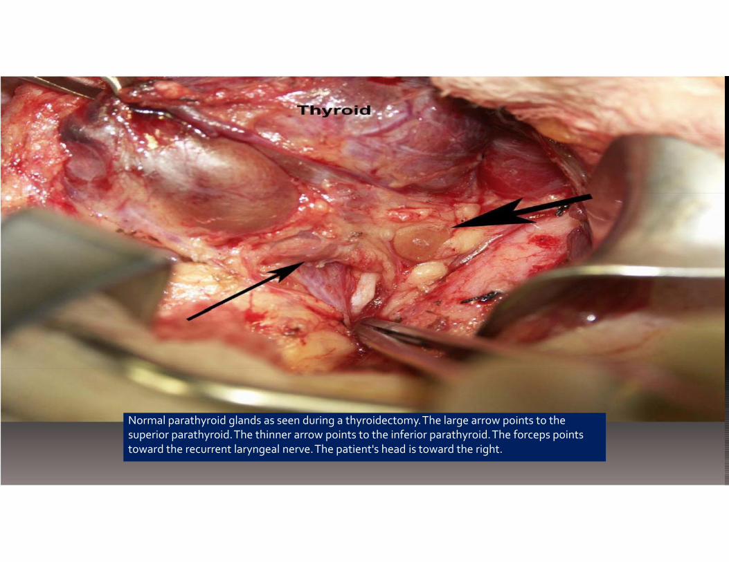
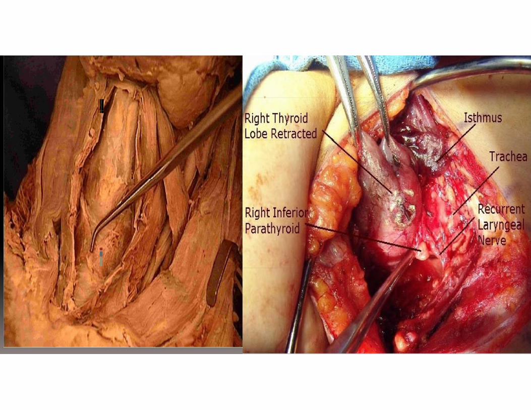
Normal parathyroid glands as seen during a thyroidectomy.The large arrow points to thesuperior parathyroid.The thinner arrow points to the inferior parathyroid.The forceps pointstoward the recurrent laryngeal nerve.The patient's head is toward the right.
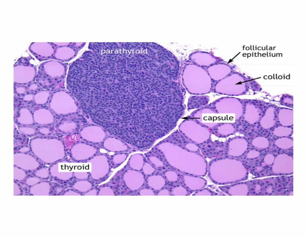

Parathyroid gland, chief and oxyphil cells
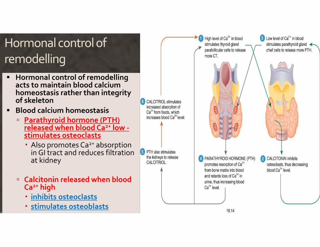
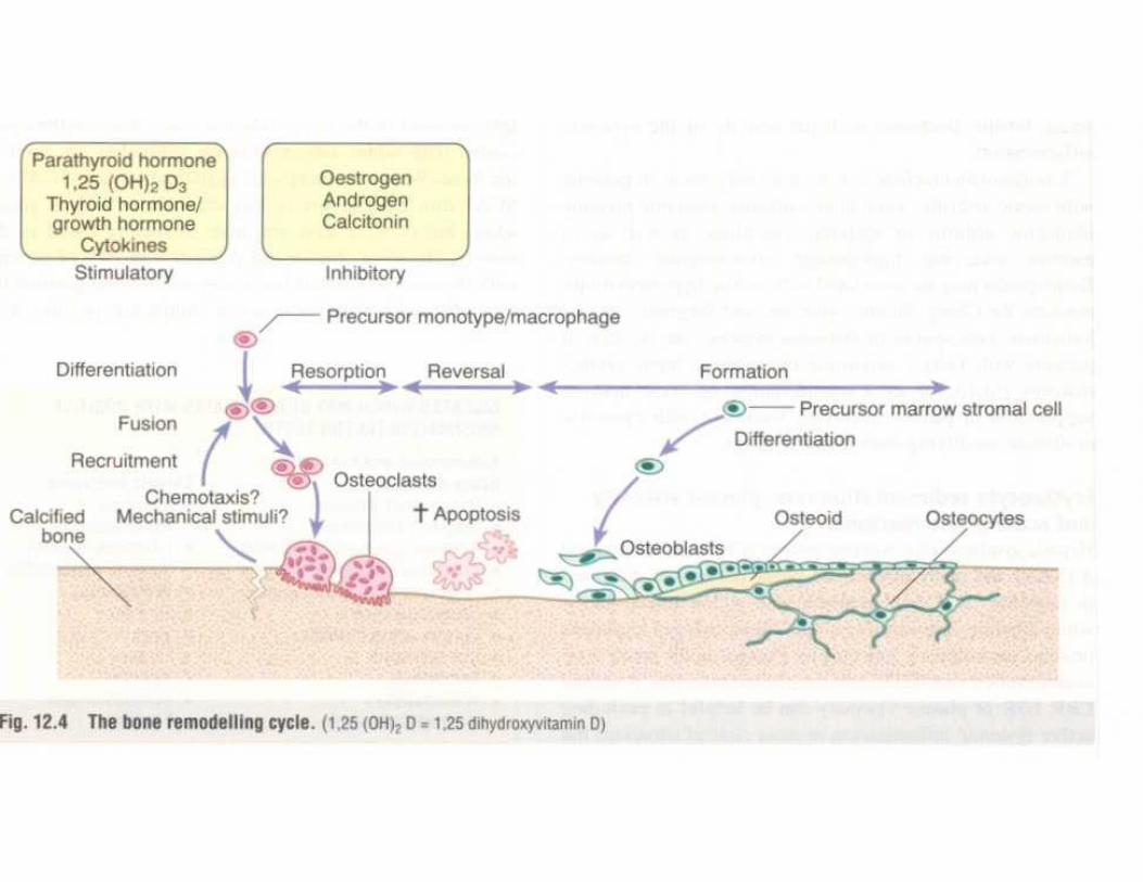
Hormonal control ofremodelling Hormonal control of remodelling
acts to maintain blood calciumhomeostasis rather than integrityof skeleton
Blood calcium homeostasis Parathyroid hormone (PTH)
released when blood Ca2+ low -stimulates osteoclasts Also promotes Ca2+ absorption
in GI tract and reduces filtrationat kidney
Calcitonin released when bloodCa2+ high inhibits osteoclasts stimulates osteoblasts

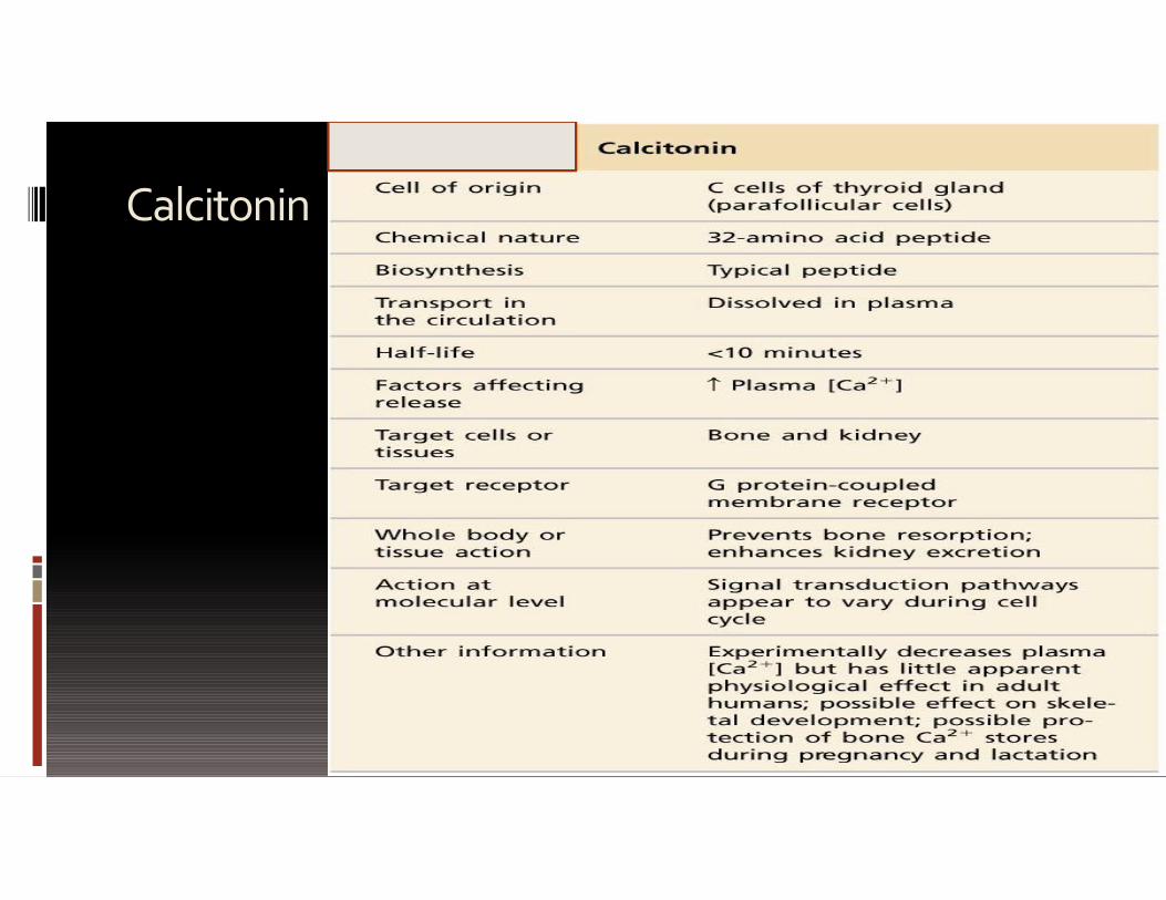
Calcitonin = thyrocalcitonin Released by the C cells = clear cells =parafollicular cells
of the thyroid gland in response to high blood [Ca2+]. Calcitonin acts to “tone down” blood calcium levels
(reduce calcemia). Calcitonin causes decreased osteoclast activity which
results in decreased break down of bone matrix anddecreased calcium being released into the blood.
Calcitonin also stimulates osteoblast activity whichmeans calcium will be taken from the blood anddeposited as bone matrix.
Notice thethyroidfollicles onthe right.The arrowindicates aC cell
• Stimulate the uptake of calcium into growing boneand the deposition of bone matrix
• Calcitonin stimulate osteoblast toproduce bone andstore calcium
Secretion of calcitonin is stimulated by:• increase in serum calcium• Gastrin and pentagastrin
More specifically, calcitonin lowers blood Ca2+ levelsin three ways:• Inhibits Ca2+ absorption by the
intestines• Inhibits osteoclast activity in bones• Inhibits renal tubular cell reabsorption
of Ca2+ allowing it to be secreted in theurine
•Vitamin D regulation• calcitonin protects against calcium loss from
skeleton during periods of calcium mobilization,such as pregnancy and, especially, lactation
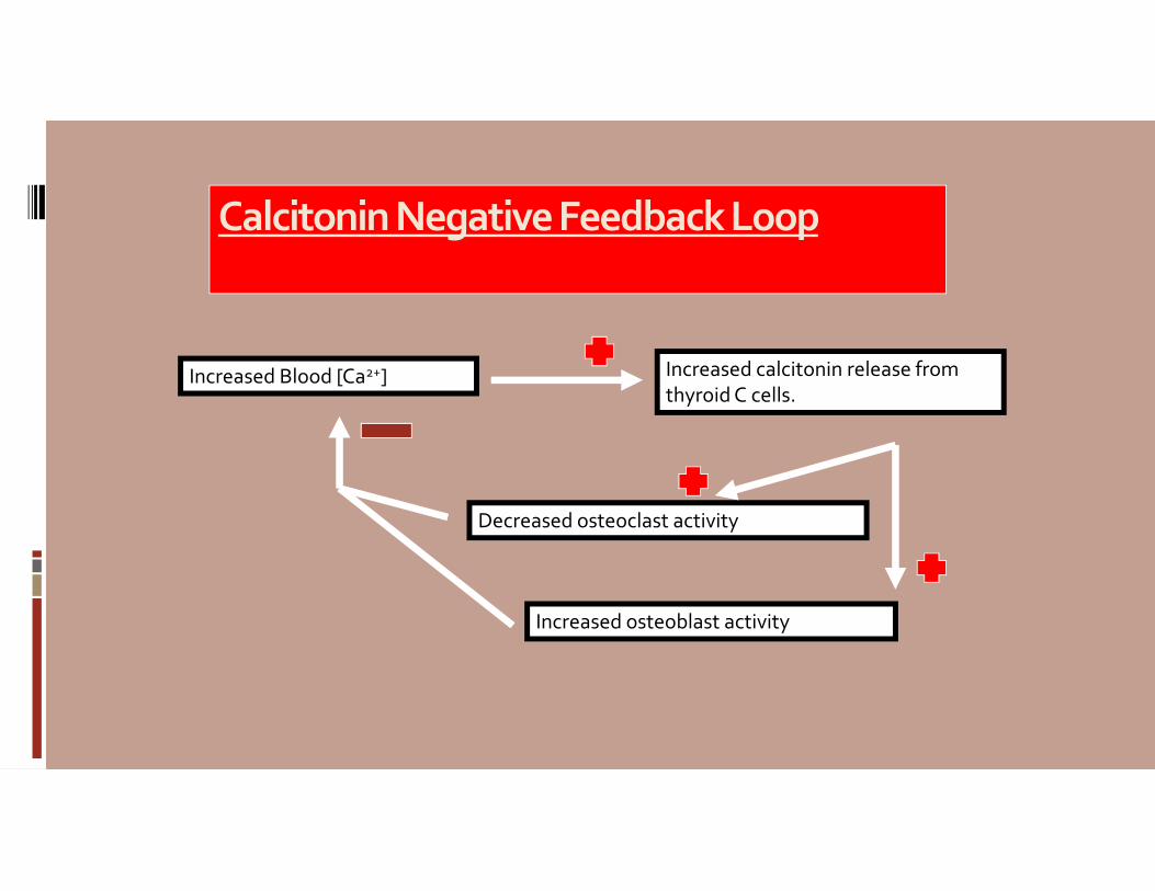
Calcitonin Negative Feedback Loop
Increased Blood [Ca2+] Increased calcitonin release fromthyroid C cells.
Increased osteoblast activity
Decreased osteoclast activity
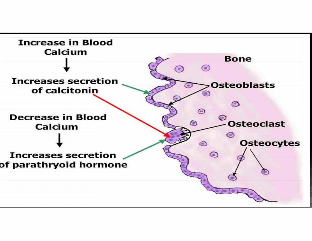
Inc.rease in BloodCaJcium
11-· creasessecretion�_
of c.,, leitoni·
•
Decrease in Bloo,dOsteoc a t.
-Cal.ci-_:m i Oste " cytes
�Incr�ase�s,ecretion�pa athryoi
·�of hormoneoo

Calcium
Important signal molecule Part of intercellular cement
that holds cells together attight junction
Cofactor in the coagulationcascade
Affects the excitability ofneurons
Calcium is the most abundant mineral in the human body.
•The average adult body contains in total approximately 1 kg,99% in the skeleton in the form of calcium phosphate salts.
•The extracellular fluid (ECF) contains approximately22.5 mmol, of which about 9 mmol is in the serum.
•Approximately 500 mmol of calcium is exchangedbetween bone and the ECF over a period of twenty-four hours
•The amount of total calcium varies with the level of serumalbumin, a protein to which calcium is bound.
The serum level of calcium is closely regulated with a normal total calcium of 2.2‐2.6 mmol/L (9‐10.5 mg/dL) and a normal ionized calcium of 1.1‐1.4 mmol/L (4.5‐ 5.6 mg/dL).

Figure 23-20
Calcium Balance in the Body
Ca2+
Small intestine
Dietarycalcium
Calciumin feces
[free Ca2+]0.001 mM
Kidney
Ca2+
in urine
Ca2+ inkidneytubules
Calcitrol(PTH, prolactin)
Activetransport
Some calcium is secretedinto the small intestine.
Cells
[Ca2+]
2.5 mM
PassivefiltrationCalcitonin
Ca2+PTH
Calcitonin
PTHCalcitriolCortisol
Bone ECF
Electrochemicalgradient
PTH = parathyroidhormone
KEY
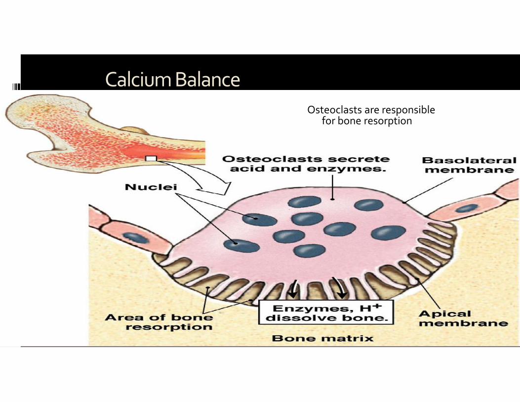
Figure 23-21
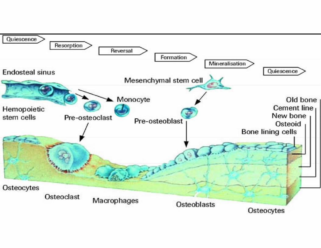
Calcium BalanceOsteoclasts are responsible
for bone resorption
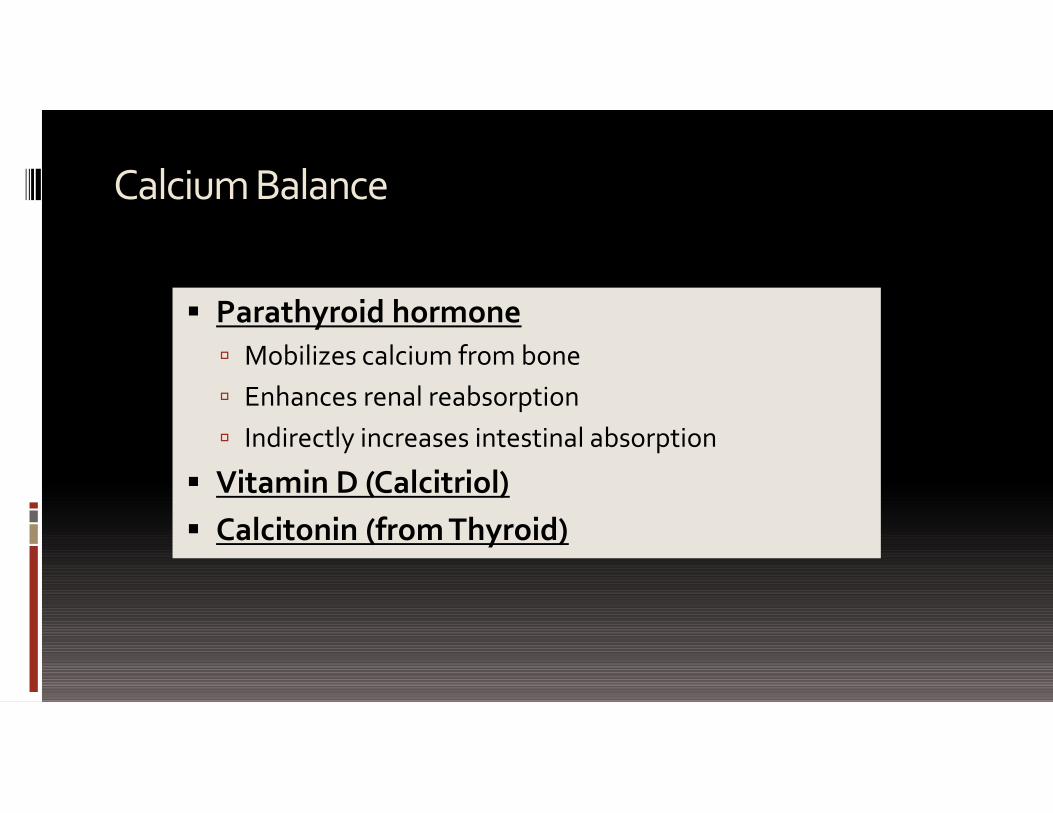
Calcium Balance
Parathyroid hormone Mobilizes calcium from bone Enhances renal reabsorption Indirectly increases intestinal absorption
Vitamin D (Calcitriol) Calcitonin (from Thyroid)
ca
ale
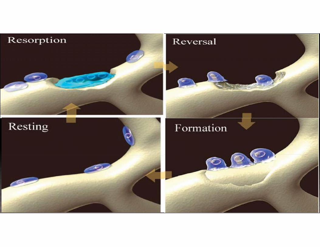
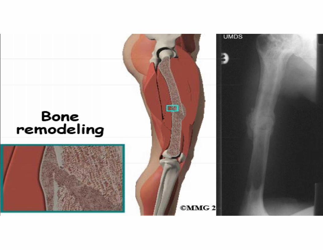
Bone homeostasis
Bones continually being remodelled 5-7% of bone mass turned over each week
Remodelling regulated by two control mechanisms: Hormonal control of blood Ca2+ homeostasis
Mechanical stress
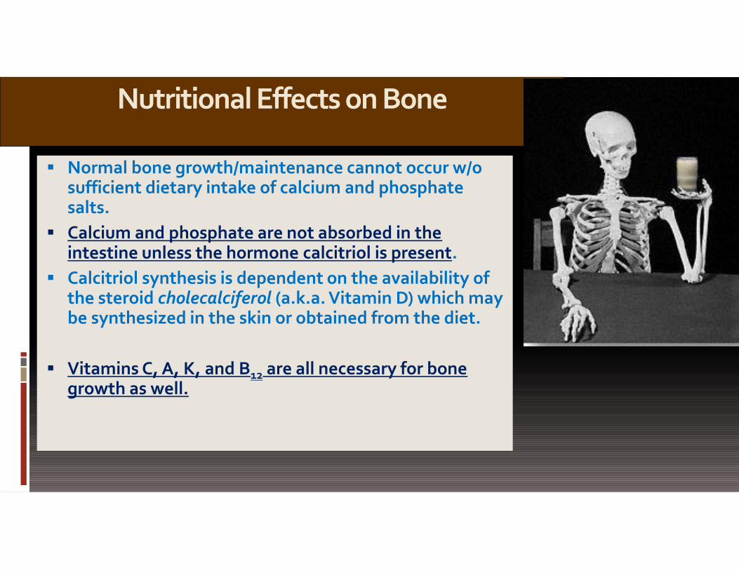
Nutritional Effects on Bone
Normal bone growth/maintenance cannot occur w/osufficient dietary intake of calcium and phosphatesalts.
Calcium and phosphate are not absorbed in theintestine unless the hormone calcitriol is present.
Calcitriol synthesis is dependent on the availability ofthe steroid cholecalciferol (a.k.a. Vitamin D) which maybe synthesized in the skin or obtained from the diet.
Vitamins C, A, K, and B12 are all necessary for bonegrowth as well.

Different Forms of Vitamin DCholecalciferolvitamin D3
Cholecalciferol: is the naturally occurring form of vitamin D.Cholecalciferol is made in large quantities in your skin when sunlight strikes your bare skin.It can also be taken as a supplement.
Calcidiol25(OH)D or 25D Calcidiol Made in Liver AFTER BEING ABSORBED BY SKIN OR INJESTION
Calcidiol (25-hydroxyvitamin D) is a prehormone in your blood that is directly made from cholecalciferol. When being testedfor vitamin D deficiency, calcidiol is the only blood test that should be drawn. When someone refers to vitamin D bloodlevels, they are referring to calcidiol levels. Your doctor can order calcidiol levels but the lab will know calcidiol as 25-hydroxyvitamin D.
Calcitriol1,25(OH)2D3 or 1,25D3
Calcitriol (1,25-dihydroxyvitamin D) is amade from calcidiol in both the kidneys and in other tissues and is the most potentsteroid hormone derived from cholecalciferol. Calcitriol has powerful anti-cancer properties. It is sometimes referred to asthe active form of vitamin D. Calcitriol levels should never be used to determine if you are deficient in vitamin D.


Calcium Balance
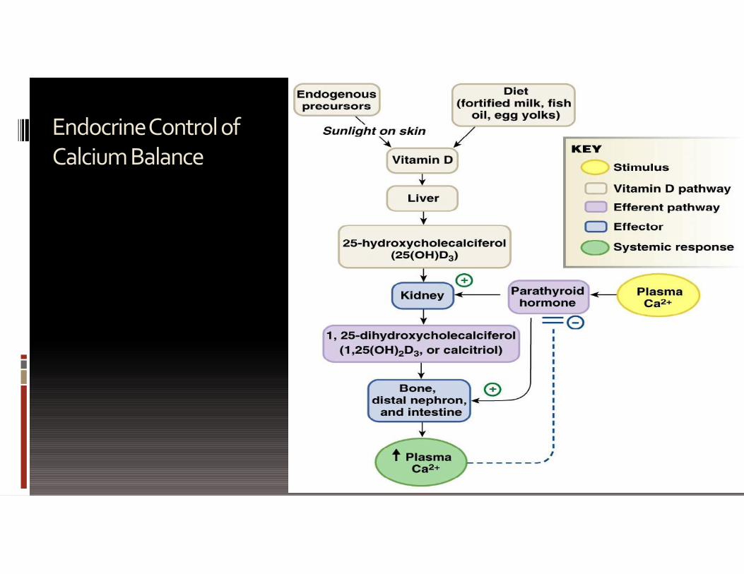
Figure 23-23
Endocrine Control ofCalcium Balance
Calcitonin

Hormonal Control of Blood Ca
Figure 6.11
PTH;calcitoninsecreted
Calcitoninstimulatescalcium saltdepositin bone
Parathyroidglands releaseparathyroidhormone (PTH)
Thyroidgland
Thyroidgland
Parathyroidglands
Osteoclastsdegrade bonematrix and releaseCa2+ into blood
Falling bloodCa2+ levels
Rising bloodCa2+ levels
Calcium homeostasis of blood: 9–11 mg/100 ml
PTH
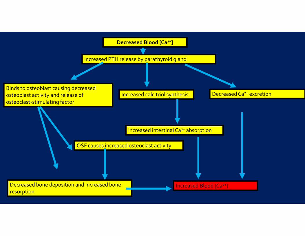
Increased PTH release by parathyroid gland
Binds to osteoblast causing decreasedosteoblast activity and release ofosteoclast-stimulating factor
OSF causes increased osteoclast activity
Decreased bone deposition and increased boneresorption
Increased calcitriol synthesis
Increased intestinal Ca2+ absorption
Decreased Ca2+ excretion
Increased Blood [Ca2+]
Decreased Blood [Ca2+]
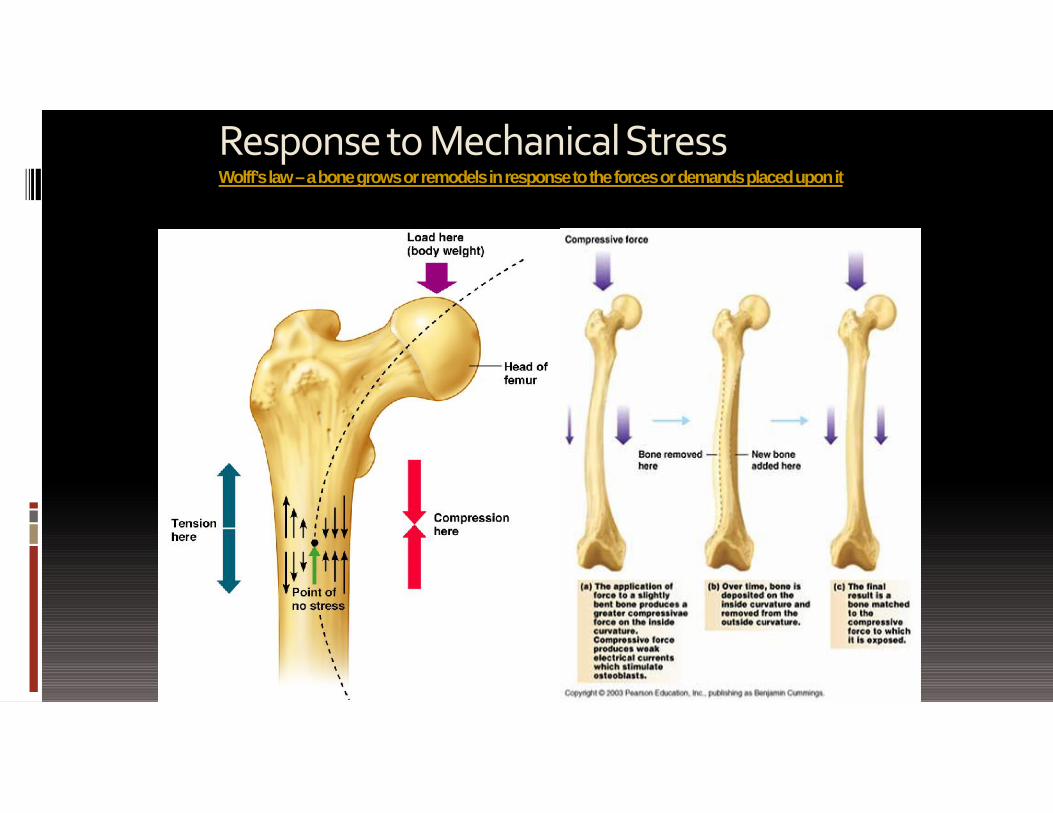
Response to MechanicalStressWolff’s law–a bone grows or remodels in response to the forces or demands placed upon it
Figure 6.12
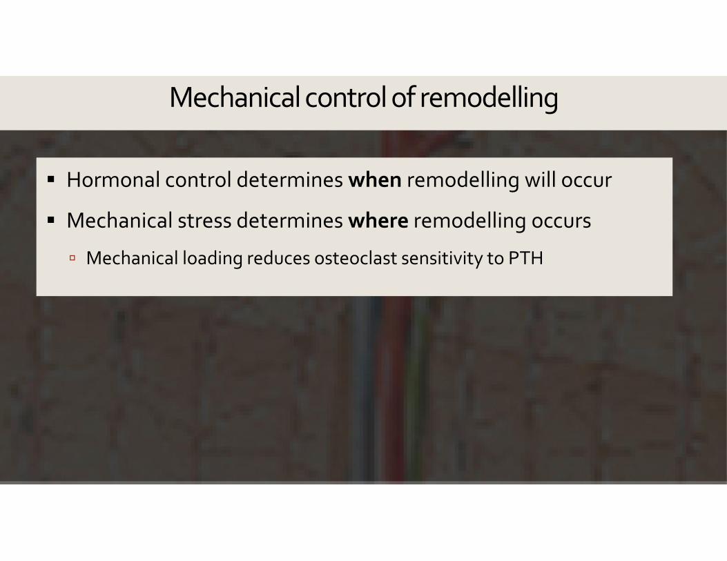
Mechanical control of remodelling
Hormonal control determines when remodelling will occur
Mechanical stress determines where remodelling occurs
Mechanical loading reduces osteoclast sensitivity to PTH

Figure 23-24
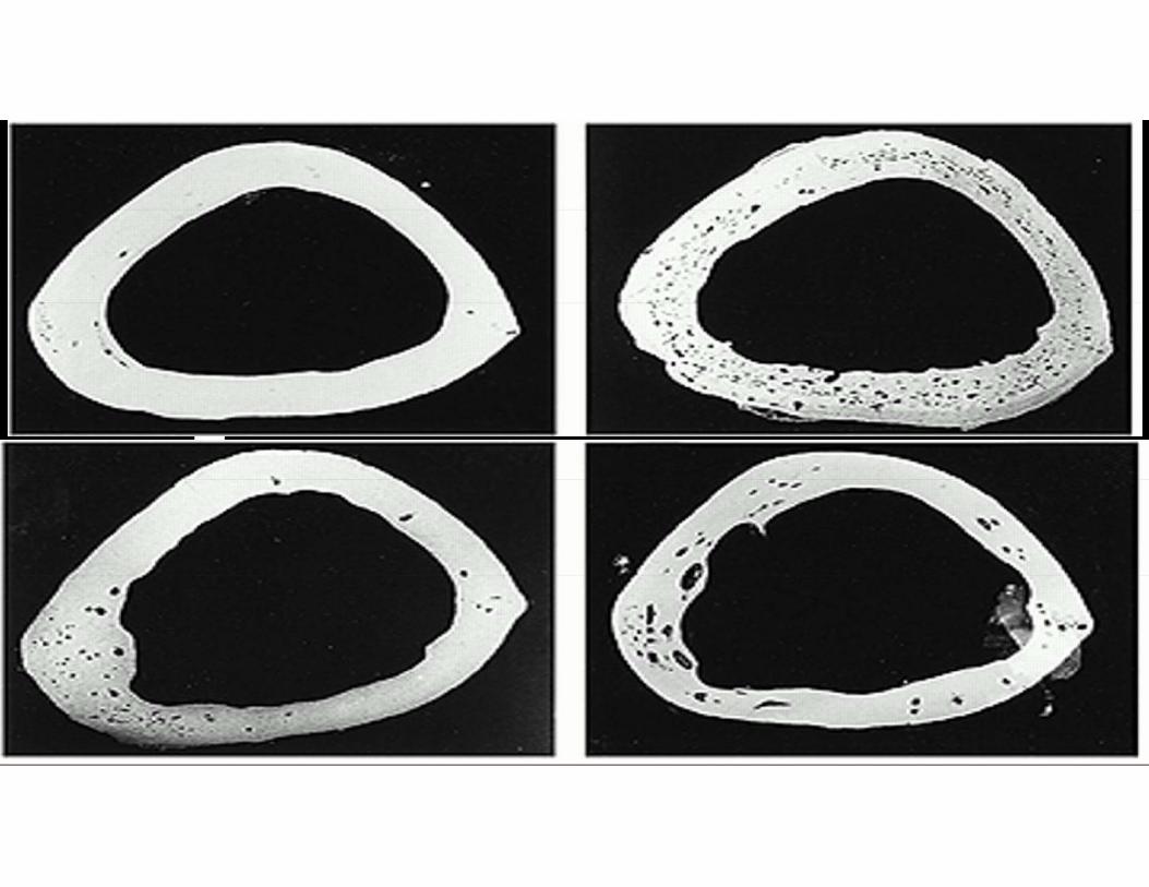
Osteoporosis Normal bone (left) and bone loss in osteoporosis (right)

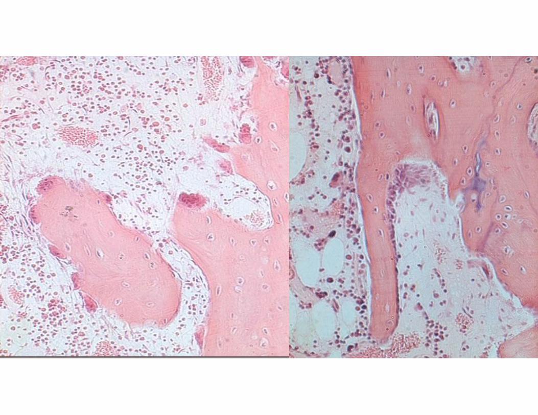
Decalcified Bone MatrixThis cross section of a long bone shows cortical bone to the right and bone marrow to the left. The white circles in themarrow are fat cells. In this preparation calcium has been removed during tissue processing.
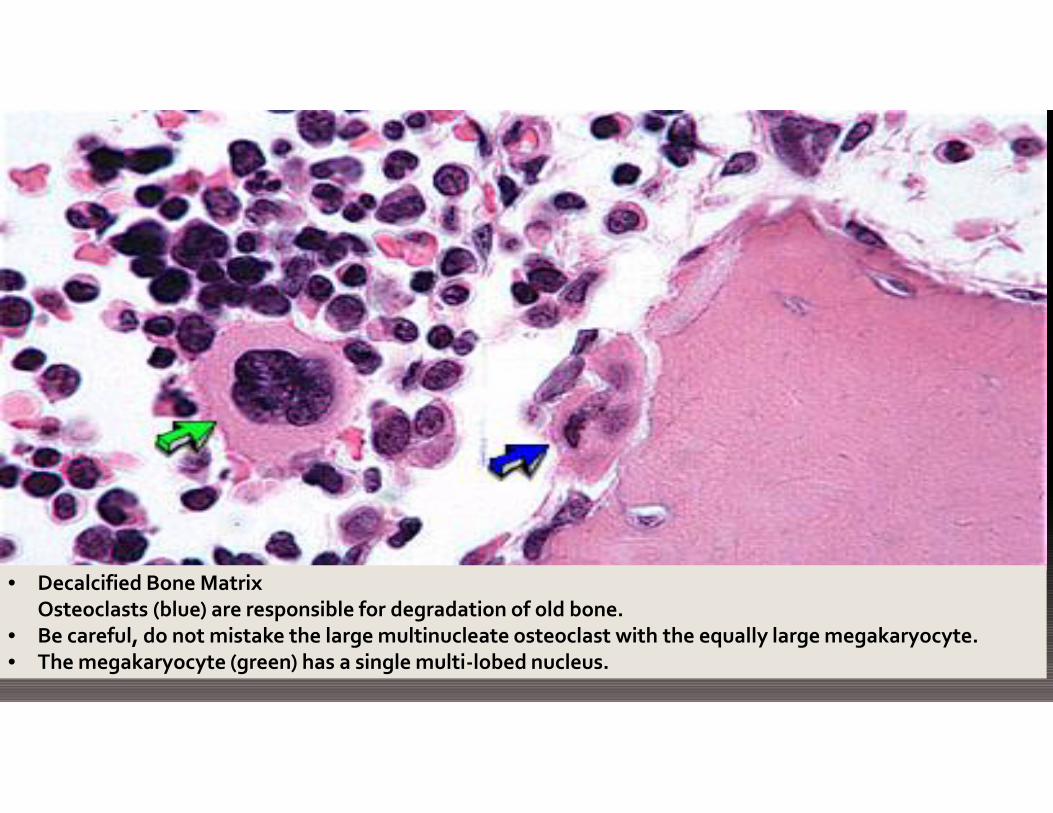
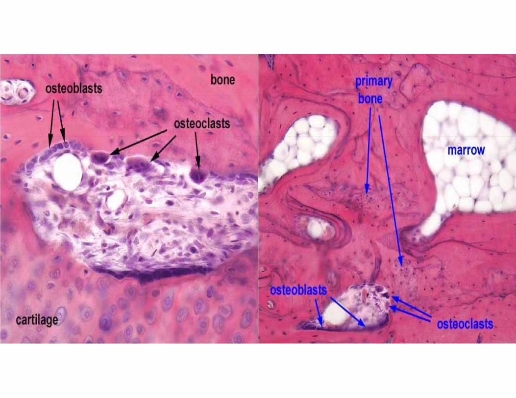
• Decalcified Bone MatrixOsteoclasts (blue) are responsible for degradation of old bone.
• Be careful, do not mistake the large multinucleate osteoclast with the equally large megakaryocyte.• The megakaryocyte (green) has a single multi-lobed nucleus.


Anna Swan with her parents


![Untitled-1 []€¦ · ENGINEERED SILVER CALCIUM TECH -PRO 71 EXCELLENT ENERGY cHioRlDE SILVER CALCIUM TEC-PRO 75023 654 h CHLORIDE ENGINEERED SILVER CALCIUM TECH -PRO . Corporate](https://static.documents.pub/doc/80x56/5f92bdfb8f40950963602274/untitled-1-engineered-silver-calcium-tech-pro-71-excellent-energy-chiorlde.jpg)


