63
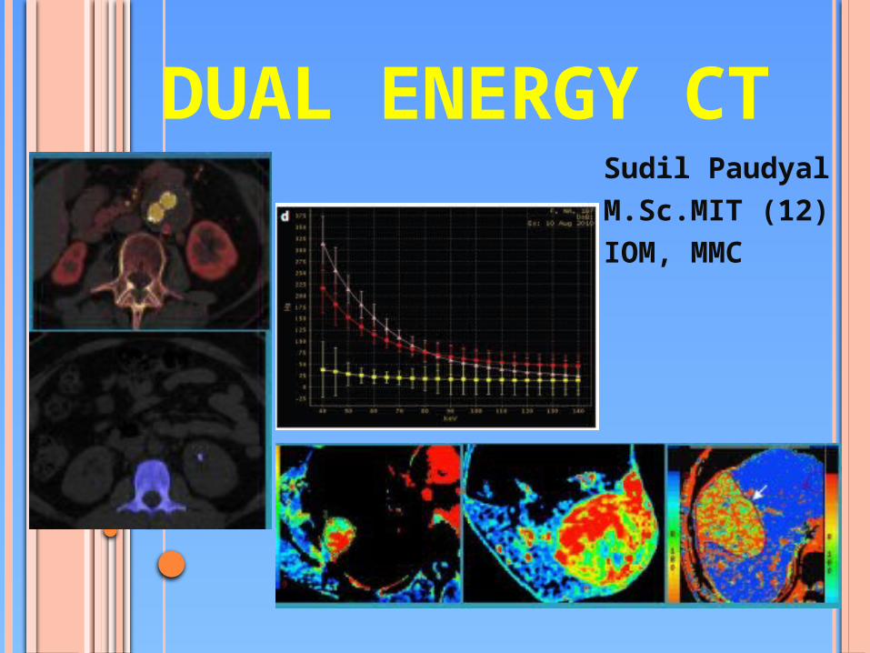
DUAL ENERGY CT Sudil Paudyal M.Sc.MIT (12) IOM, MMC 1
| Date post: | 09-Feb-2017 |
| Category: |
Health & Medicine |
| Upload: | sudil-paudyal |
| View: | 488 times |
| Download: | 0 times |
1
DUAL ENERGY CTSudil Paudyal M.Sc.MIT (12)IOM, MMC

2
SINGLE ENERGY CT

3
CHALLENGES WITH SINGLE ENERGY CT Tissue characterization Lesion detection (small lesion) Image noise and quality at low kV scanning Complex scanning protocols
Multiphase imaging Radiation dose

4
ATTENUATION ON CT Material differentiation on CT is based on x-
ray attenuation Attenuation is caused by absorption and
scattering of radiation Two main mechanisms: compton scatter and
photoelectric effect Compton effect is energy independent Photoelectric effect is strongly energy dependent
and its likelihood increases as the energy of incident photon approximates the K-shell binding energy of an electron.
Changing the kV setting results in an alteration of photon energy

5
BASIS OF DECT Attenuation is energy dependent- at 80 kVp, iodine signal is
2X of 140 kVp CT numbers donot vary with beam energy for soft tissues
but do for high “z” materials By analyzing absorption properties of different material at
different x-ray energies, materials can be distinguished. (basis material decomposition)
Human body is made up of many different elements- mainly carbon, oxygen , hydrogen , nitrogen, phosphorous and calcuim- which are arranged in many different combinations.
Hydrogen, carbon, nitrogen and oxygen have similar k- edges which are well below the energies currently used in DECT (80 kVp and 140 kVp), thus not well appreciated.
The k edges of calcium and iodine are higher than those of soft tissues and although they are lower than those, they are distinguished from soft tissues at DE imaging.

6
The settings of 80 and 140 kVp are generally used because they provide maximum difference and least overlap between the spectra with standard tubes.

PRINCIPLE OF DECTThe principle of dual energy CT is based on differential absorption of energy(Linear attenuation coefficient) at variable KVP settings.For example: Let us consider a substance (A)with K-edge at 60 kv another (B)with K-edge 130 kv, if we imagine multiple combination of A and/or B at 80KVp and 140KVp ,there will be differential attenuation at both these energy settings.The object containing larger amount of substance A will show higher attenuation at 80KVp and lower attenuation at 140 KVp, whereas object containing larger amount of substance B will show higher attenuation at 80KVp and at 140KVp as well.The CT number of blood mixed iodine becomes 1550HU similar to the value of bone at 140 kvp. so it is nearly impossible to determine weather the object of interest is bone or iodine blood mixed structure.If we take another scan of same object with on 80kvp setting , the measures CT number will be different .if the object of interest is made with bone the CT number will be 2200HU and the object is mixture of iodine and blood , the CT number will be 2800HU due to different behavior of linear attenuation co-efficient as a function of energy for different materials.

8

9
TECHNICAL APPROACHES TO DECT Five approaches : (currently only former three
are commercially available) Sequential acquisition Rapid voltage switching DSCT Layer detectors and Energy resolving or quantum counting detectors

11
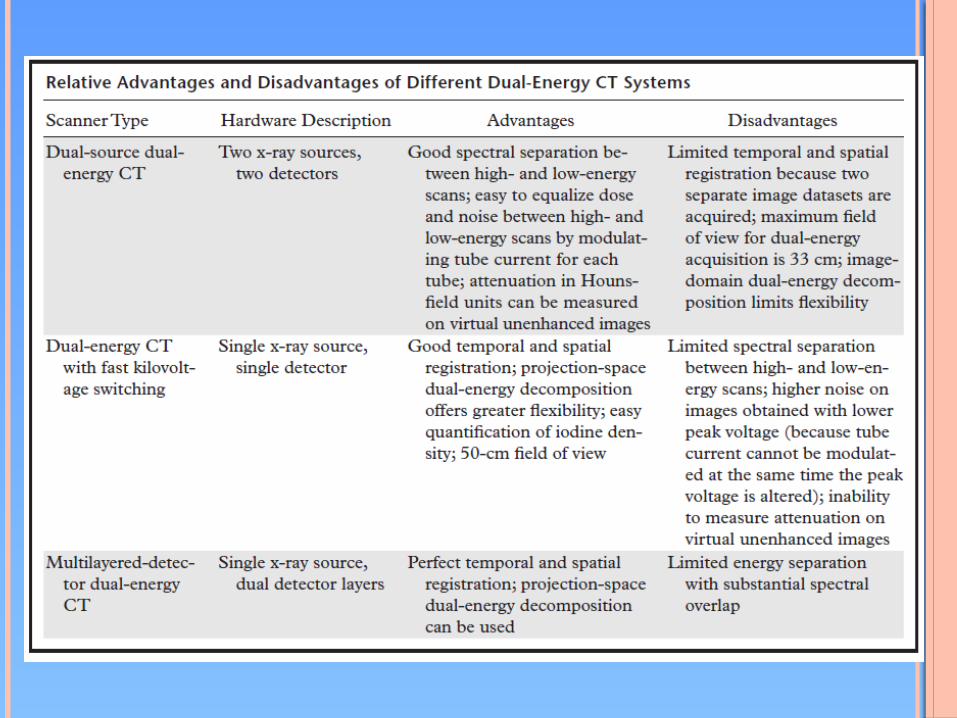
SEQUENTIAL ACQUISTION Requires least hardware effort. Achieved either as two subsequent helical
scans or as a sequence with subsequent rotations at alternating tube voltages and stepwise table feed.
Disadvantage is rather long delay between both acquisitions, causing artifacts from cardiac or respiratory motion or changes in CM opacification.
But, a viable option for clinical applications w/o CM such as metal artifact removal or kidney stone differentiation.
12
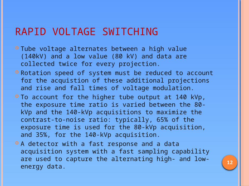
RAPID VOLTAGE SWITCHING Tube voltage alternates between a high value (140kV)
and a low value (80 kV) and data are collected twice for every projection.
Rotation speed of system must be reduced to account for the acquistion of these additional projections and rise and fall times of voltage modulation.
To account for the higher tube output at 140 kVp, the exposure time ratio is varied between the 80-kVp and the 140-kVp acquisitions to maximize the contrast-to-noise ratio: typically, 65% of the exposure time is used for the 80-kVp acquisition, and 35%, for the 140-kVp acquisition.
A detector with a fast response and a data acquisition system with a fast sampling capability are used to capture the alternating high- and low-energy data.

13
The advantages of dual-energy CT with fast kilovoltage switching are good temporal registration between high- and low-energy datasets, which are obtained nearly simultaneously, and the availability of the full 50-cm field of view for use in image analysis.
Limited photon output at low voltages which results in high noise and necessity to choose a relatively high current and consequently high dose.
So requires additional dose because other dose reduction features(ATCM) or optimized filtration are not possible.
However, because a single x-ray source is used, individual modification of the high- and low-energy x-ray beams is difficult (not yet possible on commercially available scanners), and spectral overlap is increased.

14

15
DUAL SOURCE CT Two tubes running at different voltages and corresponding
detectors mounted orthogonally in one gantry. High-energy scans are obtained at 120 or 140 kVp, and
low-energy scans are obtained simultaneously at 80 or 100 kVp.
One detector somewhat smaller than other, resulting in 33 cm FOV.
Nearly twofold investments in hardware but offers important advantages for DECT: Voltage, current and filter can be chosen independently for both
tubes to achieve an optimal contrast . Although there is an angular offset between both spiral paths,
there is no temporal offset in data acquisition because equivalent z-axis positions are scanned at same time.
Disadvantage is cross-scatter radiation, which partially hits the noncorresponding orthogonal detector and requires correction.

16

17
LAYER DETECTOR Uses an energy resolving detector with
polychromatic spectrum of one tube. The sensitivity of two layers is determined by
the scintillator material : for eg: consisting of ZnSe or CsI in the top layer and Gd2O2S in the bottom layer.
The scintillator materials determine the spectral resolution and sensitivity profiles of available materials have a rather broad overlap.
Therefore the contrast of spectral information is limited or requires high additional dose.
Not yet available for clinical use.

18
QUANTUM COUNTING DETECTOR Resolve the energy of each individual
impacting photon. Used to differentiate more than two photon
energies and is very quantum efficient. But detector materials eg; CdZnTe get
saturated rather quickly, resulting in a rapid drift of the measured signal.
Thus used to scan small animals, but cannot handle a photon flux required for clinical CT.
19

20
RADIATION EXPOSURE Direct comparison of different DECT approaches should
include both spectral contrast and dose optimally quantified as CNR per dose.
Comparison for different scanner setups is made based on Monte-Carlo simulations.
Currently a maximum spectral contrast is achieved with a DSCT with optimized voltage, current and filtration.
Technologic strategies that allow dose reduction include; tube current modulation, iterative reconstruction techniques and new detector application- specific integrated circuits (ASICs) integrating photodiode and analog digital converters.
These features offer special benefits for DECT because CNR is improved in both half-dose acquisitions with the two energy spectra so the gain in dose efficieny is even greater than single energy CT for DSCT.

21
POSTPROCESSING Two approaches:1. Data-domain or projection-space decomposition:
To subtract equivalent projections and apply filtered back projection to reconstruct the difference as spectral information.
i.e. dual energy data sets are reconstructed before images are reconstructed from high- and low-energy sinograms.
Used for material analysis of dual-energy CT images obtained with fast kilovoltage switching.
Projection-space decomposition is preferred because it enables greater flexibility in material decomposition and permits the preprocessing correction of data to minimize beam-hardening artifacts

22
2. Image-domain decomposition: First reconstruct standard CT images consisting
of voxels in HU and then to use postprocessing algorithms to extract specific spectral information from the difference between the corresponding voxels.
Dual-energy datasets are processed after the reconstruction of high- and low-energy images.
Used for material analysis of images obtained with a dual-source or dual-layer dual-energy CT scanner
IRS provides low and high kV images and a series of weighted average images. The average series integrates both acquisitions in a low noise image for immediate evaluation.
DE analysis is then performed on the dual kV series using imaging based algorithms.
23
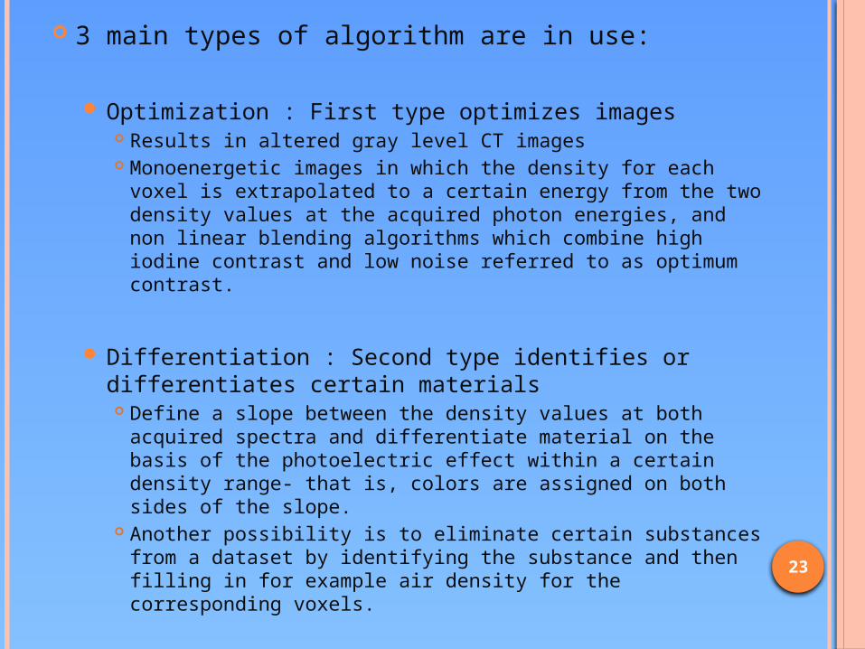
3 main types of algorithm are in use:
Optimization : First type optimizes images Results in altered gray level CT images Monoenergetic images in which the density for each voxel
is extrapolated to a certain energy from the two density values at the acquired photon energies, and non linear blending algorithms which combine high iodine contrast and low noise referred to as optimum contrast.
Differentiation : Second type identifies or differentiates certain materials Define a slope between the density values at both
acquired spectra and differentiate material on the basis of the photoelectric effect within a certain density range- that is, colors are assigned on both sides of the slope.
Another possibility is to eliminate certain substances from a dataset by identifying the substance and then filling in for example air density for the corresponding voxels.

24
• Quantification: Third type quantifies a substance in the data set.• Use a three material decomposition, quantifying one of three
materials.• A slope is defined by the density of two basic components and a
second slope is defined by the photoelectric effect of the contrast material being quantified.
• The density values measured at both energies are then interpreted as a displacement from the first slope along the second one.
• This enhancement is then color coded or is also subtracted from the image.

25
IMAGE RECONSTRUCTION In fast kV switching, 140 kV images are
reconstructed immediately at the scanner console, and are only used to verify the adequacy of anatomic coverage.
To facilitate workflow, 80 kV images are not routinely reconstructed.
Calibration correction are applied to the high and low energy datasets, which are aligned in projection space and transformed into material basis pair projections, which are then used to reconstruct two main types of images: Material density images, which provide material specific
information and Monochromatic images which provide energy selective
information.

26
Generation of material density images is based on the theory of basis material decomposition, which proposes that the attenuation coefficients of any material can be computed as a weighted sum of the attenuation coefficients of two basis materials as long as the k-edge of the material is not within the evaluated energy range.
The two basis materials should have substantially different mass attenuation coefficients; thus the material pairs chosen usually differ greatly in efffective atomic numaber.
Substances other than chosen basis materials are considered to contain combinations of both selected material densities.
Two sets of material density images are then generated each demonstrating the presence or absence of each basis material selected.

27
Two commonly selected dual energy CT basis material pairs are Iodine and calcium and Iodine and water
when iodine (high atomic number) is paired with water (low atomic number), two separate sets of images (a set of iodine density images and a set of water density images) are generated.
On the water density image, voxels that show an energy dependent change in attenuation resembiling the attenuation of iodine are removed and represented instead on the iodine density image.
The virtual unenhanced images thus generated from CE dual energy data set can provide information equivalent to that obtainable from unenhanced images.
Iodine density images are used to asses structures for areas of enhancement. Only lesions that contain iodine will show higher density.

28
29
DECT images can be processed to obtain images at any specified single photon energy. Such images are called monochromatic images.
Monochromatic projections can be generated during the processing of material density image data by calcualting the linear attenuation coefficinet of an object.
Because they are generated from projection space data, monochromatic images are less affected by beam hardening artifact and provide more accurate CT numbers than do standard CT images.
These advantages can improve the characterization of renal lesions by decreasing pseudoenhancement in simple renal cysts.

30

31
With DSDECT, images are generated by linear or nonlinear sigmoidal blending of high and low energy image datasets.
Material specific images are generated by measuring the differences in attenuation and either highlighting the pixels corresponding to the selected material or subtracting the pixels corresponding to the other materials.
For abdominal applications, a three material decomposition (soft tissue, fat and iodine) process is usually used to generate the virtual unenhanced images.
A color coded overlay image also can be generated which enables visualization of the distribution of a selected material across entire CT volume.

32

33

34
DIFFERENCE BETN VIRTUAL UNENHANCED IMAGE OBTAINED WITH DSDECT AND SSDECT.
35
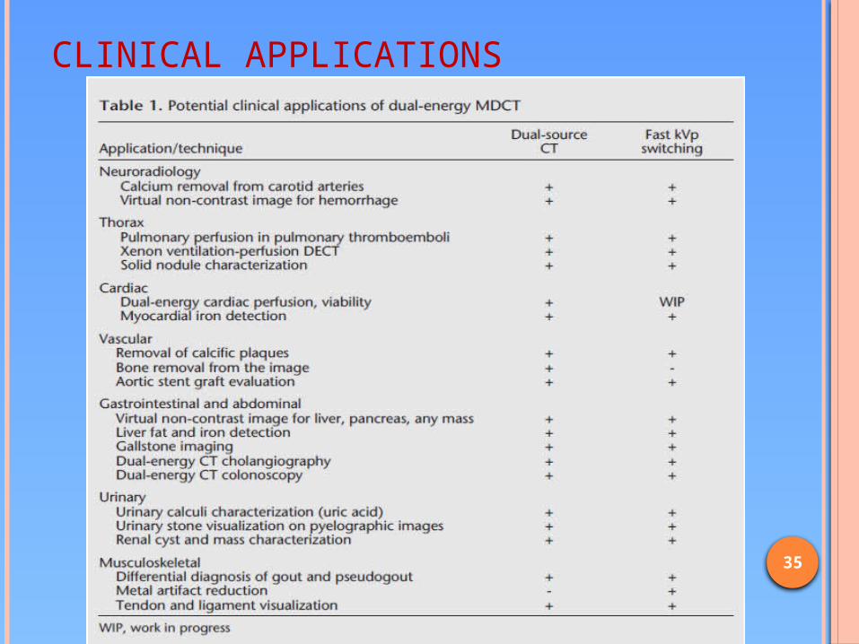
CLINICAL APPLICATIONS

36
NEURORADIOLOGY Neurological applications permit
the generation of virtual non-contrast images for the detection of brain hemorrhages in patients who undergo CTA.
Also allow the removal of bone and calcium from the carotid and brain CTA.
Possible to detect a brain hemorrhage on virtual non-contrast images.
Comparison of dual energy bone removal to digital subtraction CTA and automatic bone removal reveals superiority of dual energy technique, particularly at the level of skull base.
Radiation dose was also reduced as compared to digital subtraction CTA.

37

38
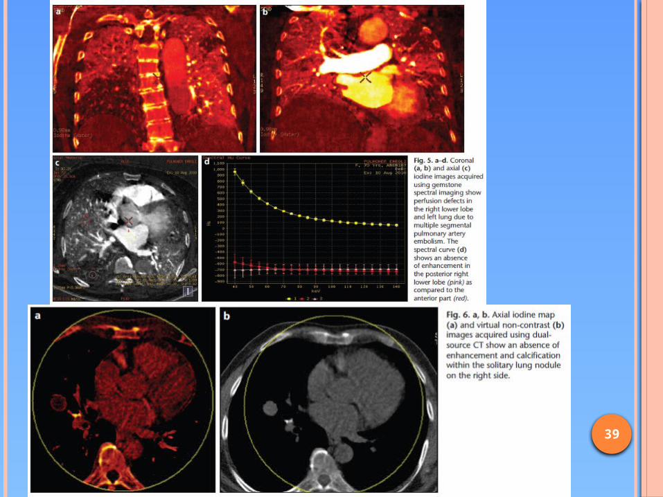
THORAX APPLICATIONS Lack of misregistration and visualization of lung perfusion and
ventilation. Misregistration avoided due to simultaneous acquisition of 80
and 140 kVp images. In pt.s with pulmonary thromboembolism, DECT may allow
the detection of subtle emboli by revealing perfusion defects. Assessment of lung perfusion can allow the visualization of
pathologies that have been perviously unknown, particularly in pt.s with ILDs, emphysema, asthma or chronic thromboembolic disease and in patients with tumors.
Xenon DECT enables collection of ventilation perfusion CT acquisitions and in future may replace ventilation perfusion scintigraphy.
Superior registration of DECT may demonstrate the presence or lack of enhancement of sub-centimeter and solitary lung nodules.
39

40
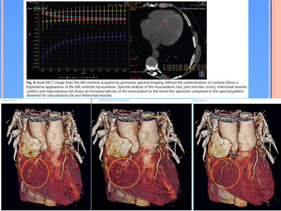
CARDIAC APPLICATIONS Dual energy perfusion with or without the
adenosine stress test, viability imaging and cardiac iron detection.
To decrease image noise, 100 and 140 kVp acquisitions are preferred.
Addition of an iterative reconstruction technique may allow a better image quality for cardiac applications.
Can be difficult in patients with elevated heart rates due to a worsening of the temporal resolution from 83 to 165 ms in DSCT.
Also for the characterization of plaques, for calcific plaque removal from coronary arteries and for evalutation of coronary stents.
41

42
VASCULAR APPLICATIONS Imaging protocol for aortic aneurysms consists of
unenhanced, arterial and venous phase images, which exposes the patient to a high radiation burden because it is repeated every six months after aortic stent grafting.
DE aortic stent graft protocol may obviate the need for unenhanced CT and iodine map images may facilitate the recognition of endoleaks.
Can also permit the faster removal of calcific plaques in large arteries and bony structures in cranial region.
However the use of this technique seems difficult for small sized arteries.
Also no sytdy has demonstrated any advantage of dual energy CTA for peripheral artery applications.

43

44
GI AND ABDOMINAL APPLICATIONS The use of dual energy has revealed that 80
kVp images demonstrate a better contrast than do 140 kVp images.
Useful for evaluation of enhanced liver lesions in the arterial phase in HCC and in hypervascular liver metastases.
Also used for the diagnosis of liver iron and fat, which display opposite spectral patterns.
The measured denisty of a liver increases with decreasing kVp and keV in patients with hepatic iron overload, but the measured density of a liver decreases with decreasing kVp and kev in patients with steatosis.
45
DE cholangiography may facilitate the detection of bile ducts and the measurement of biliary segment dimensions.
Invitro studies have been performed to characterize the dual energy of gall stones and a similar protocol may be used for patients with biliary dilation and a suspected choleduct stone.
The use of 80 kvp data obtainde by DECT may permit a better distinction of pancreatic adenocarcinoma from the adjacent normal parenchyma by increasing the conspicuity of the masses.

46

47
DECT COLONOSCOPY Obviates the non-contrast prone images from
diagnostic CT colonoscopy protocols. Colonic polyps and masses are enhanced
approx. 40-50 HU on post contrast images. Thus the enhancement of colonic masses can be differentiated from stool through the use of iodine DECT images and non cathartic DECT colonoscopy may be feasible, esp. in elderly patients.

48

49
MSK APPLICATIONS Most useful MSK application is the
differentiation of gout and pseudogout via the diagnosis of uric acid and calcium crystals in the joint space.
Tendon and ligament visualization has been proposed by CT vendors but few studies have evaluated this application.
Metal artifact reduction has been proposed for the fast kVp switching technique, wich emplyos images with a low keV.

50

51
URINARY APPLICATIONSRenal mass characterization: Paired iodine and water density images obtained at contrast enhanced
DECT can help distinguish hyper attenuating cysts from enhancing masses without the need for an unenhanced scanning phase.
The greater brightness of renal masses on iodine density images helps distinguish them from non enhancing cysts even when their attenuation measurements on CECT images are similar.
Color coded image overlays in each pixel allow easy visualization of presence or absence of enhancement.
Also water density images may be used as virtual unenhanced images to allow the detection of calcification within a renal mass.
Limitations: Virtual unenhanced images are noisier than real unenhanced images.
Because of material decomposition algorithms used, calcification in renal lesions is less conspicuous on virtual unenhanced images than on real images.
Smaller amounts of fat in renal masses are difficult to detect on virtual images and can be measured only on conventional thin section CT images.

52

53
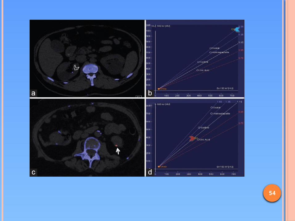
Characterization of renal stones: Different types of renal calculi; calcium based stones-
70-80%,struvite stones- 5-15%, uric acid stones 5-10%, and cystine stones 1-2.5%.
Knowledge about the composition of stones may guide decisions about theire management and shape expectations concerning the effectiveness of therapy.
Uric acid calculi can be managed with oral medications that facilitate dissolution, struvite calculi are amenable to ESWL, whereas calcium oxalate monohydrate and cystine calculi are resistant to fragementation with lithotripsy.
at dual energy, the change in attenuation between high and low energy scans can be used to differentiate types of calculi, some of which might have a similar attenuation when scanned at single energy level.
54
55
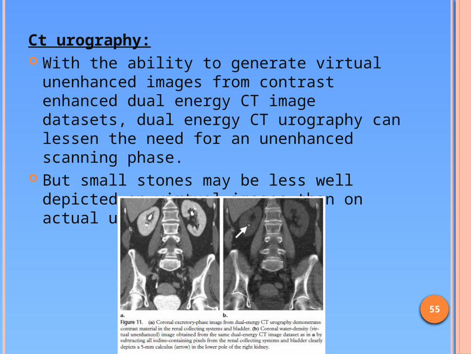
Ct urography: With the ability to generate virtual
unenhanced images from contrast enhanced dual energy CT image datasets, dual energy CT urography can lessen the need for an unenhanced scanning phase.
But small stones may be less well depicted on virtual images than on actual unenhanced images.
56
WHAT WE HAVE
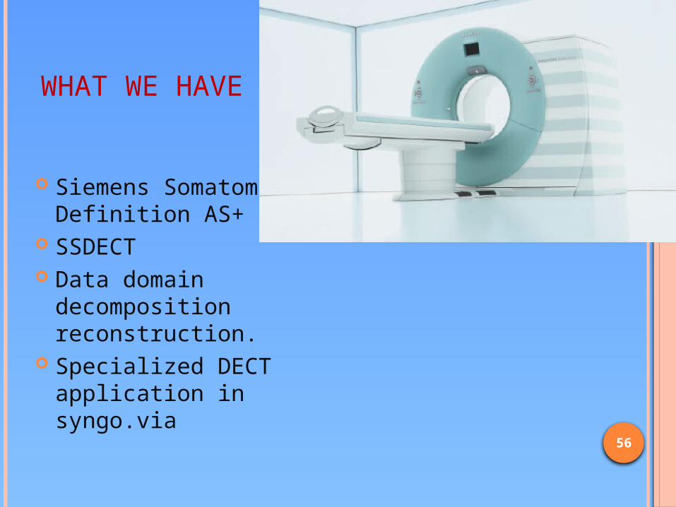
Siemens Somatom Definition AS+
SSDECT Data domain
decomposition reconstruction.
Specialized DECT application in syngo.via

57

58

59

60

61
LIMITATIONS Restriction of FOV ( with DSCT) High radiation dose
Addition of a tin filter helps to decrease Increase in radiation dose can be justified when
unenhanced images are eliminated from protocols, which may result in dose saving.
Noise in the 80 kVp images Evaluations of patients with a high BMI.
Noise constraints may result in a suboptimal image quality.
62
CONCLUSION Volumetric DECT acquisitions enables the emergence
of new applications with potential benefits. Major advantages of DECT are material
decomposition, separation of iodine from the image and prevention of misregistration particularly in thorax and abdomen, and renal mass and stone characterization in the urinary system.
The use of iterative reconstruction techniques can facilitate the wider use of DECT applications by decreasing the noise and the radiation dose.
These technologies may eventually lead to better detection and characterization of lesions in the body and objective evaluation of iodine uptake by various organs, leading to DECT becoming an alternative of PET-CT.

63
FIND MORE AT: Johnson TR; Dual energy CT: general
principles Muşturay Karçaaltıncaba, Aykut Aktaş; Dual-
energy CT revisited with multidetector CT: review of principles and clinical applications
Johnson TR, Kraus B, et.al.; Material differentiation by Dual energy CT: initial experience
Kaza RK, Plat FJ et.al.; Dual-Energy CT with Single- and Dual-Source Scanners: Current Applications in Evaluating the Genitourinary Tract
http://www.dsct.com

64
2/01/2015


