Dualfunktional polymer brushes and dual- funktional polymer nanoreactors Inauguraldissertation zur Erlangung der Würde eines Doktors der Philosophie vorgelegt der Philosophisch-Naturwissenschaftlichen Fakultät der Universität Basel von Dominik Dobrunz aus Deutschland Basel, 2013
Transcript
Dualfunktional polymer brushes and dual
funktional polymer nanoreactors
Inauguraldissertation
zur
Erlangung der Würde eines Doktors der Philosophie
vorgelegt der
Philosophisch-Naturwissenschaftlichen Fakultät
der Universität Basel
von
Dominik Dobrunz
aus
Deutschland
Basel, 2013
Genehmigt von der Philosophisch-Naturwissenschaftlichen Fakultät auf Antrag von
Prof. Dr. Wolfgang Meier
und
Prof. Dr. Andreas Taubert
Basel, den 26. Februar 2013
Prof. Dr. Jörg Schibler
Dekan
Für meine Eltern, Bernd und Hanna Dobrunz
„Jedes Naturgesetz, das sich dem Beobachter offenbart,lässt auf ein höheres, noch unerkanntes schließen.“
Alexander von Humboldt
Danksagung/Aknowlegements
Ich möchte Prof. Dr. Wolfgang Meier herzlich danken für die Möglichkeit ein spannendes und interdisziplinär ausgerichtetes Projekt in seiner Arbeitsgruppe bearbeiten können, in einer freundschaftlichen Atmosphäre und einem Freiraum, der mir die Möglichkeit zur wissenschaftlichen und persönlichen Entfaltung gab.
I want to thank Cornelia Palivan for supervising me during the vital stages of writing my paper.
Ich danke Prof. Dr. Andreas Taubert für die funktion als Koreferent und seine konstruktive Begutachtung der Arbeit.
Besten Dank auch an Prof. Dr. Thomas Pfohl für die bereitwillige Übernahme des Prüfungsvorsitzes.
Ich danke Pascal Tanner für die gute Zusammenarbeit und für die FCS Messungen.
I thank Karolina Langowska for the good atmosphere in the Lab and for her great support.
I thank Adriana-Cristina Toma for the Raman meassurements.
I am also grateful to Janne Hyötylä and Dr. Larisa Kapinos-Schneider for conducting the SPR meassurements.
Gabriele Persy für die TEM Messungen, Justina for the QCM meassurements.
Ebenso danke ich Dr. Daniela Vasquez für die Auswertung der LS Messungen.
Ich danke Dr. Jörg Braun für die interessanten wissenschaftlichen und weniger wissenschaftlichen Diskussionen.
I thank Mark Inglin for the correction of my thesis paper.
Ich danke Sven Kasper für die Unterstützung im Labor.
I want to thank the former and present members of “Gruppe Meier” for the great time we had!
Dem Schweizerischen Nationalfond und der Universität Basel danke ich für die finanzielle Unterstützung.
Ich danke meiner Familie für ihre Unterstützung.
Abstract
The design of new systems featuring multi-functionalities represents a key strategy in meeting
complex challenges in various domains including chemistry, medicine, environmental sciences, and
technology. The present thesis describes the synthesis and characterization of new nanoreactors and
polymeric brushes.
Polymer brushes were used to improve the surface properties of interfaces by attachment to a solid
supported surface. For that purpose, PEG polymers were synthesized by atom transfer radical
polymerization (ATRP). The polymeric brush was assembled on a gold surface using the grafting
method. In this way the polymeric brush provides dual functionality as it is involved both in surface
passivation and provides selective binding sites.
In a second project, nanoreactors, we described protein-containing polymer nanoreactors with dual
functionality designed for peroxynitrite degradation and oxygen transport. The vesicles were
successfully prepared using PMOXA- PDMS- PMOXA tri blockpolymers. Hemoglobin (Hb) was
encapsulated in the nanoreactors. It was used as a model protein because of its ability to provide the
dual function of oxygen transport and peroxynitrite degradation. We proved that Hb keeps its
functionality following encapsulation. The insertion of channel proteins into the polymeric
membrane of the vesicle allowed the passage of various compounds that served for the assessment
Because of their chemical nature, polymers offer a unique opportunity to create nano-sized objects
such as nanoreactors and nanocarriers, which can be used in bio- and pharmaceutical sciences.
Polymers can also be used to modify the properties of various surfaces.
The simplest polymer architecture is a linear chain of repeating single units, or monomers – a single
backbone with no branches – also referred to as a homopolymer4
In contrast to homopolymers that consist only of a single monomer, copolymers are made up of
different kinds of monomers. Copolymers can be further divided into different subgroups.
The category to which a polymer belongs depends on the positioning of the different monomers in
1
the polymer.
Copolymers that consist of two different monomers can be divided into the following types:
- Statistical copolymers, where the sequence of the different monomers, A and B, follows a
statistical rule (ABBAABBBA).
- Alternating copolymers, where the sequence of monomers consists of alternating A and B
monomers (ABABABABAB).
- Block copolymers, where the different monomers are arranged as blocks (AAAAABBBBB).
The different blocks are linked together by covalent bonds.
The use of different monomers also allows the creation of polymer architectures with different
features. When polymers comprise a main chain with one or more branches, they are referred to, for
example, as star polymers, comb polymers7, brush polymers, hyper branched polymers, and
dendrimers.10
1.1.1 Solid supported polymer brush
One area in which polymers are used is surface modification. Polymer films are thereby deposited
on a surface. Solid supported polymer films are used to improve the properties of water/surface- or
air/surface interfaces. Engineering such systems allows the formation of many different surface
properties, such as adhesion, swelling and wetability, to be optimized.
A polymer brush can be described as tethered, polymer chains that are densely attached to a sur
face.11 If the density of the polymer brushes on the surface is high, the polymers are not able to form
the same natural random-walk12 conformation as they would in solution.
In solution, polymer chains form a conformation in order to maximize the free energy of the system.
Consequently, a polymer chain on the surface prefers the same conformation as in solution.
However, if the grafting density is very low, there will not be a huge effect because the only steric
hindrance to the polymer comes from the surface and by self-avoidance of the polymers. In
contrast, if the grafting density of the polymer chain increases, repulsive forces between the
2
polymer chains also increases, due to the avoidance of overlaps. To minimize overlap, the polymer
chains are forced to stretch away from the surface, leading to a lowering of the configurational
entropy.13
Solid supported polymer films can be formed with different covalent or non-covalent methods.
1.1.2 Non-covalent methods:
There are different methods to create non-covalent bound monolayers, such as the Langmuir-
Blodgett or LBL method.
For amphiphilic copolymers, the Langmuir Blodgett method is often used. The principle of this
method is that amphiphilic copolymers form a self-assembling monolayer at the water-air interface.
The hydrophilic part enters the aqueous phase and the hydrophobic part is pushed away from the
water. The monolayer is transferred to a given surface by immersing that surface through the
monolayer into the aqueous phase.
The density of the resulting polymer brush is limited to the density of the self-assembled monolayer.
This method is often used for the preparation of monolayers. However, for the deposition of multi-
layered structures, the Layer by Layer (LbL) method has also been shown to be suitable. The
method is based on the adsorption of polymers from a polymer-containing solution. For the LbL
method, anionic and cationic charged polymers are used.
By immersing the surface alternatingly in an anionic and cationic polymer solution, a well defined
multilayer can be achieved.14
3
1.1.3 Covalent methods:
1.1.3.1 “Grafting to” method
To create covalent bound polymer brushes, two different strategies can be employed. One is the
“grafting to” method, by which a polymer is synthesized in solution, and is then attached to the
surface with an end functional group. The synthesis of these polymer chains is no different from the
synthesis of polymers in solution, except for the end functional group, which is required to attach
the polymer to the surface. Different surfaces require different end functionalized groups. To attach
polymer brushes to a gold substrate, a disulfide or a thiol group is used, because it forms a covalent
bond to the metal surface. Many different surfaces can be used, including flat surfaces such as
silicon, or surfaces having a complex shape such as nanoparticles, or cellulose17. Synthesis in
solution allows the use of a wide variety of polymerisation techniques, such as free radical-, living
radical-, ring opening- or anionic polymerisation. These result in a broad range of monomers being
polymerizable. Also, oligonucleotide brushes can be formed in this way.18 The grafting density
depends on both the molecular weight of the polymer chain and the surface properties. Additionally,
this density is influenced by some other parameters such as the reaction temperature or the grafting
time. The grafting density can be controlled by varying these parameters. It is also possible to form
a grafting gradient by creating a temperature gradient.19
One drawback of the “grafting to” method is that, compared to the “grafting from” method, the
grafting density on the surface is lower. That is why the “grafting from” method is often used.20
1.1.3.2 “Grafting from” method
In the “grafting from” method, the polymer is polymerized directly on the surface. The initiator for
the polymerisation is placed on the surface prior to the polymerisation. This can be done by
immersing the surface in an initiator solution. One of the most commonly used methods is a living
4
radical polymerisation such as Nitroxide-mediated Radical Polymerization NMP or Atom Transfer
Radical Polymerisation (ATRP). ATRP is a robust method and, compared to free radical
polymerisation, it is a much more controlled reaction. This effect decreases the problem of
polymers created during the same polymerisation process not having the exact same molar mass,
thereby leading to a smaller polydispersity. In addition, the functional group for the polymerisation
can be in one of two different states: the active and the dormant state. In the active state new
monomers add to the polymer chain. And in the dormant state the functional group is not active;
therefore, the polymer can be conserved and later reactivated, which offers the opportunity to create
diblock or triblock copolymers.22
The length of the polymer chain can be controlled by the reaction time. The grafting density
depends on the density of the initiator molecules on the surface. It is also possible to create a
grafting gradient by generating a gradient in the initiator concentration.23
The “grafting from” method is often used because it is possible to obtain a much higher grafting
density than with the “grafting to” method. The grafting density that can be achieved with the
“grafting to” method is limited, due to steric hindrance and the limited diffusion of the polymer
chains to the surface during the grafting process.24
The most advanced polymer brushes respond to stimuli such as temperature or pH change. These
stimuli cause a change in the conformation of the polymer brush.
In a polymer brush there are interactions between the individual polymer chains as well as between
the polymer chains and the solvent-like attractive interactions: van der Waals or H-bond
interactions, and repulsive interactions: steric hindrance. In a polymer brush, these interactions are
balanced and define the shape of the brush. If the strength of the interactions changes, for example
with an increase in temperature, the strength of the H-bond interactions decreases. This will change
the conformation of the polymer brush.
5
1.1.4 Responsive brushes
- Temperature responsive brushes
One example that demonstrates this effect is poly(N-isopropylacrylamide) (PNIPAM) in an aqueous
solution. PNIPAM has a lower critical solution temperature (LCST) of 30 °C. That means that
above 30 °C the hydrophobic interactions are stronger than the hydrophilic interactions, causing the
polymer to become insoluble in aqueous solution. With increasing or decreasing temperatures close
to the LCST ± 5 °C, PNIPAM bound to a surface changes its shape, as was reported by H. Yim et.
al.25
- Salt responsive brushes
There are polymer brushes such as PMAEMA that respond to a high concentration of salt in the
solvent. When adding salt, the electrostatic interactions will change and this will alter the
conformation of the polymer brush, forcing it to collapse.26
- pH responsive brushes
Changing the pH of the solvent is another way to change the structure of the brushes. Some
polymers such as poly(acrylic acid) (PAA) are weak acids or bases and they change their ionisation
state when the pH varies.26 This change influences the interactions between the polymer brush and
the solvent surrounding the polymer brush.
When the pH is below the pKa value of the polymer, the polymer is in the collapsed form. When the
pH is above the pKa value, the polymer is fully stretched.
6
1.2 Self assembly
Amphiphilic block copolymers
AB and ABA block copolymers consist of two different blocks made of different monomers. Block
copolymers have the ability to form superstructures such as vesicles, micelles or rods.
In order to form vesicles, micelles or rods, the polymer should feature amphiphilic properties,
which result from a combination of hydrophilic and hydrophobic blocks (Figure 1). Hydrophilic
blocks are readily soluble in polar solvents such as water or methanol and are not soluble in non-po
lar solvents such as hexane or toluene. Hydrophobic blocks are readily soluble in non-polar solvents
and are not soluble in polar solvents. In the absence of solvent, block copolymers also form super
structures such as laminar structures, where they are arranged in layers in order to optimize the in
teractions between the hydrophobic and the hydrophilic chains. For the formation of ordered super
structures, three key properties are involved: the overall mass of the polymer (MW), the length ratio
between the different blocks, and the interaction energy between the monomers in the different
blocks.27 Diblock copolymers contain one hydrophobic block and one hydrophilic block, while tri
block copolymers contain one hydrophobic block and two hydrophilic blocks.
The superstructures (Figure 2) are created by optimizing the hydrophilic and hydrophobic interac
tions to minimize unfavorable contact between polymer blocks and solvent molecules with different
polarities.
The driving force behind this optimization is that the system tends toward achieving the lowest en
ergy level; as a consequence, the process is also driven by a low mixing entropy.28 The formation of
vesicles, micelles, and rods is similar because all of these morphologies are solvent-dependent self-
assembled superstructures.
However, micelles strictly lack the shell-like character and encapsulated bulk solution phase of a
vesicle. Therefore, micelles are not ideal to encapsulate substances.
7
Figure 1. Schematic representation of amphiphilic copolymers AB (A) and ABA (B) Copolymers, where the hydrophobic block is represented in blue and hydrophilic block in yellow.
Figure 2. Schematic representation of vesicles prepared from AB (A) and ABA; (B) Copolymers where the hydrophobic block is represented in blue and the hydrophilic block in yellow.
8
1.3 Nanocarriers
Many substances used in medicine do not remain in the bloodstream for very long. Oral intake of a
drug is limited by the stability of the substance under proteolytic attack by the environment of the
body. The encapsulated molecules are protected from the environment of the human body and evade
the immune system to prolong the in vivo therapeutic half-life of the substrate.
Transportation of a substrate can also be limited, due to insolubility of the substance, which can be
encapsulated in nanocarriers in order to solve the problem.
Nanocarriers can provide an enhanced dissolution rate of sparingly soluble or hydrophobic drugs
because of their amphiphilic structure and can be divided into four main groups of organic
nanocarriers29: liposomes, micelles, lipidic- or polymer-based micelles or vesicles, and dendrimers.
Dendrimers30 – tree-like structures formed by the ramification of subunits around a core – are very
well established for drug delivery.
Many drug delivery studies have been carried out on lipid vesicles, for reasons of their biocompati
bility. For this reason, many substances, such as proteins, can be encapsulated in them.
There have been many studies on lipid vesicles in order to mimic Nature. It has been shown that
lipids are able to form vesicles and can also encapsulate substances under laboratory conditions.
The prepared lipid vesicles can be used as nanoreactors or nanocarriers. They provide high
biocompatibility and close interaction with host cells. In addition, liposomes are capable of
delivering both water and oil-soluble compounds, because of their amphiphilic structure. These
properties have motivated the development of liposomes as drug and protein carriers.
The main drawback to liposomes is that such vesicles have a short lifespan 35 and a tendency to
aggregate or precipitate.36 Their stability can be increased by different methods, including partial
polymerization, especially the coating of the lipid vesicles with PEG polymers, which increases the
stability37; however, stability is not as good as desired. To further increase the stability of the
9
vesicles, lipids or lipid conjugates have been replaced by polymers. The use of polymeric vesicles
constitutes a better approach because of their increased mechanical stability: block copolymer
membranes are thicker and far more stable than those of liposomes.38 The polymers used have
molecular weights more typical of a magnitude of 10 kDa), whereas lipids possess molecular
weights less than 1 kDa. Polymeric nanoparticles possess high structural integrity afforded by the
rigidity of the polymer matrix, and are thus inherently more stable than liposomes. Several different
kinds of polymers have been used to produce nanocarriers, including poly (ethylene glycol) (PEG)
and poly (lactide acid) (PLA) blocks, poly(butadiene)-poly(ethylene glycol) (PEO/PBD) 34 and
poly(ethylene glycol)-b-(caprolactone) (PEG/PCL). In addition to higher stability, polymers have
tunable properties that allow different applications due to the differing compositions of the
hydrophilic and hydrophobic blocks. These tunable properties include the ability to incorporate
proteins into the polymeric membrane, e.g. OmpF.42 Important to the functioning of many
nanocarriers is the controlled, sustained release of their cargo. Nanoreactors should be stable in
order to protect the encapsulated substance until the point where the release of the encapsulated
substrate should occur. The ability to modify the hydrophobic/hydrophilic blocks for drug targeting,
with targeting surface functionalization, permits targeted delivery.
10
1.4 Nanoreactors
In addition to nanocarriers, nanoreactors are becoming increasingly important to biological and
medical applications, because they offer the possibility for new treatments. The concept of the
nananoreactor differs from the concept of the nanocarrier, in that there is no release of the
encapsulated substance in the nanoreactor. The encapsulated substance maintains its functionality
inside the nanoreactor; therefore, the functionality of the nanoreactor depends on the activity of the
encapsulated molecule. A nanoreactor works as follows: molecules from outside enter the
nanoreactor via proteins that form tunnels that are incorporated in the copolymer membrane. Such a
tunnel can be formed e.g. by OmpF. Simple diffusing through the polymer membrane may also
occur. The entering molecules react with the molecules inside the nanoreactor. As a result, a
modification or degradation of the entering substrate occurs. The requirements of a polymer
membrane in a nanoreactor are: (i) to allow entrance of a substrate, (ii) high stability, which is
required for long lifespan; (iii) no leakage of the encapsulated substrate, and (iv) high
biocompability in order not to trigger an immune response.
1.5 Peroxynitrite degradation
Oxidative stress is related to partial reactive oxygen species (PROS) such as peroxynitrite 1O2, O 2 ·−
/HOO·, H2O2, HO·, and CO 3 ·−
These species are generated as a side reaction to cell metabolism, or are created by radiation, toxic
agents, and drugs.45
Besides ROS, oxidative stress can also be associated with toxicity from a variety of inorganic
nanoparticles or quantum dots.46-48 Oxidative stress is involved in many diseases and defects. In this
respect, peroxynitrite has been reported to be implicated in the development of neurodegenerative
disorders, cardiovascular diseases,51 and cancer.52 There are efficient biological strategies to regulate
11
ROS to reduce or prevent the formation of oxidative stress, including cellular antioxidants
(glutathione and ascorbate) and enzymes (glutathione peroxidase, glutathione transferases, catalase,
and superoxide dismutase), but these mechanisms can be overwhelmed by a high quantity of ROS.
The superoxide anion, which is present in every living cell as a result of normal metabolism53, is
problematic, because it can react with nitrogen monoxide, which plays a critical role in cell
regulation and communication. If the superoxide anion reacts with nitrogen monoxide, peroxynitrite
is formed by the diffusion-limited reaction (rate constant ~ 1x10¹⁰ M-1 s-1).54 The superoxide anion
has a short half-life of less than milliseconds55 and permeates membranes only via anion channels.56
NO is much more stable, having a half-life in the range of seconds, and it readily diffuses across
membranes57; therefore, peroxynitrite is formed in the area where the superoxide anion is formed.
The formation of large quantities of peroxynitrite can occur when a large amount of O2•– is
available. Physiological concentrations of peroxynitrite have been reported on the order of
50 µM 58; however, they have extended as high as 500 µM in some cases.59 This happens when the
combination of •NO with O2•– becomes competitive with the dismutation of O2
•– by superoxide
dismutases, which was shown, for example, in the mitochondrial respiratory complex. Peroxynitrite
is not only a strong oxidative species, it also has the ability to diffuse longer distances than the
diameter of a typical cell. For this reason, peroxynitrite can cause damage even far from its origin. It
has been shown that amino acids, nucleic acids, and membrane lipids can be modified with
peroxynitrites.60
In order to reduce the amount of peroxynitrite, there are compounds that exhibit high second-order
rate constants with peroxynitrite.61 In this respect, various proteins are capable to detoxify
peroxynitrite under natural conditions, including peroxiredoxins, Se-containing proteins (glutathion
peroxidase), and heme proteins (hemoglobin, cytochrome c oxidase, FeII cytochrome c),15. The
problem is that the concentration of antioxidant enzymes is not high enough to overcome the excess
peroxynitrite involved in metabolic dysfunction. Administration of the above antioxidant proteins is
12
expected to decrease the amount of peroxynitrite involved in pathological situations. A compound
that is active in peroxynitrite decomposition and that can be used for medical applications should
possess several properties: (i) reacting quickly with peroxynitrite; (ii) present in large amounts in
the desired biological compartment (iii) the decomposition reaction should generate products that
are not toxic by themselves and, in addition, (iv) its reaction with peroxynitrite should have a
relatively high rate constant (human hemoglobin has a rate constant of 2x10⁴M-1s-1 at pH = 7.4, at
37 ºC),64 which supports a medical application.
1.6 Blood substitutes
It is well known that the hemoglobin (Hb) inside red blood cells play an essential role in the
transport of oxygen to organs and tissues. The loss of a large amount of red blood cells can be
compensated via blood transfusions. Over the course of the last hundred years the demand for blood
transfusions has steadily increased. Stemming from short storage times 65 and safety risks associated
with cross-matching and viral infections 66, there have been many attempts to create artificial blood
with the use of hemoglobin, as will be seen below.
A limiting factor in the use of hemoglobin in both cases, however, is that, on its own, hemoglobin –
a tetramer – tends to separate into dimmers . These dimers are rapidly filtered by the kidneys and
excreted. 69 The use of purely polymeric vesicles, however, can increase lifespan significantly.
One approach to solving both mentioned problems simultaneously is to either polymerize hemoglo
bin in order to reduce dimerization 70 or to encapsulate it, as occurs naturally in red blood corpus
cles. The problem with polymerized hemoglobin is that the molecule continues to dissociate 39, thus
leading to toxic dimers. Therefore, the encapsulation of hemoglobin in a nanocarrier has been at
tempted.
In recent years various block co-polymers, including poly (ethylene glycol) (PEG) and poly (lactide
acid) (PLA) blocks, poly(butadiene)-poly(ethylene glycol) (PEO/PBD)71 and poly(ethylene glycol)-
13
b-(caprolactone) (PEG/PCL) have been used to produce nanoobjects such as vesicles that have the
ability to encapsulate Hb.
In addition to hemoglobin, other molecules such as perfluorochemicals (PFCs) have been used as a
potential oxygen carrier. The PFCs have shown promising results in animal tests
The oxygen carrying capacity of PFCs is a function of the high solubility of oxygen and carbon
dioxide in them (20 times higher than in water). The problem is that PFCs are not water soluble. For
this reason, PFCs cannot be directly injected into the blood stream and PFCs are used as suspen
sions with small size droplets of around 0.1 um. In this way, PFCs can be removed from the vascu
lar space by the reticuloendothelial system and they leave the body through the lungs.72
14
2 Materials and methods
2.1 Materials and methods for creating and analyzing the responsive
and Alexa Fluor 488-metheoglobin-containing vesicles were measured at room temperature in
19
special chambered quartz-glass holders (Lab-Tek; 8-well, NUNC A/S) that provided optimal
conditions for the measurement while reducing evaporation of the aqueous solutions. FCS
measurements were performed with a Zeiss LSM 510-META/Confocor2 confocal laser scanning
microscope (Zeiss AG, Germany) equipped with an argon laser ( = 488 nm) and a 40x water-
immersion objective (Zeiss C/Apochromat 40X, NA 1.2), with the pinhole adjusted to a diameter of
70 µm. The excitation power of the Ar laser was PL = 40 mW, and the excitation transmission at 488
nm was 3%. Fluorescence intensity fluctuations were analyzed in terms of an autocorrelation
function with the LSM 510/Confocor software package (Zeiss, AG). Spectra were recorded over
30 s, and each measurement was repeated ten times; results are reported as the average of three
independent experiments. Adsorption and bleaching effects were reduced by exchanging the sample
droplet after 5 minutes of measurement. To reduce the number of free fitting parameters, the
diffusion time of the free dye (Alexa Fluor 488), and of Alexa 488-labeled methemoglobin
(Alexa488-metHb) were independently determined and fixed in the fitting procedure.
2.2.5 Activity assays for peroxynitrite degradation and oxygen binding of Hb
The conversion of Hb was similarly established in solution and in situ inside nanoreactors by
various reactions:
a) Conversion of metHb was realized by:
i. addition of L-ascorbic acid and carbon monoxide, and
ii. addition of sodium dithionite.
i) A solution of 2 μmol methemoglobin in 1 mL PBS was converted by adding 50 L of L-ascorbic
acid solution (1 mol/L in PBS). After reacting for 10 min, the mixture was flushed with carbon
monoxide for 10 min.
ii) Methemoglobin (900 L of 50 M metHb solution) was converted by adding 100 L of sodium
dithionite solution (10 mmol/L in PBS) at room temperature. Excess sodium dithionite was
20
removed by SEC. The SEC column consisted of Sephadex G25 (300 mm) equilibrated with
phosphate buffered saline (PBS). Samples were measured both before and after removal of the
sodium dithionite excess.
b) Peroxynitrite degradation was achieved by the addition of 100 L of 10 mM peroxynitrite
solution to 900 L of a 50 µM HbO2 or deoxyHb solution (pH 7.4, room temperature). At least 8 –
10 mol ONOO- were required to completely convert 1 mol of HbO2 to metHb.
c) Oxygen binding was tested by conversion of deoxyHb to HbO2 and by conversion of HbO2 to
HbCO. DeoxyHb obtained after addition of sodium dithionite was passed through a column of
Sephadex G25 (100 mm) to remove the excess sodium dithionite, in the presence of oxygen. To
obtain HbCO, an HbO2 solution was flushed with carbon monoxide for 20 min.
The different oxidation states of hemoglobin were measured by UV-Vis spectroscopy and Raman
spectroscopy. UV-Vis measurements were performed at room temperature using a SPECORD 210
Spectrophotometer (Analytic Jena, Germany). The spectra were recorded at room temperature, in a
range from 390 nm to 1000 nm. Samples were measured immediately after preparation (within 20
s), to avoid further reaction with nitrate, a ubiquitous contaminant of ONOO- solution.
2.2.6 Raman spectroscopic characterization:
Raman spectra were recorded using an alpha300R (WITec GmbH, Ulm, Germany) confocal upright
microscope spectrometer (UHTS 300, WITec) equipped with epi-illumination. A water immersion
objective lens with glass correction (Olympus UAPON 40XW340 NA1.15WD0.25UV) was used to
focus the laser beam on the sample and to collect the Raman backscattered light. Raman scattering
was excited by a 532 nm Nd:Yag laser. Sample preparation for Raman experiments: Hemoglobin
samples were analyzed between two 0.15 mm thick glass cover slides separated by a 50 µm thick
double sided tape (Tesa, Switzerland). All measurements were performed with the laser beam
focused 20 µm below the top cover slide. The Raman signal could not be collected from
21
encapsulated hemoglobin solutions because the Rayleigh scattering from the 200 nm-size vesicles
interfered greatly with spectra acquisition. Moreover, the encapsulated Hb concentration in solution
was one order of magnitude lower than what would normally be detected using Raman
spectroscopy. We thus decided to work at a bulk concentration between 20 and 50 µM, which is
relevant to a local protein concentration within one vesicle when considering between 10 and 20
encapsulated protein molecules per polymeric vesicle. Each Raman spectrum represented the
cumulative signal of 100 measurements of 1 s integration time. Spectra were processed to remove
cosmic rays (WITec Project Software) and background corrected by subtraction of the signal
acquired with the laser off immediately after each series of 100 consecutive measurements.
2.2.7 Stopped flow spectroscopy
The kinetics of the reaction of hemoglobin with peroxynitrite was studied by single-wavelength
stopped flow spectroscopy under pseudo-first order conditions (at = 430 nm). Peroxynitrite was
always present at least at 10-fold excess, to maintain pseudo first order conditions. Measurements
were performed at room temperature using an SFM-20 instrument equipped with an MOS-200
monochromator. The degradation of peroxynitrite was measured at its absorption maximum = 302
nm. The traces correspond to the average of at least 10 single traces, and were analyzed with
Biokine32 software. The data were fitted with a second order exponential fit. In order to acquire a
better signal, the nanoreactor solution was concentrated by centrifugation (10 min at 4500 rpm).
22
3 Results and discussion
3.1 Responsive polymer brush
In material science, polymer brushes are important for the protection of surfaces. Brushes can
prevent fouling of the surfaces because they hinder the binding of other molecules to the surface.
Another application for polymer brushes that was investigated in this study was the use of
responsive brushes to create a selective layer that combines passivation abilities with the supply of
binding sites for special molecules. This selectivity of binding lends the brush the ability to detect
specific molecules. In these studies we used a polymer with PEG side chains to create the polymer
brush. We also used special, commercially available antibodies that are able to bind to the PEG
chains (Figure 3).
Figure 3. Schematic representation of the antibodies bound to the polymer brush.
3.1.1 PEG
PEG has been used for different applications, due to its renowned properties of biocompatibility and
protein resistance.78 PEG polymers have a high capacity to passivate a surface. This has been
23
demonstrated for different applications of anti-fouling surface protection, such as protection from
protein- and cell adsorption on various kinds of surfaces78, and it is also used as a part of a
copolymer for the protection of ultrafiltration membranes used in water purification.79 It can also be
used in biomedical applications80 to reduce immunogenicity and to shield drugs, prolonging their
circulation time in the blood stream. PEG reduces the tendency to aggregate and increases
resistance to proteolytic cleavage in drug targeting.80
3.1.2 PEG-binding
In this study, PEG-specific antibodies have been used as a model protein for the selective binding of
molecules to the surface and have also been developed for the measurement of PEG-modified
molecules in vivo, for drug development. These antibodies have the ability to bind to repeating units
of PEG. (Figure 4).
that passivate the surface; therefore, PEG chains have a dual-function of surface passivation and
providing binding sites for the antibodies. Another advantage is that antibodies are huge proteins
and therefore the extent of binding of the antibodies shows the degree to which binding sites are
accessible for large molecules.
Figure 4. Polymer with antibody binding sites.
24
3.1.3 Initiator
The initiator is important because, in addition to the bromide that is required for the polymerization,
the initiator also provides the thiol group for “grafting to” the surface. The first used ATRP initiator
was (BrC(CH3)2COO(CH2)11 S)2 This initiator was not used for further experiments. The problem
with this initiator was that the polymerization only leads to oligomers. As a result, therefore, the
number of repeating units of PEG chains was very low. The initiator is symmetrical and contains
two bromides that can form two radicals and is therefore able to react intermolecularly to form a
ring. For this reason, 5-bromopyridine-2-thiol was used as the initiator. The new initiator only forms
one radical, so it cannot react intermolecularly, but it has low activity and, for this reason, only a
small fraction of the monomer polymerizes. The GPC data show that he polymer only consisted of
around 20 repeating units.
3.1.4 PEGA Polymer
We selected for testing a polyPEGA polymer with a branched architecture. Instead of a linear PEG
homopolymer, in order to introduce specific binding sites for future functionalizations, knowing
that the polymer brush has the same passivation abilities no matter of formed from linear or
branched polymers (Figure 5). The PEGA polymer with desired branched architecture was
synthesized by the controlled radical polymerization of poly(ethylene glycol)methyl ether acrylate
(PEGA) monomers.
25
Figure 5. Structure of the branched polymer.
The monomer that was used for the polymerization was a poly(ethylene) glycol acrylate with 8 – 9
repeating units of PEG. This number of repeating PEG units is needed in order to provide a binding
site for the antibody. Another problem is that the polymer has a carbon backbone that can lead to
highly unspecific binding. The carbon backbone is hydrophobic; therefore, unspecific binding of
other hydrophobic molecules can occur. The PEG chains that were used are long enough to shield
the carbon backbone and prevent the unspecific binding. Polymer formation was confirmed by
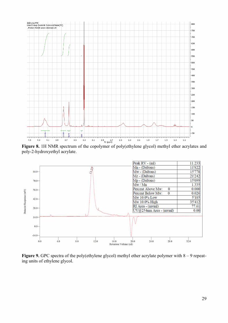
1H NMR (Figure 6). The GPC measurements showed that the polymer has an average Mn of 12000
and a PDI of 1.3, in average polyPEGA contained 20 repeating units of poly(ethylene glycol)methyl
ether acrylate (PEGA) monomer (Figure 9).
Other monomers with different numbers of PEG repeating units were tested, for example polymers
with shorter side chains with only three PEG repeating units are not adequately water soluble and
they can only be dissolved in water at low temperature, while polymers with longer side chains with
18 to 20 repeating units can be prepared, but the polydispersity is higher (Figure 7). The
poly(ethylene glycol)methyl ether acrylate (PEGA) monomers were also copolymerized with
2-hydroxyethyl acrylate (HEA) monomers (Figure 8). The addition of shorter chains may increase
the binding of antibodies, because they provide more space for the antibodies to bind. The problem
with these copolymers is that they form hydrogels. The copolymerisation is important in order to
create a polymer brush that is not specifically prepared for binding PEG-binding antibodies, but
26
also for binding other molecules. Such as tris-nitrilotriacetic acid that provides binding sites for
other molecules.
The PEG monomer can be copolymerized with other molecules that contain an acrylate or
methacrylate group that contains binding sites for other molecules. The copolymers may provide
binding sites for other molecules and maintain their surface passivation abilities and, as a result,
many different monomers can be used, because PEG is soluble in many different solvents. In order
to attach the polymer to the surface and create a polymer brush, the polymer should contain a
terminal thiol group that can be attached to a gold surface.
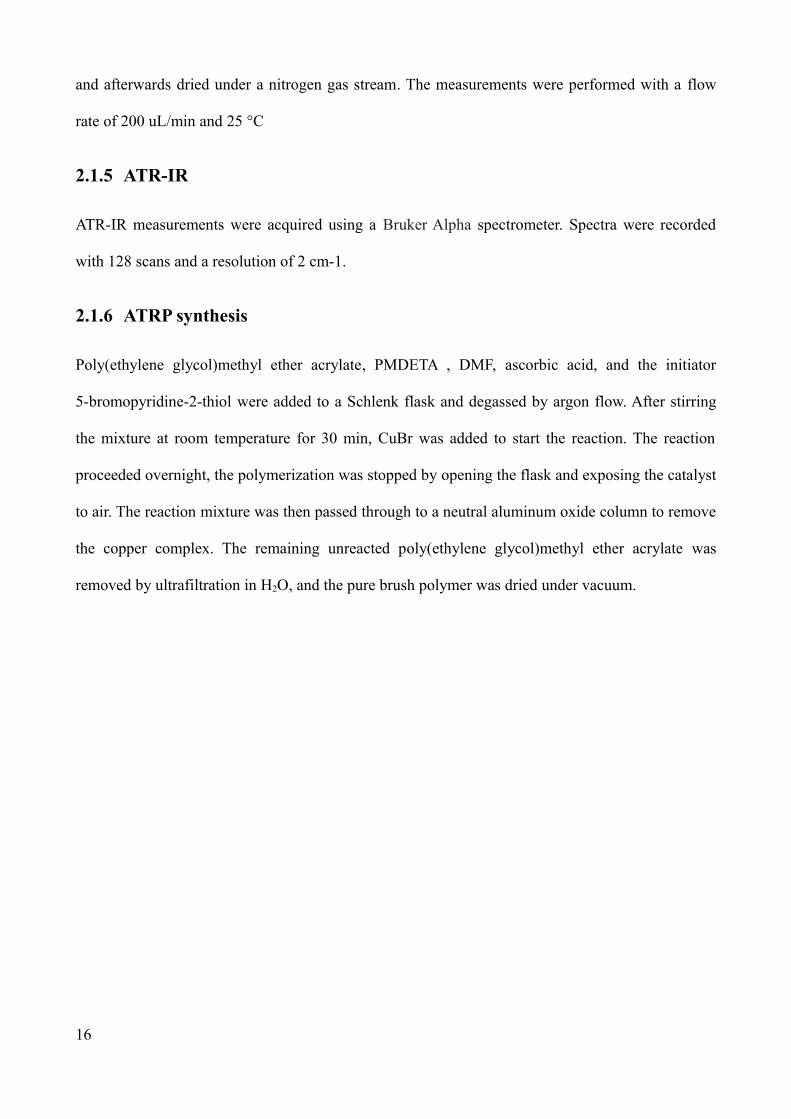
Figure 7. 1H NMR spectrum of the poly(ethylene glycol) methyl ether acrylate polymer with 8 – 9 repeating units of ethylene glycol .
27
Figure 7. 1H NMR spectrum of the poly(ethylene glycol) methyl ether acrylate polymer with 18 – 20 repeating units of ethylene glycol.
28
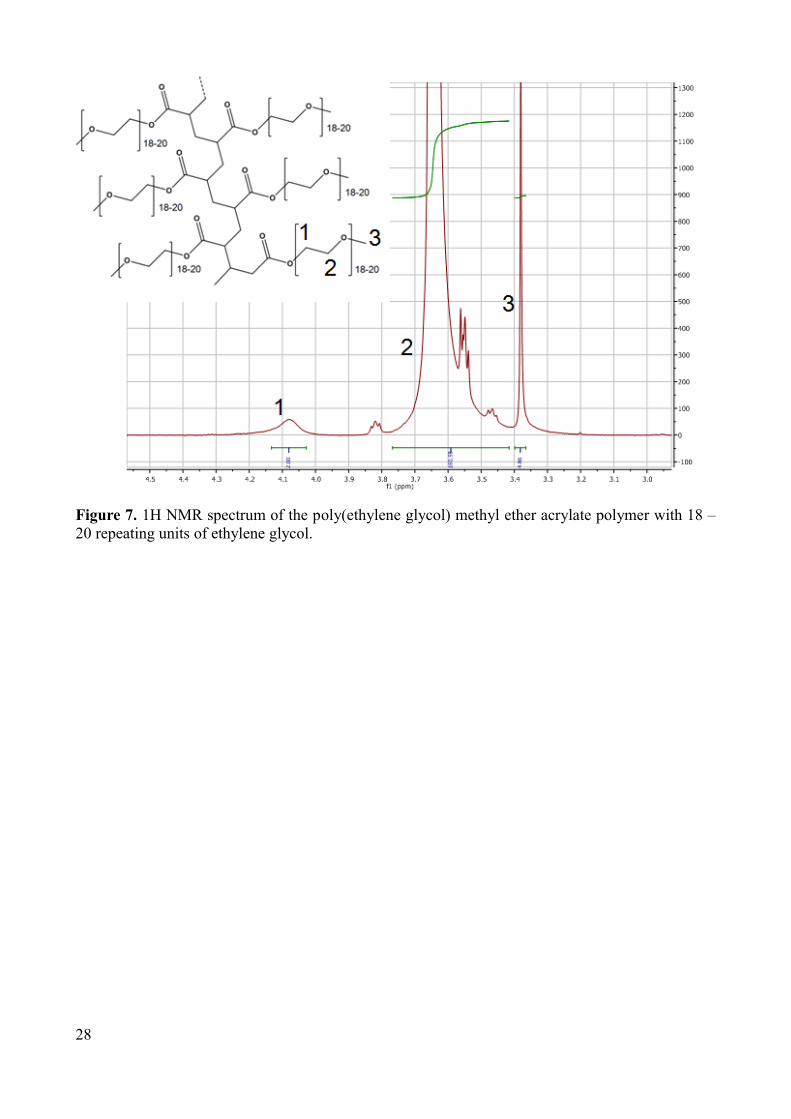
Figure 8. 1H NMR spectrum of the copolymer of poly(ethylene glycol) methyl ether acrylates and poly-2-hydroxyethyl acrylate.
Figure 9. GPC spectra of the poly(ethylene glycol) methyl ether acrylate polymer with 8 – 9 repeating units of ethylene glycol.
29
3.1.5 ARGET ATRP
The method used for the polymerization of poly(ethylene glycol)methyl ether acrylate (PEOA)
monomers was atom transfer radical polymerization ARGET ATRP (Scheme 1). This method is one
of the most successfully controlled/living radical polymerizations (CRP),83 due to its robust nature
and to the possibility of controlling molecular weight and providing a narrow molecular weight
distribution Mw/Mn, < 1.5. It enables synthesis of a wide spectrum of polymers and allows the
controlled synthesis of polymers with a branched structure with different chains, leading to different
architectures such as block-, random-, gradient-, comb-shaped-, brush-, multiarmed-, end-
functional-, and star copolymers, increasing in this way the number of multiple applications.
In addition to the wide variety of monomers that can be polymerized with ATRP it is also possible
to use a wide variety of non-organic and organic solvents.90 ATRP has been successfully used for the
polymerization of PEG-containing copolymers91-93
Scheme 1. Polymerization reaction of the monomers.
30
3.1.6 Polymer Brush
The brushes were formed on a gold surface (Figure 9). A gold surface was used because it can bind
strongly to thiols and disulfides. The polymer brush can be produced with two different methods:
the “grafting from” and the “grafting to” method. The method that was used was the “grafting to”
method because it allows the synthesis of the polymer in solution and the final polymer can then be
easily grafted onto the surface by simply deposition. The “grafting from” method was not used be
cause it is more complicated, due to the polymerization all being done on the surface. The advan
tage of the “grafting from” method is that it allows a higher density of the polymer brush. The ex
periments have shown that the grafting density with the “grafting to” method is high enough. The
brush density is an important point when it comes to the surface passivation and the binding of the
PEG-binding antibodies. The surface has to be covered with a brush that is dense enough so that no
other molecule can bind to the surface. For the binding of the antibodies on the other side, a low
grafting density is favorable because the antibodies need space to bind to the binding sites of the
polymer. The polymer that was used has a different structure than other polymers that have previ
ously been grafted on the surface. The influence of the different structure also has an influence on
the brush formation.
In order to confirm that deposition of the polymer on the surface, the sample was analyzed with at
tenuated total reflection infrared spectroscopy (ATR-IR), a blank gold slide was measured as a ref
erence, for which no adsorption bands could be detected. The spectra of the polymer monolayers on
gold were recorded. ATR-IR measurements can yield information on the structure of the polymer
that is attached to the surface. The ATR-IR measurements confirmed that the polymer is attached to
the surface. The peak at 1117 cm-1 shows the ether bonds of the PEG side chains and the bonds at
1570 cm-1 show the ester group (Figure 10).
31
Figure 10. ATR-IR spectra of the polymer brush-coated gold surface with the bonds from the ester group at 1570 cm-1 and ether bonds at 1117 cm-1
The monolayer formation was also characterized by surface plasmon resonance spectroscopy
(SPR). This optical method allows for non-invasive thin film characterization and is very sensitive
to small changes in adsorbed mass. In order to form a polymer brush on the surface, the polymer
was dissolved in PBS buffer and then deposited on the gold surface. After washing, a film remained
on the surface. The same procedure was repeated and the amount that was deposited after the sec
ond cycle was low. This showed that the surface was already covered with the polymer film. No fur
ther polymer was able to attach to the surface (Figure 11). The thickness of the polymer brush was
calculated to be 10.2 nm ± 2.5 nm. In a second step, BSA was added to the polymer film. BSA is a
protein with high, unspecific binding and it attaches to nearly any surface. The measurements
showed that BSA was not able to attach to the surface. All of the deposited BSA on the surface was
removed by flushing the surface with buffer (Figure 12). This result showed that the BSA cannot
attach to a polymer coated surface. From this, we conclude that the polymer brush has high passiva
tion ability. The passivation indicates that there is no free surface to which the BSA can bind.
32
Figure 11. SPR spectrum of the adsorption of the polymer to a gold surface. The polymer solution was deposited on the sensor (B), which changes the frequency compared to the baseline (A). Then, the polymer was able to graft onto the surface for 5 min. The sensors were then flushed again with water to remove the free polymer that could not graft to the surface (C). In a second step the polymer solution was added again to the sensor, then the polymer was able to graft onto the surface for another 30 min (D). The sensors were then flushed again with water to remove the free polymer that was not attached to the surface (E).
Figure 12. BSA adsorption to a polymer-coated gold surface
33
A commercially available PEG 20k was used as a reference to compare brush formation. It has been
reported that PEG 20k is able to passivate a gold surface and it is also able to bind PEG
antibodies.94 PEG 20k is known to form dense brushes on the surface. Our polymer showed similar
results to PEG 20k. The antibodies were placed on the polymer coated surface. The PEG side chains
provide binding points for the antibody surface. With SPR, an antibody binding curve was
observed. The SPR data shows that the antibodies can bind to the polymer coated surface (Figure
13). The equilibrium dissociation constant KD of the antibody binding was calculated by fitting Req
to the Langmuir adsorption isotherm95 giving KD = 0.77 ± 0.93 nM (Figure 14). The calculation
does not provide good results because the amount of data from the antibody binding curve that
could be used was small.
Figure 13. Antipeg binding curve for a polymer-coated surface.
34
Figure 14. Antipeg binding curve fitted to the Langmuir adsorption isotherm.
In addition to the SPR measurements, QCM measurements have also been done to analyze brush
formation and surface passivation. QCM measures mass per unit area by detecting the change in
frequency of a quartz crystal resonator. The resonance is disturbed by the addition of molecules that
bind to the surface. As a mass is deposited on the surface of the crystal, the thickness increases and
the frequency of oscillation decreases from the initial value. This frequency change can be
quantified and correlated to the mass change using Sauerbrey's equation.96 In this experiment, a
sensor with a gold surface was used. The polymer was dissolved in water at a concentration of 1
mg/mL and deposited on the sensor.
The polymer was then able to graft onto the surface for 30 min. The sensors were than flushed again
with water to remove the free polymer that wasn’t grafted to the surface. The change in the
frequency shows the amount of polymer that was attached to the surface. By comparing the
frequencies before the addition of the polymer solution and after the flushing of the sensor with
water, the mass that was attached to the surface can be obtained. The change in mass on the sensor
shows that a brush with a thickness of 6.2 nm ± 2.5nm was created. The QCM measurements show
that the polymer was bound to the surface. In a second step, BSA was added to the polymer film.
The measurements showed that BSA was not able to attach to the surface. All of the deposited BSA
on the surface was removed by flushing the surface with water (Figure 15)
35
Figure 15. QCM spectra of the adsorption of the polymer and BSA to the gold surface. The polymer solution was deposited on the sensor (B), which changed its frequency compared with the baseline (A). The polymer was then able to graft to the surface for 30 min. The sensors were then flushed again with water to remove the free polymer that wasn’t grafted to the surface (C). The BSA solution was deposited on the polymer coated sensor (D).
Then, the BSA was able to graft onto the surface for 30 min. The sensors were then flushed again
with water to remove the free BSA that wasn’t attached to the surface (E).
36
3.1.7 Conclusion: Polymer brush
Here, we have synthesized and characterized a responsive polymer brush with dual-functionality:
surface passivation and with selective binding sites for specific molecules. We selected a polymer
with branched architecture because of its ability for further modification and copolymerization. The
polymer was successfully synthesized by ARGET ATRP. The polymer structure was confirmed by
1HNMR. The GPC measurements showed that the polymer on average has 20 repeating units of the
poly(ethylene glycol)methyl ether acrylate (PEGA) monomer. The polymer was able to be
deposited on the surface via the grafting to method, while the QCM and SPR measurements showed
that a 6.2 – 10.2 nm thick polymer brush was deposited on the gold surface.
The polymer has been shown to be efficient at passivation. Surface passivation abilities were
proven by SPR and QCM. In addition to the passivation, the system also provides many possible
binding sites. An antibody binding curve was recorded by SPR, indicating clearly that the antibody
was able to bind to the polymer brush. This new system was able to achieve higher selectivity,
which means low unspecific binding and high specific binding.
37
3.2 Hemoglobin nanoreactors
The aim of the project was to generate a nanoreactor with the dual functionality of oxygen transport
and peroxynitrite reduction. Therefore, a substance that had the ability to transport oxygen and to
degrade peroxynitrite was needed. Hb was used as a model protein because Hb has the ability to
transport oxygen and reduce peroxynitrite. The advantage of Hb is that it is commercially available,
has a good stability, and its activity can be detected by UV- absorption measurements. That allows
the study of the protein inside the nanoreactor.
3.2.1 Polymers used for nanoreactor preparation
All nanoreactors used for this study were obtained from PMOXA-b-PDMS-b-PMOXA triblock
copolymer self-assembly (Figure 16). PMOXA-b-PDMS-b-PMOXA triblock copolymers were
previously reported in the literature as suitable for drug delivery, due to their increased stability and
non-toxicity. The triblock copolymer nanoreactors were proven to be impermeable to hydrophilic
cargo99 and prevented it from leaking into in the reaction medium.100 With a proven record of good
biocompatibility,101 the membrane of PMOXA-b-PDMS-b-PMOXA nanoreactors possess high
flexibility that allows for the insertion of channel proteins, such as OmpF, LamB, or Aquaporin Z
without affecting their proper functionality. The incorporation of channel proteins within the vesicle
membrane is essential to allow substances/products to penetrate through and support the
nanoreactor functions. The chemical structure of the polymer used for preparing the nanoreactors
has a major influence on both the shape and size of the nanoeactors and their ability to encapsulate
molecules. For encapsulation of hemoglobin, nanoreactors with a radius of 100 nm were preferred.
It is important that the polymer forms nanoreactors in higher numbers, high enough to encapsulate a
reasonable amount of Hb molecules. The nanoreactors were formed by copolymer self-assembly in
phosphate buffer. For the formation of nanoobjects with defined shapes such as micelles, rods,
38
vesicles or aggregates, the ratio between the length of the hydrophilic and the lengths of the
hydrophobic blocks is crucial. The selection of a specific ratio defines the shape of the nanoobjects
and their sizes. In order to determine the optimal ratio for vesicle formation, different PMOXA-b-
PDMS-b-PMOXA copolymers with various ratios of hydrophilic to hydrophobic segments were
tested. In addition to their ability to form vesicles, the polymer also has to fulfill other requirements.
It is important that the polymer not interact with the molecules that will be encapsulated. An
interaction of the polymer with the guest might interrupt vesicle formation. This kind of interaction
usually occurs if the polymer and the substances are charged. As encapsulated molecules can be pH
dependent, the encapsulation will work best at specific pH values.76 The stability of the vesicles is
important, because a nanoreactor is designed to circulate in the bloodstream for a long time. This is
why it is crucial that the polymer form mainly vesicles and less so aggregates or rods. The polymer
has to form nanoreactors with low size polydispersity. Several experiments have been conducted in
order to decide which polymer to use for further experiments. All vesicle solutions obtained from
the different PMOXA-b-PDMS-b-PMOXA copolymers were analyzed by TEM and FCS. TEM
measurements give an overall image that shows whether a polymer forms vesicles, micelles, rods or
aggregates, and also provides information about their size. In FCS, only nanoobjects that
encapsulate fluorescent active molecules can be seen. Those nanoobjects, such as micelles or rods,
that do not encapsulate fluorescent active molecules cannot be detected by FCS. FCS measurements
indicate whether the produced nanoobjects can encapsulate Hb. These methods were used to
analyze the vesicle solution formed by the different polymers and to choose the most suitable
polymer. More detailed analyses were carried out with the chosen polymer.
39
Figure 16. Chemical structure of PMOXA-b-PDMS-b-PMOXA consisting of the two hydrophilic PMOXA and one hydrophobic PDMS block. 1H NMR spectra of the PMOXA-b-PDMS-b-PMOXA copolymer.
The shape and size distribution of polymer nanoreactors can be designed by choosing the optimal
ratio of PMOXA to PDMs blocks. So even though the tested polymers only differ from the point of
view of their lengths of the hydrophobic and hydrophilic blocks, their outcomes differ greatly.
Every polymer has a specific tendency to form rods, micelles, vesicles or aggregates. This affects
the encapsulation efficiency, because micelles or rods are not able to incorporate Hb.
The first polymer that was tested was an A10B87A10 triblock copolymer. The vesicles were prepared
using film rehydration. After the preparation and purification steps the polymer solutions were
analyzed with TEM (Figure 17) and FCS. From the FCS measurements, information on the size of
the vesicles and the encapsulation efficiency were obtained. The TEM images provided information
on the shape and size of vesicles, micelles, rods or other aggregates. For the proper function of the
nanoreactors, it is important to have a vesicle solution with specific, narrow size distribution,
40
because only vesicles with a certain diameter of a minimum of 50 nm have been shown to
encapsulate hemoglobin. Because they are too small, micelles are not suitable for the encapsulation
of hemoglobin and hence they reduce the encapsulation efficiency. For A10B87A10 copolymers, the
obtained vesicle solution was not homogeneous, but contained fractions of vesicles, rods, micelles
and aggregates. Only the vesicles fraction was able to incorporate labeled Hb, as the FCS data
confirmed, while the fractions of micelles and rods formed do not encapsulate Hb.
This polymer could not be used further for the preparation of nanoreactors because of the high
percentage of aggregates that were formed. The aggregates consist of small vesicles or micelles that
stick together to form bigger structures. Aggregate formation can be caused by interactions between
the polymers or polymer with hemoglobin. Therefore, it is difficult to analyze whether labeled Hb
was successfully encapsulated into the polymer vesicles.
Figure 17. TEM images from the A10B87A10 copolymer showing vesicles, micelles and rods.
A second polymer tested was an A12B55A12 block copolymer. This was the first polymer that showed
promise for nanoreactor preparation. TEM pictures indicated that mainly vesicles were formed by
A12B55A12 self-assembly (Figure 18). The advantage of this polymer compared with those previous
tested is that it did not form large aggregates and the formed vesicles were able to incorporate
hemoglobin. In addition to the vesicles, the polymer also formed micelles and rods. Despite the
formation of micelles and rods, the main reason why this polymer was not used for further studies
41
was that there was not enough polymer to proceed with the project.
Figure 18. TEM images from the A12B55A12 copolymer showing vesicles and micelles.
The polymer tested finally was an A6B90A6 block copolymer. The polymer consists of two blocks of
six repeating units of PMOXA and 90 repeating units of PDMS. The TEM images of the A6B90A6
copolymer showed the most promising results (Figure 19). The copolymer has a large hydrophobic
block compared to the hydrophilic block. This polymer forms mainly vesicles and some micelles.
No rods or aggregates were formed. The solution contained a good amount of vesicles that were
large enough to encapsulate a reasonable amount of hemoglobin. The vesicles were larger than
those generated by the other polymers. Although all of the polymers used were able to incorporate
some quantity of Hb, some polymers, such as the A10B87A10, could not be used because of the
formation of large aggregates. The PMOXA6-PDMS90-PMOXA6 polymer has been shown to form a
narrow size distribution population in solution. Because of its ability to form mainly vesicles, this
polymer was chosen for further research. In addition to the composition of the polymer, the
preparation method also has great influence on the formation of vesicles. For the chosen polymer,
the vesicles were prepared using two different methods. The first was the film rehydration method.
For this, 5 mg of triblock copolymer were dissolved in ethanol and the solution was then evaporated
under reduced pressure. The polymer formed a thin film on the walls of a 25 ml round-bottom flask
using a rotary evaporator. After the addition of 1 ml PBS buffer to the polymer film and stirring for
42
2 h, the solution was extruded through polycarbonate membranes (pore diameter = 400 nm) in order
to generate vesicles with low size distribution. During the extrusion, the non-dissolved polymer was
also removed. Both empty vesicles and protein-containing vesicles were prepared under similar
conditions using 5 mg of polymer and PBS buffer. The vesicles were analysed by light scattering
(dynamic light scattering, DLS, and static light scattering, SLS) and transmission electron
microscopy, TEM. The other method that was used was the co-solvent method. In this method the
polymer was dissolved in a small amount of ethanol and then it was added slowly into a buffer
solution while stirring. The advantage of this approach is that it provides a more homogeneous
vesicle solution than the film rehydration method, while nearly 100% of the polymer used forms
vesicles or micelles. The problem with this method was that it only produced micelles and small
vesicles with a radius of around 25 nm (Figure 20). The vesicles were thus too small to encapsulate
Hb. This method was not used for any further experiments.
Not only does the method used have a great influence on the vesicle formation, but the conditions
(temperature, stirring time and stirring intensity) during vesicle formation has a great influence on
the final result. The different polymers behave differently and their ideal conditions depend strongly
on their hydrophilic hydrophobic block ratio. Therefore, vesicle formation was performed under
several different conditions in order to find the most suitable one for the requirements of the
nanoreactors. For most of the polymers, a long stirring time of more than 8 h was optimal because
this gave the polymer more time to self- assemble in order to form vesicles. However, extended
stirring time is not always preferable because of the mechanical force that is applied during the
stirring process. Therefore, a long stirring time is not ideal if the copolymer has a high
hydrophobicity. The polymer used has both a high hydrophobicity and a tendency to clump together
and precipitate after extended stirring. The ideal condition for nanoreactor formation was a stirring
time of around 2 h at room temperature. One disadvantage of the short stirring time is that only 50%
of the polymer forms vesicles or micelles, the rest of the polymer stays on the polymer film on the
43
bottom of the flask.
Figure 19. TEM images of A6B90A6 copolymer of empty vesicles (A), and vesicles with encapsulated metHb (B). Scale bar = 200 nm.
Figure 20. TEM images of A6B90A6 copolymer micelles that are produced by the co-solvent method.
The A6B90A6 copolymer behavior in solution was studied in more detail with DLS/SLS. The
measurements were necessary because they provided additional data to characterize nanoreactors in
solution.
Static Light Scattering (SLS). SLS is a technique that uses the intensities of scattered light at a
number of angles to derive information on the nanoobjects in the solution. From light scattering,
data on the radius of gyration Rg and the second virial coefficient A2 can be derived (Figure 21).
104-107
44
Figure 21.Second order fits of SLS data in a Guinier plot of nanoreactors (A) and empty vesicles (B).
Dynamic Light Scattering (DLS). Dynamic light scattering uses the concept of small particles
moving randomly in solution. Fluctuations in the scattering arise from the fact that small molecules
in solution undergo Brownian motion. Brownian motion causes a change in the distance between
the scattering objects in the solution. The change in the distance leads to either constructive or
destructive interference by the surrounding particles. The information on the nanoobjects is derived
from an autocorrelation function of the intensity trace recorded during the experiment (Figure 22).
45
Figure 22.Second order fits of DLS by nanoreactors (A) and empty vesicles (B).
The analysis of the samples by DLS and SLS indicates the presence of two different populations of
nanoobjects: a small fraction of spherical objects with a hydrodynamic radius (RH) of around 45 nm,
and a larger fraction of spherical objects with a hydrodynamic radius of RH= 164 ± 15.4 nm. The
small RH of around 45 nm of the first fraction indicates micelles. The light scattering from the
micelles fraction was not as dominant, and the influence of the scattering was therefore not
significant. The majority of the scattering occurs from the fraction with the larger RH of 164 nm.
The analysis of the samples by SLS showed that the fraction of the larger spherical objects has a
radius of gyration Rg of 194 ± 8.2 nm. In order to analyze whether this population consists of
vesicles, the ratios of the hydrodynamic radius RH and the radius of gyration Rg were compared as
46
RH/Rg = 0.84. A value of 0.84 indicates that the major population consists of spherical vesicles. In
order to analyze whether the encapsulation of metHb has an influence on vesicle formation, the
DLS and SLS data of empty vesicles and vesicles that contain metHb were compared. A comparison
of the DLS data shows that the presence of metHb does not influence vesicle formation. Therefore,
there is no interaction between the polymer and the metHb. The samples were also analyzed by
Transmission Electron Microscopy, TEM. The TEM micrographs indicate the presence of circular
objects with a mean radius of around 28 ± 8.5 nm and spherical objects with a mean radius of 120 ±
20 nm. From the small radius of 28 ± 8.5 nm of the first fraction, we assume them to be micelles.
The fraction of spherical objects with a mean radius of 120 ± 20 nm was expected to be vesicles.
The higher value RH obtained by LS experiments as compared to the size of the vesicles estimated
from the TEM micrographs was expected. The higher value from DLS experiments represents the
sum of the particle radii and the contribution of the particle’s surrounding hydration sphere,
whereby the TEM micrographs only show the size of the actual vesicle. The influence of the
encapsulation of metHb and the incorporation of OmpF into the polymer membrane were also
studied by TEM. TEM micrographs of metHb-containing vesicles or metHb-containing vesicles
with OmpF incorporated in the polymer membrane were compared to a solution of empty vesicles
and did not show any significant morphological change. This is in good agreement with other
protein-polymer nanoreactors based on this type of amphiphilic copolymer .74 The stability of the
vesicles was analyzed by measuring the sample again after storage for more than three weeks at 4
°C. The TEM micrographs revealed no significant changes. This suggests that the vesicles prepared
from PMOXA6-PDMS90-PMOXA6 polymers are mechanically stable.
3.2.2 Protein encapsulation efficiency
The activity of the nanoreactor is affected by the encapsulation efficiency. It is important to have a
reasonable number of Hb molecules inside the nanoreactors in order to obtain reasonable reaction
efficiency. It is therefore important to know how many Hb molecules are encapsulated inside the
47
nanoreactors.
In order to quantify the actual amount of Hb that is encapsulated inside the nanoreactors and the
remaining free Hb, the sample was further analyzed.
FCS was used to determine whether metHb was encapsulated inside the vesicles or if it was only
outside the vesicle. In addition FCS measurements performed after sample purification allowed us
to quantify the free Hb ratio and the efficiency of separation. For these measurements, metHb was
labeled with the Alexa488 fluorescent dye. The labeling was necessary because the FCS
measurements are based on the laser-induced fluorescence of excited fluorescent molecules. The
fluorescent labeled Hb molecules are measured when they pass through the confocal volume. The
signal is then auto-correlated in time and allows the calculation of the diffusion times. From the
diffusion times the size of the vesicle can be calculated, because the encapsulated fluorescent
molecule has the same diffusion time as the vesicle. The hydrodynamic radius of the vesicle can be
obtained from the diffusion time by the use of the Stokes-Einstein equation. This method has been
used for the analysis of fluorescently-labeled proteins in nanovesicles. The labeled Hb was analyzed
under free conditions and encapsulated in the nanoreactors. The diffusion time of the Alexa488 dye,
the Alexa488 labeled Hb, and the encapsulated Alexa488-labeled metHb in polymer vesicles were
measured. The diffusion times of the Alexa488-labeled metHb encapsulated in polymer vesicles
was compared to the diffusion time of the free Alexa488-labeled protein.110 The free Alexa488 dye
had a diffusion time of around 26 µs, while the Alexa488-labeled metHb featured a diffusion time
of 95 µs (Figure 23 a and b). From the diffusion time of 95 µs, the hydrodynamic radius of 2.5 nm
for the Alexa488-labeled metHb was calculated. The calculated hydrodynamic radius of Alexa488-
metHb was in good agreement with the reported value of 3.1 nm.111 The analyzed solution contained
vesicles and Alexa488-labeled metHb. The obtained data was fitted for several different
populations. The best fit that was obtained (Figure 23 c), indicating the presence of two fractions
with different diffusing times. There is a smaller fraction (2%) with a diffusing time of 95 µs and a
48
major fraction with an average diffusion time of around 6930 µs (98%). The diffusion time of 95 µs
for the smaller fraction indicates that it is the free Alexa488-labeled metHb. The major fraction with
a diffusion time of 6930 µs corresponds to vesicles with a calculated hydrodynamic radius of
around 170 nm ± 15 nm. The hydrodynamic radius obtained by the FCS measurements is in good
agreement with the LS results. To analyze whether the Hb was encapsulated in the vesicles or
whether the Hb was attached to the outside of the vesicle, Hb was added in the same concentration
as used for the nanoreactor preparation, to a vesicle solution that contained no Hb. The diffusion
time of the free Alexa488-metHb was not modified by the addition of empty vesicles. This indicates
that metHb was not attached to the outside of the polymeric membrane of the vesicles.
Figure 23. Autocorrelation function of free Alexa488 (a), Alexa488- metHb (b), and Alexa488-metHb-containing vesicles (c)
To determine the encapsulation efficiency, we first evaluated the average number of Alexa488
molecules connected to one metHb molecule. The average number was estimated by the UV
spectra. For this, the absorption spectra of the Alexa488-labeled Hb in solution was analyzed. The
absorption of the Alexa488 was compared to the absorption of the Soret band of metHb. On
average, 2.3 dye molecules were attached per metHb molecule. The number of molecules that were
encapsulated in the nanoreactor was calculated accordingly.
The number of Alexa488-metHb molecules per vesicle was estimated via brightness measurements.
49
In this respect, we compared the count rates per molecule (cpm, in kHz) of Alexa488, Alexa488-
metHb, and encapsulated Alexa488-metHb.
The free Alexa488 dye has a count rate of around 22.81CPM. It decreases to 14.59 CPM when the
dye is attached to Hb molecules. The nanoreactors have an average count rate of 829.45 CPM. The
count rate of the nanoreactors depends on the number of encapsulated Hb molecules. The count rate
corresponded to an average of 55 molecules of Alexa488 labeled Hb. The number of Alexa488-
metHb molecules per vesicle, obtained from brightness experiments (for an initial concentration of
metHb of 0.47 µM/mL) was then compared to the theoretical number of metHb/vesicle (120
molecules/vesicles), and resulted in an encapsulation efficiency of around 20.6 %.
3.2.3 Conclusion: Nanoreactors
Finding a good polymer that forms vesicles that are able to function as a nanoreactor is difficult. In
order to be used for biomedical applications, nanoreactors must fulfill some crucial requirements.
The nanoreactor has to be stable for storage; the vesicle solution has to be homogenous and the
vesicles should not form large aggregates; the vesicles must be able to encapsulate a reasonable
amount of guest, in our case modified Hb; the purification methods must be efficient enough to
remove the majority of the non-encapsulated Hb. In this study we showed that nanoreactors made of
A6B90A6 copolymer could easily be produced using the film re-hydration method. Such nanoreactors
are mechanically stable and can be stored for several weeks. The nanoreactors and the empty
vesicles did not differ by size or shape, as indicated by TEM or DLS/SLS. That shows that Hb does
not interfere with vesicle formation. The polymer mainly forms vesicles and a smaller fraction of
micelles, with the vesicles being the dominant species. The polymer did not form aggregates. The
obtained vesicles have an average radius of the120 nm. A well defined vesicle solution was
obtained. The FCS data show that the purification method removes nearly all of the non-
encapsulated Hb. A good encapsulation efficiency of around 20.6 %, with hardly any free Hb in the
solution, was determined.
50
3.2.4 Activity studies
For the preparation of nanoreactors it is not only important that the polymer be able to produce
stable vesicles, but also that they incorporate Hb. Moreover, the encapsulated, active guest should
maintain its function inside the nanoreactor. Therefore, the functionality of the nanoreactor needs to
be demonstrated. Thus, one of the key steps in this project was to investigate the activity of the
nanoreactor. The activity of the nanoreactor strongly depends on the activity of the encapsulated
molecules. One of the major differences between a nanocarrier and a nanoreactor is that the
encapsulated molecules must retain their activity inside the nanoreactor, whereas nanocarriers
require activity upon release. On the other hand, nanoreactors do not need controlled release of the
encapsulated molecule. For every nanoreactor, it is important to investigate the activity of its
encapsulated molecules. This study is important in demonstrating that the encapsulation procedure
does not damage the hemoglobin. The encapsulation could force a change in the conformation of
the hemoglobin that would cause a significant decrease in the activity when encapsulated. Another
problem could arise from strong, unspecific interactions with the polymer membrane that could also
lead to a decrease in the activity of the hemoglobin inside the nanoreactor. And yet another problem
might be that, in order to be functional, the heme pockets of the hemoglobin must still be accessible
to the chemicals that have to be detoxified. In our case, peroxynitrite must be able to enter the
nanoreactor; otherwise, no detoxification could occur.
The activity was measured by changing the oxidation state of Hb ( Scheme 2). The structural
information on the oxidation state was achieved using UV-Vis absorption spectroscopy. The Soret
absorption band was examined for metHb, HbCO, HbO2, and deoxyHb (Figure 25 A, right). The
other bands were used only to verify the reactions in the case of protein solution (Figure 24). We
did not consider the other bands (between 450 nm and 600 nm) because, even if they appear in bulk
conditions, they cannot be observed in the conditions of the nanoreactors due to the low
concentration of the protein inside nanoreactors. The transition from metHb to deoxyHb was
51
observed by a shift in the maximum of the Soret absorption band from 405 – to 430 nm (Figure 25
A right), while, for metHb to HbCO, the shift was from 405 – to 419 nm. The conversion of
deoxyHb to HbO2 after removal of sodium dithionite excess in the presence of oxygen has been
demonstrated by a shift of the maximum of the Soret absorption band from 430 – to 415 nm. 76
A study of the different oxidation states was not only important to show the activity of the
encapsulated hemoglobin, but also to understand what happens on a molecular level. Because the
different oxidation states have different activities, it is vital to change the oxidation state in a
controlled way. Not all oxidation states have the same activity and therefore it is problematic when
hemoglobin transforms into a less active state. MetHb is one of the most stable oxidation states, but
it cannot be used for the nanoreactors. MetHb cannot carry oxygen and it is not able to react with
peroxynitrite and cannot detoxify it. In the bloodstream there are many substances that prevent
HbO2 and deoxyHb from forming metHb. Without these substances, the HbO2 and the deoxyHb will
transform into the nonreactive metHb state.
52
Scheme 2. Conversion of Hb to different oxidation states in solution and in situ inside nanoreactors.
Figure 24. UV spectra of metHb (black), HbO2 (blue), HbCO (green) and deoxyHb (red) at wavelengths between 350 – 750 nm
3.2.5 Free Hemoglobin
One major problem in studying the activity of hemoglobin inside nanoreactors is the fact that the
hemoglobin concentration inside the nanoractor is very low. This limits the possibilities for
spectroscopic measurements. Another difficulty is that the vesicles scatter light in the area where
hemoglobin has the highest absorption, thereby affecting the final results. The redox-reaction of Hb
can be best studied in solution. The concentrations of Hb that can be analyzed are much higher and
53
therefore the signals obtained by the analytic methods, UV/Vis and Raman, are much more reliable.
It is possible to observe changes in the spectra that could not be observed with a nanoreactor
solution because the signal is too weak. Another advantage is that reaction conditions can be
changed. The optimization of the reaction conditions was therefore achieved in bulk solution. The
measurements of the redox-reaction with the Hb inside the nanoreactors were carried out in the
same condition as in solution. The advantage of the activity measurements in solution is also that
the results can be compared with results that are obtained by measuring the activity of hemoglobin
inside the nanoreactors. The comparison of the data is also important because it allows predicting
the influence of the polymer membrane. The permeability of the polymer membrane also has an
influence, because it can act like a barrier to entering molecules which, in turn, react with the
molecules inside the nanoreactor.
3.2.6 Hemoglobin in nanoreactors.
After the preparation the nanoreactors were purified. The purification was necessary because most
of the Hb present in the solution is not encapsulated in the nanoreactor. It is essential to have a good
purification procedure in order to get rid of the majority of the free hemoglobin. The non-
encapsulated Hb interferes with the measurements and provides incorrect readings, because the
activity measurements show the activity of the whole system and not simply that of the
encapsulated hemoglobin. Therefore, it is important to reduce the amount of nonencapsulated Hb to
a level where it does not have a significant impact. The nonencapsulated Hb was removed by SEC
as mentioned previously. The amount of nonencapsulated Hb was estimated using FCS
measurements. The remaining, nonencapsulated Hb was minimal (2%), so it had no impact on the
next measurements. Moreover, the purification method did not decrease the amount of nanoreactors
obtained.
54
3.2.7 Characterisation of nanoreactors reactivity
In contrast to the characterisation of the nanoreactors performed by TEM, DLS, and FCS, the
estimation of the activity of the encapsulated Hb is more difficult. A major drawback to the
investigation of nanoreactor activity was their low concentration and the limited amount of
encapsulated Hb that was available. The limited amount of Hb significantly reduced the variety of
analytical methods that could be used to investigate the activity of the nanoreactor. Therefore,
Raman spectroscopy could not be used for the analysis of the oxidation state of Hb in nanoreactors.
(The main analysis methods are based on absorption measurements in the UV region.)
The ability to change the oxidation state is crucial for the two key functions of the nanoreactor.
We chose to start with metHb, due to its stability and availability, and to convert it in situ, inside the
nanoreactors, to deoxyhemoglobin (deoxyHb) and carbonmonoxyhemoglobin (HbCO),
respectively, followed by the inverse reaction in the presence of peroxynitrites. Previous studies
have shown that peroxynitrite is degraded by the conversion reaction of HbO2 to metHb. The
conversion to different oxidation states of hemoglobin was studied with spectroscopic techniques,
both in bulk and inside nanoreactors. The conversion reactions in bulk served to optimize the
reaction conditions previous to the encapsulation.
In order to obtain different oxidation states of hemoglobin, several different methods to convert
metHb to HbO2, and to deoxyHb, and to HbCO(Figure 24) have been used.
3.2.8 Reducing agents
Because metHb has the highest oxidation state, there is always the need to use a reducing agent to
convert metHb to a different oxidation state. The first reducing agent used was ascorbic acid.
Ascorbic acid is present in the blood stream and is one of the natural reducing agents for Hb. The
problems with ascorbic acid are that it is not ideal for reducing metHb, because it is not strong
enough to reduce metHb by itself. Ascorbic acid has to be used in combination with other
55
substances to reduce metHb. It is also not possible to achieve every oxidation state of Hb with
ascorbic acid as the reducing agent. In order to achieve the more relevant oxidation states such as
HbO2 and deoxyHb, another reducing agent has to be used. The second problem with ascorbic acid
is that ascorbic acid is too big to penetrate the polymer membrane of the nanoreactor. It therefore
needs tunnel proteins such as OmpF to enter the nanoreactor. Because of these problems with
ascorbic acid, sodium dithionite was used as a reducing agent. Sodium dithionite is a stronger
reducing agent then ascorbic acid. Another advantage of sodium dithionite is that it can penetrate
the polymer membrane without the help of tunnel proteins. Sodium dithionite is strong enough to
reduce metHb to deoxyHb when sodium dithionite is added.
The problem with sodium dithionite are that it is not stable in aqueous solution. Thus, an excess of
sodium dithionite had to be added to the metHb solution to completely reduce it to deoxyHb.
Sodium dithionite is also a reactive species that can interact with the Hb. Therefore, if excess
sodium dithionite was not removed, a precipitate was observed after 10 min.
3.2.9 Reduction of free Hb
3.2.9.1 Reduction of metHb to HbCO
The first experiment was to test the activity of the Hb. It was converted from metHb to HbCO. This
experiment showed the activity of the Hb in terms of the redox-reaction and in terms of the ability
to bind carbon monoxide in order to investigate the binding affinity of HbCO. That showed the
ability of Hb to form complexes. The ability to form complexes is also important for gas
transportation.
The first oxidation of metHb was performed by adding ascorbic acid to the metHb solution and then
flushing it with carbon monoxide in order to produce HbCO. Previous experiments have shown that
the reduction only occurs when both ascorbic acid and carbon monoxide are present. Ascorbic acid
alone is not strong enough to completely reduce the metHb. And carbon monoxide is not able to
56
bind to metHb. In experiments where only one of these substances was present, no change in the
UV spectra was observed. The Soret band was shifted from 405 nm to 418 nm. In this reaction, two
things happened. The metHb was reduced by ascorbic acid and a complex with carbon monoxide
was formed. When the solution was flushed for a long time, the formation of precipitate was
observed. MetHb can also be converted to HbCO by adding sodium dithionite and then flushing the
solution with carbon monoxide.
3.2.9.2 Reduction of metHb to deoxyHb
The reduction of metHb to HbCO showed the activity of the Hb. But HbCO is not a reactive species
that can be used for the transport of oxygen. In order to reduce metHb to an oxidation state that can
bind oxygen, metHb was reduced to deoxyHb. DeoxyHb was obtained by adding an excess of
sodium dithionite to a metHb solution. The reduction was observed by a change in the UV spectra;
the Soret band was shifted from 405 nm to 430 nm.
3.2.9.3 Reduction of metHb to HbO2
An oxygen carrier has to be able to bind oxygen. The oxygen binding can be shown by the
formation of HbO2. HbO2 is the oxidation state of Hb in which oxygen is bound to the heme group.
The oxidation of metHb to HbO2 was more difficult, because there was no direct way to reduce
metHb to HbO2. MetHb was first reduced to deoxyHb by addition of sodium dithionite. The excess
of dithionite was then removed by SEC. The solution was then exposed to air. The oxygen from the
air binds to the deoxyHb to form HbO2. The formation of HbO2 was confirmed via UV
spectroscopy by a peak shift from 430 nm to 415 nm. The oxidation from deoxyHb to HbO2 was not
observed when the excess of dithionite was not removed. In a second experiment the procedure was
repeated in the absence of oxygen. The buffer solution was flushed with argon to remove the
oxygen. In this experiment no HbO2 formation was observed.
57
3.2.9.4 Reduction from HbO2 to deoxyHb
For an oxygen carrier, not only is the uptake of oxygen important, it is also important to release
oxygen. HbO2 can be reduced to deoxyHb by releasing the oxygen. In HbO2 the oxygen is bound to
the heme group by a complex bond. The oxygen binding to the Hb is reversible. HbO2 can be
converted to deoxyHb by removing the oxygen. The oxygen was removed from the Hbwith a
vacuum. The formation of deoxyHb was confirmed via UV spectroscopy by a peak shift from 415
nm to 430 nm. By exposing the deoxyHb solution to air the deoxHb was converted back to HbO2.
HbO2 can also be reduced to deoxyHb by adding sodium dithionite.
3.2.9.5 Conversion of HbO2 to HbCO
In addition to the reduction of HbO2 to deoxyHb, the release of oxygen can also be shown by the
conversion from HbO2 to HbCO. HbO2 was converted to HbCO by flushing with carbon monoxide.
The oxygen was replaced by carbon monoxide, which forms more stable complexes with Hb than
oxygen. This experiment demonstrates that the oxygen of the HbO2 can be removed and be replaced
with carbon monoxide. This is important because it shows that oxygen can be released in a
controlled way. The formation of HbCO was confirmed via UV spectroscopy; by a peak shift from
415 nm to 430 nm.
3.2.9.6 Raman spectra
In addition to UV-Vis spectroscopy, complementary structural information was obtained using
Raman absorption spectroscopy. The samples for the Raman measurement were prepared in the
same way as the samples for UV-Vis measurements.
Raman spectra of the different oxidation states of Hb within the frequency range 1300 – 1900 cm -1
were recorded (Figure 25 A left). The Raman spectra agree with the spectra already reported in the
literature, and the main peaks have been indexed accordingly.112-115 The changes in the Raman
spectra are associated with conformational changes of Hb. These changes are caused by the binding
58
of ligands such as oxygen, carbon monoxide or by electron transfer. We focused on the bands
between 1300 and 1400 cm-1 and the region within 1500 and 1650 cm-1.The bands between 1300
and 1400 cm-1 are named oxidation state marker bands, and the bands between 1500 and 1650 cm-1
are named spin state marker bands.113 These bands are described in literature as the best suited to
identify the different oxidation states. The change in the oxidation states of Hb can be observed by a
unique spectral shift of the relative intensities and positions of the peaks.115 When metHb is
converted to deoxyHb or HbCO, a change in the Raman peak intensity occurs in the spin state
marker band region. In both spectra of deoxyHb and HbCO the intensity of the peaks at 1555 and
1610 cm-1 increased. Therefore, the major peaks in the Raman spectrum of HbCO are largely
similar to the major peaks of the deoxyHb spectra.116 The formation of HbO2 could not be observed
by Raman spectroscopy. The samples that showed HbO2 in UV measurements showed deoxyHb in
the Raman spectra. This is due to the rapid dissociation of HbO2 to deoxHb, as already reported.117
The Raman spectra were used to confirm the oxidation state of the Hb in bulk conditions. But the
Raman spectra could not be used for the analysis of Hb inside nanoreactors, because of the low
concentration of Hb.
Although the total Hb concentration used for nanoreactor preparation was close to 30 µM, the
overall protein concentration in solution after purification was only100 nM. That concentration was
far below the Raman detection limit of 10 µM. In addition to the limited concentration of Hb,
Rayleigh scattering from the 200 – 300 nm-size vesicles interfered greatly with spectra acquisition.
Several efforts were made to overcome the concentration limitations, such as vesicle solution
concentration by centrifugation. The signal can also be increased by the co-encapsulation of silver
nanoparticles. This allows the detection of single hemoglobin molecules in the presence of silver
nanoparticles, which have the role of enhancing the weak Raman scattering signal of the molecules
absorbed on the metal surface, and this enhancement has previously been reported.118 The problem
with this method is that silver nanoparticles perturb the conformation of hemoglobin’s heme pocket.
59
Figure 25. A) Raman spectra (left) for 50 µM metHb, deoxyHb and HbCO excited at 532 nm and the corresponding UV-Vis absorption spectra (right). B) Raman spectra and corresponding UV-Vis absorbance for 20 µM metHb, HbCO and HbCO in the presence of 40 µM peroxinitrite, and C) 50 µM metHb, deoxyHb and deoxyHb in the presence of 540 µM peroxynitrite.
60
3.2.10Reduction of Hb in Nanoreactors.
After the studies of Hb in solution, the redox-reactions were repeated with a nanoreactor solution.
The reactions were done in the same manner and under the same conditions as the experiments in
bulk conditions. Only the amount of reducing agents was lowered, in order to have the same ratio as
under bulk conditions. The results were compared with the results from the redox-reactions in
solution.
3.2.10.1Reduction of metHb to HbCO
The oxidation of metHb was performed by addition of ascorbic acid to the nanoreactor solution and
it was then flushed with carbon monoxide in order to produce HbCO. The experiment was done
with nanoreactors that contain OmpF in their membranes, and with nanoreactors without OmpF
incorporated in their membranes. UV measurements showed that, for nanoreactors without OmpF,
no reaction occurred; the Hb encapsulated in nanoreactors remained unchanged as MetHb. The
reason for this is that ascorbic acid is not able to penetrate the polymer membrane of the
nanoreactor. When the nanoreactor contains OmpF, the ascorbic acid can enter the nanoreactor
through the OmpF tunnel protein. In the experiment with the OmpF-containing nanoreactors the
formation of HbCO was observed, due to the presence of ascorbic acid. This proves that OmpF is
incorporated in the polymer membrane (Figure 26). The experiment also showed that all of the Hb
is encapsulated in the nanoreactor. Otherwise, the ascorbic acid would have reacted with the free
Hb, and the formation of HbCO would have been observed in both experiments. HbCO could also
be obtained by using sodium dithionite instead of ascorbic acid. The difference is that sodium
dithionite is able to penetrate the polymeric membrane of the nanoreactor. In that case it makes no
difference if the hemoglobin is encapsulated or not.
61
Figure 26. A. Normalized UV-Vis spectra of metHb-containing nanoreactors with OmpF inserted in the membrane: before (black), and after flushing the solution with carbon monoxide (red). B. UV-Vis spectra of metHb-containing nanoreactors without OmpF inserted in the membrane: before (black), and after flushing with carbon monoxide (red).
3.2.10.2Reduction of metHb to deoxyHb
The reduction of metHb to deoxyHb can be performed in the same manner as under free conditions
by addition of sodium dithionite. This reaction is fast and leads to a complete reduction of the
metHb. The conversion can be observed by a peak shift from 405 to 430 nm. Since sodium
dithionite is used for this reaction, there is no need to incorporate OmpF in the polymeric
membrane. The same problem that occurs in free solution also occurs in nanoreactors. The problem
is that an excess of sodium dithionite has to be used in order to convert metHb to deoxyHb. If the
excess of sodium dithionite is not removed from the solution, the nanoreactors precipitate.
3.2.10.3Conversion of metHb to HbO2
For the conversion of metHb to HbO2, metHb had to be reduced to deoxyHb first. This was done in
the same way as in the previous reactions. Therefore, an excess of sodium dithionite was also added
to the metHb solution. For the oxidation from deoxyHb to HbO2 the excess of sodium dithionite
had to be removed. The removal of the sodium dithionite from a nanoreactor solution was difficult.
The problem was that, after the purification step, where the free Hb is removed, the Hb
concentration in the nanoreactor solution is very low. A second purification where the excess of
62
sodium dithionite is removed would even further decrease the Hb concentration. Therefore, the
sodium dithionite was added to the nanoreactor solution before the purification. The excess of
sodium dithionite and the non-encapsulated Hb were removed at the same time. After the removal
of the excess of dithionite the deoxyHb reacted with the oxygen present in the buffer to form HbO2
(Figure 27). If a degassed buffer was used for the purification, deoxyHb did not convert to HbO2.
Figure 27. UV spectra of metHb-containing nanoreactors: before addition of sodium dithionite (black), after addition of sodium dithionite in excess (red), and after removal of sodium dithionite excess (blue).
3.2.10.4Conversion of HbO2 to HbCO
One requirement for oxygen carriers is that the oxygen can be released from the nanoreactor in a
controlled way. In order to prove that oxygen is released from the nanoreactor, the oxygen bound to
the encapsulated HbO2 was removed. The oxygen was removed by flushing the nanoreactor solution
with carbon monoxide. Carbon monoxide binds to the Hb and replaces the oxygen. The prepared
HbO2 solution was converted to HbCO by flushing with carbon monoxide for 10 min. The oxygen
was replaced by carbon monoxide that forms more stable complexes with Hb than oxygen. The
reaction was observed by a peak shift from 415 nm to 419 nm.
63
3.2.10.5Reduction from HbO2 to deoxyHb
The reduction from HbO2 to deoxyHb was another experiment that was conducted in order to show
that oxygen can be removed from the encapsulated Hb in a controlled way. The aim was to reduce
HbO2 to deoxyHb by removing the oxygen with a vacuum. This was successfully done under bulk
conditions. But the concentration of Hb in the nanoreactor solution is extremely low; therefore, it
was not possible to remove enough oxygen from the solution to convert a significant amount of
HbO2 to deoxyHb.
3.2.11 Conclusion: metHb Reduction
The reduction of metHb to the different oxidation states can be achieved in nanoreactors in a similar
way as under free conditions. The reduction of metHb with ascorbic acid and carbon monoxide is
different from the other reducing procedures. This reaction requires that OmpF be incorporated in
the polymeric membrane of the nanoreactor. If sodium dithionite is used instead of ascorbic acid,
the conversion can be done without the incorporation of OmpF in the polymer membrane, because
sodium dithionite is able to penetrate the polymer membrane. Sodium dithionite also reacts fast
with the encapsulated Hb and reduces it to deoxyHb. The metHb can be easily converted to
deoxyHb and HbCO. Having it convert to HbO2 was a bit more difficult, because of the low
concentration of the Hb in the nanoreactor solution. The low concentration of the nanoreactor
solution made it more difficult to remove the excess of sodium dithionite by SEC. It was shown that
the hemoglobin inside the nanoreactor can be reduced to an active oxidation state. Therefore,
encapsulated Hb retained its functionality unaffected.
The biggest problem in converting Hb in nanoreactors to the different oxidation states is the low
concentration of the Hb in the nanoreactor solution. That reduces the stability with regard to
reactive species such as sodium dithionite. That could reduce the amount of intact Hb to a level at
which it cannot be detected. That also happened when the nanoreactor solution was flushed
64
extensively with carbon monoxide or oxygen. Thus, the reaction conditions have to be chosen
carefully to get a complete conversion without destroying the Hb inside the nanoreactors. Another
problem was the limited amount of nanoreactor solution that was available. This makes a
concentration of the nanoreactors through centrifugation nearly impossible. Even the concentrated
nanoreactor solution contained so little Hb that it was not suitable for most of the experiments.
With the right reaction conditions the encapsulated Hb was able to be reduced to every oxidation
state with only minimal losses.
3.2.12Peroxynitrite degradation
One of the main functions of the nanoreactors is the degradation of peroxynitrite. The degradation
of peroxynitrite occurs when the peroxynitrite reacts with the encapsulated Hb. Therefore, the
reaction between the peroxynitrite and the different oxidation states of Hb were studied.
In this reaction the Hb is oxidized and the peroxynitrite is then degraded. The previous studies on
the reduction of metHb to an active oxidation state are important for the study of the reaction with
peroxynitrite. The reaction of peroxynitrite with different oxidation states was studied extensively.
We chose to start with metHb because of its stability and availability, and its ability to be converted
in situ, inside the nanoreactors, to deoxyHb and HbO2. Previous studies showed that peroxynitrite is
degraded by the conversion reaction of HbO2 to metHb via the corresponding oxoiron(IV) of the
hemoglobin protein (Scheme 3).
Scheme 3. Peroxynitrite degradation by the conversion reaction of oxyHb (Fe(II)O2) to metHb (Fe(III)).
We studied the reaction of peroxynitrite with the different oxidation states of hemoglobin.The
experiments were performed in solution, and inside nanoreactors (Figure 28). The conversion
reactions of free hemoglobin in solution served to optimize the conditions previous to the
encapsulation.
65
Figure 28. Schematic representation of the degradation of peroxynitrite with the use of nanoreactors.
3.2.13Oxidation of deoxyHb, HbO2 and HbCO
In free solution the previously prepared deoxyHb was converted back to metHb by adding an excess
of peroxynitrite to the deoxyHb solution. The conversion was observed by a peak shift in the UV
spectra from 430 nm to 405 nm as well as by a change in the Raman spectrum. For a complete
conversion, an excess of peroxynitrite was required. The reaction of HbO2 with peroxynitrite was
similar to the reaction of peroxynitrite with deoxyHb. There was also a peak shift in the UV spectra
from 415 nm to 405 nm and a change in the Raman spectrum was observed. In addition to the two
oxidation states that were important for the function of the nanoreactor, HbCO was also oxidized
with peroxynitrite. This conversion was observed by a peak shift in the UV spectra from 419 nm to
405 nm and by a change in the Raman spectrum. All of the different oxidation states were able to be
converted to metHb by adding peroxinitrite.
After the experiments had been done under free conditions, they were also been done in
66
nanoreactors.
In nanoreactors, deoxyHb was converted back to metHb by adding an excess of peroxynitrite.
DeoxyHb was oxidized rapidly by the addition of peroxynitrite. Experiments with nanoreactors that
did not contain OmpF showed that peroxynitrite is able to pass through the polymeric membrane
without the need for tunnel proteins. In addition to deoxyHb, HbO2 was also converted back to
metHb by addition of an excess of peroxynitrite (Figure 29). HbO2 was oxidized rapidly after the
addition of peroxynitrite. HbCO was also converted back to metHb by the addition of an excess of
peroxynitrite. The reaction between peroxynitrite and the different oxidation states of Hb is a redox-
reaction, where the peroxynitrite is reduced and the different oxidation states of Hb are oxidized.
Hb is oxidized to metHb because it is the highest oxidation state. The oxidation to metHb was
observed by a peak shift in the Soret band to 405 nm under free conditions and 408 nm in
nanoreactors. The slight shift of the maximum of the absorption band of metHb from 405- to 408
nm was already observed when metHb was encapsulated in a silica matrix 123 or immobilized on a
glass surface.124 The degradation of peroxynitrite can be observed by a reduction of the peak at 302
nm. The problem with the observation of the peroxynitrite degradation is that peroxynitrite degrades
fast. This happens especially under acidic or neutral conditions, and peroxynitritre is also unstable
at higher temperatures. Because the concentration of Hb in the nanoreactor solution is really small,
it is best to not monitor the change at 302 nm; instead, the change in the absorption peak
characteristic for deoxy-Hb should be quantified.
67
Figure 29. UV spectra of HbO2-containing nanoreactors before (black), and after addition of peroxynitrite (red).
3.2.14Hb extracted from fresh blood
In our experiments, commercially available lyophilized blood was used to form nanoreactors.
In order to analyze the activity of the commercially available lyophilized blood, the commercial Hb
was compared to a sample of freshly extracted blood. The main oxidation state of the Hb in fresh
blood is HbO2. This was compared with the HbO2 that was produced by oxidation of metHb. The
fresh Hb was also converted to deoxyHb by the addition of sodium dithionite as well as converted
to metHb by the addition of peroxynitrite. The freshly extracted Hb reacted similarly to the
commercially available lyophilized blood sample.
3.2.15Conclusion: Peroxynitrite degradation
The reactions of the encapsulated Hb with the peroxynitrite in nanoreactors were in good agreement
with the experiments that were conducted under bulk condition. The peak shifts of the Soret band
that were observed under bulk conditions were also observed in the nanoreactors, while the peak
shifts in the other regions could not be observed because of the low Hb concentration. All of the
oxidation states except for metHb showed a reaction with the peroxynitrite – under free conditions
68
as well as in nanoreactors. It was shown that peroxynitrite reacts with the encapsulated Hb and is
therefore degraded. In this reaction, Hb is oxidized and the peroxynitrite is degraded. These
reactions were important to verify the function of the nanonreactors. Peroxynitrite is also able to
pass through the polymeric membrane without the need for tunnel proteins. Peroxynitrite reacts fast
with the encapsulated Hb and oxidizes it to metHb. All oxidation forms were able to be completely
oxidized to metHb, which shows that all of the encapsulated Hb was active. It was shown that the
behavior of Hb encapsulated in nanoreactors was not different to the Hb in free solution. That
proves that the nanoreactors have the ability to degrade peroxynitrite. These experiments also
showed that the previous reduction methods are reversible.
3.2.16Kinetics of peroxynitrite degradation
Using UV and Raman spectroscopy, the change in the Hb oxidation state could be shown both
under free conditions and inside the nanoreactors. That is why the two spectroscopic methods can
be employed to study how the polymeric membrane of the nanoreactor influences the Hb reaction
kinetics. The UV spectra are not adequate to prove that an Hb encapsulating nanoreactor degrades
peroxynitrite fast enough. Kinetic data were required to show how fast peroxynitrite was degraded
inside the nanoreactors. If the encapsulation of Hb in nanoreactors reduces the activity of the
encapsulated Hb too much, then it does not fulfill the purpose of peroxynitrite degradation. In order
to distinguish the mechanism of the peroxynitrite degradation, various kinetics studies on
hemoglobin, peroxynitrite, and the intermediate species that are involved have been performed.121,
The low concentration of Hb in the nanoreactor solution allowed only the study of the characteristic
heme Soret band. Because of this limitation and considering the requirements of the project-related
peroxynitrite degradation, we focused on a different approach. This study allowed us to characterize
and quantify the activity of hemoglobin-containing nanoreactors, and the overall influence of the
polymeric membrane. The study was mainly focused on the formation and degradation of deoxyHb.
69
In order to follow the reaction inside nanoreactors, we used the wavelength of the absorption
maximum of deoxyHb at 430 nm, and not the absorption bands at 586 nm or 609 nm, because of
their low extinction coefficients. This limitation prevented us from demonstrating the presence of
the ferryl intermediate already proposed by Exner.121 Despite this limitation, we were able to obtain
a rate constant value that fully characterizes the degradation process of peroxynitrite mediated by
the nanoreactor. Because of the very small difference between the rate constants for the two steps of
the reaction, the kinetic studies were performed both with Hb in solution and in nanoreactors. The
reaction kinetics was then compared in order to estimate the influence of the polymeric membrane.
The first kinetic study was related to the formation of deoxyHb from metHb. By the addition of
sodium dithionite to both free metHb and to a solution with nanoreactors containing metHb, the
metHb was reduced to deoxyHb.
The absorption from the nanoreactor solution was higher than from the solution containing free Hb,
because of light scattering from the nanoreactors. The obtained kon value for the conversion reaction
of metHb to deoxyHb in the case of free Hb (4.42 *106 M-1 s-1) was similar to that of encapsulated
Hb in the nanoreactors (kon of 4.19 * 106 M-1 s-1) (Figure 30 A). This similarity leads to the
assumption that the polymeric membrane does not influence the reaction kinetics. This result was
expected because of the permeability of the polymer membrane. In a second experiment, deoxyHb
was converted to metHb by the reaction with peroxynitrite. This experiment was done by the
addition of peroxynitrite to free deoxyHb, and to deoxyHb-containing nanoreactors. In this reaction,
a decrease in the absorption at the absorption maximum for deoxyHb of 430 nm was observed.
The decrease was observed under free conditions and inside nanoreactors. This indicates that
deoxyHb was converted to a different oxidation state. The obtained kon value of 2.75 * 104 M-1 s-1 for
free Hb in solution was also similar to the kon value of 2.28 * 104 M-1 s-1 (Figure 30 B) for Hb inside
nanoreactors. The values were also in good agreement with the values previously reported for this
reaction in free human Hb solution (2.9 x 10⁴ M-1s-1 for an Hb concentration of 3.0 µM).122 In
70
addition, the peroxynitrite degradation inside red blood cells (RBC), mediated by oxyHb present at
a 5 mM concentration, has a rate constant kon of 1.7x10⁴ M-1 s-1 at 37˚C and pH 7.4,127 which is on
the same order of magnitude as the rate constant that we obtained both for free Hb and for the
nanoreactors. Peroxynitrite degradation was also followed at 302 nm, the maximum of the
absorbance band for peroxynitrite (Figure 31) .75 The kinetic measurements show that Hb is still
active inside the nanoreactors.
Figure 30. A. Normalized trace of the conversion of encapsulated metHb (red) and free metHb (black) with 1 mmol/L sodium dithionite solution. B. Normalized trace of the conversion of encapsulated deoxyHb (red) and free deoxyHb (black) with 10 mmol/L peroxynitrite solution.
Figure 31. The degradation of peroxynitrite was analyzed with stopped flow at a wavelength of 302 nm. The peroxynitrite solution showed a strong degradation with a kon of 3.18 * 104 M-1 s-1 after addition of a deoxyHb solution. These measurements were done with unencapsulated Hb.
71
3.2.17Oxygen transport
The main advantage of using Hb for detoxification of peroxynitrite is that Hb is well known for its
ability to carry oxygen. Therefore, as hemoglobin is the active part of our nanoreactors, we also
tested them as oxygen carriers. But there are specific conditions that have to be fulfilled in order to
prove that Hb has the ability to carry oxygen. One important condition is the selected oxidation
state of the Hb. In the blood stream, HbO2 and deoxyHb are both present, and deoxyHb is able to
take up oxygen while HbO2 releases oxygen. MetHb is not able to bind oxygen. One of the main
problems with hemoglobin-based oxygen carriers is that HbO2 is rapidly oxidized to metHb when
outside of physiological conditions. In the bloodstream, reducing agents such as ascorbic acid are
present, preventing Hb from being oxidized to metHb. To avoid metHb formation inside the
nanoreactors, previous studies show that various enzymes, such as carbonic anhydrase, catalase, and
superoxide dismutase, were co-encapsulated together with hemoglobin.56 The problem with this
approach is that it increases the complexity of the system. This is problematic because the control of
the necessary enzyme amount inside the carrier is limited. Therefore, this system is extremely
difficult to optimize, especially for medical applications.
The advantage of our system is that it does not require co encapsulation of other enzymes. Our
nanoreactors feature the advantage that OmpF can be inserted in the polymer membrane. This
allows the free passage for molecules with a mass up to 600 Da into the nanoreactor. Therefore,
reducing agents such as ascorbic acid or glutathione, which are present in the plasma, 128 can
penetrate through the polymer membrane. These reducing agents support the conversion of metHb
to deoxyHb. Therefore, the presence of a co encapsulated enzyme is not required in case the
concentration of the reducing agent in vivo is sufficient. The nanoreactors can be modified by co-
encapsulation of a second biomolecule, for example NADH-cytochrome b5, known to reduce
metHb in red blood cells.129
To establish the oxygen transport ability of Hb, the uptake and release of oxygen was studied in
72
solution and in nanoreactors. The oxygen uptake was proven by the conversion of deoxyHb to
oxyHb. This was demonstrated when a solution of deoxyHb-containing nanoreactors was passed
through a column to remove the excess sodium dithionite. This experiment was done under argon
and in the presence of oxygen. The sample was then analyzed by UV-Vis. In the experiment that
was done in the presence of oxygen, the maximum of the Soret absorption band shifted to 415nm.
In the experiment done without oxygen, no peak shift was detected. The peak shift indicated the
formation of HbO2, and therefore proved that oxygen is binding to the heme pocket of the Hb inside
nanoreactors.
The release of oxygen from the Hb-containing nanoreactors was shown by flushing the
nanoreactors solution with carbon monoxide. This is a slightly modified approach as compared to
that reported by Sakai et al.130 The carbon monoxide replaces the oxygen that is bound to the heme
pocket. After the flushing, a shift in the maximum Soret absorption band was observed. That proves
that HbCO was formed both under free protein conditions, and when encapsulated in the
nanoreactors (Figure 32).
Figure 32. A. UV spectra of HbO2 solution before flushing with carbon monoxide (black), and after flushing with carbon monoxide (red). B. UV spectra in the domain of Soret absorption band of HbO2 containing nanoreactors, with OmpF inserted into the polymer membrane before flushing with carbon monoxide (black), and after flushing with carbon monoxide (red).
These experiments also showed that oxygen binding is not affected by the width of the polymeric
membrane. This is due to the chemical nature of the PMOXA-b-PDMS-b-PMOXA copolymers,
which do not interact with Hb, and to their intrinsic permeability to oxygen species, which does not
73
limit the penetration of oxygen.
We have proven the functionality of our nanoreactors separately in both the degradation of
peroxynitrites and in the transport of oxygen. These processes run inversely in terms of oxygenated
Hb species, one inducing their consumption, the other requiring them to be at a stable concentration.
Therefore, co-encapsulation of a reducing biomolecule in tandem with Hb favors the oxygen carrier
function. The co-encapsulation should be carefully optimized (so as not to obstruct the peroxynitrite
degradation function.) Depending on the intended medical application, one or the other of the
nanoreactors functions is expected to play the major role.
74
4 General Conclusion
In this thesis, we have introduced the results gathered in two different principal projects. The first
project required producing a responsive polymer brush that provides dual-functionality: surface pas
sivation and selective binding sites for specific molecules. In order to create a polymer brush with
specific functions, we used a polymer with branched architecture. The advantage of that architecture
is the potential for further modification and copolymerization, which allows for the incorporation of
specific binding sites for various molecules.
The polymer was successfully synthesized with ARGET ATRP. The polymer was deposited on the
surface via the “grafting to” method. The polymer was shown to be efficient in passivation. The
surface passivation abilities were confirmed with SPR and QCM. In addition to the passivation, the
system also provides many possible binding sites. Antibody binding was demonstrated with SPR.
Using this technique, high selectivity was achieved with interesting results in terms of low
unspecific binding and high specific binding. In the second project, functional nanoreactors were
developed. The nanoreactors have dual-functionality: the detoxification of peroxynitrites and
oxygen transport.
The degradation of peroxynitrite and oxygen transport are relevant for medical applications. The
peroxynitrite degradation is important, because peroxynitrite is a reactive oxidative species that is
involved in various pathologic conditions. In addition, the nanoreactors have the ability to carrier
oxygen and to serve as artificial blood. Nanoreactors were formed by the self-assembly of
PMOXA-b-PDMS-b-PMOXA copolymers. Hb was encapsulated in the nanoreactors. Hemoglobin
was used as a model protein, because of its ability to provide the dual functionality of transporting
oxygen and degrading peroxinitrites. It was shown that the encapsulated Hb preserved its
functionality inside the nanoreactor. The preservation of functionality is based on the mild
encapsulation procedure. The self-assembly process used for vesicle formation was done under
75
physiological conditions for hemoglobin and does not affect this protein’s activity.
The activity of the encapsulated Hb was shown by the conversion reactions involving various
oxidation states of hemoglobin, such as HbO2, HbCO and deoxyHb. The changes in oxidation states
were reversible. The high activity of the nanoreactor is not only due to the activity of the
encapsulated Hb. It was also demonstrated that channel porins can be inserted into the polymer
membrane and these channel porins serve as gates for molecules such as peroxynitrite or oxygen.
We demonstrated that encapsulated HbO2 and deoxyHb react with environmental peroxynitrite,
resulting in the degradation of peroxynitrite. The data obtained from the kinetic measurements are
in good agreement with the literature. The data also allowed a comparison of the reaction kinetics of
encapsulated Hb and free Hb. The reaction kinetics of the free Hb is not significantly different from
the reaction kinetics of the encapsulated Hb. This proves that the Hb inside the nanoreactor is active
and the reaction kinetics of the encapsulated Hb is not decreased. In addition, the ability to function
as an oxygen carrier has been proven by demonstrating that oxygen can be taken up and released
inside nanoreactors. Because of the stability of the of PMOXA-b-PDMS-b-PMOXA vesicles, the
nanoreactors maintain their function for an extended period of time.
76
5 References
1. Hagemann, R., [The Watson-Crick model of the DNA doublehelix. The history of the discovery and the role of the protein paradigm]. Acta Hist Leopoldina 2007, 48, 113-58.2. Labrude, P.; Becq, C., [Pharmacist and chemist Henri Braconnot]. Rev Hist Pharm 2003, 51, (337), 61-78.3. Crespy, D.; Bozonnet, M.; Meier, M., 100 Years of Bakelite, the Material of a 1000 Uses. Angewandte Chemie International Edition 2008, 47, (18), 3322-3328.4. Aoshima, S.; Kanaoka, S., A Renaissance in Living Cationic Polymerization. Chemical Reviews 2009, 109, (11), 5245-5287.5. Khanna, K.; Varshney, S.; Kakkar, A., Miktoarm star polymers: advances in synthesis, self-assembly, and applications. Polymer Chemistry 2010, 1, (8), 1171-1185.6. Inoue, K., Functional dendrimers, hyperbranched and star polymers. Progress in Polymer Science 2000, 25, (4), 453-571.7. Yokota, K., Periodic copolymers. Progress in Polymer Science 1999, 24, (4), 517-563.8. Jain, P.; Baker, G. L.; Bruening, M. L., Applications of polymer brushes in protein analysis and purification. Annu Rev Anal Chem 2009, 2, 387-408.9. Segawa, Y.; Higashihara, T.; Ueda, M., Hyperbranched Polymers with Controlled Degree of Branching from 0 to 100%. Journal of the American Chemical Society 2010, 132, (32), 11000-11001.10. Lee, C. C.; MacKay, J. A.; Frechet, J. M. J.; Szoka, F. C., Designing dendrimers for biological applications. Nat Biotech 2005, 23, (12), 1517-1526.11. Zhao, B.; Brittain, W. J., Polymer brushes: surface-immobilized macromolecules. Progress in Polymer Science 2000, 25, (5), 677-710.12. Kritikos, G.; Terzis, A. F., Theoretical study of polymer brushes by a new numerical mean field theory. Polymer 2007, 48, (2), 638-651.13. Brittain, W. J.; Minko, S., A structural definition of polymer brushes. Journal of Polymer Science Part A: Polymer Chemistry 2007, 45, (16), 3505-3512.14. Decher, G., Fuzzy Nanoassemblies: Toward Layered Polymeric Multicomposites. Science 1997, 277, (5330), 1232-1237.15. Kruk, M.; Dufour, B.; Celer, E. B.; Kowalewski, T.; Jaroniec, M.; Matyjaszewski, K., Grafting Monodisperse Polymer Chains from Concave Surfaces of Ordered Mesoporous Silicas. Macromolecules 2008, 41, (22), 8584-8591.16. Sofia, S. J.; Premnath, V.; Merrill, E. W., Poly(ethylene oxide) Grafted to Silicon Surfaces: Grafting Density and Protein Adsorption. Macromolecules 1998, 31, (15), 5059-5070.17. Carlmark, A.; Malmström, E. E., ATRP Grafting from Cellulose Fibers to Create Block-Copolymer Grafts. Biomacromolecules 2003, 4, (6), 1740-1745.18. Razumovitch, J.; Meier, W.; Vebert, C., A microcontact printing approach to the immobilization of oligonucleotide brushes. Biophysical Chemistry 2009, 139, (1), 70-74.19. Ionov, L.; Zdyrko, B.; Sidorenko, A.; Minko, S.; Klep, V.; Luzinov, I.; Stamm, M., Gradient Polymer Layers by “Grafting To” Approach. Macromolecular Rapid Communications 2004, 25, (1), 360-365.20. Rakhmatullina, E.; Braun, T.; Kaufmann, T.; Spillmann, H.; Malinova, V.; Meier, W., Functionalization of Gold and Silicon Surfaces by Copolymer Brushes Using Surface-Initiated ATRP. Macromolecular Chemistry and Physics 2007, 208, (12), 1283-1293.21. Pyun, J.; Kowalewski, T.; Matyjaszewski, K., Synthesis of Polymer Brushes Using Atom Transfer Radical Polymerization. Macromolecular Rapid Communications 2003, 24, (18), 1043-1059.
i
22. Rakhmatullina, E.; Mantion, A.; Bürgi, T.; Malinova, V.; Meier, W., Solid-supported amphiphilic triblock copolymer membranes grafted from gold surface. Journal of Polymer Science Part A: Polymer Chemistry 2009, 47, (1), 1-13.23. Wu, T.; Efimenko, K.; Vlcek, P.; Šubr, V.; Genzer, J., Formation and Properties of Anchored Polymers with a Gradual Variation of Grafting Densities on Flat Substrates. Macromolecules 2003, 36, (7), 2448-2453.24. Ruckenstein, E.; Li, Z. F., Surface modification and functionalization through the self-assembled monolayer and graft polymerization. Advances in Colloid and Interface Science 2005, 113, (1), 43-63.25. Yim, H.; Kent, M. S.; Mendez, S.; Lopez, G. P.; Satija, S.; Seo, Y., Effects of Grafting Density and Molecular Weight on the Temperature-Dependent Conformational Change of Poly(N-isopropylacrylamide) Grafted Chains in Water. Macromolecules 2006, 39, (9), 3420-3426.26. Lee, H.-i.; Pietrasik, J.; Sheiko, S. S.; Matyjaszewski, K., Stimuli-responsive molecular brushes. Progress in Polymer Science 2010, 35, (1–2), 24-44.27. Discher, D. E.; Eisenberg, A., Polymer Vesicles. Science 2002, 297, (5583), 967-973.28. Kita-Tokarczyk, K.; Grumelard, J.; Haefele, T.; Meier, W., Block copolymer vesicles—using concepts from polymer chemistry to mimic biomembranes. Polymer 2005, 46, (11), 3540-3563.29. López-Dávila, V.; Seifalian, A. M.; Loizidou, M., Organic nanocarriers for cancer drug delivery. Current Opinion in Pharmacology 2012, 12, (4), 414-419.30. Tomalia, D. A.; Naylor, A. M.; Goddard, W. A., Starburst Dendrimers: Molecular-Level Control of Size, Shape, Surface Chemistry, Topology, and Flexibility from Atoms to Macroscopic Matter. Angewandte Chemie International Edition in English 1990, 29, (2), 138-175.31. Twyman, L. J.; Beezer, A. E.; Esfand, R.; Hardy, M. J.; Mitchell, J. C., The synthesis of water soluble dendrimers, and their application as possible drug delivery systems. Tetrahedron Letters 1999, 40, (9), 1743-1746.32. Liu, M.; Kono, K.; Fréchet, J. M. J., Water-soluble dendritic unimolecular micelles:: Their potential as drug delivery agents. Journal of Controlled Release 2000, 65, (1–2), 121-131.33. Li, S.; Nickels, J.; Palmer, A. F., Liposome-encapsulated actin–hemoglobin (LEAcHb) artificial blood substitutes. Biomaterials 2005, 26, (17), 3759-3769.34. Arifin, D. R.; Palmer, A. F., Polymersome Encapsulated Hemoglobin: A Novel Type of Oxygen Carrier. Biomacromolecules 2005, 6, (4), 2172-2181.35. Arifin, D. R.; Palmer, A. F., Stability of Liposome Encapsulated Hemoglobin Dispersions. Artificial Cells, Blood Substitutes and Biotechnology 2005, 33, (2), 113-136.36. Yechezkel, B., Liposome application: problems and prospects. Current Opinion in Colloid & Interface Science 2001, 6, (1), 66-77.37. Sakai, H.; Tomiyama, K.-i.; Sou, K.; Takeoka, S.; Tsuchida, E., Poly(ethylene glycol)-Conjugation and Deoxygenation Enable Long-Term Preservation of Hemoglobin-Vesicles as Oxygen Carriers in a Liquid State. Bioconjugate Chemistry 2000, 11, (3), 425-432.38. Bermudez, H.; Brannan, A. K.; Hammer, D. A.; Bates, F. S.; Discher, D. E., Molecular Weight Dependence of Polymersome Membrane Structure, Elasticity, and Stability. Macromolecules 2002, 35, (21), 8203-8208.39. T. M. S. Chang, D. P., W. P. Yu, Artif. Cells, Blood, Artif. Cells, Blood Substitutes. Immobilization Biotechnol 2003, 31, 231.40. Rameez, S.; Alosta, H.; Palmer, A. F., Biocompatible and Biodegradable Polymersome Encapsulated Hemoglobin: A Potential Oxygen Carrier. Bioconjugate Chemistry 2008, 19, (5), 1025-1032.41. Zhao, J.; Liu, C.-S.; Yuan, Y.; Tao, X.-Y.; Shan, X.-Q.; Sheng, Y.; Wu, F., Preparation of hemoglobin-loaded nano-sized particles with porous structure as oxygen carriers. Biomaterials 2007, 28, (7), 1414-1422.42. Graff, A.; Fraysse-Ailhas, C.; Palivan, C. G.; Grzelakowski, M.; Friedrich, T.; Vebert, C.;
ii
Gescheidt, G.; Meier, W., Amphiphilic Copolymer Membranes Promote NADH:Ubiquinone Oxidoreductase Activity: Towards an Electron-Transfer Nanodevice. Macromolecular Chemistry and Physics 2010, 211, (2), 229-238.43. Trujillo, M.; Ferrer-Sueta, G.; Radi, R., Peroxynitrite detoxification and its biologic implications. Antioxid Redox Signal 2008, 10, (9), 1607-20.44. Burns, J.; Cooper, W.; Ferry, J.; King, D. W.; DiMento, B.; McNeill, K.; Miller, C.; Miller, W.; Peake, B.; Rusak, S.; Rose, A.; Waite, T. D., Methods for reactive oxygen species (ROS) detection in aqueous environments. Aquatic Sciences 2012, 74, (4), 683-734.45. Pillai, S.; Oresajo, C.; Hayward, J., Ultraviolet radiation and skin aging: roles of reactive oxygen species, inflammation and protease activation, and strategies for prevention of inflammation-induced matrix degradation - a review. Int J Cosmet Sci 2005, 27, (1), 17-34.46. Dandan, C.; Tingfei, X.; Jing, B., Biological effects induced by nanosilver particles: in vivo study. Biomedical Materials 2007, 2, (3), S126.47. Slemmer, J. E.; Shacka, J. J.; Sweeney, M. I.; Weber, J. T., Antioxidants and Free Radical Scavengers for the Treatment Of Stroke, Traumatic Brain Injury and Aging. Current Medicinal Chemistry 2008, 15, (4), 404-414.48. Droege, W., Oxidative Stress, Disease and Cancer. Imperial College Press 2006, 885.49. Torreilles, F.; Salman-Tabcheh, S. d.; Guérin, M.-C.; Torreilles, J., Neurodegenerative disorders: the role of peroxynitrite. Brain Research Reviews 1999, 30, (2), 153-163.50. Li, J.; Su, J.; Li, W.; Liu, W.; Altura, B. T.; Altura, B. M., Peroxynitrite induces apoptosis in canine cerebral vascular muscle cells: possible relation to neurodegenerative diseases and strokes. Neuroscience Letters 2003, 350, (3), 173-177.51. Bouloumié, A.; Bauersachs, J.; Linz, W.; Schölkens, B. A.; Wiemer, G.; Fleming, I.; Busse, R., Endothelial Dysfunction Coincides With an Enhanced Nitric Oxide Synthase Expression and Superoxide Anion Production. Hypertension 1997, 30, (4), 934-941.52. Pacher, P.; Beckman, J. S.; Liaudet, L., Nitric Oxide and Peroxynitrite in Health and Disease. Physiological Reviews 2007, 87, (1), 315-424.53. Nauser, T.; Koppenol, W. H., The Rate Constant of the Reaction of Superoxide with Nitrogen Monoxide: Approaching the Diffusion Limit. The Journal of Physical Chemistry A 2002, 106, (16), 4084-4086.54. Radi, R.; Peluffo, G.; Alvarez, M. a. N.; Naviliat, M.; Cayota, A., Unraveling peroxynitrite formation in biological systems. Free Radical Biology and Medicine 2001, 30, (5), 463-488.55. Lakshminrusimha, S.; Wiseman, D.; Black, S. M.; Russell, J. A.; Gugino, S. F.; Oishi, P.; Steinhorn, R. H.; Fineman, J. R., The role of nitric oxide synthase-derived reactive oxygen species in the altered relaxation of pulmonary arteries from lambs with increased pulmonary blood flow. Am J Physiol Heart Circ Physiol 2007, 293, (3), 18.56. Fridovich, I., Superoxide Radical and Superoxide Dismutases. Annual Review of Biochemistry 1995, 64, (1), 97-112.57. Denicola, A.; Souza, J. M.; Radi, R.; Lissi, E., Nitric Oxide Diffusion in Membranes Determined by Fluorescence Quenching. Archives of Biochemistry and Biophysics 1996, 328, (1), 208-212.58. Mihm, Michael J.; Bauer, John A., Peroxynitrite-induced inhibition and nitration of cardiac myofibrillar creatine kinase. Biochimie 2002, 84, (10), 1013-1019.59. Stachowiak, O.; Dolder, M.; Wallimann, T.; Richter, C., Mitochondrial Creatine Kinase Is a Prime Target of Peroxynitrite-induced Modification and Inactivation. Journal of Biological Chemistry 1998, 273, (27), 16694-16699.60. Szabo, C.; Ischiropoulos, H.; Radi, R., Peroxynitrite: biochemistry, pathophysiology and development of therapeutics. Nat Rev Drug Discov 2007, 6, (8), 662-680.61. Arteel, G. E.; Briviba, K.; Sies, H., Protection against peroxynitrite. FEBS letters 1999, 445, (2), 226-230.
iii
62. Rhee, S. G.; Chae, H. Z.; Kim, K., Peroxiredoxins: A historical overview and speculative preview of novel mechanisms and emerging concepts in cell signaling. Free Radical Biology and Medicine 2005, 38, (12), 1543-1552.63. Wood, Z. A.; Schröder, E.; Robin Harris, J.; Poole, L. B., Structure, mechanism and regulation of peroxiredoxins. Trends in Biochemical Sciences 2003, 28, (1), 32-40.64. Romero, N.; Radi, R.; Linares, E.; Augusto, O.; Detweiler, C. D.; Mason, R. P.; Denicola, A., Reaction of Human Hemoglobin with Peroxynitrite. Journal of Biological Chemistry 2003, 278, (45), 44049-44057.65. Cohn, S. M., The Current Status of Haemoglobin-based Blood Substitutes. Annals of Medicine 1997, 29, (5), 371-376.66. Schreiber, G. B.; Busch, M. P.; Kleinman, S. H.; Korelitz, J. J., The Risk of Transfusion-Transmitted Viral Infections. New England Journal of Medicine 1996, 334, (26), 1685-1690.67. Bunn, H. F.; Esham, W. T.; Bull, R. W., THE RENAL HANDLING OF HEMOGLOBIN. The Journal of Experimental Medicine 1969, 129, (5), 909-924.68. Chan, W. L.; Tang, N. L. S.; Yim, C. C. W.; Mac-Moune Lai, F.; Tam, M. S. C., New Features of Renal Lesion Induced by Stroma Free Hemoglobin. Toxicologic Pathology 2000, 28, (5), 635-642.69. Sanders, K. E.; Lo, J.; Sligar, S. G., Intersubunit circular permutation of human hemoglobin. Blood 2002, 100, (1), 299-305.70. Yu, B.; Liu, Z.; Chang, T. M. S., Polyhemoglobin with Different Percentage of Tetrameric Hemoglobin and Effects on Vasoactivity and Electrocardiogram. Artificial Cells, Blood Substitutes and Biotechnology 2006, 34, (2), 159-173.71. Graff, A.; Sauer, M.; Van Gelder, P.; Meier, W., Virus-assisted loading of polymer nanocontainer. Proceedings of the National Academy of Sciences 2002, 99, (8), 5064-5068.72. Tremper, K. K.; Anderson, S. T., Perfluorochemical Emulsion Oxygen Transport Fluids: A Clinical Review. Annual Review of Medicine 1985, 36, (1), 309-313.73. A. Prilipov, P. S. P., P. Van Gelder, J. P. Rosenbusch, R. Koebnik, FEMS Microbiol Lett 1998, 163, 65-72.74. Grzelakowski, M.; Onaca, O.; Rigler, P.; Kumar, M.; Meier, W., Immobilized Protein–Polymer Nanoreactors. Small 2009, 5, (22), 2545-2548.75. Uppu, R. M.; Pryor, W. A., Synthesis of Peroxynitrite in a Two-Phase System Using Isoamyl Nitrite and Hydrogen Peroxide. ANALYTICAL BIOCHEMISTRY 1996, 236, (2), 242-249.76. Sun, J.; Huang, Y.; Shi, Q.; Chen, X.; Jing, X., Oxygen Carrier Based on Hemoglobin/Poly(l-lysine)-block-poly(l-phenylalanine) Vesicles†. Langmuir 2009, 25, (24), 13726-13729.77. J.K.G, D., An introduction to Dynamics of Colloids. Elsevier 1996, 147-149.78. Dalsin, J. L.; Hu, B.-H.; Lee, B. P.; Messersmith, P. B., Mussel Adhesive Protein Mimetic Polymers for the Preparation of Nonfouling Surfaces. Journal of the American Chemical Society 2003, 125, (14), 4253-4258.79. Asatekin, A.; Kang, S.; Elimelech, M.; Mayes, A. M., Anti-fouling ultrafiltration membranes containing polyacrylonitrile-graft-poly(ethylene oxide) comb copolymer additives. Journal of Membrane Science 2007, 298, (1–2), 136-146.80. Knop, K.; Hoogenboom, R.; Fischer, D.; Schubert, U. S., Poly(ethylene glycol) in Drug Delivery: Pros and Cons as Well as Potential Alternatives. Angewandte Chemie International Edition 2010, 49, (36), 6288-6308.81. Su, Y.-C.; Chen, B.-M.; Chuang, K.-H.; Cheng, T.-L.; Roffler, S. R., Sensitive Quantification of PEGylated Compounds by Second-Generation Anti-Poly(ethylene glycol) Monoclonal Antibodies. Bioconjugate Chemistry 2010, 21, (7), 1264-1270.82. Cheng, T.-L.; Cheng, C.-M.; Chen, B.-M.; Tsao, D.-A.; Chuang, K.-H.; Hsiao, S.-W.; Lin, Y.-H.; Roffler, S. R., Monoclonal Antibody-Based Quantitation of Poly(ethylene glycol)-
iv
Derivatized Proteins, Liposomes, and Nanoparticles. Bioconjugate Chemistry 2005, 16, (5), 1225-1231.83. Controlled/Living Radical Polymerization, Copyright, Foreword. In Controlled/Living Radical Polymerization, American Chemical Society: 2000; Vol. 768, pp i-v.84. Patten, T. E.; Matyjaszewski, K., Atom Transfer Radical Polymerization and the Synthesis of Polymeric Materials. Advanced Materials 1998, 10, (12), 901-915.85. Wang, J.-S.; Matyjaszewski, K., Controlled/"living" radical polymerization. atom transfer radical polymerization in the presence of transition-metal complexes. Journal of the American Chemical Society 1995, 117, (20), 5614-5615.86. Coessens, V.; Pintauer, T.; Matyjaszewski, K., Functional polymers by atom transfer radical polymerization. Progress in Polymer Science 2001, 26, (3), 337-377.87. Matyjaszewski, K.; Ziegler, M. J.; Arehart, S. V.; Greszta, D.; Pakula, T., Gradient copolymers by atom transfer radical copolymerization. Journal of Physical Organic Chemistry 2000, 13, (12), 775-786.88. Beers, K. L.; Gaynor, S. G.; Matyjaszewski, K.; Sheiko, S. S.; Möller, M., The Synthesis of Densely Grafted Copolymers by Atom Transfer Radical Polymerization. Macromolecules 1998, 31, (26), 9413-9415.89. Matyjaszewski, K., The synthesis of functional star copolymers as an illustration of the importance of controlling polymer structures in the design of new materials. Polymer International 2003, 52, (10), 1559-1565.90. Matyjaszewski, K.; Nakagawa, Y.; Jasieczek, C. B., Polymerization of n-Butyl Acrylate by Atom Transfer Radical Polymerization. Remarkable Effect of Ethylene Carbonate and Other Solvents. Macromolecules 1998, 31, (5), 1535-1541.91. Dong, H.; Matyjaszewski, K., Thermally Responsive P(M(EO)2MA-co-OEOMA) Copolymers via AGET ATRP in Miniemulsion. Macromolecules 2010, 43, (10), 4623-4628.92. Neugebauer, D., Graft copolymers with poly(ethylene oxide) segments. Polymer International 2007, 56, (12), 1469-1498.93. Neugebauer, D.; Zhang, Y.; Pakula, T.; Sheiko, S. S.; Matyjaszewski, K., Densely-Grafted and Double-Grafted PEO Brushes via ATRP. A Route to Soft Elastomers. Macromolecules 2003, 36, (18), 6746-6755.94. Hyotyla, J. T.; Deng, J.; Lim, R. Y. H., Synthetic Protein Targeting by the Intrinsic Biorecognition Functionality of Poly(ethylene glycol) Using PEG Antibodies as Biohybrid Molecular Adaptors. ACS Nano 2011, 5, (6), 5180-5187.95. Mol, N. J.; Fischer, M. J., Surface Plasmon Resonance: A General Introduction. In 2010; Vol. 627, pp 1-14.96. Sauerbrey, G., The use of quartz oscillators for weighing thin layers and for microweighing. Zeitschrift fuer Physik 1959, (206-22), 206-22.97. Broz, P.; Ben-Haim, N.; Grzelakowski, M.; Marsch, S.; Meier, W.; Hunziker, P., Inhibition of macrophage phagocytotic activity by a receptor-targeted polymer vesicle-based drug delivery formulation of pravastatin. J Cardiovasc Pharmacol 2008, 51, (3), 246-52.98. Brož, P.; Benito, S. M.; Saw, C.; Burger, P.; Heider, H.; Pfisterer, M.; Marsch, S.; Meier, W.; Hunziker, P., Cell targeting by a generic receptor-targeted polymer nanocontainer platform. Journal of Controlled Release 2005, 102, (2), 475-488.99. Litvinchuk, S.; Lu, Z.; Rigler, P.; Hirt, T. D.; Meier, W., Calcein release from polymeric vesicles in blood plasma and PVA hydrogel. Pharm Res 2009, 26, (7), 1711-7.100. Onaca, O.; Hughes, D. W.; Balasubramanian, V.; Grzelakowski, M.; Meier, W.; Palivan, C. G., SOD Antioxidant Nanoreactors: Influence of Block Copolymer Composition on the Nanoreactor Efficiency. Macromolecular Bioscience 2010, 10, (5), 531-538.101. Baines, F. L.; Billingham, N. C.; Armes, S. P., Synthesis and Solution Properties of Water-Soluble Hydrophilic−Hydrophobic Block Copolymers. Macromolecules 1996, 29, (10), 3416-3420.
v
102. Kumar, M.; Grzelakowski, M.; Zilles, J.; Clark, M.; Meier, W., Highly permeable polymeric membranes based on the incorporation of the functional water channel protein Aquaporin Z. Proceedings of the National Academy of Sciences 2007, 104, (52), 20719-20724.103. Nardin, C.; Thoeni, S.; Widmer, J.; Winterhalter, M.; Meier, W., Nanoreactors based on (polymerized) ABA-triblock copolymer vesicles. Chemical Communications 2000, (15), 1433-1434.104. Zimm, B. H., Molecular theory of the scattering of light in fluids. Journal of Chemical Physics 1945, 13, 141-5.105. Zimm, B. H., The scattering of light and the radial distribution function of high-polymer solutions. Journal of Chemical Physics 1948, 16, 1093-9.106. Debye, P., Light scattering in solutions. Journal of Applied Physics 1944, 15, 338-42.107. Einstein, A., Opalescence theory of homogeneous liquids and liquid mixtures in the vicinity of the critical state. Annalen der Physik (Weinheim, Germany) 1910, 33, 1275-1298.108. Rigler, P.; Meier, W., Encapsulation of Fluorescent Molecules by Functionalized Polymeric Nanocontainers: Investigation by Confocal Fluorescence Imaging and Fluorescence Correlation Spectroscopy. Journal of the American Chemical Society 2005, 128, (1), 367-373.109. Axthelm, F.; Casse, O.; Koppenol, W. H.; Nauser, T.; Meier, W.; Palivan, C. G., Antioxidant Nanoreactor Based on Superoxide Dismutase Encapsulated in Superoxide-Permeable Vesicles. The Journal of Physical Chemistry B 2008, 112, (28), 8211-8217.110. Wohland, T.; Rigler, R.; Vogel, H., The Standard Deviation in Fluorescence Correlation Spectroscopy. Biophysical journal 2001, 80, (6), 2987-2999.111. Arosio, D.; Kwansa, H. E.; Gering, H.; Piszczek, G.; Bucci, E., Static and dynamic light scattering approach to the hydration of hemoglobin and its supertetramers in the presence of osmolites. Biopolymers 2002, 63, (1), 1-11.112. Torres Filho, I. P.; Terner, J.; Pittman, R. N.; Proffitt, E.; Ward, K. R., Measurement of hemoglobin oxygen saturation using Raman microspectroscopy and 532-nm excitation. Journal of Applied Physiology 2008, 104, (6), 1809-1817.113. Gaoshan Shen Gaoshan, S.; Tiarrxiu Yan Tiarrxiu, Y.; Huaimin Gu Huaimin, G.; Huajian Wei Huajian, W., Micro-Raman Spectroscopy Monitor Methemoglobin Induced by Sodium Nitrite in Whole Blood. CORD Conference Proceedings 2008, 1476-1479.114. Spiro, T. G., Biological Applications of Resonance Raman Spectroscopy: Haem Proteins. Proceedings of the Royal Society of London. Series A, Mathematical and Physical Sciences 1975, 345, (1640), 89-105.115. Spiro, T. G.; Strekas, T. C., Resonance Raman Spectra of Hemoglobin and Cytochrome c: Inverse Polarization and Vibronic Scattering. Proceedings of the National Academy of Sciences 1972, 69, (9), 2622-2626.116. Nagatomo, S.; Nagai, M.; Kitagawa, T., A New Way To Understand Quaternary Structure Changes of Hemoglobin upon Ligand Binding On the Basis of UV-Resonance Raman Evaluation of Intersubunit Interactions. Journal of the American Chemical Society 2011, 133, (26), 10101-10110.117. Yamamoto, T.; Palmer, G.; Gill, D.; Salmeen, I. T.; Rimai, L., The Valence and Spin State of Iron in Oxyhemoglobin as Inferred from Resonance Raman Spectroscopy. Journal of Biological Chemistry 1973, 248, (14), 5211-5213.118. Xu, H.; Bjerneld, E. J.; Käll, M.; Börjesson, L., Spectroscopy of Single Hemoglobin Molecules by Surface Enhanced Raman Scattering. Physical Review Letters 1999, 83, (21), 4357-4360.119. Tsuruga, M.; Matsuoka, A.; Hachimori, A.; Sugawara, Y.; Shikama, K., The Molecular Mechanism of Autoxidation for Human Oxyhemoglobin. Journal of Biological Chemistry 1998, 273, (15), 8607-8615.120. Zhang, X.; Liu, C.; Yuan, Y.; Shan, X.; Sheng, Y.; Xu, F., Reduction and suppression of methemoglobin loaded in the polymeric nanoparticles intended for blood substitutes. Journal of
vi
Biomedical Materials Research Part B: Applied Biomaterials 2008, 87B, (2), 354-363.121. Exner, M.; Herold, S., Kinetic and Mechanistic Studies of the Peroxynitrite-Mediated Oxidation of Oxymyoglobin and Oxyhemoglobin. Chemical Research in Toxicology 2000, 13, (4), 287-293.122. Ascenzi, P.; Marinis, E. D.; Masi, A. d.; Ciaccio, C.; Coletta, M., Peroxynitrite scavenging by ferryl sperm whale myoglobin and human hemoglobin. Biochemical and Biophysical Research Communications 2009, 390, (1), 27-31.123. McCool, B. A.; Cashon, R.; Karles, G.; DeSisto, W. J., Silica encapsulated hemoglobin and myoglobin powders prepared by an aqueous fast-freezing technique. Journal of Non-Crystalline Solids 2004, 333, (2), 143-149.124. Masayoshi Matsui, A. N., Akiko Takatsu, Kenji Kato, Naoki Matsuda, In Situ Observation of Reduction Behavior of Hemoglobin Molecules Adsorbed on Glass Surface. IEICE Transactions 89-C 2006, 12, 1741 - 1745.125. Herold, S., Kinetic and spectroscopic characterization of an intermediate peroxynitrite complex in the nitrogen monoxide induced oxidation of oxyhemoglobin1 Reprinted in its entirety from vol. 439 (1998) 85–88 because of errors explained on p. 80.1. FEBS letters 1999, 443, (1), 81-84.126. Li, D.-J.; Luo, H.; Wang, L.-L.; Zou, G.-L., Potential of Peroxynitrite to Promote the Conversion of Oxyhemoglobin to Methemoglobin. Acta Biochimica et Biophysica Sinica 2004, 36, (2), 87-92.127. Romero, N.; Denicola, A.; Radi, R., Red blood cells in the metabolism of nitric oxide-derived peroxynitrite. IUBMB Life 2006, 58, (10), 572-580.128. Faivre B, M. P., Labrude P, Vigneron C., Hemoglobin autooxidation/oxidation mechanisms and methemoglobin prevention or reduction processes in the bloodstream. Literature review and outline of autooxidation reaction. Artif Cells Blood Substit Immobil Biotechnol. 1998, 26, 17-26.129. Abe, K.; Sugita, Y., Properties of Cytochrome b5, and Methemoglobin Reduction in Human Erythrocytes. European Journal of Biochemistry 1979, 101, (2), 423-428.130. Sakai, H.; Sato, A.; Masuda, K.; Takeoka, S.; Tsuchida, E., Encapsulation of Concentrated Hemoglobin Solution in Phospholipid Vesicles Retards the Reaction with NO, but Not CO, by Intracellular Diffusion Barrier. Journal of Biological Chemistry 2008, 283, (3), 1508-1517.
vii
6 Abbreviations and symbols
% percent
°C degree celsius
ARGET activators regenerated by electron transfer
ATRP atom transfer radical polymerization
ATR-IR attenuated total reflection infrared (spectroscopy)
DLS Dynamic light scattering
DMF dimethylformamide
DNA deoxyribonucleic acid
FCS fluorescence correlation spectroscopy
GPC gel permeation chromatography
Hb Hemoglobin
HEA 2-hydroxyethyl acrylate
1HNMR nuclear magnetic resonance
k reaktionskonstante
kD dissoziationskonstante
kDa kilo Dalton
LbL Layer by Layer
Mn number average molecular weight
Mw weight average molecular weight
OmpF Outer membrane protein F
PBS phosphate buffered saline
PDI polydispersity index
PDMS Polydimethylsiloxan
PEG poly(ethylene glycol)
PEGA poly(ethylene glycol)methyl ether acrylate
viii
PFCs perfluorochemicals
PMOXA poly(2-methyl-2-oxazoline)
QCM Quartz crystal microbalance
Rg radius of gyration
RH hydrodynamic radius
ROS reactive oxygen species
SEC Size-exclusion chromatography
SLS Static light scattering
SOD Superoxide dismutase
SPR surface plasmon resonance (spectroscopy)
TEM transmission electron microscopy
UV UV/VIS-Spektroskopie
ix
7 Curriculum vitae
Personal Data
Name Dominik Dobrunz
Date and place of birth 26.12.1983 in Lörrach (Germany)
Nationality German
Education
05/2008 - PhD studies supervised by Prof. Dr. Wolfgang Meier
03/2013 (University of Basel, Switzerland)
04/2007 - master studies in chemistry
04/2008 (University of Basel, Switzerland)
master thesis supervised by Prof. Dr. Angelo Vedani
10/2003 - bachelor studies
03/2007 (University of Basel, Switzerland)
08/2003 A-levels (“Abitur”)
(Freie Evangelische Schule Lörrach , Germany)
The following lecturers participated in the educational training during the PhD studies:
Edwin Constable, Katarzyna Kita-Tokarzyk, Wolfgang Meier, Markus Meuwly, Cornelia Palivan,
x
Thomas Pfohl, Stefan Willitsch, Peter Hauser.
xi
8 Impact of the work
Publications
Dobrunz, D.; Toma, A. C.; Tanner, P.; Pfohl, T.; Palivan, C. G., Polymer Nanoreactors with Dual Functionality: Simultaneous Detoxification of Peroxynitrite and Oxygen Transport. Langmuir 2012.