1
Electronic Supplementary Material (ESI) for ChemComm. This journal is © The
Royal Society of Chemistry 2021
Dye-Polyoxometalate Coordination Polymer as Photodriven Electron
Pump for Photocatalytic Radical Coupling Reactions
Zheng Ming,‡a,b,c Tiexin Zhang,‡a* Wenming Tian,d Jianing Li,a Zhenhui Liu,a Renhai Liu,a Zhongmin Liu,b,c* and Chunying Duana,b*
aState Key Laboratory of Fine Chemicals, Dalian University of Technology, Dalian 116024, People’s P. R. China.
E-mail: [email protected]; [email protected]
bZhang Dayu College of Chemistry, Dalian University of Technology, Dalian 116024, P. R. China.
E-mail: [email protected]
cNational Engineering Laboratory for Methanol to Olefins, Dalian National Laboratory for Clean Energy, Dalian Institute of Chemical
Physics, Chinese Academy of Sciences, Dalian, 116023, P. R. China.
dState Key Laboratory of Molecular Reaction Dynamics and Dynamics Research Center for Energy and Environmental Materials, Dalian
Institute of Chemical Physics, Chinese Academy of Sciences, Dalian 116023, P. R. China.
‡ These author contributed equally to this work.
Table of Contents
1. Experimental Section
2. Supplementary Structural Figures
3. Characterization of Coordination Polymer
4. Comparative EPR Study
5. Comparative XPS Study
6. Dye Uptake Experiment of TPPA-Cd-SiW10V2
7. Substrate Inclusion Experiment of TPPA-Cd-SiW10V2
8. Perspective of Photocatalytic Mechanism
9. Typical Procedure for Photocatalysis by TPPA-Cd-SiW10V2
10. NMR Data of the Products
11. References
Electronic Supplementary Material (ESI) for ChemComm.This journal is © The Royal Society of Chemistry 2021
2
1. Experimental Section
Materials and Methods
The ligand tris[4(pyridin-4-yl)phenyl]amine (TPPA) was prepared according to literature procedure.1 [(n-
C4H9)4N]4[γ-H2SiV2W10O40]·H2O, the salt of Keggin POM, was synthesized according to references.2,3 Other chemical
materials were purchased from commercial sources and used without further purification unless specified.
1H NMR spectra were recorded on a Varian INOVA-400 MHz type spectrometer, with TMS as internal standard.
The powder X-ray diffraction (PXRD) patterns were collected by Rigaku D/Max-2400 X-ray diffractometer with
Cu Kα radiation (λ = 1.54056 Å). Thermogravimetric analyses (TGA) were carried out at a ramp rate of 10 oC min-1
in nitrogen flow with a SDT Q600 instrument. Scanning electron micrographs (SEM) was shot by Hitachi SU8020,
and energy dispersive spectrometer (EDS) analysis of the catalyst surface was conducted with Horiba X-max.
Fourier transform infrared (IR) spectra were recorded using ATR mode on a Nicolet iS50 spectrometer. Liquid UV-
Vis spectra were performed on a TU-1900 spectrophotometer. The solid UV−vis spectra were recorded on a Hitachi
U-4100 UV−vis−NIR spectrophotometer. The fluorescent spectra were measured on JASCO FP-6500.
Photoluminescence decay curves were recorded on Edinburgh FLS 920 stable/transient fluorescence spectrometer.
X-ray photoelectron spectroscopy (XPS) analysis was conducted with ThermoFisher ESCALAB250xi using
monochromatized Al Kα as the exciting radiation. Binding energy of C 1s at 284.8 eV was used as a reference. In
case of sampling from half-reactions as shown in Fig. S21, ESI†, the dispersed particles of specified half-reaction
were collected via filtration, then transferred into the testing panel prior to XPS examinations. The sampling
operations were carried out under N2 atmosphere. Electron paramagnetic resonance (EPR) spectra were collected
on Bruker E500; scanning frequency: 9.4117 GHz; test temperature: 293 K. In a typical procedure, 2 mg solid sample
and 0.2 mL degassed n-Hexane were added into a 4 mm thin wall quartz EPR sample tube and sealed under N2
atmosphere. The mixtures might be subjected to light radiation from a 500W Xe lamp with or without the prior
addition of specified substrates. In case of sampling from half-reactions as shown in Fig. S21, ESI†, the dispersed
particles of specified half-reaction were collected via filtration, transferred into EPR tube, and then re-dispersed in
n-Hexane prior to EPR examinations. All solvents were fully degassed before use, all the operations were performed
under N2 atmosphere. The EPR simulation was conducted with Matlab easyspin (2012) software, g‖=1.981, g┴=1.97;
A‖=151, A┴=63.
The femtosecond transient absorption (fs-TA) setup used for this study was based on a regenerative amplified
Ti: sapphire laser system from Coherent (800 nm, 35 fs, 6 mJ pulse-1, and 1 kHz repetition rate), nonlinear frequency
mixing techniques and the Femto-TA100 spectrometer (Time-Tech Spectra). Briefly, the 800 nm output pulse from
the regenerative amplifier was split in two parts with a 50% beam splitter. The transmitted part was used to pump
a TOPAS Optical Parametric Amplifier (OPA) which generates a wavelength-tunable laser pulse from 250 nm to 2.5
μm as pump beam. The reflected 800 nm beam was split again into two parts. One part with less than 10% was
attenuated with a neutral density filter and focused into a 2 mm thick sapphire window to generate a white light
continuum (WLC) from 420 nm to 800 nm used for probe beam. The probe beam was focused with an Al parabolic
reflector onto the sample (Preparation of the sample: TPPA-Cd-SiW10V2 was finely grinded and dispersed in DCM,
the suspension was transferred into a quartz cuvette and its UV-visible absorbance was adjusted to 0.5 before
further characterization). After the sample, the probe beam was collimated and then focused into a fiber-coupled
spectrometer with CMOS sensors and detected at a frequency of 1 kHz. The delay between the pump and probe
pulses was controlled by a motorized delay stage. The pump pulses were chopped by a synchronized chopper at
500 Hz and the absorbance change was calculated with two adjacent probe pulses (pump-blocked and pump-
unblocked). All experiments were performed at room temperature.
Cyclic voltammogram (CV) and electrochemical impedance spectroscopy (EIS) were measured on ZAHNER
ENNIUM Electrochemical Workstation with a typical three electrode system. A glassy carbon working electrode
(3mm diameter), a platinum-wire counter electrode and an Ag/AgCl reference electrode were used in an aqueous
acetonitrile solution of 0.1 mol L-1 tetrabutylammonium hexafluorophosphate (electrolyte, pH=7.5). In the case of
EIS examinations, crystals of TPPA-Cd-SiW10V2 were mixed with nafion and spread onto a piece of ITO conductive
glass to serve as the working electrode.
Syntheses of Coordination Polymer
Synthesis of TPPA-Cd-SiW10V2: A mixture of TPPA (0.05 mmol), [(n-C4H9)4N]4[γ-H2SiV2W10O40].H2O (0.03 mmol),
Cd(OAc)2•2H2O (0.2 mmol) were dissolved in 3 mL DMF. The resulting mixture was heated in a 25 mL Teflon-lined
autoclave at 110 oC for 36 hrs. After cooling the autoclave to room temperature, yellow dodecahedron shaped
single crystals were obtained in a yield of 50% (based on ligand TPPA). The crystals were washed with DMF, and
dried under vacuum heating before the use as photocatalyst.
3
Single Crystal X-ray Crystallography of Coordination Polymer
Single-crystal X-ray intensity data were measured on a Bruker SMART APEX CCD diffractometer (Mo–Kα radiation,
λ = 0.71073 Å) using the SMART4 and SAINT5 programs. The crystal data was solved by direct methods and further
refined by full-matrix least-squares refinements on F2 using the SHELXL version 2018.3 software,6 and an absorption
correction was performed using the SADABS program. SQUEEZE was used to remove the contributions of
disordered solvents using PLATON software.7 The framework formula was C264N32O120H192Cd6Si3V6W30, the amount
of solvent molecular was determined by the thermogravimetric analysis (TGA) to show a solvent desorption
weightlessness of 9.2%, which was correlated to 17 molecules of solvent DMF for each unit of framework.
Table S1. Crystal data and structure refinements.
Compound TPPA-Cd-SiW10V2
Empirical formula C264N32O120H192Cd6Si3V6W30. 17(C3H7NO)
Formula weight 13554.92
T/K 220
Crystal system Cubic
Space group Im-3m
a/Å 29.8105
b/Å 29.8105
c/Å 29.8105
α/o 90
β/o 90
γ/o 90
V/Å3 26492
Z 2
Dcalc/g cm-3 1.699
μ/mm-1 6.889
F(000) 12656.0
Data completeness 0.999
Rint 0.0497
Rsigma 0.0132
GOF 1.064
R [I > 2σ(I)] a R1 = 0.0776
wR2 = 0.2039
R indices (all data) b R1 = 0.1035
wR2 = 0.2385
CCDC number 2036124
a R1 = Σ||Fo| – |Fc||/Σ|Fo|; bwR2 = Σ[w(Fo2 – Fc2)2]/Σ[w(Fo2)2]1/2
4
2. Supplementary Structural Figures
Fig. S1. Ellipsoid diagram of TPPA-Cd-SiW10V2 in an asymmetric unit with labelling scheme (50% probability). Selective bond distance in TPPA-Cd-
SiW10V2 (Symmetry code: #1 1-Z, +Y, -1+X; #2 +X, +Y, -Z; #3 +X, 1-Y, -Z): W(1)-O(2) 1.646, W(1)-O(3)#1 1.890, W(1)-O(4)#2 1.889, W(2)-O(1) 1.72,
W(2)-O(4)#3 1.891, Si(1)-O(5) 1.60, Cd(1)-O(1) 2.27, Cd(1)-N(1) 2.30.
Fig. S2. Diagram of the coordination environment of Cd2+ within TPPA-Cd-SiW10V2.
Fig. S3. Diagram of the coordination environment of TPPA within TPPA-Cd-SiW10V2.
5
Fig. S4 Connecting mode of POM SiW10V2 moiety within TPPA-Cd-SiW10V2: (a) polyhedron (b) ball-and-stick representation.
Fig. S5. A simplified view of the octahedral cage in TPPA-Cd-SiW10V2.
Fig. S6. View of the three-dimensional framework of TPPA-Cd-SiW10V2 showing the interconnection of octahedral cages.
6
3. Characterization of Coordination Polymer
Fig. S7. PXRD patterns of TPPA-Cd-SiW10V2: simulated (black), experimental (blue), recycled catalyst after reactions (red), and kept in air for 1 year
(yellow).
Fig. S8. SEM images show the dodecahedron shaped crystals of TPPA-Cd-SiW10V2.
Fig. S9. Energy dispersive spectroscopy (EDS) analysis of TPPA-Cd-SiW10V2 showing the selected crystal (a) and the element distribution of V (b), W
(c) and Cd (d), respectively.
7
Fig. S10. Thermogravimetric analysis (TGA) curve of TPPA-Cd-SiW10V2 under N2 atmosphere. The weight loss of 9.5%. from 25 °C to 300 °C could be
attributed to the desorption of DMF molecules.
Fig. S11. (a) Cyclic voltammograms (CV) of free ligand TPPA, solvent:CH3CN. The redox potential around +1.02 V could be assigned to E(TPPA+./TPPA),
and the pair of peaks at +1.24 V and +1.35 V might be ascribed to E(TPPA2+/TPPA+.).7 (b) Cyclic voltammograms (CV) of POM SiW10V2, solvent: CH3CN.
Fig. S12. Cyclic voltammograms (CV) of POM SiW10V2, upon the addition of 1 equiv. amount of free ligand TPPA, solvent: CH3CN.
Fig. S13. Cyclic voltammograms (CV) of TPPA-Cd-SiW10V2 at scan rate of 300, 200, 50, 20, 10 mv/s (solvent: CH3CN).
8
Fig. S14. CV curves of 1,4-DCB 1b exhibiting the E1/2red = ca. -1.45 V, solvent: CH3CN.
Fig. S15. Absorption (black line) and emission spectra (yellow line) of free ligand TPPA, excited at 380 nm.
Fig. S16. The emission quenching experiment of free ligand TPPA in DMA upon the addition of POM SiW10V2, excited at 380 nm.
9
4. Comparative EPR Study:
TPPA-Cd-SiW10V2 was treated with excess amount of electron-donating substrate 1a and successive
photoirradiation for 1h, the EPR spectrum did not show any peaks correlated to W5+ species, but only exhibited the
signals of V4+, which was in accordance with the simulated EPR data as shown in Fig. S17, ESI†. Furthermore, after
adding the electron-accepting substrate 1,4-dicyanobenzene (1,4-DCB) 2a to this mixture, the EPR peaks of V4+ was
remarkably quenched.
Fig. S17. Comparison between the simulated (pink), experimental Radiated 1a@TPPA-Cd-SiW10V2 (red) spectra, and the spectrum after injecting
1,4-DCB (blue).
5. Comparative XPS Study:
The free ligand TPPA exhibited N1s peaks of triphenylamine (TPA) core and uncoordinated pyridyl terminal at
399.9 and 398.8, respectively. After the assembly of free TPPA ligand into coordination polymer TPPA-Cd-SiW10V2,
the N1s signal of pyridyl terminal (398.8) vanished, the pyridyl coordinated with Cd ion was found at 406.2 and
overlapped with the peak of Cd3d5/2.
Owing to the spontaneous intraframework partial charge transfer from ligand TPPA to POM moiety and the
possible background irradiation from daylight during the storage of TPPA-Cd-SiW10V2, N1s peak of the radical
cationic TPPA+. emerged at 401.9. Accordingly, the bands of V2p1/2 and W4f of POM moiety shifted slightly towards
the smaller values after assembling POM moieties into TPPA-Cd-SiW10V2, indicating the partial electron transfer
from TPPA to POM unit within TPPA-Cd-SiW10V2. The long-lived charge-separated pairs at ground state implied the
successful structural design for retarding the undesirable back electron transfer, which ensured the unidirectional
electron transfer route from external electron-donating to electron-accepting substrates mediated by the function
of “photodriven electron pump” upon irradiation.
After adding the electron-donating substrate 1a to the coordination polymer and the successive
photoirradiation, the N1s peak of radical cationic species with binding energy of 401.9 eV vanished; N1s peaks of
the neutral triphenylamine (TPA) moiety and the pyridyl coordinated with Cd ion also shifted slightly towards
smaller values, which were affected by the incoming exogeneous electrons contributed by 1a; At the same time,
the binding energy of V2p1/2 dramatically decreased from 523.6 eV to 523.1 eV while that of W4f just decreased
about 0.1~0.2 eV, which indicated the exogeneous electron mainly reduced V other than W upon irradiation.
Fig. S18. Comparison of narrow-scan X-ray photoelectron spectroscopy (XPS) of TPPA or SiW10V2 (red), TPPA-Cd-SiW10V2 (blue), and 1a@TPPA-Cd-
SiW10V2.
10
6. Dye Uptake Experiment of TPPA-Cd-SiW10V2:
TPPA-Cd-SiW10V2 (2 mg) was soaked in a methanol solution of 2′,7′-dichorofluorescein dye (24 mM, 2 mL)
overnight. The resulting crystals were washed with methanol thoroughly to remove any dye from the crystal surface
until the solution become colourless, and then dried under a stream of N2. The dried samples were digested by
concentrated hydrochloric acid, and the relevant clear solution with the light of olivine colour was diluted to 25 mL
and adjusted to a pH of 0.2. Absorption experiment was performed on a UV-vis TU-1900 spectrophotometer. The
concentration of 2′,7′-dichlorofluorescein dye was determined by comparing the UV-vis absorption with a standard
curve.
Fig. S19. UV-vis measurements of 2’,7’-dichlorofluorescein dye released from TPPA-Cd-SiW10V2; inside: The standard linear relationship between
the absorption and the concentration.
7. Substrate Inclusion Experiment of TPPA-Cd-SiW10V2:
TPPA-Cd-SiW10V2 (20 mg) was soaked in DMA solution of 1a (1.0 M, 1 mL) overnight. The resulting crystals
(1a@TPPA-Cd-SiW10V2) were rinsed with DMA on a filter paper to remove residual substrate on the crystal surface,
and then dried under a stream of N2 prior to further examinations by IR or NMR. The sample was digested with one
drop of DCl and dissolved in d6-DMSO, then 1H-NMR was tested.
Fig.S20. 1H NMR comparison of digested TPPA-Cd-SiW10V2 and 1a@TPPA-Cd-SiW10V2. The comparison revealed that TPPA-Cd-SiW10V2 could adsorb
approximately 2.5 equiv. of 1a per unit (as depicted by ratio of 2.5:1.0 for 1a/TPPA ligand). Peaks of 1a were marked by reversed red triangles.
11
Fig. S21. Colour changes of photocatalytic successive half-reactions with the intermittent feeding of substrates 1a and 2a by using TPPA-Cd-SiW10V2
or homogeneous counterparts as photocatalyst.
Fig. S22. Colour changes of photocatalytic one pot whole reaction by using TPPA-Cd-SiW10V2. (a) Yellow suspension of TPPA-Cd-SiW10V2, amine
substrate 1a, aryl nitrile 2a, and NaOAc in DMA. (b) The suspension changed to deep blue after irradiation of Xe lamp. (c) The colour changed back
to yellow after exposing the suspension to the air.
8. Perspective of Photocatalytic Mechanism
A mechanistic perspective of reaction was proposed according to literatures and shown in Fig. S23, ESI†. Upon
photoirradiation, TPPA moiety of TPPA-Cd-SiW10V2 was excited to TPPA* (E1/2ox = ca. -2.01 V vs Ag/AgCl), which
reduced SiW10V2, the adjacent electron relay. The concomitantly generated radical cationic TPPA+. (E1/2red = +1.48 V
vs Ag/AgCl, Fig. 1b ) status of ligand could abstract an electron8 upon encountering with electron-donating substrate
N-Phenylpyrolidine 1a, to afford amine radical cationic form of 1a and regenerate the neutral TPPA motif. The
neutral ligand was then excited and deeply reduced SiW10V2 into heteropolyblue by several rounds of PET process.
The in situ generated heteropolyblue was a powerful reductant, which donated electrons to the encountered
electron-accepting substrate 1,4-DCB 2a (E1/2red = -1.45 V, Fig. S14, ESI†) (or alternatively transferred electron to O2
under aerobic atmosphere) to form the corresponding radical anion of 1b as well as retrieving POM, thus furnishing
a cycle of unidirectional photodriven electron pumping process. The amine radical cation would be transformed to
ɑ-amino radical upon deprotonation by base,9 then, a radical-radical coupling could occur to generate the ɑ-aryl
amine 3a after a spontaneous releasement of cyanide (or give the formation of ɑ-carbonylation product 4a under
aerobic condition without using 2a).
Fig. S23. Proposed reaction route of photoinduced electron pump for radical coupling.
12
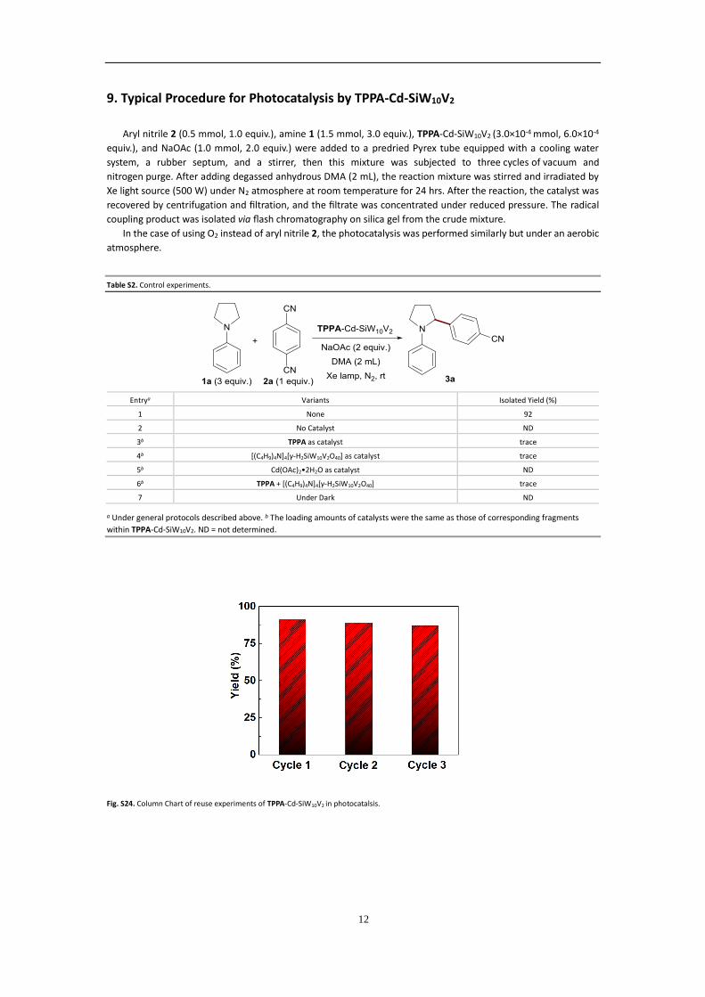
9. Typical Procedure for Photocatalysis by TPPA-Cd-SiW10V2
Aryl nitrile 2 (0.5 mmol, 1.0 equiv.), amine 1 (1.5 mmol, 3.0 equiv.), TPPA-Cd-SiW10V2 (3.0×10⁻⁴ mmol, 6.0×10⁻⁴
equiv.), and NaOAc (1.0 mmol, 2.0 equiv.) were added to a predried Pyrex tube equipped with a cooling water
system, a rubber septum, and a stirrer, then this mixture was subjected to three cycles of vacuum and
nitrogen purge. After adding degassed anhydrous DMA (2 mL), the reaction mixture was stirred and irradiated by
Xe light source (500 W) under N2 atmosphere at room temperature for 24 hrs. After the reaction, the catalyst was
recovered by centrifugation and filtration, and the filtrate was concentrated under reduced pressure. The radical
coupling product was isolated via flash chromatography on silica gel from the crude mixture.
In the case of using O2 instead of aryl nitrile 2, the photocatalysis was performed similarly but under an aerobic
atmosphere.
Table S2. Control experiments.
Entrya Variants Isolated Yield (%)
1 None 92
2 No Catalyst ND
3b TPPA as catalyst trace
4b [(C4H9)4N]4[γ-H2SiW10V2O40] as catalyst trace
5b Cd(OAc)2•2H2O as catalyst ND
6b TPPA + [(C4H9)4N]4[γ-H2SiW10V2O40] trace
7 Under Dark ND
a Under general protocols described above. b The loading amounts of catalysts were the same as those of corresponding fragments
within TPPA-Cd-SiW10V2. ND = not determined.
Fig. S24. Column Chart of reuse experiments of TPPA-Cd-SiW10V2 in photocatalsis.
13
Table S3. Detailed information of Photocatalytic radical coupling reactions.a
Entry Amine 1 Aryl Nitrile 2 Targeted Product 3 Isolated Yield (%)
1
1a 2a
3a
92
2
1b
2a
3b
82
3
1c
2a
3c
90
4 1a
2b 3d
86
5 1a
2c
3e
78
6
1a 2d
3f
85
7
1d
2a
46
a Reaction conditions: Aryl nitriles 2 (0.5 mmol, 1.0 equiv.), amines 1 (1.5 mmol, 3.0 equiv.), TPPA-Cd-SiW10V2 (3.0×10⁻⁴ mmol, 6.0×10⁻⁴
equiv.), NaOAc (1.0 mmol, 2.0 equiv.), DMA (2 mL), 500 W Xe lamp, room temperature (rt), N2 atmosphere, 24 h. Isolated yields.
Scheme S1. Carbonation of 1a. Reaction conditions: amine 1a (1.5 mmol, 3.0 equiv.), TPPA-Cd-SiW10V2 (3.0×10⁻⁴ mmol, 6.0×10⁻⁴ equiv.), NaOAc (1.0
mmol, 2.0 equiv.), DMA (2 mL), 500 W Xe lamp, room temperature (rt), O2 atmosphere, 24 h. Isolated yields.
14
10. NMR Data of the Products.
4-(1-Phenylpyrrolidin-2-yl)benzonitrile (3a)
1H NMR (400 MHz, CDCl3) δ 7.58 – 7.56 (d, J = 8.4 Hz, 2H, ArH), 7.34-7.32 (d, J = 8.0 Hz, 2H, ArH), 7.16-7.12 (dd, J = 8.8, 7.2 Hz, 2H, ArH), 6.70-
6.65 (m, 1H, ArH), 6.44-6.42 (m, 2H, ArH), 4.73 (dd, J = 8.4, 2.0 Hz, 1H, CH(Ph-4-CN)), 3.75-3.70 (m, 1H, CHAHBN), 3.45-3.39 (m, 1H, CHAHBN), 2.48-
2.38 (m, 1H, CHACHBCH(Ph-4-CN)), 2.05-1.87 (m, 3H, CHACHBCH(Ph-4-CN)). 13C NMR (126 MHz, CDCl3) δ 150.62 (CAr), 146.83 (CAr), 132.55 (CAr), 129.24
(CAr), 126.88 (CAr), 119.05 (CAr), 116.62 (CAr), 112.57 (CAr), 110.71 (CN), 62.89 (CH(Ph-4-CN)), 49.35 (NCH2), 35.99 (NCH2CH2CH2), 23.25 (NCH2CH2).
15
4-(4-Phenylmorpholin-3-yl)benzonitrile (3b)
1H NMR (400 MHz, CDCl3) δ 7.49-7.47 (d, J = 8.8 Hz, 2H, ArH), 7.43-7.40 (d, J = 8.4 Hz, 2H, ArH), 7.17-7.13 (m, 2H, ArH), 6.89-6.86 (m, 3H, ArH),
4.43-4.40 (dd, J = 8.0, 3.6 Hz, 1H, CH(Ph-4CN)), 3.99-3.94 (m, 3H, CHACHBCH(Ph-4-CN) and CH2CH2N), 3.64-3.59 (dd, J = 11.6, 8.0 Hz,1H,
CHACHBCH(Ph-4-CN)), 3.43-3.38 (m, 1H, CHAHBN), 3.15-3.08 (m, 1H, CHAHBN). 13C NMR (126 MHz, CDCl3) δ 152.06 (CAr), 146.76 (CAr), 133.93 (CAr),
130.17 (CAr), 128.69 (CAr), 122.45 (CAr), 121.18 (CAr), 118.78 (CAr), 111.16 (CN), 72.71 (CH2CH(Ph-4-CN)), 67.65 (NCH2CH2), 61.46 (CH(Ph-4-CN)), 52.66
(NCH2).
16
4-(1-Phenylpiperidin-2-yl)benzonitrile (3c)
1H NMR (400 MHz, CDCl3) δ 7.48-7.46 (d, J = 8.4 Hz, 2H, ArH), 7.38-7.36 (d, J = 8.0 Hz, 2H, ArH), 7.14-7.10 (dd, J = 8.4, 7.2 Hz, 2H, ArH), 6.87-
6.85 (d, 8.0 Hz, 2H, ArH), 6.82-6.78 (t, J = 7.6 Hz, 1H, ArH), 4.40-4.37 (dd, J = 8.0, 3.6 Hz, 1H, CH(Ph-4-CN)), 3.45-3.37 (ddd, J = 12.0, 9.6 ,4.8 Hz, 1H,
CHAHBN), 3.14-3.05 (ddd, J = 12.4, 7.2, 4.8 Hz 1H, CHAHBN), 2.00-1.93 (m, 1H, CHACHBCH(Ph-4-CN)), 1.83-1.66 (m, 4H, CH2CH2N, CHACHBCH2CH2N
and CHACHBCH(Ph-4-CN)), 1.58-1.48 (m, 1H, CHACHBCH2CH2N). 13C NMR (126 MHz, CDCl3) δ 151.72 (CAr), 150.11 (CAr), 132.28 (CAr), 129.01 (CAr),
128.08 (CAr), 121.14 (CAr), 120.23 (CAr), 120.34 (CAr), 119.10 (CAr), 110.21 (CN), 61.97 (CH(Ph-4-CN)), 52.76 (NCH2), 34.44 (CH2CH2N or CH2CH(Ph-4-
CN)), 25.82 (CH2CH2N or CH2CH(Ph-4-CN)), 22.75 (CH2CH2CH2N).
17
2-(1-Phenylpyrrolidin-2-yl)benzonitrile (3d)
1H NMR (400 MHz, CDCl3) δ 7.69 – 7.67 (d, J = 7.2 Hz, 1H, ArH), 7.44-7.47 (t, J = 7.2 Hz, 1H, ArH), 7.32-7.28 (m, 2H, ArH), 7.16-7.13 (m, 2H, ArH),
6.69-6.65 (t, J = 6.8 Hz, 1H, ArH), 6.44-6.42 (d, J = 7.6 Hz, 2H, ArH), 5.05-5.03 (d, J = 8.0 Hz, 1H, CH(Ph-2-CN)), 3.83-3.72 (m, 1H, CHAHBN), 3.46-3.44
(m, 1H, CHAHBN), 2.61-2.49(m, 1H, CHACHBCH(Ph-2-CN)), 2.04-1.99 (m, 3H, CHACHBCH(Ph-2-CN) and CH2CH2N). 13C NMR (126 MHz, CDCl3) δ 149.14
(CAr), 146.61 (CAr), 133.72 (CAr), 133.08 (CAr), 129.24 (CAr), 127.43 (CAr), 126.95 (CAr), 117.80 (CAr), 116.72 (CAr), 112.66 (CAr), 110.22 (CN), 61.66 (CH(Ph-
2-CN)), 49.51 (NCH2), 35.43 (NCH2CH2CH2), 23.41 (NCH2CH2).
18
4-(1-phenylpyrrolidin-2-yl)benzoate (3e)
1H NMR (400 MHz, CDCl3) δ 7.98-7.96 (d, J = 8.4 Hz, 2H, ArH), 7.30-7.28 (d, J = 8.0 Hz, 2H, ArH), 7.17-7.11 (dd, J = 8.4, 7.6 Hz, 2H, ArH), 6.66-
6.62 (t, J = 7.2Hz, 1H, ArH), 6.46-6.44 (d, J = 8.0 Hz, 2H, ArH), 4.76-4.73 (dd, J = 8.8, 2.0 Hz, 1H, CH(Ph-4-CO2Et)), 4.38-4.32 (q, J = 7.2 Hz, 2H, CH2CH3),
3.75-3.70 (m, 1H, CHAHBN), 3.45-3.39 (m, 1H, CHAHBN), 2.44-2.34 (1H, m , CHACHBCH(Ph-4-CO2Et)), 2.02-1.97 (m, 2H,CH2CH2N), 1.93-1.86 (1H, m ,
CHACHBCH(Ph-4-CO2Et)), 1.35 (3H, t, J = 7.0 Hz, CH2CH3). 13C NMR (126 MHz, CDCl3) δ 166.63 (CO2Et), 150.27 (CAr), 147.07 (CAr), 130.02 (CAr), 129.17
(CAr), 126.05 (CAr), 116.27 (CAr), 112.54 (CN), 63.01 (CO2CH2CH3), 60.94 (CH(Ph-4-CO2Et)), 49.32 (NCH2), 36.09 (NCH2CH2CH2), 23.29 (NCH2CH2), 14.47
(CO2CH2CH3).
19
4-(1-Phenylpyrrolidin-2-yl)pyridine (3f)
1H NMR (400 MHz, CDCl3) δ 8.51-8.49 (d, J = 6.0 Hz, 2H, ArH), 7.17-7.13 (m, 4H, ArH), 6.68-6.65 (t, J = 7.2 Hz, 1H, ArH), 6.45-6.43 (d, J = 8.0, 2H,
ArH), 4.68-4.66 (dd, J = 9.2, 2.0 Hz, 1H, CH(Py)), 3.73-3.69 (m, 1H, CHAHBN), 3.44-3.38 (m, 1H, CHAHBN), 2.47-2.37 (m, 1H, CHACHBCH(Ar)), 2.03-1.89
(m, 3H, CHACHBCH(Ar) and CH2CH2N). 13C NMR (126 MHz, CDCl3) δ 153.99 (CAr), 149.97 (CAr), 146.82 (CAr), 129.18 (CAr), 121.36 (CAr), 116.52 (CAr),
112.48 (CAr), 62.20 (CHPy), 49.23 (NCH2), 35.59 (NCH2CH2CH2), 23.22 (NCH2CH2).
20
4-(phenyl-1-pyrrolidinylmethyl)benzonitrile (isomer 1 of 3g)
1H NMR (400 MHz, CDCl3) δ 7.60-7.52 (q, J = 7.6 Hz, 4H, ArH), 7.41-7.39 (d, J = 7.5 Hz, 2H, ArH), 7.27-7.25 (d, J = 6.7 Hz, 2H, ArH), 7.20-7.17 (t,
J = 7.4 Hz, 1H, ArH), 4.21 (s, 1H, CH(Ph-4-CN)), 2.41-2.38 (t, J = 5.5 Hz, 4H, CH2CH2NCH2CH2), 1.82-1.73 (m, 4H, CH2CH2NCH2CH2). 13C NMR (126 MHz,
CDCl3) δ 149.89 (CAr), 142.98 (CAr), 132.48 (CAr), 128.79 (CAr), 128.29 (CAr), 127.61 (CAr), 127.55 (CAr), 119.06 (CAr), 110.74 (CN), 76.12 (CH(Ph-4-CN)),
53.60 (2×NCH2CH2), 23.67 (2×NCH2CH2).
21
4-[1-(phenylmethyl)-2-pyrrolidinyl]benzonitrile (isomer 2 of 3g)
1H NMR (400 MHz, CDCl3) δ 7.63-7.61 (d, J = 7.6 Hz, 2H, ArH),), 7.58-7.55 (d, J = 7.7 Hz, 2H, ArH),), 7.38-7.18 (m, 5H, ArH),), 3.77-3.74 (d, J =
13.07 Hz, 1H, CH(Ph-4-CN)), 3.49-3.45 (t, J = 8.26 Hz, 1H, CHAHBN), 3.15-3.10 (m, 2H, CHACHBCH(Ph-4-CN) and CHAHBN, 2.30-2.15 (m, 2H, Ph-CH2-N),
1.98-1.58 (m, 3H, CHACHBCH(Ph-4-CN) and CH2CH2N). The isomer 2 of 3g was not separable with 1-benzylpyrrolidine-2-carbonitrile, the
decomposition product of starting material.
1-phenyl-2-Pyrrolidinone (4a)
1H NMR (400 MHz, CDCl3) δ 7.61 (d, J = 7.9 Hz, 2H, ArH), δ 7.36 (t, J = 7.6 Hz, 2H, ArH), δ 7.14 (t, J = 7.3 Hz, 1H, ArH), δ 3.85 (t, J = 6.8 Hz, 2H,
CH2CH2N), δ 2.60 (t, J = 7.9 Hz, 2H, CH2CO), δ 2.15 (m, 2H, CH2CH2N). 13C NMR (126 MHz, CDCl3) δ 174.20 (C=O), 139.45 (CAr), 128.82 (CAr), 124.49
(CAr), 119.97 (CAr), 48.78 (NCH2), 32.77 (NCH2CH2CH2), 18.04 (NCH2CH2).
11. References:
1. M.-D. Zhang, C.-M. Di, L. Qin, X.-Q. Yao, Y.-Z. Li, Z.-J. Guo and H.-G. Zheng, Crystal Growth & Design, 2012, 12,
3957-3963.
2. J. Canny, R. Thouvenot, A. Teze, G. Herve, M. Leparuloloftus and M. T. Pope, Inorg Chem, 1991, 30, 976-981.
3. J. Canny, R. Thouvenot, A. Teze, G. Herve, M. Leparuloloftus and M. T. Pope, Inorg. Chem., 1991, 30, 976-981.
4. Z. Ming, Y. Wang, T. Zhang, L. Li, C. Duan and Z. Liu, ChemCatChem, 2020, DOI: 10.1002/cctc.202001346.
5. D. Shi, Z. Ming, Q. Wu, T. Lai, K. Zheng, C. He and J. Zhao, Inorg. Chem. Commun., 2019, 100, 125-128.
6. D. Shi, C. He, W. Sun, Z. Ming, C. Meng and C. Duan, Chem. Commun., 2016, 52, 4714-4717.
7. C. Hua, A. Baldansuren, F. Tuna, D. Collison and D. M. D'Alessandro, Inorg. Chem., 2016, 55, 7270-7280.
8. A. McNally, C. K. Prier and D. W. C. MacMillan, Science, 2011, 334, 1114-1117.
9. X. M. Zhang, S. R. Yeh, S. Hong, M. Freccero, A. Albini, D. E. Falvey and P. S. Mariano, J. Am. Chem. Soc., 1994,
116, 4211-4220.


![Welcome [] Carpet101_3_9_06.pdfWelcome . CARPET101 ... Difficult to Re-process Polymer Not recyclable into virgin Nylon ... Gauge, Fabric formation, dye technique](https://static.documents.pub/doc/80x56/5abaeeb07f8b9a297f8c4f42/welcome-carpet1013906pdfwelcome-carpet101-difficult-to-re-process.jpg)


![Polyoxometalate multi-electron transfer catalytic …...Polyoxometalate multi-electron transfer catalytic systems for water splitting Jordan M. Sumliner,[a] Hongjin Lv,[a] ... (WOC)](https://static.documents.pub/doc/80x56/5e2e7956451c664bfb5d6f31/polyoxometalate-multi-electron-transfer-catalytic-polyoxometalate-multi-electron.jpg)


