IEEE TRANSACTIONS ON SYSTEMS, MAN, AND CYBERNETICS, VOL. SMC-1, NO. 3, JULY 1971 The numerator of the right-hand side is positive since it is a probability function. By,conditions 3) and 4), (1 - q)PNCOI' < 0 qPcol' > 0. (26) Hence (1 - q)PNCO I- qPo I' < 0 (27) and dD <0. dq Then D is a monotonically decreasing function of q. Note that D(q1) < D(q2), for q1 > q2. False dismissal prob- ability pFD is defined to be D(q) PFD(q) = J PcoI(x) dx (29) co where PcOI(x) is a probability function. Hence PFD(q) is monotonically decreasing in q. By similar reasoning, PFA(q) is monotonically increasing in q. ACKNOWLEDGMENT The author is indebted to M. L. Luther and L. M. Hardy for their constructive criticism throughout the study. R. A. Schlothauer handled the programming chores. Mrs. Itsuko WeHara assisted in the computations and the data presentation, and Mrs. Nancy Norwood typed and prepared the manuscript. REFERENCES [1] J. H. Munson, "Experiments in the recognition of hand-printed text: pt. I-character recognition," in 1968 Fall Joint Computer Conf., AFIPS Conf. Proc., vol. 33. Washington, D.C.: Thompson, 1968. [2] E. M. Darling, Jr., and R. D. Joseph, "Pattern recognition from satellite altitudes," IEEE Trans. Syst. Sci. Cybern., vol. SSC-4, Mar. 1968, pp. 38-47. [3] J. G. Kawamura, "Automatic change discrimination as an aid to city planning," presented at the American Institute of Aero- nautics and Astronautics Earth Resources Observations and Information Systems Meeting, Annapolis, Md., 1970. [4] M. Schwartz, Information Transmission Modulation and Noise. New York: McGraw-Hill, 1959. [5] A. Rosenfeld, Picture Processing by Computer. New York: Academic Press, 1969. [6] R. G. Gallager, Information Theory and Reliable Communication. New York: Wiley, 1968. [7] G. S. Sebestyen, Decision-Making Processes in Pattern Recognition. New York: Macmillan, 1962. Dynamic Analysis of the Pupil with Light and Electrical Stimulation JOSEPH TERDIMAN, JAMES D. SMITH, MEMBER, IEEE, AND LAWRENCE STARK, FELLOW, IEEE Abstract-A television pupillometer and an on-line computer were used to determine the static and dynamic characteristics of the pupillary system in the cat. The characteristics of sphincter and dilator mechanisms were measured with pulse rate modulated electrical stimuli to sympathetic and parasympathetic nerves and found to be similar to the light-driven pupillary response characteristic at high frequencies. Linear and non- linear features of the response characteristics that were identified include retinal logarithmic operator, neural, neuromuscular, and mechanical saturation, high-frequency third-order dynamics of the motor component with a break frequency at 0.8 Hz, nonminimum phase transport delay of about 0.2 s, and response asymmetry to positive and negative steps of stimulation. Manuscript received March 30, 1970. This work was supported in part by the National Institutes of Health under Grant ROI NB08546-01, Neurophysiological Information Coding, by NIH Special Fellowship 5-F3-GM-33, 568-02 awarded to J. Terdiman, and by a Guggenheim Fellowship awarded to L. Stark. This work was conducted at the Department of Biomedical Engineering, Presbyterian-St. Luke's Hospital, Chicago, and the College of Engineering, University of Illinois Circle Campus, Chicago. J. Terdiman is with the Kaiser Foundation Research Institute, Oakland, Calif., and the School of Optometry, University of California, Berkeley, Calif. 94720. J. D. Smith is with the Department of Biomedical Engineering, University of Southern California, Los Angeles, Calif. L. Stark is with the University of California, Berkeley, Calif. 94720. INTRODUCTION T HE PUPILLARY system can be divided functionally into three major components: sensory, central nervous system, and motor. The sensory component is the retina, which translates light input signals into coded patterns of nerve impulses that are carried to the central nervous system by the fibers of the optic nerve. The central compo- nent serves as a neural controller that receives retinal nerve impulses from both eyes, integrates these signals spatially and temporally, and generates neural output signals to drive sympathetic and parasympathetic effectors in both eyes. The motor component consists of the sphincter and dilator muscles of the iris, reciprocally innervated by sympathetic and parasympathetic motor nerves. The motor component translates pulse-coded neural signals into pupil movement. In the sensory component retinal output signals carrying pupillary information to the central component are appar- ently indistinguishable from those carrying visual informa- tion to the lateral geniculate body and visual cortex. If this is the case, then studies conducted on the visual system 239
Transcript
IEEE TRANSACTIONS ON SYSTEMS, MAN, AND CYBERNETICS, VOL. SMC-1, NO. 3, JULY 1971
The numerator of the right-hand side is positive since it isa probability function. By,conditions 3) and 4),
(1 - q)PNCOI' < 0
qPcol' > 0. (26)Hence
(1 - q)PNCOI- qPo I' < 0 (27)and
dD <0.dq
Then D is a monotonically decreasing function of q. Notethat D(q1) < D(q2), for q1 > q2. False dismissal prob-ability pFD is defined to be
D(q)PFD(q) = J PcoI(x) dx (29)
co
where PcOI(x) is a probability function. Hence PFD(q) ismonotonically decreasing in q. By similar reasoning,PFA(q) is monotonically increasing in q.
ACKNOWLEDGMENTThe author is indebted to M. L. Luther and L. M.
Hardy for their constructive criticism throughout the study.R. A. Schlothauer handled the programming chores. Mrs.Itsuko WeHara assisted in the computations and the datapresentation, and Mrs. Nancy Norwood typed and preparedthe manuscript.
REFERENCES[1] J. H. Munson, "Experiments in the recognition of hand-printed
text: pt. I-character recognition," in 1968 Fall Joint ComputerConf., AFIPS Conf. Proc., vol. 33. Washington, D.C.: Thompson,1968.
[2] E. M. Darling, Jr., and R. D. Joseph, "Pattern recognition fromsatellite altitudes," IEEE Trans. Syst. Sci. Cybern., vol. SSC-4,Mar. 1968, pp. 38-47.
[3] J. G. Kawamura, "Automatic change discrimination as an aid tocity planning," presented at the American Institute of Aero-nautics and Astronautics Earth Resources Observations andInformation Systems Meeting, Annapolis, Md., 1970.
[4] M. Schwartz, Information Transmission Modulation and Noise.New York: McGraw-Hill, 1959.
[5] A. Rosenfeld, Picture Processing by Computer. New York:Academic Press, 1969.
[6] R. G. Gallager, Information Theory and Reliable Communication.New York: Wiley, 1968.
[7] G. S. Sebestyen, Decision-Making Processes in Pattern Recognition.New York: Macmillan, 1962.
Dynamic Analysis of the Pupil with Light
and Electrical StimulationJOSEPH TERDIMAN, JAMES D. SMITH, MEMBER, IEEE, AND LAWRENCE STARK, FELLOW, IEEE
Abstract-A television pupillometer and an on-line computer were usedto determine the static and dynamic characteristics of the pupillarysystem in the cat. The characteristics of sphincter and dilator mechanismswere measured with pulse rate modulated electrical stimuli to sympatheticand parasympathetic nerves and found to be similar to the light-drivenpupillary response characteristic at high frequencies. Linear and non-linear features of the response characteristics that were identifiedinclude retinal logarithmic operator, neural, neuromuscular, andmechanical saturation, high-frequency third-order dynamics of the motorcomponent with a break frequency at 0.8 Hz, nonminimum phasetransport delay of about 0.2 s, and response asymmetry to positive andnegative steps of stimulation.
Manuscript received March 30, 1970. This work was supported inpart by the National Institutes of Health under Grant ROI NB08546-01,Neurophysiological Information Coding, by NIH Special Fellowship5-F3-GM-33, 568-02 awarded to J. Terdiman, and by a GuggenheimFellowship awarded to L. Stark. This work was conducted at theDepartment of Biomedical Engineering, Presbyterian-St. Luke'sHospital, Chicago, and the College of Engineering, University ofIllinois Circle Campus, Chicago.
J. Terdiman is with the Kaiser Foundation Research Institute,Oakland, Calif., and the School of Optometry, University of California,Berkeley, Calif. 94720.
J. D. Smith is with the Department of Biomedical Engineering,University of Southern California, Los Angeles, Calif.
L. Stark is with the University of California, Berkeley, Calif. 94720.
INTRODUCTION
T HE PUPILLARY system can be divided functionallyinto three major components: sensory, central nervous
system, and motor. The sensory component is the retina,which translates light input signals into coded patterns ofnerve impulses that are carried to the central nervoussystem by the fibers of the optic nerve. The central compo-nent serves as a neural controller that receives retinal nerveimpulses from both eyes, integrates these signals spatiallyand temporally, and generates neural output signals to drivesympathetic and parasympathetic effectors in both eyes.The motor component consists of the sphincter and dilatormuscles of the iris, reciprocally innervated by sympatheticand parasympathetic motor nerves. The motor componenttranslates pulse-coded neural signals into pupil movement.
In the sensory component retinal output signals carryingpupillary information to the central component are appar-ently indistinguishable from those carrying visual informa-tion to the lateral geniculate body and visual cortex. Ifthis is the case, then studies conducted on the visual system
239
IEEE TRANSACTIONS ON SYSTEMS, MAN, AND CYBERNETICS, JULY 1971
may be useful in explaining certain characteristics of thepupillary system. Such pertinent studies concern therelationship of optic nerve impulse frequency to the in-tensity of retinal illumination. Enroth [9] was the first torelate light intensity to nerve impulse rate at the flicker-fusion frequency. Recently, Hughes and Maffei [15] studiedthe dynamic relationship between sinusoidal variations inlight intensity and nerve impulse rate in retinal ganglioncells of the cat. Earlier, Stark and Hermann [32] derivedtransfer relations for the variation in nerve impulsefrequency of abdominal ganglion cells of crayfish producedby sinusoidal light stimulation of its photoreceptors. Intheoretical studies Partridge [23] and Jones et al. [16] haveexamined the process of pulse-rate encoding by a neuraltransducer, i.e., receptor or effector organ.
Centrally, Nisida et al. [21], Sillito [28], and Smithet al. [29] have recorded neuronal responses to lightstimuli from the pretectal region and Edinger-Westphalnucleus. The pupillary response to electrical stimulation ofareas of the cortex, thalamus, hypothalamus, reticularformation, and other higher centers has been observed bymany investigators and used to trace the central pathway ofsympathetic and parasympathetic pupillary components[13], [14], [24].The anatomy and physiology of the motor component of
the pupillary system have been extensively described inmany species of animals. In the cat parasympathetic fibersare carried to the iris from the Edinger-Westphal nucleusby way of the oculomotor nerve, ciliary ganglion, and shortciliary nerves. Sympathetic fibers are carried to the iris fromthe sympathetic trunk by way of the superior cervicalganglion, internal carotid nerve, and long ciliary nerve.Recently, Ehinger [7], [8] obtained solid histologicalevidence of a dual sympathetic-adrenergic parasympathetic-cholinergic innervation of both the sphincter and dilatormuscles in cats. Numerous pharmacological experiments[1], [26], [27], [37] on feline iris muscle strips in vitro havedemonstrated that both sphincter and dilator have threekinds of receptors: cholinergic, alpha-adrenergic, andbeta-adrenergic, with cholinergic and beta-adrenergicreceptors predominant in sphincter muscle, and alpha-adrenergic receptors predominant in dilator muscle. In thesphincter cholinergic excitation causes contraction, andbeta-adrenergic excitation causes relaxation. In the dilatoralpha-adrenergic excitation causes contraction, and cho-linergic excitation causes relaxation. Based on thesefindings, Dennison [5] has developed an analog computermodel whose response characteristics appear to be similarto those of a cat iris.The characteristics of the overall system have been
extensively studied by many investigators, and numerousqualitative descriptions of the pupil response to variousstimuli exist in the literature. However, few quantitativestudies can be found, and there have been even fewerattempts to construct an analytic model of the pupillarysystem. Lowenstein and Loewenfeld [18] made an earlyattempt to describe some of their results by developing acrude graphical model of the pupillary reflex based on an
analysis of the shapes of various pupil response waveforms.They attributed the appearance of various portions of theamplitude and velocity response curves to the interplayof different neural and neuromuscular mechanisms andattempted to estimate the relative contributions of sym-pathetic and parasympathetic components under varyingexperimental conditions. More recently, van der Tweel andDenier van der Gon [38] conducted a semiquantitativeinvestigation in humans of the pupillary response to steps,pulses, and harmonic light stimuli. They speculated aboutthe mechanisms of various linear and nonlinear operators,but made no attempt to develop a coherent quantitativemodel.
It was not until the investigations by Stark and hisassociates into the dynamics of the pupillary controlsystem as a neurological servomechanism that an attemptwas made to devise a rational parameter model of thepupillary system that could provide a theoretical basis forthe observed characteristics of the pupil response andcould be used to predict the response under varying experi-mental conditions [25], [30]-[32]. Working only withhumans and considering the pupillary system to be afeedback control system, they employed the methodsof engineering control theory to derive a quantitativenonlinear model. Their approach to the problem, however,had to consider the human pupillary system to be a blackbox whose contents were essentially inaccessible andcould be described only by "dry dissection" techniques inwhich the characteristics of individual components arededuced from observations of their mutual interactions andtheir interaction with other neurological systems. On theother hand, by combining the control theory approachwith "wet dissection" techniques of classical neuro-physiology in experimental animals, as begun in the presentpaper, the characteristics of system components may bemeasured directly, and individual dynamic operators whoseproperties are reflected in the overall response may belocalized and described quantitatively. In addition thisprocedure enables characteristics of the pulse-coded nervesignals at intermediate points in the system to be related tomeaningful parameters of physiological input and outputsignals. In the present paper static and dynamic character-istics of the overall pupillary system and of sphincter anddilator mechanisms have been determined in the cat bymeans of light and electrical stimulation, respectively.Preliminary results have already been reported [36].
METHOD
Animal PreparationExperiments were performed on 20 cats. Since the pupil
response is extremely sensitive to anesthesia, variouspreparations were tried, including unanesthetized pre-trigeminal sections, encephale-isole sections with localinfiltration of Xylocaine, as well as lightly anesthetizedpreparations using ether, intravenous sodium pentothal, orintraperitoneal sodium pentobarbital (Diabutal). The mostsatisfactory results were usually obtained beginning about
240
TERDIMAN et al.: PUPIL WITH LIGHT AND ELECTRICAL STIMULATION
4 h following a single dose of Diabutal (20 mg/kg of weight).Body temperature was maintained at 37°C with a heatingpad and temperature controller and the EKG was mon-itored. To expose the sympathetic nerve supply to the iris,the superior cervical ganglion was located in the neck,and bipolar electrodes consisting of 5-mil teflon-insulatedplatinum wire, bared at the tips, were hooked around thepost-ganglionic internal carotid nerve. Paraffin was in-filtrated around the electrodes to hold them in position andinsulate them from the surrounding tissue. A nylon suturewas placed loosely around the nerve between electrode andganglion to facilitate sectioning later in the experiment.To expose the parasympathetic nerve supply to the iris,
the ciliary ganglion and short ciliary nerves were locatedin the orbit, using a lateral approach in which parts of thetemporalis and masseter muscles and the zygomatic archwere removed. Bipolar electrodes were placed post-ganglionically about medial and lateral branches of theshort ciliary nerves. Occasionally, only the lateral branchcould be hooked, and electrical stimulation would result inasymmetrical contraction of the lateral half of the iris.To isolate the motor portion of the pupillary system from
the influence of the central nervous system during electricalstimulation, the motor nerves could be sectioned just distalto their respective ganglia. When both sympathetic andparasympathetic pathways were interrupted, the pupilwould often dilate or constrict strongly. It then becamenecessary to apply a constant low pulse rate stimulus to thedistal segment of the short ciliary or internal carotid nerveto provide a source of tonic neural bias for the sphincter ordilator mechanism in order to reset the static pupil diameterto an operating point in an unsaturated portion of itsdynamic range. Simultaneous stimulation of the antagonistcould then be accomplished to determine its static ordynamic characteristics. In stable preparations, however, itwas found that. if the nerves were left intact to provide aphysiological source of neural bias, the dynamic character-istics of sphincter and dilator mechanisms were largelyunchanged.The animal was then placed in a stereotaxic frame, and the
ipsilateral eyeball was fixed in position with corneal suturesanchored to the frame. Both nictitating membranes weresutured, but left in place to protect the corneas, which wereexposed only during experimental runs. When light stimula-tion experiments were performed, the contralateral pupilwas dilated with Cyclogyl for open-loop light stimulation,and the consensual pupillary response was recorded.Open-loop light stimulation was performed to preventchanges in retinal flux by pupillary movement, which wouldotherwise act as a source of negative feedback and modifythe system response characteristics. The consensual pupillaryresponse is assumed to be identical to the direct response[18], [30].InstrumentationA block diagram of the experimental arrangement is
shown in Fig. 1. The light source was a glow modulator tube(Sylvania R1131C) linearly driven by a pulsewidth mod-
ulated pulse generator whose pulsewidth was proportionalto an external modulation voltage. The light beam wasdelivered to the dilated eye via a fiber optic light guide.Electrical stimulation was in the form of bipolar pulse ratemodulated pulse trains, which were generated by a physio-logical stimulator and delivered to the preparation throughan isolation transformer. Pulses were generally 1 ms induration and from 0.1 to 5 V in amplitude. The pulseamplitude was adjusted to produce a maximal pupillaryresponse. Pulses were triggered externally by the leadingedges of a square wave used as an input signal to thestimulator. The square wave was produced by a waveformgenerator whose oscillator frequency could be modulatedby an external voltage.
Pupil diameter was measured with a Biosystems televisionpupillometer. The instrument consists of an infrared-sensitive television camera with a telephoto lens, a televisionreceiver used as a monitor, and associated pupillometercircuitry. When the eye is illuminated with infrared light,the pupil appears on the monitor as a black area with asharply defined border against the lighter background of theiris. Measuring circuits in the pupillometer are triggeredby the leading edge of the pupil image and produce a signalproportional to the vertical height of the pupil image onthe monitor screen. A display of trigger points also appearson the screen parallel to the pupil border. Because of thelinearity of the instrument, a simple two-point calibrationis sufficient to determine a calibration curve. Calibrationwas performed by measuring pupil diameter and pupil-lometer voltage simultaneously at maximum diameter andagain at minimum diameter. When plotted, these two pointsdefine a straight line whose slope and intercept parametersare easily computed. These parameters are then used toconvert pupillometer voltages into values of pupil diameter.Since the pupil image and trigger pulses are monitored atall times, variations in trigger points due to drying of thecornea or disturbance of the apparatus can be recognizedat once and corrected by recalibration of the pupillometer.
241
IEEE TRANSACTIONS ON SYSTEMS, MAN, AND CYBERNETICS, JULY 1971
Because of the asymmetric shape of the cat's pupil, only thevariations in the transverse (horizontal) diameter wererecorded.Most experimental procedures were controlled by an
on-line IBM-1800 computer. Communication with thecomputer was by remote teletype and analog lines withterminals in the laboratory. The various functions of thecomputer program were selected by keying in the appro-priate code on the teletype. The computer was used tocalibrate the pupillometer, generate the desired time-varying modulation voltages for light and electrical stimula-tion, receive and store pupil response signals from thepupillometer, compute a running average pupil responseand generate a voltage in proportion to the average responsefor oscilloscope display in the laboratory, record individualand average pupil response on digital magnetic tape foroff-line analysis, and perform an on-line analysis of thedata. With on-line analysis, as soon as a smooth averageresponse waveform was obtained (usually requiring from2 to 20 cycles depending on frequency), the computer wouldbe requested to print on the teletype maximum, minimum,and average pupil diameters and time delay for stepresponses, or, from Fourier analysis, average diameter,amplitude and phase of the first harmonic component, andleast-square error of the higher harmonics (percent harmonicdistortion) for sinusoidal responses.
AnalysisIn control theory the behavior of a system may be
described in terms of static and dynamic response character-istics relating input and output variables. The staticcharacteristic or dc response curve represents their steady-state relationship for constant input values. The dynamicbehavior of a linear system for a time-varying input may bedescribed by a transfer function whose properties aregraphically illustrated by Bode diagrams of the amplituderatio and phase shift of the response relative to the input[10]. These dynamic response characteristics may beexperimentally determined by measuring the response ofthe system to a harmonic input. If the system is nonlinear,the response will, in general, be a periodic function con-sisting of the fundamental frequency plus higher harmoniccomponents. The dynamic characteristics may then berepresented by a generalized transfer function known as adescribing function, derived arbitrarily from the amplituderatio and phase shift of the first harmonic component of theresponse relative to the input. Values of amplitude andphase are determined experimentally by Fourier analysisof the response and are, in general, functions of bothfrequency and input amplitude.
In the present paper, however, the relationships ofamplitude and phase to frequency were determined for asingle representative input amplitude selected to fall withinthe unsaturated midrange of the dc characteristic. From themeasured amplitude and phase characteristics of a nonlinearsystem an approximate linear model may be derived. Thedynamic characteristics may then be separated conceptually
-i
-J 3,0
uCYJl
(a)3 5-
3
2: 259
-i
ioI 2 7,
aX 2 5-
2 3-
2 - i_ __10 100
LUMINOUS FLUX (MILLILUMENS)
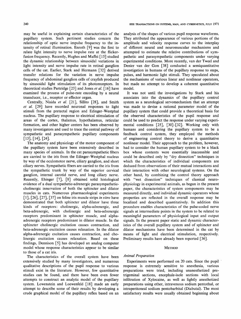
(b)Fig. 2. (a) Static pupillary response characteristic for constant flux
values. Pupil diameter as function of static retinal luminous flux,plotted on linear coordinates. Data points are experimental; curveis fitted by eye. Point P represents typical operating point fordynamic stimulation. (b) Pupil diameter as function of static retinalluminous flux, plotted on semi-logarithmic coordinates. Experimentaldata points are identical to (a); straight line is fitted by eye.
into linear and nonlinear components, where the linear partrepresents the portion of the response attributable to linearoperators in the system and the remaining nonlinearcomponent represents the deviation from linearity producedby nonlinear elements.
RESULTS
Light Stimulation: Static CharacteristicThe static characteristic of the overall pupillary system is
illustrated in Fig. 2(a). During stimulation, the consensualresponse was recorded for retinal flux values from0 to 10 mlm, and pupil diameter was measured only afterthe system had reached a steady state, allowing sufficient
242
2'5
TERDIMAN et al.: PUPIL WITH LIGHT AND ELECTRICAL STIMULATION
10
PARASYMPATHETIC
---T--- -O10 20 30 46 5u
STIMULATION RATE (PULSES/SECONDI60 70
Fig. 3. Static pupillomotor response characteristics for constantstimulus pulse rate values. Pupil diameter as independent functions ofsympathetic and parasympathetic pulse rates.
time for decay of initial transients and for retinal adaptation.As the intensity of illumination was increased, the incre-mental change in pupil diameter decreased, demonstratingthe operation of a static scale-compression nonlinearity inthe pupillary system.When plotted on semilogarithmic coordinates as in
Fig. 2(b), the experimental data points are scattered about a
straight line, which indicates that the scale-compressionoperator is approximately logarithmic in nature over therange of fluxes used. At still higher flux values the systembegins to saturate until further flux increments can produceno additional constriction. Several investigators havepreviously shown that this relationship holds in humansover several orders of flux magnitude until saturation occurs
at extreme values [19], [38]. Our results, although obtainedover a smaller flux range, confirm these findings in the cat.The illustrated characteristic is clearly not unique sincesteady-state pupil diameter is a function not only of retinalflux but of the degree of accommodation, psycho-sensorystimuli, drug levels, and other factors.
Electrical Stimulation: Static CharacteristicsThe characteristics of the pupillary system for light
stimulation represent the combined effects of neural andneuromuscular elements in sensory, central, and motorcomponents of the pupillary system. The characteristics ofthe isolated motor component can be determined bymeasuring the pupil response to post-ganglionic electricalstimulation of the motor nerves.
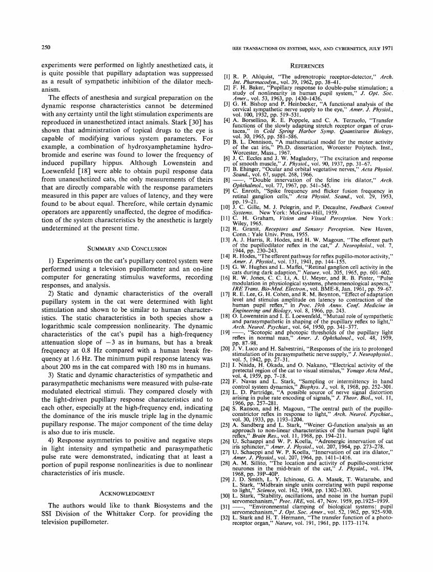
The static characteristics of sympathetic and para-
sympathetic mechanisms for constant stimulus pulse ratesfrom 0 to 60 pitts (pulses per second) are illustrated in Fig. 3.During stimulation, pupil diameters were measured on a
response plateau after initial transients had decayed andbefore the response had begun to diminish due to thedevelopment of iris muscle fatigue. Stimulation of the shortciliary nerve with trains of maximal pulses 1 ms in durationand approximately 1 V in amplitude results in a static
SYMPATHETICPULSE RATE
O Io 20 30 40 50 60 70
PARASYMPATHETIC STIMULATION RATE (PULSES/SECOND)
Fig. 4. Pupil diameter as function of parasympathetic pulse rateshowing dependence on pulse rate of simultaneous sympatheticstimulation.
characteristic for the sphincter in which pupil diameterdecreases linearly with increasing pulse rates up to about15 pitts (Fig. 3-parasympathetic curve). Minimal pupildiameter is obtained at 20-25 pitts. A similar optimal pulserate for constriction with parasympathetic stimulation wasobserved by Luco and Salvestrini [20]. At stimulus ratesabove 25 pitts the pupil begins to redilate, and the responsebegins to saturate beyond 40 pitts. In contrast, stimulationof the internal carotid nerve with trains of maximal pulses1 ms in duration and approximately 2 V in amplituderesults in a static characteristic for the dilator component inwhich pupil diameter increases monotonically with in-creasing pulse rates (Fig. 3-sympathetic curve). Thecharacteristic is approximately linear between 15 and 35pitts, but shows saturation at pulse rates above and belowthis range. In a well-isolated preparation no pupillaryresponse was ever observed due to electrical stimulation ofthe contralateral short ciliary or internal carotid nerves,even with all nerves intact. This indicates that the stimuluspathway was directly from motor nerves to iris and thatpossible stimulation of the adjacent optic nerve by currentleakage was avoided. It also indicates that the responsewas not influenced by antidromic stimulation of midbrainor higher centers.
Since the sphincter and dilator mechanisms are mechan-ically interconnected but mutually antagonistic, it is possibleto alter the static characteristics of one component byvarying the level of activation of the other. The effect ofincreased sympathetic stimulation on the static character-istic of the sphincter mechanism can be observed bysimultaneous stimulation of the short ciliary and internalcarotid nerves. In Fig. 4 static characteristics of pupildiameter as a function of parasympathetic pulse rate wereobtained for several settings of sympathetic pulse rate. Asdilator activation is progressively increased by increasingthe sympathetic pulse rate, the amount of pupil constrictionproduced by a given parasympathetic pulse rate diminishes.In addition the biphasic appearance of the unloaded
243
(f)CY
IW
1:-i-i.EOr
IW
Y-
0
-ia-DCL
./iwui
uix-j-i
CY
F-LLJx
0
-j
CLZ)CL
IEEE TRANSACTIONS ON SYSTEMS, MAN, AND CYBERNETICS, JULY 1971
sphincter characteristic is altered with increasing sym-pathetic activity, and the curve becomes essentiallymonotonic at higher sympathetic pulse rates.The fact that the characteristics of the motor components
for both sphincter and dilator mechanisms are approx-imately linear throughout much of their operating rangeindicates that the scale-comLpression operator identified inthe static characteristic for the overall system is located at apoint along the pupillary pathway between retinal photo-receptors and pupillomotor nerves. The probable site ofthis operator is postulated to be the retina [2], [33], [38].Direct neurophysiological evidence for the existence of alogarithmic operator in the retina is obtained from thework of Enroth [9], who showed that the average impulsefrequency in optic nerve fibers of the cat is directly pro-portional to the flicker-fusion frequency of a light stimulus.Since flicker-fusion frequency is proportional to thelogarithm of luminance according to the Ferry-Porterlaw [11], then the average impulse frequency is alsoproportional to the logarithm of luminance. If the staticcharacteristic of the remainder of the pupillary pathway isapproximately linear, the retinal logarithmic operator willbe reflected in the static characteristic of the overall system,resulting in the observed logarithmic relationship betweenretinal flux and pupil diameter. Furthermore, this functionalarrangement leads to the hypothesis that the average impulsefrequency in the short ciliary nerves will vary as the logarithmof retinal flux.
Light Stimulation: Dynamic ResponseBecause the static characteristic of the overall pupillary
system contains a logarithmic nonlinearity, the dynamicresponse to a sinusoidal light stimulus will also contain anonlinear component. The size of the nonlinear contributiondepends on the dc operating point and percent amplitudemodulation of the input signal. The accuracy of the linearapproximation improves as the percent modulationdecreases. Unfortunately, at high-input frequencies theattenuation of the signal is so great that, in practice, largemodulation amplitudes must be used. On the other hand,because the relationship between dc values of retinal fluxand pupil diameter is satisfied by a logarithmic function, thedeviation of the response from linearity will not be extensiveeven for moderate input amplitudes. A dc operating pointand a modulation amplitude for a sinusoidal light stimuluswere selected in the unsaturated midrange of the staticcharacteristic (point P, Fig. 2(a)). An example of thepupillary response to sinusoidal stimulation is shown inFig. 5. The light signal had a dc flux of 3 mlm and an accomponent of 1 mlm, a 33-percent modulation amplitude.
After initial transients had decayed and the pupil hadreached a steady state, periodic responses were stored andaveraged by the computer and the average response dis-played on an oscilloscope. The results of on-line Fourieranalysis of the average response at different modulationfrequencies are represented by the amplitude and p,hasecharacteristics shown in Fig. 6. The amplitude characteristic(Fig. 6(a)) is a plot of the ratio of the amplitude of the first
H _
XL a:
,U n
x_L
-iiLU)
IL
_3v
~LUHZZ-
5
3
5
41-
Fig. 5. Pupillary
.1 _
i .05
.02
I
005 -
< .002- _w
_ - 100w
(r-200
Cl)< - 300a-
-400
3 4 5 6 7 8
TIME (SECONDS)
response to sinusoidal intensity modulated lightstimulus.
02 .05 .1 .2 .5 2 5 10
FREQUENCY (HZ.)
(a)
.02 05 .2 5 2 5 10
FREQUENCY (HZ.)(b)
Fig. 6. Dynamic pupillary and pupillomotor response characteristics.(a) Amplitude characteristics for light and electrical stimulationconsist of ratio of amplitude of first harmonic component of pupildiameter variation to stimulus modulation amplitude as function offrequency, plotted on full logarithmic coordinates. (b) Phasecharacteristics for light and electrical stimulation consist of phaseshift of first harmonic component of pupil diameter variationrelative to phase of stimulus modulation waveform, plotted onsemilogarithmic coordinates.
harmonic component of pupil diameter variation to theamplitude of intensity modulation of the light stimulusas a function of frequency, and the phase characteristic is aplot of the phase shift of the first harmonic component ofthe response relative to the phase of the stimulus as afunction of frequency. The amplitude characteristic isdrawn asymptotically flat at low frequencies and is atten-uated at high frequencies falling off with an asymptoticslope of -3 (18 dB/octave) on log-log coordinates. Measure-ments of response amplitude at low frequencies (below0.1 Hz) were found to have a relatively large variance(up to + 30 percent at 0.01 Hz), resulting in a correspond-
244
2
.uu
TERDIMAN et al.: PUPIL WITH LIGHT AND ELECTRICAL STIMULATION
ingly large uncertainty in the true slope of the low-frequencyportion of the amplitude characteristic. This variance isdue to the effects of low-frequency Gaussian noise and tothe nonstationary distribution of noise amplitudes. Theseeffects become significant at low frequencies because of thelonger cycle durations and the fewer cycles of response datarecorded and averaged. The shape of the upper end of theamplitude characteristic is that of a third-order low-passfilter with a triple pole at a break frequency of 0.8 Hz,which corresponds to a time constant of 0.2 s.The experimental phase characteristic for the overall
system (Fig. 6(b)) consists of a minimum phase componentdue to the triple pole, a nonminimum phase componentdue to an average response latency of 260 ms for positiveand negative steps in light intensity, and a small residualcomponent due to nonlinearities. In contrast to theamplitude measurements, the variance of experimentalphase measurements at low frequencies was relatively small(< + 10 percent).A qualitative representation of some of the nonlinear
aspects of the pupil response to light is shown in Figs. 7and 8. The asymmetry of the pupillary response to positiveand negative steps in light intensity is illustrated in Fig. 7.The response to a positive step of 6 mlm has shorterlatency (200 ms) and a greater maximum velocity than theresponse to a negative step of equal amplitude (320 ms).Results of this asymmetry are also reflected in the responseto sinusoidal stimulation (Fig. 5) as harmonic distortionand rectification. The latter effect accounts for the observeddecrease in mean pupil diameter during sinusoidal stimula-tion as compared with pupil diameter due to a constantlight stimulus of the same average intensity [30]. Otherexperiments have shown that the latent period of the pupilresponse increases with decreasing stimulus amplitude[17], [19], [38]. Latencies of constriction as short as200 ms for a maximal stimulus and as long as 700 ms for avery small stimulus (< 1 mlm) have been found. Measure-ments of dilation latencies are less reliable because of themore gradual onset of the response, but values as short as320 ms for a maximal stimulus and as long as 800 ms for asmall stimulus have been found.
Fig. 8(a) illustrates the extent of superposition of theresponse to consecutive 200-ms pulses of light. Thisparticular experiment shows a remarkable degree of super-position for such a demonstrably nonlinear system. Fig. 8(b)shows the variation in amplitude of the response to 400-mspulses to different amplitudes. It appears to vary approx-imately as the logarithm of the pulse amplitude, a resultalso observed by Sandberg and Stark [25] and van derTweel and Denier van der Gon [38] in humans. Fig. 8(c)shows the variation in amplitude and sign of the response tonegative 8-mlm pulses of different durations. The observa-tion that the human pupil responds to short (200 ms)negative light pulses with constriction was also describedin [25]. This obviously nonlinear phenomenon is explainedby the fact that since pupillary dilation to a negative light
e _ 9~CLU
<LU
, vi 5-
z
0L--
9-
7-
-J U)
LL-Z
Lu 5-U)
z '3--i
2 3 4 5 6
TIME (SECONDS)
Fig. 7. Pupillary response to positive and negative steps in lightintensity. Note dynamic asymmetry and noise fluctuations.
r
F a
u- 2
a.~~~~~a
-J
Z -j
-J 0 2 3 4
TIME (SECONDS)
(a)
w cn-v
L -w
0- -i
x 0on
D _i 4OJ
2
3
4
4
32I.
0 2
TIME (SECONDS)
(b)
°) -I2 :ffi u:e4
ui3
U.z 3w6-
I-I
0 2TIME (SECONDS)
(c)
Fig. 8. Pupillary response to (a) consecutive light impulses, (b) lightimpulses of different amplitudes, (c) light impulses of differentdurations.
step is a much slower process with a longer latency thanpupillary constriction to a positive step, the occurrence
I~~ I
245
I
IEEE TRANSACTIONS ON SYSTEMS, MAN, AND CYBERNETICS, JULY 1971
PARASYMPATHETICPULSE RATE STEP
< _ g = 2~~~~~~~~
40
PULSE RATE STIMULUS
3 5 7 9 11 13TIME (SECONDS)
(a)
4.02
X 3-83.6
a . 3.43 3.2
F F 5_
I-~~ 5uja1:t
>-n 0
aQ.a 0 2 4 6 8 10 12 14 16 18 20T M E (seconds)
(a)
SYMPATHETICPULSE RATE STEP
50LII-J-Ji
Ij
30
25
2010
PULSE RATE STIMULUS
01
3 5 7 9 11 13
TIME (SECONDS)
(b)
Fig. 9. Pupillomotor responses to sinusoidal pulse rate modulation of(a) parasympathetic and (b) sympathetic stimulus pulse rates.
a<- 25
U< 40uj
2
t20cL0
0 2 4 6 8 10 12 14 16 18 20
T ME (seconds)
(b)Fig. 11. Pupillomotor responses to positive and negative steps in
(a) parasympathetic and (b) sympathetic stimulus pulse rates. Notedynamic asymmetries and noise fluctuations.
016~ 0
E
~~~~~~~~~~s
0122o~~0
o- 00.08
0.0
o0.04 0/
0
0 4 8 12 16 20 24MODULATION AMPLITUDE (pitts
Fig. 10. Amplitude of first harmonic component of pupil diametervariation as independent functions of stimulus modulation amplitudesof sympathetic and parasympathetic pulse rates.
of dilation can be completely masked by constriction if a
positive step follows a negative one within a sufficientlyshort interval.
Electrical Stimulation: Dynamic Response
The stimulus parameters for sinusoidal pulse ratemodulation were obtained from the static characteristicsfor sphincter and dilator mechanisms by choosing a dcoperating point and an ac amplitude of modulation withinthe linear range of activity for each motor component.Typical records of the pupil response to parasympatheticand sympathetic stimulation as shown in Fig. 9(a) and (b),respectively. The stimulus to the short ciliary nerve has a dcpulse rate of 5 pitts and an ac component of 3 pitts; hence,dynamic variations of the input signal are confined to thelinear portion of the parasympathetic static characteristic
(Fig. 3). The stimulus to the internal carotid nerve has a dcpulse rate of 25 pitts and an ac component of 10 pitts;hence, variations of this input signal lie within the linearmidrange of the sympathetic static characteristic. Thelinearity of the motor component to a dynamic electricalstimulus is demonstrated by the ac saturation curves ofFig. 10, in which the amplitude of pupil diameter variationis plotted as a function of modulation amplitude for a
given dc pulse rate.Amplitude and phase characteristics for electrical
stimulation of the motor nerves are shown in Fig. 6. Theamplitude characteristic relates the amplitude of the firstharmonic component of pupil diameter variation to theamplitude of pulse-rate modulation of the electricalstimulus. Amplitude characteristics for sympathetic andparasympathetic mechanisms were found to be nearlyidentical, differing only by a constant factor deduciblefrom the differences in the static characteristics over theoperating ranges (Fig. 3). Both curves are asymptoticallyflat at low frequencies and show high-frequency attenuation,falling off with the same asymptotic slope of -3 that was
observed for light stimulation of the overall pupillarysystem. The shape of the characteristic is that of a third-order low-pass filter, having a single pole at about 0.17 Hz(time constant of 0.94 s) and a double pole at about 1.1 Hz(time constant of 0.15 s). A single amplitude characteristicrepresenting sphincter and dilator responses is shown inFig. 6(a). This curve has been normalized by multiplicationwith a constant factor to bring its high-frequency portioninto alignment with the amplitude characteristic for theoverall system.The experimental phase characteristics for sphincter and
dilator mechanisms are shown in Fig. 6(b). They eachconsist of a minimum phase component uniquely deter-
cY x
uLJ oJL
r.
I
246
TERDIMAN et al.: PUPIL WITH LIGHT AND ELECTRICAL STIMULATION
mined by the three poles of the system, a nonminimumphase component due to response latency, and a residualnonlinear contribution. The deviation of the phase character-istics is due to slightly different latencies and nonlinearitiesfor each mechanism.
Loading of either motor component during sinusoidalstimulation by simultaneous stimulation of its antagonistat a constant pulse rate was found to have little effect on theshape of its amplitude characteristic other than a downwardshift of the curve, corresponding to attenuation of theresponse by a constant factor. There was almost no effecton the phase characteristic.Some nonlinear characteristics of the sphincter and
dilator mechanisms are seen in pupil responses to positiveand negative steps in pulse rate illustrated in Fig. 11(a) forparasympathetic stimulation and in Fig. 11(b) for sym-pathetic stimulation. The responses qualitatively show thevariations in amplitude, latency, and asymmetry of themotor components for the different pulse rates used. Withmaximal pulse-rate steps (e.g., 0-40 pitts) the minimumlatency measured for a constriction response was 180 msand for a dilation response was 230 ms. Latencies topositive and negative steps of the same amplitude wereusually equal. The effect of these nonlinearities is alsoapparent in pupil responses to sinusoidal stimulation asharmonic distortion (Fig. 9); however, little rectificationwas observed.
Step responses show that a small degree of asymmetryexists in the neuromuscular element itself. Dennison [5] andSchaeppi and Koella [26] reported a similar effect in vitrousing isolated strips of dilator and sphincter muscle. Theseobserved asymmetries must be due to nonlinear differencesin the dynamic properties of the contractile and relaxingmechanisms of iris muscle. In the responses of the sphinctermuscle to short ciliary nerve stimulation shown in Fig. 11(a),contraction time appears to be longer than relaxation time(contrary to Dennison's observations on isolated sphincterstrips) and is a function of the stimulus frequency. Thedifference between responses of the intact iris and theisolated sphincter, assuming that both preparations were ingood physiological condition, must be due to the nonlinearinteraction between the passively stretched dilator muscleand the actively contracting sphincter muscle. Thus theload on the contracting iris muscle produced by tension inits antagonist is another parameter of the response. Theeffect of varying the load on the magnitude of the response ispresented elsewhere [35]. The responses of the dilator muscleto sympathetic stimulation show similar asymmetry but to alesser degree than the sphincter muscle.The response asymmetry in the motor portion of the
pupillary system is but one contribution to the overallasymmetry of the pupillary reflex. Another well-knownsource of asymmetry is the retina, whose neuronal dischargepatterns demonstrate characteristic "on," "off," and"on-off" responses to positive and negative steps in retinalillumination. These discharge patterns are then integratedin the midbrain to produce a pupil response. The con-tribution of the midbrain to this nonlinearity is unknown.
DISCUSSIONIt has been observed that if the intensity of a light or
electrical stimulus is increased beyond a certain limit, nofurther variation in pupil size will occur. The response inthis case is said to be saturated. Saturation will occurwhenever a signal parameter exceeds the dynamic range ofone or more elements of the system. It is useful to distinguishbetween three types of saturation: neural, neuromuscular,and mechanical. Mechanical saturation occurs in the iriswhen the pupil is maximally constricted to a vertical slit orwhen the pupil is maximally dilated. This type of saturationresults when the tension in one iris muscle is so much greaterthan that in its antagonist that the size of the pupil aperturebecomes limited by geometrical constraints. Under theseconditions increased stimulation of the motor nerve supplyto the agonist, though capable of increasing muscle tension,cannot result in further pupillary movement. Neither canincreased relaxation of the antagonist alter pupil size.Saturation of the constriction response may occur as theresult of a state of low sympathetic tone in the dilatormuscle produced by drugs, deep anesthesia, or by cuttingthe sympathetic trunk, coupled with strong parasympatheticexcitation. Saturation of the dilator response may occur asa result of a state of low parasympathetic tone produced bydrugs or by cutting the short ciliary nerves, coupled withstrong sympathetic excitation.Neuromuscular saturation occurs when an increase in the
stimulus rate to an iris muscle does not produce pupillarymovement, although the pupil may not be maximallyconstricted or dilated. This condition can only arise whenadditional increases in the stimulus rate produce no furtherchanges in tetanic muscle tension due to saturation of thecontractile mechanism. This is the type of saturationillustrated in Fig. 3 by the static characteristics of thesympathetic and parasympathetic motor components.
Neural saturation may occur when the retina is stimulatedwith intense light. Above a certain maximum level, afurther increase in intensity produces no additional con-striction, although the pupil may not be maximally con-stricted. If the short ciliary nerves are then stimulatedsimultaneously, pupil size will decrease until either me-chanical or neuromuscular saturation occurs. Thus satura-tion for a light stimulus must have occurred at a pointalong the pupillary pathway central to the motor nerves.Additional examples of neural saturation are seen inhuman subjects in which the maximal constriction producedby light stimulation can be increased still further byaccommodating the lens or by application of topicalmiotic drugs. The results of numerous psycho-physicaland neurophysiological investigations [11] indicate that theretina accounts in part for neural saturation. However,Baker [2] showed that certain saturation phenomena inhumans require a light stimulus to both eyes and, therefore,are a result of integration of nereve impulses in a structurethat received inputs from both eyes, probably in themidbrain.
In the cat the amplitude characteristics for both light andelectrical stimulation approach an asymptotic slope of
247
IEEE TRANSACTIONS ON SYSTEMS, MAN, AND CYBERNETICS, JULY 1971
-3 at high frequencies and have about the same effectivebandwidth. The similarity of the curves indicates that thetransfer characteristics for the overall system are governedprimarily by the properties of structures peripheral to thepoint of electrical stimulation which comprise the motorcomponent of the pupillary system. However, to showconclusively that it is these properties and not those ofsome other sensory or cerebral mechanism that are reflectedin the overall response, it is necessary to consider two otherpoints. The first is whether pupillary response character-istics determined by light and by electrical stimulation aredirectly comparable. The second is whether sensory orcerebral mechanisms might, in themselves, pre-filterpupillary information carried by neural signals prior totheir arrival at the effector organ.To investigate the first problem, it is important to
establish whether an artifact is introduced into the responsecharacteristics by the dynamic synchronous excitation ofiris muscle fibers produced by electrical stimulation of themotor nerves, as compared with the physiological asyn-chronous activity produced by dynamic retinal illumination.Partridge [23] has studied the theoretical problem of transla-tion of physiological information from its neural representa-tion as pulse-rate modulated signals on multiple parallelasynchronous channels into a continuous physiologicalvariable (such as muscle tension). He has represented thegeneral pulse-rate translation process by a differentialequation which, when transformed into the frequencydomain, has frequency response characteristics that areessentially identical to those of a first-order sample andhold system. (Experimental evidence for the existence ofsuch a dynamic operator in a physiological system wasfound by Borsellino et al. [4] in the lobster.) In a pulse-ratemodulated system, distortion of the modulated signal bythe translation process becomes significant only when themodulating frequency approaches the mean pulse rate of thecarried signal. Similarly, the differences in the translatedsignals from parallel synchronous and asynchronouschannels become significant under the same conditions.For mean pulse rates higher than three times the systembandwidth, this distortion has been calculated to benegligible [35]. Since the effective bandwidth of the pupillarysystem in the cat is only about 2 Hz, while the averagepulse rate used to drive the iris muscles was usually aboutthree times as large, the effect of synchronous stimulationon the dynamic characteristics of the pupil response can beignored. The experimental similarity in dynamics betweenoverall and motor transfer characteristics support thisconclusion. In addition nerve impulse rates of single unitsin the pretectal nucleus monitored during sinusoidal lightstimulation have been found to vary over approximatelythe same range as the pulse rates of electrical stimuli usedto produce similar pupil response dynamics [29].The second point is concerned with the possibility that
some neural element in the sensory or central nervousportions of the pupillary system may provide an additionalstage of low-pass filtering that might limit the bandwidthof information reaching the effector. Although flicker-
fusion and other psycho-physical experiments as well assome neurophysiological data show that the visual systemis capable of responding to stimuli at frequencies greaterthan 40 Hz [9], [12], there is certainly no apparent physio-logical need for signals containing information of thisspectral distribution to be transmitted unfiltered throughthe pupillary system, whose effective bandwidth is limitedto 2 Hz by the dynamics of its motor component. If therewere another low-pass filter of first (or higher) ordersomewhere within the pupillary system, it would appearexperimentally as an additional pole in the overall dynamiccharacteristics. Since this pole is in series with the threepoles of the motor component, then, according to principlesof linear control theory, we would expect to measure anasymptotic slope of -4 for the overall amplitude character-istic. Instead, we find that both overall and motor char-acteristics have identical asymptotic slopes of -3. Thismeans that either the postulated filter does not exist or thatits presence is masked by the existence of still anotherneural operator with the characteristics of a differentiator.1The possibility that nonlinearities could mask such a pole issmall since the asymptotic slope of the overall amplitudecharacteristic remains unchanged, even when the inputmodulation amplitude is reduced to a level at which asmall signal linear approximation is valid, such as 10 percentof the dc value. In any case the dynamics of the motorcomponent would still be band-limiting. Experimentalsupport for this conclusion comes from a study of thedynamics of single neurons in the Edinger-Westphalnucleus by Smith et al. [29], who have found units capableof responding to light fluctuations well beyond the 2 Hzbandwidth of the pupil response.
It is of further interest to examine the elements of themotor component of the pupillary system in the light oftheir known physiological properties and attempt toidentify the precise mechanism whose dynamics areresponsible for the shape of the high-frequency transfercharacteristics. The functional units of the motor com-ponent distal to the points of stimulation are the short ciliarynerves, the internal carotid and long ciliary nerves, neuro-muscular junctions, and the sphincter and dilator muscles.Of these structures the motor nerves are least likely toaffect the transfer characteristics since both sympathetic andparasympathetic components have been shown to conductnerve potentials at rates well above the maximum modula-tion frequency [3], [39]. It is also doubtful that neuro-muscular transmission is a band-limiting factor inmammalian nerve-muscle preparations because of the rela-tively short synaptic delay, measured as the time between thearrival of a pre-synaptic impulse and the onset of thepost-synaptic end-plate (or junction) potential. Althoughthe time course of neuromuscular transmission has notbeen measured in iris muscle, the synaptic delay of adren-ergic fibers in the dilator muscle is probably comparable to
1 There is, in fact, some evidence that the latter hypothesis iscorrect [35].
248
TERDIMAN et al.: PUPIL WITH LIGHT AND ELECTRICAL STIMULATION
the 15-ms delay calculated by Eccles and Magladery [6]for smooth muscle of the nictitating membrane, whosesympathetic innervation and mechanical properties aresimilar to those of the dilator. Furthermore, the synapticdelay of parasympathetic cholinergic fibers in the sphinctermuscle is probably of the same order since the dynamiccharacteristics of the sphincter mechanism are nearlyidentical to those of the dilator muscle, with the exceptionof an even shorter latency. Experimental evidence thatneuromuscular transmission is not band-limiting comesfrom the fact that with parasympathetic stimulation atconstant pulse rates, pupil constriction increases up to20-25 pitts, while for sympathetic stimulation pupil dilationincreases until saturation begins to occur at 50-60 pitts.Thus the neuromuscular junction must be capable oftransmitting nerve impulses to the muscle fibers at thesepulse rates and, therefore, cannot affect the transfercharacteristics in the region of 2 Hz.The final functional unit of the motor component is the
muscle fiber. It is, therefore, the dynamics of the contractilemechanism, itself, within the muscle fiber that must accountfor the third-order lag in the transfer characteristics of thepupillary system. Why the process has the characteristics of athird-order system and its biological significance areunknown. In fact other neuromuscular control systems havebeen shown to have dynamic characteristics of a differentorder. For example, the motor component of the oculo-motor system has been described by Zuber et al. [40] asa first-order system, and Navas and Stark [22] havedemonstrated second-order response dynamics in thehuman motor coordination system while tracking a targetmoving unpredictably. It appears, then, that the factorsthat govern the order of a neuromuscular system in itslinear representation are determined, ultimately, by thebiophysical processes and geometrical constraints ofcontracting muscle fibers.Comparison of the normalized dynamic characteristics
of sphincter and dilator mechanisms indicates that they havenearly identical dynamic properties, except for differencesin latency and nonlinearities. Thus each muscle has thesame linear filtering characteristics and the same signalbandwidth for the translation of pulse coded neuralinformation into pupillary movement. Therefore, therelative contributions of each mechanism to the normalpupillary response to a physiological stimulus (i.e., light,accommodation, or psychosensory) are governed primarilyby properties of the neural controller rather than by thedynamics of the effector.
Pupillary systems in cat and human are quite similarfunctionally and in overall anatomy, except for pupilgeometry and minor differences in innervation. Comparisonof their dynamic characteristics shows that both systemshave third-order response dynamics. Amplitude character-istics of both species have high-frequency asymptoticslopes of -3, representing triple poles, located at breakfrequencies of about 0.8 Hz in the cat and 1.6 Hz in
the human. Correspondingly, the effective bandwidth of thepupillary system is about 2 Hz in the cat and 4 Hz in the
human. Response latencies to positive and negative steps inretinal illumination are not significantly different betweenthe two species. The physiological significance of thedifferences in break frequency and effective bandwidth isthat under existing experimental conditions the cat's pupilwill react more slowly than the human pupil to a given lightstimulus. Thus with identical high-frequency sinusoidal lightstimuli the pupillary response in the cat will show a greaterphase lag and the relative amplitude will show greaterattenuation than the human pupillary response. Thepossible influence of anesthesia on the dynamic character-istics is discussed in the following.Another apparent difference in response dynamics
between species is in the shapes of the low-frequencyportions of the dynamic characteristics. Sandberg andStark [25] have found that at frequencies in the neighbor-hood of 0.1 Hz the slope of the human amplitude character-istic has a value of approximately + 1. The curve rises to aplateau between 0.5 Hz and 1.0 Hz and then falls offapproaching the asymptotic slope of -3 previouslydescribed. The existence of a positive slope indicates thepresence of an operator in the pupillary system with thecharacteristics of a differentiator whose output signaldepends both on the rate of change (time derivative) andmagnitude of the input signal. This characteristic representsthe physiological process of adaptation. The minimumphase characteristic corresponding to adaptation shoulddemonstrate a positive phase shift. The site of the adaptiveelement has been identified as the retina, both from psycho-sensory studies of light adaptation and from electro-physiological recordings of single-unit activity in the cat'sretina [11], [12]. Despite the proven occurrence of retinaladaptation in the cat, an amplitude characteristic having apositive slope was never consistently demonstrated. Thereare three possible explanations for the absence of evidenceof adaptation in the dynamic characteristics.
1) A positive slope could have been obscured by thevariance of amplitude measurements at frequencies below0.1 Hz. On the other hand, the variance of phase measure-ments was extremely small, and although the experimentalphase characteristic never exhibited a phase lead, theminimum phase component did show a small positive phaseshift near 0.1 Hz.
2) Nonlinearities and the asymmetry of the responsemay have obscured the presence of an adaptive element inthe amplitude characteristic.
3) The effect of retinal adaptation may be masked bystill another linear element in the pupillary system withthe properties of a low-pass filter, whose characteristics arecomplementary to those of the differentiator over thefrequency range of interest.'The presence or absence of pupillary adaptation can be
determined directly from the step response. If adaptationdoes occur, a step increase in illumination should producerapid pupillary constriction followed by slow redilation to anew steady-state diameter smaller than the initial pupildiameter. As shown in Fig. 6, only a small degree ofpupillary adaptation was ever observed. Since these
249
IEEE TRANSACTIONS ON SYSTEMS, MAN, AND CYBERNETICS, JULY 1971
experiments were performed on lightly anesthetized cats, itis quite possible that pupillary adaptation was suppressedas a result of sympathetic inhibition of the dilator mech-anism.The effects of anesthesia and surgical preparation on the
dynamic response characteristics cannot be determinedwith any certainty until the light stimulation experiments are
reproduced in unanesthetized intact animals. Stark [30] hasshown that administration of topical drugs to the eye iscapable of modifying various system parameters. Forexample, a combination of hydroxyamphetamine hydro-bromide and eserine was found to lower the frequency ofinduced pupillary hippus. Although Lowenstein andLoewenfeld [18] were able to obtain pupil response datafrom unanesthetized cats, the only measurements of theirsthat are directly comparable with the response parametersmeasured in this paper are values of latency, and they were
found to be about equal. Therefore, while certain dynamicoperators are apparently unaffected, the degree of modifica-tion of the system characteristics by the anesthetic is largelyundetermined at the present time.
SUMMARY AND CONCLUSION
1) Experiments on the cat's pupillary control system were
performed using a television pupillometer and an on-linecomputer for generating stimulus waveforms, recordingresponses, and analysis.
2) Static and dynamic characteristics of the overallpupillary system in the cat were determined with lightstimulation and shown to be similar to human character-istics. The static characteristics in both species show a
logarithmic scale compression nonlinearity. The dynamiccharacteristics of the cat's pupil has a high-frequencyattenuation slope of -3 as in humans, but has a breakfrequency at 0.8 Hz compared with a human break fre-quency at 1.6 Hz. The minimum pupil response latency was
about 200 ms in the cat compared with 180 ms in humans.3) Static and dynamic characteristics of sympathetic and
parasympathetic mechanisms were measured with pulse-ratemodulated electrical stimuli. They compared closely withthe light-driven pupillary response characteristics and toeach other, especially at the high-frequency end, indicatingthe dominance of the iris muscle triple lag in the dynamicpupillary response. The major component of the time delayis also due to iris muscle.
4) Response asymmetries to positive and negative stepsin light intensity and sympathetic and parasympatheticpulse rate were demonstrated, indicating that at least a
portion of pupil response nonlinearities is due to nonlinearcharacteristics of iris muscle.
ACKNOWLEDGMENT
The authors would like to thank Biosystems and theSSI Division of the Whittaker Corp. for providing thetelevision pupillometer.
REFERENCES
[1] R. P. Ahlquist, "The adrenotropic receptor-detector," Arch.Int. Pharmacodyn., vol. 39, 1962, pp. 38-41.
[2] F. H. Baker, "Pupillary response to double-pulse stimulation; astudy of nonlinearity in human pupil system," J. Opt. Soc.Amer., vol. 53, 1963, pp. 1430-1436.
[3] G. H. Bishop and P. Heinbecker, "A functional analysis of thecervical sympathetic nerve supply to the eye," Amer. J. Physiol.,vol. 100, 1932, pp. 519-531.
[4] A. Borsellino, R. E. Poppele, and C. A. Terzuolo, "Transferfunctions of the slowly adapting stretch receptor organ of crus-tacea," in Cold Spring Harbor Symp. Quantitative Biology,vol. 30, 1965, pp. 581-586.
[5] B. L. Dennison, "A mathematical model for the motor activityof the cat iris," Ph.D. dissertation, Worcester Polytech. Inst.,Worcester, Mass., 1967.
[6] J. C. Eccles and J. W. Magladery, "The excitation and responseof smooth muscle," J. Physiol., vol. 90, 1937, pp. 31-67.
[7] B. Ehinger, "Ocular and orbital vegetative nerves," Acta Physiol.Scand., vol. 67, suppl. 268, 1966.
[8] "Double innervation of the feline iris dilator," Arch.Ophthalmol., vol. 77, 1967, pp. 541-545.
[9] C. Enroth, "Spike frequency and flicker fusion frequency inretinal ganglion cells," Acta Physiol. Scand., vol. 29, 1953,pp. 19-21.
[10] J. C. Gille, M. J. Pelegrin, and P. Decaulne, Feedback ControlSystems. New York: McGraw-Hill, 1959.
[11] C. H. Graham, Vision and Visual Perception. New York:Wiley, 1965.
[12] R. Granit, Receptors and Sensory Perception. New Haven,Conn.: Yale Univ. Press, 1955.
[13] A. J. Harris, R. Hodes, and H. W. Magoun, "The efferent pathof the pupillodilator reflex in the cat," J. Neutrophsiol., vol. 7,1944, pp. 230-243.
[14] R. Hodes, "The efferent pathway for reflex pupillo-motor activity,"Amer. J. Physiol., vol. 131, 1941, pp. 144-155.
[15] G. W. Hughes and L. Maffei, "Retinal ganglion cell activity in thecats during dark adaption," Nature, vol. 205, 1965, pp. 601-602.
[16] R. W. Jones, C. C. Li, A. U. Meyer, and R. B. Pinter, "Pulsemodulation in physiological systems, phenomenological aspects,"IRE Trans. Bio-Med. Electron., vol. BME-8, Jan. 1961, pp. 59-67.
[17] R. E. Lee, G. H. Cohen, and R. M. Boynton, "Effect of adaptationlevel and stimulus amplitude on latency to contraction of thehuman pupil reflex," in Proc. 19th Anna. Conf. Medicine inEngineering and Biology, vol. 8, 1966, pp. 243.
[18] 0. Lowenstein andI.E. Loewenfeld, "Mutual role of sympatheticand parasympathetic in shaping of the pupillary reflex to light,"Arch. Neurol. Psychiat., vol. 64, 1950, pp. 341-377.
[19] "Scotopic and photopic thresholds of the pupillary lightreflex in normal man," Amer. J. Ophthalmol., vol. 48, 1959,pp. 87-98.
[20] J. V. Luco and H. Salvestrini, "Responses of the iris to prolongedstimulation of its parasympathetic nerve supply," J. Neutrophysiol.,vol. 5, 1942, pp. 27-3 1.
[21] 1. Nisida, H. Okada, and 0. Nakano, "Electrical activity of thepretectal region of the cat to visual stimulus," Yonago Acta Med.,vol. 4, 1959, pp. 7-18.
[22] F. Navas and L. Stark, "Sampling or intermittency in handcontrol system dynamics," Biophys. J., vol. 8, 1968, pp. 252-301.
[23] L. D. Partridge, "A possible source of nerve signal distortionarising in pulse rate encoding of signals," J. Theor. Biol., vol. 11,1966, pp. 257-281.
[24] S. Ranson, and H. Magoun, "The central path of the pupillo-constrictor reflex in response to light," Arch. Neurol. Psychiat.,vol. 30, 1933, pp. 1193-1204.
[25] A. Sandberg and L. Stark, "Weiner G-function analysis as anapproach to non-linear characteristics of the human pupil lightreflex," Brain Res., vol. 11, 1968, pp. 194-211.
[26] U. Schaeppi and W. P. Koella, "Adrenergic innervation of catiris sphincter," Amer. J. Physiol., vol. 207, 1964, pp. 273-278.
[27] U. Schaeppi and W. P. Koella, "Innervation of cat iris dilator,"Amer. J. Physiol., vol. 207, 1964, pp. 1411-1416.
[28] A. M. Sillito, "The location and activity of pupillo-constrictorneurones in the mid-brain of the cat," J. Physiol., vol. 194,1968, pp. 39P-40P.
[29] J. D. Smith, L. Y. Ichinose, G. A. Masek, T. Watanabe, andL. Stark, "Midbrain single units correlating with pupil responseto light," Science, vol. 162, 1968, pp. 1302-1303.
[30] L. Stark, "Stability, oscillations, and noise in the human pupilservomechanism," Proc. IRE, vol. 47, Nov. 1959, pp.1925-1939.
[31] , "Environmental clamping of biological systems: pupilservomechanism," J. Opt. Soc. Amer., vol. 52, 1962, pp. 925-930.
[32] L. Stark and H. T. Hermann, "The transfer function of a photo-receptor organ," Nature, vol. 191, 1961, pp. 1173-1174.
250
IEEE TRANSACTIONS ON SYSTEMS, MAN, AND CYBERNETICS, VOL. SMC-1, NO. 3, JULY 1971
[33] L. Stark and P. M. Sherman, "A servoanalytic study of consensualpupil reflex to light," J. Neurophysiol., vol. 20, 1957, pp. 17-26.
[34] L. Stark, H. van der Tweel, and J. Redhead, "Pulse response of thepupil," Acta Physiol. Pharmacol. Neer., vol. 11, 1962, pp. 235-239.
[35] J. F. Terdiman, "Neurophysiological mechanisms in the pupillarycontrol system," Ph.D. dissertation, Univ. Illinois, Urbana, 1970.
[36] J. F. Terdiman, J. D. Smith, and L. Stark, "Pupil response tolight and electrical stimulation: static and dynamic character-istics," Brain Res., vol. 16, 1969, pp. 288-292.
[37] G. W. H. M. Van Alphen, S. L. Robinette, and F. J. Macri, "The
adrenergic receptors of the intraocular muscles of the cat,"Int. J. Neiuropharmacol., vol. 2, 1964, pp. 259-272.
[38] L. H. van der Tweel and J. J. Denier van der Gon, "The lightreflex of the normal pupil of man," Acta Physiol. Pharmacol.Neer., vol. 8, 1959, pp. 52-88.
[39] D. Whitteridge, "The transmission of impulses through the ciliaryganglion," J. Physiol., vol. 89, 1937, pp. 99-111.
[40] B. L. Zuber, J. L. Semmlow, and L. Stark, "Frequency character-istics of the saccadic eye movement," Biophys. J., vol. 8, 1968,pp. 1288-1298.
A Model of Eye Movements Induced by
Head RotationNOBORU SUGIE AND G. MELVILL JONES
Abstract-It is well known that head rotation will induce eyemovements known as rotational nystagmus, the slow phase of whichcompensates for head rotation fairly well, and the quick phase of whichtakes place intermittently in the opposite direction to the precedingslow phase. From both frequency and transient responses, it is confirmedthat the slow phase velocity is proportional to the output of the semi-circular canal, the main transducer of head rotation. The relationshipbetween the canal output and the quick phase is also discussed. A simplemodel is proposed in which the quick phase and slow phase are separatelygenerated.
In cats under controlled ether anesthesia, it is found that both phasesof the rotational nystagmus can be decomposed into primary andsecondary components, and a new model of the vestibulo-ocular systemis proposed which includes the simultaneous influence of these twocomponents. The model is analyzed to find a condition where the summedeffect of primary and secondary components of response constitutingthe slow phase of rotational ocular nystagmus can be made proportionalto the canal output. Many simulation results are presented to demonstratethe validity of the model.
INTRODUCTION
IT IS WELL KNOWN that head movements (especiallyhead rotation) elicit eye movements [1], that is, the
eyes tend to move to compensate for head motion. Thiswould be very useful if one tries to fixate one's eyes on astationary target. The nonvisual component of this responseis known as the vestibulo-ocular reflex.
Manuscript received July 10, 1970; revised December 17, 1970.This work was supported in part by the Canadian Defence ResearchBoard under Grant in Aid of Research 9910-37.
N. Sugie was with the Canadian Defence Research Board AviationMedical Research Unit, McGill University, Montreal, P. Q., Canada.He is now with the Electrotechnical Laboratory, Ministry of Inter-national Trade and Industry, Tokyo, Japan.
G. M. Jones is with the Canadian Defence Research Board Avia-tion Medical Research Unit and the Department of Physiology,McGill University, Montreal, P. Q. ,Canada.
Visual target tracking ability with the head remainingstill is known to be relatively poor in man [1]. Thus atarget motion at more than 2-3 Hz cannot be trackedsatisfactorily. However, one's head in daily life is underfrequent active and passive rotation, the highest frequencycomponent of which often exceeds 4 Hz [1], and yet undersuch a situation, one can see stationary targets fairly well.This is most likely due to the automatic compensationmechanism for head rotation, that is, the vestibulo-ocularreflex.The studies in this field have been undertaken for many
years by otolaryngologists. However, there have been fewattempts to model the system mathematically [3]. In thispaper new experimental data obtained by the presentauthors as well as data from other authors are investigatedto yield a mathematical model. In this model the eyemovements induced by head movements consist of twocomponents. The two components seem to have closeaffinity to the saccadic and smooth components in visualtarget tracking [4]. Through the model, the contribution ofeach component is analyzed to show the condition ofperfect compensation for head movements. The theoreticalanalyses are confirmed by analog computer simulation.
RESPONSES TO HEAD' ROTATION
In order to obtain the eye movements induced purely byhead rotation, the subject is asked to sit on a turntablewhich can be turned around a vertical axis with his eyesshut or covered. The horizontal eye movements can berecorded by so-called dc electro-oculography (EOG) [2].Photoelectric or contact lens method of recording cannoteasily be used in such a situation. The eye movements thusmeasured are relative to the head.