Dynamic and Ligand-Selective Interactions of Vitamin D Receptorwith Retinoid X Receptor and Cofactors in Living Cells
Mihwa Choi, Sachiko Yamada, and Makoto MakishimaDivision of Biochemistry, Department of Biomedical Sciences, Nihon University School of Medicine, Tokyo, Japan
Received June 13, 2011; accepted September 14, 2011
ABSTRACTThe vitamin D receptor (VDR) mediates vitamin D signaling innumerous physiological and pharmacological processes, in-cluding bone and calcium metabolism, cellular growth anddifferentiation, immunity, and cardiovascular function. Althoughtranscriptional regulation by VDR has been investigated inten-sively, an understanding of ligand-selective dynamic VDR con-formations remains elusive. Here, we examined ligand-dependent dynamic interactions of VDR with retinoid Xreceptor (RXR), steroid receptor coactivator 1 (SRC-1), andsilencing mediator of retinoic acid and thyroid hormone recep-tor (SMRT) in cells using fluorescence resonance energy trans-fer (FRET) and chromatin immunoprecipitation (ChIP) assays.We compared the effects of 1�,25-dihydroxyvitamin D3[1,25(OH)2D3], lithocholic acid (LCA), and (25R)-25-adamantyl-1�,25-dihydroxy-2-methylene-22,23-didehydro-19,26,27-trinor-20-epivitamin D3 (ADTT), a partial agonist/antagonist vi-
tamin D derivative. In the absence of ligand, VDR homodimerswere preferred to RXR heterodimers and were associated withSMRT. 1,25(OH)2D3 induced heterodimerization with RXR, dis-sociation of SMRT, and association of SRC-1. LCA and ADTTinduced those effects to a lesser extent at concentrations thatdid not induce expression of the VDR target gene CYP24A1 inhuman embryonic kidney (HEK) 293 cells. Unlike in HEK293cells, ADTT increased CYP24A1 expression in HCT116 cellsand increased the association of VDR and SMRT on theCYP24A1 promoter. The results indicate that ligand-selectiveconformation may lead to unique cofactor complex formation ina cell context-dependent manner. The combination of FRETand ChIP assays is a powerful tool useful in understandingligand-selective dynamic VDR conformations and the develop-ment of selective VDR modulators.
IntroductionThe vitamin D receptor (VDR; NR1I1), a member of the
nuclear receptor superfamily, mediates the biological actionof the active form of vitamin D, 1�,25-dihydroxyvitamin D3
[1,25(OH)2D3], and regulates calcium and bone homeostasis,immunity, and cellular growth and differentiation (Haussleret al., 1998). Forty-eight human nuclear receptors have beenidentified, including endocrine receptors for steroid and thy-roid hormones, metabolic sensors for fatty acids, bile acids,oxysterols and xenobiotics, and orphan receptors whose nat-ural ligands are unknown (Makishima, 2005). Like othernuclear hormone receptors, VDR is activated in a ligand-
dependent manner. On ligand binding, VDR undergoes aconformational change in the cofactor binding site and acti-vation function 2 (AF2) surface, a structural rearrangementthat results in a dynamic exchange of cofactor complexes(Makishima and Yamada, 2005). In the absence of ligand,corepressors bind to the AF2 surface, composed of portions ofhelix 3, loops 3 and 4; helices 4 and 5; and helix 11. Ligandbinding alters the AF2 surface by repositioning helix 12,reducing the affinity for corepressors and increasing affinityfor coactivator recruitment, a structural rearrangement thatallows nuclear receptors to induce transcription of specificgenes. Ligand-bound VDR not only mediates transactivationbut also can mediate transrepression in some contexts (Fujikiet al., 2005). Dynamic and coordinated interaction of VDRwith cofactor complexes is required for the efficient regula-tion of transcription. VDR binds preferentially to a vitamin Dresponse element that consists of a two-hexanucleotide (AGGTCAor a related sequence) motif as a heterodimer with the reti-noid X receptor (RXR; NR2B) (Haussler et al., 1998). Al-
This work was supported in part by the Ministry of Education, Culture,Sports, Science and Technology [Grant-in-Aid for Scientific Research on Pri-ority Areas 18077005], the Japan Society for the Promotion of Science [Grant-in-Aid for JSPS Fellows 19-07197] (to M.C.); and the Naito Foundation.
Article, publication date, and citation information can be found athttp://molpharm.aspetjournals.org.
though RXR acts as a receptor for 9-cis retinoic acid, theVDR-RXR heterodimer is not permissive to RXR ligand acti-vation. VDR is highly expressed in target organs that medi-ate calcium homeostasis, such as the intestine, bone, kidney,and parathyroid glands. VDR response elements have beenidentified in regulatory regions of many target genes, includ-ing vitamin D 24-hydroxylase (CYP24A1), calbindin D9k,and transient receptor potential vanilloid type 6 (Choi andMakishima, 2009). Genes involved in xenobiotic metabolism,inflammation, and cell growth are also regulated by VDRactivation (Nagpal et al., 2005).
An understanding of the physiological and pharmacologi-cal properties of 1,25(OH)2D3 reveals that VDR is a promis-ing drug target in the treatment of cancers, autoimmunediseases, infections, and cardiovascular diseases as well asbone and mineral disorders (Choi and Makishima, 2009). Anumber of vitamin D derivatives have been synthesized andevaluated for therapeutic application (Carlberg, 2003). Al-though they have been used successfully in the treatment ofbone, mineral, and skin disorders, adverse effects (hypercal-cemia in particular) limit their clinical application. There-fore, the development of VDR ligands that lack hypercalce-mic action is required to realize the potential of VDR-targeted therapy. The molecular basis of function-selective ornonhypercalcemic VDR ligands can be tested with in vitroand in vivo assays, including VDR interaction, regulation ofcofactor recruitment, pharmacokinetics, and cell type- or tis-sue-selective action (Choi and Makishima, 2009). With animproved understanding of the mechanisms of VDR signal-ing, the possibility of identifying VDR ligands with selectiveaction is emerging.
Fluorescence resonance energy transfer (FRET), a method tomonitor protein-protein interaction, has been successfully usedin studies of nuclear receptor dimerization and cofactor inter-action for the estrogen receptor, androgen receptor, retinoic acidreceptor, and peroxisome proliferator-activated receptor in liv-ing cells (Llopis et al., 2000; Bai and Giguere, 2003; Feige et al.,2005; Schaufele et al., 2005). Ligand-selective interactions ofVDR with RXR and cofactors have been investigated usingtechniques such as the mammalian two-hybrid assay and aglutathione transferase (GST) pull-down assay (Perakyla et al.,2005; Ma et al., 2006; Inaba et al., 2007). In this study, weapplied FRET in living cells to evaluate the interaction of VDRwith RXR� and cofactors stimulated by 1,25(OH)2D3, (25R)-25-adamantyl-1�,25-dihydroxy-2-methylene-22,23-didehydro-19,26,27-trinor-20-epivitamin D3 (ADTT), and lithocholic acid(LCA). ADTT is a synthetic vitamin D derivative that showspartial agonist/antagonist activity (Nakabayashi et al., 2008).LCA is a secondary bile acid that acts as an additional physio-logical VDR agonist (Makishima et al., 2002). Our results showthat 1,25(OH)2D3, ADTT, and LCA induce distinct complexes ofVDR with RXR and cofactor fragments and that these com-plexes are recruited to the CYP24A1 promoter in a cell type-specific manner. Our findings provide evidence for the dynamicregulation of ligand-selective VDR-cofactor complexes in vivo.
Materials and MethodsChemical Compounds. 1,25(OH)2D3 was purchased from Wako
Pure Chemical Industries (Osaka, Japan), and LCA was from Naca-lai Tesque, Inc. (Kyoto, Japan). ADTT was synthesized in our labo-ratory (Nakabayashi et al., 2008).
Plasmids. A fragment of human VDR (amino acids 2–427; GenBankaccession no. NM_000376) was inserted into Clontech pAmCyan1-C1 and pEYFP-C1 (Takara Bio Inc., Otsu, Japan) to make pAmCyan-VDR and pEYFP-VDR, respectively. Fragments of RXR� (aminoacids 2–462; GenBank accession no. NM_002957), nuclear receptor-interacting domain (ID) 1 to 3 (amino acids 595–771) and ID4 (aminoacids 1345–1441) of steroid receptor coactivator 1 (SRC-1) (GenBankaccession no. NM_003743), and ID1 (amino acids 2096–2182), ID2(amino acids 2278–2514), and ID1 � ID2 (amino acids 2096–2514) ofsilencing mediator of retinoic acid and thyroid hormone receptor(SMRT) (GenBank accession no. NM_006312) were inserted intopEYFP-C1 to make pEYFP-RXR, pEYFP-SRC-1 (ID1–ID3), pEYFP-SRC-1 (ID4), pEYFP-SMRT (ID1), pEYFP-SMRT (ID2), and pEYFP-SMRT (ID1 � ID2), respectively. Fragments of SRC-1 (ID4), SMRT(ID1), SMRT (ID2), and SMRT (ID1 � ID2) were inserted into thepCMX-GAL4 vector to make pCMX-GAL4-SRC-1 (ID4), pCMX-GAL4-SMRT (ID1), pCMX-GAL4-SMRT (ID2), and pCMX-GAL4-SMRT (ID1 � ID2), respectively (Igarashi et al., 2007; Inaba et al.,2007). pCMX-VDR, pCMX-VP16-VDR, pCMX-GAL4-SRC-1 (ID1–ID3; amino acids 595–771), Sppx3-tk-LUC, and MH100(UAS)x4-tk-LUC were previously reported (Igarashi et al., 2007; Inaba et al.,2007). All plasmids were sequenced before use to verify DNA se-quence fidelity.
Cell Culture and Transfection Assays. HEK293 cells, coloncarcinoma HCT116 cells, immortalized keratinocyte HaCaT cells,and monkey kidney COS-7 cells were cultured in Dulbecco’s modifiedEagle’s medium containing 10% fetal bovine serum, 100 units/mlpenicillin, and 100 �g/ml streptomycin at 37°C in a humidifiedincubator containing 5% CO2. Human osteosarcoma MG63 cells weremaintained in minimum essential medium containing 10% fetalbovine serum.
For FRET, cells were seeded in six-well plates or glass-bottomeddishes and transfected with 1 �g of each fluorescent protein expres-sion plasmid using FuGENE HD (Roche Applied Science, Indianap-olis, IN) according to the manufacturer’s instructions. For luciferasereporter assays, transfection in HEK293 cells was performed viacalcium phosphate coprecipitation (Inaba et al., 2007). Transfectionused 50 ng of a reporter plasmid (Sppx3-tk-LUC for VDR orMH100(UAS)x4-tk-LUC for GAL4), 15 ng of each expression plas-mid, and 10 ng of pCMX-�-galactosidase for each well of 96-well plate(Igarashi et al., 2007; Inaba et al., 2007). Luciferase data werenormalized to an internal �-galactosidase control.
GST Pull-Down Assay. GST fusion proteins were expressed inBL21 DE3 cells (Promega Corporation, Madison, WI) and purifiedusing glutathione Sepharose 4B (GE Healthcare, Chalfont St. Giles,Buckinghamshire, UK). 35S-labeled proteins were generated using aTNT Quick-Coupled Transcription/Translation System (PromegaCorporation). GST pull-down assays were performed as reportedpreviously (Inaba et al., 2007). GST proteins were incubated withreticulocyte lysate containing 35S-labeled proteins and were treatedwith test compounds for 2 h at 4°C.
Immunoprecipitation and Immunoblotting. HEK293 cellswere transfected with pCMX-FLAG-VDR in combination with pCMX-GAL4-SRC-1 (ID1–ID3) or pCMX-GAL4-SRC-1 (ID4) and were treatedwith ligand for 1 h. Cell lysates were subjected to immunoprecipitationwith anti-FLAG antibody (Sigma-Aldrich, St. Louis, MO). Immunocom-plexes were separated by SDS-polyacrylamide gel electrophoresis,transferred to a membrane, probed with anti-GAL4 antibody (SantaCruz Biotechnology, Inc., Santa Cruz, CA), and visualized with en-hanced chemiluminescence (GE Healthcare).
Reverse Transcription and Quantitative Real-Time PCRAnalysis. Total RNAs from cells were prepared using an RNAgentsTotal RNA Isolation System (Promega, Madison, WI), and cDNAswere synthesized with an ImProm-II Reverse Transcription System(Promega). Quantitative real-time PCR was performed on an ABIPRISM 7000 Sequence Detection System (Applied Biosystems, Fos-ter City, CA) with Power SYBR Green PCR Master Mix (AppliedBiosystems) (Nishida et al., 2009). Primers were as follows: CYP24,
5�-GTT TGG GAG GAT GAT GGT CAC T-3� and 5�-AGT GTG TCCCTG CCA GAC CTT-3�; cyclophilin A, 5�-GGA GAT GGC ACA GGAGGA A-3� and 5�-GCC CGT AGT GCT TCA GTT T-3�. mRNA valueswere normalized to an amount of cyclophilin A mRNA.
FRET. FRET measurements were performed with a spectrofluo-rophotometer as described previously with minor modifications(Baneyx et al., 2001; Erickson et al., 2003). HEK293 cells weretransfected with expression plasmids for AmCyan (excitation maxi-mum, 453 nm; emission maximum, 486 nm) and EYFP (excitationmaximum, 513 nm; emission maximum, 527 nm). Forty-eight hoursafter transfections, ligands were added. After treatment for 30 or 60min, cells were washed in ice-cold phosphate-buffered saline, soni-cated, and centrifuged. Fluorescence of supernatants was measuredwith an RF-1500 spectrofluorophotometer (Shimadzu Corporation,Kyoto, Japan). Fluorescent emission spectra were recorded withexcited AmCyan at 450 nm. FRET was calculated as a ratio of theemission maximum of EYFP to that of AmCyan (526 � 2 nm/488 �2 nm) as reported previously for FRET from europium to allophyco-cyanin (Makishima et al., 1999).
For FRET in living cells, COS-7 cells were transfected with expres-sion plasmids for fluorescent fusion proteins and were cultured on aglass-bottomed dish. Forty-eight hours after transfection, ligands wereadded. Cells were washed with phosphate-buffered saline, replaced inphenol red-free Dulbecco’s modified Eagle’s medium, and observed witha TCS SP-5 fluorescence confocal microscope (Leica Microsystems, Wet-zlar, Germany) with a Plan-Apochromat 63�/1.4 numerical aperture oilobjective lens (Leica Microsystems). Excitation light intensities werecalibrated using an objective with a laser power meter. Cell imageswere acquired in exciting AmCyan from 458 nm as described previously(Zwart et al., 2007). Emission spectra were detected with a photomul-tiplier tube. A pinhole was set to 1 Airy unit with a scanning frequencyof 1000 Hz.
Chromatin Immunoprecipitation Assays. Chromatin immu-noprecipitation (ChIP) assays were performed as described previ-ously with minor modifications (Shang et al., 2000; Matsunawa etal., 2009). Cells were fixed with 1% formaldehyde for 10 min at roomtemperature. ChIP was performed with a ChIP Assay Kit (Millipore,Billerica, MA) and anti-VDR, anti-RXR, anti-SRC-1, anti-SMRT, orcontrol IgG antibodies (Santa Cruz Biotechnology). For sequentialChIP, immune complexes were first eluted by incubation with 10mM dithiothreitol at room temperature for 30 min (Shang et al.,2000). After dilution, eluted samples were incubated with a secondantibody overnight at 4°C. After purification of DNA from the im-munoprecipitated chromatin complexes, quantitative PCR was pre-formed using Power SYBR Green PCR Master Mix (Applied Biosys-tems) with primers for CYP24A1 (5�-CAT CGC GAT TGT GCAAGC-3� and 5�-CAA TGA GCA CGC AGA GG-3�).
Statistics. Values are shown as means � 1 S.D. The unpaired two-group Student’s t test was performed to assess significant differences.
ResultsInduction of FRET between AmCyan-VDR and EYFP-
RXR by VDR Ligands. To establish a FRET assay for theinteraction of VDR, RXR and cofactors, we examined spec-troscopic properties of four fluorescent chromophores: twovariants of green fluorescent protein (enhanced cyan fluores-cent protein and EYFP) and two coral fluorescent proteins(AmCyan and ZsYellow). The four fluorescent proteins wereexpressed individually or as a donor-acceptor pair (enhancedcyan fluorescent protein and EYFP, AmCyan and EYFP, orAmCyan and ZsYellow) in HEK293 cells, and the fluorescentspectra of the cell lysates were analyzed. Spectral analysis ofthe cell lysate showed acceptable parameters for the AmCyanand EYFP pair (data not shown). We examined ligand-in-duced transactivation of VDR fused to AmCyan or EYFP by
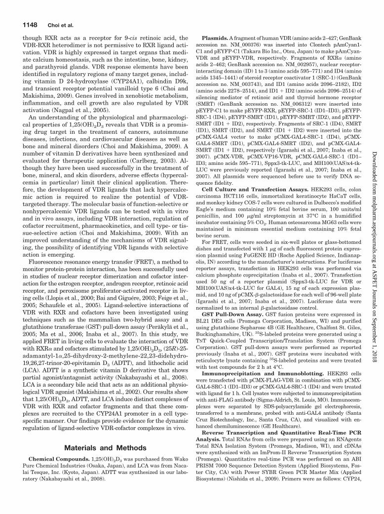
a luciferase reporter assay. We transiently transfectedHEK293 cells with an expression vector for AmCyan-VDR orEYFP-VDR (Fig. 1A) and a luciferase reporter containing aVDR-responsive direct repeat-3 element from the mouse osteopon-tin promoter (Igarashi et al., 2007) and measured 1,25(OH)2D3-dependent luciferase activity. 1,25(OH)2D3 (100 nM) effectivelyinduced transactivation of wild-type VDR, AmCyan-VDR, andEYFP-VDR but not of control empty vector, AmCyan, or EYFP(Fig. 1B). These results show that ligand-dependent transactiva-tion of AmCyan-VDR and EYFP-VDR is comparable with that ofwild-type VDR.
Prufer et al. has reported that GFP-VDR and RXR�-bluefluorescent proteins form heterodimers in the absence ofligand and that 1,25(OH)2D3 treatment induces the forma-tion of multiple nuclear foci of heterodimers (Prufer et al.,2000). We have reported a ligand-independent interaction ofVDR with RXR, which is enhanced by 1,25(OH)2D3 using amammalian two-hybrid assay (Inaba et al., 2007). We inves-tigated the interaction of VDR with RXR using a FRETfluorophore pair of AmCyan-VDR and EYFP-RXR (Fig. 2A).Although exciting AmCyan at 450 nm results in emission at488 nm unless energy is transferred to EYFP, FRET fromAmCyan to EYFP yields the acceptor emission at 526 nm,resulting in an increased ratio of 526 nm/488 nm. First, wetransfected HEK293 cells with AmCyan-VDR and EYFP-RXR expression plasmids and measured emission spectra inthe cell lysates. Emission spectra at 450 nm excitationshowed an intensity ratio (526 nm/488 nm) of 0.70 � 0.08(Table 1). An intensity ratio of a parent fluorophore pairof AmCyan and EYFP was 0.55 � 0.10 (p � 0.01 versusAmCyan-VDR and EYFP-RXR pair) and that of an AmCyan-VDR and EYFP pair was 0.60 � 0.10 (p � 0.05 versus
Fig. 1. Ligand-dependent transactivation of fluorescent-fused VDRs. A,AmCyan- and EYFP-fused VDRs. Numbers indicate amino acids. B, transacti-vation of AmCyan- and EYFP-fused VDRs. HEK293 cells were transfected withpCMX (control), pCMX-VDR, pAmCyan, pAmCyan-VDR, pEYFP, or pEYFP-VDR, and Sppx3-tk-LUC and treated with ethanol (control) or 100 nM1,25(OH)2D3. Values represent the means � S.D. of triplicate assays. ���, p �0.001.
Dynamic Interactions of VDR with RXR and Cofactors 1149
AmCyan-VDR and EYFP-RXR pair), indicating weak associ-ation of AmCyan-VDR and EYFP-RXR in the absence ofligand. When cells were treated with 1,25(OH)2D3 (100 nM)for 30 min, an emission spectrum showed an increased in-tensity ratio of 0.90 � 0.10 (Fig. 2B). The increased FRET incells treated with 1,25(OH)2D3 was no longer present at a60-min time point.
Next, we expressed AmCyan-VDR and EYFP-RXR in COS-7cells and examined FRET in living cells using confocal micros-copy. Upon excitation at 458 nm, the cytoplasm showed anemission spectrum with a negligible FRET, and 1,25(OH)2D3
treatment did not induce FRET (Fig. 2C), indicating that het-erodimerization of VDR and RXR is negligible in the cytoplasm.In the nucleus, the emission spectrum showed two distinctpeaks at 488 and 526 nm at nearly a 1:1 ratio, indicating thatFRET occurs between AmCyan-VDR and EYFP-RXR in the
absence of ligand. 1,25(OH)2D3 treatment increased the inten-sity ratio (526 nm/488 nm) from 1.00 to 1.13 (Fig. 2C). Thus,1,25(OH)2D3 binding to VDR enhances heterodimerization ofnuclear VDR and RXR.
VDR homodimerization has been reported (Cheskis andFreedman, 1994; Lemon and Freedman, 1996), but the exis-tence of VDR homodimers in vivo is controversial. We examinedVDR homodimerization by evaluating FRET between AmCyan-VDR and EYFP-VDR (Fig. 1A). In the absence of ligand, anemission spectrum in lysates of HEK293 cells expressingAmCyan-VDR and EYFP-VDR revealed an intensity ratio (526nm/488 nm) of 0.94 � 0.10, indicating a significant interactioncompared with that of AmCyan-VDR and EYFP (0.60 � 0.10;p � 0.05) (Table 1). 1,25(OH)2D3 treatment for 30 min de-creased FRET and this effect disappeared at 60 min (Fig. 3).Thus, 1,25(OH)2D3 binding to VDR induces heterodimerizationwith RXR and inhibits homodimerization in cells.
ADTT is a synthetic vitamin D derivative that acts as apartial agonist/antagonist for VDR and LCA is a weak en-dogenous agonist (Makishima et al., 2002; Nakabayashi etal., 2008) (Fig. 4A). ADTT (10 �M) and LCA (10 �M) inducedVDR transactivation (Fig. 4B). LCA (10 �M) but not ADTT(10 �M) increased a FRET signal between AmCyan-VDR andEYFP-RXR (Fig. 4C). The rank order of transactivation is thesame as that of VDR-RXR heterodimerization: 1,25(OH)2D3
(100 nM) � LCA (10 �M) � ADTT (10 �M).Ligand-Dependent Interactions of VDR with Cofac-
tors. Upon ligand binding, nuclear receptors undergo confor-
Fig. 2. 1,25(OH)2D3 induces FRET between AmCyan-VDR and EYFP-RXR.A, principle of FRET. Exciting AmCyan at 450 nm results in emission at 488nm, unless energy is transferred to EYFP. Energy transfer depends on thedistance between AmCyan and EYFP. An increased EYFP emission (at 526nm) at the expense of AmCyan emission can occur as the result of interactionbetween AmCyan-VDR and EYFP-RXR. Numbers indicate amino acids. B,1,25(OH)2D3 increases FRET between AmCyan-VDR and EYFP-RXR.HEK293 cells were transfected with pAmCyan-VDR and pEYFP-RXR andtreated with ethanol (control) or 100 nM 1,25(OH)2D3 for 30 or 60 min. Celllysates were subjected to a spectrophotometer for a FRET assay. Valuesrepresent the means � S.D. of septuplicate assays. ���, p � 0.001. C,induction of FRET between AmCyan-VDR and EYFP-RXR in the nucleus ofCOS-7 cells. COS-7 cells were transfected with pAmCyan-VDR and pEYFP-RXR and treated with ethanol (control) or 100 nM 1,25(OH)2D3 for 30 min.Emission spectra were measured in the cytoplasm and nucleus with afluorescence microscope. The experiments were repeated with similarresults.
TABLE 1The ratio of 526 nm/488 nm at 450 nm excitationHEK293 cells were transfected with expression vectors for the indicated fluorescentproteins. Forty-eight hours after transfection, cell lysates were subjected to a spec-trophotometer for FRET analysis. Values represent the means � S.D. of triplicate ormore assays.
Fig. 3. 1,25(OH)2D3 decreases VDR homodimerization. HEK293 cellswere transfected with pAmCyan-VDR and pEYFP-VDR and treated withethanol (control) or 100 nM 1,25(OH)2D3 for 30 or 60 min. Cell lysateswere subjected to a spectrophotometer for a FRET assay. Values repre-sent the means � S.D. of septuplicate assays. �, p � 0.05.
mational changes in the cofactor binding site and AF2 sur-face that result in the dissociation of corepressors andrecruitment of coactivators (Rosenfeld et al., 2006). The p160family proteins such as SRC-1 are well characterized coacti-vators that bind to the AF2 surface and transmit the alloste-ric signal of ligand binding to a chromatin remodeling sys-tem. We examined the effect of ligands on the interaction ofVDR with SRC-1 using a mammalian two-hybrid assay and aFRET assay. We generated GAL4 fusions of SRC-1 (ID1–ID3)and SRC-1 (ID4) for the mammalian two-hybrid assay andtheir EYFP fusions for a FRET assay (Fig. 5A). EYFP-SRC-1(ID1–ID3) contains three LXXLL motifs and has been shownto interact with VDR in a ligand-dependent manner (Adachiet al., 2004; Inaba et al., 2007). Results from a mammaliantwo-hybrid assay showed that 1,25(OH)2D3, and to a lesserextent ADTT and LCA, effectively induced the interaction ofVDR with the SRC-1 (ID1–ID3) fragment (Fig. 5B).1,25(OH)2D3, and ADTT increased FRET between AmCyan-VDR and EYFP-SRC-1 (ID1–ID3) at 30 min, and these asso-ciations disappeared at 60 min (Fig. 5C). LCA did notstabilize FRET between AmCyan-VDR and EYFP-SRC-1(ID1–ID3). A SRC-1 domain including ID4 has been reportedto be necessary for coactivation of the mineralocorticoid re-ceptor (Li et al., 2005). In the mammalian two-hybrid assay,1,25(OH)2D3 but not ADTT or LCA induced the interaction ofVDR with SRC-1 (ID4) (Fig. 5D). The FRET assay did notdetect ligand-induced interaction between these proteins at30 min (Fig. 5E). It is noteworthy that FRET was detected incells treated with 1,25(OH)2D3, ADTT and LCA at 60 min.Luciferase activity in a mammalian two-hybrid assay reflectsthe amount of luciferase protein in cultured cells and mayrepresent an integrated value of the interaction over time. By
contrast, the FRET assay detects the dynamic interaction ofproteins at the selected 30- and 60-min time points. Discrep-ancies in the results between the mammalian two-hybridassay and the FRET assay may be due to the two assays’differing ability to measure the strength of interaction overtime. To further examine the interaction of VDR and SRC-1(ID4), we performed GST pull-down and immunoprecipita-tion assays. 1,25(OH)2D3 induced the interaction of GAL4-SRC-1 (ID1–ID3) and GAL4-SRC-1 (ID4) with GST-VDR butnot with GST (Fig. 5F). GAL4 control proteins did not bind toGST-VDR in the presence or absence of 1,25(OH)2D3 (datanot shown). GAL4 fusion proteins of SRC-1 (ID1–ID3) andSRC-1 (ID4) bound to FLAG-VDR in cells, and 1,25(OH)2D3
treatment enhanced these interactions (Fig. 5G). Thus,SRC-1 (ID4), like SRC-1 (ID1–ID3), binds to VDR in a ligand-dependent manner.
The corepressor SMRT has been reported to mediate tran-scriptional repression by unliganded VDR (Kim et al., 2009).SMRT has bipartite IDs (ID1 and ID2), each of which con-tains a LI/XXI/VI box (Fig. 6A). We generated GAL4 andEYFP fusions of SMRT (ID1), SMRT (ID2), and SMRT (ID1 �ID2). Treatment with 1,25(OH)2D3 and ADTT but not LCAdecreased the association of VP16-VDR and GAL4-SMRT(ID1) (Fig. 6B). An intensity ratio of a fluorophore pair ofAmCyan-VDR and EYFP-SMRT (ID1) was 0.88 � 0.10 (p �0.01 versus AmCyan-VDR and EYFP pair), indicating inter-action of these proteins (Table 1). 1,25(OH)2D3 treatment didnot change the FRET emission (Fig. 6C). ADTT, LCA, and toa lesser extent 1,25(OH)2D3 decreased the interaction be-tween VP16-VDR and GAL4-SMRT (ID2) (Fig. 6D). AmCyan-VDR and EYFP-SMRT (ID2) showed a strong FRET signal to1.41 � 0.41 (p � 0.001 versus AmCyan-VDR and EYFP pair)(Table 1), and 1,25(OH)2D3, ADTT, and LCA decreasedFRET (Fig. 6E). Treatment with 1,25(OH)2D3, ADTT, andLCA decreased the association of VP16-VDR and GAL4-SMRT (ID1 � ID2) (Fig. 6F). Although 1,25(OH)2D3, but notADTT or LCA, decreased FRET between AmCyan-VDR andEYFP-SMRT (ID1 � ID2) at 30 min, FRET signals weredecreased in cells treated with 1,25(OH)2D3, ADTT and LCAfor 60 min (Fig. 6G). Thus, SMRT IDs dissociate from VDR ina ligand-selective manner.
Ligand-Selective Recruitment of VDR, RXR, and Co-factors to an Endogenous Gene Promoter. We comparedthe effects of 1,25(OH)2D3, ADTT, and LCA on the expression ofan endogenous VDR target, CYP24A1, in kidney epithelium-derived HEK293 cells, osteoblast-derived MG63 cells, intestinalmucosa-derived HCT116 cells, and skin keratinocyte-derivedHaCaT cells. As reported previously (Inaba et al., 2007),1,25(OH)2D3 effectively induced CYP24A1 expression in all ofthese cell lines (Fig. 7A). Although ADTT was not effective inHEK293 cells, it increased CYP24A1 expression in HCT116cells, HaCaT cells, and, to a lesser extent, MG63 cells. The effectof ADTT (10 �M) was weaker than that of 1,25(OH)2D3 (100nM), indicating that ADTT is a partial agonist, consistent withVDR transactivation data (Fig. 4B) and a crystal structure ofVDR and ADTT (Nakabayashi et al., 2008). LCA (10 �M)slightly increased CYP24A1 expression in HaCaT cells, but notin HEK293 cells, MG63 cells, or HaCaT cells (Fig. 7A), althoughit induced VDR transactivation in a luciferase reporter assay(Fig. 4B).
Next, we examined the recruitment of VDR, RXR, SRC-1, andSMRT to the CYP24A1 promoter using a ChIP assay in
Fig. 4. Effect of 1,25(OH)2D3, ADTT, and LCA on VDR-RXR het-erodimerization. A, chemical structures of 1,25(OH)2D3, ADTT, and LCA.B, transactivation of VDR. HEK293 cells were transfected with pCMX-VDR and Sppx3-tk-LUC and treated with ethanol (control), 100 nM1,25(OH)2D3, 10 �M ADTT, or 10 �M LCA. Values represent the means �S.D. of triplicate assays. C, FRET between AmCyan-VDR and EYFP-RXR. HEK293 cells were transfected with pAmCyan-VDR and pEYFP-RXR and treated with ethanol (control), 100 nM 1,25(OH)2D3, 10 �MADTT, or 10 �M LCA for 30 min. Cell lysates were subjected to aspectrophotometer for a FRET assay. Values represent the means � S.D.of quadruplicate assays. �, p � 0.05; ���, p � 0.001.
Dynamic Interactions of VDR with RXR and Cofactors 1151
HEK293 cells and HCT116 cells, because ADTT and LCA hadno effect in HEK293 cells but ADTT exhibited agonist activityin HCT116 cells (Fig. 7A). Treatment with 1,25(OH)2D3 for 30and 60 min increased the occupancy of VDR, RXR, and SRC-1on the CYP24A1 promoter in HEK293 cells (Fig. 7B). The1,25(OH)2D3-dependent recruitment of these proteins dimin-ished at 120 min, consistent with the cyclic recruitment ofVDR-RXR and coactivators that has been reported previously(Kim et al., 2005; Vaisanen et al., 2005). SMRT associated withthe CYP24A1 promoter region in the absence of ligand and1,25(OH)2D3 treatment decreased SMRT association (Fig. 7B).In contrast, ADTT treatment resulted in decreased recruitmentof VDR to the CYP24A1 promoter (Fig. 7C). ADTT decreasedSMRT association without increasing recruitment of RXR orSRC-1. LCA increased association of VDR and decreased that ofSMRT. Thus, ADTT and LCA induce the recruitment of recep-tors and cofactors differently from 1,25(OH)2D3. Similar to re-sults in HEK293 cells, 1,25(OH)2D3 increased the association ofVDR, RXR, and SRC-1 and decreased that of SMRT on theCYP24A1 promoter in HCT116 cells (Fig. 7D). Although LCAinduced dissociation of SMRT, it did not effectively recruit VDR,RXR, and SRC-1. It is noteworthy that ADTT increased theassociation of RXR and SMRT but not of VDR and SRC-1 inHCT116 cells. The interaction of SMRT to the CYP24A1 pro-moter was further examined with sequential ChIP analysis.Nuclear lysates of HCT116 cells were immunoprecipitated withanti-VDR antibody and re-ChIP was performed with anti-SMRT antibody. ADTT increased formation of the VDR- andSMRT-containing complex on the CYP24A1 promoter inHCT116 cells, whereas 1,25(OH)2D3 did not (Fig. 7E). Thesefindings suggest that ADTT and LCA are cell type-specific VDRmodulators and that they exhibit selective recruitment of re-ceptors and cofactors in a context-dependent manner.
DiscussionIn this study, we observed ligand-selective dynamic interac-
tions of VDR with RXR, SRC-1, and SMRT using FRET andChIP assays. We used the AmCyan and EYFP fluorophore pairfor FRET assays in living cells and cell lysates. In the absenceof ligand, VDR preferentially formed homodimers and associ-ated with SMRT (Table 1). 1,25(OH)2D3 treatment inducedVDR-RXR heterodimerization rather than VDR homodimeriza-tion (Fig. 2 and 3), consistent with previous reports (Cheskisand Freedman, 1994; Lemon and Freedman, 1996). FRET as-says showed VDR-cofactor interactions, dissociation of SMRT,and recruitment of SRC-1, similar to the results of mammaliantwo-hybrid and ChIP assays (Fig. 5, 6, and 7). 1,25(OH)2D3-dependent dissociation of SMRT (ID1) was observed in a mam-malian two-hybrid assay but not in a FRET assay (Fig. 6).1,25(OH)2D3 decreased the FRET signal between VDR andSMRT (ID2) and induced a weak dissociation of SMRT (ID2) inthe mammalian two-hybrid assay. VDR and RXR have beenreported to interact with ID1 and ID2 of SMRT, respectively(Hu et al., 2001; Kim et al., 2009). Our results suggest that1,25(OH)2D3 binding first induces dissociation of ID2 from RXR
Fig. 5. Effect of 1,25(OH)2D3, ADTT, and LCA on the VDR-SRC-1interaction. A, EYFP-SRC-1 fusion proteins. Numbers indicate aminoacids. B, a mammalian two-hybrid assay for VDR and SRC-1 (ID1–ID3) interaction. C, FRET between AmCyan-VDR and EYFP-SRC-1(ID1–ID3). D, a mammalian two-hybrid assay for VDR and SRC-1 (ID4)interaction. E, FRET between AmCyan-VDR and EYFP-SRC-1 (ID4).For mammalian two-hybrid assays (B and D), HEK293 cells weretransfected with pCMX-VP16-VDR; pCMX-GAL4-SRC-1 (ID1–ID3) orpCMX-GAL4-SRC-1 (ID4); and MH100(UAS)x4-tk-LUC and treatedwith ethanol (control), 100 nM 1,25(OH)2D3, 10 �M ADTT, or 10 �MLCA. For FRET assays (C and E), HEK293 cells were transfected withpAmCyan-VDR and pEYFP-SRC-1 (ID1–ID3) or pEYFP-SRC-1 (ID4)and treated with ethanol (control), 100 nM 1,25(OH)2D3, 10 �M ADTT,or 10 �M LCA for 30 and 60 min. Cell lysates were subjected to aspectrophotometer for a FRET assay. Values represent the means �S.D. of triplicate assays. �, p � 0.05; �, p � 0.01; ���, p � 0.001. F, GSTpull-down assays were performed to evaluate interactions betweenVDR and SRC-1 fragments. Control GST or GST-VDR proteins wereincubated with 35S-labeled GAL4-SRC-1 (ID1–ID3) or GAL4-SRC-1(ID4) in the presence of ethanol (C) or 100 nM 1,25(OH)2D3 (D3). G, invivo interaction of VDR with SRC-1 fragments. HEK293 cells were
transfected with pCMX-FLAG-VDR in combination with pCMX-GAL4-SRC-1 (ID1–ID3) or pCMX-GAL4-SRC-1 (ID4) and treated with ethanol(Cont) or 100 nM 1,25(OH)2D3 (D3) for 1 h. Protein complexes in celllysates were immunoprecipitated with anti-FLAG antibody and thenimmunoblotted with anti-GAL4 antibody. IP, immunoprecipitation; IB,immunoblotting.
in the VDR-RXR heterodimer and subsequently release of ID1from VDR. The interaction of ID2 and RXR may play a principalrole in the binding of the VDR-RXR heterodimer and SMRT incells. Ligand-dependent dissociation of SMRT (ID1 � ID2) fromVDR was different from those of SMRT (ID1) and SMRT (ID2)(Fig. 6), suggesting combined effects of ID1 and ID2 domains ofSMRT on interaction with VDR-RXR heterodimer. Consistentwith previous reports (Tagami et al., 1998; Pathrose et al., 2002;Adachi et al., 2004; Inaba et al., 2007), 1,25(OH)2D3 was shownto induce association of VDR and SRC-1 (ID1–ID3) in mamma-lian two-hybrid and FRET assays (Fig. 5). In addition,1,25(OH)2D3 induced interaction between VDR and SRC-1(ID4). SRC-1 (ID4) is necessary for coactivation of the miner-alocorticoid receptor (Li et al., 2005). We found that VDR bounddirectly to SRC-1 (ID4) in a ligand-dependent manner (Fig. 5).Because a FRET assay showed 1,25(OH)2D3-induced associa-tion of VDR and SRC-1 (ID4) at 60 min and not at 30 min (Fig.5), this domain may play an accessory role in VDR-RXR coacti-vation. Thus, FRET assays are useful in the detection of time-dependent protein-protein interactions. The role of ID4 frag-ment in functional interaction between SRC-1 and VDR isunder investigation.
LCA is a secondary bile acid that acts as a weak VDRagonist and interacts with the VDR ligand-binding pocket ina mode distinct from that of 1,25(OH)2D3 (Makishima et al.,
2002; Adachi et al., 2004). ADTT is a synthetic vitamin Dderivative that acts as a VDR partial agonist/antagonist (Na-kabayashi et al., 2008). LCA (10 �M) increased FRET be-tween VDR and RXR but not between VDR and SRC-1 (ID1–ID3), whereas ADTT (10 �M) induced FRET between VDRand SRC-1 (ID1–ID3) but not between VDR and RXR (Figs. 4and 5). ADTT and LCA at 10 �M were less effective than1,25(OH)2D3 (100 nM) in VDR transactivation in a luciferasereporter assay (Fig. 4) and did not induce endogenousCYP24A1 mRNA expression in HEK293 cells (Fig. 7). Nei-ther ADTT nor LCA increased occupancy of RXR and SRC-1on the CYP24A1 promoter (Fig. 7). Both heterodimerizationwith RXR and formation of an active conformation with co-activators are necessary for ligand-dependent VDR transac-tivation (Prufer et al., 2000; Pathrose et al., 2002). Thesefindings suggest that the conformation induced by ADTT andLCA is not sufficient to recruit a stable complex of VDR, RXR,and cofactors to a target gene needed to induce effectivetranscription. ADTT but not LCA induced dissociation ofSMRT (ID1), and both ligands decreased association of VDRwith SMRT (ID2) and SMRT (ID1 � ID2) (Fig. 6). VDR antago-nists induce dissociation of corepressors as well as agonists (Inabaet al., 2007). Corepressor dissociation is suggested to reflect aligand-dependent conformational change, and additional factors
Fig. 6. Effect of 1,25(OH)2D3, ADTT, and LCA on VDR-SMRT interaction. A, EYFP-SMRT fusion proteins. Numbers indicate amino acids. B, amammalian two-hybrid assay for VDR and SMRT (ID1) interaction. C, FRET between AmCyan-VDR and EYFP-SMRT (ID1). D, a mammaliantwo-hybrid assay for VDR and SMRT (ID2) interaction. E, FRET between AmCyan-VDR and EYFP-SMRT (ID2). F, a mammalian two-hybrid assayfor VDR and SMRT (ID1 � ID2) interaction. G, FRET between AmCyan-VDR and EYFP-SMRT (ID1 � ID2). For mammalian two-hybrid assays (B,D, and F), HEK293 cells were transfected with pCMX-VP16-VDR, pCMX-GAL4-SMRT (ID1), pCMX-GAL4-SMRT (ID2), or pCMX-GAL4-SMRT(ID1 � ID2), and MH100(UAS)x4-tk-LUC, and treated with ethanol (control), 100 nM 1,25(OH)2D3, 10 �M ADTT, or 10 �M LCA. For FRET assays(C, E, and G), HEK293 cells were transfected with pAmCyan-VDR and pEYFP-SMRT (ID1), pEYFP-SMRT (ID2), or pEYFP-SMRT (ID1 � ID2) andtreated with ethanol (control), 100 nM 1,25(OH)2D3, 10 �M ADTT, or 10 �M LCA for 30 min (C, E, and G) and 60 min (G). Cell lysates were subjectedto a spectrophotometer for a FRET assay. Values represent the means � S.D. of triplicate assays (B–D, F, and G) or septuplicate assays (E). �, p �0.05; �, p � 0.01; ���, p � 0.001.
Dynamic Interactions of VDR with RXR and Cofactors 1153
such as stable complex with RXR and coactivators may be re-quired for efficient transactivation.
Like AD47, another partial agonist/antagonist having anadamantane ring side chain (Inaba et al., 2007), ADTTinduced endogenous CYP24A1 expression in HCT116 cellsbut not HEK293 cells (Fig. 7). ADTT, like 1,25(OH)2D3 andLCA, decreased SMRT association on the CYP24A1 pro-moter in HEK293 cells, consistent with the results inmammalian two-hybrid and FRET assays. Unexpectedly,ADTT increased association of RXR and SMRT but not ofVDR and SRC-1 on the CYP24A1 promoter in HCT116cells. Although different occupancies of VDR and RXR on aCYP24A1 promoter region were reported (Matilainen etal., 2010), the mechanism of discrepancy in the recruit-ment of VDR and RXR remains unclear. ADTT may inducea complex of VDR-RXR heterodimer and cofactors thatdecreases the efficiency of ChIP with anti-VDR antibody.In a sequential ChIP assay, ADTT, but not 1,25(OH)2D3,induced a complex of VDR and SMRT on the promoter.
These findings suggest that ADTT induces a cofactor com-plex in a cell context-dependent manner. ADTT and1,25(OH)2D3 may induce distinct VDR conformations, re-sulting in the selective recruitment of cofactors. The subsetof involved cofactors may be dependent on cell type-specificexpression of cofactor proteins and other cellular environ-ments. 1,25(OH)2D3 recruits SMRT to the CYP24A1 pro-moter, and the corepressor is involved in negative tran-scriptional regulation (Sanchez-Martínez et al., 2008). Wedid not observe corecruitment of SRC-1 and SMRT to theCYP24A1 promoter in HCT116 cells. Ligand-dependentcorepressor recruitment may be in a different phase fromthat of coactivators through a cyclic cell-selective pattern.Whereas 1,25(OH)2D3 and LCA repress expression of thehuman cholesterol 7�-hydroxylase gene by recruiting SMRTto the VDR-RXR heterodimer (Han et al., 2010), SMRT hasbeen reported to be involved in coactivation of estrogen re-ceptor-� with SRC-3 on the progesterone receptor gene pro-moter (Karmakar et al., 2010). Further studies are needed to
Fig. 7. Association of VDR, RXR, SRC-1, and SMRT on the CYP24A1 promoter in cells treated with 1,25(OH)2D3, ADTT and LCA. A, expression ofthe CYP24A1 gene in HEK293 cells, MG63 cells, HCT116 cells, and HaCaT cells. Cells were treated with ethanol (control), 100 nM 1,25(OH)2D3(1,25D3), 10 �M ADTT, or 10 �M LCA for 16 h. CYP24A1 mRNA levels were evaluated by quantitative real-time PCR. Values represent the means �S.D. of triplicate assays. B and C, occupancy of VDR, RXR, SRC-1, and SMRT on the CYP24A1 promoter in HEK293 cells treated with 1,25(OH)2D3(B), ADTT, or LCA (C). HEK293 cells were treated with ethanol (C), 100 nM 1,25(OH)2D3, 10 �M ADTT, or 10 �M LCA for 30, 60 or 120 min.Occupancy of the indicated proteins on the CYP24A1 promoter were examined with ChIP assays using control IgG, anti-VDR, anti-RXR, anti-SRC-1,or anti-SMRT antibodies. Occupancy (percentage) is relative to the input values. D, occupancy of VDR, RXR, SRC-1, and SMRT on the CYP24A1promoter in HCT116 cells treated with 1,25(OH)2D3, ADTT, or LCA. HCT116 cells were treated with ethanol (C), 100 nM 1,25(OH)2D3, 10 �M ADTT,or 10 �M LCA for 30, 60 or 120 min, and ChIP assays were performed as in B and C. E, sequential ChIP with VDR and SMRT in HCT116 cells. ChIPsamples with anti-VDR antibody as in D were next subjected with ChIP with anti-SMRT antibody. Values represent the means � S.D. of triplicateassays. �, p � 0.05; �, p � 0.01; ���, p � 0.001.
elucidate the mechanism of agonist-dependent recruitmentof SMRT and its functional relevance.
More than 2000 vitamin D derivatives have been synthe-sized and evaluated for potential therapeutic application(Carlberg, 2003). Although they have been used successfullyin the treatment of bone, mineral and skin disorders, adverseeffects, particularly hypercalcemia, limit the clinical applica-tion of vitamin D and its synthetic derivatives in the man-agement of other diseases, such as cancer, autoimmunity,infection, and cardiovascular disease (Choi and Makishima,2009). VDR interaction and cofactor recruitment as well aspharmacokinetics are key factors in designing VDR ligandswith selective activity. In this study, we provided evidence forligand-selective VDR conformations and cofactor recruitmentusing FRET and ChIP assays. These techniques will be use-ful in the further development of selective VDR modulators.
Acknowledgments
We thank Dr. Atsuko Iwane of the Graduate School of FrontierBiosciences, Osaka University, Osaka, Japan; Dr. Hiroki Nagase andDr. Makoto Kimura of Nihon University Advanced Research Insti-tute for the Science and Humanities, Tokyo, Japan; Dr. KazumichiKuroda and Toshikatsu Shibata of the Division of Microbiology,Department of Pathology and Microbiology, Nihon University Schoolof Medicine; Sergej Popov, Daisuke Akagi, and other members of theMakishima laboratory for technical assistance and helpful com-ments; and Dr. Andrew I. Shulman for editorial assistance.
Authorship Contributions
Participated in research design: Choi, Yamada, and Makishima.Conducted experiments: Choi.Contributed new reagents or analytic tools: Choi and Yamada.Performed data analysis: Choi, Yamada, and Makishima.Wrote or contributed to the writing of the manuscript: Choi,
Yamada, and Makishima.
ReferencesAdachi R, Shulman AI, Yamamoto K, Shimomura I, Yamada S, Mangelsdorf DJ, and
Makishima M (2004) Structural determinants for vitamin D receptor response toendocrine and xenobiotic signals. Mol Endocrinol 18:43–52.
Bai Y and Giguere V (2003) Isoform-selective interactions between estrogen recep-tors and steroid receptor coactivators promoted by estradiol and ErbB-2 signalingin living cells. Mol Endocrinol 17:589–599.
Baneyx G, Baugh L, and Vogel V (2001) Coexisting conformations of fibronectin incell culture imaged using fluorescence resonance energy transfer. Proc Natl AcadSci USA 98:14464–14468.
Carlberg C (2003) Molecular basis of the selective activity of vitamin D analogues.J Cell Biochem 88:274–281.
Cheskis B and Freedman LP (1994) Ligand modulates the conversion of DNA-boundvitamin D3 receptor (VDR) homodimers into VDR-retinoid X receptor het-erodimers. Mol Cell Biol 14:3329–3338.
Choi M and Makishima M (2009) Therapeutic applications for novel non-hypercalcemic vitamin D receptor ligands. Expert Opin Ther Pat 19:593–606.
Erickson MG, Moon DL, and Yue DT (2003) DsRed as a potential FRET partner withCFP and GFP. Biophys J 85:599–611.
Feige JN, Gelman L, Tudor C, Engelborghs Y, Wahli W, and Desvergne B (2005)Fluorescence imaging reveals the nuclear behavior of peroxisome proliferator-activated receptor/retinoid X receptor heterodimers in the absence and presence ofligand. J Biol Chem 280:17880–17890.
Fujiki R, Kim MS, Sasaki Y, Yoshimura K, Kitagawa H, and Kato S (2005) Ligand-induced transrepression by VDR through association of WSTF with acetylatedhistones. EMBO J 24:3881–3894.
Han S, Li T, Ellis E, Strom S, and Chiang JY (2010) A novel bile acid-activatedvitamin D receptor signaling in human hepatocytes. Mol Endocrinol 24:1151–1164.
Haussler MR, Whitfield GK, Haussler CA, Hsieh JC, Thompson PD, Selznick SH,Dominguez CE, and Jurutka PW (1998) The nuclear vitamin D receptor: biologicaland molecular regulatory properties revealed. J Bone Miner Res 13:325–349.
Hu X, Li Y, and Lazar MA (2001) Determinants of CoRNR-dependent repressioncomplex assembly on nuclear hormone receptors. Mol Cell Biol 21:1747–1758.
Igarashi M, Yoshimoto N, Yamamoto K, Shimizu M, Ishizawa M, Makishima M,DeLuca HF, and Yamada S (2007) Identification of a highly potent vitamin Dreceptor antagonist: (25S)-26-adamantyl-25-hydroxy-2-methylene-22,23-
Inaba Y, Yamamoto K, Yoshimoto N, Matsunawa M, Uno S, Yamada S, and Mak-ishima M (2007) Vitamin D3 derivatives with adamantane or lactone ring sidechains are cell type-selective vitamin D receptor modulators. Mol Pharmacol71:1298–1311.
Karmakar S, Gao T, Pace MC, Oesterreich S, and Smith CL (2010) Cooperativeactivation of cyclin D1 and progesterone receptor gene expression by the SRC-3coactivator and SMRT corepressor. Mol Endocrinol 24:1187–1202.
Kim JY, Son YL, and Lee YC (2009) Involvement of SMRT corepressor in transcrip-tional repression by the vitamin D receptor. Mol Endocrinol 23:251–264.
Kim S, Shevde NK, and Pike JW (2005) 1,25-Dihydroxyvitamin D3 stimulates cyclicvitamin D receptor/retinoid X receptor DNA-binding, co-activator recruitment, andhistone acetylation in intact osteoblasts. J Bone Miner Res 20:305–317.
Lemon BD and Freedman LP (1996) Selective effects of ligands on vitamin D3receptor- and retinoid X receptor-mediated gene activation in vivo. Mol Cell Biol16:1006–1016.
Li Y, Suino K, Daugherty J, and Xu HE (2005) Structural and biochemical mecha-nisms for the specificity of hormone binding and coactivator assembly by miner-alocorticoid receptor. Mol Cell 19:367–380.
Llopis J, Westin S, Ricote M, Wang Z, Cho CY, Kurokawa R, Mullen TM, Rose DW,Rosenfeld MG, Tsien RY, et al. (2000) Ligand-dependent interactions of coactiva-tors steroid receptor coactivator-1 and peroxisome proliferator-activated receptorbinding protein with nuclear hormone receptors can be imaged in live cells and arerequired for transcription. Proc Natl Acad Sci USA 97:4363–4368.
Ma Y, Khalifa B, Yee YK, Lu J, Memezawa A, Savkur RS, Yamamoto Y, Chinta-lacharuvu SR, Yamaoka K, Stayrook KR, et al. (2006) Identification and charac-terization of noncalcemic, tissue-selective, nonsecosteroidal vitamin D receptormodulators. J Clin Invest 116:892–904.
Makishima M (2005) Nuclear receptors as targets for drug development: regulationof cholesterol and bile acid metabolism by nuclear receptors. J Pharmacol Sci97:177–183.
Makishima M, Lu TT, Xie W, Whitfield GK, Domoto H, Evans RM, Haussler MR, andMangelsdorf DJ (2002) Vitamin D receptor as an intestinal bile acid sensor.Science 296:1313–1316.
Makishima M, Okamoto AY, Repa JJ, Tu H, Learned RM, Luk A, Hull MV, LustigKD, Mangelsdorf DJ, and Shan B (1999) Identification of a nuclear receptor for bileacids. Science 284:1362–1365.
Makishima M and Yamada S (2005) Targeting the vitamin D receptor: advances indrug discovery. Expert Opin Ther Pat 15:1133–1145.
Matilainen JM, Malinen M, Turunen MM, Carlberg C, and Vaisanen S (2010) Thenumber of vitamin D receptor binding sites defines the different vitamin D re-sponsiveness of the CYP24 gene in malignant and normal mammary cells. J BiolChem 285:24174–24183.
Matsunawa M, Amano Y, Endo K, Uno S, Sakaki T, Yamada S, and Makishima M(2009) The aryl hydrocarbon receptor activator benzo[a]pyrene enhances vitaminD3 catabolism in macrophages. Toxicol Sci 109:50–58.
Nagpal S, Na S, and Rathnachalam R (2005) Noncalcemic actions of vitamin Dreceptor ligands. Endocr Rev 26:662–687.
Nakabayashi M, Yamada S, Yoshimoto N, Tanaka T, Igarashi M, Ikura T, Ito N,Makishima M, Tokiwa H, DeLuca HF, et al. (2008) Crystal structures of ratvitamin D receptor bound to adamantyl vitamin D analogs: structural basis forvitamin D receptor antagonism and partial agonism. J Med Chem 51:5320–5329.
Nishida S, Ozeki J, and Makishima M (2009) Modulation of bile acid metabolism by1�-hydroxyvitamin D3 administration in mice. Drug Metab Dispos 37:2037–2044.
Pathrose P, Barmina O, Chang CY, McDonnell DP, Shevde NK, and Pike JW (2002)Inhibition of 1,25-dihydroxyvitamin D3-dependent transcription by syntheticLXXLL peptide antagonists that target the activation domains of the vitamin Dand retinoid X receptors. J Bone Miner Res 17:2196–2205.
Perakyla M, Malinen M, Herzig KH, and Carlberg C (2005) Gene regulatory poten-tial of nonsteroidal vitamin D receptor ligands. Mol Endocrinol 19:2060–2073.
Prufer K, Racz A, Lin GC, and Barsony J (2000) Dimerization with retinoid Xreceptors promotes nuclear localization and subnuclear targeting of vitamin Dreceptors. J Biol Chem 275:41114–41123.
Rosenfeld MG, Lunyak VV, and Glass CK (2006) Sensors and signals: a coactivator/corepressor/epigenetic code for integrating signal-dependent programs of tran-scriptional response. Genes Dev 20:1405–1428.
Sanchez-Martínez R, Zambrano A, Castillo AI, and Aranda A (2008) Vitamin D-de-pendent recruitment of corepressors to vitamin D/retinoid X receptor het-erodimers. Mol Cell Biol 28:3817–3829.
Schaufele F, Carbonell X, Guerbadot M, Borngraeber S, Chapman MS, Ma AA,Miner JN, and Diamond MI (2005) The structural basis of androgen receptoractivation: Intramolecular and intermolecular amino-carboxy interactions. ProcNatl Acad Sci USA 102:9802–9807.
Shang Y, Hu X, DiRenzo J, Lazar MA, and Brown M (2000) Cofactor dynamics andsufficiency in estrogen receptor-regulated transcription. Cell 103:843–852.
Tagami T, Lutz WH, Kumar R, and Jameson JL (1998) The interaction of the vitaminD receptor with nuclear receptor corepressors and coactivators. Biochem BiophysRes Commun 253:358–363.
Vaisanen S, Dunlop TW, Sinkkonen L, Frank C, and Carlberg C (2005) Spatio-temporal activation of chromatin on the human CYP24 gene promoter in thepresence of 1�,25-dihydroxyvitamin D3. J Mol Biol 350:65–77.
Zwart W, Griekspoor A, Berno V, Lakeman K, Jalink K, Mancini M, Neefjes J, andMichalides R (2007) PKA-induced resistance to tamoxifen is associated with analtered orientation of ER� towards co-activator SRC-1. EMBO J 26:3534–3544.
Address correspondence to: Makoto Makishima, Division of Biochemistry, De-partment of Biomedical Sciences, Nihon University School of Medicine, Itabashi-ku,Tokyo 173-8610, Japan. E-mail: [email protected]
Dynamic Interactions of VDR with RXR and Cofactors 1155


![Imaging of Vascular Inflammation With · [11C]-PK11195, a selective ligand for peripheral benzodiazepine receptors expressed in activated macrophages, can be used to image vascular](https://static.documents.pub/doc/80x56/5f7643b5cbfe9b2e8666b454/imaging-of-vascular-iniammation-with-11c-pk11195-a-selective-ligand-for-peripheral.jpg)
