Page 1
Page S1
Dynamic Supramolecular Complexation by Shapeshifting Organic Molecules
Alexander R. Lippert, Vasken L. Keleshian, and Jeffrey W. Bode*Roy and Diana Vagelos Laboratories, Department of Chemistry, University of Pennsylvania
Philadelphia, Pennsylvania 19104
Supporting Information
General Methods. All reactions utilizing air- or moisture-sensitive reagents were performed in dried glassware
under an atmosphere of dry N2. THF and CH2Cl2 were distilled over CaH2. Hexamethyldisilazane was distilled
from KOH. iso-Butyl chloroformate was distilled under N2. Other reagents were used without further
purification. Thin layer chromatography (TLC) was performed on Merck precoated plates (silica gel 60 F254,
Art 5715, 0.25 mm) and were visualized by fluorescence quenching under UV light or by staining with
phosphomolybdic acid. Silica-gel preparative thin-layer chromatography (PTLC) was performed using plates
prepared from Merck Kieselgel 60 PF254 (Art 7747). Column chromatography was performed on E. Merck
Silica Gel 60 (230–400 Mesh) using a forced flow of 0.5–1.0 bar. 1H NMR (500 MHz) and
13C NMR (125
MHz) were measured on a Bruker Avance 500 spectrometer. Chemical shifts are expressed in parts per million
(PPM) downfield from residual solvent peaks and coupling constants are reported as Hertz (Hz). Splitting
patterns are indicated as follows: br, broad; s, singlet; d, doublet; t, triplet; q, quartet; m, multiplet; dd, doublet
of doublets; dt, doublet of triplets. Infrared (IR) spectra were recorded on a JASCO FT/IR-4100
spectrophotometer and are reported as wavenumber (cm–1
).
Page 2
Page S2
A. Synthetic Protocols and Tabulated Spectra
O
OOEt
iBuO
O
4
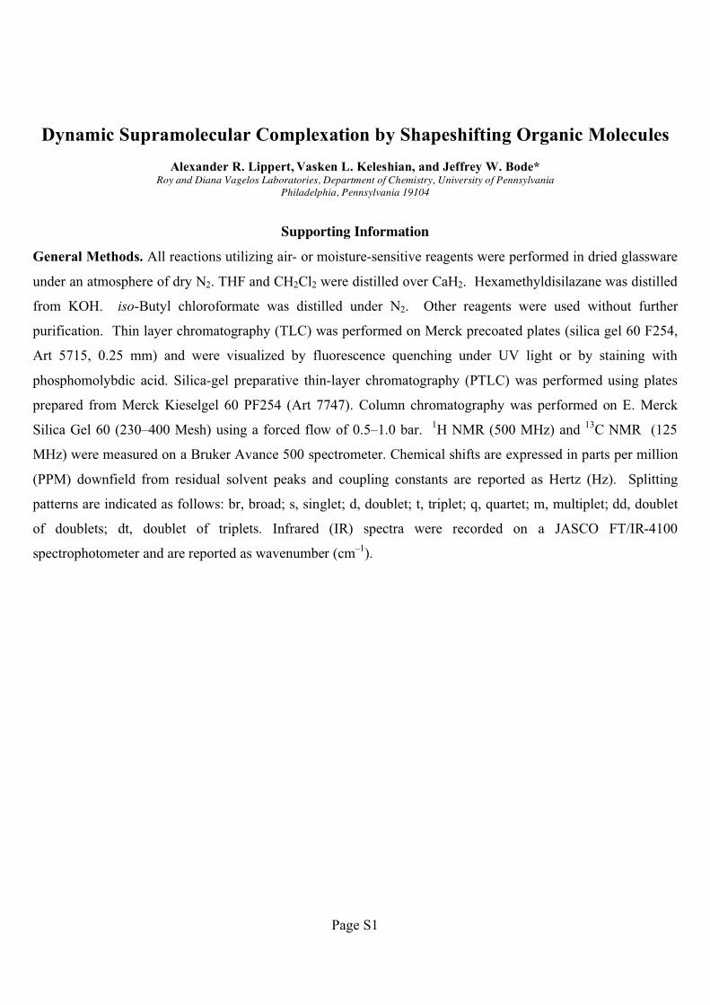
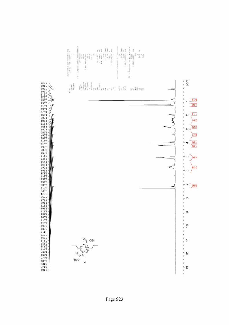
Bisallyl Bullvalene 4: Hexamethyldisilazane (0.55 mL, 0.26 mmol, 3.1 equiv) was added to a solution of 2.5M
n-butyl lithium in hexane (0.10 mL, 0.25 mmol, 3.0 equiv) in THF at –78 °C and allowed to warm to 0 °C.
After stirring for 20 min at 0 °C, the reaction was cooled to –78 °C and bullvalone 31 (25.0 mg, 0.0838 mmol,
1.0 equiv) was added as a solution in 3 x 0.8 mL THF and stirred for 15 min. iso-Butyl chloroformate (110 μL,
0.840 mmol, 10 equiv) was added and the reaction was allowed to warm to rt. After 2 h 45 min, the reaction
was poured into 10 mL sat NH4Cl and extracted with 3 x 10 mL CH2Cl2. The combined organic layers were
washed with brine, dried over Na2SO4, filtered, and concentrated. Isolation of three major bands by silica
chromatography (20:1 hexanes:EtOAc � 10:1 hexanes:EtOAc) yielded bisallyl bullvalene 4 (16.2 mg, 49%) as
a clear oil. 1H NMR (500 MHz, (CD3)2SO, 120 °C) � 5.90–5.70 (m, 2H), 5.10–5.00 (m, 4H), 4.16 (q, 2H, J =
7.5 Hz), 3.95 (d, 2H, J = 6.5 Hz), 2.75–2.60 (m, 3H), 1.95 (quintet, 1H, J = 6.5 Hz), 1.25 (t, 3H, J = 6.5 Hz),
0.93 (d, 6H, J = 6.5 Hz);1H NMR (500 MHz, (CD3)2SO, 30 °C)* � 7.05 (m, 1H), 5.85–5.68 (m, 3H), 5.2–5.0
(m, 4H), 4.12–4.09 (m, 2H), 3.91 (d, 2H, J = 6.0 Hz), 2.90–2.70 (m, 4H), 2.50–2.30 (m, 2H), 1.91 (quintet, 1H,
J = 6.5 Hz), 1.24–1.19 (m, 1H), 0.89 (d, 6H, J = 7 Hz);1H NMR (500 MHz, CDCl3, 30 °C)* � 7.27 (m, 1H),
5.79–5.67 (m, 3H), 5.11–5.04 (m, 4H), 4.20–4.16 (m, 2H), 3.97 (d, 2H, J = 9.5 Hz), 3.40 (m, 1H), 2.93–2.80
(m, 4H), 2.70–2.35 (m, 3H), 1.98 (quintet, 1H, J = 6.5 Hz), 1.30–1.24 (m, 1H), 0.92 (m, 6H); 13
C NMR (125
MHz, CDCl3) � 165.6, 153.6, 153.4, 137.4, 136.0, 135.9, 135.8, 135.6, 135.5, 134.9, 134.6, 134.5, 120.8, 120.6,
117.5, 117.4, 117.3, 117.2, 116.9, 116.8, 116.8, 116.7, 116.6, 116.4, 116.3, 74.5, 60.8, 60.7, 60.7, 45.6, 45.2,
44.8, 44.6, 44.4, 43.9, 43.8, 43.7, 43.6, 43.5, 38.8, 38.3, 37.1, 36.8, 36.0, 35.9, 31.9, 29.7, 29.4, 29.2, 29.0, 28.0,
27.8, 27.7, 27.4, 27.3, 25.8, 25.7, 22.7, 22.1, 21.0, 19.7, 18.9, 18.8, 14.3, 14.2, 14.1; IR (thin film) � 2958.3,
2919.7, 2849.3, 1756.8, 1707.6, 1463.2, 1238.6 cm-1
; HRMS (ESI) calcd for C24H30O5 [M+H]+ 399.2171, found
399.2176.
* Only major peaks are reported. See variable temperature spectra in Figure S12 on page S15 (in (CD3)2SO)
and spectra on page S21 (in CDCl3).
[1] A. R. Lippert, J. Kaeobamrung, J. W. Bode, J. Am. Chem. Soc. 2006, 128, 14738–14739.
Page 3
Page S3
N
HNN
NH
tButBu
tBu
tBu
tButBu
NH
O
6
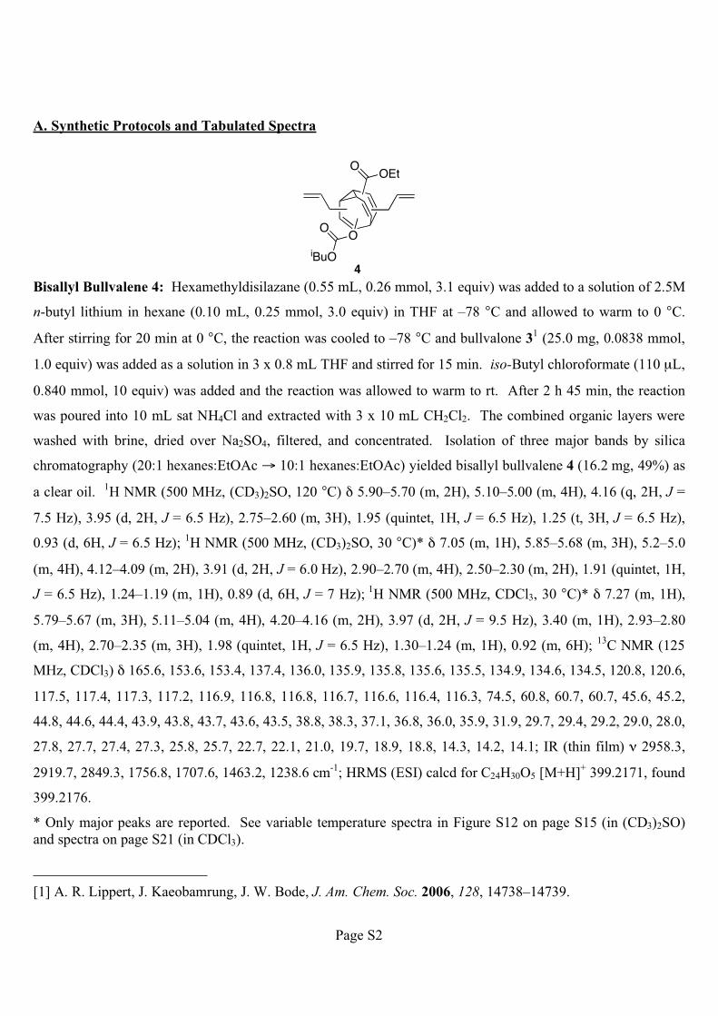
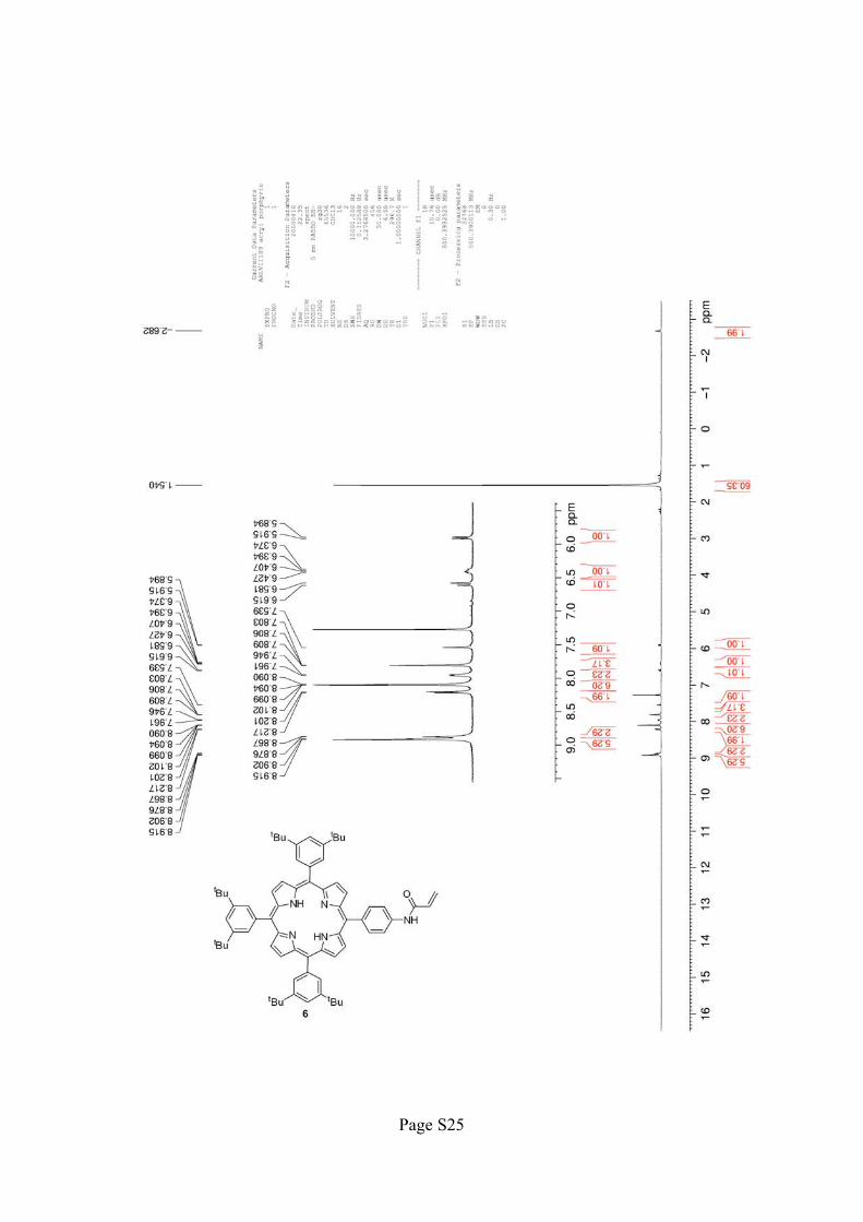
Acrylamide Porphyrin 6: Acrylic acid (0.57 mL, 0.83 mmol, 6.9 equiv) was added to a solution of thionyl
chloride (57 �L, 0.79 mmol, 6.6 equiv) in 0.50 mL of dimethylacetamide at –10 °C. After stirring for 5 min, a
solution of amino porphyrin 52 (119.8 mg, 0.1202 mmol, 1.0 equiv) in 4 mL dimethylacetamide was added to
the reaction mixture over 10 min, giving a green solution. Upon complete addition, the reaction mixture was
allowed to warm and stirred at rt. After 1 h, the reaction was quenched by the addition of 10 mL of distilled
water causing a purple solid to precipitate, which was filtered and washed with water. The solid was dissolved
in CH2Cl2, washed through the filter, poured onto brine, and extracted into 3 x 15 mL CH2Cl2. The combined
organic layers were dried over Na2SO4, filtered, and concentrated. The crude product was purified by silica gel
chromatography (hexane � 5:1 hexane:EtOAc) to yield the acrylamide porphyrin 6 (94.0 mg, 77%) as a red oil.
1H NMR (500 MHz, CDCl3) � 8.91 (m, 6H), 8.87 (d, 2H, J = 4.5 Hz), 8.21 (d, 2H, J = 8 Hz), 8.10 (dd, 6H, J =
4 Hz, 1.5 Hz), 7.95 (d, 2H, J = 7.5 Hz), 7.81 (m, 3H), 7.54 (s, 1H), 6.60 (d, 1H, J = 17 Hz), 6.41 (m, 1H), 5.90
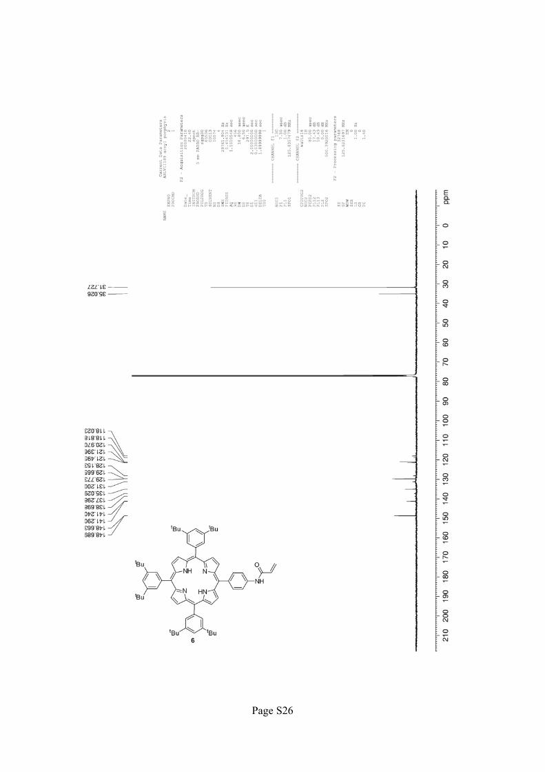
(d, 1H, J = 10.5 Hz), 1.54 (s, 54H), –2.68 (s, 2H); 13
C NMR (125 MHz, CDCl3) � 148.7, 141.3, 138.7, 137.3,
135.0, 131.2, 129.8, 129.7, 128.2, 121.5, 121.4, 121.0, 119.0, 118.0, 35.0, 31.7; IR (thin film) � 3318.4, 2951.2,
2869.1, 2910.1, 1586.0, 1592.9 cm-1
; HRMS (ESI) calcd for C71H81N5O [M+H]+ 1020.6520, found 1020.6490.
[2] H. Imahori, K. Hagiwara, M. Aoki, T. Akiyama, S. Taniguchi, T. Okada, M. Shirakawa, Y. Sakata, J. Am.
Chem. Soc. 1996, 118, 11771–11782.
Page 4
Page S4
NHN
N
NH
tBu
tBu
tBu
tBu
tBu
tBu
HN
O
O
O OEt
iBuO
O
NHN
N
NH
tBu
tBu
tBu
tBu
tBu
tBu
NH
O
1
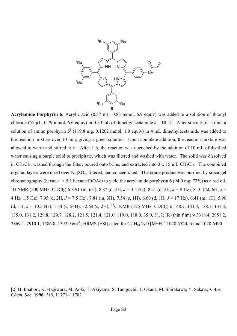
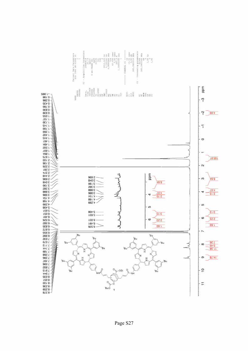
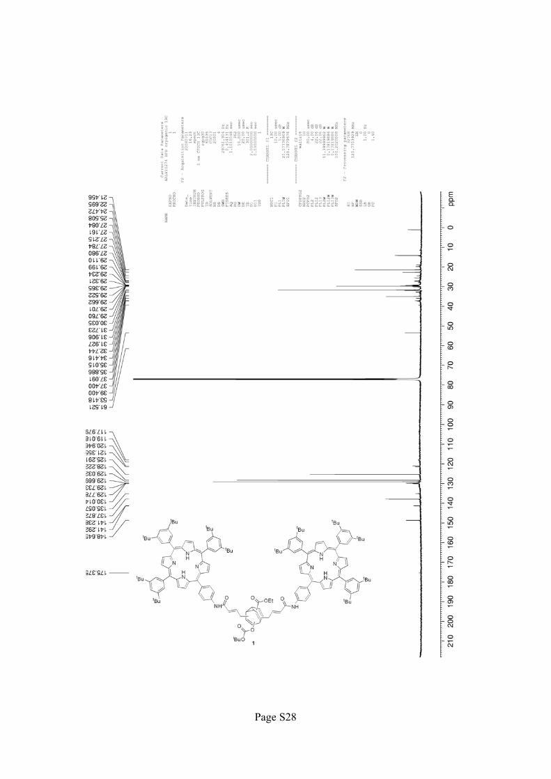
Bisporphyrin Bullvalene 1: Acrylamide porphyrin 6 (21.3 mg, 0.0209 mmol, 2.5 equiv) and bisallyl
bullvalene 4 (3.3 mg, 0.0084 mmol, 1.0 equiv) were dissolved in 0.42 mL CH2Cl2. Grubbs’ 2nd
generation
catalyst3 (4.3 mg, 0.0051 mmol, 0.61 equiv) was added, the flask was sealed, and the reaction was heated at 40
°C for 16 h. The reaction mixture was concentrated and purified by PTLC (11:1 toluene:EtOAc) to yield the
bisporphyrin bullvalene 1 (3.8 mg, 19%) as a red oil. 1H NMR (500 MHz, C6D5CD3) � 9.0–8.85 (m, 14H),
8.24–8.16 (m, 12H), 8.14–8.00 (m, 6H), 7.93–7.85 (m, 8H), 6.53–6.49 (m, 2H), 6.15–5.80 (m, 2H), 5.65–5.41
(m, 3H), 4.30–3.75 (m, 6H), 3.38–2.70 (m, 6H), 1.49–1.43 (m, 108H), 1.14 (t, 3H, J = 7.5 Hz), 0.84 (d, 6H, J =
22 Hz), –2.02 (br s, 4H);13
C NMR (125 MHz, CDCl3) � 175.4, 148.6, 141.3, 141.2, 137.9, 135.1, 130.0, 129.8,
128.2, 125.3, 121.4, 120.9, 119.0 (br), 118.0 (br), 61.5 (br), 39.4 (br), 37.4 (br), 37.1, 35.9, 35.0, 34.4, 32.7,
31.9, 31.7, 30.0, 29.8, 29.7, 29.6, 29.5, 29.4, 29.3, 29.2, 29.1, 28.0, 27.8, 27.2, 27.1, 25.5, 24.5, 22.7, 21.5, 19.7
(br), 19.3, 18.9, 14.4, 14.1 cm-1
; IR (thin film) � 3315.5, 2954.9, 2923.6, 2853.2, 1753.9, 1665.7, 1592.4,
1525.4, 1475.3, 1466.6, 1362.9, 1260.3, 1362.9, 1260.3, 1246.8 cm-1
; HRMS (MALDI) calcd for
(C162H185N10O7)+, 2382.44; found 2382.83.
[3] M. Scholl, S. Ding, C. W. Lee, R. H. Grubbs, Org. Lett. 1999, 1, 953–956.
Page 5
Page S5
N
HNN
NH
tBu
tBu
tBu
tBu
tBu
tBu
NH
O
N
NH N
HN
tBu tBu
tBu
tBu
tBu tBu
HN
OO
EtO
O
2
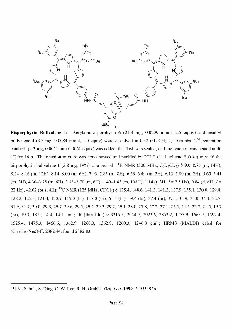
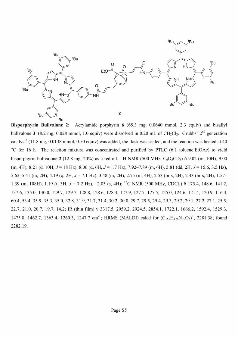
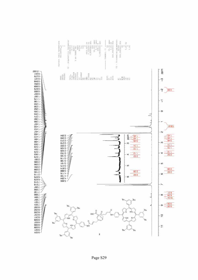
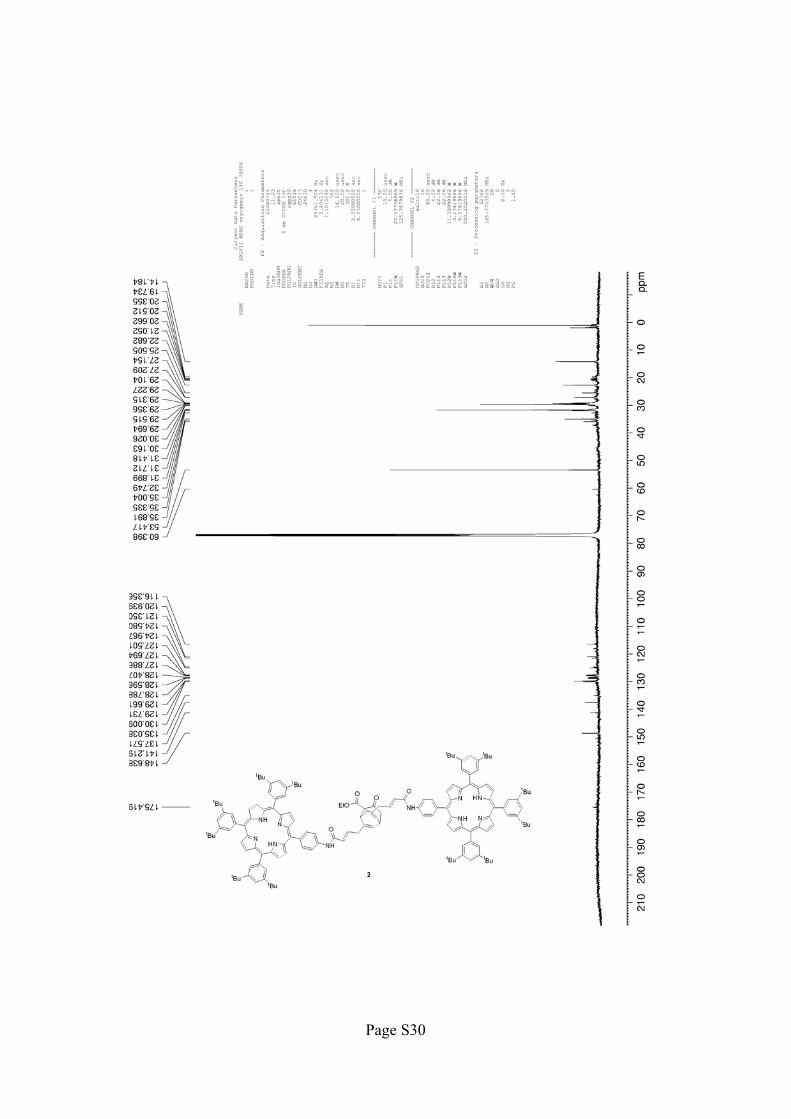
Bisporphyrin Bullvalone 2: Acrylamide porphyrin 6 (65.3 mg, 0.0640 mmol, 2.3 equiv) and bisallyl
bullvalone 31
(8.2 mg, 0.028 mmol, 1.0 equiv) were dissolved in 0.20 mL of CH2Cl2. Grubbs’ 2nd
generation
catalyst3 (11.8 mg, 0.0138 mmol, 0.50 equiv) was added, the flask was sealed, and the reaction was heated at 40
°C for 16 h. The reaction mixture was concentrated and purified by PTLC (6:1 toluene:EtOAc) to yield
bisporphyrin bullvalone 2 (12.8 mg, 20%) as a red oil. 1H NMR (500 MHz, C6D5CD3) � 9.02 (m, 10H), 9.00
(m, 4H), 8.21 (d, 10H, J = 18 Hz), 8.06 (d, 6H, J = 1.7 Hz), 7.92–7.89 (m, 6H), 5.81 (dd, 2H, J = 15.6, 3.5 Hz),
5.62–5.41 (m, 2H), 4.19 (q, 2H, J = 7.1 Hz), 3.48 (m, 2H), 2.75 (m, 4H), 2.53 (br s, 2H), 2.43 (br s, 2H), 1.57–
1.39 (m, 108H), 1.19 (t, 3H, J = 7.2 Hz), –2.03 (s, 4H);13
C NMR (500 MHz, CDCl3) � 175.4, 148.6, 141.2,
137.6, 135.0, 130.0, 129.7, 129.7, 128.8, 128.6, 128.4, 127.9, 127.7, 127.5, 125.0, 124.6, 121.4, 120.9, 116.4,
60.4, 53.4, 35.9, 35.3, 35.0, 32.8, 31.9, 31.7, 31.4, 30.2, 30.0, 29.7, 29.5, 29.4, 29.3, 29.2, 29.1, 27.2, 27.1, 25.5,
22.7, 21.0, 20.7, 19.7, 14.2; IR (thin film) � 3317.5, 2959.2, 2924.5, 2854.1, 1722.1, 1666.2, 1592.4, 1529.3,
1475.8, 1462.7, 1363.4, 1260.3, 1247.7 cm-1
; HRMS (MALDI) calcd for (C157H176N10O5)+, 2281.38; found
2282.19.
Page 6
Page S6
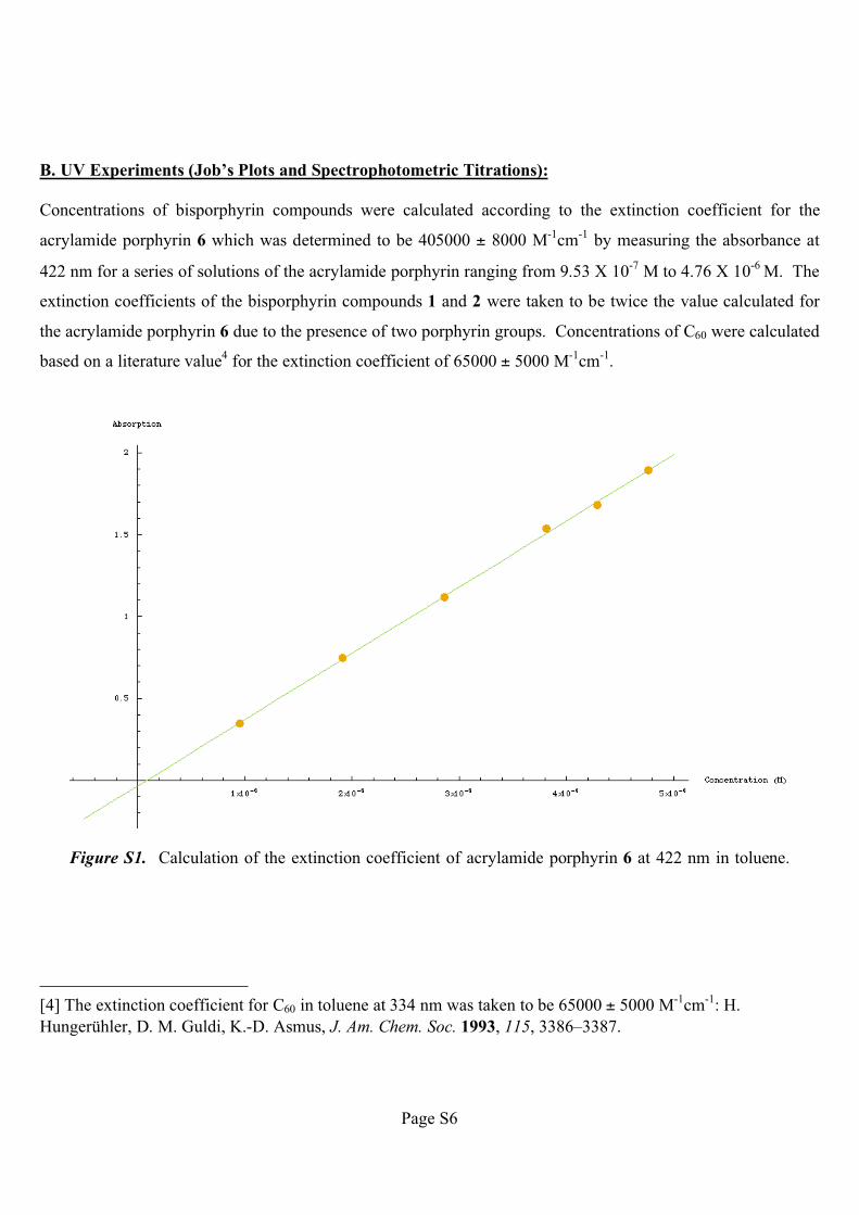
B. UV Experiments (Job’s Plots and Spectrophotometric Titrations):
Concentrations of bisporphyrin compounds were calculated according to the extinction coefficient for the
acrylamide porphyrin 6 which was determined to be 405000 ± 8000 M-1
cm-1
by measuring the absorbance at
422 nm for a series of solutions of the acrylamide porphyrin ranging from 9.53 X 10⁻⁷
M to 4.76 X 10⁻⁶
M. The
extinction coefficients of the bisporphyrin compounds 1 and 2 were taken to be twice the value calculated for
the acrylamide porphyrin 6 due to the presence of two porphyrin groups. Concentrations of C60 were calculated
based on a literature value4 for the extinction coefficient of 65000 ± 5000 M
-1cm
-1.
Figure S1. Calculation of the extinction coefficient of acrylamide porphyrin 6 at 422 nm in toluene.
[4] The extinction coefficient for C60 in toluene at 334 nm was taken to be 65000 ± 5000 M-1
cm-1
: H.
Hungerühler, D. M. Guldi, K.-D. Asmus, J. Am. Chem. Soc. 1993, 115, 3386–3387.
Page 7
Page S7
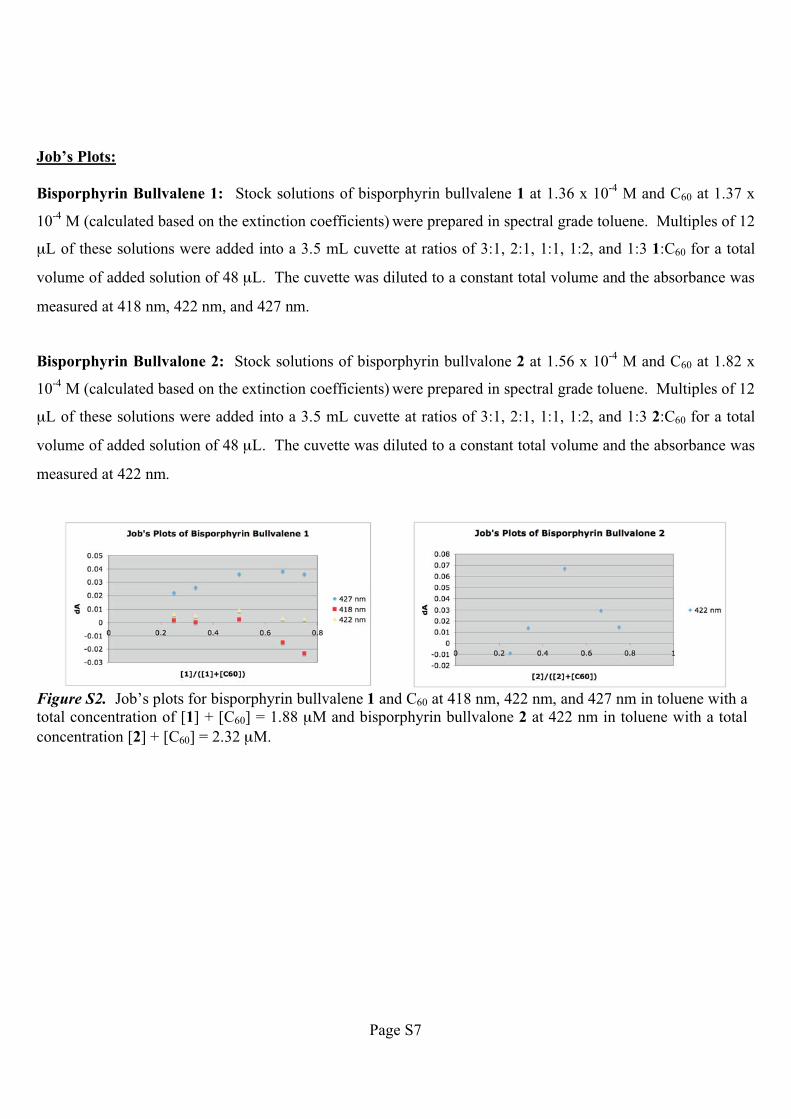
Job’s Plots:
Bisporphyrin Bullvalene 1: Stock solutions of bisporphyrin bullvalene 1 at 1.36 x 10⁻⁴
M and C60 at 1.37 x
10-4
M (calculated based on the extinction coefficients) were prepared in spectral grade toluene. Multiples of 12
μL of these solutions were added into a 3.5 mL cuvette at ratios of 3:1, 2:1, 1:1, 1:2, and 1:3 1:C60 for a total
volume of added solution of 48 μL. The cuvette was diluted to a constant total volume and the absorbance was
measured at 418 nm, 422 nm, and 427 nm.
Bisporphyrin Bullvalone 2: Stock solutions of bisporphyrin bullvalone 2 at 1.56 x 10⁻⁴
M and C60 at 1.82 x
10-4
M (calculated based on the extinction coefficients) were prepared in spectral grade toluene. Multiples of 12
μL of these solutions were added into a 3.5 mL cuvette at ratios of 3:1, 2:1, 1:1, 1:2, and 1:3 2:C60 for a total
volume of added solution of 48 μL. The cuvette was diluted to a constant total volume and the absorbance was
measured at 422 nm.
Figure S2. Job’s plots for bisporphyrin bullvalene 1 and C60 at 418 nm, 422 nm, and 427 nm in toluene with a
total concentration of [1] + [C60] = 1.88 μM and bisporphyrin bullvalone 2 at 422 nm in toluene with a total
concentration [2] + [C60] = 2.32 μM.
Page 8
Page S8
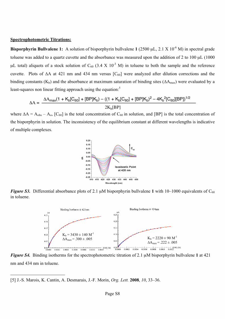
Spectrophotometric Titrations:
Bisporphyrin Bullvalene 1: A solution of bisporphyrin bullvalene 1 (2500 μL, 2.1 X 10⁻⁶
M) in spectral grade
toluene was added to a quartz cuvette and the absorbance was measured upon the addition of 2 to 100 μL (1000
μL total) aliquots of a stock solution of C60 (3.4 X 10⁻³
M) in toluene to both the sample and the reference
cuvette. Plots of �A at 421 nm and 434 nm versus [C60] were analyzed after dilution corrections and the
binding constants (Kb) and the absorbance at maximum saturation of binding sites (�Amax) were evaluated by a
least-squares non linear fitting approach using the equation:5
�� =��max(1 + Kb[C60] + [BP]Kb) – ((1 + Kb[C60] + [BP]Kb)
2 – 4Kb2[C60][BP])
1/2
2Kb[BP]
where �A = Aobs – Ao, [C60] is the total concentration of C60 in solution, and [BP] is the total concentration of
the bisporphyrin in solution. The inconsistency of the equilibrium constant at different wavelengths is indicative
of multiple complexes.
Figure S3. Differential absorbance plots of 2.1 μM bisporphyrin bullvalene 1 with 10–1000 equivalents of C60
in toluene.
Figure S4. Binding isotherms for the spectrophotometric titration of 2.1 μM bisporphyrin bullvalene 1 at 421
nm and 434 nm in toluene.
[5] J.-S. Marois, K. Cantin, A. Desmarais, J.-F. Morin, Org. Lett. 2008, 10, 33–36.
Kb = 3430 ± 140 M-1
�Amax = .300 ± .005 Kb = 2220 ± 90 M-1
�Amax = .222 ± .005
Page 9
Page S9
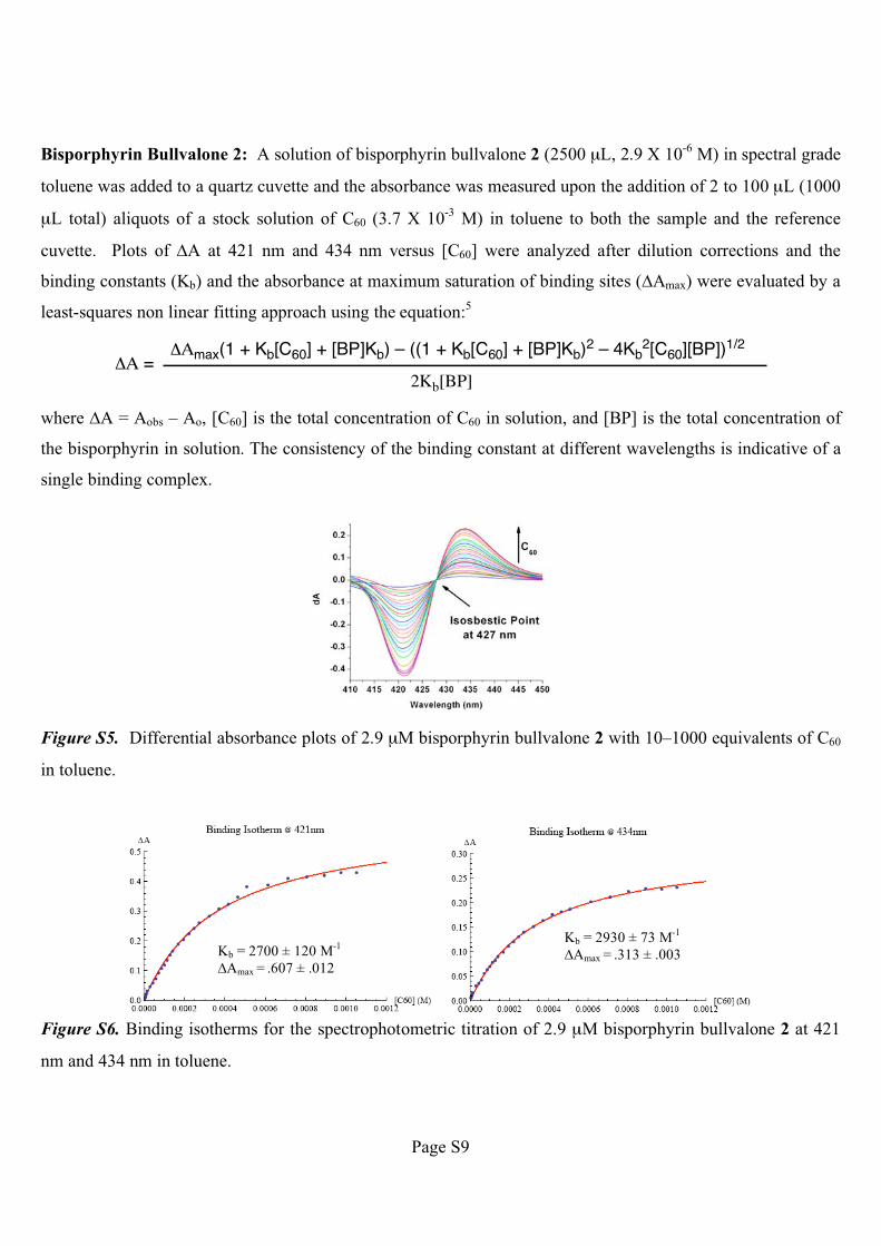
Bisporphyrin Bullvalone 2: A solution of bisporphyrin bullvalone 2 (2500 μL, 2.9 X 10⁻⁶
M) in spectral grade
toluene was added to a quartz cuvette and the absorbance was measured upon the addition of 2 to 100 μL (1000
μL total) aliquots of a stock solution of C60 (3.7 X 10⁻³
M) in toluene to both the sample and the reference
cuvette. Plots of �A at 421 nm and 434 nm versus [C60] were analyzed after dilution corrections and the
binding constants (Kb) and the absorbance at maximum saturation of binding sites (�Amax) were evaluated by a
least-squares non linear fitting approach using the equation:5
�� =��max(1 + Kb[C60] + [BP]Kb) – ((1 + Kb[C60] + [BP]Kb)
2 – 4Kb2[C60][BP])
1/2
2Kb[BP]
where �A = Aobs – Ao, [C60] is the total concentration of C60 in solution, and [BP] is the total concentration of
the bisporphyrin in solution. The consistency of the binding constant at different wavelengths is indicative of a
single binding complex.
Figure S5. Differential absorbance plots of 2.9 μM bisporphyrin bullvalone 2 with 10–1000 equivalents of C60
in toluene.
Figure S6. Binding isotherms for the spectrophotometric titration of 2.9 μM bisporphyrin bullvalone 2 at 421
nm and 434 nm in toluene.
Kb = 2700 ± 120 M-1
�Amax = .607 ± .012
Kb = 2930 ± 73 M-1
�Amax = .313 ± .003
Page 10
Page S10
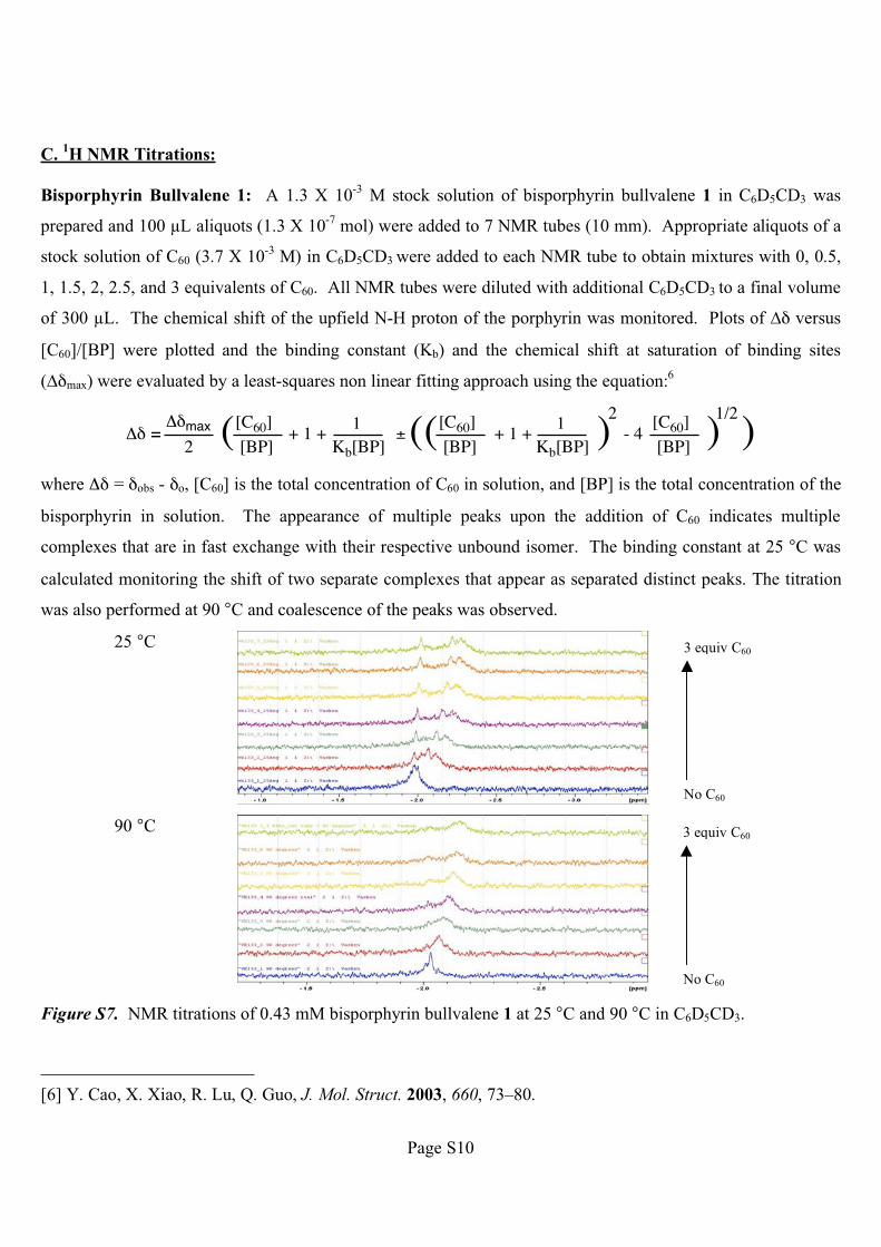
C. 1H NMR Titrations:
Bisporphyrin Bullvalene 1: A 1.3 X 10⁻³
M stock solution of bisporphyrin bullvalene 1 in C6D5CD3 was
prepared and 100 �L aliquots (1.3 X 10⁻⁷
mol) were added to 7 NMR tubes (10 mm). Appropriate aliquots of a
stock solution of C60 (3.7 X 10⁻³
M) in C6D5CD3 were added to each NMR tube to obtain mixtures with 0, 0.5,
1, 1.5, 2, 2.5, and 3 equivalents of C60. All NMR tubes were diluted with additional C6D5CD3 to a final volume
of 300 �L. The chemical shift of the upfield N-H proton of the porphyrin was monitored. Plots of �� versus
[C60]/[BP] were plotted and the binding constant (Kb) and the chemical shift at saturation of binding sites
(��max) were evaluated by a least-squares non linear fitting approach using the equation:6
�� =��max ( + 1 +
1Kb[BP]
±(( + 1 +1
Kb[BP] )2
- 4 )1/2
2
[C60]
[BP]
[C60]
[BP]
[C60]
[BP] )where �� = �obs - �o, [C60] is the total concentration of C60 in solution, and [BP] is the total concentration of the
bisporphyrin in solution. The appearance of multiple peaks upon the addition of C60 indicates multiple
complexes that are in fast exchange with their respective unbound isomer. The binding constant at 25 °C was
calculated monitoring the shift of two separate complexes that appear as separated distinct peaks. The titration
was also performed at 90 °C and coalescence of the peaks was observed.
25 °C
90 °C
Figure S7. NMR titrations of 0.43 mM bisporphyrin bullvalene 1 at 25 °C and 90 °C in C6D5CD3.
[6] Y. Cao, X. Xiao, R. Lu, Q. Guo, J. Mol. Struct. 2003, 660, 73–80.
3 equiv C60
No C60
3 equiv C60
No C60
Page 11
Page S11
Figure S8. Binding isotherms of the NMR titration of 0.43 mM bisporphyrin bullvalene 1 at 25 °C and 90 °C in
C6D5CD3.
Stated Assumptions: Our calculation of the binding constants for bisporphyrin bullvalene 1 requires two key
assumptions. The first assumption is that the observed �� upon addition of C60 is predominately due to the
change in the ratio of bound and unbound bisporphyrin compounds and not due to any change in the relative
ratio of valence isomers. We believe this assumption is valid because (1) the rate of exchange of valence
isomers (which coalesce at 90 °C) is much slower than the rate of exchange of the bound and unbound
bisporphyrin compounds (which coalesce at –50 °C) and (2) the observed difference in the chemical shift
between the observed peaks (�� ~ 0.05 ppm, see peak d and e in Figure 3b) at 25 °C changes very little (< 0.009
ppm) as increasing amounts of C60 are added. The second assumption is that the concentrations of each binding
isomer are 100% of the total concentration of bisporphyrin bullvalene 1 used in the experiment. In actuality, the
population of each binding isomer is a difficult to measure fraction of this value, which would lead to higher
calculated affinities. For example, the binding affinities for the complex giving rise to peak d in Figure 3b
would be 4037 ± 578 M-1
, 6056 ± 867 M-1
, and 12111 ± 1734 M-1
if we assume that the concentration, [BP], of
this binding isomer is 75%, 50%, and 25%, respectively, of the amount of bisporphyrin bullvalene 1 added.
Kb = 3030 ± 430
��max = .319 ± .014
Kb = 6770 ± 1800
��max = .313 ± .016
Kb = 920 ± 100
��max = .257 ± .014
Page 12
Page S12
The same analysis for peak e in Figure 3b yields values of 9033 ± 2425 M-1
, 13550 ± 3637 M-1
, and 27099 ±
7274 M-1
if we assume that the concentration, [BP], of this binding isomer is 75%, 50%, and 25%, respectively,
of the amount of bisporphyrin bullvalene 1 added. Our best estimates for these relative populations based on 1H
NMR integrations of the N-H peak indicate that the two isomers are in approximately a 1:1 ratio, which would
give binding constants of 6056 ± 867 M-1
for peak d and 13550 ± 3637 M-1
for peak e.
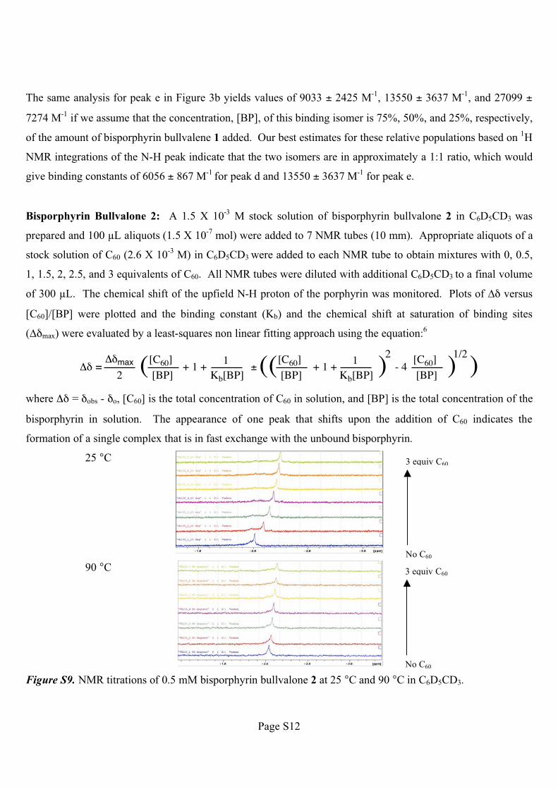
Bisporphyrin Bullvalone 2: A 1.5 X 10⁻³
M stock solution of bisporphyrin bullvalone 2 in C6D5CD3 was
prepared and 100 �L aliquots (1.5 X 10⁻⁷
mol) were added to 7 NMR tubes (10 mm). Appropriate aliquots of a
stock solution of C60 (2.6 X 10⁻³
M) in C6D5CD3 were added to each NMR tube to obtain mixtures with 0, 0.5,
1, 1.5, 2, 2.5, and 3 equivalents of C60. All NMR tubes were diluted with additional C6D5CD3 to a final volume
of 300 �L. The chemical shift of the upfield N-H proton of the porphyrin was monitored. Plots of �� versus
[C60]/[BP] were plotted and the binding constant (Kb) and the chemical shift at saturation of binding sites
(��max) were evaluated by a least-squares non linear fitting approach using the equation:6
�� =��max ( + 1 +
1Kb[BP]
±(( + 1 +1
Kb[BP] )2
- 4 )1/2
2
[C60]
[BP]
[C60]
[BP]
[C60]
[BP] )where �� = �obs - �o, [C60] is the total concentration of C60 in solution, and [BP] is the total concentration of the
bisporphyrin in solution. The appearance of one peak that shifts upon the addition of C60 indicates the
formation of a single complex that is in fast exchange with the unbound bisporphyrin.
25 °C
90 °C
Figure S9. NMR titrations of 0.5 mM bisporphyrin bullvalone 2 at 25 °C and 90 °C in C6D5CD3.
3 equiv C60
No C60
3 equiv C60
No C60
Page 13
Page S13
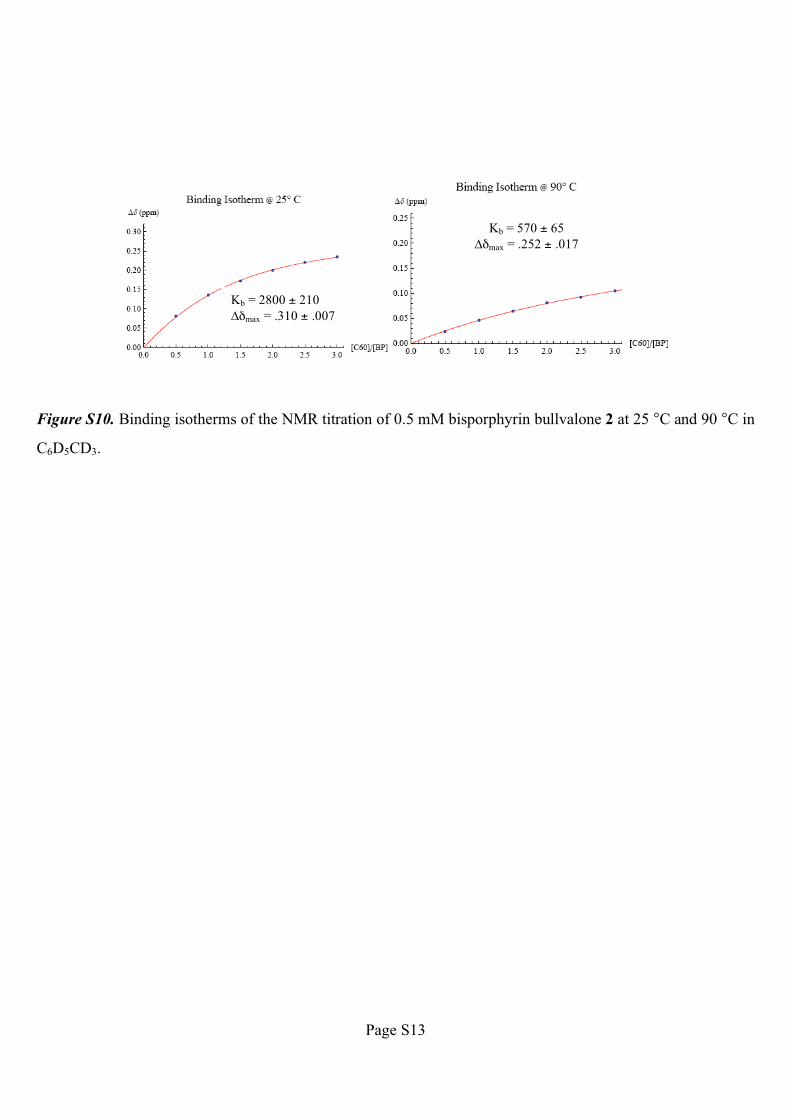
Figure S10. Binding isotherms of the NMR titration of 0.5 mM bisporphyrin bullvalone 2 at 25 °C and 90 °C in
C6D5CD3.
Kb = 2800 ± 210
��max = .310 ± .007
Kb = 570 ± 65
��max = .252 ± .017
Page 14
Page S14
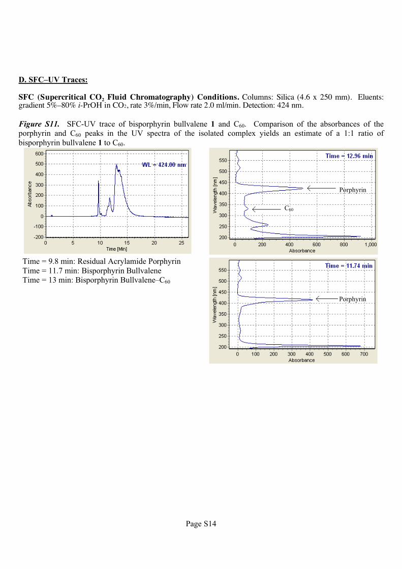
D. SFC–UV Traces:
SFC (Supercritical CO2 Fluid Chromatography) Conditions. Columns: Silica (4.6 x 250 mm). Eluents: gradient 5%–80% i-PrOH in CO2, rate 3%/min, Flow rate 2.0 ml/min. Detection: 424 nm.
Figure S11. SFC-UV trace of bisporphyrin bullvalene 1 and C60. Comparison of the absorbances of the
porphyrin and C60 peaks in the UV spectra of the isolated complex yields an estimate of a 1:1 ratio of
bisporphyrin bullvalene 1 to C60.
Time = 9.8 min: Residual Acrylamide Porphyrin
Time = 11.7 min: Bisporphyrin Bullvalene
Time = 13 min: Bisporphyrin Bullvalene–C60
Porphyrin
Porphyrin
C60
Page 15
Page S15
E. UV Spectra
Figure S12. UV spectra of 2.1 μM bisporphyrin bullvalene 1 in spectral grade toluene.
Figure S13. UV spectra of 2.9 μM bisporphyrin bullvalene 2 in spectral grade toluene.
Page 16
Page S16
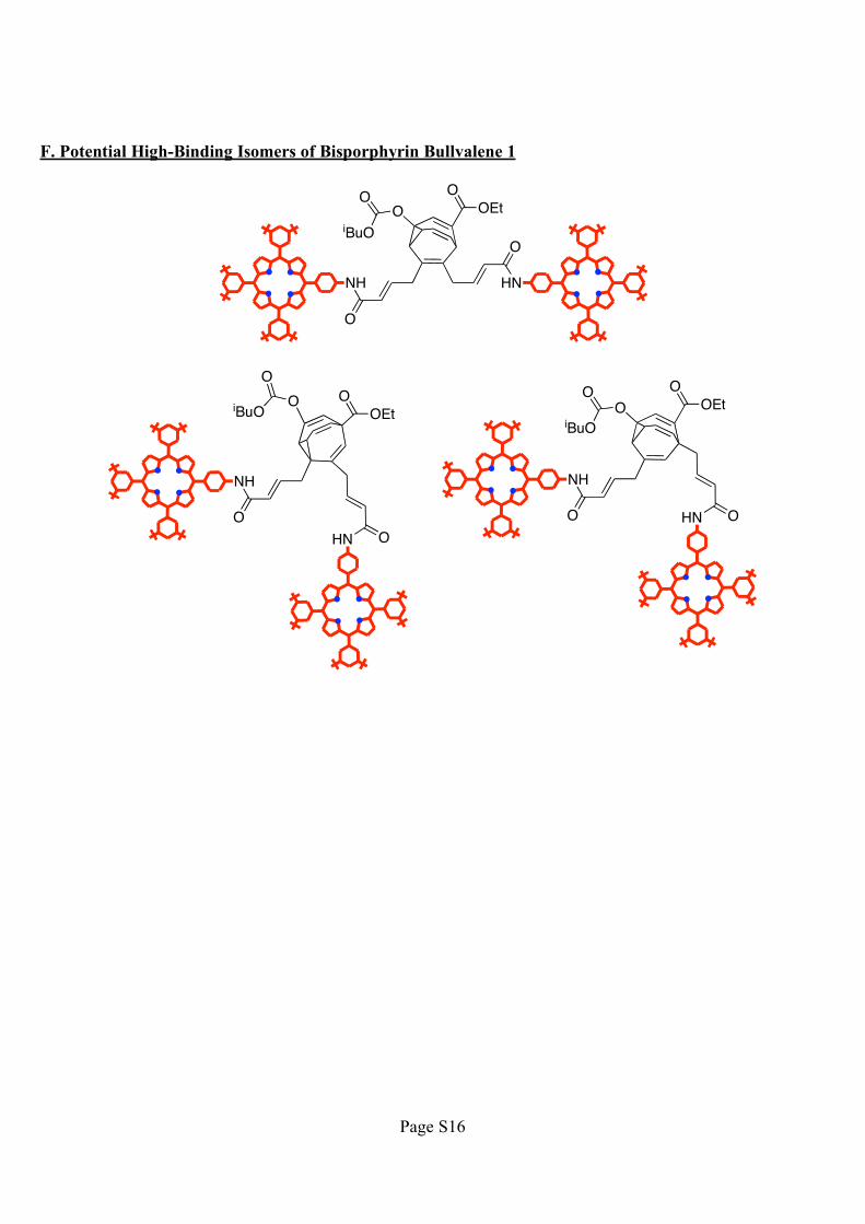
F. Potential High-Binding Isomers of Bisporphyrin Bullvalene 1
O
O
OO
iBuO
OEtO
NH HN
OO
O
O
iBuO OEtO
NH
HN
OO
OO
iBuO
OEtO
NH
HN
Page 17
Page S17
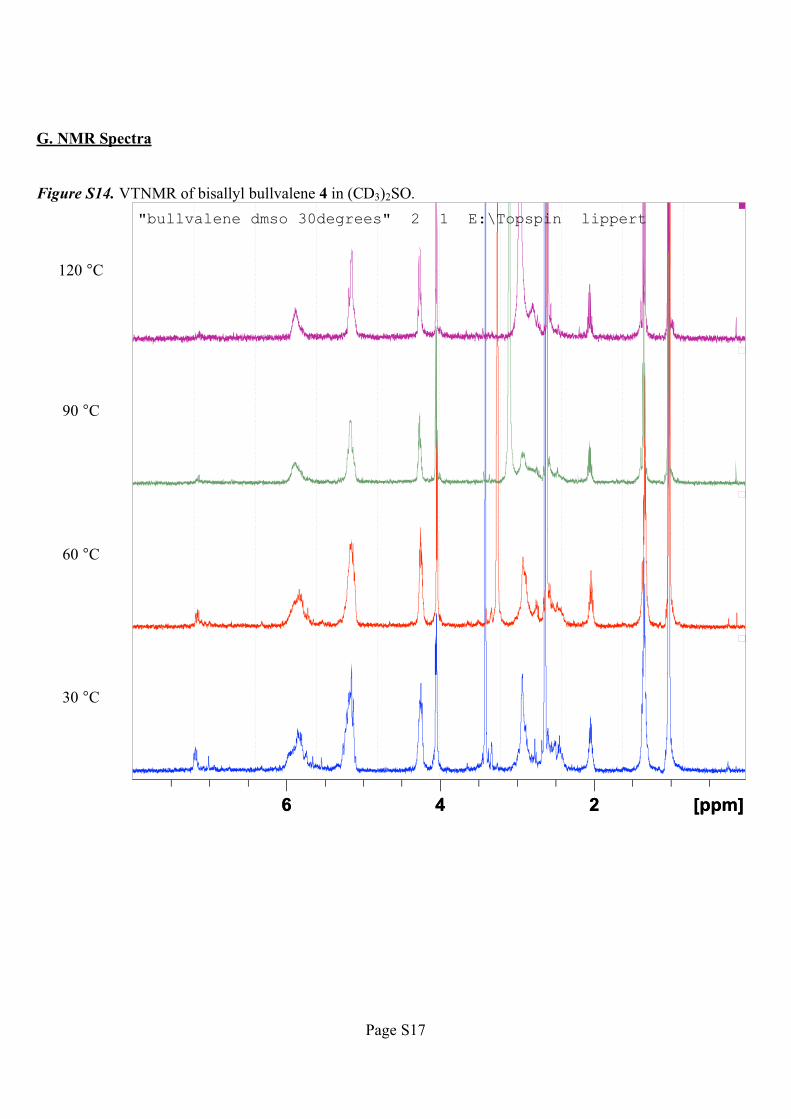
G. NMR Spectra
Figure S14. VTNMR of bisallyl bullvalene 4 in (CD3)2SO.
120 °C
90 °C
60 °C
30 °C
[ppm] 6 4 2
"bullvalene dmso 30degrees" 2 1 E:\Topspin lippert
[ppm] 6 4 2
"bullvalene dmso 30degrees" 2 1 E:\Topspin lippert
Page 18
Page S18
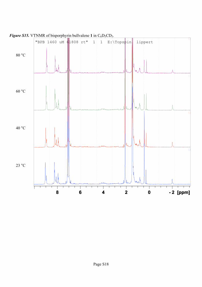
Figure S15. VTNMR of bisporphyrin bullvalene 1 in C6D5CD3.
80 °C
60 °C
40 °C
23 °C
[ppm] 8 6 4 2 0 - 2
"BPB 1460 uM 61808 rt" 1 1 E:\Topspin lippert
[ppm] 8 6 4 2 0 - 2
"BPB 1460 uM 61808 rt" 1 1 E:\Topspin lippert
Page 19
Page S19
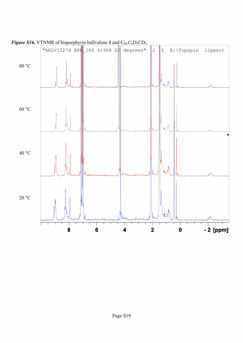
Figure S16. VTNMR of bisporphyrin bullvalene 1 and C60 C6D5CD3.
80 °C
60 °C
40 °C
20 °C
[ppm] 8 6 4 2 0 - 2
"ARLVII274 BPB_C60 61908 20 degrees" 2 1 E:\Topspin lippert
[ppm] 8 6 4 2 0 - 2
"ARLVII274 BPB_C60 61908 20 degrees" 2 1 E:\Topspin lippert
Page 20
Page S20
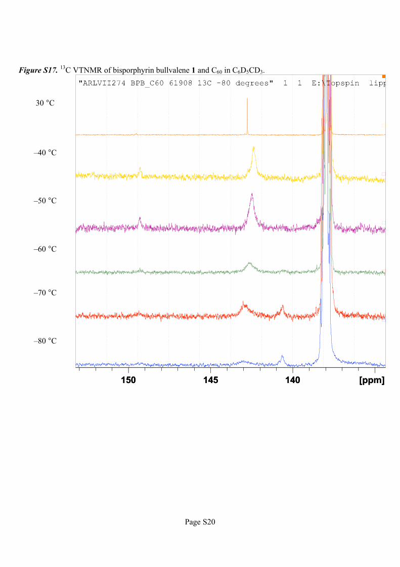
Figure S17.13
C VTNMR of bisporphyrin bullvalene 1 and C60 in C6D5CD3.
30 °C
–40 °C
–50 °C
–60 °C
–70 °C
–80 °C
[ppm] 150 145 140 [ppm] 150 145 140
Page 21
Page S21
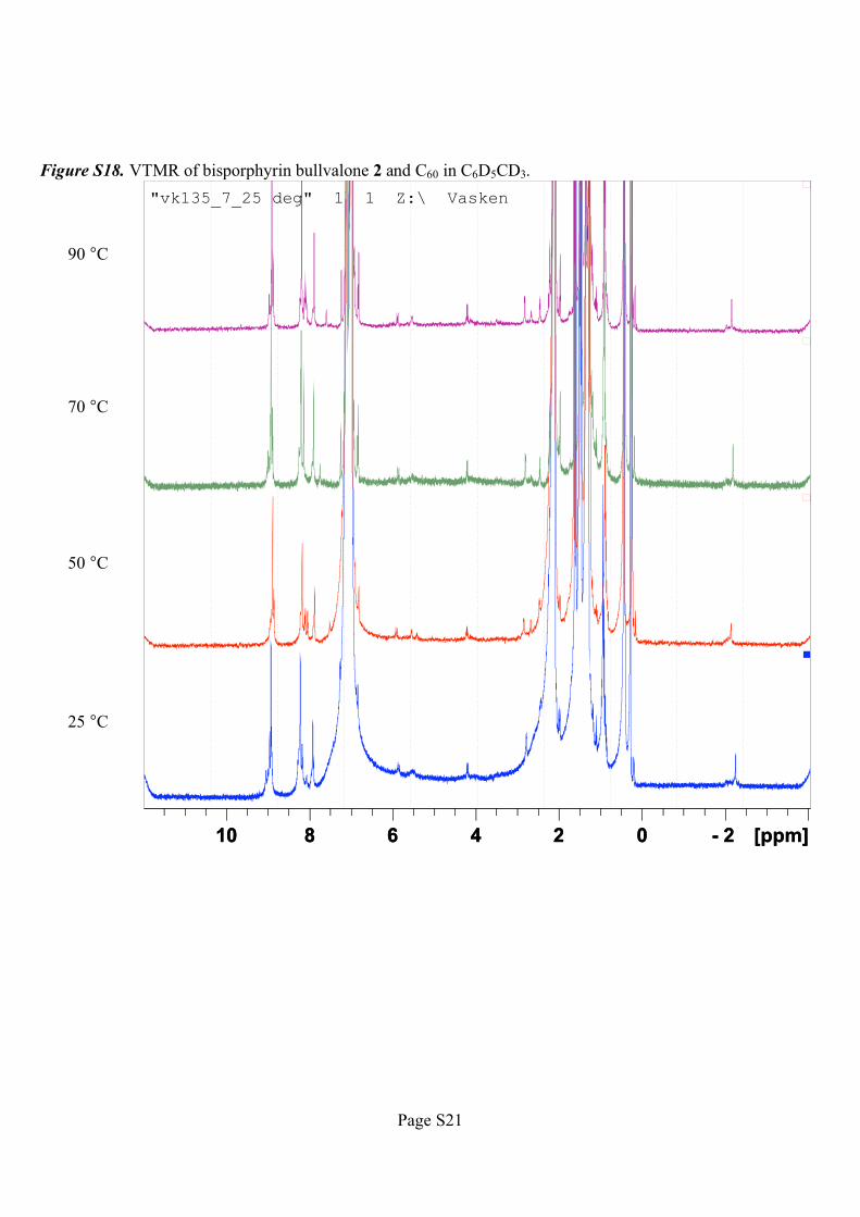
Figure S18. VTMR of bisporphyrin bullvalone 2 and C60 in C6D5CD3.
90 °C
70 °C
50 °C
25 °C
[ppm] 10 8 6 4 2 0 - 2
"vk135_7_25 deg" 1 1 Z:\ Vasken
[ppm] 10 8 6 4 2 0 - 2
"vk135_7_25 deg" 1 1 Z:\ Vasken
Page 22
Page S22
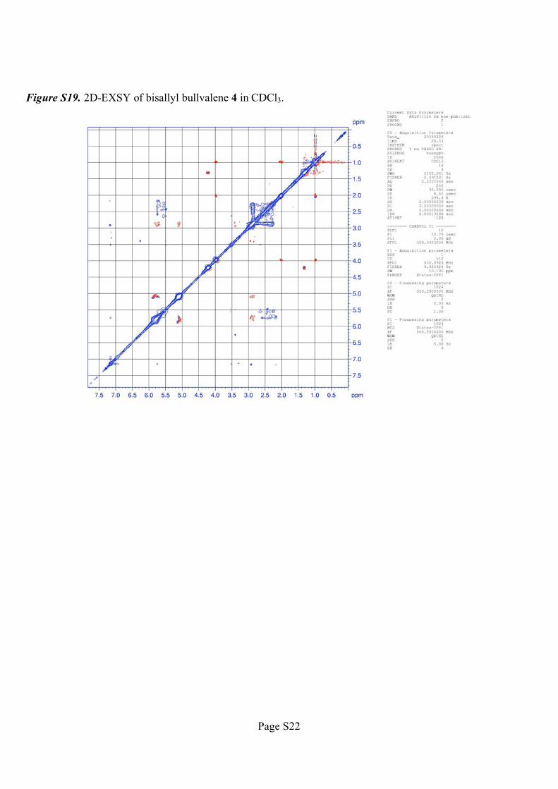
Figure S19. 2D-EXSY of bisallyl bullvalene 4 in CDCl3.


![87 Complexation Of P -Sulphonatocalix[4]Arene Complexation ...jnca.iau-saveh.ac.ir/Files/Journal/2014-05-21_12.15.39_e.pdf · 89 Complexation Of P -Sulphonatocalix[4]Arene of between](https://static.documents.pub/doc/80x56/5e14cb4e271e02747b0fae8f/87-complexation-of-p-sulphonatocalix4arene-complexation-jncaiau-savehacirfilesjournal2014-05-21121539epdf.jpg)
![Regular Article PHYSICAL CHEMISTRY RESEARCH Iranian ... · supramolecular chemistry for ion sensing and selective complexation [6-9]. A kind of non-covalent interaction is the hydrogen](https://static.documents.pub/doc/80x56/5f73ae70083928332f3b75a9/regular-article-physical-chemistry-research-iranian-supramolecular-chemistry.jpg)

![2014 OPEN ACCESS sensors - pdfs.semanticscholar.org · anion complexation, sensing and/or transport have gained much relevance within the area of supramolecular chemistry [7–11].](https://static.documents.pub/doc/80x56/5e46724fc713ba37307d0ea7/2014-open-access-sensors-pdfs-anion-complexation-sensing-andor-transport-have.jpg)


