Dynamics of Hippocampal Neurogenesis in Adult Humans Kirsty L. Spalding, 1,8 Olaf Bergmann, 1,8 Kanar Alkass, 1,2 Samuel Bernard, 3 Mehran Salehpour, 4 Hagen B. Huttner, 1,5 Emil Bostro ¨ m, 1 Isabelle Westerlund, 1 Ce ´ line Vial, 3 Bruce A. Buchholz, 6 Go ¨ ran Possnert, 4 Deborah C. Mash, 7 Henrik Druid, 2 and Jonas Frise ´n 1, * 1 Department of Cell and Molecular Biology 2 Department of Oncology-Pathology Karolinska Institutet, 171 77 Stockholm, Sweden 3 Institut Camille Jordan, CNRS UMR 5208, University of Lyon, 69622 Villeurbanne, France 4 Department of Physics and Astronomy, Ion Physics, Uppsala University, 751 20 Sweden 5 Department of Neurology, University of Erlangen-Nuremberg, Schwabachanlage 6, 91054 Erlangen, Germany 6 Center for Accelerator Mass Spectrometry, Lawrence Livermore National Laboratory, 7000 East Avenue L-397, Livermore, CA 94550, USA 7 Department of Neurology, Miller School of Medicine, University of Miami, Miami, FL 33136, USA 8 These authors contributed equally to this work *Correspondence: [email protected]http://dx.doi.org/10.1016/j.cell.2013.05.002 SUMMARY Adult-born hippocampal neurons are important for cognitive plasticity in rodents. There is evidence for hippocampal neurogenesis in adult humans, although whether its extent is sufficient to have func- tional significance has been questioned. We have assessed the generation of hippocampal cells in humans by measuring the concentration of nuclear- bomb-test-derived 14 C in genomic DNA, and we present an integrated model of the cell turnover dy- namics. We found that a large subpopulation of hip- pocampal neurons constituting one-third of the neu- rons is subject to exchange. In adult humans, 700 new neurons are added in each hippocampus per day, corresponding to an annual turnover of 1.75% of the neurons within the renewing fraction, with a modest decline during aging. We conclude that neu- rons are generated throughout adulthood and that the rates are comparable in middle-aged humans and mice, suggesting that adult hippocampal neuro- genesis may contribute to human brain function. INTRODUCTION New neurons integrate throughout life in the hippocampus and olfactory bulb of most mammals. The newborn neurons have enhanced synaptic plasticity for a limited time after their diff- erentiation (Ge et al., 2007; Schmidt-Hieber et al., 2004), which is critical for their role in mediating pattern separation in memory formation and cognition in rodents (Clelland et al., 2009; Naka- shiba et al., 2012; Sahay et al., 2011). It has long been debated whether adult neurogenesis decreased during primate evolution and whether there is sufficient generation of neurons in adult hu- mans to contribute to brain function (Kempermann, 2012; Rakic, 1985). A seminal study by Eriksson et al., 1998, provided the only direct evidence to date for adult neurogenesis in humans, although it did not enable researchers to assess the number of new neurons generated or the dynamics of this process. To estimate the extent of adult neurogenesis in humans, recent studies have quantified the number of cells expressing the neuronal precursor (neuroblast) marker doublecortin (DCX) in the subventricular zone, which gives rise to olfactory bulb neu- rons, and in the dentate gyrus of the hippocampus (Knoth et al., 2010; Sanai et al., 2011; Wang et al., 2011). Very similar dy- namics have been revealed in these two regions, which contain a large number of neuroblasts shortly after birth that decreases sharply during the first postnatal year and then declines more moderately through childhood and adult life (Go ¨ ritz and Frise ´ n, 2012; Knoth et al., 2010; Sanai et al., 2011; Wang et al., 2011). The decrease in neuroblast numbers in the subventricular zone and their migratory path suggested that there is negligible, if any, adult olfactory bulb neurogenesis in humans (Arellano and Rakic, 2011; Sanai et al., 2011; Wang et al., 2011). Retrospective birth dating established that olfactory bulb neurons are as old as the individual, and, if there is any addition of neurons in the adult human olfactory bulb, less than 1% of the neurons are exchanged over a century (Bergmann et al., 2012). It appears un- likely that adult olfactory bulb neurogenesis has any functional significance in humans. The similar decline in neuroblast numbers in the subventricular zone and the hippocampus poses the question of whether there is postnatal hippocampal neuro- genesis in humans to an extent that may have an impact on brain function. Analysis of the number of neuronal progenitor cells gives an in- direct indication of the possible extent of neurogenesis. How- ever, it does not provide information as to whether neuroblasts differentiate and integrate as mature neurons. This is evident from the studies of the subventricular zone and olfactory bulb, where the generation of neuroblasts does not result in the detectable integration of new neurons in the olfactory bulb (Berg- mann et al., 2012). The strategies used to study the generation of Cell 153, 1219–1227, June 6, 2013 ª2013 Elsevier Inc. 1219
Transcript
Dynamics of Hippocampal Neurogenesisin Adult HumansKirsty L. Spalding,1,8 Olaf Bergmann,1,8 Kanar Alkass,1,2 Samuel Bernard,3 Mehran Salehpour,4 Hagen B. Huttner,1,5
Emil Bostrom,1 Isabelle Westerlund,1 Celine Vial,3 Bruce A. Buchholz,6 Goran Possnert,4 Deborah C. Mash,7
Henrik Druid,2 and Jonas Frisen1,*1Department of Cell and Molecular Biology2Department of Oncology-PathologyKarolinska Institutet, 171 77 Stockholm, Sweden3Institut Camille Jordan, CNRS UMR 5208, University of Lyon, 69622 Villeurbanne, France4Department of Physics and Astronomy, Ion Physics, Uppsala University, 751 20 Sweden5Department of Neurology, University of Erlangen-Nuremberg, Schwabachanlage 6, 91054 Erlangen, Germany6Center for Accelerator Mass Spectrometry, Lawrence Livermore National Laboratory, 7000 East Avenue L-397, Livermore, CA 94550, USA7Department of Neurology, Miller School of Medicine, University of Miami, Miami, FL 33136, USA8These authors contributed equally to this work*Correspondence: [email protected]
http://dx.doi.org/10.1016/j.cell.2013.05.002
SUMMARY
Adult-born hippocampal neurons are important forcognitive plasticity in rodents. There is evidencefor hippocampal neurogenesis in adult humans,although whether its extent is sufficient to have func-tional significance has been questioned. We haveassessed the generation of hippocampal cells inhumans by measuring the concentration of nuclear-bomb-test-derived 14C in genomic DNA, and wepresent an integrated model of the cell turnover dy-namics. We found that a large subpopulation of hip-pocampal neurons constituting one-third of the neu-rons is subject to exchange. In adult humans, 700new neurons are added in each hippocampus perday, corresponding to an annual turnover of 1.75%of the neurons within the renewing fraction, with amodest decline during aging. We conclude that neu-rons are generated throughout adulthood and thatthe rates are comparable in middle-aged humansand mice, suggesting that adult hippocampal neuro-genesis may contribute to human brain function.
INTRODUCTION
New neurons integrate throughout life in the hippocampus and
olfactory bulb of most mammals. The newborn neurons have
enhanced synaptic plasticity for a limited time after their diff-
erentiation (Ge et al., 2007; Schmidt-Hieber et al., 2004), which
is critical for their role in mediating pattern separation in memory
formation and cognition in rodents (Clelland et al., 2009; Naka-
shiba et al., 2012; Sahay et al., 2011). It has long been debated
whether adult neurogenesis decreased during primate evolution
and whether there is sufficient generation of neurons in adult hu-
mans to contribute to brain function (Kempermann, 2012; Rakic,
1985). A seminal study by Eriksson et al., 1998, provided the only
direct evidence to date for adult neurogenesis in humans,
although it did not enable researchers to assess the number of
new neurons generated or the dynamics of this process.
To estimate the extent of adult neurogenesis in humans,
recent studies have quantified the number of cells expressing
the neuronal precursor (neuroblast) marker doublecortin (DCX)
in the subventricular zone, which gives rise to olfactory bulb neu-
rons, and in the dentate gyrus of the hippocampus (Knoth et al.,
2010; Sanai et al., 2011; Wang et al., 2011). Very similar dy-
namics have been revealed in these two regions, which contain
a large number of neuroblasts shortly after birth that decreases
sharply during the first postnatal year and then declines more
moderately through childhood and adult life (Goritz and Frisen,
2012; Knoth et al., 2010; Sanai et al., 2011; Wang et al., 2011).
The decrease in neuroblast numbers in the subventricular zone
and their migratory path suggested that there is negligible, if
any, adult olfactory bulb neurogenesis in humans (Arellano and
Rakic, 2011; Sanai et al., 2011; Wang et al., 2011). Retrospective
birth dating established that olfactory bulb neurons are as old as
the individual, and, if there is any addition of neurons in the adult
human olfactory bulb, less than 1% of the neurons are
exchanged over a century (Bergmann et al., 2012). It appears un-
likely that adult olfactory bulb neurogenesis has any functional
significance in humans. The similar decline in neuroblast
numbers in the subventricular zone and the hippocampus poses
the question of whether there is postnatal hippocampal neuro-
genesis in humans to an extent that may have an impact on brain
function.
Analysis of the number of neuronal progenitor cells gives an in-
direct indication of the possible extent of neurogenesis. How-
ever, it does not provide information as to whether neuroblasts
differentiate and integrate as mature neurons. This is evident
from the studies of the subventricular zone and olfactory bulb,
where the generation of neuroblasts does not result in the
detectable integration of new neurons in the olfactory bulb (Berg-
mann et al., 2012). The strategies used to study the generation of
Cell 153, 1219–1227, June 6, 2013 ª2013 Elsevier Inc. 1219
type control antibody (A) or an antibody against the
neuron-specific epitope (NeuN) (B), and the
neuronal and nonneural populations were isolated
by flow cytometry. The sorting gate for neuronal
nuclei is indicated.
See also Figure S1.
mature neurons in experimental animals are not readily appli-
cable to humans. To be able to study cell turnover dynamics in
humans, we have developed a strategy to retrospectively birth
date cells (Spalding et al., 2005a). This strategy takes advantage
of the elevated atmospheric 14C levels caused by above-ground
nuclear bomb testing in 1955–1963 during the Cold War (De
Vries, 1958; Nydal and Lovseth, 1965). Since the Partial Nuclear
Test Ban Treaty in 1963, atmospheric levels of 14C have declined
because of uptake by the biotope and diffusion from the atmo-
sphere (Levin and Kromer, 2004; Levin et al., 2010). 14C in the at-
mosphere reacts with oxygen to form CO2, which is taken up by
plants in photosynthesis. Whenwe eat plants, or animals that live
off plants, we take up 14C, meaning that atmospheric 14C levels
are mirrored in the human body at all times (Harkness, 1972;
Libby et al., 1964; Spalding et al., 2005b). When a cell goes
through mitosis and duplicates its chromosomes, it integrates14C in the synthesized genomic DNA with a concentration corre-
sponding to that in the atmosphere at the time, creating a date
mark in the DNA (Spalding et al., 2005a). The cumulative nature
of 14C integration makes the method especially suited for estab-
lishing the kinetics of slowly turning over cell populations. The
accuracy of individual datings is approximately ±1.5 years
(Spalding et al., 2005b), but higher accuracy is reached by inte-
grating data from many independent measurements.
We have retrospectively birth dated hippocampal cells, and
we provide an integrated model for adult hippocampal neuro-
genesis in humans. We report that there is substantial neurogen-
esis in the human hippocampus throughout life, to an extent
comparable to that in the middle-aged mouse, thus supporting
the idea that adult hippocampal neurogenesis may contribute
to human brain function.
RESULTS
Retrospective Birth Dating of Cells from the HumanHippocampusCell nuclei were isolated by gradient centrifugation from
dissected human postmortem hippocampi. The nuclei were
incubated with antibodies against the neuron-specific nuclear
epitope NeuN, and neuronal and nonneuronal nuclei were iso-
lated by flow cytometry (Figure 1 and Figure S1 available online)
1220 Cell 153, 1219–1227, June 6, 2013 ª2013 Elsevier Inc.
(Bergmann et al., 2012; Bhardwaj et al.,
2006; Spalding et al., 2005a). The 14C
concentration in genomic DNA from
hippocampal neurons (n = 55) and non-
neuronal cells (n = 65) was measured by accelerator mass spec-
trometry (AMS) in subjects 19–92 years of age (14C data are given
in Table S1).
Standard AMS analysis requires samples corresponding to
about 1 mg of carbon. The total amount of carbon in genomic
DNA samples from hippocampal cell populations, after cell sort-
ing and purifications steps, is typically in the range 10–20 mg,
necessitating a different approach. Consequently, we developed
an experimental method with a sample preparation setup and
laboratory procedure to address various critical issues, including
reliability and accuracy (Salehpour et al., 2013).
To infer the cell turnover dynamics in the adult hippocampus,
we fitted several mathematical models, or scenarios, with
increasing detail to the 14C data. All scenarios were based on
birth and death processes by which cells can die or be added
to a cell population. A scenario defines a set of rules for how cells
are born, die, or renew; i.e., it sets whether there should bemore,
less, or equal amounts of birth and death and which cells will die
preferentially or renew, etc. For each of these scenarios, a set of
parameters quantifies the extent of renewal. The mathematical
model tracks the chronological age of each cell and the age of
the person with a variable nðt; aÞ, with the cell density (units in
cells per year) of age a in a person age t. The evolution of the
cell density is given by a biological transport equation, which
moves cells along age as time progresses with a loss term ac-
counting for cell death:
vnðt; aÞvt
+vnðt; aÞda
= � gðt; aÞnðt; aÞ:
An initial condition describing the cell population at birth and a
boundary condition describing how new cells are added are sup-
plemented to the transport equation to solve the problem fully
(equations are given in the Extended Experimental Procedures).
Solving the problem allows the prediction of the 14C level for a
sample by integrating the solution nðt; aÞ along the atmospheric14C curve between the birth and death of the individual. By
comparing the model prediction to all neuronal or nonneuronal
cell data, best parameter sets for each scenario were found.
The best scenarios were selected on the basis of Akaike informa-
tion criterion (AIC); i.e., their goodness of fit and their level of
Figure 2. Turnover Dynamics of Nonneuronal Cells
(A) Schematic illustration of the representation of the measured 14C concentration in genomic DNA. The black line indicates the 14C concentration in the at-
mosphere at different time points in the last century. Individually measured 14C concentrations in the genomic DNA of human hippocampal cells are plotted at the
time of the subject’s birth (vertical lines), before (green dot) or after the 14C bomb spike (orange dot). 14C concentrations above the bomb curve (subjects born
before the bomb peak) and data points below the bomb curve (subjects born after the nuclear tests) indicate cellular turnover.
(B) The 14C concentrations of genomic DNA from nonneuronal cells demonstrate postnatal cell turnover in subjects born before and after the bomb spike.
(C) Individual turnover rates for Nonneuronal cells computed on the basis of individual data fitting. Individual turnover rate calculations are sensitive to deviations
in measured 14C and values <0.001 or >1.5 were excluded from the plot, but the full data are given in Table S1.
(D) Nonneuronal average cell age estimates of cells within the renewing fraction are depicted (red curve). The dashed line represents a no-cell-turnover scenario.
See also Figure S2 and Table S2.
detail. For scenario A (constant turnover) and scenario 2POP
(constant turnover in a fraction of cells), individual turnover rates
could also be estimated.
Turnover of Nonneuronal Cells in the Adult HumanHippocampusFirst, we assessed the turnover dynamics of nonneuronal
(NeuN�) cells in the human hippocampus. The 14C concentra-
tion in genomic DNA corresponded to time points after the birth
of the individuals (Figures 2A and 2B), establishing the turnover
of nonneuronal cells in the human hippocampus. Mathematical
modeling of 14C data allowed a detailed analysis of the dynamics
of cell turnover (Bergmann et al., 2009; Bergmann et al., 2012;
Spalding et al., 2008). By fitting the models to the data, we can
infer how much cell renewal is needed to reproduce the
observed 14C levels and whether the renewal is restricted to a
subpopulation (see Figure S2 and the Extended Experimental
Procedures). The best model, based on AIC, was scenario
2POP, in which a fraction of the population is renewing and the
other is not. In this scenario, cells within the renewing fraction
are set to turn over at a constant rate throughout life. This sce-
nario indicated that a large proportion of the nonneuronal cells
(51%, 95% confidence interval [CI] [22%–88%]) are continu-
ously exchanged. The median turnover rate within the subpopu-
lation of nonneuronal cells undergoing exchange is 3.5% per
year (Figure 2C and Table S2). Individual turnover estimates sug-
gest that there is a decline in the turnover of nonneuronal cells
during aging (r =�0.35, p = 0.04). The average age of nonneuro-
nal cells within the renewing fraction at different ages of an indi-
vidual is shown in Figure 2D.
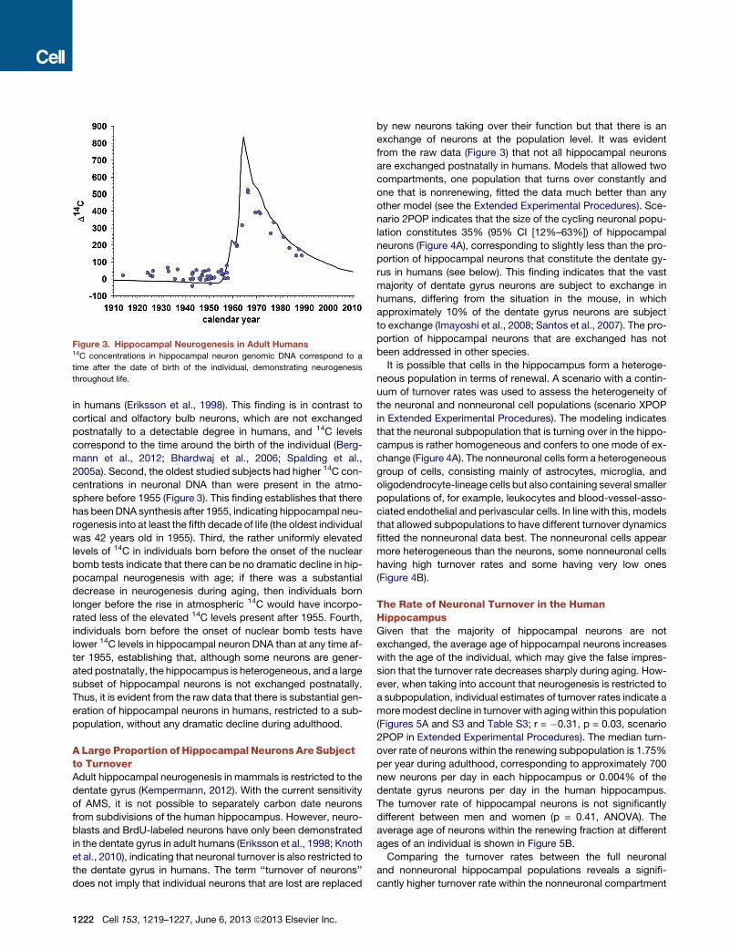
Hippocampal Neurogenesis in Adult HumansNext, we analyzed the 14C concentration in neuronal genomic
DNA. One can draw several conclusions regarding hippocampal
neurogenesis from the raw data (Figure 3). First, the 14C concen-
tration in genomic DNA of hippocampal neurons corresponds to
the concentration in the atmosphere after the birth of the individ-
ual, confirming the postnatal generation of hippocampal neurons
Cell 153, 1219–1227, June 6, 2013 ª2013 Elsevier Inc. 1221
Figure 3. Hippocampal Neurogenesis in Adult Humans14C concentrations in hippocampal neuron genomic DNA correspond to a
time after the date of birth of the individual, demonstrating neurogenesis
throughout life.
in humans (Eriksson et al., 1998). This finding is in contrast to
cortical and olfactory bulb neurons, which are not exchanged
postnatally to a detectable degree in humans, and 14C levels
correspond to the time around the birth of the individual (Berg-
mann et al., 2012; Bhardwaj et al., 2006; Spalding et al.,
2005a). Second, the oldest studied subjects had higher 14C con-
centrations in neuronal DNA than were present in the atmo-
sphere before 1955 (Figure 3). This finding establishes that there
has beenDNA synthesis after 1955, indicating hippocampal neu-
rogenesis into at least the fifth decade of life (the oldest individual
was 42 years old in 1955). Third, the rather uniformly elevated
levels of 14C in individuals born before the onset of the nuclear
bomb tests indicate that there can be no dramatic decline in hip-
pocampal neurogenesis with age; if there was a substantial
decrease in neurogenesis during aging, then individuals born
longer before the rise in atmospheric 14C would have incorpo-
rated less of the elevated 14C levels present after 1955. Fourth,
individuals born before the onset of nuclear bomb tests have
lower 14C levels in hippocampal neuron DNA than at any time af-
ter 1955, establishing that, although some neurons are gener-
ated postnatally, the hippocampus is heterogeneous, and a large
subset of hippocampal neurons is not exchanged postnatally.
Thus, it is evident from the raw data that there is substantial gen-
eration of hippocampal neurons in humans, restricted to a sub-
population, without any dramatic decline during adulthood.
A Large Proportion of Hippocampal Neurons Are Subjectto TurnoverAdult hippocampal neurogenesis in mammals is restricted to the
dentate gyrus (Kempermann, 2012). With the current sensitivity
of AMS, it is not possible to separately carbon date neurons
from subdivisions of the human hippocampus. However, neuro-
blasts and BrdU-labeled neurons have only been demonstrated
in the dentate gyrus in adult humans (Eriksson et al., 1998; Knoth
et al., 2010), indicating that neuronal turnover is also restricted to
the dentate gyrus in humans. The term ‘‘turnover of neurons’’
does not imply that individual neurons that are lost are replaced
1222 Cell 153, 1219–1227, June 6, 2013 ª2013 Elsevier Inc.
by new neurons taking over their function but that there is an
exchange of neurons at the population level. It was evident
from the raw data (Figure 3) that not all hippocampal neurons
are exchanged postnatally in humans. Models that allowed two
compartments, one population that turns over constantly and
one that is nonrenewing, fitted the data much better than any
other model (see the Extended Experimental Procedures). Sce-
nario 2POP indicates that the size of the cycling neuronal popu-
lation constitutes 35% (95% CI [12%–63%]) of hippocampal
neurons (Figure 4A), corresponding to slightly less than the pro-
portion of hippocampal neurons that constitute the dentate gy-
rus in humans (see below). This finding indicates that the vast
majority of dentate gyrus neurons are subject to exchange in
humans, differing from the situation in the mouse, in which
approximately 10% of the dentate gyrus neurons are subject
to exchange (Imayoshi et al., 2008; Santos et al., 2007). The pro-
portion of hippocampal neurons that are exchanged has not
been addressed in other species.
It is possible that cells in the hippocampus form a heteroge-
neous population in terms of renewal. A scenario with a contin-
uum of turnover rates was used to assess the heterogeneity of
the neuronal and nonneuronal cell populations (scenario XPOP
in Extended Experimental Procedures). The modeling indicates
that the neuronal subpopulation that is turning over in the hippo-
campus is rather homogeneous and confers to one mode of ex-
change (Figure 4A). The nonneuronal cells form a heterogeneous
group of cells, consisting mainly of astrocytes, microglia, and
oligodendrocyte-lineage cells but also containing several smaller
populations of, for example, leukocytes and blood-vessel-asso-
ciated endothelial and perivascular cells. In line with this, models
that allowed subpopulations to have different turnover dynamics
fitted the nonneuronal data best. The nonneuronal cells appear
more heterogeneous than the neurons, some nonneuronal cells
having high turnover rates and some having very low ones
(Figure 4B).
The Rate of Neuronal Turnover in the HumanHippocampusGiven that the majority of hippocampal neurons are not
exchanged, the average age of hippocampal neurons increases
with the age of the individual, which may give the false impres-
sion that the turnover rate decreases sharply during aging. How-
ever, when taking into account that neurogenesis is restricted to
a subpopulation, individual estimates of turnover rates indicate a
moremodest decline in turnover with agingwithin this population
(Figures 5A and S3 and Table S3; r = �0.31, p = 0.03, scenario
2POP in Extended Experimental Procedures). The median turn-
over rate of neurons within the renewing subpopulation is 1.75%
per year during adulthood, corresponding to approximately 700
new neurons per day in each hippocampus or 0.004% of the
dentate gyrus neurons per day in the human hippocampus.
The turnover rate of hippocampal neurons is not significantly
different between men and women (p = 0.41, ANOVA). The
average age of neurons within the renewing fraction at different
ages of an individual is shown in Figure 5B.
Comparing the turnover rates between the full neuronal
and nonneuronal hippocampal populations reveals a signifi-
cantly higher turnover rate within the nonneuronal compartment
Figure 4. Subpopulation Dynamics of Hippocampal Neurons and Nonneurons
(A) The Hill function indicates that the fraction of neurons being exchanged is homogenous and confers to one mode of exchange.
(B) In line with a nonneuronal population comprised of several cell types, the Hill function indicates that the nonneuronal cells form a heterogeneous group, some
subpopulations having high turnover rates and some having very low ones. The z axis indicates different possible solutions compatible with the data. Only
solutions with a good fit are shown, and those with the highest probability are indicated in red. Solutions with a lower probability in are indicated in blue.
(p < 23 10�5, Wilcoxon signed-rank test, scenario A in Extended
Experimental Procedures). This finding is largely explained by a
larger subset of cells turning over within the nonneuronal popu-
lation than within the neuronal population, and, when comparing
the turnover rates specifically within the respective sub-
populations that are subject to cellular exchange, there was no
significant difference in turnover rates between the neuronal
and nonneuronal populations (p = 0.054, Wilcoxon signed-rank
test, scenario 2POP in Extended Experimental Procedures).
However, given that nonneuronal cells are more abundant than
neurons in the human hippocampus (Figure 1A), a larger number
of nonneuronal cells in absolute numbers are generated.
There was no correlation between the neuronal and nonneuro-
nal turnover rates within individuals older than 50 years (r =
�0.14, p = 0.58, scenario 2POP in Extended Experimental Pro-
cedures), suggesting that the generation of these different cell
types is regulated independently, as in the mouse (Steiner
et al., 2004). However, there was a correlation in young individ-
uals (<50 years, r = �0.62, p = 0.003). The interindividual varia-
tion in the turnover rate of neurons and nonneuronal cells in
the hippocampus is similar, having a median absolute deviation
of 0.0226 and 0.0158 per year, respectively. The interindividual
variation may appear largest in the younger subjects, but this
is a consequence of the shallow slope of the atmospheric 14C
levels in recent times, which provides less resolution and, there-
fore, introduces higher variability.
An IntegratedModel of Neuronal Dynamics in theHumanHippocampusThe determination of the fraction of neurons that is subject to ex-
change in the human hippocampus and their turnover rate
makes it possible to infer the age of the full complement of neu-
rons in individuals of different ages. The hippocampus is a
mosaic of neurons of different ages, given the large fraction of
cells remaining from development and neurons generated at
different times throughout life. Stereological quantifications
have revealed a decrease in the number of hippocampal neurons
during aging in humans, the dentate gyrus being least affected
(Figure S4). A relative increase in the proportion of neurons in
the renewing fraction with age fits the 14C data well.
The most detailed model, scenario 2POPEd, provides a global
picture of the dynamics of neuronal turnover. Nonrenewing neu-
rons die without being replaced, resulting in a slow decrease in
neuron level throughout life. Within the renewing neuron popula-
tion, young cells die faster, leading to a neuron age distribution
with fewer middle-aged cells than would be expected if all neu-
rons were as likely to be replaced. One observation from the
modeling is that adult-born neurons are preferentially lost and
do not survive as long as the neurons generated during develop-
ment. The half-life of a neuron in the renewing fraction is 7.1
years—103 shorter than in the nonrenewing fraction. Although
it is known that adult-born neurons integrate long term in ro-
dents, whether they last for the remainder of the animal’s life
has not been studied, although the available data are compatible
with a preferential loss of adult-born neurons (Imayoshi et al.,
2008; Kempermann et al., 2003; Ninkovic et al., 2007). The inte-
grated model of the dynamics of hippocampal neuron numbers
and exchange in humans is shown in Figure 6.
DISCUSSION
Newborn neurons in the adult hippocampus have distinct
features for a limited period after their differentiation that give
them a key role in pattern separation and cognitive adaptability
in rodents. We have birth dated hippocampal cells in order
to assess whether adult neurogenesis occurs to a significant
extent in adult humans, and we provide a detailed view of the
cell turnover dynamics. There is substantial neurogenesis
throughout life in the human hippocampus, and only a modest
decline in neurogenesis occurs during aging. There is a
Cell 153, 1219–1227, June 6, 2013 ª2013 Elsevier Inc. 1223
Figure 5. Neuronal Turnover Dynamics in
the Human Hippocampus
(A) Individual turnover rates for neuronal cells
within the renewing fractionwere computed on the
basis of individual data fitting. The number of DCX-
positive cells per mm2 in the dentate gyrus (data
from Knoth et al., 2010) shows a similar modest
decline during adult ages as the computed
neuronal turnover rates. Straight lines depict linear
regression curves, and the regression line for DCX
cell counts was calculated for individuals 10 years
and older. Individual turnover rate calculations are
sensitive to deviations in measured 14C and values
<0.001 or >1.5 were excluded from the plot, but
the full data are given in Table S1.
(B) The average age of the neurons within the renewing fraction (blue curve). The dashed line represents the no-cell-turnover scenario.
See also Figure S3 and Table S3.
preferential loss of adult-born neurons, and a larger proportion of
hippocampal neurons are subject to exchange in humans in
comparison to the mouse. Nonneuronal cells have more hetero-
geneous turnover dynamics than hippocampal neurons.
It is important to consider whether DNA repair may contribute
to 14C integration in hippocampal cells. DNA damage and repair
are largely restricted to proliferating cells and are believed to be
several orders of magnitude below the level that is detectable by14C dating in postmitotic cells (Spalding et al., 2005a). DNA
repair during cell proliferation will not affect the assessment of
cell generation, given that 14C integrates in DNA at a concentra-
tion corresponding to that in the atmosphere during mitosis. We
have not found any measurable 14C integration in the DNA of
cortical, cerebellar, or olfactory bulb neurons over many de-
cades in humans (Bergmann et al., 2012; Bhardwaj et al.,
2006; Spalding et al., 2005a). Not even neurons surviving at
the perimeter of an ischemic cortical stroke, a situation where
there is substantial DNA damage and repair, incorporate suffi-
cient 14C to be detected (H.B.H., O.B., M.S., G.P., and J.F.,
our unpublished data). The dynamics of 14C integration in the
DNA of hippocampal neurons does not appear to be compatible
with any pattern of DNA repair previously described; a large frac-
tion of hippocampal neurons (35%) would have to exchange
their entire genome by DNA repair during the lifetime of an indi-
vidual, whereas there would be no detectable DNA repair in the
remaining hippocampal neurons. In contrast, the number of neu-
roblasts reported in the adult human dentate gyrus (Knoth et al.,
2010) is sufficient to give rise to the number of new neurons indi-
cated by the 14C analysis, and the decline in neurogenesis
closely parallels the decrease in the number of neuroblasts (Fig-
ure 5A). Thus, the 14C concentration in genomic DNA of hippo-
campal neurons is likely to accurately reflect neurogenesis.
Retrospective birth dating reveals that what appears as small
numbers of neuroblasts present in adulthood (Knoth et al., 2010)
give rise to a substantial number of new neurons over time in the
hippocampus. In this context, it is interesting that the similar den-
sity of neuroblasts in the subventricular zone to that in the hippo-
campal dentate gyrus does not result in any detectable addition
of new neurons to the olfactory bulb (Bergmann et al., 2012; Gor-
itz and Frisen, 2012; Knoth et al., 2010; Sanai et al., 2011; Wang
et al., 2011). Thus, the lack of olfactory bulb neurogenesis ap-
pears to be a consequence of an absence of migration and/or
1224 Cell 153, 1219–1227, June 6, 2013 ª2013 Elsevier Inc.
integration of new neurons in the olfactory bulb rather than a
lack of generation of neuroblasts.
There are some distinct differences in the pattern of adult hip-
pocampal neurogenesis in humans compared to that in rodents,
in which this process has been most extensively characterized.
First, a much larger proportion of hippocampal neurons are
subject to exchange in humans. In mice, 10% of the neurons in
the dentate gyrus are added in adulthood and subject to ex-
change (Imayoshi et al., 2008; Ninkovic et al., 2007). In humans,
approximately one-third of the hippocampal neurons turn over,
corresponding to the vast majority of the dentate gyrus neurons.
Second, although hippocampal neurogenesis declines with age
in both rodents and humans, the relative decline during adult-
hood appears smaller in humans in comparison to mice. Com-
parisons of the kinetics of the age-dependent decline in hippo-
campal neurogenesis between different species have revealed
a similar chronology rather than correlating to developmental
milestones (Amrein et al., 2011). In line with this, the most dra-
matic decrease in the number of neuroblasts in the dentate gyrus
occurs during the first postnatal months in both mice and hu-
mans (Ben Abdallah et al., 2010; Knoth et al., 2010). An effect
of this is that young adult mice are still in the most steeply
declining phase of neurogenesis, making the relative decrease
in neurogenesis during adult life much larger in mice than in hu-
mans. Although there is an approximately 10-fold decrease in
neurogenesis from 2 to 9 months of age in mice (Ben Abdallah
et al., 2010), there is an approximate 4-fold decline during the
entire adult lifespan in humans (Figure 5A). Third, the impact of
adult neurogenesis on the total number of neurons in the dentate
gyrus differs between rodents and humans. Hippocampal neuro-
genesis inmice and rats is additive and results in a net increase in
the number of dentate gyrus neurons with age (Bayer, 1985; Im-
ayoshi et al., 2008; Kempermann et al., 2003; Ninkovic et al.,
2007). This is not the case in humans, where there is a net loss
of dentate gyrus neurons during adult life. Although the decrease
in neuronal numbers is less pronounced in the dentate gyrus
than in other subdivisions of the human hippocampus, the gen-
eration of new neurons does not keep up with neuronal loss (Fig-
ure 6). Computational models have indicated that the addition of
new neurons to the circuitry, along with loss of older redundant
cells and enhanced synaptic plasticity, can maximize the effect
of the new neurons, whereas an isolated exchange of neurons
Figure 6. An Integrated Model of the Number and Age of Neurons in
the Human Hippocampus
A schematic illustration of the number of neurons in the dentate gyrus (above
thewhite line) and other subdivisions of the hippocampus (below thewhite line)
and the age of neurons within the dentate gyrus at different ages. The total
number of neurons declines with age in the hippocampus, though the dentate
gyrus is relatively spared. The dentate gyrus is composed of a declining
fraction of cells generated during development (black), which is gradually re-
placed by postnatally generated cells. For a given age of the person, post-
natally generated cells are shown with different shades of gray indicating
decade intervals, the lightest gray being cells generated during the last
decade, and cells one shade darker being generated 10 to 20 years ago, etc.
This way, at age 15 among postnatally generated cells, only cells generated
0 to10 years ago and 10 to 20 years ago are present. Read vertically for a fixed
age of the person, the cell age distribution goes from oldest cells (black) to the
youngest ones (light gray). Read horizontally, the fraction of adult-born cells
(nonblack) increases with age. The model is based on scenario 2POPEd. The
figure was generated with the following parameters: initial fraction of renewing
neurons, 0.31; death rate of the nonrenewing neurons, 0.0035 per year; death
rate of newborn neurons, 0.11 per year; and cell age at which the death rate
has reduced by half, 19 years. The parameter set was selected among the 3%
best out of 33 105 parameter sets explored with a Markov chain Monte Carlo
algorithm and consistent with scenario 2POP.
See also Figure S4.
would have less influence (Appleby et al., 2011). The adult
generation of neurons serves to uphold a pool of neurons with
specific functional properties rather than replacing individual
neurons that are lost. Therefore, the continuous generation of
new neurons in the adult human hippocampus may have an
additive role functionally in the circuitry, although more neurons
are lost than generated.
Can the number of new neurons generated in the adult human
hippocampus have functional significance? An indication of this
may be gained by comparing the extent of adult neurogenesis in
humans with that in other species, in particular the mouse, in
which most experiments on the function of adult hippocampal
neurogenesis have been carried out. It is difficult to make direct
comparisons because there are several factors influencing the
potential impact of newborn neurons that may vary between
species; for example, the total number of cells in the circuitry
and for how long newborn neurons have distinct features. The
best measure of adult neurogenesis when comparing across
speciesmay be the relative proportion of newborn to old neurons
(Kempermann, 2012). We conclude that 0.004% of the dentate
gyrus neurons are exchanged daily in adult humans, which can
be compared to 0.03%–0.06% per day in 2-month-old mice
and 0.004%–0.02% per day in 5- to 16-year-old macaque mon-
keys (Jabes et al., 2010; Kempermann et al., 1997; Kornack and
Rakic, 1999). Hippocampal neurogenesis has been estimated to
decrease approximately 10-fold between 2 and 9 months of age
in the mouse (Ben Abdallah et al., 2010), indicating that the rate
of neurogenesis in adult humans may correspond to that of a
9-month-old mouse. Along with the extended period of imma-
ture features of the adult-born neurons in nonhuman primates
(Kohler et al., 2011), and potentially humans, the relative propor-
tion of adult-born neurons with unique functions in the human
hippocampus may not be smaller than that in a middle-aged
mouse. Therefore, the extent of neurogenesis in the adult human
hippocampus may be sufficient to convey similar functions as in
themouse, in which adult neurogenesis is important for cognitive
adaptability.
Adult-born hippocampal neurons have enhanced synaptic
plasticity for a period of time after their differentiation (Ge
et al., 2007; Schmidt-Hieber et al., 2004). This, along with the
fact that the dentate gyrus acts as a bottleneck in the network,
allows a small proportion of neurons to have a substantial influ-
ence on the circuitry and hippocampal function. The new neu-
rons are required for efficient pattern separation and the ability
to distinguish and store similar experiences as distinct mem-
ories, whereas the old granule cells are necessary for pattern
completion, which serves to associate similar memories to
each other (Clelland et al., 2009; Nakashiba et al., 2012; Sahay
et al., 2011). Failing pattern separation may result in generaliza-
tion, a common feature in anxiety and depression in humans
(Kheirbek et al., 2012). There are a number of indications that
implicate reduced neurogenesis in psychiatric disease, but it
has been difficult to explore whether there is a link in humans
(Eisch and Petrik, 2012). We find considerable interindividual
variation in this study, and the assessment of hippocampal neu-
rogenesis, along with the reconstruction of medical histories,
may reveal whether reduced neurogenesis is associated with
psychiatric disease in humans.
EXPERIMENTAL PROCEDURES
Tissue Collection
After informed consent from the individual or next-of-kin was given, tissues
were procured from autopsy cases from 2000 to 2012 from the Swedish Na-
tional Department of Forensic Medicine, the Department of Neurology of the
Miller School of Medicine Brain Endowment Bank, and the Department of Pa-
thology of the University of Debrecen. Ethical permission for this study was
granted by Regional Ethics Committee of Sweden (No 02-418, 2005/185,
2006/1029-31/2, 2006/189-31, 2010-313/31-3), the institutional review board
of the University of Miami Miller School of Medicine (FWA00002247;
IRB00005622), and the local institutional review board of the medical faculty
in Debrecen, Hungary. Whole hippocampi were dissected and analyzed. Brain
tissue was frozen and stored at �80�C until further analysis.
Nuclei Isolation
Tissue samples were thawed and Dounce homogenized in 10 ml lysis buffer
(0.32 M sucrose, 5 mM CaCl2, 3 mM magnesium acetate, 0.1 mM EDTA,
10 mM Tris-HCl [pH 8.0], 0.1% Triton X-100, and 1 mM DTT). Homogenized
samples were suspended in 20 ml of sucrose solution (1.7 M sucrose, 3 mM
magnesium acetate, 1 mM DTT, and 10 mM Tris-HCl [pH 8.0]), layered onto
Cell 153, 1219–1227, June 6, 2013 ª2013 Elsevier Inc. 1225
a cushion of 10 ml sucrose solution, and centrifuged at 36,5003 g for 2.4 hr at
4�C. The isolated nuclei were resuspended in nuclei storage buffer (10 mMTris
[pH = 7.2], 2 mMMgCl2, 70 mM KCl, and 15% sucrose) for consecutive immu-
nostaining and flow cytometry analysis.
FACS Sorting and Analysis
Isolated nuclei were stainedwithmouse NeuN (A-60) (Millipore, 1:1,000). NeuN