Dynamics of Root Microorganisms in Closed Hydroponic Cropping Systems Anna Karin Rosberg Faculty of Landscape Architecture, Horticulture and Crop Production Science Department of Biosystems and Technology Alnarp Doctoral Thesis Swedish University of Agricultural Sciences Alnarp 2014
Transcript
Dynamics of Root Microorganisms in Closed Hydroponic Cropping Systems
Anna Karin Rosberg Faculty of Landscape Architecture, Horticulture and Crop Production Science
Department of Biosystems and Technology Alnarp
Doctoral Thesis Swedish University of Agricultural Sciences
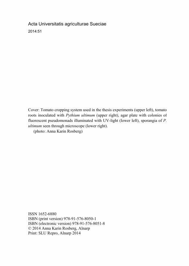
Cover: Tomato cropping system used in the thesis experiments (upper left), tomato roots inoculated with Pythium ultimum (upper right), agar plate with colonies of fluorescent pseudomonads illuminated with UV-light (lower left), sporangia of P. ultimum seen through microscope (lower right).
(photo: Anna Karin Rosberg)
Dynamics of Root Microorganisms in Closed Hydroponic Cropping Systems.
Abstract Greenhouse production of vegetables in closed hydroponic cropping systems is a resource-efficient technique for the production of high-quality produce with a high yield per unit area. While this type of cropping system allows savings in terms of water and nutrient use, the recirculation of water increases the risk of pathogen dispersal. Oomycetous pathogens in particular, such as Pythium, thrive in aquatic environments and are among the most destructive pathogens. Root pathogen outbreaks could be controlled by sustaining a stable and high level of general microbiota. It is therefore of utmost importance to understand how biotic and abiotic factors affect the indigenous root-associated microbiota.
In the present thesis, the impact of pathogen inoculation, plant age, nutrient availability and use of plant protection products on microbial communities associated with root and nutrient solution was investigated. It was demonstrated that all these factors had an effect on the indigenous microorganisms. Plant age in particular was a significant driver of microbial community structure due to its significant impact on root exudation patterns. Using 454-pyrosequencing, the effect on bacterial communities of the inoculation of P. ultimum was visualized. Although the effect was strongest at the fruit-bearing plant stage, clear differences were seen also in the seedling and flowering stages. Nutrient availability was revealed to have an impact on the number of colony-forming units of bacteria, fungi and fluorescent pseudomonads. Organic carbon specifically was shown to be the main driving force of microbial growth. However, not only the quantity, but also the quality and ratios between the different organic and inorganic nutrients had an effect on microbial numbers. Through DGGE analysis, a shift in the bacterial community structure was seen after the addition of a fungicide in the nutrient solution. The microorganisms present in the nutrient solution were not able to degrade the fungicide.
This thesis also demonstrates the importance of combining culture-dependent and culture-independent methods in the analysis of microbial communities.
Author’s address: Anna Karin Rosberg, SLU, Department of Biosystems and Technology, Microbial Horticulture Unit, P.O. Box 103, 230 53 Alnarp, Sweden E-mail: [email protected]
Dedication To David and Gabriel
Contents List of Publications 7
Abbreviations 10
1 Introduction 12
2 Background 13 2.1 Hydroponic systems 13 2.2 Microbiota in hydroponic systems 14
2.2.1 Disinfection 15 2.2.2 Disease suppression 16 2.2.3 Differences in microbiota depending on the growing medium 17 2.2.4 Oomycetes in closed systems with tomato 18
2.3 Root exudation 20 2.4 Temporal and spatial shifts in microbial communities 22 2.5 Abiotic factors affecting the structure of rhizosphere microbial
communities 24 2.6 Microbial community analysis 25
3 Objectives 27
4 Materials and Methods 29 4.1 Phytotron and greenhouse experiments 29
4.1.1 Plant material (Papers I-III) 29 4.1.2 Oomycete inoculum preparation and inoculation (Papers I-III) 30
4.2 Sampling from a commercial greenhouse 31 4.3 Analyses 32
4.3.1 Plant analyses (Papers I-III) 32 4.3.2 Nutrient solution analyses (Papers II, III and IV) 32 4.3.3 Harvesting of plants and extraction of microbiota (Papers I-III) 33 4.3.4 Culture-dependent microbiological analyses (Papers I-IV) 33 4.3.5 Culture-independent microbiological analyses (Papers I, II and IV)35 4.3.6 Statistical analyses 36
5 Results and Discussion 37 5.1 Impact of a root pathogen on root-associated microbial communities 37 5.2 Impact of plant age on root-associated microbial communities 40
5.3 Impact of available nutrients on nutrient solution-associated microbial communities 41
5.4 Impact of plant protection products on nutrient solution-associated microbial communities 44
6 Conclusions 46
References 48
Acknowledgements 61
7
List of Publications This thesis is based on the work contained in the following papers, referred to in the text by Roman numerals:
I Rosberg, A.K., Gruyer, N., Hultberg, M., Wohanka, W. & Alsanius, B.W. (2014). Monitoring rhizosphere microbial communities in healthy and Pythium ultimum inoculated tomato plants in soilless growing systems. Scientia Horticulturae 173, 106-113.
II Rosberg, A.K., Wohanka, W., Hultberg, M. & Alsanius B.W. (2014). Impacts of Pythium ultimum inoculation on tomato root-associated bacterial communities, as examined by 454 pyrosequencing (submitted).
III Rosberg, A.K., Wohanka, W., Hultberg, M. & Alsanius B.W. (2014). Organic carbon and micronutrients have an effect on microbial dynamics in nutrient solutions of diseased and non-diseased hydroponic systems (manuscript).
IV Alsanius, B.W., Bergstrand, K-J., Burleigh, S., Gruyer, N. & Rosberg, A.K. (2013). Persistence of fenhexamid in the nutrient solution of a closed cropping system. Agricultural Water Management 127, 25-30.
V Alsanius, B.W., Rosberg, A.K., Hultberg, M., Khalil, S. & Jung, V. (2014). Understanding and utilizing naturally-occurring microbes against plant pathogens in irrigation reservoirs. In: Hong, C. et al. Biology, Detection, and Management of Plant Pathogens in Irrigation Water. Chapter 28; 347-364. APS (in press).
Papers I and IV are reproduced with the kind permission of Elsevier.
8
Paper V is kindly reproduced with the permission of APS PRESS, from Biology, Detection, and Management of Plant Pathogens in Irrigation Water, eds. C. X Hong, G. W. Moorman, W. Wohanka, and C. Büttner, Copyright 2014 by The American Phytopathological Society.
9
The contribution of Anna Karin Rosberg to the papers included in this thesis was as follows:
I Planned the experiment with co-authors. Performed the experimental work and evaluated the data. Wrote the manuscript with co-authors.
II Planned the experiment with co-authors. Performed the experimental work and evaluated the data. Wrote the manuscript with co-authors.
III Planned the experiment with co-authors. Performed the experimental work and evaluated the data. Wrote the manuscript with co-authors.
IV Participated in planning the experiments and performing the statistical analysis. Wrote the manuscript with co-authors.
V Participated in creating the carbon pool model, and wrote the manuscript with the main author.
10
Abbreviations ATP adenosine triphosphate CFU colony forming unit CLPP community level physiological profiling CMA corn meal agar DAPG 2,4-diacetylphloroglucinol DFT deep flow technique DGGE denaturing gradient gel electrophoresis DNA deoxyribonucleic acid DOC dissolved organic carbon EC electrical conductivity KB King’s B agar MA malt extract agar MDA multiple displacement amplification NFT nutrient film technique PCR polymerase chain reaction PDA potato dextrose agar PLFA phospholipid fatty acid PM phenotypic microarray rRNA ribosomal ribonucleic acid TIC total inorganic carbon TOC total organic carbon T-RFLP terminal restriction fragment length polymorphism TSA tryptic soy agar UV ultraviolet
11
12
1 Introduction The production of vegetables in closed hydroponic cropping systems is a resource-efficient way of producing crops of high quality, with a high productivity in terms of yield per unit area (Stanghellini & Montero, 2012). Compared to fields, water savings in greenhouse production are in the order of 70-90 % for the same crops. This applies to closed systems in particular (Raviv & Lieth, 2008). One of the main concerns related to closed systems is the potential spread of root pathogens. With the recirculation of nutrient solution, a single infected plant could potentially be the source of infection for the entire crop (Postma et al., 2008). Oomycetous pathogens in particular, such as Pythium and Phytophthora, are the most destructive diseases due to their production of motile zoospores that thrive in aquatic environments (Stanghellini & Rasmussen, 1994). Root pathogen outbreaks could be controlled by sustaining a stable and high level of general microbiota (Hoitink et al., 1991). It is therefore of the utmost importance to understand how biotic and abiotic factors affect the indigenous, root-associated microbiota.
In this thesis, the root and nutrient solution-associated microbial communities of tomato plants grown in closed hydroponic cropping systems were investigated. The microbial communities were investigated particularly for changes in community structure caused by the introduction of a pathogen, plant age, organic and inorganic nutrient availability, and the effects of the use of plant protection products. This thesis project was funded by the Swedish Research Council for Environment, Agricultural Sciences and Spatial Planning (Formas). It was conducted within the scope of the Microbial Horticulture (µHort) research school, Formas, Stockholm, Sweden.
13
2 Background
2.1 Hydroponic systems
Hydroponics is a term that was coined in the 1930s to describe all methods of growing plants in liquid media for commercial purposes (Gericke, 1937). From the outset, hydroponic systems were used purely to look at plant nutrition and identify essential plant nutrients. Eventually, a growing medium was added to support and aerate plant roots; the growing medium had to be inert in order not to interfere with the nutrient composition, hence gravel and sand were commonly used. This type of growing system attracted a lot of attention early on, but due to the lack of automation and technology at the time, it took many years before it became economically sustainable (Jones, 1982). It was not until the 1970s that there was renewed interest in hydroponics. Many greenhouse growers experienced problems with soil fatigue from using the same soils for several years. The introduction of rockwool as a growing medium opened up the use of hydroponic systems in commercial settings. For the last four decades there has been considerable expansion in hydroponics. In the Netherlands, for example, the area of hydroponically-grown crops has increased from 5 ha in 1976 (Van Os, 1982) to almost 8,000 ha in 2013 (Voogt et al., 2013), mainly due to its independence from soil and soil-related problems, and the possibility of precise nutrient control (Jones, 1982).
Soilless greenhouse cropping systems can be divided into open and closed systems. In open systems the drained, nutrient-enriched irrigation water is discharged into the soil, catchment or sewage treatment plant. As much as 40 % of the nitrogen supplied to the plants is estimated to be lost with the drainage water (Dorais & Dube, 2011; Hansson, 2003). This drainage contributes significantly to the eutrophication of water courses and estuarine areas in the vicinity of the greenhouse, as well as to groundwater pollution. In closed systems, the drainage is collected, mixed with fresh water, readjusted to
14
the accurate nutrient composition and redistributed to the plants. The use of closed systems is therefore a solution to the issues of nutrient and pesticide leakage from greenhouse production. European legislation demands a substantial reduction in nutrient release (EU, 2000), therefore growers are being encouraged to change from open to closed irrigation systems. Such changes require high investment costs, however up to 30-40 % and 50-60 % of fertilizer and water costs respectively can be saved through recirculation (Peet & Welles, 2005). The two greatest concerns associated with closed irrigation systems are the accumulation of salts in the water and the potential spread of plant root pathogens, where the presence of just one infected plant will put the entire crop at risk (Postma et al., 2008). Oomycetous pathogens in particular, such as Pythium and Phytophthora, can easily spread and propagate explosively under favorable conditions, causing serious damage (Macdonald et al., 1994; Stanghellini & Rasmussen, 1994).
2.2 Microbiota in hydroponic systems
Initially, it was expected that soilless systems would be the solution to evading root pathogens. It has since been shown that the diversity of root-infecting microorganisms is less than in soil. However root diseases do occur and are sometimes more serious in soilless systems (Stanghellini & Rasmussen, 1994). Before plants are introduced into soilless systems, the numbers of viable counts are low, but already within twenty hours of planting, high numbers of heterotrophic bacteria and fungi develop in the nutrient solution (Berkelmann et al., 1994). The number of bacteria can be as much as 1010 colony-forming units (CFU) per gram root. The number of fungi is usually lower, but tends to vary over the season more than bacteria (Waechter-Kristensen et al., 1994). This contamination of microorganisms in soilless systems can come from plant material, growing media, insects, workers in the greenhouse and irrigation water (Postma et al., 2008). The colonization is often triggered by environmental conditions, such as steady temperature and moisture regimes, and by the fact that root colonizers are easily spread by the recirculating water. The availability of water in hydroponic systems favors all zoosporic pathogens, with the temperature of the nutrient solution being the most important environmental factor controlling the proliferation of these root diseases (Stanghellini & Rasmussen, 1994). Pythium, which in soil-grown cucumbers and tomatoes is not considered to be a serious threat, has become severe in hydroponic conditions. It seems that rockwool, the most common growing medium in soilless culture, is exceptionally conducive to Pythium diseases. This is due to its high water content relative to other types of growing media,
15
such as coir or pumice (Van der Gaag & Wever, 2005). It has also been suggested that the relatively low microbial diversity in hydroponic systems compared to soil (Menzies et al., 2005) leads to an increased severity of opportunistic pathogens, such as Pythium (Paulitz, 1997). Both Craft and Nelson (1996) and Chen et al. (1988) have demonstrated the association of high microbial activity with suppression of Pythium diseases. Once a zoosporic pathogen comes into the system, it will multiply and spread by self-dispersal through the recirculating solution and by root-to-root contact (Stanghellini & Rasmussen, 1994). In periods of stress, such as during fruiting when the temperature is high and there is a low concentration of dissolved oxygen in the nutrient solution, disease epidemics can take place (Cherif et al., 1997; Stanghellini & Rasmussen, 1994; Gold & Stanghellini, 1985).
2.2.1 Disinfection
In closed hydroponic systems, the risk of pathogen dispersal can be limited by the use of different disinfection techniques. The techniques are divided into active and passive methods. The active methods are, for example, UV radiation, ozone and heat treatment. One of the weaknesses of many active methods is that they also reduce the beneficial microorganisms. Zhang and Tu (2000) found that UV radiation reduced bacterial flora, but that Pythium was not reduced; instead, Pythium increased due to the lack of competition from the general microbiota. For Pythium to be eradicated, high doses of UV radiation were needed. The efficiency of the method was also highly dependent on the UV dose. This was also seen by Buyanovsky et al. (1981), who found that three hours of UV radiation per day on the nutrient solution was more efficient in reducing the microbiota than one hour per day. Three hours per day was also as efficient as having continuous UV radiation. Ozone has been shown to be efficient in eliminating pathogens such as Fusarium oxysporum and tomato mosaic virus (Igura et al., 2004; Runia, 1994). With this method too, however, its efficiency is dependent on the dose and length of treatment. With heat treatment, the returned drainage water is heated to 95 °C for a minimum of 10 s. This temperature is generally high enough to remove most microorganisms, viruses and nematodes. However, spore-forming bacteria will survive (Os, 2009).
Slow filtration is an example of a passive method. Here, the nutrient solution is filtered slowly through a growing medium, e.g. sand, rockwool or ground volcanic stone, where it is thought that mechanical and biological factors are responsible for the elimination of pathogens. Many pathogenic bacteria and fungi are efficiently eliminated this way, and plenty of beneficial
16
organisms stay in the filter where they are active in suppressing pathogens (Bergstrand, 2010; Brand & Wohanka, 2001; Ehret et al., 2001). Another type of passive method is the use of constructed wetlands filled with ground volcanic stone and implanted with common cattail (Typha latifolia), which has proven efficient in reducing P. ultimum in tomato production (Gruyer et al., 2011).
2.2.2 Disease suppression
Suppressiveness in soil has been defined by Baker and Cook (1974) as “soils in which the pathogen does not establish or persist, establishes but causes little or no damage, or establishes and causes disease for a while but thereafter the disease is less important, although the pathogen may still persist in the soil”. Two types of mechanisms of suppressiveness have been distinguished: i) general suppression and ii) specific suppression. In general suppression, the entire microbial biomass competes with the plant pathogenic microorganisms for nutrient and space resources, hence a reduction in the germination and growth of the pathogens. Specific suppression, on the other hand, is due to specific individual populations or groups of microorganisms. The main characteristic of specific suppression is that the suppressiveness is transferable (Cook & Baker, 1983). Factors thought to influence disease suppression are the activity of the total microbiota, the diversity of the microbial population, the presence of specific types of antagonists, the production of antimicrobial compounds, as well as the microbial-induced disease resistance of the plants (Clematis et al., 2009; Minuto et al., 2007; Weller et al., 2007; Postma et al., 2005; Weller et al., 2002; Hultberg et al., 2000; Postma et al., 2000; Ongena et al., 1999; Tu et al., 1999; Paulitz, 1997; Stanghellini & Miller, 1997).
Several studies (Clematis et al., 2009; Minuto et al., 2007; Postma et al., 2005; Postma et al., 2000; Tu et al., 1999) report on suppressiveness against root pathogens in soilless cropping systems as well. Few growing media have been described based on their disease suppressive properties. However, differences in the composition of microbial communities have been found when root samples from two different growing media used for tomato production were analyzed (Khalil, 2001). The differences found were most likely due to the diverging chemical and physical properties of the two growing media, but could also reflect differences in potential suppressiveness. It has been shown that the microbiota of re-used rockwool slabs, used originally for cucumber production, was able to suppress Pythium aphanidermatum in a new cucumber crop, while new, sterile rockwool slabs were not. New rockwool slabs provide a microbiological vacuum that allows a fast multiplication of
17
pioneer, pathogenic Pythium species (Postma, 2004). They are, however, inferior competitors in the presence of a fully developed microbial community (Sutton et al., 2006).
There are several ways in which the suppressiveness of growing media can be improved, e.g. by adding antagonistic microorganisms (Castano et al., 2013; Khalil et al., 2011; Wohanka et al., 2008; Paulitz, 1997; Nemec et al., 1996) or by nutritional amendments (St Martin & Brathwaite, 2012; van der Gaag et al., 2007; Chen et al., 1987). Compared to soil, in greenhouse production there is a possibility of providing the perfect environmental conditions needed to suit antagonistic agents (Paulitz & Belanger, 2001), yet the efficiency of the biocontrol agents varies depending on the growing media used (Khalil et al., 2009). The efficiency of the biocontrol agent is also dependent on its mode of introduction into the cropping system. Antagonistic microorganisms have proven to be most efficient when they have been introduced into the system in the same way as the pathogen, i.e. if the pathogen was introduced through the nutrient solution then the antagonist should be introduced in the same way (Rattink & Postma, 1996).
A combination of adding antagonistic microorganisms and using a disinfection technique to clean the nutrient solution is also a possible approach to increasing suppressiveness. The disinfection of the nutrient solution has to be gentle in order not to eradicate the beneficial organisms along with the pathogens. Slow filtration has proven to be a suitable disinfection technique with positive effects in combination with oomycete and fungal and bacterial biocontrol agents (Deniel et al., 2004; Garibaldi et al., 2003; Rey et al., 1999).
2.2.3 Differences in microbiota depending on the growing medium
Considering the differences in soilless growing media in terms of physical and chemical properties, it is likely that their microbial community structures are very different too. This was shown by Borrero et al. (2006), who found a clear difference in carbon utilization using community-level physiological profiling (CLPP) when comparing the microbiota of peat and compost. In soilless growing systems there can be inorganic growing media such as rockwool, perlite, pumice and vermiculite, and organic growing media such as coir, peat and wood fibers. Differences in terms of the microbiota inhabiting these two groups of growing media were identified by Koohakan et al. (2004). They found that inorganic growing media were mainly colonized by bacteria, while the organic growing media had a larger fungal population. Similar findings were established by Khalil and Alsanius (2001), where higher amounts of bacteria were found in rockwool than in peat, and the reverse was found with regard to fungi. Although it is not known exactly why there are differences in
18
the microbial communities in different growing media, it is thought that it could be related to the type of organic compounds available to the microorganisms (Koohakan et al., 2004).
Cropping systems without growing media, such as nutrient film and deep flow techniques, also have a high microbial diversity. Despite the simplicity of these systems compared to soil-based growing systems, analyses have shown that bacterial diversity in the rhizoplane of roses was as great as when grown in soil (Chave et al., 2008).
The conduciveness of different growing media to root pathogens differs. Experiments with cucumber showed that the number of diseased plants was significantly higher in rockwool than in pumice or coir. The pathogen responsible for the diseased plants was Pythium aphanidermatum, a common pathogen in cucumber and tomato. The water content of the growing media tested was highest in rockwool (Van der Gaag & Wever, 2005). Since high water content is favorable for the formation and spreading of zoospores (Duniway, 1979), this factor was described as the most important. With high water availability, the zoospores can move easily in the growing medium and reach more locations where they can infect roots. The height of the growing medium and also the type are important tools that can be used to control the water content in the root zone in order to decrease disease incidence (Van der Gaag & Wever, 2005).
In a comparison of rockwool, coir, nutrient film technique (NFT) and deep flow technique (DFT), Pythium was found to be highest in NFT. The total number of bacteria was significantly lower in DFT than in all the other growing systems. The tests were performed both on plant roots and in the nutrient solution. In the same experiment, it was also established that differences occurred in the microbial communities colonizing roots and nutrient solutions. The structure and diversity of the microbiota also differed between the cropping systems used (Koohakan et al., 2004).
2.2.4 Oomycetes in closed systems with tomato
Within the Oomycota class, there are both pathogenic and non-pathogenic organisms present in hydroponic systems. The main species are Pythium and Phytophthora. Oomycetes were previously grouped with fungi owing to their filamentous growth and the presence of multinucleate hyphae. They now belong to the Chromista kingdom and the phylum Stramenopiles. The factor separating them from real fungi is the differences in the composition of cell walls, which contain cellulose instead of chitin. They have flagellate cells and a diploid, sexually reproductive phase. In most fungi this phase has been reduced, but in oomycetes it is dominant. Ecologically they are similar to fungi
19
with respect to mass of hyphal growth formation and decomposition of dead organic material in aquatic habitats (Madigan et al., 2009).
Various species of Pythium are known to infect plants in hydroponic systems, with the main species being P. aphanidermatum, P. dissotocum, P. ultimum var. ultimum, P. ultimum var. sporangiferum and members of Pythium group F. Except for P. ultimum, all these species produce an abundance of zoospores. They are all able to produce oospores in infected roots and in the rhizosphere. The infection has several ways of entering the greenhouse: i) airborne dust, ii) soil, seeds, and plant fragments on tools and equipment, iii) footwear, iv) water used to prepare the nutrient solution and v) growing medium. A common way of introducing the infection is through transplants. Many growers buy small transplants instead of sowing the plants themselves, enhancing the risk of infection. Greenhouse equipment, i.e. pipes, tubing, nutrient solution tanks etc., are common sources of infection. Even when the systems have been treated with disinfectants, it is still common to find Pythium. Owing to their thick cell walls, the oospores of Pythium are resistant to chemical disinfectants and are frequently found in biofilms in hydroponic plumbing.
Pythium infects plant roots with mycelia and zoospores. The primary infection is predominantly through zoospores produced by sporangia, which are formed by germinating oospores, or by mycelium. Subsequent infections are usually caused by zoospores formed by sporangia on infected roots. With P. ultimum, which has a poor zoospore production, mycelium is the primary source of inoculum. The actual infection starts with zoospore encystment on the root surface. It produces a thick cell wall, adheres to the root, germinates through a germ tube and then penetrates the root surface. In general, root tips, elongation zones and young root hairs are frequently penetrated. However, some Pythium species are able to infect all parts of young roots. Direct penetration with penetration pegs, fine hyphae and enzymatic action has been observed with P. aphanidermatum. Wounded root tissues are easy entryways for pathogens, such as damage caused by insects and larvae, as well as sites of emergence of lateral roots. Root mucilage is an important factor promoting Pythium populations in the root zone. P. ultimum is known to penetrate roots where there is increased mucilage production in junctions of cortical cells, zones of elongation and at the base of lateral roots and root hairs.
Root colonization of Pythium is both inter- and intracellular. The infection is commonly biotrophic at first, and then develops into a necrotrophic stage. In the biotrophic stage, it is possible to isolate the pathogen from the roots, but there are no visible symptoms. In the necrotrophic stage, the roots become discolored. The color depends on both the plant and Pythium species, but is
20
generally brownish. Symptoms on the upper parts of the plants can include stunted shoots, wilted leaves and smaller and fewer fruits. In many plants the foliage stays green until the roots are heavily infected and have started to decay. At the first signs of wilting, the wilting itself is often temporary, commonly in combination with high temperatures in the greenhouse. Eventually, however, the plants will wilt permanently (Sutton et al., 2006).
In commercial tomato production, two stages of Pythium infection have been observed. The first stage is at the start of the crop until June, when a small population of Pythium is regularly detected. In July and August and at the end of the cropping season, this infection increases rapidly (Rey et al., 2001). In many cases the infection occurs without symptoms, but could also lead to a reduced yield and premature plant death (Tu et al., 1999).
Among the Phytophthora species, P. cryptogea and P. nicotianae var. nicotianae are well-known pathogens of tomato plants in hydroponic systems. As with Pythium, they cause root rots, damping-off of seedlings and rots of lower stems. Plants infected by Phytophthora show symptoms of drought and starvation. They are quickly weakened and thus susceptible to infection by other pathogens. When there is root infection, many of the small roots die quickly, while the older roots get necrotic lesions (Agrios, 2005). The zoospores of Phytophthora are produced inside sporangia, which are formed terminally on hyphae. The pathogen can infect plants not only by zoospores, but also by the sporangia which can germinate directly by means of germ tubes (Crous et al., 2009).
2.3 Root exudation
Plant roots exude a vast array of different compounds into the rhizosphere. The type and amount of compounds released depend on plant species, cultivar, age and environmental conditions (Rovira, 1959). Most of the compounds are normal plant constituents produced by photosynthesis and other plant processes. The substances exuded are sugars, amino acids, organic acids, fatty acids, aromatic acids, aliphatic acids, sterols, hormones, vitamins, enzymes, phenolics and nucleotides. The suggested functions of the root exudates are that they assist in the acquisition of nutrients and water and protect against physical stress, pathogens, toxic elements and competition from other plants, as well as help establish beneficial relationships (Uren, 2001).
The mode of exudation of plant roots is thought to be through passive diffusion and active secretion. Low molecular weight compounds that are soluble in water, such as sugars, amino acids, carboxylic acids and phenolics, are passively transported along the concentration gradient between the
21
cytoplasm of the intact root cells and the external solution. The concentration of solutes in the cytoplasm is in the mM range, while that in the solution is usually in the µM range, creating a rather steep concentration gradient. Exudation of high amounts of specific compounds, however, is thought to be through controlled release. For example, plants such as wheat and maize exude organic acid anions through anion channels which are activated by high levels of aluminum. The organic acids chelate the aluminum and thereby protect the roots (Ma et al., 2001). Anion channels, such as ATP channels for proton release and K+ channels for potassium ions, are involved. Another mode of active exudation, primarily for high molecular weight compounds such as polysaccharides, mucilage and proteins, is exocytosis by vesicle transport (Neumann & Römheld, 2001).
The most active site of exudation is the area immediately behind the root tip. In hydroponic culture, as much as 98 % of the exuded sugars and sloughed organic matter come from the root apex (Griffin et al., 1976). The plant already starts to exude carbon as a seed; as the seed imbibes, it exudes water-soluble carbon compounds and gases, which continues as the roots and shoots emerge (Whipps, 1990). It has been shown that seedlings exude the lowest amount of exudates. As the plant grows, the exudation rate increases until it sets flowers, and as it matures the rate decreases again (Aulakh et al., 2001). Another correlation between exudation and growth stage was made by Garcia et al. (2001) who showed that actively growing root systems excrete more. The same was found by Prikryl and Vancura (1980). Plants are known to distribute assimilates from photosynthesis to actively growing parts, therefore the roots act as sinks until the plant starts to set flowers and fruits (Heuvelink & Dorais, 2005). Therefore the roots have a large amount of carbon to exude during the vegetative phase.
A rough estimate of exudation amounts has been ascertained by Uren (2001) who claimed that approximately 10 % of net fixed carbon is allocated to the root. The main part of this 10 % is thought to be root debris, and a minor part is root exudates. In terms of actual amounts, the values for rhizodeposition in nutrient solution culture trials have not exceeded 600 mg g-1 root dry weight. In most cases, the measured volumes were much lower (Whipps, 1990).
Intact roots exude small amounts of organic material. However, there are many factors that have an effect on root permeability, such as high and low temperatures, light intensity, anaerobic conditions, nutrient deficiencies and the presence of microorganisms (Larcher, 2003; Neumann & Römheld, 2001; Rovira, 1969).
Plants can exude both antimicrobial substances and substances that stimulate microbial growth. As an example of antimicrobial substances, it is
22
known that some plants can generate reactive oxygen species which places stress on the colonizing microbes and leads to a selective pressure on the rhizosphere microbiota (Apel & Hirt, 2004). Secondary plant root metabolites (Bais et al., 2005) and quorum sensing inhibitors (Teplitski et al., 2000) have also been found in root exudates as a response to the presence of bacteria and their signaling compounds. In contrast, many of the root exudates, such as sugars and amino acids, stimulate the chemotactic response of bacteria and encourage colonization. There are many independent evidences claiming that roots selectively stimulate specific microorganisms and thereby create unique rhizosphere communities depending on plant species (Micallef et al., 2009; Broeckling et al., 2008; Sharma et al., 2005; Grayston et al., 1998).
The exudates of tomato roots have been investigated by several researchers. Kamilova et al. (2006) recently discovered that the total amount of organic acids exuded by the roots is much higher than the amount of sugars, and that exudation of organic acids and sugars increases as the plants grow. They also found that citric acid, succinic acid and malic acid are the major organic acids, while fructose, glucose and xylose are the major sugars. In the same experiment it was shown that tomato roots exuded five times more sugars and organic acids when the plants were grown in rockwool compared to glass beads (Kamilova et al., 2006), indicating that the growing medium is an important factor for root exudation and subsequent colonization. Aspartic acid, glutamic acid, isoleucine, leucine and lysine are the major amino acids exuded from tomato seedlings. Other amino acids were found, although in low concentrations (Simons et al., 1997). Other studies claim that the major amino acids in root exudates of tomato are glycine, serine and alanine (Lesuffleur et al., 2007; Vancura & Hovadik, 1965).
2.4 Temporal and spatial shifts in microbial communities
Several studies report temporal and spatial shifts in microbial communities. The causes of these shifts vary from pathogen attack to changes in plant stage and seasonal weather changes. It is a complex system with many possible influences, and one that is not easily explained.
Vallance et al. (2009) found that fungal populations in the rhizosphere of tomatoes grown in soilless culture were more complex towards the end of the cropping season than at the beginning. This was the case both for plants inoculated with the biocontrol agent Pythium oligandrum and for the control plants. Over a six-month growing period, samples were taken on three occasions and the genetic structures of the fungal populations differed significantly from one another at all three samplings. The changes in fungal
23
populations over the cropping season were most likely induced by changes in the plant root exudates depending on plant stage. This hypothesis is strengthened by a study by Mougel et al. (2006) in which the microbial community of the roots was investigated at five different plant stages (three vegetative and two reproductive) of Medicago truncatula. The most significant community shifts were observed at the transition from vegetative to reproductive phase. It was also shown that there were differences in how the bacterial communities changed compared to the fungal communities. During the vegetative stages the bacterial communities shifted at each plant development stage, whereas the fungal communities did not vary significantly. In the transition from vegetative to reproductive stage, both communities shifted considerably. At the last reproductive stage, the bacterial community had almost returned to what it looked like during the first vegetative stage, i.e. the system is able to return to a steady state after a perturbation. The fungal community also changed significantly in the last plant stage. However, it looked very different from all of the other stages. This indicates that the plant has a large rhizosphere effect (Mougel et al., 2006).
In a study carried out by Hagn et al. (2008), in which cucumbers were grown in compost-amended peat, the bacterial and fungal populations were found to be different when the growing medium was infected with P. ultimum compared to non-infected ones. Of the bacterial populations, Actinobacteria and α-Proteobacteria were the dominant classes in the presence of P. ultimum, while the control was dominated by γ-/δ-Proteobacteria. The γ-Proteobacteria comprises pseudomonads among other things, which are typical biocontrol organisms against Pythium. However, these were not present in high amounts in the pathogen-inoculated samples. The fungal community was more greatly affected by the pathogen than the bacterial community. More than 70 % of the fungal sequences in the control were linked to Homobasidiomycetes, whereas none of these were found in the Pythium-inoculated samples. From these data it was assumed that the presence of P. ultimum induced shifts in the microbial communities in the growing medium. The actual cause of the shifts was suggested to be a consequence of root damage by the pathogen, resulting in nutrient leakage, which in turn leads to secondary colonization (Hagn et al., 2008).
General bacterial and fungal populations, as well as Pythium, have been shown to increase over the cropping season in the nutrient solution and on the roots of tomato plants grown in closed rockwool and peat systems (Tu et al., 1999; Khalil & Alsanius, 2001).
24
2.5 Abiotic factors affecting the structure of rhizosphere microbial communities
Microbial growth occurs in response to changes in the environment. Therefore it is important to understand the relationship between the physical and chemical factors in the environment and the microorganisms’ response to these. Several physical and chemical properties have been shown to correlate with differences seen in different microbial communities, e.g. pH, percentage of organic carbon, electrical conductivity and cation exchange capacity (Andrew et al., 2012). Andrew et al. (2012) found that the level of available carbon was the limiting factor driving microbial diversity in desert soils. Neither the percentage of nitrogen present nor the C:N ratio showed any significant correlations with the rhizosphere communities. Li et al. (2012) saw an increase in gene copy numbers of Bacteria, Archaea and fungi in river sediment samples with high DOC concentrations compared to those with lower concentrations. Similarly, Eiler et al. (2003) showed that the bacterial biomass in cultures increased linearly with increasing dissolved organic carbon (DOC) concentrations. Organic carbon is not the only limiting factor in microbial growth; mineral nutrients, such as nitrogen and phosphorus are also required (Fagerbakke et al., 1996). The addition of micronutrients to systems where biocontrol agents are used has been shown to increase their efficiency (Khalil & Olsson, 2013; Duffy & Defago, 1997). The effect on microbial community structure, however, is unknown. It has been shown that soil pH has as great an impact on microbial biomass as soil carbon and nitrogen concentrations (Wardle, 1992). Lauber et al. (2009) found that soil pH was significantly correlated with changes in soil microbial communities, and that specific bacterial classes were associated with specific pH ranges in soil samples taken from across North and South America. The pH has a clear effect on the ratio of fungi to bacteria, where the respiratory activity of fungi increases under acidic conditions while that of bacteria decreases (Blagodatskaya & Anderson, 1998).
The availability of water is a major factor controlling the survival and activity of microorganisms. Hueso et al. (2012) showed that soil microbial biomass and activity were severely affected by drought. The structure of the microbial communities was also affected by different water regimes, where a high moisture content results in >50 % greater fungal-to-bacterial ratio (Williams & Rice, 2007). Temperature increases corresponding to those expected from climate change (4 °C in central Europe) had no impact on microbial community structure or biomass, but increased the level of microbial metabolic activity (Schindlbacher et al., 2011). In the event of more extreme temperature changes, the species of the microbial communities that are
25
genetically better suited to extreme temperatures will outcompete the less adaptable ones (Barcenas-Moreno et al., 2009).
The use of both chemical and botanical pesticides has been shown to have a considerable impact on soil microbial community structures (Spyrou et al., 2009). Despite strict regulations on pesticide registration to ensure they are only toxic to the target organism, this is rarely the case in practice (Johnsen et al., 2001).
2.6 Microbial community analysis
There are several methods available for investigating microbial communities. These are commonly divided into two general types: culture dependent and culture independent. The plate count method is an example of a culture-dependent technique, in which microorganisms from natural samples can be grown on solid nutrient media. However, different microorganisms in a natural sample can have very different requirements concerning nutrient and temperature conditions. This means that only a small number of organisms can be cultured under laboratory conditions (Madigan et al., 2009). By comparing plate counts with direct microscopic counts of the same samples, it has been estimated that only 0.1-1 % of all microorganisms will be revealed by the plate count method ((Hugenholtz & Pace, 1996; Torsvik et al., 1990).
A commonly used culture-dependent technique for analyzing microbial communities is community-level physiological profiling (CLPP) (Houlden et al., 2008; Söderberg et al., 2004; Khalil & Alsanius, 2001; Waechter-Kristensen et al., 1999; Zak et al., 1994; Garland & Mills, 1991). The system is provided by Biolog (Hayward, CA, USA) and is based on the utilization of specific organic nutrient sources by either specific species or entire microbial populations using different sole organic compounds. The utilization of the sole nutrient sources is detected through the reduction of a tetrazolium dye, and the color change is measured spectrophotometrically. The number of sources utilized, as well as the level of utilization, can be used for comparison between different samples. Multivariate statistics are commonly used to analyze the utilization profile data (Garland, 1997). The method has been proven to differentiate spatial and temporal shifts in microbial communities consistently (Suda et al., 2009; Houlden et al., 2008; Correa et al., 2007; Khalil & Alsanius, 2001; Garland, 1997).
Culture-dependent techniques are considered inadequate to reflect the entire rhizosphere microbiota. Hence it is advised that culture-dependent and culture-independent methods are combined to reveal more complete information regarding microbial communities (Shade et al., 2012; Edenborn & Sexstone,
26
2007; Nybroe et al., 2007). Several culture-independent methods have been used to study microbial communities, both DNA based and non-DNA based. Among the non-DNA based are phospholipid fatty acid analysis (PLFA) (Söderberg et al., 2004; Khalil & Alsanius, 2001) and fatty acid methyl ester (FAME) (Ibekwe & Kennedy, 1999), where the presence of specific signature fatty acids reveals the abundance of specific groups of microorganisms (Kent & Triplett, 2002).
A number of DNA-based techniques have been applied to analyze rhizosphere microbial communities, e.g. terminal restriction fragment length polymorphism (T-RFLP) (Zhang et al., 2011; Smalla et al., 2007), denaturing gradient gel electrophoresis (DGGE) (Smalla et al., 2007; Calvo-Bado et al., 2006; Postma et al., 2005), and 454-pyrosequencing (Bakker et al., 2014; Carvalhais et al., 2013; Yu et al., 2012; Gottel et al., 2011). In DGGE, polymerase chain reaction (PCR) products are separated on a gel containing a chemical denaturant, which allows for separation of DNA fragments of the same length on the basis of their differing GC content and distribution. The community profiles from different samples can thus be compared and the level of similarity calculated (O’Callaghan et al., 2006). High-throughput sequencing, such as 454-pyrosequencing, is a culture-independent technique that allows rapid characterization of microbial communities. Compared to Sanger sequencing, 454-pyrosequencing requires only 10 % of the costs for the same number of sequences, with only a fraction of the time needed (Jones, 2010). Another advantage over Sanger sequencing is that high-throughput sequencing requires no gels or capillaries to separate products by size. The principle of 454-pyrosequencing is that genomic DNA fragments are bound individually to microscopic beads, and thereafter clonally amplified by emulsion PCR. The beads are then transferred to picotiter plates, in which each well holds a single bead. The amplified DNA is subsequently sequenced by adding the four nucleotides (A-C-T-G) one by one. When a nucleotide is incorporated into the complementary template sequence, pyrophosphate is released and transformed into adenosine tri-phosphate (ATP). ATP reacts with luciferase for the oxidation of luciferin, resulting in light being emitted from the respective wells. The sequences of the DNA fragments are determined through the light signals emitted from the wells (Thomas et al., 2012; Jones, 2010; Madigan et al., 2009).
27
3 Objectives The main objective of the present thesis was to investigate changes occurring in the root-associated microbial communities of tomato plants in closed hydroponic cropping systems, caused by biotic (pathogen) and abiotic (nutrient concentration, plant protection products) factors. The more specific objectives were:
to determine if community-level physiological profiling (CLPP) can be
used to monitor changes in the rhizosphere microbial communities inflicted by a pathogen (Paper I)
to characterize the bacterial communities of non-inoculated and Pythium ultimum-inoculated tomato plants (Paper II)
to characterize the role of organic carbon in microbial growth in the nutrient solution (Paper III)
to determine if fluctuations in inorganic nutrient concentration affect microbial growth in the nutrient solution (Paper III)
to characterize changes in the microbial communities associated with reused nutrient solution caused by use of plant protection products (Paper IV).
The hypotheses included in the present thesis were as follows:
the microbial communities of roots change in the presence of a pathogen
(Papers I and II) certain organic nutrient sources are used more quickly in the presence of
a pathogen and thus act as signature compound candidates (Paper I) the microbial community structure is affected by plant age (Paper I)
28
there is an increase in the number of bacteria associated with the roots and nutrient solution of P. ultimum-inoculated plants compared with non-inoculated plants (Paper II)
organic carbon is a driving force in microbial growth (Paper III) fluctuations in organic carbon and inorganic mineral nutrient
composition affect microbial growth (Paper III) roots infected with a pathogen will release more organic carbon than
healthy roots (Paper III) pesticides used in closed hydroponic greenhouse systems to counteract
foliar diseases do not persist in the nutrient solution beyond the waiting period (Paper IV).
29
4 Materials and Methods
4.1 Phytotron and greenhouse experiments
4.1.1 Plant material (Papers I-III)
For the experiments conducted in Papers I-III, the tomato cultivar Tiesto was used. Seeds were germinated in Petri dishes and subsequently transferred to hydroponic containers filled with nutrient solution in a static aerated culture. The tomato seedlings were supported by a polyethylene foam mat and the nutrient solution was continuously aerated. To provide the plants with optimal conditions, they were placed in a phytotron chamber for the first six weeks of the experiments. For the trials on fruit-bearing plants in Papers I and II, however, the seedlings were placed in the greenhouse directly after germination. The nutrient solution was prepared in accordance with Sonneveld and Straver (1989) and contained KNO3 8.01 mM, NH4NO3 0.6 mM, MgSO4 1.88 mM, Ca(NO3)2 4.2 mM, Fe-EDTA 18.73 µM, MnSO4*7H2O 6.25 µM, ZnSO4*7H2O 12.5 µM, H3BO3 31.25 µM, CuCl2*2H2O 0.94 µM, and Na2MoO4*2H2O 0.63 µM. The ion strength of the nutrient solution was gradually increased during the first five weeks from EC 1.5 at planting, reaching EC 2.5 at week 5. After the initial six weeks, the plants were moved to the greenhouse for spacing and transferred to larger growing containers. Information about climate settings is provided in Table 1.
Table 1
Temperature Light Light source Photoperiod
Phytotron 19 °C day; 18 °C night 400 µmol m-2 s-1 Fluorescent lamps 16 h Greenhouse 19 °C day; 18 °C night 200 W m-2 High-pressure sodium 12 h*
*Provided when natural light conditions were low
30
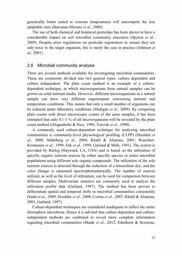
Figure 1. Experimental set-up in the phytotron and in the greenhouse (Photo: Anna Karin Rosberg).
4.1.2 Oomycete inoculum preparation and inoculation (Papers I-III)
For plant inoculation, a kanamycin-resistant Pythium ultimum isolate was used (Hultberg, 1999). The isolate was kept on potato dextrose agar (PDA) supplemented with kanamycin (200 µg mL-1) during laboratory storage. To avoid loss of pathogenicity, the isolate was repeatedly re-isolated from diseased tomato seedlings. For inoculation of the tomato plants, mycelium was used in the form of entire mycelium mats and as a mycelium fragment suspension for inoculation of the seedlings in the Petri dishes. The mycelium mats were produced, as described by Hultberg et al. (2011), by taking mycelium of P. ultimum growing on PDA and transferring it to water agar plates and leaving it to grow for two days. Small plugs were then taken from the periphery of the colonies and placed individually in Petri dishes containing 15 mL of cleared V8 broth (Table 2), and incubated at 25 °C in darkness for three days. The mycelium mats were then washed twice in a sterile mineral solution and once in sterile deionized water before being used to inoculate the plants. For the mycelium fragment suspension, mycelium mats were mixed with sterile deionized water using a high-frequency blender (Polytron, Kinematica GmbH, Switzerland) to produce a suspension with a mycelial fragment concentration of 2.7*107 L-1.
Half of the tomato plants were inoculated with P. ultimum, and the other half were used as healthy control plants. In Papers I and II the seedlings were inoculated five days after sowing, the flowering plants were inoculated two and six weeks after the sowing date, and the fruit-bearing plants were inoculated 14 and 16 weeks after the sowing date. In Paper III all plants used in the experiment were inoculated three and six weeks after sowing. The seedlings were inoculated by giving each Petri dish five mL of the mycelium fragment suspension, while the plants growing in nutrient solution were given one mycelial mat each in their respective growing containers. The fruit-bearing
31
plants in Papers I and II were inoculated with three mycelial mats each at both inoculation events to ensure heavy infection considering the large plant size.
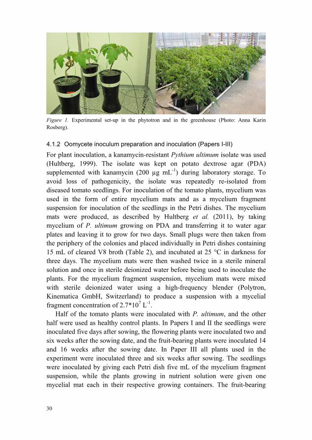
Table 2. Overview of solutions used for inoculum preparation and recovery from roots
V8 broth Mineral solution Wash solution
11.5 g oatmeal 125 mL V8 500 mL deionized water 0.94 g CaCO2
0.145 g Ca(NO3)2 0.012 g MgSO4 0.099 g KH2PO4 0.016 g FeCl3 1 L deionized water
1 g peptone from meat (enzymatic digest: Fluka Chemika, Buchs, Switzerland) 2 g Na-hexametaphosphate (Fluka Chemika, Buchs, Switzerland) 1 L deionized water
Figure 2. Left to right: mycelium mat of P. ultimum grown in a Petri dish; tomato roots inoculated with P. ultimum; non-inoculated tomato roots (Photos: Anna Karin Rosberg)
4.2 Sampling from a commercial greenhouse
In Paper IV samples of nutrient solution were collected from a commercial greenhouse in the south of Sweden with a tomato production area of 0.4 ha. The greenhouse was equipped with a closed hydroponic system in which the plants were grown in plastic containers filled with pumice. Fertigation was supplied to the plants through drip irrigation, and drained nutrient solution was collected in an open gutter system. The drainage was filtered through a slow filter consisting of mineral wool and supplemented with fresh water, as well as adjusted for pH and EC, before it was resupplied to the crop. At the point of sampling, the nutrient solution had been recycled since the start of the cropping season without any discharges.
32
4.3 Analyses
4.3.1 Plant analyses (Papers I-III)
Destructive plant analyses were performed each week for Paper III, and on three occasions for Papers I and II, to study the possible physiological consequences of inoculation with a root pathogen. The analyses included fresh and dry weight of roots, leaves, stems and fruits. For the dry weight measurements, the plant parts were placed in aluminum foil envelopes and placed in a drying cabinet at 105 °C for five days. Leaf area was measured using a LI-COR Li 3100 Area Meter (LI-COR Instruments Inc., USA).
4.3.2 Nutrient solution analyses (Papers II, III and IV)
Nutrient content The analysis of nutrient concentration (mg L-1) in the nutrient solution was performed by the accredited laboratory LMI AB (Helsingborg, Sweden). The method used was inductively coupled plasma optical emission spectrometry (ICP-OES). Nutrient solution samples were taken from both non-inoculated and P. ultimum-inoculated plants (Paper III).
Organic carbon In Papers II and III, the level of total organic carbon (TOC, mg L-1) and dissolved organic carbon (DOC) was measured in the nutrient solution of each individual plant using the HACH LANGE system (TOC cuvette test LCK 385, HACH LANGE GmbH, Düsseldorf, Germany). A sample of the nutrient solution was added to a cuvette and then placed in a shaker (TOC-X5) for 5 min where all of the total inorganic carbon (TIC) was expelled. After this the cuvette was placed in a heating block (LT 200, HACH LANGE GmbH, Düsseldorf, Germany) set to 100 °C for 120 min. During this time the TOC is oxidized to CO2 which passes through a membrane and into an indicator cuvette where it causes a color change. The color reaction was read using a photometer (Xion 500, HACH LANGE GmbH, Düsseldorf, Germany).
Ultra-performance liquid chromatography tandem mass spectrometry (UPLC-MS/MS) For quantification of fenhexamid in the nutrient solution samples taken from the commercial greenhouse, ultra-performance liquid chromatography tandem mass spectrometry (UPLC-MS/MS) was undertaken by the accredited laboratory Eurofins Food and Agro Testing (Kristianstad, Sweden).
33
4.3.3 Harvesting of plants and extraction of microbiota (Papers I-III)
For Papers I and II, plants were harvested on three occasions – as seedlings, flowering plants and fruit-bearing plants – while in Paper III plants were harvested each week. The roots were washed as described in Papers I-III and used for plate counts, community-level physiological profiling (CLPP), denaturing gradient gel electrophoresis (DGGE) and 454-pyrosequencing.
Plate counts Aliquots from the root wash solution and from the nutrient solution obtained from non-inoculated and P. ultimum-inoculated plants were dilution plated on different nutrient media targeting different microorganisms (Table 3). Viable bacteria and fungi were enumerated as colony-forming units (CFU) ml-1and g dw-1 (Papers I-IV).
Community-level physiological profiling (CLPP) In Paper I, microbial utilization of organic compounds was studied using Phenotype MicroArray (PM) panels PM1, PM2A and PM3B (Biolog Inc., Haywood, CA, USA). The PM panels are microtiter plates with 96 wells: one control well and 95 wells containing a single organic nutrient source. PM panels 1 and 2A contained different carbon compounds and panel 3B contained nitrogen compounds. A tetrazolium dye was added to the microtiter plates along with the inoculation of the root microbiota, which gave rise to a color change as the microbiota metabolized the organic compounds. The number of organic compounds and the level of utilization were read spectrophotometrically. For details concerning inoculation, incubation and data collection, see Paper I.
34
Table 3. Overview of plate count analysis media used for enumeration of different microbial groups
R2A Tryptic soy agar (TSA) King’s B agar Corn meal agar (CMA) Malt extract agar (MA) Potato dextrose agar (PDA)
Target organism total aerobic counts (nutrient solution)
Incubation temperature (°C) 25 25 25 22 25 Room temperature
35
4.3.5 Culture-independent microbiological analyses (Papers I, II and IV)
Denaturing gradient gel electrophoresis (DGGE) The rhizosphere bacterial communities of non-inoculated and P. ultimum-inoculated tomato plants (Paper I) and of nutrient solution from a tomato culture with and without the addition of a fungicide (active substance fenhexamid) (Paper IV) were investigated using PCR-DGGE. Samples of the wash solution from roots and from the nutrient solution were processed as described in Papers I and IV respectively. The obtained pellets were used for extraction of genomic DNA (DNeasy Plant Mini Kit, Cat. No. 69104, Qiagen GmbH, Hilden, Germany). Bacterial amplifications were performed using the primers F968 and R1378 for primary PCR, and with the forward primer GC-F968 containing a GC-clamp for the secondary PCR (Muyzer et al., 1993).
The PCR products were loaded on acrylamide gels (8 %) containing a linear gradient of 45-60 % denaturant. The gels ran at 70 V in 1xTAE buffer at 60 °C for 16 h, using a CTV 400 System (Cat. No. 700-71149, VWR International, Stockholm, Sweden). The gels were stained using 1xTAE buffer and SybrGold.
454-pyrosequencing The rhizosphere bacterial communities of non-inoculated and P. ultimum-inoculated tomato plants were investigated using 454-pyrosequencing in Paper II. Samples from the wash solution of roots were processed as described in Paper II. The obtained pellets were used for the extraction of genomic DNA (DNeasy Plant Mini Kit, Cat. No. 69104, Qiagen GmbH, Hilden, Germany). For pyrosequencing, the DNA extractions of the five replicates from each treatment were pooled to form one 16S rRNA library for each treatment and plant stage (Paper II). Due to the low concentration of DNA in the samples, a multiple displacement amplification (MDA) was carried out on the pooled DNA samples using the illustra GenomiPhi V2 DNA amplification kit (GE Health Care Life Sciences, Uppsala, Sweden). After MDA, a DNA concentration of 415-1250 ng µL-1 was achieved, measured using a NanoDrop ND-1000 Spectrophotometer (NanoDrop Technologies, Wilmington, USA).
The DNA samples were sent to LGC Genomics GmbH, Berlin, Germany for pyrosequencing analysis where the Roche 454 FLX-Titanium chemistry system was used (Roche/454 Life Sciences, Bradford, CT 06405, USA). The primers used by LGC were GM3 and 926R, targeting the V1-V5 region of the 16S gene.
36
4.3.6 Statistical analyses
Basic statistical analyses (Papers I-IV) were performed using Minitab Statistical Software version 16.1.0 (Minitab Inc., USA). PCR-DGGE gels were analyzed with the GelCompar II program versions 4.1 (Paper I) and 6.5 (Paper IV) (Applied Maths, Sint-Martens-Latem, Belgium). Discriminant analysis of data from the CLPP in Paper II was carried out with SAS v. 9.2 (Institute Inc., Cary, NC, USA). Analysis of the 454-pyrosequencing data was performed by the bioinformatics services at LGC Genomics using QIIME Virtual Box v. 1.6.0. The PAleontological STatistics software package (PAST) version 3.0 was used for additional calculations on the pyrosequencing data. A detailed description of the statistical methods used is given in each individual paper.
37
5 Results and Discussion
5.1 Impact of a root pathogen on root-associated microbial communities
The rhizosphere is a place of great microbial activity closely linked to the status of the plant (Mendes et al., 2013; Hinsinger et al., 2009; Jones & Hinsinger, 2008). Damage to plants caused by necrotrophic pathogens, such as P. ultimum, have been shown to activate the jasmonic acid defense pathway. This activation alters the composition of the root exudates and consequently the rhizosphere microbial communities (Carvalhais et al., 2013). A range of studies have compared the rhizosphere microbiota of diseased and non-diseased plants in soil and compost (Zhu et al., 2013; Yu et al., 2012; Zhang et al., 2011; Hagn et al., 2008; Filion et al., 2004; Gardener & Weller, 2001), and found differences in the microbial communities due to pathogen attack. For soilless cultures, few studies deal with both quantitative and qualitative differences related to pathogen infection.
In the present thesis, the effect of pathogen attack on microbial communities associated with the roots of tomato plants in soilless culture was examined. Differences were seen in the microbial community structure between non-inoculated and P. ultimum-inoculated tomato plants through DGGE, CLPP (Paper I) and 454-pyrosequencing (Paper II). There were, however, significant individual differences between the replicates, which were clearly seen in the DGGE results (Paper I). When the replicates were run individually on the gel, there was no clear treatment effect. However, when the replicates for each treatment were pooled, the distinct treatment effects became evident. The pooling of samples has been shown to omit many species and thus comparisons of individual samples portray a more accurate picture of the species’ diversity (Manter et al., 2010). It should, however, be borne in mind that DGGE is a method that has been estimated to detect only those microbial
38
community members that represent ≥1-2 % of the population in an environmental sample (Muyzer et al., 1993). This implies that even with replicates and pseudo-replicates, some of the microbial community will still remain undetected. The differences seen between treatments of the pooled samples are therefore most likely to reflect differences among the species of highest abundance.
With CLPP it was found that the presence of P. ultimum did change the utilization of different carbon sources, and significant differences between the treatments were found in this respect. Despite considerable differences between replicates with this method as well, there was an abundance of carbon sources with significantly different utilization on a biochemical basis (Table 2, Paper I). This fact provides good evidence that the microbial communities did indeed change due to pathogen attack. Considering the results from DGGE of the individual samples, the change in the microbial communities might not have been structural, but could have been in relation to activity and function. According to Blagodatskaya and Kuzyakov (2013), the active part of the total microbiota is approximately 0.1-2 %, while the fraction of potentially active microorganisms is 10-60 %. The potentially active microorganisms have a quick response to the influx of new substrates, such as a potential difference in root exudates due to pathogen attack. DGGE and other DNA-based methods cannot differentiate between active and non-active microorganisms (Bodrossy et al., 2006).
When the same samples were subjected to 454-pyrosequencing, clear differences in bacterial community structure were seen between the non-inoculated and P. ultimum-inoculated tomato plants. This is illustrated in Figure 3. The root-associated microbial communities were highly dominated by Proteobacteria. This has also been found by Calmin et al. (2008) and Deery (2012) in the rhizosphere of tomato, and in the rhizosphere of many other crops by Filion et al. (2004), Hagn et al. (2008), Zhang et al. (2011), and Zhu et al. (2013). Despite a high number of reads for each sample, ranging from 11, 283 to 51,453, the results do not reflect an entirely accurate view of the bacterial communities. Replicates are essential due to the heterogeneity of microbial communities, and without an estimate of variability no conclusion can be drawn (Prosser, 2010). However, the present results give a general understanding and a trend analysis of the microorganisms present in the rhizosphere of non-inoculated and P. ultimum-inoculated tomato plants.
39
Figure 3. Results from 454-pyrosequencing of the bacteria present in the rhizosphere of non-inoculated and P-ultimum-inoculated tomato plants, at class level. Left column: non-inoculated treatment; right column: P. ultimum-inoculated treatment.
Seedlings
Flowering
Fruit-bearing
40
5.2 Impact of plant age on root-associated microbial communities
The impact of plant age on rhizosphere microbial communities has been shown by several authors (Micallef et al., 2009; Houlden et al., 2008; Marschner et al., 2002). It is a well-established fact that root exudation changes with plant age (Rovira, 1959), hence changes in microbial community structures are most likely caused by changes in root exudation patterns (Micallef et al., 2009; Marschner et al., 2002).
In the present thesis, the effects on the microbial communities related to plant age were found using DGGE, CLPP (Paper I) and 454-pyrosequencing (Paper II). In contrast to the effect of a pathogen, the effect of plant age was clearly seen with DGGE when individual replicates were compared (Paper I). At the seedling and flowering plant stage, clear clusters were formed. At the flowering stage, however, the replicates were more diverse. Postma et al. (2000) demonstrated that the complexity of bacterial communities increased with plant age, resulting in a decrease of replicate similarity. This also seems to be the case in the present thesis.
Figure 4. Ordination plot of principal components (PC) 1 and 2 from community-level physiological profiling (CLPP) of root microorganisms on tomato seedlings (∆, ▲), and flowering (○, ●) and fruit-bearing (□, ■) plants. CLPP was established from carbon substrate utilization patterns obtained using PM panel 2 (Biolog). Filled and open symbols indicate inoculated (P. ultimum) and non-inoculated plants respectively.
When the results of the CLPP were subjected to PCA, clear clusters with respect to plant physiological stages became apparent (Figure 4). With this analysis the microbial community structure of seedlings showed the highest
1050-5-10
5.0
2.5
0.0
-2.5
-5.0
-7.5
PC 1 (37.0 % )
PC 2
(13.
4 %
)
41
diversity between replicates. This high variability between seedling replicates could be due to the fact that each replicate, consisting of 100 seeds, was grown in a sealed Petri dish, inhibiting the migration of microorganisms from the surrounding environment. The microbiota developing in each Petri dish was therefore a result of microorganisms already present on the seeds and in the water. At the flowering and fruit-bearing stages, however, the plants were grown in open containers and subjected to a nutrient solution. Despite the large number of carbon sources for which the utilization was significantly different between the treatments, few of these carbon sources reoccurred in two plant stages, and none reoccurred in all three. This implies that the microbial community structure, activity or function was different at the different plant stages.
With 454-pyrosequencing the differences in bacterial communities between the plant stages became apparent. The microbiota formed clear groups with respect to plant age in the PCA. Similar findings were made by Zhang et al. (2011) who saw clear differences in bacterial communities at the different plant physiological stages of cotton. Bacterial diversity initially increased and then decreased at plant maturity, while in the present thesis the bacterial species’ richness and diversity increased with plant age. As illustrated in Figure 3, not only the classes of bacteria differed, but the species’ richness also increased with plant age. Although there was a high number of reads per sample, rarefaction analysis showed that the samples from the fruit-bearing stage did not reach saturation. Hence it can be assumed that the species’ richness and the complexity of the microbial communities are greater than the result suggests.
5.3 Impact of available nutrients on nutrient solution-associated microbial communities
The first two sections of this chapter have focused on the root, which is the main producer of carbon in the hydroponic cropping systems used in the present thesis. The following two sections will focus on the microorganisms present in the nutrient solution (Papers III-V). These are the main consumers and transformers of the carbon provided by the roots. As seen in Figure 5, the carbon in hydroponic cropping systems is provided by the macrophytes, i.e. plants, as well as by phytoplankton and biofilms that develop on surfaces such as pipes and growing containers (Paper V). If a growing medium is used, then depending on its origin, this could also act as a supplier of carbon. The main users of the carbon pool are free-living microorganisms and microorganisms associated with biofilms.
42
Organic carbon is the dominant energy source in the root environment that is available to heterotrophic microorganisms, and is also the major constituent of the microbial cell (Madigan et al., 2009). Organic carbon, however, is not the only nutrient needed for microbial growth. Mineral nutrients, such as nitrogen and phosphorous, as well as micronutrients are also required (Duffy & Defago, 1997; Fagerbakke et al., 1996). The number of microorganisms inhabiting the rhizoplane and nutrient solution fluctuates during the growing season in hydroponic cropping systems with tomato (Khalil & Alsanius, 2001). There are few explanations as to why these fluctuations occur, other than the impact of plant physiological age discussed in the previous section.
In the present thesis, a study was conducted regarding the interactivities between available organic carbon and mineral nutrients in the nutrient solution from microbially-colonized hydroponic cropping systems (Paper III). Regression analysis indicated that the level of organic carbon was highly correlated with the number of bacteria, fungi and fluorescent pseudomonads. The correlation values were particularly high in the non-inoculated treatment, with R2 values as high as 95.9 for the correlation between organic carbon and fluorescent pseudomonads. Despite differences in the concentration of TOC and DOC, both fungi and fluorescent pseudomonads were equally correlated with TOC and DOC. The concentrations of TOC and DOC were quite similar (Paper III; Table 3) but in the last five weeks of measurements, DOC did not increase at the same rate as TOC. While TOC consists of plant organic matter, microorganisms and DOC, DOC contains the low-molecular weight compounds readily available for the microbiota, as well as some high-molecular weight compounds of hydrophilic and acidic origin (Wetzel, 1983). The increasing difference in TOC and DOC therefore indicates that a larger part of the organic carbon became organically bound to roots, microorganisms and microfauna, for example. The high level of DOC measured means that carbon alone was not sufficient as a nutrient source, but that other nutrient sources had a limiting effect on the number of microorganisms. The high levels of DOC could also be an indication that the available organic carbon compounds were in a form that the resident microbiota could not degrade (Alsanius & Jung, 2004). It has been found that carbon-to-nitrogen ratios (C:N) are important for microbial growth, with a C:N ratio of 15 preferred by bacteria and a C:N ratio of 75 by fungi (Rousk & Bååth, 2007). In the present study, the C:N ratios were more favorable to fungi. This could be seen in the numbers of CFU, which increased over time for fungi but stayed at a relatively stable level for the bacteria.
43
Figure 5. Flow of organic carbon in a hydroponic growing system where the autochthonous and allochthonous carbon is provided by macrophytes, phytoplankton, biofilms and carbon sources associated with the cropping system. The consumers of carbon are biofilms and free-living microorganisms, while the use of UV or ozone leads to a removal of carbon from the system (Paper V; Figure 28.2).
44
The quantity and quality of nutrients are both important. Hultberg and Alsanius (2008) saw that production of the antibiotic 2,4-diacetylphloroglucinol (DAPG) by the biocontrol agent Pseudomonas fluorescens Pf-5 was highly dependent on the type of nitrogen source provided. Similarly, micronutrients are known to act as catalysts for the production of secondary metabolites. It has been shown that production is highly sensitive to fluctuations in micronutrient availability (Zweifel et al., 1993).
The type of carbon source available is also important. From the CLPP study in the present thesis (Paper I), it was seen that not all carbon sources available were utilized at the same rate. This could be an effect of adaptation by the microorganisms to the carbon sources to which they had not previously been exposed. Given time, all sources were used to some degree. There were differences in the utilization patterns, both with respect to treatment and to plant age. This implies that there were changes in the microbial community structures or that the active parts of the communities changed due to differences in root exudation patterns as a result of pathogen infection and the ageing of the plants.
5.4 Impact of plant protection products on nutrient solution-associated microbial communities
Disturbances of rhizosphere microbial communities through the use of plant protection products have been shown in several studies (Spyrou et al., 2009; Borzi et al., 2007; Engelen et al., 1998). Depending on the pesticide used, the effect has been found to act either to eliminate certain organisms or to stimulate their growth. The use of fenhexamid in soil has been shown to exhibit a selective pressure on some microbial communities where a number of microorganisms disappeared and a few increased. It was hypothesized that the species increasing in numbers after the addition of the pesticide were those able to degrade xenobiotics (Borzi et al., 2007).
In the present study (Paper IV), the number of CFUs obtained through plate counts differed between untreated and fenhexamid-treated nutrient solutions. With the presence of fenhexamid, the number of bacterial CFUs started to decrease after just two days. Without fenhexamid, differences in plate counts were first seen after eight days. However, at the end of the exposure time, the number of bacterial counts was higher in the fenhexamid-supplemented medium compared to the control. For the fungal flora, counts dropped immediately after exposure to fenhexamid and did not recover for the length of the experiment. This was to be expected since fenhexamid is a fungicide with
45
Botrytis as the main target. Through DGGE analysis, a shift in the bacterial community structure was seen after eight days.
Pesticides which are dissolved in water are available as substrates for the microorganisms, and are metabolized through enzymatic hydrolysis, oxidation and reduction. Both free-living microorganisms and those associated with biofilms are involved (Katagi, 2013). The level of fenhexamid in the nutrient solution was unaffected after 21 days of incubation. Hence no degradation by the microorganisms occurred. The increase in bacterial counts was therefore not due to the utilization of fenhexamid, but was more likely an effect of mineralization of the organic carbon from the microorganisms that did not survive the pesticide treatment, predominantly fungi. Previous studies have demonstrated similar findings where a lack of degradation was revealed after 112 days of incubation in natural river water (Abbate et al., 2007). In the same study, fenhexamid had a DT50 of 1.2 days in the presence of an active microbiota, while in the absence of microorganisms, fenhexamid proved to be stable. The degradation of the insecticide fenthion increased in the presence of plants and plant-associated microorganisms (Cripe et al., 1989). The absence of plants in the experiments of Paper IV might have been a factor limiting the degradation of fenhexamid.
46
6 Conclusions In the present thesis, it has been demonstrated that root-associated and nutrient solution-associated microbial community structures are affected by plant age, pathogen attack, the availability of organic and inorganic nutrients, and the use of plant protection products. Plant age in particular seemed to have the greatest impact on community structure.
The combined use of culture-dependent and culture-independent techniques proved successful, since different parts of the microbial communities were visualized with different methods. The methods used, however, did not target the active parts of the communities. There should be a focus in future studies on microbial activity and function through the use of metabolomics and metaproteomics. Changes induced by abiotic and biotic factors are more likely to be seen in microbial activity rather than in qualitative, structural alterations. The activity and function of the microbial communities were partly seen with the use of CLPP, where it was demonstrated that the utilization of sole carbon sources differed between treatments and between plant physiological stages. This could imply changes in the activity of different microbial species caused by pathogen inoculation.
The rhizosphere is one of the most complex ecosystems on earth. Hence, additional environmental factors, such as temperature, light intensity and oxygen saturation of the nutrient solution, should be monitored closely in future studies in order to understand their combined effect on microbial communities.
47
48
References Abbate, C., Borzi, D., Caboni, P., Baglieri, A. & Gennari, M. (2007). Behavior of
fenhexamid in soil and water. Journal of Environmental Science and Health Part B-Pesticides Food Contaminants and Agricultural Wastes, 42(7), pp. 843-849.
Agrios, G.N. (2005). Plant pathology. 5. ed. San Diego: Elsevier Academic Press. Alsanius, B.W. & Jung, V. (2004). Potential of utilization of selected organic
compounds by the Mi-croflora inhabiting the effluent nutrient solution of closed green-house house irrigation systems. European Journal of Horticultural Science, 69(3), pp. 96-103.
Andrew, D.R., Fitak, R.R., Munguia-Vega, A., Racolta, A., Martinson, V.G. & Dontsova, K. (2012). Abiotic Factors Shape Microbial Diversity in Sonoran Desert Soils. Applied and Environmental Microbiology, 78(21), pp. 7527-7537.
Apel, K. & Hirt, H. (2004). Reactive oxygen species: Metabolism, oxidative stress, and signal transduction. Annual Review of Plant Biology, 55, pp. 373-399.
Aulakh, M.S., Wassmann, R., Bueno, C., Kreuzwieser, J. & Rennenberg, H. (2001). Characterization of root exudates at different growth stages of ten rice (Oryza sativa L.) cultivars. Plant Biology, 3(2), pp. 139-148.
Bais, H.P., Prithiviraj, B., Jha, A.K., Ausubel, F.M. & Vivanco, J.M. (2005). Mediation of pathogen resistance by exudation of antimicrobials from roots. Nature, 434(7030), pp. 217-221.
Baker, K.F. & Cook, R.J. (1974). Biological control of plant pathogens. University of Michigan: American Phytopathological Society.
Bakker, M.G., Schlatter, D.C., Otto-Hanson, L. & Kinkel, L.L. (2014). Diffuse symbioses: roles of plant-plant, plant-microbe and microbe-microbe interactions in structuring the soil microbiome. Molecular Ecology, 23(6), pp. 1571-1583.
Barcenas-Moreno, G., Gomez-Brandon, M., Rousk, J. & Bååth, E. (2009). Adaptation of soil microbial communities to temperature: comparison of fungi and bacteria in a laboratory experiment. Global Change Biology, 15(12), pp. 2950-2957.
49
Bergstrand, K.-J. (2010). Approaches for mitigating the environmental impact of greenhouse horticulture. Diss. Alnarp, Sweden: Swedish University of Agricultural Sciences.
Berkelmann, B., Wohanka, W. & Wolf, G.A. (1994). Characterization of the bacterial flora in circulating nutrient solutions of a hydroponic system with rockwool. Acta Horticulturae, 361, pp. 372-381.
Blagodatskaya, E. & Kuzyakov, Y. (2013). Active microorganisms in soil: Critical review of estimation criteria and approaches. Soil Biology & Biochemistry, 67, pp. 192-211.
Blagodatskaya, E.V. & Anderson, T.H. (1998). Interactive effects of pH and substrate quality on the fungal-to-bacterial ratio and QCO(2) of microbial communities in forest soils. Soil Biology & Biochemistry, 30(10-11), pp. 1269-1274.
Bodrossy, L., Stralis-Pavese, N., Konrad-Koszler, M., Weilharter, A., Reichenauer, T.G., Schofer, D. & Sessitsch, A. (2006). mRNA-based parallel detection of active methanotroph populations by use of a diagnostic microarray. Applied and Environmental Microbiology, 72(2), pp. 1672-1676.
Borrero, C., Ordovas, J., Trillas, M.I. & Aviles, M. (2006). Tomato Fusarium wilt suppressiveness. The relationship between the organic plant growth media and their microbial communities as characterised by Biolog (R). Soil Biology & Biochemistry, 38(7), pp. 1631-1637.
Borzi, D., Abbate, C., Martin-Laurent, F., El Azhari, N. & Gennari, M. (2007). Studies on the response of soil microflora to the application of the fungicide fenhexamid. International Journal of Environmental Analytical Chemistry, 87(13-14), pp. 949-956.
Brand, T. & Wohanka, W. (2001). Importance and characterization of the biological component in slow filters. Acta Horticulturae, 554, pp. 313-321.
Broeckling, C.D., Broz, A.K., Bergelson, J., Manter, D.K. & Vivanco, J.M. (2008). Root exudates regulate soil fungal community composition and diversty. Applied and Environmental Microbiology, 74(3), pp. 738-744.
Buyanovsky, G., Gale, J. & Degani, N. (1981). UV-radiation for the inactivation of microorganisms in hydroponics. Plant and Soil, 60(1), pp. 131-136.
Calmin, G., Dennler, G., Belbahri, L., Wigger, A. & Lefort, F. (2008). Molecular identification of microbial communities in the recycled nutrient solution of a tomato glasshouse soil-less culture. Open Horticulture Journal, 1, pp. 7-14.
Calvo-Bado, L.A., Petch, G., Parsons, N.R., Morgan, J.A.W., Pettitt, T.R. & Whipps, J.M. (2006). Microbial community responses associated with the development of oomycete plant pathogens on tomato roots in soilless growing systems. Journal of Applied Microbiology, 100(6), pp. 1194-1207.
Carvalhais, L.C., Dennis, P.G., Badri, D.V., Tyson, G.W., Vivanco, J.M. & Schenk, P.M. (2013). Activation of the jasmonic acid plant defence pathway alters the composition of rhizosphere bacterial communities. Plos One, 8(2).
50
Castano, R., Borrero, C., Trillas, M.I. & Aviles, M. (2013). Selection of biological control agents against tomato Fusarium wilt and evaluation in greenhouse conditions of two selected agents in three growing media. Biocontrol, 58(1), pp. 105-116.
Chave, M., Dabert, P., Brun, R., Godon, J.J. & Poncet, C. (2008). Dynamics of rhizoplane bacterial communities subjected to physicochemical treatments in hydroponic crops. Crop Protection, 27(3-5), pp. 418-426.
Chen, W., Hoitink, H.A.J., Schmitthenner, A.F. & Tuovinen, O.H. (1988). The role of microbial activity in suppression of damping-off caused by Pythium ultimum. Phytopathology, 78(3), pp. 314-322.
Chen, W.D., Hoitink, H.A.J. & Schmitthenner, A.F. (1987). Factors affecting suppression of Pythium damping-off in container media amended with composts. Phytopathology, 77(5), pp. 755-760.
Cherif, M., Tirilly, Y. & Belanger, R.R. (1997). Effect of oxygen concentration on plant growth, lipid peroxidation, and receptivity of tomato roots to Pythium F under hydroponic conditions. European Journal of Plant Pathology, 103(3), pp. 255-264.
Clematis, F., Minuto, A., Gullino, M.L. & Garibaldi, A. (2009). Suppressiveness to Fusarium oxysporum f. sp. radicis lycopersici in re-used perlite and perlite-peat substrates in soilless tomatoes. Biological Control, 48(2), pp. 108-114.
Cook, R.J. & Baker, K.F. (1983). The nature and practice of biological control of plant pathogens. St Paul, MN, USA: The American Phytopathological Society.
Correa, O.S., Romero, A.M., Montecchia, M.S. & Soria, M.A. (2007). Tomato genotype and Azospirillum inoculation modulate the changes in bacterial communities associated with roots and leaves. Journal of Applied Microbiology, 102(3), pp. 781-786.
Craft, C.M. & Nelson, E.B. (1996). Microbial properties of composts that suppress damping-off and root rot of creeping bentgrass caused by Pythium graminicola. Applied and Environmental Microbiology, 62(5), pp. 1550-1557.
Cripe, C.R., Oneill, E.J., Woods, M.E., Gilliam, W.T. & Pritchard, P.H. (1989). Fate OofF fenthion in salt-marsh environments: 1. Factors affecting biotic and abiotic degradation rates in water and sediment. Environmental Toxicology and Chemistry, 8(9), pp. 747-758.
Crous, P.W., Verkley, G.J.M., Groenewald, J.Z. & Samson, R.A. (2009). Fungal Biodiversity. CBS Laboratory Manual series. Utrecht, Netherlands: CBS KNAW Fungal Biodiversity Centre.
Deery, S.J. (2012). Monitoring rhizosphere microbial communities of tomato. Diss. Nottingham, UK: University of Nottingham.
Deniel, F., Rey, P., Cherif, M., Guillou, A. & Tirilly, Y. (2004). Indigenous bacteria with antagonistic and plant growth-promoting activities improve slow filtration efficiency in soilless cultivation. Canadian Journal of Microbiology, 50(7), pp. 499-508.
51
Dorais, M. & Dube, Y. (2011). Closed greenhouse systems: multiphase recycling of solid and liquid organic waste. Chronica Horticulturae, 51(1), pp. 12-19.
Duffy, B.K. & Defago, G. (1997). Zinc improves biocontrol of Fusarium crown and root rot of tomato by Pseudomonas fluorescens and represses the production of pathogen metabolites inhibitory to bacterial antibiotic biosynthesis. Phytopathology, 87(12), pp. 1250-1257.
Duniway, J.M. (1979). Water relations of water molds. Annual Review of Phytopathology, 17, pp. 431-460.
Ehret, D.L., Alsanius, B., Wohanka, W., Menzies, J.G. & Utkhede, R. (2001). Disinfestation of recirculating nutrient solutions in greenhouse horticulture. Agronomie, 21(4), pp. 323-339.
Eiler, A., Langenheder, S., Bertilsson, S. & Tranvik, L.J. (2003). Heterotrophic bacterial growth efficiency and community structure at different natural organic carbon concentrations. Applied and Environmental Microbiology, 69(7), pp. 3701-3709.
Engelen, B., Meinken, K., von Wintzintgerode, F., Heuer, H., Malkomes, H.P. & Backhaus, H. (1998). Monitoring impact of a pesticide treatment on bacterial soil communities by metabolic and genetic fingerprinting in addition to conventional testing procedures. Applied and Environmental Microbiology, 64(8), pp. 2814-2821.
EU Water Framework Directive (2000). Fagerbakke, K.M., Heldal, M. & Norland, S. (1996). Content of carbon, nitrogen,
oxygen, sulfur and phosphorus in native aquatic and cultured bacteria. Aquatic Microbial Ecology, 10(1), pp. 15-27.
Filion, M., Hamelin, R.C., Bernier, L. & St-Arnaud, M. (2004). Molecular profiling of rhizosphere microbial communities associated with healthy and diseased black spruce (Picea mariana) seedlings grown in a nursery. Applied and Environmental Microbiology, 70(6), pp. 3541-3551.
Garcia, J.A.L., Barbas, C., Probanza, A., Barrientos, M.L. & Manero, F.J.G. (2001). Low molecular weight organic acids and fatty acids in root exudates of two Lupinus cultivars at flowering and fruiting stages. Phytochemical Analysis, 12(5), pp. 305-311.
Gardener, B.B.M. & Weller, D.M. (2001). Changes in populations of rhizosphere bacteria associated with take-all disease of wheat. Applied and Environmental Microbiology, 67(10), pp. 4414-4425.
Garibaldi, A., Minuto, A., Grasso, V. & Gullino, M.L. (2003). Application of selected antagonistic strains against Phytophthora cryptogea on gerbera in closed soilless systems with disinfection by slow sand filtration. Crop Protection, 22(8), pp. 1053-1061.
Garland, J.L. (1997). Analysis and interpretation of community-level physiological profiles in microbial ecology. Fems Microbiology Ecology, 24(4), pp. 289-300.
52
Garland, J.L. & Mills, A.L. (1991). Classification and characterization of heterotrophic microbial communities on the basis of patterns of community-level sole-carbon-source utilization. Applied and Environmental Microbiology, 57(8), pp. 2351-2359.
Gericke, W.F. (1937). Hydroponics - crop production in liquid culture media. Science, 85(2198), pp. 177-8.
Gold, S.E. & Stanghellini, M.E. (1985). Effects of temperature on Pythium root-rot of spinach grown under hydroponic conditions. Phytopathology, 75(3), pp. 333-337.
Gottel, N.R., Castro, H.F., Kerley, M., Yang, Z., Pelletier, D.A., Podar, M., Karpinets, T., Uberbacher, E., Tuskan, G.A., Vilgalys, R., Doktycz, M.J. & Schadt, C.W. (2011). Distinct microbial communities within the endosphere and rhizosphere of Populus deltoides roots across contrasting soil types. Applied and Environmental Microbiology, 77(17), pp. 5934-5944.
Grayston, S.J., Wang, S.Q., Campbell, C.D. & Edwards, A.C. (1998). Selective influence of plant species on microbial diversity in the rhizosphere. Soil Biology & Biochemistry, 30(3), pp. 369-378.
Griffin, C.J., Hale, M.G. & Shay, F.J. (1976). Nature and quantity of sloughed organic matter produced by roots of axenic peanut plants. Soil Biology and Biochemistry, 8(1), pp. 29-32.
Gruyer, N., Dorais, M., Zagury, G.J. & Alsanius, B.W. (2011). Effects of using water treated by artificial wetlands on root rot suppression and tomato growth. Horttechnology, 21(6), pp. 759-766.
Hagn, A., Engel, M., Kleikamp, B., Munch, J.C., Schloter, M. & Bruns, C. (2008). Microbial community shifts in Pythium ultimum-inoculated suppressive substrates. Biology and Fertility of Soils, 44(3), pp. 481-490.
Hansson, T. (2003). Dräneringsvatten i växthus. (Jordbruksinformation, 16): Jordbruksverket.
Heuvelink, E. & Dorais, M. (2005). Crop growth and yield. In: Heuvelink, E. (ed. Tomatoes. Oxfordshire: CABI Publishing, pp. 85-144.
Hinsinger, P., Bengough, A.G., Vetterlein, D. & Young, I.M. (2009). Rhizosphere: biophysics, biogeochemistry and ecological relevance. Plant and Soil, 321(1-2), pp. 117-152.
Hoitink, H.A.J., Inbar, Y. & Boehm, M.J. (1991). Status of compost-amended potting mixes naturally suppressive to soilborne diseases of floricultural crops. Plant Disease, 75(9), pp. 869-873.
Houlden, A., Timms-Wilson, T.M., Day, M.J. & Bailey, M.J. (2008). Influence of plant developmental stage on microbial community structure and activity in the rhizosphere of three field crops. Fems Microbiology Ecology, 65(2), pp. 193-201.
Hueso, S., Garcia, C. & Hernandez, T. (2012). Severe drought conditions modify the microbial community structure, size and activity in amended and unamended soils. Soil Biology & Biochemistry, 50, pp. 167-173.
Hugenholtz, P. & Pace, N.R. (1996). Identifying microbial diversity in the natural environment: A molecular phylogenetic approach. Trends in Biotechnology, 14(6), pp. 190-197.
53
Hultberg, M. (1999). Seed bacterization with Pseudomonas fluorescens and interactions with Pythium ultimum on tomato in soilless systems. Diss. Alnarp: Swedish University of Agricultural Sciences.
Hultberg, M., Alsanius, B. & Sundin, P. (2000). In vivo and in vitro interactions between Pseudomonas fluorescens and Pythium ultimum in the suppression of damping-off in tomato seedlings. Biological Control, 19(1), pp. 1-8.
Hultberg, M. & Alsanius, B.W. (2008). Influence of Nitrogen Source on 2,4-diacetylphloroglucinol Production by the Biocontrol Strain Pf-5. The open microbiology journal, 2, pp. 74-8.
Hultberg, M., Holmkvist, A. & Alsanius, B. (2011). Strategies for administration of biosurfactant-producing pseudomonads for biocontrol in closed hydroponic systems. Crop Protection, 30(8), pp. 995-999.
Ibekwe, A.M. & Kennedy, A.C. (1999). Fatty acid methyl ester (FAME) profiles as a tool to investigate community structure of two agricultural sails. Plant and Soil, 206(2), pp. 151-161.
Igura, N., Fujii, M., Shimoda, M. & Hayakawa, I. (2004). Research note: Inactivation efficiency of ozonated water for Fusarium oxysporum conidia under hydroponic greenhouse conditions. Ozone-Science & Engineering, 26(5), pp. 517-521.
Johnsen, K., Jacobsen, C.S., Torsvik, V. & Sorensen, J. (2001). Pesticide effects on bacterial diversity in agricultural soils - a review. Biology and Fertility of Soils, 33(6), pp. 443-453.
Jones, D.L. & Hinsinger, P. (2008). The rhizosphere: complex by design. Plant and Soil, 312(1-2), pp. 1-6.
Jones, J.B. (1982). Hydroponics - its history and use in plant nutrition studies. Journal of Plant Nutrition, 5(8), pp. 1003-1030.
Jones, W.J. (2010). High-throughput sequencing and metagenomics. Estuaries and Coasts, 33(4), pp. 944-952.
Kamilova, F., Kravchenko, L.V., Shaposhnikov, A.I., Azarova, T., Makarova, N. & Lugtenberg, B. (2006). Organic acids, sugars, and L-tryptophane in exudates of vegetables growing on stonewool and their effects on activities of rhizosphere bacteria. Molecular Plant-Microbe Interactions, 19(3), pp. 250-256.
Katagi, T. (2013). Aerobic microbial transformation of pesticides in surface water. Journal of Pesticide Science, 38(1-2), pp. 10-26.
Kent, A.D. & Triplett, E.W. (2002). Microbial communities and their interactions in soil and rhizosphere ecosystems. Annual Review of Microbiology, 56, pp. 211-236.
Khalil, S. (2001). Microflora in the root environment of hydroponically grown tomato: Methods for assessment and effects of introduced bacteria and Pythium ultimum. Diss. Alnarp: Swedish University of Agricultural Sciences.
Khalil, S. & Alsanius, B.W. (2001). Dynamics of the indigenous microflora inhabiting the root zone and the nutrient solution of tomato in a commercial closed greenhouse system. Gartenbauwissenschaft, 66(4), pp. 188-198.
54
Khalil, S., Hultberg, M. & Alsanius, B.W. (2009). Effects of growing medium on the interactions between biocontrol agents and tomato root pathogens in a closed hydroponic system. Journal of Horticultural Science & Biotechnology, 84(5), pp. 489-494.
Khalil, S., Hultberg, M. & Alsanius, B.W. (2011). Interactions between growing media and biocontrol agents in closed hydroponic systems. Acta Horticulturae, 891, pp. 51-57.
Khalil, S. & Olsson, O. (2013). Influence of micronutrients on the biological control of Pythium ultimum in a closed soilless system. Canadian Journal of Plant Protection, 1(4), pp. 134-141.
Koohakan, P., Ikeda, H., Jeanaksorn, T., Tojo, M., Kusakari, S.I., Okada, K. & Sato, S. (2004). Evaluation of the indigenous microorganisms in soilless culture: occurrence and quantitative characteristics in the different growing systems. Scientia Horticulturae, 101(1-2), pp. 179-188.
Larcher, W. (2003). Physiological plant ecology: Ecophysiology and stress physiology of functional groups. 4. ed.
Lauber, C.L., Hamady, M., Knight, R. & Fierer, N. (2009). Pyrosequencing-Based Assessment of Soil pH as a Predictor of Soil Bacterial Community Structure at the Continental Scale. Applied and Environmental Microbiology, 75(15), pp. 5111-5120.
Lesuffleur, F., Paynel, F., Bataille, M.-P., Le Deunff, E. & Cliquet, J.-B. (2007). Root amino acid exudation: measurement of high efflux rates of glycine and serine from six different plant species. Plant and Soil, 294(1-2), pp. 235-246.
Li, D., Sharp, J.O., Saikaly, P.E., Ali, S., Alidina, M., Alarawi, M.S., Keller, S., Hoppe-Jones, C. & Drewes, J.E. (2012). Dissolved organic carbon influences microbial community composition and diversity in managed aquifer recharge systems. Applied and Environmental Microbiology, 78(19), pp. 6819-6828.
Ma, J.F., Ryan, P.R. & Delhaize, E. (2001). Aluminium tolerance in plants and the complexing role of organic acids. Trends in Plant Science, 6(6), pp. 273-278.
Macdonald, J.D., Alishtayeh, M.S., Kabashima, J. & Stites, J. (1994). Occurrence of Phytophthora species in recirculated nursery irrigation effluents. Plant Disease, 78(6), pp. 607-611.
Madigan, M.T., Martinko, J.M., Dunlap, P.V. & Clark, D.P. (eds) (2009). Brock Biology of microorganisms. 12 ed. San Francisco, USA: Pearsons Education Inc.
Manter, D.K., Weir, T.L. & Vivanco, J.M. (2010). Negative Effects of Sample Pooling on PCR-Based Estimates of Soil Microbial Richness and Community Structure. Applied and Environmental Microbiology, 76(7), pp. 2086-2090.
Marschner, P., Neumann, G., Kania, A., Weiskopf, L. & Lieberei, R. (2002). Spatial and temporal dynamics of the microbial community structure in the rhizosphere of cluster roots of white lupin (Lupinus albus L.). Plant and Soil, 246(2), pp. 167-174.
55
Mendes, R., Garbeva, P. & Raaijmakers, J.M. (2013). The rhizosphere microbiome: significance of plant beneficial, plant pathogenic, and human pathogenic microorganisms. Fems Microbiology Reviews, 37(5), pp. 634-663.
Menzies, J.G., Ehret, D.L., Koch, C., Hall, J.W., Seifert, K.A., Bissett, J. & Barr, D.J.S. (2005). Fungi associated with roots of cucumber grown in different greenhouse root substrates. Canadian Journal of Botany-Revue Canadienne De Botanique, 83(1), pp. 80-92.
Micallef, S.A., Channer, S., Shiaris, M.P. & Colon-Carmona, A. (2009). Plant age and genotype impact the progression of bacterial community succession in the Arabidopsis rhizosphere. Plant signaling & behavior, 4(8), pp. 777-80.
Minuto, A., Clematis, F., Gullino, M.L. & Garibaldi, A. (2007). Induced suppressiveness to Fusarium oxysporum f.sp. radicis-lycopersici in rockwool substrate used in closed soilless systems. Phytoparasitica, 35(1), pp. 77-85.
Mougel, C., Offre, P., Ranjard, L., Corberand, T., Gamalero, E., Robin, C. & Lemanceau, P. (2006). Dynamic of the genetic structure of bacterial and fungal communities at different developmental stages of Medicago truncatula Gaertn. cv. Jemalong line J5. New Phytologist, 170(1), pp. 165-175.
Muyzer, G., Dewaal, E.C. & Uitterlinden, A.G. (1993). Profiling of complex microbial-populations by denaturing gradient gel-electrophoresis analysis of polymerase chain reaction-amplified genes-coding for 16S ribosomal-RNA. Applied and Environmental Microbiology, 59(3), pp. 695-700.
Nemec, S., Datnoff, L.E. & Strandberg, J. (1996). Efficacy of biocontrol agents in planting mixes to colonize plant roots and control root diseases of vegetables and citrus. Crop Protection, 15(8), pp. 735-742.
Neumann, G. & Römheld, V. (2001). The release of root exudates as affected by the plant’s physiological status. In: Pinton, R., Varanini, Z. & Nannipieri, P. (eds) The rhizosphere – Biochemistry and organic substances at the soil-plant interface. New York: Marcel Dekker Inc., pp. 41-93.
Nybroe, O., Brandt, K.K., Nicolaisen, M.H. & Sorensen, J. (2007). Methods to detect and quantify bacteria in soil. In: van Elsas, J.D., Jansson, J.K. & Trevors, J.T. (eds) Modern Soil Microbiology. 2. ed. Boca Raton, FL, USA: CRC Press, pp. 283-316.
O’Callaghan, M., Lorenz, N. & Gerard, E.M. (2006). Characterization of phylloplane and rhizosphere microbial populations using PCR and denaturing gradient gel electrophoreses (DGGE). In: Cooper, J.E. & Rao, J.R. (eds) Molecular approaches to soil, rhizosphere and plant microorganism analysis. Belfast, UK: CABI, pp. 99-115.
Ongena, M., Daayf, F., Jacques, P., Thonart, P., Benhamou, N., Paulitz, T.C., Cornelis, P., Koedam, N. & Belanger, R.R. (1999). Protection of cucumber against Pythium root rot by fluorescent pseudomonads: predominant role of induced resistance over siderophores and antibiosis. Plant Pathology, 48(1), pp. 66-76.
56
Os, E.A.v. (2009). Comparison of some chemical and non-chemical treatments to disinfect a recirculating nutrient solution. Acta Horticulturae, 843, pp. 229-234.
Paulitz, T.C. (1997). Biological control of root pathogens in soilless and hydroponic systems. Hortscience, 32(2), pp. 193-196.
Paulitz, T.C. & Belanger, R.R. (2001). Biological control in greenhouse systems. Annual Review of Phytopathology, 39, pp. 103-133.
Peet, M.M. & Welles, G. (2005). Greenhouse tomato production. In: Heuvelink, E. (ed. Tomatoes. London, UK: CABI Publishing, pp. 257-304.
Postma, J. (2004). Suppressiveness of root pathogens in closed cultivation systems. Acta Horticulturae, 644, pp. 503-510.
Postma, J., Geraats, B.P.J., Pastoor, R. & van Elsas, J.D. (2005). Characterization of the microbial community involved in the suppression of Pythium aphanidermatum in cucumber grown on rockwool. Phytopathology, 95(7), pp. 808-818.
Postma, J., Van Os, E.A. & Bonants, P.J.M. (2008). Pathogen detection and management strategies in soilless plant growing systems. In: Raviv, M. & Lieth, J.H. (eds) Soilless culture - theory and practice. USA: Elsevier BV, pp. 425-458.
Postma, J., Willemsen-de Klein, M. & van Elsas, J.D. (2000). Effect of the indigenous microflora on the development of root and crown rot caused by Pythium aphanidermatum in cucumber grown on rockwool. Phytopathology, 90(2), pp. 125-133.
Prikryl, Z. & Vancura, V. (1980). Root exudates of plants .6. Wheat root exudation as dependent on growth, concentration gradient of exudates and the prescense of bacteria. Plant and Soil, 57(1), pp. 69-83.
Prosser, J.I. (2010). Replicate or lie. Environmental Microbiology, 12(7), pp. 1806-1810.
Rattink, H. & Postma, J. (1996). Biological control of Fusarium wilt in carnations on a recirculation system by a nonpathogenic Fusarium oxysporum isolate. Mededelingen Faculteit Landbouwkundige en Toegepaste Biologische Wetenschappen, 61(2A&B,3), pp. 491-498.
Raviv, M. & Lieth, J.H. (2008). Significance of soilless culture in agriculture. In: Lieth, J.H. & Raviv, M. (eds) Soilless culture. Amsterdam: Elsevier, pp. 1-12.
Rey, P., Deniel, F., Vasseur, V., Tirilly, Y. & Benhamou, N. (2001). Evolution of Pythium spp. populations in soilless cultures and their control by active disinfecting methods. Acta Horticulturae, 554, pp. 341-348.
Rey, P., Picard, K., Deniel, F., Benhamou, N. & Tirilly, Y. (1999). Development of an IPM system in soilless culture by using slow sand filtration and a biocontrol fungus, Pythium oligandrum. In: Lenteren, J.C.v. (ed. Bulletin OILB/SROP22), pp. 205-208.
Rousk, J. & Bååth, E. (2007). Fungal and bacterial growth in soil with plant materials of different C/N ratios. Fems Microbiology Ecology, 62(3), pp. 258-267.
57
Rovira, A.D. (1959). Plant root excretions in relation to the rhizosphere effect. IV. Influence of plant species, age of plant, light, temperature, and calcium nutrition on exudation. Plant and Soil, 11, pp. 53-64.
Rovira, A.D. (1969). Plant root exudates. Botanical Review, 35(1), pp. 35-57. Runia, W.T. (1994). Disinfection of recirculation water from closed cultivation
systems with ozone. Acta Horticulturae, 361, pp. 388-396. Schindlbacher, A., Rodler, A., Kuffner, M., Kitzler, B., Sessitsch, A. &
Zechmeister-Boltenstern, S. (2011). Experimental warming effects on the microbial community of a temperate mountain forest soil. Soil Biology & Biochemistry, 43(7), pp. 1417-1425.
Shade, A., Hogan, C.S., Klimowicz, A.K., Linske, M., McManus, P.S. & Handelsman, J. (2012). Culturing captures members of the soil rare biosphere. Environmental Microbiology, 14(9), pp. 2247-2252.
Sharma, S., Aneja, M.K., Mayer, J., Munch, J.C. & Schloter, M. (2005). Characterization of bacterial community structure in rhizosphere soil of grain legumes. Microbial Ecology, 49(3), pp. 407-415.
Simons, M., Permentier, H.P., deWeger, L.A., Wijffelman, C.A. & Lugtenberg, B.J.J. (1997). Amino acid synthesis is necessary for tomato root colonization by Pseudomonas fluorescens strain WCS365. Molecular Plant-Microbe Interactions, 10(1), pp. 102-106.
Smalla, K., Oros-Sichler, M., Milling, A., Heuer, H., Baumgarte, S., Becker, R., Neuber, G., Kropf, S., Ulrich, A. & Tebbe, C.C. (2007). Bacterial diversity of soils assessed by DGGE, T-RFLP and SSCP fingerprints of PCR-amplified 16S rRNA gene fragments: Do the different methods provide similar results? Journal of Microbiological Methods, 69(3), pp. 470-479.
Söderberg, K.H., Probanza, A., Jumpponen, A. & Bååth, E. (2004). The microbial community in the rhizosphere determined by community-level physiological profiles (CLPP) and direct soil- and cfu-PLFA techniques. Applied Soil Ecology, 25(2), pp. 135-145.
Sonneveld, C. & Straver, N. (1989). Nutrient solution for vegetables and flowers grown in water or substrates. Voedingsoplossingen glastuinbow. Aalsmeer/Naaldwijk, Netherlands: Research station for floriculture and glasshouse vegetables.
Spyrou, I.M., Karpouzas, D.G. & Menkissoglu-Spiroudi, U. (2009). Do Botanical Pesticides Alter the Structure of the Soil Microbial Community? Microbial Ecology, 58(4), pp. 715-727.
St Martin, C.C.G. & Brathwaite, R.A.I. (2012). Compost and compost tea: Principles and prospects as substrates and soil-borne disease management strategies in soil-less vegetable production. Biological Agriculture & Horticulture, 28(1), pp. 1-33.
Stanghellini, C. & Montero, J.I. (2012). Resource use efficiency in protected cultivation: towards the greenhouse with zero emissions. Acta Horticulturae, 927, pp. 91-100.
Stanghellini, M.E. & Miller, R.M. (1997). Their identity and potential efficacy in the biological control of zoosporic plant pathogens. Plant Disease, 81(1), pp. 4-12.
58
Stanghellini, M.E. & Rasmussen, S.L. (1994). Hydroponics - a solution for zoosporic pathogens. Plant Disease, 78(12), pp. 1129-1138.
Suda, W., Nagasaki, A. & Shishido, M. (2009). Powdery mildew-infection changes bacterial community composition in the phyllosphere. Microbes and Environments, 24(3), pp. 217-223.
Sutton, J.C., Sopher, C.R., Owen-Going, T.N., Liu, W., Grodzinski, B., Hall, J.C., Benchimol, R.L. & Liu, W.Z. (2006). Etiology and epidemiology of Pythium root rot in hydroponic crops: current knowledge and perspectives. Summa Phytopathologica, 32(4), pp. 307-321.
Teplitski, M., Robinson, J.B. & Bauer, W.D. (2000). Plants secrete substances that mimic bacterial N-acyl homoserine lactone signal activities and affect population density-dependent behaviors in associated bacteria. Molecular Plant-Microbe Interactions, 13(6), pp. 637-648.
Thomas, T., Gilbert, J. & Meyer, F. (2012). Metagenomics - a guide from sampling to data analysis. Microbial informatics and experimentation, 2(1), pp. 3-3.
Torsvik, V., Goksoyr, J. & Daae, F.L. (1990). High diversity in DNA of soil bacteria. Applied and Environmental Microbiology, 56(3), pp. 782-787.
Tu, J.C., Papadopoulos, A.P., Hao, X. & Zheng, J. (1999). The relationship of Pythium root rot and rhizosphere microorganisms in a closed circulating and an open system in rockwool culture of tomato. Acta Horticulturae, 481, pp. 577-585.
Uren, N.C. (2001). Types, amounts, and possible functions of compounds released into the rhizosphere by soil-grown plants. In: Pinton, R., Varanini, Z. & Nannipieri, P. (eds) The rhizosphere – Biochemistry and organic substances at the soil-plant interface. New York, USA: Marcel Dekker Inc., pp. 19-40.
Vallance, J., Le Floch, G., Deniel, F., Barbier, G., Levesque, C.A. & Rey, P. (2009). Influence of Pythium oligandrum biocontrol on fungal and oomycete population dynamics in the rhizosphere. Applied and Environmental Microbiology, 75(14), pp. 4790-4800.
van der Gaag, D.J., van Noort, F.R., Stapel-Cuijpers, L.H.M., de Kreij, C., Termorshuizen, A.J., van Rijn, E., Zmora-Nahum, S. & Chen, Y. (2007). The use of green waste compost in peat-based potting mixtures: Fertilization and suppressiveness against soilborne diseases. Scientia Horticulturae, 114(4), pp. 289-297.
Van der Gaag, D.J. & Wever, G. (2005). Conduciveness of different soilless growing media to Pythium root and crown rot of cucumber under near-commercial conditions. European Journal of Plant Pathology, 112(1), pp. 31-41.
Van Os, E.A. (1982). Dutch developments in soilless culture. Outlook on Agriculture, 11(4), pp. 165-171.
Vancura, V. & Hovadik, A. (1965). Root exudates of plants II. Composition of root exudates of some vegetables. Plant and Soil, 22, pp. 21-32.
Voogt, W., Beerling, E., Blok, C., van der Maas, B. & van Os, E. (2013). The road to sustainable water and nutrient managment in soil-less culture in Dutch greenhouse horticulture. Proceedings NUTRIHORT, pp. 52-58.
59
Waechter-Kristensen, B., Caspersen, S., Adalsteinsson, S., Sundin, P. & Jensen, P. (1999). Organic compounds and micro-organisms in closed, hydroponic culture: occurrence and effects on plant growth and mineral nutrition. Acta Horticulturae, 481, pp. 197-204.
Waechter-Kristensen, B., Gertsson, U.E. & Sundin, P. (1994). Prospects for microbial stabilization in the hydroponic culture of tomato using circulating nutrient solution. Acta Horticulturae, 361, pp. 382-387.
Wardle, D.A. (1992). A comparative assessment of factors which influence microbial biomass carbon and nitrogen levels in soil. Biological Reviews, 67, pp. 321-358.
Weller, D.M., Landa, B.B., Mavrodi, O.V., Schroeder, K.L., De La Fuente, L., Bankhead, S.B., Molar, R.A., Bonsall, R.F., Mavrodi, D.V. & Thomashow, L.S. (2007). Role of 2,4-diacetylphloroglucinol-producing fluorescent Pseudomonas spp. in the defense of plant roots. Plant Biology, 9(1), pp. 4-20.
Weller, D.M., Raaijmakers, J.M., Gardener, B.B.M. & Thomashow, L.S. (2002). Microbial populations responsible for specific soil suppressiveness to plant pathogens. Annual Review of Phytopathology, 40, pp. 309-348.
59-97. Williams, M.A. & Rice, C.W. (2007). Seven years of enhanced water availability
influences the physiological, structural, and functional attributes of a soil microbial community. Applied Soil Ecology, 35(3), pp. 535-545.
Wohanka, W., Bodenburg, S. & Molitor, H.D. (2008). Disease suppressiveness and microbial communities of flax straw compost. Acta Horticulturae, 779, pp. 51-57.
Yu, L., Nicolaisen, M., Larsen, J. & Ravnskov, S. (2012). Molecular characterization of root-associated fungal communities in relation to health status of Pisum sativum using barcoded pyrosequencing. Plant and Soil, 357(1-2), pp. 395-405.
Zak, J.C., Willig, M.R., Moorhead, D.L. & Wildman, H.G. (1994). Functional diversity of microbial communities - a quantitative approach. Soil Biology & Biochemistry, 26(9), pp. 1101-1108.
Zhang, W. & Tu, J.C. (2000). Effect of ultraviolet disinfection of hydroponic solutions on Pythium root rot and non-target bacteria. European Journal of Plant Pathology, 106(5), pp. 415-421.
Zhang, Y., Du, B.-H., Jin, Z.-g., Li, Z.-h., Song, H.-n. & Ding, Y.-Q. (2011). Analysis of bacterial communities in rhizosphere soil of healthy and diseased cotton (Gossypium sp.) at different plant growth stages. Plant and Soil, 339(1-2), pp. 447-455.
Zhu, Y., Tian, J., Shi, F., Su, L., Liu, K., Xiang, M. & Liu, X. (2013). Rhizosphere bacterial communities associated with healthy and Heterodera glycines-infected soybean roots. European Journal of Soil Biology, 58, pp. 32-37.
Zweifel, U.L., Norrman, B. & Hagstrom, A. (1993). Consumption of dissolved organic-carbon by marine-bacteria and demand for inorganic nutrients. Marine Ecology Progress Series, 101(1-2), pp. 23-32.
60
61
Acknowledgements First of all I would like to thank my main supervisor Beatrix Alsanius for giving me the opportunity to work in this project. You have always been encouraging and believed in me. You have given me the opportunity to work independently, but have also happily stood beside me in the lab when I needed support. Thank you for all of the enjoyable discussions about life and science, and for providing me with great coffee! I am grateful to my co-supervisor Malin Hultberg for patiently answering all of my questions regarding the laboratory work in the beginning of my studies. I would also like to thank you for putting so much time and effort into reading and helping me structure the manuscripts. I would like to thank my co-supervisor Walter Wohanka for your support and wise input to the project, especially regarding the writing. I would also like to thank Rolf Larsen who was part of the supervisor group during the first two years, and who helped me wrap my head around mathematical models. I am grateful to The Swedish Research Council for Environment, Agricultural Sciences and Spatial Planning (Formas) for funding this project. I am also grateful to the Swedish University of Agricultural Sciences in Alnarp, and to The Royal Swedish Academy of Agriculture and Forestry (KSLA), for providing me with the funds to go abroad to conferences and courses. I would also like to thank: Crister Olsson, Sammar Khalil and Maria Grudén for making lab work more enjoyable! Stephen Burleigh for all the help with the DGGE. Karl-Johan
62
Bergstrand for never hesitating to lend a helping hand. Lars Mogren for making my office greener, and for always having time for chats about anything and everything. Håkan Asp, Helene Larsson Jönsson, Peter Andersson and Elisabeth Marling for all the fun around the coffee table! Göran Nilsson for all your help in the phytotrone. The Microhort research school. Thanks to Christine Ahlström, Nicolas Gruyer, Therése Bengtsson, Justine Sylla, Karl-Johan Bergstrand, Mehboob Alam, Liaqat Ali, Srisailam Marupakula, Srivathsa Nallanchakravarthula, Yosefe Amha, Samareh Gharaie and Rahel Hartmann for the good times during the courses and for all the new friendships! Christine Ahlström for being an amazing friend and office mate, and for always being able to put things into new light. Bengt Persson for being my excellent mentor who have cleared a lot of time in his busy calendar listening to me ramble on about life. Finally I would like to express my gratitude to my parents (Kerstin and Tommy) and my sisters (Martina and Boel) and their beautiful families, for your endless support, encouragement and love. To David, for always standing by my side and cheering me on. To Gabriel, for giving me a new meaning and perspective in life by your pure existence. Words are not enough.