Dysregulation of the complement cascade in the hSOD1 G93A transgenic mouse model of amyotrophic lateral sclerosis Lee et al. Lee et al. Journal of Neuroinflammation 2013, 10:119 http://www.jneuroinflammation.com/content/10/1/119
Transcript
Dysregulation of the complement cascadein the hSOD1G93A transgenic mouse modelof amyotrophic lateral sclerosisLee et al.
Lee et al. Journal of Neuroinflammation 2013, 10:119http://www.jneuroinflammation.com/content/10/1/119
JOURNAL OF NEUROINFLAMMATION
Lee et al. Journal of Neuroinflammation 2013, 10:119http://www.jneuroinflammation.com/content/10/1/119
RESEARCH Open Access
Dysregulation of the complement cascadein the hSOD1G93A transgenic mouse modelof amyotrophic lateral sclerosisJohn D Lee1, Nur A Kamaruzaman1, Jenny NT Fung1, Stephen M Taylor1, Bradley J Turner3, Julie D Atkin4,Trent M Woodruff1* and Peter G Noakes1,2*
Abstract
Background: Components of the innate immune complement system have been implicated in the pathogenesisof amyotrophic lateral sclerosis (ALS); however, a comprehensive examination of complement expression in thisdisease has not been performed. This study therefore aimed to determine the expression of complement components(C1qB, C4, factor B, C3/C3b, C5 and CD88) and regulators (CD55 and CD59a) in the lumbar spinal cord of hSOD1G93A
mice during defined disease stages.
Methods: hSOD1G93A and wild-type mice were examined at four different ages of disease progression. mRNA andprotein expression of complement components and regulators were examined using quantitative PCR, westernblotting and ELISA. Localisation of complement components within lumbar spinal cord was investigated usingimmunohistochemistry. Statistical differences between hSOD1G93A and wild-type mice were analysed using a two-tailedt-test at each stage of disease progression.
Results: We found several early complement factors increased as disease progressed, whilst complement regulatorsdecreased; suggesting overall increased complement activation through the classical or alternative pathways inhSOD1G93A mice. CD88 was also increased during disease progression, with immunolocalisation demonstratingexpression on motor neurons and increasing expression on microglia surrounding the regions of motor neuron death.
Conclusions: These results indicate that local complement activation and increased expression of CD88 may contributeto motor neuron death and ALS pathology in the hSOD1G93A mouse. Hence, reducing complement-inducedinflammation could be an important therapeutic strategy to treat ALS.
BackgroundAmyotrophic lateral sclerosis (ALS) is a late-onset neuro-degenerative disorder characterised by selective loss ofupper motor neurons within the motor cortex, and of α-motor neurons of the spinal cord and brainstem [1]. Thisresults in symptoms of muscle weakness and atrophy ofskeletal muscles, leading to paralysis and eventual deathdue to failure of respiratory muscles [2]. The mechanisms
* Correspondence: [email protected]; [email protected] of Biomedical Sciences, University of Queensland, Brisbane, St LuciaQLD 4072, Australia2Queensland Brain Institute, University of Queensland, Brisbane, St Lucia QLD4072, AustraliaFull list of author information is available at the end of the article
leading to ALS are still unclear but there are compellingdata that suggest neuroinflammation may contribute to thedisease progression of ALS [1,3,4]. These data include thepresence of reactive microglia and astrocytes, infiltration ofT lymphocytes and upregulation of cyclooxygenase 2 andprostaglandin E2 in the spinal cord of ALS patients andanimal models [5-9]. The classical complement system isalso implicated in ALS pathology, as studies have shownactivation fragments of complement components C1q, C3and C4 are increased in the serum, cerebrospinal fluid andneurological tissue (including spinal cord and motorcortex) of ALS patients [4].In addition to evidence suggesting complement in-
volvement in human ALS pathology, several studies have
. This is an Open Access article distributed under the terms of the Creativeommons.org/licenses/by/2.0), which permits unrestricted use, distribution, andiginal work is properly cited.
Table 1 Different stages defined in amyotrophic lateralsclerosis
Stage Age Phenotype
Pre-symptomatic 30 days postnatal No signs of motor deficit
Onset 70 days postnatal Initial signs of motor deficit(grip strength)
Mid-symptomatic 130 days postnatal Weakness in hind-limb and tremorwhen suspended by the tail
End 150 to 175 dayspostnatal
Full paralysis of lower limbs andloss of righting reflex
Lee et al. Journal of Neuroinflammation 2013, 10:119 Page 2 of 14http://www.jneuroinflammation.com/content/10/1/119
demonstrated the involvement of complement factorsin animal models of ALS. Upregulation of the classicalpathway complement components C1q and C4, as wellas of the central factor C3, has been shown in humanSOD1 transgenic rodent models of ALS [4]. Other studieshave also shown upregulation of the major proinflam-matory C5a receptor, CD88, during disease progression[10,11]. In addition, our group has shown that chronic ad-ministration of a specific CD88 antagonist in hSOD1G93A
transgenic rats delayed the onset of motor symptoms andincreased survival compared with untreated animals [11].Overall, these studies indicate that overactivation of thecomplement system and increased C5a-CD88 signallingcontribute to the progression of disease in these animalmodels of ALS.In the present study, we examined the expression
and cellular location of major complement factors andregulators during defined disease stages in hSOD1G93A
mice in order to provide a comprehensive overview ofcomplement’s involvement in ALS. Additionally, giventhe importance of C5a in disease pathology in ALSmodels [10,11], we also examined mRNA, protein levels,and the cellular localisation of C5, C5a and its cognatereceptor, CD88, during disease progression. Our find-ings demonstrate a global dysregulation of complement,as disease progressed in these murine models of humanALS.
MethodsEthical statementAll experimental procedures were approved by theUniversity of Queensland Animal Ethics Committee (Per-mit Number: 227–09), and complied with the policies andregulations regarding animal experimentation and otherethical matters [12]. They were conducted in accordancewith the Queensland Government Animal Research Act2001, associated Animal Care and Protection Regulations(2002 and 2008), and the Australian Code of Practice forthe Care and Use of Animals for Scientific Purposes, 7thEdition (National Health and Medical Research Council,2004). ARRIVE guidelines have been followed in the prep-aration of the manuscript.
AnimalsTransgenic hSOD1G93A mice (B6-Cg-Tg (SOD1-G93A)1Gur/J) were obtained from Jackson Laboratory (BarHarbor, ME, USA) and were bred on a C57BL/6J back-ground to produce hSOD1G93A mice and wild-type (WT)control mice. These hSOD1G93A mice carry a high copynumber of the mutated allele of the human (h) SOD1 gene.Female hSOD1G93A and WT mice at four predefined stagesof ALS were used in this study (Table 1). By the end stageof ALS, hSOD1G93A mice display significant signs of hind-limb weakness, paralysis and loss of the righting reflex. All
mice were anaesthetised with intraperitoneal injection ofzolazapam (50 mg/kg, Zoletil; Lyppard, Melbourne, VIC,Australia) and xylazine (10 mg/kg, Xylazil; Lyppard) priorto the collection of tissue samples.
Weight measurements and behavioural testshSOD1G93A and WT mice were weighed weekly at thesame time of day (4:00 p.m.), from 42 days of age untilend-stage when they lose their righting reflex. Twoneuromotor tests, the Rota-rod and hind-limb gripstrength test, were conducted on hSOD1G93A and age-matched WT mice (detailed below). These tests wereperformed blinded to genotype.
Rota-rod testMice were tested for their motor coordination from42 days of age using Rota-rod apparatus (Ugo Basile,Comerio, VA,Italy) at a constant speed of 20 rpm. Eachmouse was given three attempts and the longest latencyto fall was recorded; 180 seconds was chosen as the arbi-trary cutoff time. One week prior to the test, mice weretrained twice to remain on the Rota-rod apparatus to ex-clude differences in motivation and motor learning. Inthe training phase, mice were placed on the Rota-rod at aconstant speed of 20 rpm for a maximum duration of240 seconds [13].
Hind-limb grip strength testA digital force gauge (Ugo Basile) was used to measuremaximal muscle grip strength. The mice were held bytheir tail and lowered until the mice grasped the T-barconnected to the digital force gauge with their hindlimbs. The tail is lowered until the body is horizontaland the mice are pulled away from the T-bar with asmooth steady pull until both of their hind limbs re-leased the bar. The strength of the grip was measured ingram force. Each mouse was given 10 attempts and themaximum grip strength was recorded.
ImmunohistochemistryhSOD1G93A and WT mice were fixed by transcardiacperfusion with 2% sodium nitrite buffer (Ajax Finechem
Lee et al. Journal of Neuroinflammation 2013, 10:119 Page 3 of 14http://www.jneuroinflammation.com/content/10/1/119
Pty Ltd, Cheltenham, VIC, Australia) followed by 4%paraformaldehyde (Sigma, St. Louis, MO, USA) in 0.1 Mphosphate buffer pH 7.4. The lumbar spinal cords werecollected and then placed in 4% paraformaldehyde foranother 2 hours at 4°C. The lumbar spinal cords wereembedded in optimal cutting temperature compound(Sakura Finetek, Torrance, CA, USA,) and snap frozenin liquid nitrogen. Serial transverse cryosections (16 μm)were collected on Superfrostplus slides (Menzel-Glaser,Braunschweig, Germany) for estimation of motor neuronnumbers and fluorescence immunohistochemistry.For the motor neuron numbers in the spinal cord, the
sections are stained using 0.1% thionin (v/v) in aceticacid buffer solution (Sigma) for 3 minutes and processedas per our previous studies [14]. The lumbar lateralmotor column extending from the second lumbar dor-sal root ganglia to the sixth lumbar dorsal root gangliawas selected from our serial spinal sections, with theaid of the mouse spinal cord atlas [15]. Alpha-motorneurons within the lumbar lateral motor column wereidentified and counted on both sides of the spinal cordin every 10th section following previous guidelines [16-18].The mouse genotypes were not made available to theresearcher (JDL) conducting the counts until it wascompleted.Fluorescence double-labelling was performed to local-
ise the expression of C1q, C3b, C5 and its receptorCD88 with specific cell-type markers for motor neurons,astrocytes and microglia. All sections were rehydrated inPBS pH 7.4 and blocked in PBS containing 3% bovineserum albumin (BSA) or 3% donkey serum and 1% BSAfor 1 hour at room temperature. Sections were then in-cubated overnight at 4°C with a combination of anti-bodies outlined in Table 2. All primary antibodies werediluted in PBS containing 1% BSA or 1% donkey serum.These sections were then washed 3× 10 minutes withPBS prior to incubation with an appropriate Alexa conju-gated secondary cocktail: Alexa 555 goat anti-rat, Alexa555 goat anti-mouse, Alexa 594 donkey anti-rat, Alexa 555
Table 2 Summary of antibodies used for immunohistochemis
donkey anti-mouse, Alexa 488 goat anti-mouse, Alexa 488goat anti-rabbit, Alexa 488 goat anti-rat and Alexa 488donkey anti-goat (Invitrogen, Life Technologies, Mulgrave,VIC, Australia). All secondary antibodies were diluted inPBS containing 1% BSA or 1% donkey serum (1:1,000 forAlexa 555/594 and 1:600 for Alexa 488). Following 3× 5minutes washes in PBS, the sections were incubated for 5minutes in 4,6-diamidino-2-phenylindole (Invitrogen, LifeTechnologies). All sections were mounted with ProlongGold Anti-Fade medium (Invitrogen, Life Technologies).IgG-negative controls (mouse IgG2a and rat IgG2a; AbDSerotec, Kidlington, UK) were used in place of primaryantibodies to give a measure of nonspecific backgroundstaining. These IgG control antibodies were used at thesame concentrations as and were of the same species asthe primary antibodies listed above. Images were takenwith a Zeiss LSM Meta 510 upright confocal microscopeusing a Plan-Apochromat 63× oil objective (Carl Zeiss Inc.,Oberkochen, Germany).Quantification of immunofluorescence for C1q and
C3b was performed on ~25 to 35 lumbar spinal cordsections (per animal; n = 3) spaced 160 μm apartand expressed as the percentage immunoreactive areaper section. Staining procedures and image exposureswere all standardised between genotype and betweensections.
Real-time quantitative PCRLumbar spinal cords from hSOD1G93A and WT micewere collected into RNAlater (Ambion, Life Technologies)and stored at −20°C for subsequent quantitative PCRanalysis. Total RNA was isolated using an RNeasy LipidTissue extraction kit according to the manufacturer’s in-structions (QIAGEN Inc, Alameda, CA, USA). After thetotal RNA was purified using Turbo DNAse treatment(Ambion, Life Technologies), cDNA was synthesised usingthe Stratagene RT kit (Agilent Technologies Inc, SantaClara CA, USA). Commercially available gene-specificTaqMan probes (Applied Biosystems, Life Technologies)
Lee et al. Journal of Neuroinflammation 2013, 10:119 Page 4 of 14http://www.jneuroinflammation.com/content/10/1/119
were used to amplify target gene of interest. All probesused are listed in Table 3. Relative target gene expressionto glyceraldehyde-3-phosphate dehydrogenase (GAPDH)was determined using the formula 2–ΔCT, where ΔCt = (Cttarget gene – Ct GAPDH) [19]. Final measures are pre-sented as relative levels of gene expression in hSOD1G93A
mice compared with expression in WTcontrols.
Western blot analysisLumbar spinal homogenates from hSOD1G93A and WTmice were resolved on a 10% SDS-PAGE gel and electro-transferred onto nitrocellulose membranes. Membraneswere blocked in 2.5% skim-milk–Tris-buffered solution–0.1% Tween 20 for CD88 and 5% BSA–Tris bufferedsolution–0.1% Tween 20 for CD55 and were incubatedovernight with one of the following antibodies; anti-CD88 (1:1,000; BMA Biomedical, Augst, Switzerland),or anti-CD55 (1:1,000; Hycult Biotechnology). All pri-mary antibodies were diluted in 5% BSA–Tris buff-ered solution–0.1% Tween 20. Anti-CD88 was detectedwith goat anti-chicken horseradish peroxidase (HRP)(1:15,000; GE Healthcare, Pittsburgh, PA, USA) andanti-CD55 was detected with goat anti-rat HRP (1:10,000;GE Healthcare). These secondary antibodies were de-tected by enhanced chemiluminescence (ECL; Amersham,Pittsburgh, PA, USA). Blots were stripped and re-probed with anti-GAPDH, (1:15,000; Millipore, Billerica,MA, USA) and detected with sheep anti-mouse HRP(1:4,000; GE Healthcare) to ensure equal protein loading.Densitometric analyses of immunoreactive bands wereperformed by deducting background pixels from thegrey-scale pixel density of the band multiplied by thearea value using Image J software [20]. The integratedpixel value for each band was normalised to its corre-sponding anti-GAPDH band. The normalised integratedpixel values of hSOD1G93A bands were compared with WTbands.
In situ hybridisationSynthesis of digoxigenin-labelled probes was performedusing digoxigenin RNA labelling mix according tothe manufacturer’s instructions (Roche, Brisbane, QLD,Australia) using PCR-amplified cDNA templates generatedwith primers specific for CD88: forward, TAATACGACTCACTATAGGGATCATCTACTCGGTGGTGTTCCand reverse, AATTAACCCTCACTAAAGGGGAGAGACCTTAGGAGTCGTCCA. Lumbar spinal cords fromhSOD1G93A and WT mice were collected and fixed over-night in 4% paraformaldehyde at 4°C. Samples wereprocessed and embedded in optimal cutting temperaturecompound (Sakura Finetek), sectioned at 16 μm andprobed with CD88 riboprobes and sense control as previ-ously described [21].
Enzyme-linked immunosorbent assayNinety-six-well plates (Greiner Bio-One, Frickenhausen,Germany) were precoated with monoclonal rat anti-mouse C5a capture antibody (Clone I52 – 1486; BDPharmingen, San Diego, CA, USA) diluted in coatingbuffer (100 μM, NaHCO3, 34 μM Na2CO3, pH 9.5) over-night at 4°C in a sealed humidified container. This cap-ture antibody is specific for a neo-epitope exposed onlyin mouse C5a/C5a desArg and does not cross-react withC5 [22,23]. Following the plate being blocked for 1 hourat room temperature with assay diluent (10% FCS/PBS),C5a standard and lumbar spinal cord homogenateswas incubated for 2 hours at room temperature. Theplates were subsequently incubated with biotinylatedrat anti-mouse C5a detection antibody (clone I52-278;BD Pharmingen) for 1 hour at room temperature, andthen incubated with streptavidin–HRP conjugate for30 minutes at room temperature. Tetramethylbenzidine(Sigma) substrate was used as the chromogen and theplate was read at 450 nm. Levels of C5a in lumbarspinal cord samples were adjusted to micrograms perprotein and expressed as nanograms of C5a per micro-gram of protein.
Statistical analysisAll measures were performed using GraphPad Prism5.0 (GraphPad Software Inc., San Diego, CA, USA).The statistical differences between hSOD1G93A and WTmice for body weight, Rota-rod test and hind-limbgrip strength test were analysed using a two-tailedStudent’s t test at each time point. For the resultsfrom motor neuron counts, quantitative real-time PCR,western blotting, ELISA, statistical differences betweenhSOD1G93A and WT mice were analysed using a two-tailed t test at each stage of disease progression. Alldata are presented as mean ± standard error of themean and differences were considered significant whenP ≤0.05.
Lee et al. Journal of Neuroinflammation 2013, 10:119 Page 5 of 14http://www.jneuroinflammation.com/content/10/1/119
ResultsMotor deficits in hSOD1G93A mice correlate with lumbarmotor neuron loss during disease progressionTo monitor the decline in neuromotor performance andloss of motor neurons during ALS progression inhSOD1G93A mice, body weights, motor behavioural testsand motor neuron counts were performed. The onset ofdisease was defined as a stage in which a neuromotordeficit was measurable. In this study, hSOD1G93A miceshowed a decrease in their body weight, hind-limb gripstrength and Rota-rod performance when compared withWT mice. The weight of the hSOD1G93A mice reached themaximum at 133 days of age and was significantly de-creased when compared with WT mice at 140 days (meanbody weight, hSOD1G93A = 20.4 ± 0.23 g and WT = 22.3 ±0.25 g, n = 9, *P < 0.05, #P < 0.001; arrow in Additionalfile 1: Figure S1A).The Rota-rod is a widely used measure of neuromotor
performance in hSOD1G93A mice [24]. In our study,both hSOD1G93A and WT mice remained on the Rota-rod for the full duration of the test until 119 days whenhSOD1G93A mice showed ~30% reduction in the timeremained on the Rota-rod (n = 12, *P < 0.05, #P < 0.001;arrow in Additional file 1: Figure S1B). Next, we mea-sured maximal hind-limb grip strength as an alternatemeasure of neuromotor function [25,26]. At 70 days,hSOD1G93A mice showed a significant reduction in gripstrength (~35% reduction; n = 12, *P <0.05, +P <0.01,#P <0.001) when compared with WT mice (arrow inAdditional file 1: Figure S1C). Our results suggest thatthe hind-limb grip strength test is a more sensitive meas-ure of detecting motor deficit symptoms in hSOD1G93A
mice compared with weight loss and Rota-rod perform-ance [26]. The Rota-rod test mainly evaluates balance andcoordination, and does not necessarily reflect muscle de-nervation (that is, loss of muscle function [27]). By con-trast, the decline in grip strength in the hSOD1G93A miceclosely correlated with the onset of lumbar motor neuronloss at 70 days (n = 6, ***P <0.001; in Additional file 1:Figure S1D). Post 70 days, we observed further declinesin hind-limb grip strength presumably resulting from aprogressive drop in lumbar motor neuron numbers upto end stage (in Additional file 1: Figure S1C, D).
Components of the classical and alternate pathwaysof complement are upregulated along with decreasedexpression levels of complement regulators inhSOD1G93A micePrevious studies have identified various members of thecomplement system that are upregulated in ALS and inALS animal models, but it is unclear which of the majorcomplement pathways are being activated. To investigatethis further, we examined the mRNA and protein levelsfor some of the key initiators of the complement pathways,
the classical, alternate and lectin pathways, as well as themajor complement regulators CD55 and CD59a [28].The expression levels of initiating components of the
classical pathway (C1qB and C4), the alternative pathway(factor B), the lectin pathway (mannose binding lectin 1and 2), the central component common to all pathways,C3, and the complement regulators CD55 and CD59awere measured in the lumbar spinal cord of hSOD1G93A
mice during disease progression of ALS (30 to 175 days).This was achieved using one of or a combination of thefollowing: quantitative real-time PCR, immunofluores-cence and/or western blotting. C1qB and C4 transcriptswere significantly increased by 1.2-fold and 1.3-fold atonset, 1.7-fold and 2.9-fold at mid-symptomatic disease,and 13.1-fold and 10.7-fold by end stage of disease inhSOD1G93A mice when compared with WT mice re-spectively (n = 6, *P <0.05, **P <0.01 and ***P <0.001;Figures 1A and 2D). Upregulation of C1q at end stagewas confirmed using immunofluorescence, where therewas marked increase in hSOD1G93A mice compared withWT mice (Figure 1B). We also observed that the markedincrease of C1q in hSOD1G93A mice was localised tomotor neurons and activated microglia (white arrowsin Figure 1L, N (detailed in 1U), and white arrows inFigure 1R, T (detailed in 1W)), compared with WTwhere little to no C1q was observed (Figure 1C, D, E andFigure 1I, J, K). We did not observe C1q on glial fibrillaryacidic protein (GFAP) expressing astrocytes in eitherhSOD1G93A or WT mice (Figure 1O, P, Q (detailed in 1V)for hSOD1G93A mice; Figure 1F, G, H for WT mice).Factor B showed a similar activation profile to that of C1qBand C4; namely, there was a 2.2-fold increase in mRNA atmid-symptomatic disease, and by end stage of disease, therewas a 6.0-fold increase in hSOD1G93A mice respectivelycompared with WT mice (n = 6, *P <0.05, ***P <0.001;Figure 2E).The central component of complement system C3 was
also increased in hSOD1G93A mice, but its expressionprofile only dramatically increased by end stage inhSOD1G93A mice when compared with WT. Specifically,we observed 1.8-fold and 1.6-fold increases at onset andmid-symptomatic disease, with a dramatic 10.2-fold in-crease in C3 mRNA by end stage of disease when com-pared with WT mice (n = 6, *P <0.05 and ***P <0.001;Figure 3A). We then examined the expression and local-isation of its activation fragment C3b in hSOD1G93A andWT mice at end stage. Increased immunolabelling forC3b was observed in the lumbar spinal cords ofhSOD1G93A mice compared with WT mice at end stage(Figure 3B). In hSOD1G93A mice, C3b deposition appearedprimarily on motor neurons and microglia (white arrows inFigure 3L, N (detailed in Figure 3U), and white arrows inFigure 3R, T (detailed in Figure 3W) compared with WT,where there was little to no C3b staining (Figure 3C, D, E,
Figure 1 Expression and localisation of C1q in hSOD1G93A and wild-type mice during disease progression. (A) mRNA expression profile ofC1qB in lumbar spinal cord of hSOD1G93A mice relative to wild-type (WT) mice. Dashed line, baseline expression in WT controls at each time point.(B) Degree of immunolabelling for C1q significantly increased in the lumbar spinal cord of hSOD1G93A mice at end stage when compared with WTmice. (A, B) Data expressed as mean ± standard error of the mean (n = 6 mice/group; *P <0.05, **P <0.01, ***P <0.001, Student t test). (C) to (T)Double immunolabelling of C1q (red) with cellular markers (green) for motor neurons (ChAT; (C) to (E) WT mice, (L) to (N) hSOD1G93A mice),astrocyte (glial fibrillary acidic protein (GFAP); (F) to (H) WT mice, (O) to (Q) hSOD1G93A mice), and microglia (Iba-1; (I) to (K) WT mice, (R) to (T)hSOD1G93A mice) in the ventral lumbar spinal cord of WT and hSOD1G93A mice (end stage). There was minimal expression of C1q in WT (C, F and I)with marked increase in hSOD1G93A mice (L, O and R). In hSOD1G93A mice, C1q was co-localised with ChAT-positive motor neurons (white arrowsin (L) and (N) (detailed in U)). There was little to no co-localisation of C1q with GFAP-positive astrocytes (Q (detailed in V)), and minimal co-localisationwith Iba-1-labelled microglia (white arrows in R and T (detailed inW)). PS, pre-symptomatic; OS, onset; MS, mid-symptomatic; ES, end-stage. Scale bar forall panels = 20 μm.
Lee et al. Journal of Neuroinflammation 2013, 10:119 Page 6 of 14http://www.jneuroinflammation.com/content/10/1/119
and white arrows in Figure 3I, K). We did not observe C3bstaining on GFAP expressing astrocytes in either hSODG39A
or WT mice (Figure 3O, P, Q (detailed in Figure 3V) forhSOD1G93A mice and Figure 3F, G, H for WT mice).Changes in mRNA expression level of mannose binding lec-tin 1 and mannose binding lectin 2, which are the initiatingcomponents of the lectin pathway, were not detectable in ei-ther hSOD1G93A or WT mice (data not shown), suggestingthis pathway plays a minor role in this model.The regulators of complement system CD55 and CD59a
were also investigated, because they are important inmaintaining homeostasis and keeping the complement sys-tem in its proper physiological state. Specifically, CD55 andCD59a negatively regulate complement activation by accel-erating C3 convertase decay and inhibiting the assembly ofmembrane attack complex respectively [29,30]. CD55
mRNA expression was initially increased by 1.4-fold at pre-symptomatic, and decreased at mid-symptomatic and endstage of disease by 0.4-fold and 0.5-fold respectively whencompared with WT (n=6, *P <0.05 and ***P <0.001;Figure 2A). This was confirmed at protein level using west-ern blotting, where a 41 kDa CD55 immunoreactive bandwas observed in all stages of hSOD1G93A mice and their re-spective WT mice (Figure 2B, upper panel). Semi-quantitative densitometry analyses of these bands withrespect to GAPDH loading controls (Figure 2B, lowerpanel), revealed increased CD55 protein in the lumbarspinal cord of hSOD1G93A mice by 3.2-fold at pre-symptomatic and decreased by 0.7-fold at mid-symptomatic and 0.6-fold at end stage respectively whencompared with WT mice (n = 4, *P <0.05 and ***P <0.001;Figure 2C). CD59a mRNA was also increased initially at
Figure 2 Altered expression of complement components in hSOD1G93A and wild-type mice at different ages of disease progression.(A) mRNA expression of CD55 in the lumbar spinal cord of hSOD1G93A transgenic mice relative to age-matched wild-type (WT) mice at fourdifferent ages. (B) Representative western blot of CD55 with glyceraldehyde-3-phosphate dehydrogenase (GAPDH) in the lumbar spinal cord ofhSOD1G93A mice (SOD1) relative to age-matched WT mice, at different ages of disease progression. (C) Protein expression of CD55 determined bysemi-quantitative densitometry in the lumbar spinal cord of hSOD1G93A mice relative to age-matched WT mice at four different ages. (D) to (F)mRNA expressions of C4 (classical pathway, D), factor B (alternative pathway, E) and CD59a (regulator, F) in lumbar spinal cord of hSOD1G93A
mice relative to age-matched WT mice at four different ages. (A, C, D, E, F) Dashed lines, baseline expressions in WT controls at each time point.Data expressed as mean ± standard error of the mean (n = 6 mice/group; *P <0.05, **P <0.01, ***P <0.001, Student t test).
Lee et al. Journal of Neuroinflammation 2013, 10:119 Page 7 of 14http://www.jneuroinflammation.com/content/10/1/119
onset by 1.3-fold and decreased at end stage of disease by0.2-fold when compared with WT mice (n = 6, *P <0.05and **P <0.01; Figure 2F).Collectively, the above results suggest that regulation of
complement system is perturbed, which leads to activationof classical and alternate pathways of complement systemin the lumbar spinal cord of hSOD1G93A mice, which maycontribute to the disease progression of ALS.
C5 is expressed by motor neurons but is not alteredin hSOD1G93A miceC5a, the ligand for CD88, is rapidly generated from its pre-cursor protein C5 following complement activation [31].We therefore examined the mRNA expression of C5 andprotein levels of C5a in hSOD1G93A and WT mice by quan-titative real-time PCR and ELISA respectively. C5 mRNA
expression levels did not change in hSODG93A mice whencompared with WT mice over the four ages examined(Figure 4A). Intriguingly, when we examined the protein ex-pression levels of C5a we noted a steady decline in C5alevels with increasing postnatal age in both hSOD1G93A andWT mice; however, by disease end stage the levels of C5awere significantly lower in hSOD1G93A mice when com-pared with WT mice (n = 6, *P <0.05; Figure 4B).Next, we immunostained lumbar spinal cords from
hSOD1G93A and WT mice for C5 with specific cellularmarkers for motor neurons (anti-ChAT), astrocytes (anti-GFAP), and microglia (anti-CD11b). C5 was clearly presentin ChAT-positive lumbar motor neurons from end-stagehSOD1G93A and WT mice (white arrows in Figure 4C, D),but not in GFAP-positive astrocytes (Figure 4E, F). Formicroglia, we did not see any C5 in CD11b-positive
Figure 3 Localisation and expression of C3/C3b in hSOD1G93A and wild-type mice during disease progression. (A) mRNA expressionprofile of C3 in lumbar spinal cord of hSOD1G93A mice relative to wild-type (WT) mice. Dashed line, baseline expression in WT controls at eachtime point. (B) Degree of immunolabelling for C3b significantly increased in the lumbar spinal cord of hSOD1G93A mice at end stage whencompared with WT mice. (A, B) Data expressed as mean ± standard error of the mean (n = 6 mice/group; *P <0.05, **P <0.01, ***P <0.001,Student t test). (C) to (T) Double immunolabelling of C3b (red) with cellular markers (green) for motor neurons (ChAT; (C) to (E) WT mice, (L) to(N) hSOD1G93A mice), astrocyte (glial fibrillary acidic protein (GFAP); (F) to (H) WT mice, (O) to (Q) for hSOD1G93A mice), and microglia (Iba-1; (I) to(K) WT mice, (R) to (T) hSOD1G93A mice) in the ventral lumbar spinal cord of WT and hSOD1G93A mice (end stage). C3b immunolabelling wasabsent on motor neurons in WT mice (C to E), but was present on motor neurons in hSOD1G93A mice (white arrows in L and N (detailed in U)).There was minimal co-localisation of C3b with Iba-1-labelled microglia in WT (white arrows in I and K). In hSOD1G93A mice immunolabelling ofC3b was evident in Iba-1-labelled microglia (white arrows, R and T (detailed in W)). There was no co-localisation with C3b and GFAP-positiveastrocytes in WT and hSOD1G93A mice (F to H for WT, O to Q (detailed on V) for hSOD1G93A mice). PS, pre-symptomatic; OS, onset; MS, mid-symptomatic and ES, end-stage. Scale bars for all panels = 20 μM.
Lee et al. Journal of Neuroinflammation 2013, 10:119 Page 8 of 14http://www.jneuroinflammation.com/content/10/1/119
microglia in WT spinal cords (Figure 4G), but we did seesome activated microglia (enlarged cell shape with thicken-ing of proximal processes and decrease in ramification ofdistal branches [32]) expressing low amounts of C5 in thespinal cords from end-stage hSOD1G93A mice (whitearrows; Figure 4H).
CD88 is upregulated during disease progressionin hSOD1G93A micePrevious studies have shown an increase in CD88 expres-sion in multiple rodent models of ALS [10,11]; hence thisstudy aimed to investigate whether there were similardifferences in expression of CD88 between hSOD1G93A
and WT mice during disease progression of ALS.
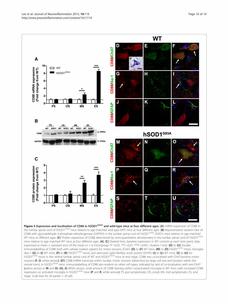
The mRNA expression levels for CD88 in the lumbarspinal cord of hSOD1G93A mice were normalised and com-pared with WT mice during disease progression (30 to 175days) using quantitative real-time PCR. CD88 expressionwas increased by 2.3-fold at mid-symptomatic, and by 8.6-fold at end stage of disease respectively compared withWT mice (n = 6, *P <0.05 and ***P <0.001; Figure 5A). Theprotein expression level of CD88 in the lumbar spinalcords of hSOD1G93A and WT mice were measured usingwestern blotting. A 45 kDa CD88 immunoreactive bandwas observed in all stages of hSOD1G93A mice and theirrespective WT mice (Figure 5B, upper panel). Semi-quantitative densitometry analyses of these bands with re-spect to GAPDH loading controls (Figure 5B, lower panel)revealed increased CD88 protein in the lumbar spinal cord
Figure 4 Expression and localisation of C5 and C5a in hSOD1G93A and wild-type mice during disease progression. (A) mRNA expressionprofile of C5 in lumbar spinal cord of hSOD1G93A mice relative to wild-type (WT) mice. Dashed line, baseline expression in WT controls at eachtime point. (B) Protein expression of C5a in the lumbar spinal cord of hSOD1G93A mice has decreased by end stage (ES) when compared with WTmice. (A, B) Data expressed as mean ± standard error of the mean (n = 6 mice/group; *P <0.05, Student t test). (C) to (H) Double immunolabelling for C5(red) with cellular makers (green) for motor neurons (ChAT; C and D, arrows), astrocytes (glial fibrillary acidic protein (GFAP), E and F), and microglia(CD11b; G and H), in the ventral lumbar spinal cord region of hSOD1G93A mice (D, F and H) and WT mice (C, E and G) at end stage. Co-localisation of C5with these cellular markers is seen as a merge of green and red (for example, white arrows in C, D, and H). PS, pre-symptomatic; OS, onset; MS,mid-symptomatic; ES, end-stage. Scale bar for C to H = 20 μm.
Lee et al. Journal of Neuroinflammation 2013, 10:119 Page 9 of 14http://www.jneuroinflammation.com/content/10/1/119
of hSOD1G93A mice by 2.6-fold and 3.7-fold atmid-symptomatic and end stage respectively whencompared with WT mice (n = 6, *P <0.05 and ***P <0.001;Figure 5C).
CD88 is localised to motor neurons and activatedmicroglia with minimal localisation to astrocytesin hSOD1G93A miceNext, we aimed to determine the cellular localisation ofCD88 that has contributed to the increased expressionseen in hSOD1G93A mice. To achieve this, we performedimmunolabelling for CD88 on lumbar spinal cord sec-tions from hSOD1G93A mice and WT mice. These sec-tions were immunostained for CD88 and with specificcellular markers to identify motor neurons (anti-ChAT),microglia (anti-Iba-1) and astrocytes (anti-GFAP).In WT mice, CD88 staining was observed on lumbar
motor neurons (Figure 5D). CD88-stained cells werereadily identified as motor neurons due to their largesize, location and distinctive morphology [17]. This wasconfirmed by double-labelling with the motor neuronmarker ChAT (white arrows in Figure 5F). CD88 immu-nostaining was localised predominantly to the motorneurons’ soma (Figure 5D, E, F). To further support these
immunohistochemical findings, we confirmed that motorneurons expressed CD88 mRNA transcripts by in-situ hy-bridisation (Figure 5D′, inset). Next, we examined whetherCD88 immunostaining was present on surrounding micro-glia and astrocytes in sections of WT lumbar spinal cords.We observed minimal co-localisation of CD88 to Iba-1-positive microglia (white arrows in Figure 5G,I), but noneto GFAP-positive astrocytes (Figure 5J, K, L).Following this demonstration of CD88 immunolabelling
in WT lumbar spinal cord, we then investigated CD88cellular localisation in hSOD1G93A mice. CD88 was alsoexpressed on the few remaining motor neurons seen at theend-stage of disease (white arrow in Figure 5O). By con-trast to WT lumbar spinal cords, we observed prominentCD88 immunostaining on other cellular structures sur-rounding motor neurons in the lumbar spinal cords ofhSOD1G93A mice by the end stage of disease (for example,yellow arrows in Figure 5M, O).This additional CD88 immunoreactivity was investi-
gated in hSOD1G93A end-stage mice, using microgliamarker Iba-1 and astrocyte marker GFAP. By contrast toIba-1-positive microglia in WT mice, where these cellsexpressed little observable CD88 and appeared to benonactivated (that is, small size with slender processes;
Figure 5 Expression and localisation of CD88 in hSOD1G93A and wild-type mice at four different ages. (A) mRNA expression of CD88 inthe lumbar spinal cord of hSOD1G93A mice relative to age-matched wild-type (WT) mice at four different ages. (B) Representative western blot ofCD88 with glyceraldehyde-3-phosphate dehydrogenase (GAPDH) in the lumbar spinal cord of hSOD1G93A (SOD1) mice relative to age-matchedWT mice at different ages. (C) Protein expression of CD88 determined by semi-quantitative densitometry in the lumbar spinal cord of hSOD1G93A
mice relative to age-matched WT mice at four different ages. (A), (C) Dashed lines, baseline expression in WT controls at each time point; dataexpressed as mean ± standard error of the mean (n = 6 mice/group; *P <0.05, **P <0.01, ***P <0.001, Student t test). (D) to (U) Doubleimmunolabelling of CD88 (red) with cellular markers (green) for motor neurons (ChAT; (D) to (F) WT mice, (M) to (O) hSOD1G93A mice), microglia(Iba-1; (G) to (I) WT mice, (P) to (R) hSOD1G93A mice), and astrocytes (glial fibrillary acidic protein (GFAP); (J) to (L) WT mice, (S) to (U) forhSOD1G93A mice) in the ventral lumbar spinal cord of WT and hSOD1G93A mice at end stage. CD88 was co-localised with ChAT-positive motorneurons (F, O, white arrows). (D′) CD88 mRNA transcript within lumbar motor neurons (determine by large cell size and location within theventral horn). In hSOD1G93A mice, immunolabelling of CD88 also evident on other cell types, indicated by lack of co-localisation with anti-ChAT(yellow arrows in M and O). (G), (I) White arrows, small amount of CD88 staining within nonactivated microglia in WT mice, with increased CD88expression on activated microglia in hSOD1G93A mice (P and R, white arrows). PS, pre-symptomatic; OS, onset; MS, mid-symptomatic; ES, endstage. Scale bars for all panels = 20 μM.
Lee et al. Journal of Neuroinflammation 2013, 10:119 Page 10 of 14http://www.jneuroinflammation.com/content/10/1/119
Lee et al. Journal of Neuroinflammation 2013, 10:119 Page 11 of 14http://www.jneuroinflammation.com/content/10/1/119
[32]; Figure 5H, I), Iba-1-positive microglia in hSOD1G93A
mice demonstrated an activated morphology with in-creased expression of CD88 (white arrows in Figure 5P, R).As expected, astrocytes were seen to increase in num-
bers as disease progressed in hSOD1G93A mice [33]. Thiswas noted by increased GFAP immunolabelling in thelumbar spinal cord of hSOD1G93A mice at end stagecompared with WT mice (Figure 5K, L for WT, andFigure 5T, U for hSOD1G93A mice). Minimal CD88 co-localisation to GFAP-positive astrocytes was observed inWT mice (Figure 5J, K, L) and at the end stage of diseasein hSOD1G93A mice (Figure 5S, T, U).
DiscussionWhile the pathogenesis of ALS is still unclear, there ispersuasive evidence that complement factors are in-volved in promoting disease progression. Previous stud-ies have demonstrated that C1q and C3 mRNAexpression are significantly increased during ALS pro-gression in hSOD1G93A mice [4]. In addition,upregulation of CD88 has also been observed in numer-ous neurodegenerative diseases in rodents [34-37], in-cluding ALS [10,11], so it is plausible to propose thatthe complement system could be involved in the patho-physiology of ALS. The present study demonstrates thatcomponents of the classical and alternative complementpathways are upregulated during the course of diseaseprogression in hSOD1G93A mice, and that C5a receptorCD88 expression level is also increased. In addition, wefound a reduction in two major regulatory inhibitors ofcomplement activation as the disease worsened, which issuggestive of a progressive dysregulation of complementin this model. Furthermore, we show that C5, the pre-cursor of C5a, is expressed predominantly by motorneurons, suggesting that diseased motor neurons couldbe a major source of C5a generation during disease pro-gression. This local complement self-signalling in thecentral nervous system might therefore contribute tomotor neuron death in hSOD1G93A mice as shown pre-viously for cortical neurons [22]. Taken together, our re-sults indicate that motor neurons may generate C5aunder stress, and that this may promote self-damageunder disease conditions that exist in ALS.
Classical and alternate complement pathways areactivated in ALS progression in hSOD1G93A miceThe present study provided evidence for the activationof classical (C1qB and C4) and alternate (factor B) path-ways of the complement system in the lumbar spinalcord of hSOD1G93A mice during ALS disease progres-sion. This is consistent with numerous studies in mousemodels of ALS and human patients where increasedlevels of C1q, C3 and C4 have been found [4,38]. Further-more this study also extended upregulation of C1qB and
C3 mRNA expression in previous studies to protein levelsand localisation where C1q and C3b immunolabelling wasincreased in hSOD1G93A mice and expressed on motorneurons and microglia compared with WT. This may sug-gest that upregulation of these components could assist inthe removal of dying motor neurons via opsonisation, dur-ing disease progression in hSOD1G93A mice [39].It is also possible that cell fragments or protein aggre-
gates from dying motor neurons could lead to comple-ment activation in the degenerating spinal cord [28]. Inour study, complement activation was seen at disease on-set (P70) and was restricted to the areas of motor neurondeath in the spinal cord of hSOD1G93A mice. Other studieshave demonstrated that complement components C1qand C3b are located at the neuromuscular junction duringthe early stages of disease (P47) in hSOD1G93A mice [40].These findings are consistent with the idea that C1q andC3b may contribute to the cellular destruction of motornerve terminals in these mice [41-43]. Taken together,these findings and our own are consistent with the hypoth-esis that the early loss of motor neuron terminals isfollowed by the subsequent death of motor neurons withinthe spinal cord. However, there is current debate about theinitiating site of degeneration in the cortical-motor system(upper motor neurons [44]) versus peripheral (neuromus-cular junction [41]). Future studies could contrast comple-ment activation temporally at these different sites todetermine the initiating site of complement-mediatedneurodegeneration.We also showed decreased mRNA expression levels
of complement regulators CD55 and CD59a at laterstages of the disease, which suggests that the homeo-stasis of the complement system is perturbed, whichmay lead to dysregulation and overactivation of thecomplement system. This supports other studies,which have shown that deficiency in CD55 andCD59a exacerbates neuronal degeneration [45-47].Our findings are also consistent with those ofHeurich and colleagues [40], where decreased (butnonsignificant) levels of CD55 mRNA were observedduring the later stages of disease in hSOD1G93A mice.In the present study, we also confirmed the mRNAchanges in CD55 at the protein level, which similarlyshowed decreased CD55 levels at later stages of dis-ease. Interestingly, we also observed an initial in-crease in the mRNA expression levels of CD55 andCD59a during early stages of disease. This may indicatean early negative feedback mechanism to delay theactivation of complement in host cells, but this needsfurther investigation.Collectively, our data add further support to the no-
tion that complement activation may play an importantrole in accelerating motor neuron loss and ultimately inprogression of ALS.
Lee et al. Journal of Neuroinflammation 2013, 10:119 Page 12 of 14http://www.jneuroinflammation.com/content/10/1/119
C5 is expressed by wild-type and hSOD1G93A motorneurons during disease progressionC5, the precursor of C5a, is expressed by motor neuronsin both WT and hSOD1G93A mice. This suggests thatmotor neurons are a major source of C5 generation inthis tissue. We recently showed murine cortical neuronsalso expressed endogenous C5, and generated C5a in re-sponse to ischaemia, which contributed to neuronal celldeath [22]. It is plausible the same phenomenon is oc-curring in the hSOD1G93A mouse, where stressed anddying motor neurons generate their own C5a, to act inan autocrine fashion by binding to CD88, present onthese neurons, to promote their death. Indeed, C5a hasbeen suggested to directly cause neuronal cell death in aseparate model of ALS [10]. C5a protein level in thelumbar spinal cord only appeared to be significantly de-creased by end stage in hSOD1G93A mice when com-pared with WT mice. This could be due to fewer motorneurons (a source of C5/C5a), and the increased CD88receptor levels by surrounding activated microglia asdisease progressed. Increased CD88 on these cells wouldact to internalise and degrade C5a post activation of itsreceptor [48], which could account for the decline inC5a levels over time. The consequence of C5a–CD88signalling in WT motor neurons is not yet fully under-stood and will need further investigation.
C5a receptor CD88 is upregulated during diseaseprogression in hSOD1G93A miceThe present study provided evidence for a patho-physiological role of CD88 in hSOD1G93A mice. Specific-ally, a significant increase in CD88 protein was observedat mid-symptomatic and end stage in hSOD1G93A mice.This is in agreement with our previous studies conductedin hSOD1G93A rats [11]. The increase in CD88 pro-tein in hSOD1G93A mice also parallels observationsin other models of neurodegenerative diseases, such asHuntington’s disease and Alzheimer’s disease [35,36].Taken together, this suggests that complement activa-tion is a generalised response to neuronal injury inneurodegenerative diseases. Furthermore, the ability ofCD88 antagonists to attenuate both neurodegenerationand disease progression in rat models of ALS andHuntington’s disease, and in mouse models of Alzheimer’sdisease, further suggests that increased CD88 activation ac-tively contributes to neurodegeneration [11,37,49].In addition to an increase in CD88 in hSOD1G93A
mice, the present study observed CD88 on motor neu-rons in WT mice, as well as in hSOD1G93A mice. Thefact that CD88 was found on WT motor neurons sug-gests that it may play a non-inflammatory role in motorneuron function. Indeed, studies including our own,have shown CD88 is also present on other neuronswithin the brains of WT adult mice, including pyramidal
neurons in the CA subfields of the hippocampus andneocortex, and Purkinje cells in the cerebellum [50,51];and CD88 has also been documented to be expressed onhuman motor neurons [10]. Hence the physiological sig-nificance of C5a receptor presence in motor neuronsawaits further study.Previous studies have shown that motor neuron death in
animal models of ALS is exacerbated by toxic signals em-anating from non-neuronal neighbouring cells (astrocytesand microglia), via an inflammatory response that acceler-ates disease progression [52,53]. The present study alsoshowed upregulation of CD88 on activated microglia, butminimal expression on astrocytes in hSOD1G93A mice. Thelatter is in contrast to our previous study conducted inhSOD1G93A rats, where CD88 was expressed primarily onproliferating astrocytes [11], but is in support of otherstudies that show CD88 on microglia in other neurodegen-erative diseases [36,54] The differential expression of CD88on these proliferating glial cells between hSOD1G93A ratand mouse models may suggest a differential role forCD88 in these two species.The strong co-localisation of CD88 with activated
microglia in hSOD1G93A mice suggests that CD88-activated microglia contribute to the propagation of dis-ease as opposed to the aetiology of the disease. This issupported by previous studies where transplanted WTmicroglia produced no delay of disease onset but sur-vival was greatly extended through slowing of diseaseprogression in hSOD1G93A mice [55]. The exact mech-anism by which C5a–CD88 signalling in microglia playsa role in neurodegeneration is still unknown, but mayinvolve the release of reactive oxygen species throughNADPH oxidase, or proinflammatory cytokines, whichhave been shown to be upregulated in ALS [56].
ConclusionsIn summary, the present study has demonstrated theupregulation of classical and alternative pathway com-plement components, together with decreased levelsof complement regulators, suggesting that comple-ment activation and/or its dysregulation may play animportant role in motor neuron loss and ultimately inprogression of ALS. Expression of C5a receptor CD88was upregulated in hSOD1G93A mice, and the in-creased expression of CD88 in end-stage hSOD1G93A
mice appears to be due to increased microglial CD88expression. Taken together, these results indicate thatCD88 may play an important role in the pathophysiologyof ALS. These results pave the way for preliminarypharmacological experiments using specific downstreamcomplement inhibitors in hSOD1G93A mice, such as C5inhibitors or antagonists to C5a receptor that could con-ceivably be extended to humans with positive therapeuticoutcomes.
Lee et al. Journal of Neuroinflammation 2013, 10:119 Page 13 of 14http://www.jneuroinflammation.com/content/10/1/119
Additional file
Additional file 1: Figure S1. Showing the decline in motor performanceduring ALS progression correlates with lumbar motor neuron loss in thelumbar spinal cord of hSOD1G93A mice. (A) Significant weight loss ofhSOD1G93A mice when compared with wild-type (WT) control mice at 140days of age (arrow, n = 12, *P <0.05, #P <0.001, Student t test). (B), (C)Significant reduction in time spent on Rota-rod and hind-limb grip strengthfor hSOD1G93A versus WT mice, at 119 days and 70 days respectively (arrows,n = 12, *P <0.05, +P <0.01, #P <0.001, Student t test). (D) Lumbar motorneuron loss in hSOD1G93A mice when compared with WT control mice at 70days of age onwards (n = 6, ***P <0.001, Student t test). The decline in motorneuron number at 70 days correlates with the onset of loss of hind limbmuscle strength at this same age (C). Data expressed as mean ± standarderror of the mean. PS, pre-symptomatic (30 days postnatal (P30)); OS, onset(70 days postnatal (P70)); MS, mid-symptomatic (130 days postnatal (P130));ES, end-stage (175 days postnatal (P175)).
Competing interestsThe authors declare that they have no competing interests.
Authors’ contributionsPGN, SMT and TMW conceived the project. JDL performed most of theexperiments. NAK and JNT performed additional experiments. JDA and BJTprovided additional tissues. JDL, PGN and TMW wrote the paper. All authorsread and made comment on the manuscript during its drafting. All authorsread and approved the final manuscript.
AcknowledgementsThe authors would like to thank Maryam Shayegh and Mary White for theirtechnical support and David Simmons for assistance in in situ hybridisation.JDL holds an APA scholarship from the Australian government. The workwas funded by project grant APP1004455 from NHMRC to PGN and TMW,and by a MNDRIA project grant to PGN, SMT and TMW. BJT is supported bygrants APP1008910 from NHMRC and MNDRIA, and JDA by grantsAPP1006141 and APP1030513from the NHMRC and grants from MNDRIA.
Author details1School of Biomedical Sciences, University of Queensland, Brisbane, St LuciaQLD 4072, Australia. 2Queensland Brain Institute, University of Queensland,Brisbane, St Lucia QLD 4072, Australia. 3Florey Institute of Neuroscience &Mental Health, University of Melbourne, Parkville VIC 3010, Australia.4Department of Biochemistry, La Trobe Institute for Molecular Science, LaTrobe University, Bundoora VIC 3083, Australia.
Received: 1 August 2013 Accepted: 6 September 2013Published: 26 September 2013
References1. Bruijn LI, Miller TM, Cleveland DW: Unraveling the mechanisms involved in
motor neuron degeneration in ALS. Annu Rev Neurosci 2004, 27:723–749.2. Cozzolino M, Ferri A, Carri MT: Amyotrophic lateral sclerosis: from current
developments in the laboratory to clinical implications. Antioxid RedoxSignal 2008, 10:405–443.
3. Woodruff TM, Costantini KJ, Taylor SM, Noakes PG: Role of complement inmotor neuron disease: animal models and therapeutic potential ofcomplement inhibitors. Adv Exp Med Biol 2008, 632:143–158.
4. Lee JD, Lee JY, Taylor SM, Noakes PG, Woodruff TM: Innate Immunity inALS. In Amyotrophic Lateral Sclerosis. Edited by Maurer MH. Croatia: InTech;2012:393–412.
5. Almer G, Guegan C, Teismann P, Naini A, Rosoklija G, Hays AP, Chen C,Przedborski S: Increased expression of the pro-inflammatory enzyme
cyclooxygenase-2 in amyotrophic lateral sclerosis. Ann Neurol 2001,49:176–185.
6. Almer G, Teismann P, Stevic Z, Halaschek-Wiener J, Deecke L, Kostic V,Przedborski S: Increased levels of the pro-inflammatory prostaglandinPGE2 in CSF from ALS patients. Neurology 2002, 58:1277–1279.
9. Zhao W, Beers DR, Liao B, Henkel JS, Appel SH: Regulatory T lymphocytesfrom ALS mice suppress microglia and effector T lymphocytes throughdifferent cytokine-mediated mechanisms. Neurobiol Dis 2012, 48:418–428.
10. Humayun S, Gohar M, Volkening K, Moisse K, Leystra-Lantz C, Mepham J,McLean J, Strong MJ: The complement factor C5a receptor is upregulatedin NFL−/− mouse motor neurons. J Neuroimmunol 2009, 210:52–62.
11. Woodruff TM, Costantini KJ, Crane JW, Atkin JD, Monk PN, Taylor SM,Noakes PG: The complement factor C5a contributes to pathology in a ratmodel of amyotrophic lateral sclerosis. J Immunol 2008, 181:8727–8734.
12. Drummond M, Sorenson C: Nasty or nice? A perspective on the use ofhealth technology assessment in the United Kingdom. Value Health 2009,12(Suppl 2):S8–S13.
13. Zhou C, Zhao CP, Zhang C, Wu GY, Xiong F: A method comparison inmonitoring disease progression of G93A mouse model of ALS.Amyotroph Lateral Scler 2007, 8:366–372.
14. Fogarty MJ, Smallcombe KL, Yanagawa Y, Obata K, Bellingham MC, Noakes PG:Genetic deficiency of GABA differentially regulates respiratory and non-respiratory motor neuron development. PLoS One 2013, 8:e56257.
16. Banks GB, Chau TN, Bartlett SE, Noakes PG: Promotion of motoneuronsurvival and branching in rapsyn-deficient mice. J Comp Neurol 2001,429:156–165.
17. Banks GB, Kanjhan R, Wiese S, Kneussel M, Wong LM, O'Sullivan G, Sendtner M,Bellingham MC, Betz H, Noakes PG: Glycinergic and GABAergic synapticactivity differentially regulate motoneuron survival and skeletal muscleinnervation. J Neurosci 2005, 25:1249–1259.
18. Clarke PG, Oppenheim RW: Neuron death in vertebrate development:in vitro methods. Methods Cell Biol 1995, 46:277–321.
19. Livak KJ, Schmittgen TD: Analysis of relative gene expression data usingreal-time quantitative PCR and the 2(−Delta Delta C(T)) method.Methods 2001, 25:402–408.
20. Abramoff MD, Magalhaes PJ, Ram SJ: Image processing with ImageJ.Biophoton Int 2004, 11:36–42.
21. Simmons DG, Rawn S, Davies A, Hughes M, Cross JC: Spatial and temporalexpression of the 23 murine prolactin/placental lactogen-related genesis not associated with their position in the locus. BMC Genomics 2008,9:352.
22. Pavlovski D, Thundyil J, Monk PN, Wetsel RA, Taylor SM, Woodruff TM:Generation of complement component C5a by ischemic neuronspromotes neuronal apoptosis. Faseb J 2012, 26:3680–3690.
23. Wu MC, Brennan FH, Lynch JP, Mantovani S, Phipps S, Wetsel RA,Ruitenberg MJ, Taylor SM, Woodruff TM: The receptor for complementcomponent C3a mediates protection from intestinal ischemia-reperfusion injuries by inhibiting neutrophil mobilization. Proc Natl AcadSci U S A 2013, 110:9439–9444.
24. Mead RJ, Bennett EJ, Kennerley AJ, Sharp P, Sunyach C, Kasher P, Berwick J,Pettmann B, Battaglia G, Azzouz M, Grierson A, Shaw PJ: Optimised andrapid pre-clinical screening in the SOD1(G93A) transgenic mouse modelof amyotrophic lateral sclerosis (ALS). PLoS One 2011, 6:e23244.
25. Lepore AC, Haenggeli C, Gasmi M, Bishop KM, Bartus RT, Maragakis NJ,Rothstein JD: Intraparenchymal spinal cord delivery of adeno-associatedvirus IGF-1 is protective in the SOD1G93A model of ALS. Brain Res 2007,1185:256–265.
26. Schafer S, Hermans E: Reassessment of motor-behavioural test analysesenables the detection of early disease-onset in a transgenic mousemodel of amyotrophic lateral sclerosis. Behav Brain Res 2011, 225:7–14.
27. Weydt P, Hong SY, Kliot M, Moller T: Assessing disease onset andprogression in the SOD1 mouse model of ALS. Neuroreport 2003,14:1051–1054.
Lee et al. Journal of Neuroinflammation 2013, 10:119 Page 14 of 14http://www.jneuroinflammation.com/content/10/1/119
28. Woodruff TM, Ager RR, Tenner AJ, Noakes PG, Taylor SM: The role of thecomplement system and the activation fragment C5a in the centralnervous system. Neuromolecular Med 2010, 12:179–192.
29. Spendlove I, Ramage JM, Bradley R, Harris C, Durrant LG: Complementdecay accelerating factor (DAF)/CD55 in cancer. Cancer ImmunolImmunother 2006, 55:987–995.
30. Kimberley FC, Sivasankar B, Paul Morgan B: Alternative roles for CD59.Mol Immunol 2007, 44:73–81.
32. Raivich G: Like cops on the beat: the active role of resting microglia.Trends Neurosci 2005, 28:571–573.
33. Barbeito LH, Pehar M, Cassina P, Vargas MR, Peluffo H, Viera L, Estevez AG,Beckman JS: A role for astrocytes in motor neuron loss in amyotrophiclateral sclerosis. Brain Res Brain Res Rev 2004, 47:263–274.
34. Yasojima K, Schwab C, McGeer EG, McGeer PL: Up-regulated productionand activation of the complement system in Alzheimer's disease brain.Am J Pathol 1999, 154:927–936.
35. Singhrao SK, Neal JW, Morgan BP, Gasque P: Increased complementbiosynthesis by microglia and complement activation on neurons inHuntington's disease. Exp Neurol 1999, 159:362–376.
36. Ager RR, Fonseca MI, Chu SH, Sanderson SD, Taylor SM, Woodruff TM,Tenner AJ: Microglial C5aR (CD88) expression correlates with amyloid-beta deposition in murine models of Alzheimer's disease. J Neurochem2010, 113:389–401.
37. Woodruff TM, Crane JW, Proctor LM, Buller KM, Shek AB, de Vos K, Pollitt S,Williams HM, Shiels IA, Monk PN, Taylor SM: Therapeutic activity of C5areceptor antagonists in a rat model of neurodegeneration. FASEB J 2006,20:1407–1417.
38. Chiu IM, Phatnani H, Kuligowski M, Tapia JC, Carrasco MA, Zhang M,Maniatis T, Carroll MC: Activation of innate and humoral immunity in theperipheral nervous system of ALS transgenic mice. Proc Natl Acad Sci U S A2009, 106:20960–20965.
39. Stevens B, Allen NJ, Vazquez LE, Howell GR, Christopherson KS, Nouri N,Micheva KD, Mehalow AK, Huberman AD, Stafford B, Sher A, Litke AM,Lambris JD, Smith SJ, John SW, Barres BA: The classical complementcascade mediates CNS synapse elimination. Cell 2007, 131:1164–1178.
40. Heurich B, El Idrissi NB, Donev RM, Petri S, Claus P, Neal J, Morgan BP,Ramaglia V: Complement upregulation and activation on motor neuronsand neuromuscular junction in the SOD1 G93A mouse model of familialamyotrophic lateral sclerosis. J Neuroimmunol 2011, 235:104–109.
42. Cappello V, Vezzoli E, Righi M, Fossati M, Mariotti R, Crespi A, Patruno M,Bentivoglio M, Pietrini G, Francolini M: Analysis of neuromuscular junctionsand effects of anabolic steroid administration in the SOD1G93A mousemodel of ALS. Mol Cell Neurosci 2012, 51:12–21.
44. Vucic S, Nicholson GA, Kiernan MC: Cortical hyperexcitability mayprecede the onset of familial amyotrophic lateral sclerosis. Brain 2008,131:1540–1550.
45. Wang Y, Li Y, Dalle Lucca SL, Simovic M, Tsokos GC, Dalle Lucca JJ: Decayaccelerating factor (CD55) protects neuronal cells from chemicalhypoxia-induced injury. J Neuroinflammation 2010, 7:24.
46. Britschgi M, Takeda-Uchimura Y, Rockenstein E, Johns H, Masliah E, Wyss-Coray T: Deficiency of terminal complement pathway inhibitor promotesneuronal tau pathology and degeneration in mice. J Neuroinflammation2012, 9:220.
47. Stahel PF, Flierl MA, Morgan BP, Persigehl I, Stoll C, Conrad C, Touban BM,Smith WR, Beauchamp K, Schmidt OI, Ertel W, Leinhase I: Absence ofthe complement regulatory molecule CD59a leads to exacerbatedneuropathology after traumatic brain injury in mice. J Neuroinflammation2009, 6:2.
48. Oppermann M, Gotze O: Plasma clearance of the human C5a anaphylatoxinby binding to leucocyte C5a receptors. Immunology 1994, 82:516–521.
49. Fonseca MI, Ager RR, Chu SH, Yazan O, Sanderson SD, LaFerla FM, Taylor SM,Woodruff TM, Tenner AJ: Treatment with a C5aR antagonist decreases
pathology and enhances behavioral performance in murine models ofAlzheimer's disease. J Immunol 2009, 183:1375–1383.
50. O'Barr SA, Caguioa J, Gruol D, Perkins G, Ember JA, Hugli T, Cooper NR:Neuronal expression of a functional receptor for the C5a complementactivation fragment. J Immunol 2001, 166:4154–4162.
51. Crane JW, Baiquni GP, Sullivan RK, Lee JD, Sah P, Taylor SM, Noakes PG,Woodruff TM: The C5a anaphylatoxin receptor CD88 is expressed inpresynaptic terminals of hippocampal mossy fibres. J Neuroinflammation2009, 6:34.
52. Kreutzberg GW: Microglia: a sensor for pathological events in the CNS.Trends Neurosci 1996, 19:312–318.
53. Nagai M, Re DB, Nagata T, Chalazonitis A, Jessell TM, Wichterle H,Przedborski S: Astrocytes expressing ALS-linked mutated SOD1 releasefactors selectively toxic to motor neurons. Nat Neurosci 2007, 10:615–622.
54. Griffin RS, Costigan M, Brenner GJ, Ma CH, Scholz J, Moss A, Allchorne AJ,Stahl GL, Woolf CJ: Complement induction in spinal cord microglia resultsin anaphylatoxin C5a-mediated pain hypersensitivity. J Neurosci 2007,27:8699–8708.
55. Beers DR, Henkel JS, Xiao Q, Zhao W, Wang J, Yen AA, Siklos L, McKercher SR,Appel SH: Wild-type microglia extend survival in PU.1 knockout mice withfamilial amyotrophic lateral sclerosis. Proc Natl Acad Sci U S A 2006,103:16021–16026.
56. Wu DC, Re DB, Nagai M, Ischiropoulos H, Przedborski S: The inflammatoryNADPH oxidase enzyme modulates motor neuron degeneration inamyotrophic lateral sclerosis mice. Proc Natl Acad Sci U S A 2006,103:12132–12137.
doi:10.1186/1742-2094-10-119Cite this article as: Lee et al.: Dysregulation of the complement cascadein the hSOD1G93A transgenic mouse model of amyotrophic lateralsclerosis. Journal of Neuroinflammation 2013 10:119.
Submit your next manuscript to BioMed Centraland take full advantage of:
• Convenient online submission
• Thorough peer review
• No space constraints or color figure charges
• Immediate publication on acceptance
• Inclusion in PubMed, CAS, Scopus and Google Scholar
• Research which is freely available for redistribution
Submit your manuscript at www.biomedcentral.com/submit