14


Back
7
Membrane Potential
John Koester
Steven A. Siegelbaum
INFORMATION IS CARRIED WITHIN and between neurons by electrical and chemical signals. Transient electrical signals are particularly important for carrying
time-sensitive information rapidly and over long distances. These electrical signals—receptor potentials, synaptic potentials, and action potentials—are all
produced by temporary changes in the current flow into and out of the cell that drive the electrical potential across the cell membrane away from its resting value.
This current flow is controlled by ion channels in the cell membrane. We can distinguish two types of ion channels—resting and gated—by their distinctive roles in
neuronal signaling. Resting channels normally are open and are not influenced significantly by extrinsic factors, such as the potential across the membrane. They
are primarily important in maintaining the resting membrane potential, the electrical potential across the membrane in the absence of signaling. Most gated
channels, in contrast, are closed when the membrane is at rest. Their probability of opening is regulated by the three factors we considered in the last chapter:
changes in membrane potential, ligand binding, or membrane stretch.
In this and succeeding chapters we consider how transient electrical signals are generated in the neuron. We begin by discussing how resting ion channels
establish and maintain the resting potential. We also briefly describe the mechanism by which the resting potential can be perturbed, giving rise to transient
electrical signals such as the action potential. In Chapter 8 we shall consider how the passive properties of neurons—their resistive and capacitive
characteristics—contribute to local signaling within the neuron. In Chapter 9 we shall examine how voltage-gated Na+, K+, and Ca2+ channels generate the action
potential, the electrical signal conveyed along the axon. Synaptic and receptor potentials are considered in Chapters 10,11,12,13 in the context of synaptic
signaling between neurons.
P.126
The Resting Membrane Potential Results From the Separation of Charges Across the Cell
Membrane
Every neuron has a separation of charges across its cell membrane consisting of a thin cloud of positive and negative ions spread over the inner and outer
surfaces of the cell membrane (Figure 7-1). At rest a nerve cell has an excess of positive charges on the outside of the membrane and an excess of negative
charges on the inside. This separation of charge is maintained because the lipid bilayer of the membrane blocks the diffusion of ions, as explained in Chapter 6.
The charge separation gives rise to a difference of electrical potential, or voltage, across the membrane called the membrane potential. The membrane potential
(Vm) is defined as
where Vin is the potential on the inside of the cell and Vout the potential on the outside.
The membrane potential of a cell at rest is called the resting membrane potential. Since, by convention, the potential outside the cell is defined as zero, the
resting potential (Vr) is equal to Vin. Its usual range in neurons is -60 mV to -70 mV. All electrical signaling involves brief changes from the resting membrane
potential due to alterations in the flow of electrical current across the cell membrane resulting from the opening and closing of ion channels.
The electric current that flows into and out of the cell is carried by ions, both positively charged (cations) and negatively charged (anions). The direction of
current flow is conventionally defined as the direction of net movement of positive charge. Thus, in an ionic solution cations move in the direction of the electric
current, anions in the opposite direction. Whenever there is a net flow of cations or anions into or out of the cell, the charge separation across the resting
membrane is disturbed, altering the polarization of the membrane. A reduction of charge separation, leading to a less negative membrane potential, is called
depolarization. An increase in charge separation, leading to a more negative membrane potential, is called hyperpolarization. Changes in membrane potential
that do not lead to the opening of gated ion channels, are called electrotonic potentials and are said to be passive responsives of the membrane. Hyperpolarizing
responses are almost always passive, as are small depolarizations. However, when depolarization approaches a critical level, called the threshold, the cell
responds actively with the opening of voltage-gated ion channels, which at threshold produces an all-or-none action potential (Box 7-1).
Figure 7-1 The membrane potential results from a separation of positive and negative charges across the cell membrane. The excess of positive
charges (red circles) outside the membrane and negative charges (blue circles) inside the membrane of a nerve cell at rest represents a small fraction of the
total number of ions inside and outside cell.
We begin examining the membrane potential by analyzing how the passive flux of individual ion species through resting channels generates the resting potential.
We shall then be able to understand how the selective gating of different types of ion channels generates the action potential, as well as the receptor and
synaptic potentials.

The Resting Membrane Potential Is Determined by Resting Ion Channels
No single ion species is distributed equally on the two sides of a nerve cell membrane. Of the four most abundant ions found on either side of the cell membrane,
Na+ and Cl- are more concentrated outside the cell, and K+ and organic anions (A-) are more concentrated inside. The organic anions are primarily amino acids
and proteins. Table 7-1 shows the distribution of these ions inside and outside one particularly well-studied nerve cell process, the giant axon of the squid, whose
blood has a salt concentration similar to sea water. Although the absolute values of the ionic concentrations for vertebrate nerve cells are two- to threefold lower
than those for the squid giant axon, the concentration gradients (the ratio of the external ion concentration to internal ion concentration) are about the same.
The unequal distribution of ions raises several important questions. How do ionic gradients contribute to the resting membrane potential? How are they
maintained? What prevents the ionic gradients from dissipating by diffusion of ions across the membrane through
P.127
P.128
the passive (resting) channels? These questions are interrelated, and we shall answer them by considering two examples of membrane permeability: the resting
membrane of glial cells, which is permeable to only one species of ions, and the resting membrane of nerve cells, which is permeable to three. For the purposes
of this discussion we shall consider only the resting ion channels, which are always open.
Box 7-1 Recording the Membrane Potential
Reliable techniques for recording the electrical potential across cell membranes were developed in the late 1940s. These techniques allow accurate
recordings of both the resting and the action potentials and make use of glass micropipettes filled with a concentrated salt solution that serve as electrodes.
These microelectrodes are placed on either side of the cell membrane. Wires inserted into the back ends of the pipettes are connected via an amplifier to an
oscilloscope, which displays the amplitude of the membrane potential in volts. Because the tip diameter of a microelectrode is very small (<1 µM), it can be
inserted into a cell with relatively little damage to the cell membrane.
Figure 7-2A The recording setup.
When both electrodes are outside the cell no electrical potential difference is recorded. But as soon as one microelectrode is inserted into the cell the
oscilloscope shows a steady voltage, the resting membrane potential. In most nerve cells at rest the membrane potential is around -65 mV.
Figure 7-2B Oscilloscope display.
The membrane potential can be experimentally changed using a current generator connected to a second pair of electrodes—one intracellular and one
extracellular. When the intracellular electrode is made positive with respect to the extracellular one, a pulse of positive current from the generator will cause
current to flow into the neuron from the intracellular electrode. This current returns to the extracellular electrode by flowing outward across the membrane.
As a result, the inside of the membrane becomes more positive while the outside of the membrane becomes more negative. This progressive decrease in the
normal separation of charge is called depolarization.

Figure 7-2C Depolarization.
Small depolarizing current pulses evoke purely electrotonic (passive) potentials in the cell—the size of the change in potential is proportional to the size of
the current pulses. However, sufficiently large depolarizing current triggers the opening of voltage-gated ion channels. The opening of these channels leads
to the action potential, which differs from electrotonic potentials not only in the way in which it is generated but also in magnitude and duration.
Reversing the direction of current flow—making the intracellular electrode negative with respect to the extracellular electrode—makes the membrane
potential more negative. This increase in charge separation is called hyperpolarization.
Figure 7-2D Hyperpolarization.
The responses of the cell to hyperpolarization are usually purely electrotonic—as the size of the current pulse increases, the hyperpolarization increases
proportionately. Hyperpolarization does not trigger an active response in the cell.
Table 7-1 Distribution of the Major Ions Across a Neuronal Membrane at Rest: the Giant Axon of the Squid
Species of ion Concentration in cytoplasm (mM) Concentration in extracellular fluid (mM) Equilibrium potential1 (mV)
K+ 400 20-75
Na+ 50 440+55
Cl- 52 560-60
A- (organic anions) 385 ——
1 The membrane potential at which there is no net flux of the ion species across the cell membrane.
Resting Channels in Glial Cells Are Selective for Potassium Only
A membrane's overall selectivity for individual ion species is determined by the relative proportions of the various types of ion channels in the cell that are open.
The simplest case is that of the glial cell, which has a resting potential of about -75 mV. Here, the vast majority of resting channels in the membrane are
permeable only to K+. As a result, the glial cell membrane at rest is almost exclusively permeable to K+ ions. A glial cell has a high concentration of K+ and
negatively charged organic anions on the inside and a high concentration of Na+ and Cl- on the outside.
How do these ionic gradients generate the membrane potential of the glial cell? Because K+ ions are present at a high concentration inside the cell and glial cells
are selectively permeable to them, K+ ions tend to diffuse from inside to outside the cell, down their chemical concentration gradient. As a result, the outside of
the membrane accumulates a positive charge (due to the slight excess of K+) and the inside a negative charge (because of the deficit of K+ and the resulting
slight excess of anions). Since opposite charges attract each other, the excess positive charges on the outside and the excess negative charges on the inside
collect locally on either surface of the membrane (see Figure 7-1).
The diffusion of K+ out of the cell is self-limiting. The separation of charge resulting from the diffusion of K+ gives rise to an electrical potential difference:

positive outside, negative inside. The more K+ continues to flow, the more charge will be separated and the greater will be the potential difference. Since K+ is
positively charged, this potential difference tends to oppose the further efflux of K+. Thus, ions are subject to two forces driving them across the membrane: (1)
a chemical driving force that depends on the concentration gradient across the membrane and (2) an electrical driving force that depends on the electrical
potential difference across the membrane. Once K+ diffusion has proceeded to a certain point, a potential develops across the membrane at which the electrical
force driving K+ into the cell exactly balances the chemical force driving K+ ions out of the cell. That is, the outward movement of K+ (driven by its concentration
gradient) is equal to the inward movement of K+ (driven by the electrical potential difference across the membrane). This potential is called the potassium
equilibrium potential, EK (Figure 7-3). In a cell permeable only to K+ ions, EK determines the resting membrane potential, which in most glial cells is about -75
mV.
The equilibrium potential for any ion X can be calculated from an equation derived in 1888 from basic thermodynamic principles by the German physical chemist
Walter Nernst:
where R is the gas constant, T the temperature (in degrees Kelvin), z the valence of the ion, F the Faraday constant, and [X]o and [X]i are the concentrations of
the ion outside and inside of the cell. (To be precise, chemical activities should be used rather than concentrations.)
Since RT/F is 25 mV at 25°C (room temperature), and the constant for converting from natural logarithms
P.129
to base 10 logarithms is 2.3, the Nernst equation can also be written as:
Figure 7-3 The flux of K+ across the membrane is determined by both the K+ concentration gradient and the electrical potential across the
membrane.
A. In a cell permeable only to K+ the resting potential is generated by the efflux of K+ down its concentration gradient.
B. The continued efflux of K+ builds up an excess of positive charge on the outside of the cell and leaves behind on the inside an excess of negative charge. This
buildup of charge leads to a potential difference across the membrane that impedes the further efflux of K+, so that eventually an equilibrium is reached: the
electrical and chemical driving forces are equal and opposite, and as many K+ ions move in as move out.
Thus, for K+, since z = +1 and given the concentrations inside and outside the squid axon in Table 7-1:
The Nernst equation can be used to find the equilibrium potential of any ion that is present on both sides of a membrane permeable to that ion (the potential is
sometimes called the Nernst potential). The Na+, K+, and Cl- equilibrium potentials for the distributions of ions across the squid axon are given in Table 7-1.
In our discussion so far we have treated the generation of the resting potential by the diffusion of ions down their chemical gradients as a passive mechanism,
one that does not require the expenditure of energy by the cell, for example through hydrolysis of ATP. However, as we shall see below, energy (and ATP
hydrolysis) is required to set up the initial concentration gradients and to maintain them during the activity of a neuron.
Resting Channels in Nerve Cells Are Selective for Several Ion Species
Measurements of the resting membrane potential with intracellular electrodes and flux studies using radioactive tracers show that, unlike glial cells, nerve cells at
rest are permeable to Na+ and Cl- ions in addition to K+ ions. Of the abundant ion species in nerve cells only the large organic anions (A-)—negatively charged
proteins and amino acids—are unable to permeate the cell membrane. How can the concentration gradients for the three permeant ions (Na+, K+, and Cl-) be
maintained across the membrane of a single cell, and how do these three gradients interact to determine the cell's resting membrane potential?
To answer these questions, it will be easiest to examine first only the diffusion of K+ and Na+. Let us return to the simple example of a cell having only K+
channels, with concentration gradients for K+, Na+, Cl-, and A- as shown in Table 7-1. Under these conditions the resting membrane potential, Vr, is determined
solely by the K+ concentration gradient and will be equal to Ek (-75 mV) (Figure 7-4A).
Now consider what happens if a few resting Na+ channels are added to the membrane, making it slightly

P.130
P.131
permeable to Na+. Two forces act on Na+ to drive it into the cell. First, Na+ is more concentrated outside than inside and therefore it tends to flow into the cell
down its chemical concentration gradient. Second, Na+ is driven into the cell by the negative electrical potential difference across the membrane (Figure 7-4B).
The influx of positive charge (Na+) depolarizes the cell, but only slightly from the K+ equilibrium potential (-75 mV). The new membrane potential does not come
close to the Na+ equilibrium potential of +55 mV because there are many more resting K+ channels than Na+ channels in the membrane.

Figure 7-4 The resting potential of a cell is determined by the relative proportion of different types of ion channels that are open, together with
the value of their equilibrium (Nernst) potentials. In this simplified diagram the channels shown represent the entire complement of K+ or Na+ channels in
the cell membrane. The lengths of the arrows within the channels represent the relative amplitudes of the electrical (red) and chemical (blue) driving forces
acting on Na+ and K+. The lengths of the arrows on the right denote the net driving force on a particular ion (that is, the sum of the electrical and chemical
driving forces) and the relative sizes of the different net ion fluxes. Three hypothetical situations are illustrated.
A. In a resting cell in which only K+ permeant channels are present, K+ ions are in equilibrium and Vm = EK.
B. Adding a few Na+ channels to the resting membrane at a given time allows Na+ ions to diffuse into the cell, and this influx begins to depolarize the
membrane.
C. The resting potential settles at a new resting potential, which is the value of Vm where /Na = -/K. In this example the aggregate conductance of the K+
channels is much greater than that of the Na+ channels because the K+ channels are more numerous. As a result, a relatively small net driving force for K+ ions
drives a current equal and opposite to the Na+ current driven by the much larger net driving force for Na+ ions. This is a steady-state condition, in which neither
Na+ nor K+ is in equilibrium but the net flux of charge is null.
D. Illustration of membrane voltage changes during the hypothetical situations considered in A, B, and C.
As soon as the membrane potential begins to depolarize from the value of the K+ equilibrium potential, K+ flux is no longer in equilibrium across the membrane.
The reduction in the negative electrical force driving K+ into the cell means that there will be a net efflux of K+ out of the cell, tending to counteract the Na+
influx. The more the membrane potential is depolarized and moves away from the K+ equilibrium potential, the greater is the electrochemical force driving K+ out
of the cell and consequently the greater is the K+ efflux. Eventually, the membrane potential reaches a new resting potential at which the outward movement of K
+ just balances the inward movement of Na+ (Figure 7-4C). This balance point (usually -60 mV) is far from the Na+ equilibrium potential (+55 mV) and is only
slightly more positive than the equilibrium potential for K+ (-75 mV).
To understand how this balance point is determined, bear in mind that the magnitude of the flux of an ion across a cell membrane is the product of its
electrochemical driving force (the sum of the electrical driving force and the chemical driving force due to the concentration gradient) and the conductance of the
membrane to the ion:
A cell has relatively few resting Na+ channels so at rest the conductance to Na+ is quite low. Thus, despite the large chemical and electrical forces driving Na+
into the cell, the influx of Na+ is small. In contrast, since there are many resting K+ channels, the membrane conductance of K+ is relatively large. As a result,
the small net outward force acting on K+ at the resting membrane potential is enough to produce a K+ efflux equal to the Na+ influx.
Passive Flux of Sodium and Potassium Is Balanced by Active Pumping of the Ions
For a cell to have a steady resting membrane potential the charge separation across the membrane must be maintained constant over time. That is, the influx of
positive charge must be balanced by the efflux of positive charge. If these fluxes were not equal, the charge separation across the membrane, and thus the
membrane potential, would vary continually. As we have seen, the passive movement of K+ out of the cell through resting channels balances the passive
movement of Na+ into the cell. However, these steady ion leaks cannot be allowed to continue unopposed for any appreciable length of time because the Na+
and K+ gradients would eventually run down, reducing the resting membrane potential.
Dissipation of ionic gradients is prevented by the Na+-K+ pump, which moves Na+ and K+ against their net electrochemical gradients: it extrudes Na+ from the
cell while taking in K+. The pump therefore requires energy to run. The energy comes from the hydrolysis of ATP. Thus, at the resting membrane potential the
cell is not in equilibrium but rather in a steady state: there is a continuous passive influx of Na+ and efflux of K+ through resting channels that is exactly
counterbalanced by the Na+-K+ pump.
The Na+-K+ pump is a large membrane-spanning protein with catalytic binding sites for Na+, K+, and ATP. The sites for Na+ and ATP are located on its
intracellular surface and the sites for K+ on its extracellular surface. With each cycle the pump hydrolyzes one molecule of ATP. It then uses this energy to
extrude three Na+ ions and bring in two K+ ions. The unequal flux of Na+ and K+ ions causes the pump to generate a net outward ionic current. Thus, the pump
is said to be electrogenic. This pump-driven outward flux of positive charge tends to hyperpolarize the membrane to a somewhat more negative potential than
would be achieved by the simple passive-diffusion mechanisms discussed above.
Chloride Ions May Be Passively Distributed
So far we have ignored the contribution of chloride (Cl-) to the resting potential, even though many nerve cells have Cl- channels that are open in the resting
membrane. This simplification is valid for nerve cells that do not have a mechanism for active transport of Cl- against an electrochemical gradient. In these cells
the resting potential is ultimately determined by K+ and Na+ fluxes because the intracellular concentrations of K+and Na+ are fixed by active transport (the Na+-K
+ pump), whereas the Cl- concentration inside the cell is affected only by passive forces (electrical potential and concentration gradient). Therefore, the
movement of Cl- ions tends toward equilibrium across the membrane, so that ECl is equal to the resting potential, Vr, and there is no net Cl- flux at rest.
P.132
In many nerve cells the Cl- gradient is controlled by an integral membrane protein called a Cl- transporter. Like the Na+-K+ pump it catalyzes the movement of
ions across the membrane against an electrochemical gradient without forming a continuous pore. Unlike the Na+-K+ pump, the transport process does not
require the hydrolysis of ATP. Although no chemical bond energy is utilized in the transport process, the Cl- transporter can move Cl- against its electrochemical
gradient by utilizing the energy stored in a preexisting ionic concentration gradient for a different type of ion—a process known as secondary active transport. For
example, one type of Cl- transporter couples the outward movement of one Cl- ion to the outward movement of one K+ ion. Since the electrochemical gradient
for K+ is outward, the energetically favorable outward K+ flux is able to drive the energetically unfavorable outward Cl- flux. As a result, the outside-to-inside
ratio of Cl- is greater than would result from passive diffusion alone. The effect of increasing the Cl- gradient is to make the equilibrium potential for Cl- ions more
negative than the resting membrane potential overall. (Remember, the valence (z) of Cl- is -1.)
The Balance of Ion Fluxes That Gives Rise to the Resting Membrane Potential Is Abolished
During the Action Potential
In the nerve cell at rest the steady Na+ influx is balanced by a steady K+ efflux, so that the membrane potential is constant. This balance changes, however,
when the membrane is depolarized past the threshold for generating an action potential. Once the membrane potential reaches this threshold, voltage-gated Na+
channels open rapidly. The resultant increase in membrane permeability to Na+ causes the Na+ influx to exceed the K+ efflux, creating a net influx of positive

charge that causes further depolarization. The increase in depolarization causes still more voltage-gated Na+ channels to open, resulting in a greater influx of Na
+, which accelerates the depolarization even further.
This regenerative, positive feedback cycle develops explosively, driving the membrane potential toward the Na+ equilibrium potential of +55 mV:
However, the membrane potential never quite reaches that point because K+ efflux continues throughout the depolarization. A slight influx of Cl- into the cell also
counteracts the depolarizing tendency of the Na+ influx. Nevertheless, so many voltage-gated Na+ channels open during the rising phase of the action potential
that the cell's permeability to Na+ is much greater than to either Cl- or K+. Thus, at the peak of the action potential the membrane potential approaches the Na+
equilibrium potential, just as at rest (when permeability to K+ is predominant) the membrane potential tends to approach the K+ equilibrium potential.
The membrane potential would remain at this large positive value near the Na+ equilibrium potential indefinitely but for two processes that repolarize the
membrane, thus terminating the action potential. First, as the depolarization continues, the population of voltage-gated Na+ channels gradually closes by the
process of inactivation (see Chapters 6 and 9). Second, opening of the voltage-gated K+ channels causes the K+ efflux to gradually increase. The increase in K+
permeability is slower than the increase in Na+ permeability because of the slower rate of opening of the voltage-gated K+ channels. The delayed increase in K+
efflux combines with a decrease in Na+ influx to produce a net efflux of positive charge from the cell, which continues until the cell has repolarized to its resting
membrane potential.
The Contributions of Different Ions to the Resting Membrane Potential Can Be Quantified by the
Goldman Equation
Although Na+ and K+ fluxes set the value of the resting potential, Vm is not equal to either EK or ENa but lies between them. As a general rule, when Vm is
determined by two or more species of ions, the influence of each species is determined not only by the concentrations of the ion inside and outside the cell but
also by the ease with which the ion crosses the membrane. In terms of electrical current flow, the membrane's conductance (1/resistance) provides a convenient
measure of how readily the ion crosses the membrane. Another convenient measure is the permeability (P) of the membrane to that ion in units of velocity, cm/
s. This measure is similar to that of a diffusion constant, which measures the rate of solute movement in solution. The dependence of membrane potential on
ionic permeability and concentration is given quantitatively by the Goldman equation:
Goldman Equation
This equation applies only when Vm is not changing. It states that the greater the concentration of a particular ion species and the greater its membrane
permeability,
P.133
the greater its role in determining the membrane potential. In the limit, when permeability to one ion is exceptionally high, the Goldman equation reduces to the
Nernst equation for that ion. For example, if PK >>PCl or PNa, as in glial cells, the equation becomes
Alan Hodgkin and Bernard Katz used the Goldman equation to analyze changes in membrane potential. They first measured the variation in membrane potential
of a squid giant axon while systematically changing the extracellular concentrations of Na+, Cl-, and K+. They found that if Vm is measured shortly after the
extracellular concentration is changed (before the internal ionic concentrations are altered), [K+]o has a strong effect on the resting potential, [Cl-]o has a
moderate effect, and [Na+]o has little effect. The data for the membrane at rest could be fit accurately by the Goldman equation using the following permeability
ratios:
At the peak of the action potential, however, the variation of Vm with external ionic concentrations was fit best if a quite different set of permeability ratios were
assumed:1

Figure 7-5 Electrical properties of a single K+ channel.
A. A single K+ channel can be represented as a conductor or resistor (conductance, γ, is the inverse of resistance, r).
B. The current-voltage relation for a single K+ channel in the absence of a concentration gradient. The slope of the relation is equal to γK.
Figure 7-6 Chemical and electrical forces contribute to current flow.
A. A concentration gradient for K+ gives rise to an electromotive force, with a value equal to the K+ Nernst potential. This can be represented by a battery, EK.
In this circuit the battery is in series with a conductor, γK, representing the conductance of a channel that is selectively permeable to K+ ions.
B. The current-voltage relation for a K+ channel in the presence of both electrical and chemical driving forces. The potential at which the current is zero is equal
to the K+ Nernst potential.
For these values of permeabilities the Goldman equation approaches the Nernst equation for Na+:
Thus at the peak of the action potential, when the membrane is much more permeable to Na+ than to any other ion, Vm approaches ENa, the Nernst potential for
Na+.
P.134
However, the finite permeability of the membrane to K+ and Cl- results in K+ efflux and Cl- influx that oppose Na+ influx, thereby preventing Vm from quite
reaching ENa.

Figure 7-7 All of the passive K+ channels in a nerve cell membrane can be lumped into a single equivalent electrical structure comprising a
battery (EK) in series with a conductor (gK). The conductance is gK = NK × γK, where N K is the number of passive K+ channels and γ K is the conductance
of a single K+ channel.
The Functional Properties of the Neuron Can Be Represented in an Electrical Equivalent Circuit
The Goldman equation is limited because it cannot be used to determine how rapidly the membrane potential changes in response to a change in permeability.
Moreover, it is inconvenient for determining the magnitude of the individual Na+, K+, and Cl- currents. This information can be obtained with a simple
mathematical model derived from electrical circuits. Within this model, called an equivalent circuit, all of the important functional properties of the neuron are
represented by an electrical circuit consisting only of conductors or resistors (representing the ion channels), batteries (representing the concentration gradients
of relevant ions), and capacitors (the ability of the membrane to store charge). Equivalent circuits provide us with an intuitive understanding as well as a
quantitative description of how current flow due to the movement of ions generates signals in nerve cells. The first step in developing a circuit is to relate the
membrane's discrete physical properties to its electrical properties. (A review of elementary circuit theory in Appendix A may be helpful before proceeding to the
discussion that follows.)
Each Ion Channel Acts as a Conductor and Battery in Parallel
As described in Chapter 6, the lipid bilayer of the membrane is a poor conductor of ionic current because it is not permeable to ions. Even a large potential
difference will produce practically no current flow across a pure lipid bilayer. Consider the cell body of a typical spinal motor neuron, which has a membrane area
of about 10-4 cm2. If the membrane were composed solely of lipid bilayer, its electrical conductance would be only about 1 pS. In reality, however, the
membrane contains thousands of resting ion channels through which ions constantly diffuse, so that the actual conductance of the membrane at rest is about
40,000 pS or 40 × 10-9 S, ie, 40,000 times greater than it would be if no ion channels were present.
In an equivalent circuit each K+ channel can be represented as a resistor or conductor of ionic current with a single-channel conductance of γK (remember,
conductance = 1/resistance) (Figure 7-5). If there were no K+ concentration gradient, the current through the K+ channel would be given by Ohm's law: iK = γK
× Vm. Since there is normally a K+ concentration gradient, there will be a chemical force driving K+ across the membrane. In the equivalent circuit this chemical
force is represented by a battery, whose electromotive force is given by the Nernst potential for K+, EK (Figure 7-6). (A source of electrical potential is called an
electromotive force and an electromotive force generated by a difference in chemical potentials is called a battery.)
Figure 7-8 Each population of ion channels selective for Na+, K+, or Cl- can be represented by a battery in series with a conductor. Note the
directions of poles of batteries, indicating a negative electromotive force for K+ and Cl- and a positive one for Na+.
Figure 7-9 The passive current flow in a neuron can be modeled using an electrical equivalent circuit. The circuit includes elements representing the
ion-selective membrane channels and the short-circuit pathways provided by the cytoplasm and extracellular fluid.
P.135
In the absence of voltage across the membrane the normal K+ concentration gradient will cause an outward K+ current flow. According to our conventions for
electrical current flow an outward movement of positive charge corresponds to a positive electric current. From the Nernst equation, we also saw that when the
concentration gradient for a positively charged ion, such as K+, is directed outward (ie, there is a higher K+ concentration inside than outside the cell), the
equilibrium potential for that ion is negative. Thus, the K+ current that flows solely because of its concentration gradient is given by iK = -γK × EK (the negative
sign is required because a negative equilibrium potential produces a positive current).

Finally, for a real neuron that has both a membrane voltage and K+ concentration gradient, the net K+ current is given by the sum of the currents due to the
electrical and chemical driving forces:
The term Vm - EK is called the electrochemical driving force. It determines the direction of ionic current flow and (along with the conductance) the magnitude of
current flow. This equation is a modified form of Ohm's law that takes into account that ionic current flow through a membrane is determined not only by the
voltage across the membrane but also by the ionic concentration gradients.
So far we have used two terms to indicate the ability of ions to cross membranes: permeability and conductance. Although they are related, we should be careful
not to confuse them. The permeability of a membrane to an ion is an intrinsic property of the membrane that is a measure of the ease with which the ion passes
through the membrane (in units of cm/s). Permeability depends only on the types and numbers of ion channels present in the membrane. Conductance, on the
other hand, measures the ability of the membrane (or channel) to carry electrical current (in units of 1/ohms). Since current is carried by ions, the conductance
of a membrane will depend not only on the properties of the membrane but also on the concentration of ions in solution. A membrane can have a very high
permeability to K+ ions, but if there is no K+ in solution there can be no K+ current flow and so the conductance of the membrane will be zero. In practice,
permeability is used in the Goldman equation whereas conductance is used in electrical measurements and equivalent circuits.
A cell membrane has many resting K+ channels, all of which can be combined into a single equivalent circuit consisting of a conductor in series with a battery
(Figure 7-7). In this equivalent circuit the total conductance of all the K+ channels (gK), ie, the K+ conductance of the cell membrane in its resting state, is equal
to the number N of resting K+ channels multiplied by the conductance of an individual K+ channel (γK):
Since the battery in this equivalent circuit depends solely on the concentration gradient for K+ and is independent of the number of K+ channels, its value is the
equilibrium potential for K+, EK (Figure 7-7).
Figure 7-10 Under steady state conditions the passive Na- and K+ currents are balanced by active Na+ and K+ fluxes (I′Na and I′K) driven by the
Na+-K+ pump. The lipid bilayer endows the membrane with electrical capacitance (C m). Note I′Na is 50% greater than IK (and therefore INa is 50% greater
than IK) since the Na+-K- pump transports three Na+ ions out for every two K+ ions it transports into the cell.
Box 7-2 Using the Equivalent Circuit Model to Calculate Resting Membrane Potential
The equivalent circuit model of the resting membrane can be used to calculate the resting potential. To simplify the calculation we shall initially ignore Cl-
channels and begin with just two types of passive channels, K+ and Na+, as illustrated in Figure 7-11. Moreover, we ignore the electrogenic influence of the
Na+-K+ pump because it is small. Because we will consider only steady-state conditions, where Vm is not changing, we can also ignore membrane
capacitance. (Membrane capacitance and its delaying effect on changes in Vm are discussed in Chapter 8.) Because there are more passive channels for K+
than for Na+, the membrane conductance for current flow carried by K+ is much greater than that for Na+. In the equivalent circuit in Figure 7-11, gK is 20
times higher than gNa (10 × 10-6 S compared to 0.5 × 10⁻⁶ S). Given these values and the values of EK and ENa, the membrane potential, Vm, is calculated
as follows.
Since Vm is constant in the resting state, the net current must be zero, otherwise the separation of positive and negative charges across the membrane
would change, causing Vm to change. Therefore INa is equal and opposite to IK:
or
We can easily calculate INa and IK in two steps. First, we add up the separate potential differences across the Na+ and K+ branches of the circuit. Going from
the inside to the outside across the Na+ branch, the total potential difference is the sum of the potential differences across ENa and across gNa:*
Similarly, for the K+ conductance branch
Next, we rearrange and solve for I:
As these equations illustrate, the ionic current through each conductance branch is equal to the conductance of that branch multiplied by the net electrical
driving force. For example, the conductance for the K+ branch is proportional to the number of open K+ channels, and the driving force is equal to the
difference between Vm and EK. If Vm is more positive than EK (-75 mV), the driving force is positive (outward); if Vm is more negative than EK, the driving
force is negative (inward).
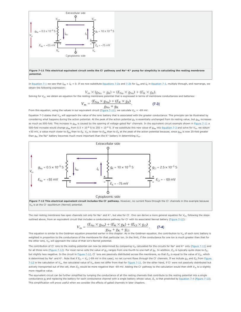
Figure 7-11 This electrical equivalent circuit omits the Cl- pathway and Na+-K+ pump for simplicity in calculating the resting membrane
potential.
In Equation 7-1 we saw that INa + IK = 0. If we now substitute Equations 7-2a and 7-2b for INa and IK in Equation 7-1, multiply through, and rearrange, we
obtain the following expression:
Solving for Vm, we obtain an equation for the resting membrane potential that is expressed in terms of membrane conductances and batteries:
From this equation, using the values in our equivalent circuit (Figure 7-11), we calculate Vm = -69 mV.
Equation 7-3 states that Vm will approach the value of the ionic battery that is associated with the greater conductance. This principle can be illustrated by
considering what happens during the action potential. At the peak of the action potential gK is essentially unchanged from its resting value, but gNa increases
as much as 500-fold. This increase in gNa is caused by the opening of voltage-gated Na+ channels. In the equivalent circuit example shown in Figure 7-11 a
500-fold increase would change gNa from 0.5 × 10⁻⁶ S to 250 × 10-6 S. If we substitute this new value of gNa into Equation 7-3 and solve for Vm, we obtain
+50 mV, a value much closer to ENa than to EK. Vm is closer to ENa than to EK at the peak of the action potential because, since gNa is now 25-fold greater
than gK, the Na+ battery becomes much more important than the K+ battery in determining Vm.
Figure 7-12 This electrical equivalent circuit includes the Cl- pathway. However, no current flows through the Cl- channels in this example because
Vm is at the Cl- equilibrium (Nernst) potential.
The real resting membrane has open channels not only for Na+ and K+, but also for Cl-. One can derive a more general equation for Vm, following the steps
outlined above, from an equivalent circuit that includes a conductance pathway for Cl- with its associated Nernst battery (Figure 7-12):
This equation is similar to the Goldman equation presented earlier in this chapter. As in the Goldman equation, the contribution to Vm of each ionic battery is
weighted in proportion to the conductance of the membrane for that particular ion. In the limit, if the conductance for one ion is much greater than that for
the other ions, Vm will approach the value of that ion's Nernst potential.
The contribution of Cl- ions to the resting potential can now be determined by comparing Vm calculated for the circuits for Na+ and K+ only (Figure 7-11) and
for all three ions (Figure 7-12). For most nerve cells the value of gCl ranges from one-fourth to one-half of gK. In addition, ECl is typically quite close to EK,
but slightly less negative. In the circuit in Figure 7-12, Cl- ions are passively distributed across the membrane, so that ECl is equal to the value of Vm, which
is determined by Na+ and K+. Note that if ECl = Vm (-69 mV in this case), no net current flows through the Cl- channels. If we include gCl and ECl from Figure
7-12 in the calculation of Vm, the calculated value of Vm does not differ from that for Figure 7-11. On the other hand, if Cl- were not passively distributed but
actively transported out of the cell, then ECl would be more negative than -69 mV. Adding the Cl- pathway to the calculation would then shift Vm to a slightly
more negative value.
The equivalent circuit can be further simplified by lumping the conductance of all the resting channels that contribute to the resting potential into a single
conductance gl and replacing the battery for each conductance channel with a single battery whose value, El, is that predicted by Equation 7-4 (Figure 7-13).
This simplification will prove useful when we consider the effects of gated channels in later chapters.

Figure 7-13 The complement of Na+, K+, and Cl- resting channels can be represented by a single equivalent conductance and battery. In this
simplified equivalent circuit the total resting membrane conductance gl = gCl + gNa + gK, and the electromotive force or battery (El) is the resting potential
predicted by Equation 7-4.
P.136
P.137
P.138
An Equivalent Circuit Model of the Membrane Includes Batteries, Conductors, a Capacitor, and a
Current Generator
Like the population of resting K+ channels, all the resting Na+ channels can be represented by a single conductor in series with a single battery, as can the
resting Cl- channels (Figure 7-8). Since the K+, Na+, and Cl- channels account for the bulk of the passive ionic current through the membrane in the cell at rest,
we can calculate the resting potential by incorporating these three channels into a simple equivalent circuit of a neuron.
To construct this circuit we need only connect the elements representing each type of channel at their two ends with elements representing the extracellular fluid
and cytoplasm. The extracellular fluid and cytoplasm are both excellent conductors because they have relatively large cross-sectional areas and many ions
available to carry charge. Both can be approximated by a short circuit— a conductor with zero resistance (Figure 7-9).
The equivalent circuit of the neuron can be made more accurate by adding a current generator. As described earlier in this chapter, steady fluxes of Na+ and K+
ions through the passive membrane channels are exactly counterbalanced by active ion fluxes driven by the Na+-K+ pump, which extrudes three Na+ ions from
the cell for every two K+ ions it pumps in. This electrogenic ATP-dependent pump, which keeps the ionic batteries charged, can be added to the equivalent circuit
in the form of a current generator (Figure 7-10).
Finally, we can complete the equivalent circuit of the neuron by incorporating its capacitance, the third important passive electrical property of the neuron.
Capacitance is the property of an electric nonconductor (insulator) that permits the storage of charge when opposite surfaces of the nonconductor are maintained
at a difference of potential. For the neuron, the nonconductor (or capacitor) is the cell membrane, which separates the cytoplasm and extracellular fluid, both of
which are highly conductive environments. Strictly speaking, the membrane is a leaky capacitor because it is penetrated by ion channels. However, since the
density of the ion channels is low, the insulating portion of the membrane—the lipid bilayer—occupies at least 100 times the area of all the ion channels
combined. Membrane capacitance is included in the equivalent circuit in Figure 7-10.
The electrical potential difference across a capacitor, V, is expressed as:
where Q is the excess of positive or negative charges on each side of the capacitor and C is the capacitance. Capacitance is measured in units of farads, F, where
a charge separation of 1 coulomb across a 1 farad capacitor produces a 1 volt potential difference.
A typical value of membrane capacitance for a nerve cell is about 1 µF/cm2 of membrane area. The excess of positive and negative charges separated by the
membrane of a spherical cell body with a diameter of 50 µm and a resting potential of -60 mV is 29 × 106 ions. Although this number may seem large, it
represents only a tiny fraction (1/200,000) of the total number of positive or negative charges in solution within the cytoplasm. The bulk of the cytoplasm and
the bulk of the extracellular fluid are electroneutral.
The use of the equivalent circuit model of the neuron to analyze neuronal properties quantitatively is illustrated in Box 7-2.
An Overall View
The lipid bilayer, which is virtually impermeant to ions, is an insulator separating two conducting solutions, the cytoplasm and the extracellular fluid. Ions can
cross the lipid bilayer only by passing through ion channels in the cell membrane. When the cell is at rest, the passive ionic fluxes into and out of the cell are
balanced, so that the charge separation across the membrane remains constant and the membrane potential remains at its resting value.
The value of the resting membrane potential in nerve cells is determined primarily by resting channels selective for K+, Cl-, and Na+. In general, the membrane
potential will be closest to the equilibrium (Nernst) potential of the ion (or ions) with the greatest membrane permeability. The permeability for an ion species is
proportional to the number of open channels that allow passage of that ion.
At rest, the membrane potential is close to the Nernst potential for K+, the ion to which the membrane is most permeable. The membrane is also somewhat
permeable to Na+, however, and therefore an influx of Na+ drives the membrane potential slightly positive to the K+ Nernst potential. At this potential the
electrical and chemical driving forces acting on K+ are no longer in balance, so K+ diffuses out of the cell. These two passive fluxes are each counterbalanced by
active fluxes driven by the Na+-K+ pump.
Chloride is actively pumped out of some, but not all, cells. When it is not, it is passively distributed so as to be at equilibrium inside and outside the cell. Under
most physiological conditions the bulk concentrations of Na+, K+, and Cl- inside and outside the cell are constant. During signaling the changes in membrane
potential
P.139

(action potentials, synaptic potentials, and receptor potentials) are caused by substantial changes in the membrane's relative permeabilities to these three ions,
not by changes in the bulk concentrations of ions, which are negligible. These changes in permeability, caused by the opening of gated ion channels, cause
changes in the net charge separation across the membrane.
Selected Readings
Finkelstein A, Mauro A. 1977. Physical principles and formalisms of electrical excitability. In: ER Kandel (ed). Handbook of Physiology: A Critical,
Comprehensive Presentation of Physiological Knowledge and Concepts, Sect. 1, The Nervous System. Vol. 1, Cellular Biology of Neurons, Part 1, pp. 161-
213. Bethesda, MD: American Physiological Society.
Hille B. 1992. Ionic Channels of Excitable Membranes, 2nd ed. Sunderland, MA: Sinauer.
Hodgkin AL. 1992. Chance and Design. Cambridge: Cambridge Univ. Press.
References
Bernstein J. [1902] 1979. Investigations on the thermodynamics of bioelectric currents. Pflügers Arch 92:521-562. Translated in: GR Kepner (ed). Cell
Membrane Permeability and Transport, pp. 184-210. Stroudsburg, PA: Dowden, Hutchinson & Ross.
Goldman DE. 1943. Potential, impedance, and rectification in membranes. J Gen Physiol 27:37–60.
Hodgkin AL, Katz B. 1949. The effect of sodium ions on the electrical activity of the giant axon of the squid. J Physiol (Lond) 108:37–77.
Nernst W. [1888] 1979. On the kinetics of substances in solution. Z Physik Chem. 2:613-622, 634-637. Translated in: GR Kepner (ed). Cell Membrane
Permeability and Transport, pp. 174-183. Stroudsburg, PA: Dowden, Hutchinson & Ross.
Orkand RK. 1977. Glial cells. In: ER Kandel (ed). Handbook of Physiology: A Critical, Comprehensive Presentation of Physiological Knowledge and Concepts,
Sect. 1, The Nervous System. Vol. 1, Cellular Biology of Neurons, Part 2, pp. 855-875. Bethesda, MD: American Physiological Society.
Siegel GJ, Agranoff BW, Albers RW (eds). 1999. Basic Neurochemistry: Molecular, Cellular, and Medical Aspects, 6th ed, Philadelphia: Lippincott-Raven.
1At the peak of the action potential threr is an instant in time when Vm is not changing and the Goldman equation is applicable.
*Because we have defined Vm as Vin - Vout, the following convention must be used for these equations. Outward current (in this case IK) is positive and inward
current is negative. Batteries with their positive poles toward the inside of the membrane (eg, ENa) are given positive values in the equations. The reverse is true
for batteries that have their negative poles toward the inside, such as the K+ battery.


![index [materials.usask.ca]materials.usask.ca/files/Index.pdfDeperming. See Demagnetization Depletion capacitance, 553, 637 Depletion region. See pn junction Depolarizing field, 737–738](https://static.documents.pub/doc/80x56/60ca86db1309a8697b49aa63/index-deperming-see-demagnetization-depletion-capacitance-553-637-depletion.jpg)
