Institute of Cancer Research Repository https://publications.icr.ac.uk Please direct all emails to: [email protected]This is an author produced version of an article that appears in: The internet address for this paper is: NATURE https://publications.icr.ac.uk/12228/ Published text: E Ruark, K Snape, et al (2013), Mosaic PPM1D mutations are associated with predisposition to breast and ovarian cancer, Nature, Vol. 493(7432), 406-410
Transcript
Institute of Cancer Research Repository https://publications.icr.ac.uk
This is an author produced version of an article that appears in:
The internet address for this paper is:
NATURE
https://publications.icr.ac.uk/12228/
Published text:
E Ruark, K Snape, et al (2013), Mosaic PPM1D mutations are associated with predisposition to breast and ovarian cancer, Nature, Vol. 493(7432), 406-410
1
Mosaic PPM1D mutations are associated with predisposition to breast and
ovarian cancer
Elise Ruark1*, Katie Snape1*, Peter Humburg2*, Chey Loveday1, Ilirjana Bajrami3, Rachel Brough3,4, Daniel Nava Rodrigues3, Anthony Renwick1, Sheila Seal1, Emma Ramsay1, Silvana Del Vecchio Duarte1, Manuel A. Rivas2,5, Margaret Warren-Perry1, Anna Zachariou1, Adriana Campion-Flora3, Sandra Hanks1, Anne Murray1, Naser Ansari Pour1, Jenny Douglas1, Lorna Gregory2, Andrew Rimmer2, Neil M. Walker6, Tsun-Po Yang7, Julian W. Adlard8, Julian Barwell9, Jonathan Berg10, Angela F. Brady11, Carole Brewer12, Glen Brice13, Cyril Chapman14, Jackie Cook15, Rosemarie Davidson16, Alan Donaldson17, Fiona Douglas18, Diana Eccles19, D. Gareth Evans20, Lynn Greenhalgh21, Alex Henderson18, Louise Izatt22, Ajith Kumar23, Fiona Lalloo24, Zosia Miedzybrodzka25, Patrick J. Morrison26, Joan Paterson27, Mary Porteous28, Mark T. Rogers29, Susan Shanley30, Lisa Walker31, Martin Gore32, Richard Houlston1, Matthew A. Brown33, Mark J. Caufield34, Panagiotis Deloukas7, Mark I. McCarthy2,35,36, John A. Todd6, The Breast and Ovarian Cancer Susceptibility Collaboration (BOCS)37, Wellcome Trust Case Control Consortium37, Clare Turnbull1,30, Jorge S. Reis-Filho3, Alan Ashworth3, Antonis C. Antoniou38, Christopher J. Lord3, Peter Donnelly2,39& Nazneen Rahman1,30
1Division of Genetics & Epidemiology, The Institute of Cancer Research, Sutton, SM2 5NG, UK 2The Wellcome Trust Centre for Human Genetics, University of Oxford, Oxford, OX3 7BN, UK 3The Breakthrough Breast Cancer Research Centre, The Institute of Cancer Research, London, SW3 6JB, UK 4Cancer Research UK Gene Function Laboratory, The Institute of Cancer Research, London, SW3 6JB, UK 5Nuffield Department of Clinical Medicine, University of Oxford, Oxford, OX3 7LD, UK 6Juvenile Diabetes Research Foundation/Wellcome Trust Diabetes and Inflammation Laboratory, Cambridge Institute for Medical Research, University of Cambridge, Addenbrooke’s Hospital, Cambridge, CB2 0XY, UK 7The Wellcome Trust Sanger Institute, Wellcome Trust Genome Campus, Hinxton, Cambridge, CB10 1SA, UK 8Yorkshire Regional Genetics Service, Chapel Allerton Hospital, Leeds, LS7 4SA, UK 9Leicestershire Genetics Centre, University Hospitals of Leicester NHS Trust, LE1 5WW, UK 10Human genetics, Division of Medical Sciences, University of Dundee, DD1 9SY, UK 11NW Thames Regional Genetics Service, Kennedy Galton Centre, London, HA1 3UJ, UK 12Peninsula Regional Genetics Service, Royal Devon & Exeter Hospital, Exeter, EX1 2ED, UK 13SW Thames Regional Genetics Service, St George's Hospital, London, SW17 0RE, UK 14West Midlands Regional Genetics Service, Birmingham Women's Hospital, Birmingham, B15 2TG, UK 15Sheffield Regional Genetics Service, Sheffield Children's NHS Foundation Trust, S10 2TH, UK 16West of Scotland Regional Genetics Service, Laboratory Medicine, Southern General Hospital, Glasgow, G51 4TF, UK 17South Western Regional Genetics Service, University Hospitals of Bristol NHS Foundation Trust, BS2 8EG, UK
2
18Northern Genetics Service, Newcastle upon Tyne Hospitals NHS Foundation Trust, NE1 3BZ, UK 19Faculty of Medicine, University of Southampton, Southampton University Hospitals NHS Trust, SO16 5YA, UK 20Genetic Medicine, Manchester Academic Health Science Centre, St. Mary's Hospital, Manchester M13 9WL, UK 21 Merseyside and Cheshire Clinical Genetics Service, Liverpool Women's NHS Foundation Trust, Liverpool, L8 7SS, UK 22SE Thames Regional Genetics Service, Guy's and St Thomas NHS Foundation Trust, London, SE1 9RT, UK 23NE Thames Regional Genetics Service, Great Ormond St Hospital, London, WC1N 3JH, UK 24University Dept of Medical Genetics & Regional Genetics Service, St Mary's Hospital, Manchester, M13 9WL, UK 25University of Aberdeen and North of Scotland Clinical Genetics Service, Aberdeen Royal Infirmary, AB25 2ZA, UK 26Northern Ireland Regional Genetics Service, Belfast HSC Trust, Department of Medical Genetics, Queen's University Belfast, BT9 7AB, UK 27 East Anglian Regional Genetics Service, Cambridge University Hospitals NHS Foundation Trust, CB2 0QQ, UK 28South East of Scotland Clinical Genetics Service, Western General Hospital, Edinburgh, EH4 2XU, UK 29All Wales Medical Genetics Service, University Hospital of Wales, Cardiff, CF14 4XW, UK 30Dept of Cancer Genetics, Royal Marsden NHS Foundation Trust, Sutton, SM2 5PT, UK 31Oxford Regional Genetics Service, Oxford University Hospitals NHS Trust, Oxford, OX3 7LJ, UK 32Dept of Gynaecologic Oncology, Royal Marsden NHS Foundation Trust, London, SW3 6JJ, UK 33University of Queensland Diamantina Institute, University of Queensland, Princess Alexandra Hospital, Woolloongabba, Brisbane, 4102, Australia 34Clinical Pharmacology and Barts and The London Genome Centre, William Harvey Research Institute, Barts and The London School of Medicine and Dentistry, Queen Mary University of London, London, EC1M 6BQ, UK. 35Oxford Centre for Diabetes, Endocrinology and Medicine, University of Oxford, Churchill Hospital, Oxford, OX3 7LI, UK. 36Oxford NIHR Biomedical Research Centre, Churchill Hospital, Oxford, OX3 7LI, UK. 37The members of these consortia are listed in the Supplementary Material 38Centre for Cancer Genetic Epidemiology, Department of Public Health and Primary Care, University of Cambridge, Cambridge, CB1 8RN, UK. 39Department of Statistics, University of Oxford, Oxford, OX1 3TG, UK *Joint first author
3
ABSTRACT
Improved sequencing technologies offer unprecedented opportunities for investigating the
role of rare genetic variation in common disease. However, there are considerable challenges
with respect to study design, data analysis and replication. Here, using pooled next-generation
sequencing of 507 genes implicated in the repair of DNA in 1,150 samples, an analytical
strategy focussed on protein truncating variants (PTVs) and a large-scale sequencing case-
control replication experiment in 13,642 individuals, we show that rare PTVs in the p53
inducible protein phosphatase PPM1D are associated with predisposition to breast cancer and
to ovarian cancer. PPM1D PTV mutations were present in 25/7781 cases vs 1/5861 controls;
P = 1.12x10⁻⁵, which included 18 mutations in 6,912 individuals with breast cancer; P =
2.42x10⁻⁴ and 12 mutations in 1,121 individuals with ovarian cancer; P = 3.10x10⁻⁹. Notably,
all the identified PPM1D PTVs were mosaic in lymphocyte DNA and clustered within a 370
bp region in the final exon of the gene, C-terminal to the phosphatase catalytic domain.
Functional studies demonstrated that the mutations result in enhanced suppression of p53 in
response to ionising radiation exposure, suggesting the mutant alleles encode hyperactive
PPM1D isoforms. Thus, although the mutations cause premature protein truncation, they do
not result in the simple loss-of-function typically associated with this class of variant, but
instead likely have a gain-of-function effect. Our results have implications for the detection
and management of breast and ovarian cancer risk. More generally, these data provide new
insights into the role of rare and of mosaic genetic variants in common conditions, and the
utility of sequencing in their identification.
4
TEXT
There is strong evidence that rare genetic variation is important in breast and ovarian cancer
predisposition1,2. In the 1990s, genome-wide linkage analysis and positional cloning led to
the identification of the DNA repair genes BRCA1 and BRCA2, rare mutations of which
confer substantial risks of both diseases1,2. More recently, through case-control resequencing
studies of candidate genes we, and others, have discovered rare variants that confer moderate
risks of breast and/or ovarian cancer3-9. These cancers are therefore exemplars of the rare
variant-common disease hypothesis.
The successful studies to date have focussed on genes encoding proteins involved in DNA
repair such as PALB2, ATM, CHEK2, BRIP1, RAD51C and RAD51D3-9. These genes are
characterised by multiple, very rare, loss-of-function mutations, usually protein truncating
variants (PTVs), which predispose carriers to breast and/or ovarian cancer3-9. To further
investigate the role of DNA repair genes in cancer susceptibility, we sequenced 507 genes
(the ‘DNA repair panel’) in 1,150 individuals with breast cancer from the UK, 69 of whom
also had ovarian cancer (Supplementary Table 1, Supplementary Fig. 1). To maximise time,
sample and cost efficiency we used a pooled approach combining 200 ng of DNA from each
of 24 individuals into a single pool which we hybridised to a custom pulldown containing the
DNA repair panel (Supplementary Table 2). We performed sequencing using an Illumina
HiSeq2000 which generated a minimum coverage per pool of 480x for ≥ 90% of the target
region (Supplementary Fig. 2). Sequence variants were called using Syzygy10, the
performance of which was evaluated using previously generated data in a subset of the
samples. The sensitivity of base substitution calling was 99.6% (439/439 common variants
and 24/26 rare variants that were present in 1/24 individuals in a pool). The sensitivity of
insertion/deletion calling was 94.4% (51/54 rare insertion/deletions present in 1/24
individuals in a pool, Supplementary Table 3).
We next considered the 34,564 sequence variants called by Syzygy. We first focussed on
PTVs because of the strong association of this class of mutation with disease. In total, 1,044
PTVs were called by Syzygy and we used a ‘PTV prioritisation method’ to stratify the genes
according to the number of different, rare truncating mutations present within the samples11.
PPM1D showed the strongest signal in this analysis, and we confirmed by Sanger sequencing
5
that five individuals carried different PPM1D PTVs. Two of these individuals had ovarian
cancer in addition to breast cancer.
To further explore the role of PPM1D in breast and ovarian cancer susceptibility we next
performed a case-control Sanger sequencing analysis of PPM1D in a total of 13,642
individuals; 7,781 unrelated individuals with breast and/or ovarian cancer and 5,861
population controls (Supplementary Table 1). We initially sequenced all PPM1D exons and
intron-exon boundaries but after completing this analysis in 3,803 samples we noted that all
10 PTV mutations identified occurred within the last exon of PPM1D, and this clustering was
highly significant (P = 8.2x10⁻⁶). We thus analysed the remaining 9,839 samples for this
mutation cluster region (MCR), identifying a further 16 PTVs (Supplementary Table 1, Fig.
1). In total we identified 25 PPM1D PTVs in individuals with breast and/or ovarian cancer
and 1 in controls (P = 1.12x10⁻⁵, Fig. 1, Fig. 2a and Supplementary Table 4). This included 18
mutations in 6,912 individuals with breast cancer (P = 2.42x10⁻⁴) and 12 mutations in 1,121
individuals with ovarian cancer (P = 3.10x10⁻⁹). The histological features of the cancers in
PPM1D mutation carriers were diverse, and five individuals had both breast and ovarian
cancer (Supplementary Table 5). The case series included 773 individuals with mutations in
BRCA1 or BRCA2 (termed ‘BRCA1/2 mutation carriers’), four of whom also carried PTVs in
PPM1D (4/773 vs 1/5861 controls, P = 8.30x10⁻⁴). We also identified a total of 16 non-
synonymous, 14 synonymous and one intronic variant across the cases and controls; there
was no evidence for an association with cancer for these variant classes (Supplementary
Table 6).
The Sanger sequencing chromatograms for the PPM1D PTVs were unusual for heterozygous
mutations as the mutant allele was considerably and consistently lower than the wildtype
allele, suggesting the mutations were mosaic in lymphocyte DNA (Fig. 2a and Supplementary
Fig. 3). This contrasted with the non-truncating variants which all had normal sequencing
profiles. DNA from saliva was available for two individuals and the PTVs were present at
similar amplitude to that identified in the corresponding blood derived DNA (Supplementary
Fig 3). To further confirm the PTV mutations were bona fide we used two additional
and Supplementary Table 4) and multiplex ligation-dependent probe amplification (MLPA)13
(Supplementary Fig. 5 and Supplementary Table 7). For the deep PCR amplicon sequencing
we generated Nextera libraries of pooled PCR products covering BRCA1, BRCA2 and the
6
PPM1D mutation, which we sequenced using an Illumina MiSeq generating a median
coverage of 3387x across the PPM1D mutation (Supplementary Fig. 4 and Supplementary
Table 4). This confirmed the PPM1D PTVs were present at a lower proportion than
heterozygous polymorphisms in BRCA1 and BRCA2, with a median mutant read percentage
of 16% (range 5-34%). Additionally, we sequenced the original DNA repair panel in six cases
individually (i.e. unpooled), which again confirmed the mutations were present, but mosaic
(Supplementary Fig. 6 and Supplementary Table 4). For three samples we had data from both
the deep PCR amplicon sequencing and the DNA repair panel which gave identical mutation
percentage results (Supplementary Table 4). Finally, family studies were also consistent with
mosaicism; none of 14 relatives carried the PPM1D mutation identified in the proband. Most
compellingly, for each of probands 17 and 24, we identified two offspring that had inherited
different maternal haplotypes at the PPM1D locus, but neither offspring carried the relevant
maternal PPM1D mutation, demonstrating that the mutations were either not present, or
mosaic in the germline of the probands (Fig. 2c).
PPM1D (protein phosphatase, Mg2+/Mn2+dependent 1D), also known as WIP1 (Wild-type
p53-induced phosphatase 1) was first identified in a screen for p53 target genes induced by
ionising radiation14. PPM1D encodes a 605 amino acid protein with a N-terminal phosphatase
catalytic domain and a C-terminal domain that contains a putative nuclear localisation signal
(Fig. 1)15. PPM1D transcription is upregulated in response to various types of DNA damage
in a p53-dependent manner. Once upregulated, PPM1D has been shown to dephosphorylate
and downregulate several targets, particularly proteins associated with the ATM/ATR-
initiated DNA damage response (DDR) and including tumour suppressors with a proven role
in cancer susceptibility such as p5316, ATM17 and CHK218. Thus it has been proposed that a
primary role of PPM1D is as a homeostatic regulator of the DDR, facilitating return of cells
to their normal state after repair of damaged DNA16. There is also accumulating evidence that
PPM1D is involved in oncogenesis15. PPM1D amplification and overexpression has been
demonstrated in multiple human tumours19, including breast cancers20 and ovarian clear cell
carcinoma21, and is a promising therapeutic target21-23.
The clustering of PTVs within the 370 bp region corresponding to amino acids 420-546,
which is downstream of the phosphatase catalytic domain but precedes or disrupts the nuclear
localisation signal,24 suggests the PTVs are not acting as simple loss-of-function mutations
(Fig. 1). Moreover, all the PTVs were in the last exon and thus predicted to evade nonsense-
7
mediated RNA decay and to result in a truncated protein that retains the phosphatase catalytic
domain, rather than in haploinsufficiency24,25. We confirmed this experimentally for three
mutations (Fig 2a). To investigate the effect of PPM1D PTVs we generated cDNA expression
constructs representing two mutant alleles (PPM1D c.1384C>T; case 6 and PPM1D
c.1420delC; case 7) and tested their ability to suppress p53 activation in response to ionising
radiation (IR) exposure. As expected, the normal elevation of p53 levels after IR exposure
was moderately suppressed in human U2-OS tumour cells transfected with a wildtype
PPM1D expression construct, matching previous observations15,16 (Fig. 3). The suppression
of p53 was enhanced in cells transfected with the mutant PPM1D expression constructs
suggesting that each of these alleles encodes a hyperactive PPM1D isoform, i.e. consistent
with a gain-of-function rather than a loss-of-function effect (Fig. 3). Similar effects were also
observed in HeLa and 293 cells (Supplementary Fig. 7).
To investigate the mechanism of oncogenesis in PPM1D PTV mutation carriers we analysed
eight tumours from five individuals. Intriguingly, the PPM1D mutations were not detectable
in any of the tumours by Sanger sequencing or MLPA (Supplementary Fig. 8). Through
microsatellite analysis we confirmed that the tumours were from the correct individuals and
demonstrated loss of heterozygosity at the PPM1D locus in seven of eight tumours, though
there was no evidence of PPM1D copy number alteration (Supplementary Fig. 8 and
Supplementary Table 8). We microdissected stromal tissue from the ovarian tumour in four
cases and undertook deep sequencing across the PPM1D PTV in blood, tumour and stromal
DNA. Each mutation was present in the blood, at similar level to that detected previously,
absent from the tumour and either absent (two cases) or present at very low level (5/915 reads
and 4/5793 reads) in the stroma, consistent with lymphocyte contamination (Supplementary
Fig. 8 and Supplementary Table 5).
These data are intriguing and strongly suggest the mechanism of cancer association in
PPM1D mutation carriers differs from that in carriers of mutations in other DNA repair genes
associated with predisposition to these cancers. There are several potential explanations. It is
possible the mutation was present in the cell of cancer origin but was subsequently lost,
perhaps because a PPM1D mutation can act as a driver to initiate oncogenesis, but is not
required, or is detrimental to the progression of the resulting cancer. The allele loss we
observed at the PPM1D locus could be interpreted as supportive of this hypothesis, but it
should be noted that it is not known if the lost allele carried the PPM1D PTV, and loss in this
8
region of 17q is common in these cancers. Alternatively, the absence of the PPM1D mutation
in the tumour could be because oncogenesis is being driven by the mutation in circulating
blood cells. Another possibility is that the PPM1D mutations are not directly involved in
causing breast or ovarian cancer. For example, they could be a separate manifestation of an
underlying lesion, perhaps one that causes genomic instability, which can lead to selection
and clonal expansion of cells with PPM1D PTVs and also to cancers in other tissues. Clearly,
further studies will be required to explain the mechanism of oncogenesis in PPM1D mutation
carriers.
Irrespective of the mechanism of the association, our data demonstrate that individuals with
mosaic PPM1D PTVs in the mutation cluster region are at increased risk of cancer. The
association is not explicable by increasing age, unlike recently reported mosaic chromosomal
abnormalities (Supplementary Table 5)26,27. To estimate the cancer risks we undertook a
retrospective cohort analysis using information on breast and ovarian cancer occurrence in the
6,577 unrelated individuals negative for BRCA1/2 mutations and controls, by modelling the
retrospective likelihood of the observed mutation status conditional on the disease phenotype,
as previously described8,28. This approach adjusts for our ascertainment of cases with more
extreme phenotypes such as young age of onset or bilateral breast cancer, which we utilise to
empower gene discovery3-6,8,9,29. The relative risk of breast cancer for PPM1D PTV carriers
was estimated to be 2.7 (95% CI: 1.3-5.3; P = 5.38x10⁻³), which translates to approximately
23% cumulative risk by age 80. The relative risk of ovarian cancer was estimated to be 11.5
(95% CI: 4.3-30.4; P = 9.95x10⁻⁷), which translates to approximately 18% cumulative risk by
age 80. It is noteworthy that we included an unselected hospital-based series of 322 ovarian
cancer patients in whom we identified five PPM1D PTVs, suggesting that 1-2% of ovarian
cancer patients may harbour mosaic PPM1D mutations.
The frequency of PPM1D PTVs in BRCA1/2 mutation carriers with breast and/or ovarian
cancer was also significantly different from population controls (4/773 vs 1/5861; P =
8.30x10⁻⁴) and similar to that in cases of breast and/or ovarian cancer without BRCA1/2
mutations (4/773 vs 21/6634; P = 0.56), suggesting that PPM1D PTVs are also associated
with increased risks of cancer in BRCA1/2 mutation carriers. Studies of unselected,
population-based cancer patients and of larger series of BRCA1/2 mutation carriers would be
of value to extend our observations, and to further explore the prevalence and cancer risks
associated with PPM1D mutations.
9
These data provide new insights into ovarian and breast cancer, potentially identifying a
novel class of genetic defect that lies somewhere between classic germline genetic
predisposition mutations and tumour-specific somatic events. It is also highly plausible that
PPM1D mutations are associated with other cancers, and broad evaluation of individuals with
other tumour types would be of interest. More generally, the clinical implications of a mosaic
cancer predisposition marker that is genetic, but not hereditary, and that is detectable in the
blood but not the tumour(s) it is associated with are rather profound, particularly if this
phenomenon is observed in other genes/contexts.
Our results also provide insights into genetic variation, particularly in relation to the nature
and impact of rare gene mutations associated with disease. Given the truncating mutations we
report likely have a gain-of-function effect, the widespread interchangeable use of the terms
‘truncating mutation’ and ‘loss-of-function mutation’ is inappropriate. We believe a more
descriptive term such as ‘protein truncating variant’ (PTV), which does not imply the
functional consequence of the mutation, is preferable. We also provide evidence that mosaic
mutations can have relevance to common disease. Such variants are challenging to detect by
Sanger sequencing, but are detectable by next-generation sequencing approaches. It is
therefore likely that further examples of mosaic disease-associated mutations will be
forthcoming, though studies to define the frequency and characteristics of mosaic mutations
in control individuals will be essential, to ensure the implications of such variants in case
series are correctly interpreted. Finally, although newer sequencing technologies are making
large-scale whole-genome sequencing experiments ever more feasible, it is likely that
focussed sequencing experiments with tailored design and analytical prioritisation strategies,
such as those employed here, will have utility over the next few years.
Methods Summary
Lymphocyte DNA from 8,046 individuals affected with breast and/or ovarian cancer and
5,861 population-based controls were included. A custom pulldown that included 507 genes
(DNA repair panel) was designed using the Agilent SureSelect Target Enrichment system and
sequenced in samples from 1,150 women, in pools of 24 samples, with an Illumina
HiSeq2000. Sequence reads were mapped to the human reference genome (hg19) using BWA
(version 0.5.6). Variant calling was undertaken with Syzygy (version 1.2.4)10. Primers for
PPM1D Sanger sequencing were designed using Exon-Primer. Amplicons were sequenced
10
using the BigDye Terminator Cycle sequencing kit and an ABI3730 automated sequencer
(ABI PerkinElmer). Deep PCR amplicon sequencing was undertaken by amplifying the
PPM1D mutation cluster region, BRCA1 and BRCA2 using a Multiplex PCR Kit (Qiagen),
preparing libraries with Nextera technology11 and sequencing on an Illumina MiSeq.
Sequencing of the DNA repair panel in indexed samples from six PPM1D PTV carriers was
undertaken using Illumina TruSeq kits for library preparation and an Illumina HiSeq2000. For
these latter experiments, sequence reads were mapped using Stampy version 1.0.14 and
variants were called with Platypus (http://www.well.ox.ac.uk/platypus). MLPA was
undertaken using the SALSA MLPA probe mix P200 (MRC Holland). Microsatellite analysis
was undertaken with 5’6-FAM tagged primer pairs. PCR products were run on a 3730xL
genetic analyser (ABI PerkinElmer) and data were analysed using GeneMarker v1.51
(SoftGenetics). The U2OS, HeLa and HEK293 (p53 wildtype) cell lines were transfected
with a plasmid containing full-length wildtype PPM1D cDNA and the PPM1D open reading
frame subcloned into pCMV6-AN-HA (Origene), generating a construct that could express a
PPM1D-N-terminal HA epitope fusion protein. Mutations were introduced using the
QuickChange II XL Site-Directed Mutagenesis Kit (Stratagene). Statistical analyses were
performed using the stats package in R. Cancer risks were estimated within a retrospective
cohort analysis framework8,28.
References
1 Turnbull, C. & Rahman, N. Genetic predisposition to breast cancer: past, present, and future. Annu Rev Genomics Hum Genet 9, 321-345 (2008).
2 Gayther, S. A. & Pharoah, P. D. The inherited genetics of ovarian and endometrial cancer. Curr Opin Genet Dev 20, 231-238 (2010).
3 Rahman, N. et al. PALB2, which encodes a BRCA2-interacting protein, is a breast cancer susceptibility gene. Nat Genet 39, 165-167 (2007).
4 Renwick, A. et al. ATM mutations that cause ataxia-telangiectasia are breast cancer susceptibility alleles. Nat Genet 38, 873-875 (2006).
5 Seal, S. et al. Truncating mutations in the Fanconi anemia J gene BRIP1 are low-penetrance breast cancer susceptibility alleles. Nat Genet 38, 1239-1241 (2006).
6 Meijers-Heijboer, H. et al. Low-penetrance susceptibility to breast cancer due to CHEK2(*)1100delC in noncarriers of BRCA1 or BRCA2 mutations. Nat Genet 31, 55-59 (2002).
7 Meindl, A. et al. Germline mutations in breast and ovarian cancer pedigrees establish RAD51C as a human cancer susceptibility gene. Nat Genet 42, 410-414 (2010).
8 Loveday, C. et al. Germline RAD51C mutations confer susceptibility to ovarian cancer. Nat Genet 44, 475-476 (2012).
11
9 Loveday, C. et al. Germline mutations in RAD51D confer susceptibility to ovarian cancer. Nat Genet 43, 879-882 (2011).
10 Rivas, M. A. et al. Deep resequencing of GWAS loci identifies independent rare variants associated with inflammatory bowel disease. Nat Genet 43, 1066-1073 (2011).
11 Snape, K. et al. Predisposition gene identification in common cancers by exome sequencing: insights from familial breast cancer. Breast Cancer Res Treat 134, 429-433 (2012).
12 Caruccio, N. Preparation of next-generation sequencing libraries using Nextera technology: simultaneous DNA fragmentation and adaptor tagging by in vitro transposition. Methods Mol Biol 733, 241-255 (2011).
13 Schouten, J. P. et al. Relative quantification of 40 nucleic acid sequences by multiplex ligation-dependent probe amplification. Nucleic Acids Res 30, e57 (2002).
14 Fiscella, M. et al. Wip1, a novel human protein phosphatase that is induced in response to ionizing radiation in a p53-dependent manner. Proc Natl Acad Sci U S A 94, 6048-6053 (1997).
15 Lu, X. et al. The type 2C phosphatase Wip1: an oncogenic regulator of tumor suppressor and DNA damage response pathways. Cancer Metastasis Rev 27, 123-135 (2008).
16 Lu, X., Nguyen, T. A. & Donehower, L. A. Reversal of the ATM/ATR-mediated DNA damage response by the oncogenic phosphatase PPM1D. Cell Cycle 4, 1060-1064 (2005).
17 Shreeram, S. et al. Regulation of ATM/p53-dependent suppression of myc-induced lymphomas by Wip1 phosphatase. J Exp Med 203, 2793-2799 (2006).
18 Fujimoto, H. et al. Regulation of the antioncogenic Chk2 kinase by the oncogenic Wip1 phosphatase. Cell Death Differ 13, 1170-1180 (2006).
19 Bulavin, D. V. et al. Amplification of PPM1D in human tumors abrogates p53 tumor-suppressor activity. Nat Genet 31, 210-215 (2002).
20 Natrajan, R. et al. Tiling path genomic profiling of grade 3 invasive ductal breast cancers. Clin Cancer Res 15, 2711-2722 (2009).
21 Tan, D. S. et al. PPM1D is a potential therapeutic target in ovarian clear cell carcinomas. Clin Cancer Res 15, 2269-2280 (2009).
22 Rayter, S. et al. A chemical inhibitor of PPM1D that selectively kills cells overexpressing PPM1D. Oncogene 27, 1036-1044 (2008).
23 Hayashi, R. et al. Optimization of a cyclic peptide inhibitor of Ser/Thr phosphatase PPM1D (Wip1). Biochemistry 50, 4537-4549 (2011).
24 Chuman, Y. et al. PPM1D430, a novel alternative splicing variant of the human PPM1D, can dephosphorylate p53 and exhibits specific tissue expression. J Biochem 145, 1-12 (2009).
25 Silva, A. L., Ribeiro, P., Inacio, A., Liebhaber, S. A. & Romao, L. Proximity of the poly(A)-binding protein to a premature termination codon inhibits mammalian nonsense-mediated mRNA decay. RNA 14, 563-576 (2008).
12
26 Laurie CC. et al. Detectable clonal mosaicism from birth to old age and its relationship to cancer. Nat Genet. 44, 642-650 (2012).
27 Jacobs KB et al.. Detectable clonal mosaicism and its relationship to aging and cancer. Nat Genet 44, 651-658 (2012).
28 Barnes, D. et al. Evaluation of association methods for analysing modifiers of disease risk in carriers of high risk mutations. Genet. Epidemiol. 36, 274-291 (2012).
29 Antoniou, A. C. & Easton, D. F. Polygenic inheritance of breast cancer: Implications for design of association studies. Genet Epidemiol 25, 190-202 (2003).
Acknowledgements We thank all the subjects and families that participated in the research and D. Dudakia, J. Bull, R. Linger for their assistance in recruitment. We are indebted to Mike Stratton for discussions of the data and to Ann Strydom for extensive editorial assistance. We thank the High-Throughput Genomics Group at the Wellcome Trust Centre for Human Genetics, Oxford (funded by Wellcome Trust grant reference 090532/Z/09/Z and MRC Hub grant G0900747 91070) for the generation of the Phase 1 Sequencing data. This work was funded by the Institute of Cancer Research, The Wellcome Trust, Cancer Research UK and Breakthrough Breast Cancer. We acknowledge support by the RMH-ICR National Institute for Health Research (NIHR) Specialist Biomedical Research Centre for Cancer. We acknowledge the use of DNA from the British 1958 Birth Cohort collection funded by the MRC grant G0000934 and the Wellcome Trust grant 068545/Z/02. A.C.A. is a Cancer Research UK Senior Cancer Research Fellow (C12292/A11174). P.D. is supported by a Wolfson-Royal Society Merit Award. K.S. is supported by the Michael and Betty Kadoorie Cancer Genetics Research Programme. Author Contributions E.R., K.S., P.H., N.W., T-P.Y., M.B., M.C., C.T., J.T., M.Mc., P.De., P.Do. and N.R (chair) are the WTCCC exon-resequencing group who devised and funded Phase 1. J.W.A., J.Ba., J.Be., A.F.B., C.B., G.B., C.C., J.C., R.D., A.D., F.D., D.E., D.G.E., L.G., A.H., L.I., A.K., F.L., Z.M., P.J.M., J.P., M.P., M.T.R., S.Sh., L.W. and N.R. are centre leads of the Breast and Ovarian Cancer Susceptibility Collaboration, which is coordinated by M.W.P and A.Z. A full list of the WTCCC and BOCS consortia are in the Supplementary Material. R.H and M.G assembled the unselected ovarian cancer series. L.G. coordinated Phase 1 sequencing. P.H, M.R. and P.Do. undertook analysis of the pooled DNA repair panel. J.D., A.M., S.Se., S.H. and E.R. undertook NGS sequencing and analysis. S.Se. E.R., S.DVS., N.A.P., A.Re., K.S., C.L. and J.D. undertook Sanger sequencing, MLPA, and tumour microsatellite analysis. E.R. undertook sample selection and data analyses with C.T. A.C.A. wrote the risk analysis software and oversaw the penetrance analysis which was performed by E.R. A.Ri. provided and optimised Platypus. D.N R., A.C-F. and J.S.R-F. undertook histopathological analyses and performed microdissections. I.B., R.B., C.J.L. and A.A. undertook functional analyses. E.R., K.S. and N.R. managed and oversaw all aspects of the study and wrote the manuscript. Author Information Reprints and permissions information is available at www.nature.com/reprints. There are no competing financial interests for any of the authors. Correspondence and requests for materials should be addressed to Nazneen Rahman at [email protected]
13
Fig. 1. Clustering of cancer predisposing mutations in PPM1D.
a, PPM1D mutations and cancer phenotype. b, PPM1D gene with region targeted by
mutations (mutation cluster region) in blue; c, PPM1D protein showing position of mutation
cluster region downstream of the phosphatase domain and upstream/overlapping the nuclear
localisation signal (NLS); d, mutation cluster region showing position of mutations. The
numbers above give the position of the mutations and correspond to the IDs in panel (a). Ov
ca, ovarian cancer; br ca, breast cancer; bil br ca, bilateral breast cancer.
Fig. 2. PPM1D mutations are mosaic in lymphocyte DNA.
a, Sanger sequencing traces showing mutant allele is lower in genomic DNA extracted from
peripheral blood lymphocytes (gDNA) than typical for heterozygous mutations. The cDNA
analysis demonstrates the mutations lead to truncated products not nmRNA decay. b, deep
was 99.6% for base substitutions and 94.4% for rare indels. Frequency estimation for rare
variants was assessed by evaluation of 39 BRCA1 and BRCA2 variants at a frequency of one
per pool. Syzygy correctly estimated the frequency in 33 of the 35 variants it detected,
incorrectly estimating the frequency at two per pool for the remaining two variants.
Deep PCR amplicon sequencing data
For the deep PCR amplicon sequencing and the indexed DNA repair panel sequencing in six
individuals, sequence reads were mapped to the human reference genome (hg19) using
Stampy version 1.0.148. Duplicate reads were flagged using Picard version 1.60
(http://picard.sourceforge.net). Variant calling was performed with Platypus version 0.1.9
(http://www.well.ox.ac.uk/platypus)9. The mutant read percentage was calculated as the
proportion of total reads at the variant location that contained the variant, with a minimum
mutant read percentage threshold of 5%.
Variant Annotation
Annotation for all experiments was undertaken with reference to CCDS transcripts from
EnsEMBL version 65 identified using a custom Perl script (Supplementary Table 2). Variant
calls were annotated for changes with respect to the chosen transcript and assigned a
consequence type from the list used by EnsEMBL.
PTV Prioritisation Method
This is a gene-based (rather than the more typical variant-based) strategy that aims to
prioritise potential disease-associated genes for follow-up by leveraging two properties of
protein truncating variants: (1) the strong association of rare truncating variants with disease,
and (2) collapsibility; different PTVs within a gene typically result in the same functional
effect and can be combined equally. We implemented the method using the stats package in
R. We first outputted all the predicted protein truncating variants: stop gains, coding
frameshifts and essential splice site variants (-2, -1, +1, +2, +5). For this experiment we
defined ‘rare’ as PTVs that were seen only once in the DNA repair panel data. We next
stratified the genes according to the number of different, rare singleton PTVs called. We
excluded genes for which samples had been included as positive controls (Supplementary
19
Table 3). PPM1D was the top gene in this analysis. We are undertaking further analyses and
follow-up of the DNA repair panel data which we aspire to present in a separate publication.
MLPA
We designed 22 probe pairs targeting PPM1D PTVs (n=18), wildtype PPM1D (n=2),
wildtype BRCA1 (n=1) and wildtype CEP112 (n=1) (Supplementary Table 7). We added the
synthetic probes to the SALSA MLPA probe mix P200 (MRC Holland). MLPA reactions
were performed in triplicate according to the manufacturer’s instructions. MLPA was
undertaken in lymphocyte DNA from 17 probands and in eight tumour DNA samples (from
five individuals). In brief, probes were hybridised to 150 ng of denatured DNA, amplified by
PCR, and separated on an ABI 3130 Genetic Analyzer (Applied Biosystems). Data were
analysed using GeneMarker v1.51 software (SoftGenetics).
Microsatellite analysis
We used 5’6- FAM tagged primer pairs and PCR conditions for 17q microsatellite analysis as
listed in Supplementary Table 8. 10 μl of a mastermix of 30 μl ROX size standard and 1ml
HiDi formamide were added to each reaction post PCR, denatured at 95ºC for 5 minutes, and
cooled at -20ºC for 5 minutes. Reactions were run on a 3730xL genetic analyser (Applied
Biosystems) under the fragment analysis protocol. Data were analysed using GeneMarker
v1.51 software (SoftGenetics). Microsatellite analysis was undertaken in lymphocyte DNA
from 13 individuals from eight families, and in eight tumour DNA samples and four stroma
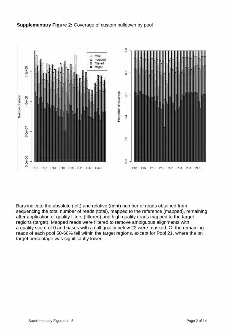
DNA samples from five individuals. Of note, one of these cases (17) harbours both BRCA1
and PPM1D mutations. Both genes are located at chromosome 17q and it is the wild-type
BRCA1 allele that is reduced in the tumours and therefore the relevance of the loss of
heterozygosity with respect to PPM1D is difficult to deduce.
Cell line and plasmid constructs
The U2OS, HeLa and HEK293 (all p53 wildtype) cell lines were obtained from the American
Type Culture Collection (ATCC). Cells were cultured and maintained according to the
supplier’s instructions. Cells were transfected with plasmid DNA using Lipofectamine 2000
(Invitrogen). A plasmid containing full-length wildtype PPM1D cDNA (pCMV6 entry-
PPM1D) was obtained from Origene, and the PPM1D open reading frame (ORF) subcloned
into pCMV6-AN-HA (Origene), generating a construct that could express a PPM1D - N-
terminal HA epitope fusion protein. Truncating mutations were introduced into the PPM1D
20
ORF of this construct using the QuickChange II XL Site-Directed Mutagenesis Kit
(Stratagene). To generate the following mutants, we used the following DNA amplification
primers:
PPM1D mutant 1 (c.1384C>T),
forward primer GAGAGAATGTCTAAGGTGTAGTC,
reverse primer GACTACACCTTAGACATTCTCTC,
PPM1D mutant 2 (c.1420delC),
forward primer GATCCAGAACCATTGAAG,
reverse primer CTTCAATGGTTCTGGATC.
Western Blot Analysis of P53 levels
U2OS, HeLa and HEK293 cells were transfected with PPM1D expression constructs and 24
hours after transfection, cells were exposed to gamma irradiation (5 Gy) from an X ray
source. Whole cell lysates were generated from transfected cells after irradiation (at 30
minute and four hour time points) and subjected to protein electrophoresis. Immunoblotting
of electrophoresed lysates was performed using antibodies specific for p53 (9282S - Cell
Signaling Technology) and actin (sc-1616, Santa Cruz Biotech).
Frequency and Risk Estimation
Statistical analyses were performed using the stats package in R. The significance of mutation
clustering was modelled under a binomial distribution where the probability of observing a
mutation in the last exon, which comprises 31% of the coding sequence, was 0.31. The
frequency in BRCA1/BRCA2 carriers and non-carriers was compared using a two-sided test of
proportions. Risk estimation was implemented using a competing risks retrospective
likelihood model incorporating age at onset according to a proportional hazards model. Since
individuals screened for PPM1D mutations were selected on the basis of both personal and
family history of breast or ovarian cancer, standard methods of analysis that ignore the
sampling frame would yield biased estimates of the risk ratios. To address this, we analysed
data within a retrospective cohort approach by modelling the conditional likelihood of the
observed genotypes given the disease phenotypes, using information on breast and ovarian
cancer occurrence in the set of 6,577 unrelated individuals negative for BRCA1/2 mutations
(BRCA1/2 mutation-positive individuals from the BOCS series and all the unselected ovarian
case series were excluded) and controls. Male controls were included in the analysis, but were
not considered to be at risk of developing breast or ovarian cancer. We assumed a competing
21
risks model, under which, each individual was at risk of developing breast or ovarian cancer.
This has been shown to provide unbiased estimates of the risk ratios for breast and ovarian
cancer where a genetic variant may be associated with one or both of the diseases10. We
estimated the PPM1D mutation carrier frequency in the population and breast and ovarian
cancer risk ratios simultaneously. Since mutation screened probands may have been selected
on the basis of bilateral breast cancer diagnosis or on the basis of both breast and ovarian
cancer diagnosis we allowed for the risks of breast or ovarian cancer diagnosis after the first
cancer diagnosis, including the risk of contralateral breast cancer. This model assumes that
the increased breast cancer (including contralateral) or ovarian cancer risk after the first
cancer diagnosis is entirely due to the susceptibility as defined by the model, with no
additional variation in risk. Site-specific cancer risks were assumed to be independent
conditional on genotype. Therefore the incidence of cancer at the second site was assumed to
be the same as if the preceding cancer had not occurred, with the exception of contralateral
breast cancer incidence after the first breast cancer, which was assumed to be half the overall
breast cancer incidence, since only one breast was at risk. In all models females were
censored at age 80 years. We assumed that the breast and ovarian cancer incidences depend
on the underlying PPM1D genotype through models of the form: λ(t) = λ0(t)exp(βx)where
λ0(t) is the baseline incidence at age t in non-mutation carriers, β is the log risk ratio
associated with the mutation and x takes value 0 for non-mutation carriers and 1 for mutation
carriers. The overall breast and ovarian cancer incidences, over all genotypes, were
constrained to agree with the population incidences for England and Wales in the period of
1993-199711, as described previously12.13. The models were parameterised in terms of the
mutation frequencies and log-risk ratios for breast and ovarian cancer. Parameters were
estimated using maximum likelihood estimation and were implemented in the pedigree
analysis software MENDEL14. The variances of the parameters were obtained by inverting
the observed information matrix. To obtain confidence intervals for the risk ratios and
perform hypothesis testing, log risk ratios were assumed to be normally distributed. A Wald
test-statistic was used to test the null hypothesis that β=0 for both breast and ovarian cancer.
Since PPM1D mutations were not found to segregate within families, we did not take into
account precise family histories or pedigree information and therefore did not incorporate the
effects of other susceptibility genes.
22
References for Online Methods 1 Tavassoli, F. A. & Devilee, P. in Pathology and Genetics of Tumours of the Breast and
Female Genital Organs (IARC Press, Lyon, France, 2003).
2 Hernandez, L. et al. Genomic and mutational profiling of ductal carcinomas in situ and matched adjacent invasive breast cancers reveals intra-tumour genetic heterogeneity and clonal selection. J. Pathol. 227, 42-52 (2012).
3 Gnirke, A. et al. Solution hybrid selection with ultra-long oligonucleotides for massively parallel targeted sequencing. Nat. Biotechnol. 27, 182-189 (2009).
4 Li, H. & Durbin, R. Fast and accurate short read alignment with Burrows-Wheeler transform. Bioinformatics 25, 1754-1760 (2009).
5 Caruccio, N. Preparation of next-generation sequencing libraries using Nextera technology: simultaneous DNA fragmentation and adaptor tagging by in vitro transposition. Methods Mol. Biol. 733, 241-255 (2011).
6 Rivas, M. A. et al. Deep resequencing of GWAS loci identifies independent rare variants associated with inflammatory bowel disease. Nat. Genet. 43, 1066-1073 (2011).
7 Turnbull, C. et al. Genome-wide association study identifies five new breast cancer susceptibility loci. Nat. Genet. 42, 504-507 (2010).
8 Lunter, G. & Goodson, M. Stampy: a statistical algorithm for sensitive and fast mapping of Illumina sequence reads. Genome Res. 21, 936-939 (2011).
9 Rimmer, A., Mathieson, I., Lunter, G. & McVean, G. Platypus: An Integrated Variant Caller (www.well.ox.ac.uk/platypus) (2012).
10 Barnes, D. et al. Evaluation of association methods for analysing modifiers of disease risk in carriers of high risk mutations. Genet. Epidemiol. 36, 274-291 (2012).
11 Cancer incidence in five continents. Volume VIII IARC Sci. Publ. 1-781 (2002).
12 Antoniou, A. C. et al. Evidence for further breast cancer susceptibility genes in addition to BRCA1 and BRCA2 in a population-based study. Genet. Epidemiol. 21, 1-18 (2001).
13 Antoniou, A. C. & Easton, D. F. Polygenic inheritance of breast cancer: Implications for design of association studies. Genet. Epidemiol. 25, 190-202 (2003).
14 Lange, K., Weeks, D. & Boehnke, M. Programs for Pedigree Analysis: MENDEL, FISHER, and dGENE. Genet. Epidemiol. 5, 471-472 (1988).
ID PPM1D mutation Cancer (age in yrs)
1a c.1270_1363dup94 Ov ca (64), Bil br ca (43,56)
2 c.1272delGGinsC Br ca (34)
3 a c.1337C>G_p.S446X Ov ca (43), Bladder ca (55)
4 a c.1340delA Br ca (46)
5 c.1340delA Br ca (65)
6 c.1384C>T_p.Q462X Br ca (59)
7 c.1420delC Ov ca (68), Br ca (71)
8 c.1430delA Br ca (44)
9 c.1434C>A_p.C478X Br ca (40)
10 c.1448delC Br ca (41)
11 c.1451delT Ov ca (67)
12 c.1451delT Bil br ca (61,76)
13 c.1451T>G_p.L484X Br ca (65)
14 c.1455_1456delGA Br ca (70)
15 c.1465delT Ov ca (60), Bil br ca (50,55)
16 c.1518delT Ov ca (69)
17 b c.1519delG Ov ca (40), Bil br ca (36,40)
18 c.1535delA Br ca (46)
19 c.1536insG Ov ca (47)
20 c.1538delT Ov ca (60) Br ca (55)
21 c.1538_1551del14 Ov ca (41)
22 c.1589delC Ov ca (69) Colorectal (69)
23 c.1600_1601delTT Br ca (62)
24 c.1613T>A_p.L538X Br ca (63)
25 c.1637_1638dupTG Ov ca (76)
26 c.1412delC control
a PPM1D mutations
b PPM1D gene
c PPM1D protein
d PPM1D mutation cluster region
420 546 Mutation Cluster Region
AAA 98
Phosphatase domain NLS
Exon1 Exon 2 Exon 6 Exon 3 Exon 4 Exon 5
Mutation Cluster Region
1261
Case 24: c.1613T>A_p.L538X
Case 6 Case 15 Case 17
Case 20 Case 22 Case 24
b
a Case 20: c.1538delT Case 23: c.1600_1601delTT
c Case 17 Case 24
293T
p53
yH2AX
Actin
52kDA —
14kDA —
38kDA —
EMPTY PPM1D
WT PPM1D
c.1384 C>T PPM1D
c.1420 delC
-IR 0.5 4 -IR 0.5 4 -IR 0.5 4 -IR 0.5 4
a)
Method PPM1D full gene Sanger sequencing
Samples 2456 cases 1347 controls
PPM1D PTVs 10 cases 0 control
b)
Method PPM1D mutation cluster region (MCR) Sanger sequencing
Samples 5325 cases 4514 controls
PPM1D PTVs 15 cases 1 control
Phase 2— case-control PPM1D sequencing
Method NGS of custom pulldown including 507 DNA repair genes
Samples 1150 cases (in 48 pools of 24 samples)
PPM1D PTVs 5 cases
Phase 1— case only DNA repair panel sequencing
Supplementary Figure 1. Samples, sequencing methods and PPM1D PTVs identified in different phases of the experiment
Supplementary Figures 1 - 8 Page 1 of 14
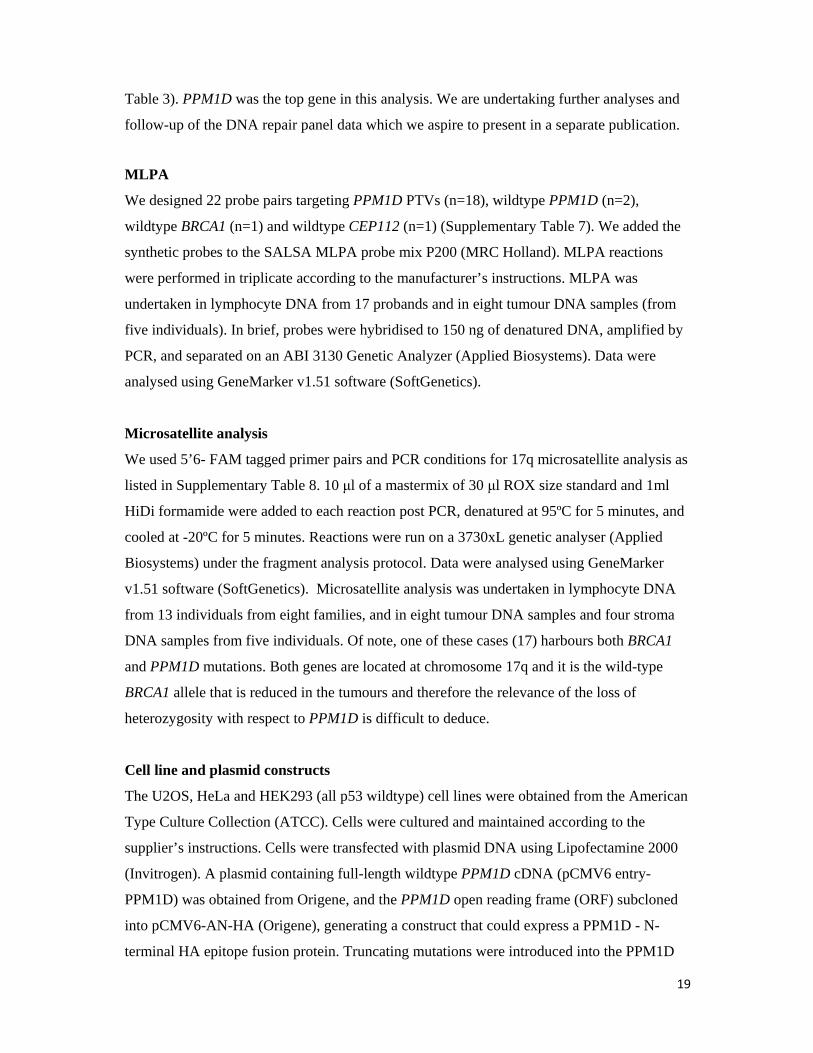
Supplementary Figure 2: Coverage of custom pulldown by pool
Bars indicate the absolute (left) and relative (right) number of reads obtained from sequencing the total number of reads (total), mapped to the reference (mapped), remaining after application of quality filters (filtered) and high quality reads mapped to the target regions (target). Mapped reads were filtered to remove ambiguous alignments with a quality score of 0 and bases with a call quality below 22 were masked. Of the remaining reads of each pool 50-60% fell within the target regions, except for Pool 21, where the on target percentage was significantly lower.
Supplementary Figures 1 - 8 Page 2 of 14
CASE 2: c.1272delGGinsC
Mutant
Wildtype
CASE 1: c.1270_c.1363dup94
CASE 3: c.1337C>G_p.S446X
CASE 4: c.1340delA
Mutant
Wildtype
CASE 5: c.1340delA
Mutant
Wildtype
CASE 8: c.1430delA*
CASE 7: c.1420delC
CASE 12: c.1451delT*
CASE 6: c.1384C>T_p.Q462X
CASE 9: c.1434C>A_p.C478X
CASE 10: c.1448delC*
CASE 11: c.1451delT*
Supplementary Figure 3. Sanger sequencing chromatograms for 26 PPM1D mutations
Supplementary Figures 1 - 8 Page 3 of 14
Mutant
Wildtype
Mutant
Wildtype
Mutant
Wildtype
CASE16: c.1518delT
CASE13: c.1451T>G_p.L484X
CASE14: c.1455_1456delGA
CASE15: c.1465delT
CASE17: c.1519delG*
CASE18: c.1535delA*
CASE19: c.1536insG
CASE20: blood c.1538delT
CASE21: c.1538_1551del14
CASE22: c.1589delC
CASE20: saliva c.1538delT
CASE23: c.1600_1601delTT
Supplementary Figures 1 - 8 Page 4 of 14
Mutant
Wildtype
The mutant allele is lower than typical of heterozygous mutations, consistent with mosaicism. *indicates that the reverse sequencing trace is presented.
26: Control c.1412delC
CASE25: c.1637_1638dupTG
CASE24: blood c.1613T>A_p.L538X
CASE24: saliva c.1613T>A_p.L538X
Supplementary Figures 1 - 8 Page 5 of 14
Case 1 Case 2 Case 3 Case 4M
utan
t rea
d pe
rcen
tage
Mut
ant r
ead
perc
enta
ge
Mut
ant r
ead
perc
enta
ge
Mut
ant r
ead
perc
enta
ge
Coverage Coverage Coverage Coverage
Case 5 Case 6 Case 8 Case 9
Mut
ant r
ead
perc
enta
ge
Mut
ant r
ead
perc
enta
ge
Mut
ant r
ead
perc
enta
ge
Mut
ant r
ead
perc
enta
ge
Coverage Coverage Coverage Coverage
Case 11 Case 12 Case 13 Case 15
Mut
ant r
ead
perc
enta
ge
Mut
ant r
ead
perc
enta
ge
Mut
ant r
ead
perc
enta
ge
Mut
ant r
ead
perc
enta
geCoverage Coverage Coverage Coverage
Case 17 Case 18 Case 20 Case 21
Mut
ant r
ead
perc
enta
ge
Mut
ant r
ead
perc
enta
ge
Mut
ant r
ead
perc
enta
ge
Mut
ant r
ead
perc
enta
ge
Coverage Coverage Coverage Coverage
Case 22 Case 23 Case 24 Case 26
Mut
ant r
ead
perc
enta
ge
Mut
ant r
ead
perc
enta
ge
Mut
ant r
ead
perc
enta
ge
Mut
ant r
ead
perc
enta
ge
Coverage Coverage Coverage Coverage
Case 25
Mutant read percentage is calculated as the proportion of reads containing the variant. The red dot indicates the PPM1D mutation. In case 2, the complex indel was called as two different mutations and thus two red dots. Variants were censored at 5%. All mutations have a consistently lower mutant read percentage, indicating mosaicism. Open dots represent variants in BRCA1 or BRCA2.
Supplementary Figure 4. Deep PCR amplicon sequencing of BRCA1, BRCA2 and PPM1D cluster region showing mosaic mutations.
Supplementary Figures 1 - 8 Page 6 of 14
CASE 1: c.1270_1363dup94 CASE 6: c.1384C>T_p.Q462X
Supplementary Figure 6. DNA repair panel individual sequencing in six PPM1D mutation carriers.
Mutant read percentage was calculated as in Supplementary Figure 4. Read coverage was recorded at all variant sites, counting only bases within reads with a mapping quality of at least 20 and a base quality of at least 22. A window around the mutation containing at least 50 variants with similar coverage was identified. The red dot indicates the PPM1D mutation. Open dots represent other variants in the custom pulldown which were not validated. All PPM1D mutations were consistently lower, indicating mosaicism. Mutant read percentages for cases 15, 20 and 21 matched those in Supplementary Figure 4.
Supplementary Figures 1 - 8 Page 8 of 14
Supplementary Figure 7: The effect of mutant PPM1D isoforms on p53 activation
293T
EMPTY
-IR 0.5 4 ‐IR 0.5 4 ‐IR 0.5 4 ‐IR 0.5 4
PPM1Dc.1420delC
PPM1Dc. 1384C>T
PPM1DWT
p53Panel 1
Hela
ActinPanel 2
Panel 2
EMPTY
-IR 0.5 4 ‐IR 0.5 4 ‐IR 0.5 4 ‐IR 0.5 4
PPM1Dc.1420delC
PPM1Dc. 1384C>T
PPM1DWT
Actin
p53 Panel 1
p53 wild type HeLa and HEK293 cells were transfected with PPM1D cDNA expression constructs and exposed to ionising irradiation (5 Grays). At 30 minute and four hour intervals after IR exposure whole cell lysates were generated and western blotted to estimate the IR induced activation of p53. Western blots showing p53 and actin(loading control) protein levels at different times (in hours) after IR exposure are shown. ‘Empty’ represents cells transfected with an empty expression construct, ‘PPM1D WT’ represents cells transfected with a wild type PPM1D cDNA expression construct and ‘PPM1D c.1384C>T’ and ‘PPM1D c.1420delC’ represent cells transfected with mutant PPM1D cDNA constructs. The suppression of p53 was enhanced in cells transfected with the mutant constructs suggesting these alleles encode hyperactive PPM1D isoforms.
Microsatellite data demonstrates two alleles in DNA from lymphocytes and stroma and loss of heterozygosity in tumours in cases 1, 11, 17 and 20. There is no loss of heterozygosity in the tumour DNA from case 15. The Sanger sequencing and MLPA data demonstrate that the mosaic mutations in lymphocyte DNA are not detectable in stromal or tumour DNA. The data from deep PCR amplicon sequencing of tumour and stromal DNA is presented in Supplementary Table 5.
Supplementary Figures 1 - 8 Page 14 of 14
The Wellcome Trust Case Control Consortium
The following individuals are part of the Wellcome Trust Case Control Consortium+ Jan Aerts1, Tariq Ahmad2, Hazel Arbury1, Anthony Attwood1,3,4, Adam Auton5, Stephen G Ball6,
Anthony J Balmforth6, Chris Barnes1, Jeffrey C Barrett1, Inês Barroso1, Anne Barton7, Amanda J Bennett8, Sanjeev Bhaskar1, Katarzyna Blaszczyk9, John Bowes7, Oliver J Brand8,10, Peter S Braund11, Francesca Bredin12, Gerome Breen13,14, Morris J Brown15, Ian N Bruce7, Jaswinder Bull16, Oliver S Burren17, John Burton1, Jake Byrnes18, Sian Caesar19, Niall Cardin5, Chris M Clee1, Alison J Coffey1, John MC Connell20, Donald F Conrad1, Jason D Cooper17, Anna F Dominiczak20, Kate Downes17, Hazel E Drummond21, Darshna Dudakia16, Andrew Dunham1, Bernadette Ebbs16, Diana Eccles22, Sarah Edkins1, Cathryn Edwards23, Anna Elliot16, Paul Emery24, David M Evans25, Gareth Evans26, Steve Eyre7, Anne Farmer14, I Nicol Ferrier27,
Edward Flynn7, Alistair Forbes28, Liz Forty29, Jayne A Franklyn10,30, Timothy M Frayling2, Rachel M Freathy2, Eleni Giannoulatou5, Paul Gilbert7, Katherine Gordon-Smith19,29, Emma Gray1, Elaine Green29, Chris J Groves8, Detelina Grozeva29, Rhian Gwilliam1, Naomi Hammond1,
Matt Hardy17, Pile Harrison31, Neelam Hassanali8, Husam Hebaishi1, Sarah Hines16, Anne Hinks7,
Graham A Hitman32, Lynne Hocking33, Chris Holmes5, Eleanor Howard1, Philip Howard34,
Joanna MM Howson17, Debbie Hughes16, Sarah Hunt1, John D Isaacs35, Mahim Jain18, Derek P Jewell36, Toby Johnson34, Jennifer D Jolley3,4, Ian R Jones29, Lisa A Jones19, George Kirov29,
Cordelia F Langford1, Hana Lango-Allen2, G Mark Lathrop37, James Lee12, Kate L Lee34, Charlie Lees21, Kevin Lewis1, Cecilia M Lindgren8,18, Meeta Maisuria-Armer17, Julian Maller18, John Mansfield38, Jonathan L Marchini5, Paul Martin7, Dunecan CO Massey12, Wendy L McArdle39,
Peter McGuffin14, Kirsten E McLay1, Gil McVean5,18, Alex Mentzer40, Michael L Mimmack1,
Ann E Morgan41, Andrew P Morris18, Craig Mowat42, Patricia B Munroe34, Simon Myers18,
William Newman26, Elaine R Nimmo21, Michael C O'Donovan29, Abiodun Onipinla34, Nigel R Ovington17, Michael J Owen29, Kimmo Palin1, Aarno Palotie1, Kirstie Parnell2, Richard Pearson8, John RB Perry2,18, Anne Phillips42, Vincent Plagnol17, Natalie J Prescott9, Inga Prokopenko8,18,
Michael A Quail1, Suzanne Rafelt11, Nigel W Rayner8,18, David M Reid33, Anthony Renwick16,
Susan M Ring39, Neil Robertson8,18, Samuel Robson1, Ellie Russell29, David St Clair13, Jennifer G Sambrook3,4, Jeremy D Sanderson40, Stephen J Sawcer43, Helen Schuilenburg17, Carol E Scott1,
Sheila Seal16, Sue Shaw-Hawkins34, Beverley M Shields2, Matthew J Simmonds8,10, Debbie J Smyth17, Elilan Somaskantharajah1, Katarina Spanova16, Sophia Steer44, Jonathan Stephens3,4,
Helen E Stevens17, Kathy Stirrups1, Millicent A Stone45,46, David P Strachan47, Zhan Su5,
Deborah PM Symmons7, John R Thompson48, Wendy Thomson7, Martin D Tobin48, Mary E Travers8, Clare Turnbull16, Damjan Vukcevic18, Louise V Wain48, Mark Walker49, Neil M Walker17, Chris Wallace17, Margaret Warren-Perry16, Nicholas A Watkins3,4, John Webster50,
Michael N Weedon2, Anthony G Wilson51, Matthew Woodburn17, B Paul Wordsworth52, Chris Yau5, Allan H Young27,53, Eleftheria Zeggini1, Matthew A Brown52,54, Paul R Burton48, Mark J Caulfield34, Alastair Compston43, Martin Farrall55, Stephen CL Gough8,10,30, Alistair S Hall6, Andrew T Hattersley2,56, Adrian VS Hill18, Christopher G Mathew9, Marcus Pembrey57, Jack Satsangi21, Michael R Stratton1,16, Jane Worthington7, Matthew E Hurles1, Audrey Duncanson58, Willem H Ouwehand1,3,4, Miles Parkes12, Nazneen Rahman16, John A Todd17, Nilesh J Samani11,59, Dominic P Kwiatkowski1,18, Mark I McCarthy8,18,60, Nick Craddock29, Panos Deloukas1, Peter Donnelly5,18.
1 The Wellcome Trust Sanger Institute, Wellcome Trust Genome Campus, Hinxton, Cambridge, CB10 1SA UK. 2 Genetics of Complex Traits, Peninsula College of Medicine and Dentistry University of Exeter, EX1 2LU, UK. 3 Department of Haematology, University of Cambridge, Long Road, Cambridge, CB2 0PT, UK. 4 National Health Service Blood and Transplant, Cambridge Centre, Long Road, Cambridge CB2 0PT, UK. 5 Department of Statistics, University of Oxford, 1 South Parks Road, Oxford, OX1 3TG, UK. 6 Multidisciplinary Cardiovascular Research Centre (MCRC), Leeds Institute of Genetics, Health and
Therapeutics (LIGHT), University of Leeds, Leeds, LS2 9JT, UK. 7 arc Epidemiology Unit, Stopford Building, University of Manchester, Oxford Road, Manchester, M13 9PT,
UK. 8 Oxford Centre for Diabetes, Endocrinology and Medicine, University of Oxford, Churchill Hospital, Oxford
OX3 7LJ, UK. 9 Department of Medical and Molecular Genetics, King’s College London School of Medicine, 8th Floor Guy’s
Tower, Guy’s Hospital, London, SE1 9RT, UK. 10 Centre for Endocrinology, Diabetes and Metabolism, Institute of Biomedical Research, University of
Birmingham, Birmingham, B15 2TT, UK. 11 Department of Cardiovascular Sciences, University of Leicester, Glenfield Hospital, Groby Road, Leicester
LE3 9QP, UK. 12 IBD Genetics Research Group, Addenbrooke's Hospital, Cambridge, CB2 0QQ, UK. 13 University of Aberdeen, Institute of Medical Sciences, Foresterhill, Aberdeen AB25 2ZD, UK. 14 SGDP, The Institute of Psychiatry, King's College London, De Crespigny Park, Denmark Hill, London SE5
8AF, UK. 15 Clinical Pharmacology Unit, University of Cambridge, Addenbrookes Hospital, Hills Road, Cambridge CB2
2QQ, UK. 16 Section of Cancer Genetics, Institute of Cancer Research, 15 Cotswold Road, Sutton SM2 5NG, UK. 17 Juvenile Diabetes Research Foundation/Wellcome Trust Diabetes and Inflammation Laboratory, Department
of Medical Genetics, Cambridge Institute for Medical Research, University of Cambridge, Wellcome Trust/MRC Building, Cambridge CB2 0XY, UK.
18 The Wellcome Trust Centre for Human Genetics, University of Oxford, Roosevelt Drive, Oxford OX3 7BN, UK.
19 Department of Psychiatry, University of Birmingham, National Centre for Mental Health, 25 Vincent Drive, Birmingham, B15 2FG, UK.
20 BHF Glasgow Cardiovascular Research Centre, University of Glasgow, 126 University Place, Glasgow, G12 8TA, UK.
21 Gastrointestinal Unit, Division of Medical Sciences, School of Molecular and Clinical Medicine, University of Edinburgh, Western General Hospital, Edinburgh EH4 2XU, UK.
22 Faculty of Medicine, University of Southampton and Wessex Clinical Genetics Service, UHSFT, Southampton, UK. SO16 5YA
23 Endoscopy Regional Training Unit, Torbay Hospital, Torbay TQ2 7AA, UK. 24 Academic Unit of Musculoskeletal Disease, University of Leeds, Chapel Allerton Hospital, Leeds, West
Yorkshire LS7 4SA, UK. 25 MRC Centre for Causal Analyses in Translational Epidemiology, Department of Social Medicine, University
of Bristol, Bristol, BS8 2BN, UK. 26 Department of Medical Genetics, Manchester Academic Health Science Centre (MAHSC), University of
Manchester, Manchester M13 0JH, UK. 27 School of Neurology, Neurobiology and Psychiatry, Royal Victoria Infirmary, Queen Victoria Road,
Newcastle upon Tyne, NE1 4LP, UK. 28 Institute for Digestive Diseases, University College London Hospitals Trust, London NW1 2BU, UK. 29 MRC Centre for Neuropsychiatric Genetics and Genomics, School of Medicine, Cardiff University, Heath
Park, Cardiff, CF14 4XN, UK. 30 University Hospital Birmingham NHS Foundation Trust, Birmingham, B15 2TT, UK. 31 University of Oxford, Institute of Musculoskeletal Sciences, Botnar Research Centre, Oxford, OX3 7LD, UK. 32 Centre for Diabetes and Metabolic Medicine, Barts and The London, Royal London Hospital, Whitechapel,
London, E1 1BB, UK. 33 Bone Research Group, Department of Medicine and Therapeutics, University of Aberdeen, Aberdeen, AB25
2ZD, UK.
34 Clinical Pharmacology and Barts and The London Genome Centre, William Harvey Research Institute, Barts and The London School of Medicine and Dentistry, Queen Mary University of London, Charterhouse Square, London EC1M 6BQ, UK.
35 Institute of Cellular Medicine, Musculoskeletal Research Group, 4th Floor, Catherine Cookson Building, The Medical School, Framlington Place, Newcastle upon Tyne, NE2 4HH, UK.
36 Gastroenterology Unit, Radcliffe Infirmary, University of Oxford, Oxford, OX2 6HE, UK. 37 Centre National de Genotypage, 2, Rue Gaston Cremieux, Evry, Paris 91057, France. 38 Department of Gastroenterology & Hepatology, University of Newcastle upon Tyne, Royal Victoria Infirmary,
Newcastle upon Tyne NE1 4LP, UK. 39 ALSPAC Laboratory, Department of Social Medicine, University of Bristol, BS8 2BN, UK. 40 Division of Nutritional Sciences, King's College London School of Biomedical and Health Sciences, London
SE1 9NH, UK. 41 NIHR-Leeds Musculoskeletal Biomedical Research Unit, University of Leeds, Chapel Allerton Hospital,
Leeds, West Yorkshire LS7 4SA, UK. 42 Department of General Internal Medicine, Ninewells Hospital and Medical School, Ninewells Avenue, Dundee
DD1 9SY, UK. 43 Department of Clinical Neurosciences, University of Cambridge, Addenbrooke's Hospital, Hills Road,
Cambridge, CB2 2QQ, UK. 44 Clinical and Academic Rheumatology, Kings College Hosptal National Health Service Foundation Trust,
Denmark Hill, London SE5 9RS, UK. 45 University of Toronto, St. Michael's Hospital, 30 Bond Street, Toronto, Ontario M5B 1W8, Canada. 46 University of Bath, Claverdon, Norwood House, Room 5.11a Bath Somerset BA2 7AY, UK. 47 Division of Community Health Sciences, St George's, University of London, London SW17 0RE, UK. 48 Departments of Health Sciences and Genetics, University of Leicester, 217 Adrian Building, University Road,
Leicester, LE1 7RH, UK. 49 Diabetes Research Group, School of Clinical Medical Sciences, Newcastle University, Framlington Place,
Newcastle upon Tyne NE2 4HH, UK. 50 Medicine and Therapeutics, Aberdeen Royal Infirmary, Foresterhill, Aberdeen, Grampian AB9 2ZB, UK. 51 School of Medicine and Biomedical Sciences, University of Sheffield, Sheffield, S10 2JF, UK. 52 Nuffield Department of Orthopaedics, Rheumatology and Musculoskeletal Sciences, Nuffield Orthopaedic
Centre, University of Oxford, Windmill Road, Headington, Oxford, OX3 7LD, UK. 53 UBC Institute of Mental Health, 430-5950 University Boulevard Vancouver, British Columbia, V6T 1Z3,
Canada. 54 Diamantina Institute of Cancer, Immunology and Metabolic Medicine, Princess Alexandra Hospital, University
of Queensland, Ipswich Road, Woolloongabba, Brisbane, Queensland, 4102, Australia. 55 Cardiovascular Medicine, University of Oxford, Wellcome Trust Centre for Human Genetics, Roosevelt Drive,
Oxford OX3 7BN, UK. 56 Genetics of Diabetes, Peninsula College of Medicine and Dentistry, University of Exeter, Barrack Road,
Exeter, EX2 5DW, UK. 57 Clinical and Molecular Genetics Unit, Institute of Child Health, University College London, 30 Guilford
Street, London WC1N 1EH, UK. 58 The Wellcome Trust, Gibbs Building, 215 Euston Road, London NW1 2BE, UK. 59 Leicester NIHR Biomedical Research Unit in Cardiovascular Disease, Glenfield Hospital, Leicester, LE3 9QP,
UK. 60 Oxford NIHR Biomedical Research Centre, Churchill Hospital, Oxford, OX3 7LJ, UK.
The Breast and Ovarian Cancer Susceptibility Collaboration (BOCS)
The following individuals are part of Breast and Ovarian Cancer Susceptibility Collaboration (BOCS)
A. Ardern-Jones, J. Adlard, M. Ahmed, G. Attard, K. Bailey, E. Bancroft, C. Bardsley, D. Barton, J. Barwell, L. Baxter, R. Belk, J. Berg, B. Bernhard, T. Bishop, L. Boyes, N. Bradshaw, A.F. Brady, S. Brant, C. Brewer, G. Brice, G. Bromilow, C. Brooks, A. Bruce, B. Bulman, L. Burgess, J. Campbell, N. Canham, B. Castle, R. Cetnarskyj, C. Chapman, O. Claber, N. Coates, T. Cole, A. Collins, J. Cook, S. Coulson, G. Crawford, D. Cruger, C. Cummings, L. D’Mello, R. Davidson, L. Day, L. de Silva, B. Dell, C. Dolling, A. Donaldson, H. Dorkins, F. Douglas, S. Downing, S. Drummond, C. Dubras, J. Dunlop, S. Durrell, D. Eccles, C. Eddy, M. Edwards, E. Edwards, J. Edwardson, R. Eeles, I. Ellis, F. Elmslie, G. Evans, B. Gibbens, C. Gardiner, N. Ghali, C. Giblin, S. Gibson, S. Goff, S. Goodman, D. Goudie, L. Greenhalgh, J. Greer, H. Gregory, D. Halliday, R. Hardy, C. Hartigan, T. Heaton, A. Henderson, C. Higgins, S. Hodgson, T. Holt, T. Homfray, D. Horrigan, C. Houghton, R.S. Houlston, L. Hughes, V. Hunt, L. Irvine, L. Izatt, C. Jacobs, S. James, M. James, L. Jeffers, I. Jobson, W. Jones, M.J. Kennedy, S. Kenwrick, C. Kightley, C. Kirk, L. Kirk, E. Kivuva, K. Kohut, M. Kosicka-Slawinska, A. Kulkarni, A. Kumar, F. Lalloo, N. Lambord, C. Langman, P. Leonard, S. Levene, S. Locker, P. Logan, M. Longmuir, A. Lucassen, V. Lyus, A. Magee, A. Male, S. Mansour, D. McBride, E. McCann, V. McConnell, M. McEntagart, C. McKeown, L. McLeish, D. McLeod, A. Melville, L. Mercer, C. Mercer, Z. Miedzybrodzka, A. Mitra, P. J. Morrison, V. Murday, A. Murray, K. Myhill, J. Myring, E. O'Hara, J. Paterson, P. Pearson, G. Pichert, K. Platt, M. Porteous, C. Pottinger, S. Price, L. Protheroe, S. Pugh, O. Quarrell, K. Randhawa, C. Riddick, L. Robertson, A. Robinson, V. Roffey-Johnson, M. Rogers, S. Rose, S. Rowe, A. Schofield, N. Rahman, S. Saya, G. Scott, J. Scott, A. Searle, S. Shanley, S. Sharif, A. Shaw, J. Shaw, J. Shea-Simonds, L. Side, J. Sillibourne, K. Simon, S. Simpson, S. Slater, S. Smalley, K. Smith, L. Snadden, K. Snape, J. Soloway, Y. Stait, B. Stayner, M. Steel, C. Steel, H. Stewart, D. Stirling, M. Thomas, S. Thomas, S. Tomkins, H. Turner, E. Tyler, A. Vandersteen, E. Wakeling, F. Waldrup, L. Walker, C. Watt, S. Watts, A. Webber, C. Whyte, J. Wiggins, E. Williams, L. Winchester.