AbstractBackground: Early prediction of outcome after cardiac arrest (CA) may influence treatment strategies. Existing
biomarkers are of limited value. Therefore, we sought to determine whether analysis of the transcriptome of circulating blood cells may help to predict outcome after CA.
Methods: Consecutive comatose patients resuscitated after CA and treated with hypothermia were enrolled in this study. Blood samples were drawn 48 hours after CA for gene expression analyses by microarrays and quantitative PCR. Neurological outcome at 6 months was evaluated using the cerebral performance category (CPC). Patients with CPC 1-2 were considered having favorable neurological outcome, whereas patients with CPC 3-5 were considered as having poor outcome.
Results: The initial cohort consisted of 35 patients. Microarrays revealed a biosignature associated with neurological outcome after CA. 582 genes were differentially expressed between patients with favorable neurological outcome (n=21) and patients with poor neurological outcome (n=14). Bioinformatic analyses revealed significant associations between these genes and neuronal damage. Prediction analysis of microarrays identified the chemokine (C-X3-C motif) receptor 1 (CX3CR1) as a candidate prognostic biomarker. CX3CR1 was up-regulated in patients with favorable outcome (2-fold, P<0.001) and predicted neurological outcome with an AUC of 0.92 (95% CI [0.83; 1.00]). In a second cohort of 45 patients, CX3CR1 was increased in patients with favorable outcome (1.6-fold, P=0.003) and predicted outcome with an AUC of 0.76 (95% CI [0.60; 0.92]). Multivariate analyses identified CX3CR1, neuron-specific enolase and acute myocardial infarction at the time of CA as significant predictors of survival. These variables had additive value to predict survival (AUC 0.84).
Conclusion: We identified CX3CR1 as a new candidate prognostic biomarker after CA. Further studies are required to confirm its predictive value.
The Chemokine (C-X3-C Motif) Receptor 1 is a Candidate Prognostic Biomarker after Cardiac ArrestPascal Stammet1, Melanie Kirchmeyer2, Lu Zhang2, Daniel R. Wagner2,3 and Yvan Devaux2*1Department of Anaesthesia and Intensive Care, Centre Hospitalier, Luxembourg, L-1210, Luxembourg 2Laboratory of Cardiovascular Research, Centre de Recherche Public-Sante, Luxembourg, L-1150, Luxembourg3Division of Cardiology, Centre Hospitalier, Luxembourg, L-1210, Luxembourg
IntroductionCardiovascular disease is the most common cause of death in
developed countries, despite a substantial reduction in age-adjusted rates of death from cardiovascular causes over the past decades. Of all cardiac deaths, approximately 50% are sudden. Sudden cardiac death – cardiac arrest (CA) – is the most common lethal manifestation of heart disease [1-3].
Therapeutic hypothermia (TH) has been shown in randomized trials to improve outcome of patients resuscitated from out-of-hospital ventricular fibrillation [4,5]. Induction of TH after return of spontaneous circulation has been associated with improved functional recovery and a clear benefit in neurologic outcome and mortality [4,5]. Indeed, hypothermia decreases cerebral oxygen demand and thus provides protection from ongoing cerebral ischemia. TH also reduces the glutamate level, the production of free radicals [6], and the intracranial pressure [7].
Today, few methods exist to predict outcome in the early stage after CA. Biochemical markers of brain injury like S-100B and serum neuron-specific enolase (NSE) have been used to predict neurological outcome. However, their predictive value was determined in trials without hypothermia [8-11] and was found to be of limited benefit in patients treated with TH [12,13]. Therefore, there is an unmet clinical need to find new prognostic biomarkers after CA.
We hypothesized that gene expression profiles from circulating
blood cells may be used to identify new prognostic biomarkers after CA.
Materials and MethodsPatients
All adult comatose patients (Glasgow coma score <8) successfully resuscitated from CA admitted to our institution, were included in this study, regardless of the cause of CA and the initial rhythm. Exclusion criteria were age under 18 years, no therapeutic hypothermia performed, terminal disease and traumatic cardiac arrest. The study protocol was approved by the National Ethics Committee and informed consent was obtained from the next of kin. This was a single-centre, prospective, observational study. All data were recorded according to the Utstein-style template [14].
Journal of Clinical & Experimental CardiologyJo
urna
l of C
linica
l & Experimental Cardiology
ISSN: 2155-9880
Citation: Stammet P, Kirchmeyer M, Zhang L, Wagner DR, Devaux Y (2012) The Chemokine (C-X3-C Motif) Receptor 1 is a Candidate Prognostic Biomarker after Cardiac Arrest. J Clinic Experiment Cardiol S2:004. doi:10.4172/2155-9880.S2-004
Page 2 of 7
J Clinic Experiment Cardiol Cardiac Biomarkers ISSN:2155-9880 JCEC, an open access journal
Treatment of the patients was performed according to standard protocols. All patients were treated with TH as soon as possible [15] after arrival in the catheterization laboratory or in the intensive care unit (ICU). Target temperature was 33 ± 1°C. Induction of hypothermia was done either by using cold IV fluids [16] or an iv-cooling device (Coolgard® with Icy-cath® ZOLL Medical Corp. MA, USA) [17]. After 24 h, rewarming was started at a maximum rate of 0.5°C h-1. Core temperature was controlled by an indwelling bladder catheter and a second, rectal, probe serving as control. Sedation was done by a standardized protocol using Midazolam (max. 0.2 mg kg-1 h-1) and Fentanyl (max. 1.5 µg kg-1 h-1).
At the end of the ICU stay, neurological examination was performed using the Glasgow coma scale and the cerebral performance category score (CPC) [18]. CPC1 and CPC2 were regarded as good outcome, whereas CPC3, 4 and 5 were considered as poor outcome. At 6 months, a clinical neurological examination was done by physicians uninvolved in the ICU management of the patient. At the end of hypothermia, blood samples were drawn in PAXgene™ tubes (BD Biosciences, Erembodegem, Belgium) and tubes containing a clot activator (BD Biosciences). Total RNA extracted from PAXgene™ tubes was used to study gene expression by microarrays and PCR. NSE was measured in serum using an electrochemiluminescent immunoassay (ECLIA, Roche, Basel, Switzerland) performed on the on the Cobas e601 analyzer® (Roche, Basel, Switzerland) with a detection limit of <0.05ng/ml.
Microarrays
Transcriptomic analysis of blood cells was performed as previously described [19,20]. Total RNA was extracted from PAXgene™ tubes using the PAXgene™ blood RNA kit (Qiagen, Venlo, Netherlands). RNA was subjected to additional purification and concentration with the RNeasy® MinElute™ kit (Qiagen). RNA quantity was measured with the ND-1000 spectrophotometer (NanoDrop® Technologies, Wilmington, USA). RNA quality was evaluated with the 2100 Bioanalyzer® apparatus (Agilent Technologies, Massy, France) and RNA 6000 Nano chips. Only high quality RNA (OD260/OD280 > 1.9 and OD260/OD230 > 1.7) and un-degraded RNA was retained for further analysis. The Universal Human Reference RNA (Stratagene Europe, Amsterdam, The Netherlands) was used in conjunction with patient’s RNA to provide an internal reference for comparisons of relative gene expression levels across arrays. Messenger RNAs were amplified with the Amino Allyl MessageAmp™ kit (Ambion®, Cambridgeshire, United Kingdom), starting with 1µg of total RNA. 5µg of amino allyl aRNA were labeled with Cy3 or Cy5 (Amersham, Buckinghamshire, United Kingdom). Dye coupling was evaluated using the ND-1000 spectrophotometer. Coupling yield >5% was a prerequisite for further analysis. 750ng of each amino allyl aRNA labeled Cy3 or Cy5 (reference RNA or patient RNA) were mixed and hybridized on long oligonucleotide microarrays covering 25,000 genes [21]. 4 microarray replicates and a dye-swap were performed. Scanning was achieved with an Axon 4000B scanner and data were acquired with the GenePix Pro 6® software (Molecular Devices, Berks, UK). Spot finding and raw data quantification were performed using MAIA® software [22]. A Lowess non linear normalization was completed and genes not present in at least 3 microarrays of 4 were filtered out. Data are available at the Gene Expression Omnibus (www.ncbi.nlm.nih.gov/geo/) under the accession number GSE29540.
Prior to statistical analysis, genes not present in at least 50% of the patients were removed. Supervised analysis was achieved with the Significance Analysis of Microarrays (SAM) software [23] using 2-class
unpaired test and 100 permutations. Classification value of microarrays data was evaluated using Prediction Analysis of Microarrays (PAM) software [24]. The Database for Annotation, Visualization and Integrated Discovery (DAVID) [25] and the GOMIner resource [26] were used for functional analysis of microarray data. Heat maps were obtained with Cluster 3.0 and Java Treeview [27].
Quantitative PCR
Total RNA extracted from PAXgene™ tubes using the PAXgene™ blood RNA kit (Qiagen) was subjected to a second step of purification and concentration with the RNeasy® MinElute™ kit (Qiagen). 1µg of total RNA was reverse-transcribed using the Superscript® II Reverse Transcriptase (Invitrogen, Merelbeke, Belgium). PCR primers were designed using the Beacon Designer software (Premier Biosoft, Palo Alto, USA) and were chosen to encompass an intron. PCR was performed using the iCycler® and the IQTM SYBR® Green Supermix (Biorad, Nazareth, Belgium). 1/10 dilutions of cDNA were used. PCR conditions were as follows: 3 min at 95°C, 30 sec at 95°C and 1 min annealing (40-fold). Optimal annealing temperature was determined for each primer pair. Melting point analysis was obtained after 80 cycles for 10 sec from 55°C up to 95°C. Each run included negative reaction controls. GAPDH was used as housekeeping genes for normalization. Expression levels were calculated by the relative quantification method (ΔΔCt) using the Genex software (Biorad) which takes into account primer pair efficiency.
Statistical analysis
The primary end-point of the study was the neurological status at 6 months after resuscitation. The secondary end-point was mortality. The ability of CX3CR1 to predict end-points was evaluated using receiver-operating characteristic (ROC) curves with the corresponding area under the curve (AUC). Multiple logistic regression models proceeding by backward elimination were used to identify clinical covariates with predictive value. Logistic regression models proceeding with 10-fold cross-validation were used to determine the added value of multiple clinical covariates.
Kaplan-Meier curves and the LogRank statistic were used for survival analysis, in which patients were distributed in tertiles of biomarker values. To isolate the tertiles that differ from each other, all pairwise multiple comparison procedure was applied (Holm-Sidak method).
Data normality was assessed using the Shapiro-Wilk test. Comparisons between 2 groups were performed using t-test or Mann-Whitney Rank Sum test. Fisher’s exact test was used for categorical variables. Correlations were determined using the Spearman test. Statistical tests were performed with the Sigma Plot v11.0 software.
ResultsPatients
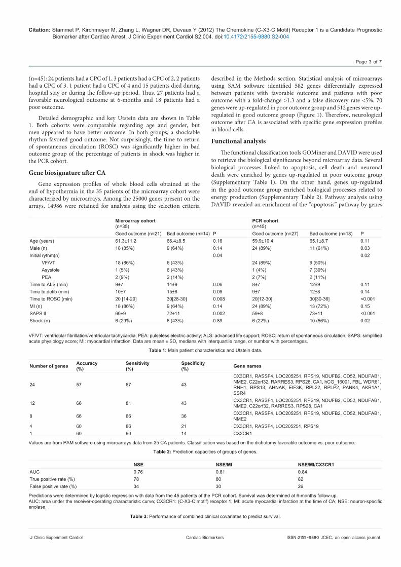
An initial cohort, named “microarray cohort”, consisted of 35 patients. At 6 months post resuscitation, 18 patients had a CPC of 1, 3 patients had a CPC of 2, 2 patients had a CPC of 3, 1 patients had a CPC of 4, and 11 patients died during hospital stay or follow-up (CPC 5). Patients were dichotomized into good neurological outcome, as attested by CPC 1-2 (n=21), and poor neurological outcome, as attested by CPC 3-5 (n=14).
A second group of patients, named “PCR cohort”, was formed of the 35 patients of the microarray cohort and 10 additional patients
Citation: Stammet P, Kirchmeyer M, Zhang L, Wagner DR, Devaux Y (2012) The Chemokine (C-X3-C Motif) Receptor 1 is a Candidate Prognostic Biomarker after Cardiac Arrest. J Clinic Experiment Cardiol S2:004. doi:10.4172/2155-9880.S2-004
Page 3 of 7
J Clinic Experiment Cardiol Cardiac Biomarkers ISSN:2155-9880 JCEC, an open access journal
(n=45): 24 patients had a CPC of 1, 3 patients had a CPC of 2, 2 patients had a CPC of 3, 1 patient had a CPC of 4 and 15 patients died during hospital stay or during the follow-up period. Thus, 27 patients had a favorable neurological outcome at 6-months and 18 patients had a poor outcome.
Detailed demographic and key Utstein data are shown in Table 1. Both cohorts were comparable regarding age and gender, but men appeared to have better outcome. In both groups, a shockable rhythm favored good outcome. Not surprisingly, the time to return of spontaneous circulation (ROSC) was significantly higher in bad outcome group of the percentage of patients in shock was higher in the PCR cohort.
Gene biosignature after CA
Gene expression profiles of whole blood cells obtained at the end of hypothermia in the 35 patients of the microarray cohort were characterized by microarrays. Among the 25000 genes present on the arrays, 14986 were retained for analysis using the selection criteria
described in the Methods section. Statistical analysis of microarrays using SAM software identified 582 genes differentially expressed between patients with favorable outcome and patients with poor outcome with a fold-change >1.3 and a false discovery rate <5%. 70 genes were up-regulated in poor outcome group and 512 genes were up-regulated in good outcome group (Figure 1). Therefore, neurological outcome after CA is associated with specific gene expression profiles in blood cells.
Functional analysis
The functional classification tools GOMiner and DAVID were used to retrieve the biological significance beyond microarray data. Several biological processes linked to apoptosis, cell death and neuronal death were enriched by genes up-regulated in poor outcome group (Supplementary Table 1). On the other hand, genes up-regulated in the good outcome group enriched biological processes related to energy production (Supplementary Table 2). Pathway analysis using DAVID revealed an enrichment of the “apoptosis” pathway by genes
Microarray cohort(n=35)
PCR cohort(n=45)
Good outcome (n=21) Bad outcome (n=14) P Good outcome (n=27) Bad outcome (n=18) PAge (years) 61.3±11.2 66.4±8.5 0.16 59.9±10.4 65.1±8.7 0.11Male (n) 18 (85%) 9 (64%) 0.14 24 (89%) 11 (61%) 0.03Initial rythm(n) 0.04 0.02
Time to ALS (min) 9±7 14±9 0.06 8±7 12±9 0.11Time to defib (min) 10±7 15±8 0.09 9±7 12±8 0.14Time to ROSC (min) 20 [14-29] 30[28-30] 0.008 20[12-30] 30[30-36] <0.001MI (n) 18 (86%) 9 (64%) 0.14 24 (89%) 13 (72%) 0.15SAPS II 60±9 72±11 0.002 59±8 73±11 <0.001Shock (n) 6 (29%) 6 (43%) 0.89 6 (22%) 10 (56%) 0.02
VF/VT: ventricular fibrillation/ventricular tachycardia; PEA: pulseless electric activity; ALS: advanced life support; ROSC: return of spontaneous circulation; SAPS: simplified acute physiology score; MI: myocardial infarction. Data are mean ± SD, medians with interquartile range, or number with percentages.
Table 1: Main patient characteristics and Utstein data.
Values are from PAM software using microarrays data from 35 CA patients. Classification was based on the dichotomy favorable outcome vs. poor outcome.
Table 2: Prediction capacities of groups of genes.
Predictions were determined by logistic regression with data from the 45 patients of the PCR cohort. Survival was determined at 6-months follow-up.AUC: area under the receiver-operating characteristic curve; CX3CR1: (C-X3-C motif) receptor 1; MI: acute myocardial infarction at the time of CA; NSE: neuron-specific enolase.
Table 3: Performance of combined clinical covariates to predict survival.
Citation: Stammet P, Kirchmeyer M, Zhang L, Wagner DR, Devaux Y (2012) The Chemokine (C-X3-C Motif) Receptor 1 is a Candidate Prognostic Biomarker after Cardiac Arrest. J Clinic Experiment Cardiol S2:004. doi:10.4172/2155-9880.S2-004
Page 4 of 7
J Clinic Experiment Cardiol Cardiac Biomarkers ISSN:2155-9880 JCEC, an open access journal
up-regulated in the poor outcome group (Supplementary Table 3). The pathway “oxidative phosphorylation” was enriched by genes up-regulated in the good outcome group (Supplementary Table 4). These observations strengthened our hypothesis that gene expression of blood cells may reflect neurological damage induced by CA.
Selection of genes with predictive value
We used the PAM software to identify genes and groups of genes able to predict neurological outcome after CA. Several groups of genes were identified and one gene was included in all groups, CX3CR1 (Table 2). CX3CR1 is the chemokine (C-X3-C motif) receptor 1, a member of the superfamily of chemokines and their receptors involved in leukocyte migration and adhesion. CX3CR1 provided a high sensitivity (90%) and a low specificity (14%) (Table 2). We focused further investigations on CX3CR1.
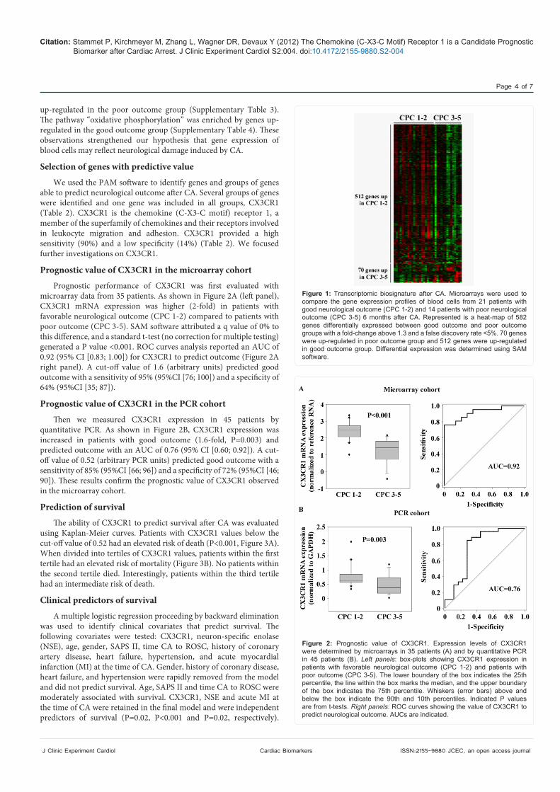
Prognostic value of CX3CR1 in the microarray cohort
Prognostic performance of CX3CR1 was first evaluated with microarray data from 35 patients. As shown in Figure 2A (left panel), CX3CR1 mRNA expression was higher (2-fold) in patients with favorable neurological outcome (CPC 1-2) compared to patients with poor outcome (CPC 3-5). SAM software attributed a q value of 0% to this difference, and a standard t-test (no correction for multiple testing) generated a P value <0.001. ROC curves analysis reported an AUC of 0.92 (95% CI [0.83; 1.00]) for CX3CR1 to predict outcome (Figure 2A right panel). A cut-off value of 1.6 (arbitrary units) predicted good outcome with a sensitivity of 95% (95%CI [76; 100]) and a specificity of 64% (95%CI [35; 87]).
Prognostic value of CX3CR1 in the PCR cohort
Then we measured CX3CR1 expression in 45 patients by quantitative PCR. As shown in Figure 2B, CX3CR1 expression was increased in patients with good outcome (1.6-fold, P=0.003) and predicted outcome with an AUC of 0.76 (95% CI [0.60; 0.92]). A cut-off value of 0.52 (arbitrary PCR units) predicted good outcome with a sensitivity of 85% (95%CI [66; 96]) and a specificity of 72% (95%CI [46; 90]). These results confirm the prognostic value of CX3CR1 observed in the microarray cohort.
Prediction of survival
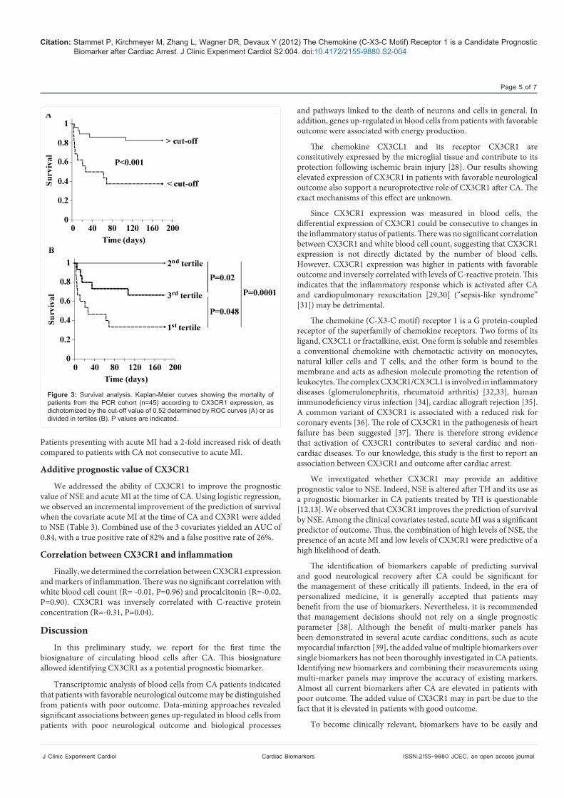
The ability of CX3CR1 to predict survival after CA was evaluated using Kaplan-Meier curves. Patients with CX3CR1 values below the cut-off value of 0.52 had an elevated risk of death (P<0.001, Figure 3A). When divided into tertiles of CX3CR1 values, patients within the first tertile had an elevated risk of mortality (Figure 3B). No patients within the second tertile died. Interestingly, patients within the third tertile had an intermediate risk of death.
Clinical predictors of survival
A multiple logistic regression proceeding by backward elimination was used to identify clinical covariates that predict survival. The following covariates were tested: CX3CR1, neuron-specific enolase (NSE), age, gender, SAPS II, time CA to ROSC, history of coronary artery disease, heart failure, hypertension, and acute myocardial infarction (MI) at the time of CA. Gender, history of coronary disease, heart failure, and hypertension were rapidly removed from the model and did not predict survival. Age, SAPS II and time CA to ROSC were moderately associated with survival. CX3CR1, NSE and acute MI at the time of CA were retained in the final model and were independent predictors of survival (P=0.02, P<0.001 and P=0.02, respectively).
Figure 1: Transcriptomic biosignature after CA. Microarrays were used to compare the gene expression profiles of blood cells from 21 patients with good neurological outcome (CPC 1-2) and 14 patients with poor neurological outcome (CPC 3-5) 6 months after CA. Represented is a heat-map of 582 genes differentially expressed between good outcome and poor outcome groups with a fold-change above 1.3 and a false discovery rate <5%. 70 genes were up-regulated in poor outcome group and 512 genes were up-regulated in good outcome group. Differential expression was determined using SAM software.
Figure 2: Prognostic value of CX3CR1. Expression levels of CX3CR1 were determined by microarrays in 35 patients (A) and by quantitative PCR in 45 patients (B). Left panels: box-plots showing CX3CR1 expression in patients with favorable neurological outcome (CPC 1-2) and patients with poor outcome (CPC 3-5). The lower boundary of the box indicates the 25th percentile, the line within the box marks the median, and the upper boundary of the box indicates the 75th percentile. Whiskers (error bars) above and below the box indicate the 90th and 10th percentiles. Indicated P values are from t-tests. Right panels: ROC curves showing the value of CX3CR1 to predict neurological outcome. AUCs are indicated.
Citation: Stammet P, Kirchmeyer M, Zhang L, Wagner DR, Devaux Y (2012) The Chemokine (C-X3-C Motif) Receptor 1 is a Candidate Prognostic Biomarker after Cardiac Arrest. J Clinic Experiment Cardiol S2:004. doi:10.4172/2155-9880.S2-004
Page 5 of 7
J Clinic Experiment Cardiol Cardiac Biomarkers ISSN:2155-9880 JCEC, an open access journal
Patients presenting with acute MI had a 2-fold increased risk of death compared to patients with CA not consecutive to acute MI.
Additive prognostic value of CX3CR1
We addressed the ability of CX3CR1 to improve the prognostic value of NSE and acute MI at the time of CA. Using logistic regression, we observed an incremental improvement of the prediction of survival when the covariate acute MI at the time of CA and CX3R1 were added to NSE (Table 3). Combined use of the 3 covariates yielded an AUC of 0.84, with a true positive rate of 82% and a false positive rate of 26%.
Correlation between CX3CR1 and inflammation
Finally, we determined the correlation between CX3CR1 expression and markers of inflammation. There was no significant correlation with white blood cell count (R= -0.01, P=0.96) and procalcitonin (R=-0.02, P=0.90). CX3CR1 was inversely correlated with C-reactive protein concentration (R=-0.31, P=0.04).
DiscussionIn this preliminary study, we report for the first time the
biosignature of circulating blood cells after CA. This biosignature allowed identifying CX3CR1 as a potential prognostic biomarker.
Transcriptomic analysis of blood cells from CA patients indicated that patients with favorable neurological outcome may be distinguished from patients with poor outcome. Data-mining approaches revealed significant associations between genes up-regulated in blood cells from patients with poor neurological outcome and biological processes
and pathways linked to the death of neurons and cells in general. In addition, genes up-regulated in blood cells from patients with favorable outcome were associated with energy production.
The chemokine CX3CL1 and its receptor CX3CR1 are constitutively expressed by the microglial tissue and contribute to its protection following ischemic brain injury [28]. Our results showing elevated expression of CX3CR1 in patients with favorable neurological outcome also support a neuroprotective role of CX3CR1 after CA. The exact mechanisms of this effect are unknown.
Since CX3CR1 expression was measured in blood cells, the differential expression of CX3CR1 could be consecutive to changes in the inflammatory status of patients. There was no significant correlation between CX3CR1 and white blood cell count, suggesting that CX3CR1 expression is not directly dictated by the number of blood cells. However, CX3CR1 expression was higher in patients with favorable outcome and inversely correlated with levels of C-reactive protein. This indicates that the inflammatory response which is activated after CA and cardiopulmonary resuscitation [29,30] (“sepsis-like syndrome” [31]) may be detrimental.
The chemokine (C-X3-C motif) receptor 1 is a G protein-coupled receptor of the superfamily of chemokine receptors. Two forms of its ligand, CX3CL1 or fractalkine, exist. One form is soluble and resembles a conventional chemokine with chemotactic activity on monocytes, natural killer cells and T cells, and the other form is bound to the membrane and acts as adhesion molecule promoting the retention of leukocytes. The complex CX3CR1/CX3CL1 is involved in inflammatory diseases (glomerulonephritis, rheumatoid arthritis) [32,33], human immunodeficiency virus infection [34], cardiac allograft rejection [35]. A common variant of CX3CR1 is associated with a reduced risk for coronary events [36]. The role of CX3CR1 in the pathogenesis of heart failure has been suggested [37]. There is therefore strong evidence that activation of CX3CR1 contributes to several cardiac and non-cardiac diseases. To our knowledge, this study is the first to report an association between CX3CR1 and outcome after cardiac arrest.
We investigated whether CX3CR1 may provide an additive prognostic value to NSE. Indeed, NSE is altered after TH and its use as a prognostic biomarker in CA patients treated by TH is questionable [12,13]. We observed that CX3CR1 improves the prediction of survival by NSE. Among the clinical covariates tested, acute MI was a significant predictor of outcome. Thus, the combination of high levels of NSE, the presence of an acute MI and low levels of CX3CR1 were predictive of a high likelihood of death.
The identification of biomarkers capable of predicting survival and good neurological recovery after CA could be significant for the management of these critically ill patients. Indeed, in the era of personalized medicine, it is generally accepted that patients may benefit from the use of biomarkers. Nevertheless, it is recommended that management decisions should not rely on a single prognostic parameter [38]. Although the benefit of multi-marker panels has been demonstrated in several acute cardiac conditions, such as acute myocardial infarction [39], the added value of multiple biomarkers over single biomarkers has not been thoroughly investigated in CA patients. Identifying new biomarkers and combining their measurements using multi-marker panels may improve the accuracy of existing markers. Almost all current biomarkers after CA are elevated in patients with poor outcome. The added value of CX3CR1 may in part be due to the fact that it is elevated in patients with good outcome.
To become clinically relevant, biomarkers have to be easily and
Figure 3: Survival analysis. Kaplan-Meier curves showing the mortality of patients from the PCR cohort (n=45) according to CX3CR1 expression, as dichotomized by the cut-off value of 0.52 determined by ROC curves (A) or as divided in tertiles (B). P values are indicated.
Citation: Stammet P, Kirchmeyer M, Zhang L, Wagner DR, Devaux Y (2012) The Chemokine (C-X3-C Motif) Receptor 1 is a Candidate Prognostic Biomarker after Cardiac Arrest. J Clinic Experiment Cardiol S2:004. doi:10.4172/2155-9880.S2-004
Page 6 of 7
J Clinic Experiment Cardiol Cardiac Biomarkers ISSN:2155-9880 JCEC, an open access journal
quickly measurable. In the present study, CX3CR1 was quantified by PCR, a technique that is time-consuming but which may become faster and automated in the future.
Future studies on large populations of cardiac arrest patients are needed to determine: (1) the kinetics of biomarkers after hypothermia, (2) the effect of hypothermia on biomarker levels, (3) the cut-offs for optimal prediction, (4) the added value of combined biomarkers, (5) the effect of biomarker determination on patient care and outcome.
This study is limited by a small population size. In addition, the PCR cohort used for validation purposes was not an independent cohort since it enrolled the patients of the microarray cohort and 10 additional patients.
In conclusion, we have identified CX3CR1 as a new candidate prognostic biomarker after CA which may be further evaluated in larger patient populations.
Acknowledgments
We thank Bernadette Leners, Malou Gloesener and Loredana Jacobs for expert technical assistance. This study was supported by grants from the Society for Research on Cardiovascular Diseases, the Ministry of Culture, Higher Education and Research, and the National Funds of Research of Luxembourg. M.K. was recipient of a fellowship from the National Funds of Research of Luxembourg (grant # TR-PDR-BFR07-145).
References
1. de Vreede-Swagemakers JJ, Gorgels AP, Dubois-Arbouw WI, van Ree JW, Daemen MJ, et al. (1997) Out-of-hospital cardiac arrest in the 1990’s: a population-based study in the Maastricht area on incidence, characteristics and survival. J Am Coll Cardiol 30: 1500-1505.
2. Nichol G, Rumsfeld J, Eigel B, Abella BS, Labarthe D, et al. (2008) Essential features of designating out-of-hospital cardiac arrest as a reportable event: a scientific statement from the American Heart Association Emergency Cardiovascular Care Committee; Council on Cardiopulmonary, Perioperative, and Critical Care; Council on Cardiovascular Nursing; Council on Clinical Cardiology; and Quality of Care and Outcomes Research Interdisciplinary Working Group. Circulation 117: 2299-2308.
3. Straus SM, Bleumink GS, Dieleman JP, van der Lei J, Stricker BH, et al. (2004) The incidence of sudden cardiac death in the general population. J Clin Epidemiol 57: 98-102.
4. Bernard SA, Gray TW, Buist MD, Jones BM, Silvester W, et al. (2002) Treatment of comatose survivors of out-of-hospital cardiac arrest with induced hypothermia. N Engl J Med 346: 557-563.
5. Hypothermia after Cardiac Arrest Study Group (2002) Mild therapeutic hypothermia to improve the neurologic outcome after cardiac arrest. N Engl J Med 346: 549-556.
6. Busto R, Globus MY, Dietrich WD, Martinez E, Valdes I, et al. (1989) Effect of mild hypothermia on ischemia-induced release of neurotransmitters and free fatty acids in rat brain. Stroke 20: 904-910.
7. Morimoto Y, Kemmotsu O, Kitami K, Matsubara I, Tedo I (1993) Acute brain swelling after out-of-hospital cardiac arrest: pathogenesis and outcome. Crit Care Med 21: 104-110.
8. Meynaar IA, Oudemans-van Straaten HM, van der Wetering J, Verlooy P, Slaats EH, et al. (2003) Serum neuron-specific enolase predicts outcome in post-anoxic coma: a prospective cohort study. Intensive Care Med 29: 189-195.
9. Reisinger J, Hollinger K, Lang W, Steiner C, Winter T, et al. (2007) Prediction of neurological outcome after cardiopulmonary resuscitation by serial determination of serum neuron-specific enolase. Eur Heart J 28: 52-58.
10. Schoerkhuber W, Kittler H, Sterz F, Behringer W, Holzer M, et al. (1999) Time course of serum neuron-specific enolase. A predictor of neurological outcome in patients resuscitated from cardiac arrest. Stroke 30: 1598-1603.
11. Zandbergen EG, Koelman JH, de Haan RJ, Hijdra A, PROPAC-Study Group (2006) SSEPs and prognosis in postanoxic coma: only short or also long latency responses? Neurology 67: 583-586.
12. Tiainen M, Roine RO, Pettila V, Takkunen O (2003) Serum neuron-specific enolase and S-100B protein in cardiac arrest patients treated with hypothermia. Stroke 34: 2881-2886.
13. Steffen IG, Hasper D, Ploner CJ, Schefold JC, Dietz E, et al. (2010) Mild therapeutic hypothermia alters neuron specific enolase as an outcome predictor after resuscitation: 97 prospective hypothermia patients compared to 133 historical non-hypothermia patients. Crit Care 14: R69.
14. Jacobs I, Nadkarni V, Bahr J, Berg RA, Billi JE, et al. (2004) Cardiac arrest and cardiopulmonary resuscitation outcome reports: update and simplification of the Utstein templates for resuscitation registries. A statement for healthcare professionals from a task force of the international liaison committee on resuscitation (American Heart Association, European Resuscitation Council, Australian Resuscitation Council, New Zealand Resuscitation Council, Heart and Stroke Foundation of Canada, InterAmerican Heart Foundation, Resuscitation Council of Southern Africa). Resuscitation 63: 233-249.
15. Nolan JP, Deakin CD, Soar J, Bottiger BW, Smith G (2005) European Resuscitation Council guidelines for resuscitation 2005. Section 4. Adult advanced life support. Resuscitation 67 Suppl 1: S39-86.
16. Bernard S, Buist M, Monteiro O, Smith K (2003) Induced hypothermia using large volume, ice-cold intravenous fluid in comatose survivors of out-of-hospital cardiac arrest: a preliminary report. Resuscitation 56: 9-13.
17. Pichon N, Amiel JB, Francois B, Dugard A, Etchecopar C, et al. (2007) Efficacy of and tolerance to mild induced hypothermia after out-of-hospital cardiac arrest using an endovascular cooling system. Crit Care 11: R71.
18. Booth CM, Boone RH, Tomlinson G, Detsky AS (2004) Is this patient dead, vegetative, or severely neurologically impaired? Assessing outcome for comatose survivors of cardiac arrest. JAMA 291: 870-879.
19. Devaux Y, Azuaje F, Vausort M, Yvorra C, Wagner DR (2010) Integrated protein network and microarray analysis to identify potential biomarkers after myocardial infarction. Funct Integr Genomics 10: 329-337.
20. Devaux Y, Bousquenaud M, Rodius S, Marie PY, Maskali F, et al. (2011) Transforming growth factor beta receptor 1 is a new candidate prognostic biomarker after acute myocardial infarction. BMC Medical Genomics 4: 83.
21. Le Brigand K, Russell R, Moreilhon C, Rouillard JM, Jost B, et al. (2006) An open-access long oligonucleotide microarray resource for analysis of the human and mouse transcriptomes. Nucleic Acids Res 34: e87.
22. Novikov E, Barillot E (2007) Software package for automatic microarray image analysis (MAIA). Bioinformatics 23: 639-640.
23. Tusher VG, Tibshirani R, Chu G (2001) Significance analysis of microarrays applied to the ionizing radiation response. Proc Natl Acad Sci U S A 98: 5116-5121.
24. Tibshirani R, Hastie T, Narasimhan B, Chu G (2002) Diagnosis of multiple cancer types by shrunken centroids of gene expression. Proc Natl Acad Sci U S A 99: 6567-6572.
25. Huang da W, Sherman BT, Tan Q, Collins JR, Alvord WG, et al. (2007) The DAVID Gene Functional Classification Tool: a novel biological module-centric algorithm to functionally analyze large gene lists. Genome Biol 8: R183.
26. Zeeberg BR, Feng W, Wang G, Wang MD, Fojo AT, et al. (2003) GoMiner: a resource for biological interpretation of genomic and proteomic data. Genome Biol 4: R28.
27. de Hoon MJ, Imoto S, Nolan J, Miyano S (2004) Open source clustering software. Bioinformatics 20: 1453-1454.
28. Cipriani R, Villa P, Chece G, Lauro C, Paladini A, et al. (2011) CX3CL1 Is Neuroprotective in Permanent Focal Cerebral Ischemia in Rodents. J Neurosci 31: 16327-16335.
29. Geppert A, Zorn G, Karth GD, Haumer M, Gwechenberger M, et al. (2000) Soluble selectins and the systemic inflammatory response syndrome after successful cardiopulmonary resuscitation. Crit Care Med 28: 2360-2365.
30. Bottiger BW, Motsch J, Braun V, Martin E, Kirschfink M (2002) Marked activation of complement and leukocytes and an increase in the concentrations of soluble endothelial adhesion molecules during cardiopulmonary resuscitation and early reperfusion after cardiac arrest in humans. Crit Care Med 30: 2473-2480.
31. Adrie C, Adib-Conquy M, Laurent I, Monchi M, Vinsonneau C, et al. (2002) Successful cardiopulmonary resuscitation after cardiac arrest as a “sepsis-like” syndrome. Circulation 106: 562-568.
Citation: Stammet P, Kirchmeyer M, Zhang L, Wagner DR, Devaux Y (2012) The Chemokine (C-X3-C Motif) Receptor 1 is a Candidate Prognostic Biomarker after Cardiac Arrest. J Clinic Experiment Cardiol S2:004. doi:10.4172/2155-9880.S2-004
Page 7 of 7
J Clinic Experiment Cardiol Cardiac Biomarkers ISSN:2155-9880 JCEC, an open access journal
32. Stievano L, Piovan E, Amadori A (2004) C and CX3C chemokines: cell sources and physiopathological implications. Crit Rev Immunol 24: 205-228.
33. Umehara H, Bloom ET, Okazaki T, Nagano Y, Yoshie O, et al. (2004) Fractalkine in vascular biology: from basic research to clinical disease. Arterioscler Thromb Vasc Biol 24: 34-40.
34. Faure S, Meyer L, Costagliola D, Vaneensberghe C, Genin E, et al. (2000) Rapid progression to AIDS in HIV+ individuals with a structural variant of the chemokine receptor CX3CR1. Science 287: 2274-2277.
35. Robinson LA, Nataraj C, Thomas DW, Howell DN, Griffiths R, et al. (2000) A role for fractalkine and its receptor (CX3CR1) in cardiac allograft rejection. J Immunol 165: 6067-6072.
36. McDermott DH, Fong AM, Yang Q, Sechler JM, Cupples LA, et al. (2003)
Chemokine receptor mutant CX3CR1-M280 has impaired adhesive function and correlates with protection from cardiovascular disease in humans. J Clin Invest 111: 1241-1250.
37. Husberg C, Nygard S, Finsen AV, Damas JK, Frigessi A, et al. (2008) Cytokine expression profiling of the myocardium reveals a role for CX3CL1 (fractalkine) in heart failure. J Mol Cell Cardiol 45: 261-269.
38. Samaniego EA, Persoon S, Wijman CA (2011) Prognosis after cardiac arrest and hypothermia: a new paradigm. Curr Neurol Neurosci Rep 11: 111-119.
39. Sabatine MS, Morrow DA, de Lemos JA, Gibson CM, Murphy SA, et al. (2002) Multimarker approach to risk stratification in non-ST elevation acute coronary syndromes: simultaneous assessment of troponin I, C-reactive protein, and B-type natriuretic peptide. Circulation 105: 1760-1763.
Thisarticlewasoriginallypublishedinaspecialissue,Cardiac Biomarkers handled by Editor(s). Dr. Virginija Jazbutyte, Institute for Molecular andTranslationalTherapyStrategies,Germany