94 DOI: http://doi.org/10.4038/sljm.v30i1.277 Sri Lanka Journal of Medicine Vol. 30 No.1, 2021 Sri Lanka Journal of Medicine SLJM Brief Report Citation: Perera SDC, 2021. Early onset acute lymphocytic interstitial pneumonitis (ALIP) in COVID-19 pathophysiology; A narrative review. Sri Lanka Journal of Medicine, 30(1), pp 94-98 DOI: http://doi.org/10.4038/sljm.v30i1.277 Early onset acute lymphocytic interstitial pneumonitis (ALIP) in COVID-19 pathophysiology: Preliminary lung autopsy findings. Perera SDC Abstract Sri Lanka is one of few countries which has performed post mortem examinations in deaths due to SARS CoV- 2 virus (COVID-19) infection. At the office of the Judicial Medical Officer of National Institute of Infectious Diseases (NIID)/Infectious Disease Hospital (IDH) we have performed over sixty (60) post mortem examinations of deceased persons who were confirmed positive for COVID-19 by Real Time - Polymerase Chain reaction (RT-PCR) test performed before or after death. They included a majority of deaths due to natural causes and few deaths due to unnatural causes such as accidents, suicides and homicides. The first autopsy was performed on 04 th of May 2020. We have observed acute lymphocytic interstitial pneumonitis (ALIP) as a potentially fatal complication in COVID-19 infection at the initial stages of lung involvement. This phenomenon of early onset ALIP has not been reported in the literature of COVID-19 lung pathophysiology to date. This is a narrative of our histological findings. Our attempt is to highlight the importance early onset ALIP as a pathological entity in COVID-19 lung involvement, which will enhance the clinicians to better understand the pathophysiology of this new illness and modify their treatment strategies. Keywords: Interstitial pneumonitis, Hyaline membrane, Diffuse alveolar damage, ARDS, viral pneumonia, COVID INTRODUCTION Pathophysiology of COVID-19 lung involvement is complex, multi-factorial and the global understanding on same among clinicians and researchers is evolving. Acute alveolar inflammation, ARDS, diffuse alveolar damage, secondary bacterial infections, organizing pneumonia have all been described as pathophysiological mechanism at different stages of illness 1 . National Institute of Infectious Diseases, Sri Lanka Correspondence: Dr. S D Channa Perera Consultant Judicial Medical Officer Consultant Forensic Pathologist Office of the Judicial Medical officer, National Institute of Infectious Diseases, Infectious Disease Hospital (IDH), Angoda, Sri Lanka. E-mail: [email protected]Received: 2021-04-28 Accepted revised version: 2021-06-10 Published: 2021-06-30 This work is licensed under a Creative Commons Attribution 4.0 International License (CC BY)
Transcript
94
DOI: http://doi.org/10.4038/sljm.v30i1.277 Sri Lanka Journal of Medicine Vol. 30 No.1, 2021
Sri Lanka Journal of Medicine SLJM
Brief Report Citation: Perera SDC, 2021. Early onset acute lymphocytic interstitial pneumonitis (ALIP) in COVID-19 pathophysiology; A narrative review. Sri Lanka Journal of Medicine, 30(1), pp 94-98 DOI: http://doi.org/10.4038/sljm.v30i1.277
Early onset acute lymphocytic interstitial pneumonitis (ALIP) in COVID-19 pathophysiology: Preliminary lung autopsy findings.
Perera SDC
Abstract
Sri Lanka is one of few countries which has performed post mortem examinations in deaths due to SARS CoV-2 virus (COVID-19) infection. At the office of the Judicial Medical Officer of National Institute of Infectious Diseases (NIID)/Infectious Disease Hospital (IDH) we have performed over sixty (60) post mortem examinations of deceased persons who were confirmed positive for COVID-19 by Real Time - Polymerase Chain reaction (RT-PCR) test performed before or after death. They included a majority of deaths due to natural causes and few deaths due to unnatural causes such as accidents, suicides and homicides. The first autopsy was performed on 04th of May 2020. We have observed acute lymphocytic interstitial pneumonitis (ALIP) as a potentially fatal complication in COVID-19 infection at the initial stages of lung involvement. This phenomenon of early onset ALIP has not been reported in the literature of COVID-19 lung pathophysiology to date. This is a narrative of our histological findings. Our attempt is to highlight the importance early onset ALIP as a pathological entity in COVID-19 lung involvement, which will enhance the clinicians to better understand the pathophysiology of this new illness and modify their treatment strategies.
Pathophysiology of COVID-19 lung involvement is complex, multi-factorial and the global understanding on same among clinicians and researchers is evolving. Acute alveolar inflammation, ARDS, diffuse alveolar damage,
secondary bacterial infections, organizing pneumonia have all been described as pathophysiological mechanism at different stages of illness1.
National Institute of Infectious Diseases, Sri Lanka
Correspondence: Dr. S D Channa Perera Consultant Judicial Medical Officer Consultant Forensic Pathologist Office of the Judicial Medical officer, National Institute of Infectious Diseases, Infectious Disease Hospital (IDH), Angoda, Sri Lanka. E-mail: [email protected]
Acute Lymphocytic Interstitial Pneumonitis Sri Lanka Journal of Medicine Vol.30 No.1,2021
in COVID-19
95
Early ALIP as a clinical entity in COVID-19 lung disease has not been reported before. However, we have identified this phenomenon by studying histology of the post mortem examinations performed in deceased persons who were infected with COVID-19. We have observed Early ALIP as a potentially fatal complication in COVID-19 infection at the initial stage of lung involvement. We believe that early identification and treatment of this condition could prevent or minimize the effects of severe lung damage and disease progression. Our attempt is to highlight the importance of early clinical diagnosis of ALIP, which will enhance the clinicians to modify their treatment of COVID-19 lung disease. Based on our autopsy experience in COVID-19, we have formulated the following histopathological classification. Larger observational studies would be needed to clarify the sequential nature of the stages mentioned. Stage 1: Upper respiratory tract infection. Stage 2: Lower respiratory tract infection.
Stage 3: Secondary bacterial pneumonia. Stage 4: Systemic hypercoagulable stage. Stage 1: Upper respiratory tract infection Majority of persons infected with COVID-19 virus are either asymptomatic or mildly symptomatic. They may show symptoms of upper respiratory infection such as a fever, sneezing, dry cough, loss of smell and body aches etc. Large number of persons in society who are actually infected would not even notice that they have contracted COVID-19 infection. Most of the patients recover at this stage with or without specific treatment or complications. However, patients with certain co-morbidities, could be seen to develop worsening of complications of existing disease at this stage. It has been assumed that COVID-19 infection may play an aggravating role in such patients. Commonly, COVID-19 infection could even be an incidental finding at post mortem examinations.
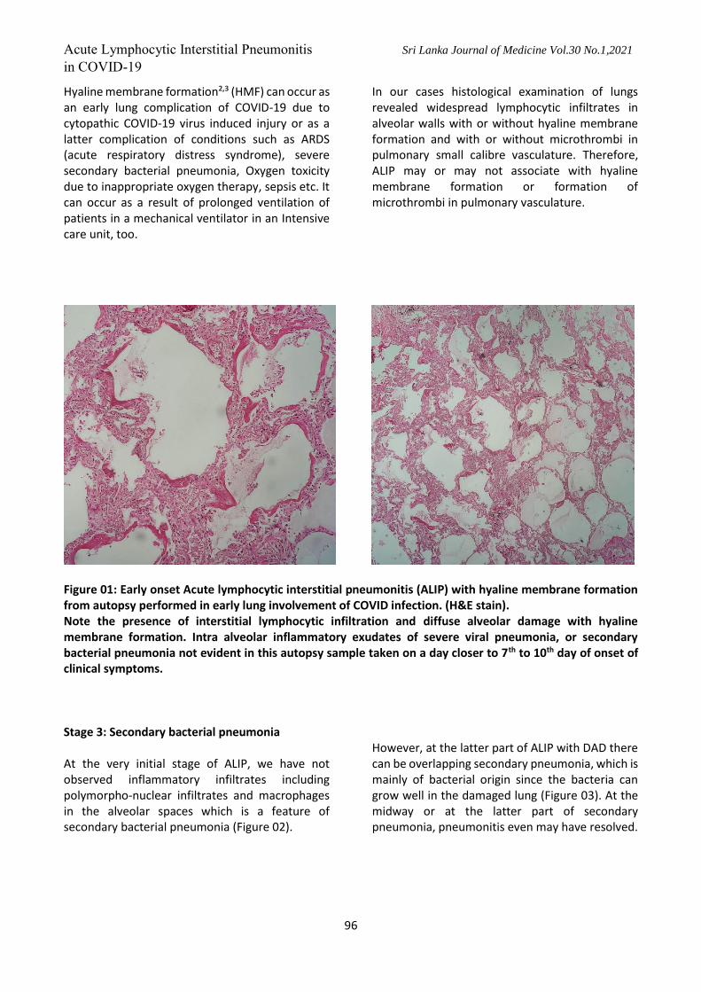
Stage 2: Lower respiratory tract infection A small percentage of patients could develop lower respiratory symptoms. Those who are vulnerable to this condition are the elderly, debilitated people, immunocompromised persons including those infected with the human immunodeficiency virus (HIV), persons with cancer, persons undergoing chemotherapy, radiotherapy, and are undergoing treatment following transplantation, etc. Furthermore, persons with comorbidities such as hypertension, diabetes, bronchial asthma, heart disease and pregnant mothers also belong to this vulnerable group. The two types of cells that line the alveolar wall are Type I pneumocytes which are squamous cells and type II pneumocytes which are cuboidal cells. Type II pneumocytes produce surfactant that keeps the surface tension of the alveoli and prevent them getting collapsed2. At the initial stage of lower respiratory tract infections, in histological sections we have observed ALIP and DAD with hyaline membrane formation (Figure 01). Our understanding is that ALIP in COVID-19 could occur at the end of 1st week to the second week of infection. However, the dating of these complications need more studies. At this stage there could also be formation of micro thrombi in pulmonary vasculature. DAD may occur with ALIP leading to destruction of the alveolar wall. Damage to type II pneumocytes may cause alveolar collapse due to reduced production of surfactant which is important for the stability of alveolar wall. Type II pneumocytes have the ability to regenerate themselves as well as forming type I pneumocytes whereas type I pneumocytes does not have the ability to regenerate2. Therefore, regeneration of type II pneumocytes reconstruct the damaged alveolar wall. The initial stage of diffuse alveolar damage (DAD) is a reversible condition which should not be neglected to prevent it from advancing into irreversible stage2.
Acute Lymphocytic Interstitial Pneumonitis Sri Lanka Journal of Medicine Vol.30 No.1,2021
in COVID-19
96
Hyaline membrane formation2,3 (HMF) can occur as an early lung complication of COVID-19 due to cytopathic COVID-19 virus induced injury or as a latter complication of conditions such as ARDS (acute respiratory distress syndrome), severe secondary bacterial pneumonia, Oxygen toxicity due to inappropriate oxygen therapy, sepsis etc. It can occur as a result of prolonged ventilation of patients in a mechanical ventilator in an Intensive care unit, too.
In our cases histological examination of lungs revealed widespread lymphocytic infiltrates in alveolar walls with or without hyaline membrane formation and with or without microthrombi in pulmonary small calibre vasculature. Therefore, ALIP may or may not associate with hyaline membrane formation or formation of microthrombi in pulmonary vasculature.
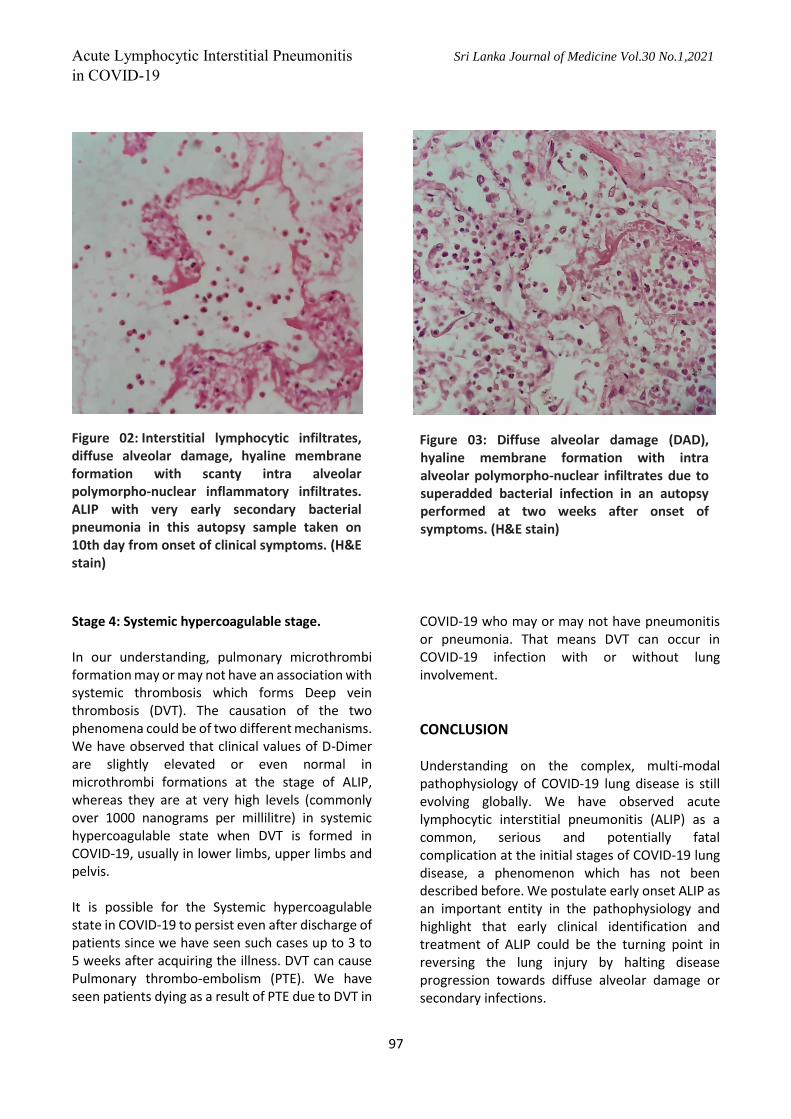
Figure 01: Early onset Acute lymphocytic interstitial pneumonitis (ALIP) with hyaline membrane formation from autopsy performed in early lung involvement of COVID infection. (H&E stain). Note the presence of interstitial lymphocytic infiltration and diffuse alveolar damage with hyaline membrane formation. Intra alveolar inflammatory exudates of severe viral pneumonia, or secondary bacterial pneumonia not evident in this autopsy sample taken on a day closer to 7th to 10th day of onset of clinical symptoms. Stage 3: Secondary bacterial pneumonia At the very initial stage of ALIP, we have not observed inflammatory infiltrates including polymorpho-nuclear infiltrates and macrophages in the alveolar spaces which is a feature of secondary bacterial pneumonia (Figure 02).
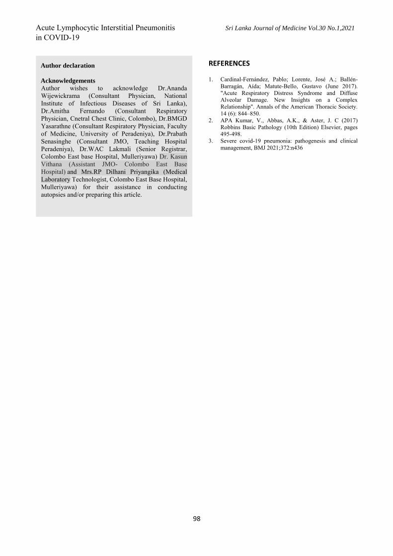
However, at the latter part of ALIP with DAD there can be overlapping secondary pneumonia, which is mainly of bacterial origin since the bacteria can grow well in the damaged lung (Figure 03). At the midway or at the latter part of secondary pneumonia, pneumonitis even may have resolved.
Acute Lymphocytic Interstitial Pneumonitis Sri Lanka Journal of Medicine Vol.30 No.1,2021
in COVID-19
97
Stage 4: Systemic hypercoagulable stage. In our understanding, pulmonary microthrombi formation may or may not have an association with systemic thrombosis which forms Deep vein thrombosis (DVT). The causation of the two phenomena could be of two different mechanisms. We have observed that clinical values of D-Dimer are slightly elevated or even normal in microthrombi formations at the stage of ALIP, whereas they are at very high levels (commonly over 1000 nanograms per millilitre) in systemic hypercoagulable state when DVT is formed in COVID-19, usually in lower limbs, upper limbs and pelvis. It is possible for the Systemic hypercoagulable state in COVID-19 to persist even after discharge of patients since we have seen such cases up to 3 to 5 weeks after acquiring the illness. DVT can cause Pulmonary thrombo-embolism (PTE). We have seen patients dying as a result of PTE due to DVT in
COVID-19 who may or may not have pneumonitis or pneumonia. That means DVT can occur in COVID-19 infection with or without lung involvement.
CONCLUSION Understanding on the complex, multi-modal pathophysiology of COVID-19 lung disease is still evolving globally. We have observed acute lymphocytic interstitial pneumonitis (ALIP) as a common, serious and potentially fatal complication at the initial stages of COVID-19 lung disease, a phenomenon which has not been described before. We postulate early onset ALIP as an important entity in the pathophysiology and highlight that early clinical identification and treatment of ALIP could be the turning point in reversing the lung injury by halting disease progression towards diffuse alveolar damage or secondary infections.
Figure 02: Interstitial lymphocytic infiltrates, diffuse alveolar damage, hyaline membrane formation with scanty intra alveolar polymorpho-nuclear inflammatory infiltrates. ALIP with very early secondary bacterial pneumonia in this autopsy sample taken on 10th day from onset of clinical symptoms. (H&E stain)
Figure 03: Diffuse alveolar damage (DAD), hyaline membrane formation with intra alveolar polymorpho-nuclear infiltrates due to superadded bacterial infection in an autopsy performed at two weeks after onset of symptoms. (H&E stain)
Acute Lymphocytic Interstitial Pneumonitis Sri Lanka Journal of Medicine Vol.30 No.1,2021
in COVID-19
98
REFERENCES
1. Cardinal-Fernández, Pablo; Lorente, José A.; Ballén-Barragán, Aída; Matute-Bello, Gustavo (June 2017). "Acute Respiratory Distress Syndrome and Diffuse Alveolar Damage. New Insights on a Complex Relationship". Annals of the American Thoracic Society. 14 (6): 844–850.
2. APA Kumar, V., Abbas, A.K., & Aster, J. C (2017) Robbins Basic Pathology (10th Edition) Elsevier, pages 495-498.
3. Severe covid-19 pneumonia: pathogenesis and clinical management, BMJ 2021;372:n436
Author declaration
Acknowledgements
Author wishes to acknowledge Dr.Ananda Wijewickrama (Consultant Physician, National Institute of Infectious Diseases of Sri Lanka), Dr.Amitha Fernando (Consultant Respiratory Physician, Cnetral Chest Clinic, Colombo), Dr.BMGD Yasarathne (Consultant Respiratory Physician, Faculty of Medicine, University of Peradeniya), Dr.Prabath Senasinghe (Consultant JMO, Teaching Hospital Peradeniya), Dr.WAC Lakmali (Senior Registrar, Colombo East base Hospital, Mulleriyawa) Dr. Kasun Vithana (Assistant JMO- Colombo East Base Hospital) and Mrs.RP Dilhani Priyangika (Medical Laboratory Technologist, Colombo East Base Hospital, Mulleriyawa) for their assistance in conducting autopsies and/or preparing this article.