ORIGINAL RESEARCH
Early Response of Bone Marrow Osteoprogenitors to SkeletalUnloading and Sclerostin Antibody
Mohammad Shahnazari • Thomas Wronski •
Vivian Chu • Alyssa Williams • Alicia Leeper •
Marina Stolina • Hua Zhu Ke • Bernard Halloran
Received: 24 February 2012 / Accepted: 16 April 2012 / Published online: 27 May 2012
� Springer Science+Business Media, LLC 2012
Abstract Sclerostin functions as an antagonist to Wnt
signaling and inhibits bone-forming activity. We studied
the effects of skeletal unloading and treatment with
sclerostin antibody (Scl-Ab) on mesenchymal stem cell,
osteoprogenitor and osteoclast precursor pools, and their
relationship to bone formation and resorption. Male
C57BL/6 mice (5-months-old) were hind limb unloaded for
1 week or allowed normal ambulation and treated with Scl-
Ab (25 mg/kg, s.c. injections on days 1 and 4) or placebo.
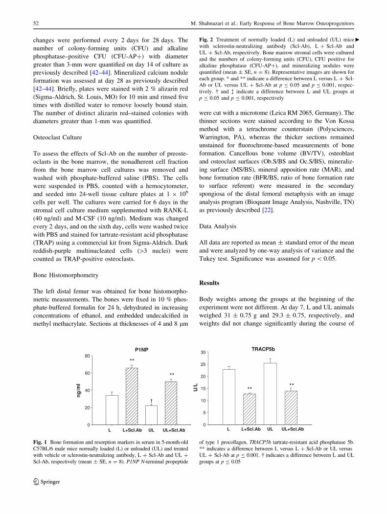
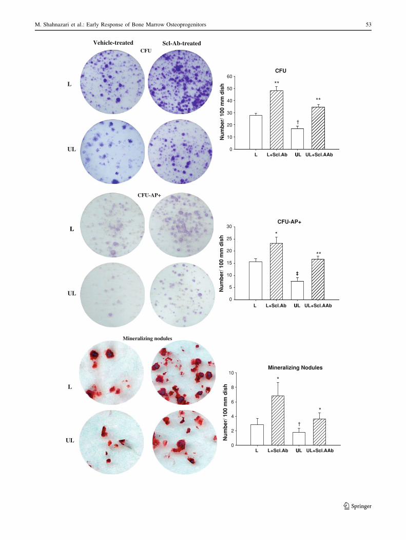
Unloading decreased the serum concentration of bone
formation marker P1NP (-35 %), number of colony-
forming units (CFU) (-38 %), alkaline phosphatase–
positive CFUs (CFU-AP?) (-51 %), and calcified nodules
(-35 %); and resulted in a fourfold increase in the number
of osteoclast precursors. The effects of Scl-Ab treatment on
unloaded and normally loaded mice were nearly identical;
Scl-Ab increased serum P1NP and the number of CFU,
CFU-AP?, and calcified nodules in ex vivo cultures; and
increased osteoblast and bone mineralizing surfaces in
vivo. Although the marrow-derived osteoclast precursor
population increased with Scl-Ab, the bone osteoclast
surface did not change, and the serum concentration of
osteoclast activity marker TRACP5b decreased. Our data
suggest that short-term Scl-Ab treatment can prevent the
decrease in osteoprogenitor population associated with
skeletal unloading and increase osteoblast surface and bone
mineralizing surface in unloaded animals. The anabolic
effects of Scl-Ab treatment on bone are preserved during
skeletal unloading. These findings suggest that Scl-Ab
treatment can both increase bone formation and decrease
bone resorption, and provide a new means for prevention
and treatment of disuse osteoporosis.
Keywords Bone � Osteoblast � Osteoclast � Sclerostin �Skeletal unloading
Targeting the Wnt signaling pathway to augment bone
formation has been the focus of numerous recent studies
[1–4]. Wnt pathways are involved in coordinating proper
bone development, formation, and growth, both before and
after birth [5, 6]. Signaling by Wnt proteins is antagonized
by sclerostin, which is expressed mainly by osteocytes and
functions to inhibit bone formation [7, 8]. Targeted dele-
tion of the sclerostin gene increases the osteoblast surface
(Ob.S/BS), mineralizing surface (MS/BS), bone formation
rate (BFR), and bone volume (BV/TV) [9]. Treatment of
adult rats with sclerostin antibody (Scl-Ab) for 3–5 weeks
is reported to increase MS/BS and BFR [10] and of
estrogen-deficient rats to increase Ob.S/BS, MS/BS, BFR,
and BV/TV [11]. These data overwhelmingly demonstrate
the anabolic effect of decreasing sclerostin activity. The
effects of blocking sclerostin activity on bone resorption
are less clear; the ratio of ratio of osteoclast surface to bone
Thomas Wronski received research funding from Amgen Inc. Marina
Stolina and Hua Zhu Ke are employed by Amgen. Marina Stolina,
Hua Zhu Ke, and Thomas Wronski have stock ownership in Amgen.
Other authors have stated that they have no conflict of interest.
M. Shahnazari � V. Chu � B. Halloran (&)
Division of Endocrinology, Veterans Affairs Medical Center,
University of California, San Francisco, CA 94121, USA
e-mail: [email protected]
T. Wronski � A. Williams � A. Leeper
Department of Physiological Sciences, College of Veterinary
Medicine, University of Florida, Gainesville, FL 32610-0125,
USA
M. Stolina � H. Z. Ke
Department of Metabolic Disorders, Amgen Inc., One Amgen
Center Drive, Thousand Oaks, CA 91320, USA
123
Calcif Tissue Int (2012) 91:50–58
DOI 10.1007/s00223-012-9610-9
surface (Oc.S/BS) has been reported to decrease [11] or
remain unchanged [9]. In postmenopausal women, Scl-Ab
was shown to increase bone formation markers and bone
mineral density with a concurrent reduction in serum type I
collagen C-telopeptides, a marker of bone resorption [4].
That sclerostin regulates bone resorption is supported by
studies that suggest that sclerostin may direct hematopoi-
etic cell lineage allocation, stimulate osteoclast recruit-
ment, and increase activity [12–16].
Skeletal disuse or loss of weight bearing inhibits bone
formation and induces osteopenia in both humans and
animals [17–21]. These effects are associated with a
reduction in Ob.S/BS and MS/BS [18, 22–27]. In animal
models, hind limb unloading increases sclerostin expres-
sion in bone, suggesting a mechanism by which skeletal
unloading down-regulates osteogenesis and decreases bone
formation [28]. In vitro mechanical loading of osteoblastic
cell lines has been shown to decrease expression of scle-
rostin mRNA and protein [29, 30]. The effects of skeletal
unloading on bone resorption are less clear, with reports
indicating that unloading in mice and rats increases
osteoclast number and bone resorption [18, 31, 32] or has
no effect [22, 24–26, 33–35]. In the adult human, resorp-
tion markers are increased significantly with loss of weight
bearing [17, 36–38].
Data on the effects of Scl-Ab treatment during skeletal
unloading are limited. Tian et al. [39] reported that long-
term treatment with Scl-Ab (twice per week for 4 weeks)
increased bone mass by stimulating bone formation and
inhibiting bone resorption in an immobilized female rat
model. However, the early response of bone to unloading
and Scl-Ab treatment has received little attention, and the
effects of unloading and Scl-Ab on the osteoprogenitor and
osteoclast precursor populations have not been studied. We
studied the effects of unloading and treatment with a
neutralizing Scl-Ab on the mesenchymal stem cell, osteo-
progenitor, and osteoclast precursor pools, as well as their
relationship to bone formation and resorption using the
mouse hind limb unloading model. Our results suggest that
treatment with Scl-Ab greatly increases osteoprogenitor
number and bone formation, causes an unexpected increase
in the size of the osteoclast precursor pool, but greatly
suppresses bone resorption.
Materials and Methods
Animal Protocol
Thirty-two male C57BL/6 mice, 5-months-old (n = 8/
experimental group), were obtained from Jackson Labo-
ratories (Sacramento, CA). The animals were housed in air-
filtered, humidity- and temperature-controlled rooms with
equal 12 h light–12 h dark cycles and fed a standard mouse
diet. They were allowed to acclimate in our animal care
facility for 1 week before the experiment. The animal
protocol for the study was in accordance with the National
Institutes of Health’s Guide for the Care and Use of Lab-
oratory Animals and approved by the Animal Care and Use
Committee at the Veterans Affairs Medical Center, San
Francisco. Skeletal unloading was induced using the hind
limb elevation or tail suspension model [22, 40]. Animals
were divided into four groups of eight animals each. They
were either normally loaded (L) or unloaded (UL)
for 1 week and treated with either vehicle or Scl-Ab
(25 mg/kg, days 1 and 4) at a dose known to increase bone
formation [10, 11, 39]. The mice were injected subcuta-
neously with calcein (10 mg/kg) and demeclocycline
(20 mg/kg) 7 and 2 days before euthanasia, respectively, to
label bone mineralizing surfaces and measure bone for-
mation. At the time of euthanasia, serum samples were
collected, and tibiae and femurs were dissected for bone
marrow cell culture and histomorphometry.
In separate studies, mice were loaded or unloaded for 1,
2, and 4 weeks to establish the time course of the effects of
unloading on the osteoprogenitor and osteoclast precursor
pools. One week of unloading induced the greatest changes
in osteoprogenitor and osteoclast precursor number (data
not shown), causing us to chose this time to study the
effects of Scl-Ab treatment on the response of bone to
unloading.
Measurement of Bone Biomarkers in Serum
The bone formation marker N-terminal propeptide of type
1 collage (P1NP) and the bone resorption marker tartrate-
resistant acid phosphatase 5b (TRACP5b) were measured
in serum by enzyme immunoassay using commercial kits
provided by IDS (Fountain Hills, AZ). Both assays were
performed in duplicate and in accordance with the manu-
facturer’s instructions. Serum was separated and stored at
-80 �C until processed.
Osteoblast Culture
Tibiae and femurs were cleaned of adherent tissues and
diaphyseal bone marrow stromal cells were collected and
plated at 1.3 9 105 nucleated cell/cm2 in a 100-mm plate
as previously described [41]. On day 2, nonadherent cells
were removed and cultured under osteoclast induction
conditions as described under below. The culture medium
in the adherent cells was changed to secondary medium
(aMEM supplemented with 10 % fetal bovine serum, 1 %
antibiotics, 0.1 % Fungizone, 50 lg/100 ml L-ascorbic
acid, and 3 mM b-glycerophosphate) on day 5 to induce
mesenchymal cells to form osteoblasts. Subsequent media
M. Shahnazari et al.: Early Response of Bone Marrow Osteoprogenitors 51
123
changes were performed every 2 days for 28 days. The
number of colony-forming units (CFU) and alkaline
phosphatase–positive CFU (CFU-AP?) with diameter
greater than 3-mm were quantified on day 14 of culture as
previously described [42–44]. Mineralized calcium nodule
formation was assessed at day 28 as previously described
[42–44]. Briefly, plates were stained with 2 % alizarin red
(Sigma-Aldrich, St. Louis, MO) for 10 min and rinsed five
times with distilled water to remove loosely bound stain.
The number of distinct alizarin red–stained colonies with
diameters greater than 1-mm was quantified.
Osteoclast Culture
To assess the effects of Scl-Ab on the number of preoste-
oclasts in the bone marrow, the nonadherent cell fraction
from the bone marrow cell cultures was removed and
washed with phosphate-buffered saline (PBS). The cells
were suspended in PBS, counted with a hemocytometer,
and seeded into 24-well tissue culture plates at 1 9 106
cells per well. The cultures were carried for 6 days in the
stromal cell culture medium supplemented with RANK-L
(40 ng/ml) and M-CSF (10 ng/ml). Medium was changed
every 2 days, and on the sixth day, cells were washed twice
with PBS and stained for tartrate-resistant acid phosphatase
(TRAP) using a commercial kit from Sigma-Aldrich. Dark
reddish-purple multinucleated cells ([3 nuclei) were
counted as TRAP-positive osteoclasts.
Bone Histomorphometry
The left distal femur was obtained for bone histomorpho-
metric measurements. The bones were fixed in 10 % phos-
phate-buffered formalin for 24 h, dehydrated in increasing
concentrations of ethanol, and embedded undecalcified in
methyl methacrylate. Sections at thicknesses of 4 and 8 lm
were cut with a microtome (Leica RM 2065, Germany). The
thinner sections were stained according to the Von Kossa
method with a tetrachrome counterstain (Polysciences,
Warrington, PA), whereas the thicker sections remained
unstained for fluorochrome-based measurements of bone
formation. Cancellous bone volume (BV/TV), osteoblast
and osteoclast surfaces (Ob.S/BS and Oc.S/BS), mineraliz-
ing surface (MS/BS), mineral apposition rate (MAR), and
bone formation rate (BFR/BS, ratio of bone formation rate
to surface referent) were measured in the secondary
spongiosa of the distal femoral metaphysis with an image
analysis program (Bioquant Image Analysis, Nashville, TN)
as previously described [22].
Data Analysis
All data are reported as mean ± standard error of the mean
and were analyzed by one-way analysis of variance and the
Tukey test. Significance was assumed for p \ 0.05.
Results
Body weights among the groups at the beginning of the
experiment were not different. At day 7, L and UL animals
weighed 31 ± 0.75 g and 29.3 ± 0.75, respectively, and
weights did not change significantly during the course of
P1NP
ng
/ml
0
20
40
60
80
†
TRACP5b
L L+Scl.Ab UL UL+Scl.AbL L+Scl.Ab UL UL+Scl.Ab
U/L
0
5
10
15
20
25
30
**
**
****
Fig. 1 Bone formation and resorption markers in serum in 5-month-old
C57BL/6 male mice normally loaded (L) or unloaded (UL) and treated
with vehicle or sclerostin-neutralizing antibody, L ? Scl-Ab and UL ?
Scl-Ab, respectively (mean ± SE, n = 8). P1NP N-terminal propeptide
of type 1 procollagen, TRACP5b tartrate-resistant acid phosphatase 5b.
** indicates a difference between L versus L ? Scl-Ab or UL versus
UL ? Scl-Ab at p B 0.001. � indicates a difference between L and UL
groups at p B 0.05
Fig. 2 Treatment of normally loaded (L) and unloaded (UL) mice
with sclerostin-neutralizing antibody (Scl-Ab), L ? Scl-Ab and
UL ? Scl-Ab, respectively. Bone marrow stromal cells were cultured
and the numbers of colony-forming units (CFU), CFU positive for
alkaline phosphatase (CFU-AP?), and mineralizing nodules were
quantified (mean ± SE, n = 8). Representative images are shown for
each group. * and ** indicate a difference between L versus L ? Scl-
Ab or UL versus UL ? Scl-Ab at p B 0.05 and p B 0.001, respec-
tively. � and � indicate a difference between L and UL groups at
p B 0.05 and p B 0.001, respectively
c
52 M. Shahnazari et al.: Early Response of Bone Marrow Osteoprogenitors
123
UL
L
L
UL
Vehicle-treated
L
CFUScl-Ab-treated
Nu
mb
er/ 1
00 m
m d
ish
0
5
10
15
20
25
30N
um
ber
/ 100
mm
dis
h
0
10
20
30
40
50
60
L
CFU-AP+
‡
CFU
L+Scl.Ab U
‡
UL UL+Scl.AAb
L L+Scl.Ab UUL UL+Scl.AAb
†
Mineralizing nodules
L
UL
Mineralizing Nodules
Nu
mb
er/ 1
00 m
m d
ish
0
2
4
6
8
10
*
*
†
**
**
**
*
CFU-AP+
M. Shahnazari et al.: Early Response of Bone Marrow Osteoprogenitors 53
123
the experiment. L ? Scl-Ab and UL ? Scl-Ab animals
weighed 31.4 ± 0.57 and 30 ± 0.76 g, respectively, and
weights between these groups did not differ during the
course of the experiment.
The serum concentration of the bone formation marker
P1NP was 35 % lower in UL mice than in L mice
(p \ 0.05), whereas the serum concentration of the bone
resorption marker TRACP5b was not affected after 1 week
of unloading (Fig. 1). Treatment with Scl-Ab increased the
serum concentration of P1NP (twofold, p \ 0.0001) and
decreased the serum concentration of TRACP5b (50 %,
p \ 0.0001) in both UL and L mice.
One week of skeletal unloading induced a decrease in
CFU (-38 %, p \ 0.05), CFU-AP? (-51 %, p \ 0.001),
and calcified nodules (-35 %, p \ 0.05); and resulted in a
fourfold increase in the number of osteoclasts formed from
the nonadherent population of marrow stromal cells
(osteoclast precursors) (Figs. 2, 3). Treatment of unloaded
animals with Scl-Ab resulted in twofold increases in CFU,
CFU-AP?, and calcified nodules. Similar increases in
these variables were observed in L mice receiving Scl-Ab.
Scl-Ab treatment had no effect on osteoclast precursor
number in UL mice but increased the number in the L
group by more than twofold (Fig. 3).
The BV/TV, trabecular number, and trabecular separa-
tion were not significantly affected by unloading or Scl-Ab
treatment (Table 1). Although trabecular thickness did not
change significantly in response to Scl-Ab in normally L
mice, it increased in UL mice.
Osteoclast surface/bone surface was increased by 55 %
with 1 week of unloading, but the difference did not reach
significance. No difference in Ob.S/BS was observed
between L and UL groups (Table 1). The means of the
MAR and BFR/BS were 31 and 36 % lower in UL when
compared to L mice, but these changes did not reach sig-
nificance. Treatment of UL mice with Scl-Ab resulted in
more than a fourfold increase in Ob.S/BS (p and le; 0.001)
and a twofold increase in MS/BS (p \ 0.05). The BFR/BS
tended to increase ([twofold), but not significantly.
Treatment of L mice with Scl-Ab increased Ob.S/BS
(p \ 0.001) but did not significantly affect the MS/BS or
BFR/BS. Oc.S/BS did not differ among the groups. No
effects of either unloading or treatment with Scl-Ab were
observed for MAR.
Discussion
Our goal was to examine the early effects of Scl-Ab
treatment on the mesenchymal stem cell, osteoprogenitor,
and osteoclast precursor pools, as well as their relationship
to bone formation and resorption in normally loaded and
unloaded mice. The results suggest that Scl-Ab treatment
has rapid and profound effects on the osteoprogenitor and
osteoclast precursor populations in both loaded and
unloaded mice, and that the early anabolic response to Scl-
Ab is preserved during skeletal unloading, a finding con-
sistent with previous reports [45].
Osteoclast
L
UL
Osteoclast
L L+Scl.Ab UL UL+Scl.Ab
Nu
mb
er/ w
ell
0
5
10
15
20
25
*
‡
Vehicle-treated Scl-Ab-treated
Fig. 3 Treatment of normally loaded (L) and unloaded (UL) mice
with sclerostin-neutralizing antibody, L ? Scl-Ab and UL ? Scl-Ab,
respectively. Bone marrow nonadherent cell populations were
cultured and osteoclast formation (TRAP ? multinucleated cells,
n [ 5 nuclei) were quantified ex vivo (mean ± SE, n = 8). Repre-
sentative images are shown for each group. * indicates a difference
between L vs. L ? Scl-Ab at p B 0.05. � indicates a difference
between L and UL groups at p B 0.001
54 M. Shahnazari et al.: Early Response of Bone Marrow Osteoprogenitors
123
Hind limb unloading is accompanied by a reduction in
the number of osteoblast progenitors in the bone marrow,
Ob.S/BS, MS/BS, BFR, and BV/TV [18, 22, 33, 35, 46]. In
our study, we also found a reduction in the number of
osteoprogenitors and bone formation as judged by the
serum concentration of P1NP, but we did not observe
changes in Ob.S/BS, MS/BS, BFR, or BV/TV. The absence
of significant changes in these variables may in part reflect
the short duration of our study. Our data show that
unloading induces rapid changes in the mesenchymal stem
cells (MSC) and osteoprogenitor populations. The MSC
serves as a pool of cells for recruitment to the osteoblast
lineage. To change the bone surface populations, cells must
be recruited from the MSC pool, stimulated to migrate to
active bone surfaces, and induced to mature into func-
tioning osteoblasts. Data from our model are consistent
with the notion that although the MSC and osteoprogenitor
populations have changed by 7 days, longer periods of
unloading may be required to detect significant changes in
the Ob.S/BS, MS/BS, BFR, and BV/TV.
Skeletal unloading is also reported to be accompanied
by an increase in Oc.S/BS [31, 32], although other inves-
tigators found no change in Oc.S/BS [34, 35]. Whether a
change in Oc.S/BS is observed depends on the length of
time of unloading and the model used. 1 week of skeletal
unloading in mice has been reported to increase Oc.S/BS
and numbers of TRAP? multinucleated cells from bone
marrow cultures [31], whereas 2 weeks of unloading in
growing rats [22] or 5 weeks of unloading in adult
rats [24] did not change the osteoclast surface. In our
study, we also found an increase in TRAP? multinu-
cleated cells from cultured marrow, but we did not
observe significant changes in either Oc.S/BS or serum
TRACP5b.
Like the Ob.S/BS, this may reflect the short duration of
our study. By 7 days, the osteoclast precursor population
may have changed, but not enough time may have elapsed
to significantly alter the Oc.S/BS. The findings that there
are rapid changes in the MSC, osteoprogenitor, and
osteoclast precursor cell populations after skeletal unload-
ing suggests that there are regulatory pathways linking
bone strain and osteocyte activity to the stem and precur-
sor cell populations that give rise to osteoblasts and
osteoclasts.
Hind limb unloading is associated with an increase in
expression of sclerostin and a decrease in expression of
Wnt target genes [47]. Our data show that Scl-Ab treatment
in vivo greatly increases the number of MSC, CFU-AP?,
and calcified nodules in both normally loaded and unloaded
mice. Loss of weight bearing does not impair Scl-Ab-
induced augmentation of the MSC and osteoprogenitor
populations. These data suggest that sclerostin may not
only regulate recruitment of cells into the osteoblast line-
age, but also may regulate the MSC pool size.
Previous rat studies have reported that Scl-Ab treatment
increases Ob.S/BS, MS/BS, MAR, BFR, and BV/TV
[10, 11]. The changes in BFR seem to be primarily linked
to an increase in MS/BS. The results of our studies are
similar with respect to osteoblast and MS/BS. Osteoblast
surface was greatly increased in both unloaded and nor-
mally loaded mice treated with Scl-Ab, and MS/BS was
either increased (unloaded mice) or tended to be increased
(normally loaded mice). Unlike previous studies where the
MAR was measured after 5 weeks of treatment [10], the
MAR in our study after 1 week of treatment seemed to be
unresponsive. The reason for this is not clear, but it likely
reflects the relatively short duration of our study. Normally
a relatively long time is required to change BV/TV, and we
Table 1 Bone histomorphometric parameters from distal femoral metaphysis in mice treated with Scl-Ab (mean ± SE, n = 8)
Femoral metaphyses Experimental group
L L ? Scl-Ab UL UL ? Scl-Ab
BV/TV (%) 7.6 ± 0.4 8.6 ± 0.6 8.0 ± 0.8 9.0 ± 0.6
Tb.Th. (lm) 16.7 ± 1 17.9 ± 0.9 16.5 ± 0.4 19.6 ± 0.7*
Tb.N. (1/mm) 5.4 ± 0.1 5.6 ± 0.2 5.6 ± 0.3 5.5 ± 0.3
Tb.Sp. (lm) 172 ± 4.6 163 ± 6 170 ± 14 171 ± 10
MS/BS (%) 12.3 ± 2.6 20.1 ± 3.4 11.4 ± 2.6 24.0 ± 4.9*
MAR (lm/d) 1.44 ± 0.30 1.70 ± 0.20 1.0 ± 0.17 1.16 ± 0.19
BFR/BS (nm3/nm2/d) 22.6 ± 8.0 35.6 ± 8.5 14.5 ± 5.0 32.3 ± 9.2
Oc.S/BS (%) 0.67 ± 0.2 0.43 ± 0.1 1.0 ± 0.3 0.8 ± 0.2
Ob.S/BS (%) 15.1 ± 3.2 46.7 ± 4.5** 12.6 ± 4.4 57.0 ± 6.9**
L loaded, UL unloaded, Scl-Ab sclerostin neutralizing antibody, BV/TV ratio of bone volume to total volume, Tb.Th. trabecular thickness, Tb.N.trabecular number, Tb.Sp. trabecular separation, MS/BS ratio of mineralizing surface to bone surface, MAR mineral apposition rate, BFR/BS ratio
of bone formation rate to surface referent, Oc.S/BS ratio of osteoclast surface to bone surface, Ob.S/BS ratio of osteoblast surface to bone surface
Difference between L versus L ? Scl-Ab or UL versus UL ? Scl-Ab is shown for * p B 0.05 and ** p B 0.001
M. Shahnazari et al.: Early Response of Bone Marrow Osteoprogenitors 55
123
did not expect to find a Scl-Ab effect after only 1 week of
treatment.
The changes in CFU-AP? and calcium nodule number
induced by Scl-Ab treatment and unloading were propor-
tional to the changes in CFU or MSC number, suggesting
that the increase in osteoprogenitor number in treated mice
may reflect the increase in the MSC number. Scl-Ab
treatment seems to increase the MSC pool size but to have
less of an effect on the propensity of these cells to form
osteoblasts in vitro. In vivo, however, our data do not
exclude the possibility that neutralization of sclerostin
activity may promote recruitment of osteoprogenitors into
the osteoblast lineage. It may also stimulate migration of
osteoblasts to the bone surface and bone-forming activity.
Scl-Ab treatment of normally loaded and unloaded mice
also increased the osteoclast precursor population. This
unexpected result suggests that Wnt signaling may play an
important role in regulation of the osteoclast precursor pool
[13]. Although limited, there are data directly linking
LRP5/6 expression and hematopoietic stem cell function
[48, 49], and canonical Wnt signaling has been shown to
alter the differentiation potential of Lin-CD34?CD1a-
human thymic progenitors [14]. Sclerostin may also act
indirectly to regulate the osteoclast precursor pool.
Recruitment of cells into the hematopoietic lineages is a
dynamic process, and if the rate of recruitment from the
osteoclast precursor pool into the osteoclast lineage is
slowed, it is conceivable that this might result in an
abnormal accumulation of preosteoclasts. Stimulation of
Wnt/b-catenin signaling in osteoblasts has been shown to
decrease osteoclast formation by up-regulating osteopro-
tegerin (OPG) and down-regulating RANK-L [12, 13, 15,
16]. Our results are consistent with the idea that inhibition
of sclerostin action through treatment with the Scl-Ab
stimulates Wnt signaling in the osteoblast, thereby
increasing the OPG/RANK-L ratio and inhibiting osteo-
clastogenesis. Sclerostin may also influence migration to
and maintenance of the osteoclast population on the bone
surface. Spencer et al. [13] report that LRP6 is expressed in
mature osteoclasts. If sclerostin impairs migration, reduces
osteoclast stability on the bone surface, or both, it might
account, in part, for the apparent anomaly between osteo-
clast precursor number and Oc.S/BS.
Oc.S/BS has been reported to decrease [11] or remain
unchanged in response to blocking sclerostin [9]. Our cell
data are similar and suggest that Scl-Ab treatment for
1 week has little or no effect on Oc.S/BS. Serum
TRACP5b, however, was decreased, suggesting that
although osteoclast number may not change, osteoclast
activity per cell may decrease. Serum CTX (C-terminal
telopeptides of type I collagen, another marker of bone
resorption) has been reported to be unchanged after Scl-Ab
treatment, a finding consistent with the maintenance of a
normal Oc.S/BS in these studies [10]. The reason for
the discrepancy between the serum concentrations of
TRACP5b and CTX is not clear but may reflect the nature
of these variables. TRAP is expressed by cells other than
osteoclasts and may not in all instances accurately reflect
breakdown of bone.
The dose of Scl-Ab we chose was based on previous
animal studies showing efficacy on bone formation [10, 11,
39]. In future studies, it will be important to determine the
effects of lower and higher doses of Scl-Ab on osteoclast
formation and activity. Therapeutically, there may be
other doses that are more efficacious for regulating bone
resorption.
Collectively, our data suggest that sclerostin may func-
tion not only to regulate the osteoprogenitor pool size and
bone formation, but also to regulate the osteoclast precur-
sor pool size and osteoclast activity. Despite the increase in
osteoclast precursors, osteoclast number on the bone sur-
face remained unchanged and serum TRACP5b activity
clearly decreased, suggesting that Scl-Ab treatment may
act to block resorption by inhibiting recruitment of cells
from the osteoclast precursor population into the OC
lineage, impairing migration to the bone surface, sup-
pressing osteoclast fusion and activity, or causing an
increase in osteoclast turnover. These findings suggest a
new paradigm where the hematopoietic cell population and
lineage allocation are regulated by sclerostin. The benefi-
cial effects of Scl-Ab treatment in patients with low bone
mass may be mediated through increased formation and,
despite an increase in osteoclast precursors, decreased bone
resorption. Future studies are suggested to investigate the
effects of Scl-Ab on osteoclast formation in vitro to
examine the direct effects of inhibiting sclerostin on
osteoclast precursors in addition to the effects poten-
tially mediated through osteoblasts and their associated
cytokines.
In summary, our data suggest that skeletal unloading
causes a rapid decrease in the MSC and osteoprogenitor
populations, and a rapid increase in the osteoclast precursor
population in the bone marrow. Because of the short-term
nature of our studies (7 days), these changes may not be
reflected in changes in BFR/BS and bone volume. Scl-Ab
treatment in both unloaded and normally loaded mice
markedly increased MSC, osteoprogenitor, and osteoclast
precursor numbers and osteoblast surface, but did not affect
osteoclast surface. Serum concentrations of P1NP and
TRACP5b in unloaded and normally loaded, and treated
and untreated mice confirmed the pro-anabolic effects of
Scl-Ab treatment on bone and demonstrated that the ana-
bolic response of bone to Scl-Ab is preserved during
skeletal unloading or loss of weight bearing. These findings
suggest that Scl-Ab treatment may be useful in the pre-
vention and treatment of disuse osteoporosis.
56 M. Shahnazari et al.: Early Response of Bone Marrow Osteoprogenitors
123
Acknowledgments This work was supported by the Veterans
Affairs Merit Review program, NASA, and the Northern California
Institute for Research and Education.
References
1. Yaccoby S, Ling W, Zhan F, Walker R, Barlogie B, Shaughnessy
JD Jr (2007) Antibody-based inhibition of DKK1 suppresses
tumor-induced bone resorption and multiple myeloma growth in
vivo. Blood 109:2106–2111
2. Bodine P, Stauffer B, Ponce-de-Leon H, Bhat B, Mangine A,
Seestaller-Wehr L (2007) A small molecule inhibitor of the Wnt
antagonist secreted frizzled-related protein (SFRP)-1 stimulates
bone formation. Abstracts of the 29th Annual Meeting of the
American Society for Bone and Mineral Research
3. Kulkarni NH, Wei T, Kumar A, Dow ER, Stewart TR, Shou J,
N’Cho M, Sterchi DL, Gitter BD, Higgs RE, Halladay DL, En-
gler TA, Martin TJ, Bryant HU, Ma YL, Onyia JE (2007)
Changes in osteoblast, chondrocyte, and adipocyte lineages
mediate the bone anabolic actions of PTH and small molecule
GSK-3 inhibitor. J Cell Biochem 102:1504–1518
4. Padhi D, Jang G, Stouch B, Fang L, Posvar E (2011) Single-dose,
placebo-controlled, randomized study of AMG 785, a sclerostin
monoclonal antibody. J Bone Miner Res 26:19–26
5. Bennett CN, Longo KA, Wright WS, Suva LJ, Lane TF, Han-
kenson KD, MacDougald OA (2005) Regulation of osteoblasto-
genesis and bone mass by Wnt10b. Proc Natl Acad Sci U S A
102:3324–3329
6. Andrade AC, Nilsson O, Barnes KM, Baron J (2007) Wnt gene
expression in the post-natal growth plate: regulation with chon-
drocyte differentiation. Bone 40:1361–1369
7. Poole KE, van Bezooijen RL, Loveridge N, Hamersma H, Pap-
apoulos SE, Lowik CW, Reeve J (2005) Sclerostin is a delayed
secreted product of osteocytes that inhibits bone formation.
FASEB J 19:1842–1844
8. Irie K, Ejiri S, Sakakura Y, Shibui T, Yajima T (2008) Matrix
mineralization as a trigger for osteocyte maturation. J Histochem
Cytochem 56:561–567
9. Li X, Ominsky MS, Niu QT, Sun N, Daugherty B, D’Agostin D,
Kurahara C, Gao Y, Cao J, Gong J, Asuncion F, Barrero M,
Warmington K, Dwyer D, Stolina M, Morony S, Sarosi I, Kos-
tenuik PJ, Lacey DL, Simonet WS, Ke HZ, Paszty C (2008)
Targeted deletion of the sclerostin gene in mice results in
increased bone formation and bone strength. J Bone Miner Res
23:860–869
10. Li X, Warmington KS, Niu QT, Asuncion FJ, Barrero M, Grisanti
M, Dwyer D, Stouch B, Thway TM, Stolina M, Ominsky MS,
Kostenuik PJ, Simonet WS, Paszty C, Ke HZ (2010) Inhibition of
sclerostin by monoclonal antibody increases bone formation,
bone mass, and bone strength in aged male rats. J Bone Miner
Res 25:2647–2656
11. Li X, Ominsky MS, Warmington KS, Morony S, Gong J, Cao J,
Gao Y, Shalhoub V, Tipton B, Haldankar R, Chen Q, Winters A,
Boone T, Geng Z, Niu QT, Ke HZ, Kostenuik PJ, Simonet WS,
Lacey DL, Paszty C (2009) Sclerostin antibody treatment
increases bone formation, bone mass, and bone strength in a rat
model of postmenopausal osteoporosis. J Bone Miner Res 24:
578–588
12. Glass DA 2nd, Bialek P, Ahn JD, Starbuck M, Patel MS, Clevers
H, Taketo MM, Long F, McMahon AP, Lang RA, Karsenty G
(2005) Canonical Wnt signaling in differentiated osteoblasts
controls osteoclast differentiation. Dev Cell 8:751–764
13. Spencer GJ, Utting JC, Etheridge SL, Arnett TR, Genever PG
(2006) Wnt signalling in osteoblasts regulates expression of the
receptor activator of NFkappaB ligand and inhibits osteoclasto-
genesis in vitro. J Cell Sci 119:1283–1296
14. Valencia J, Hernandez-Lopez C, Martinez VG, Hidalgo L, Zapata
AG, Vicente A, Varas A, Sacedon R (2010) Transient beta-
catenin stabilization modifies lineage output from human thymic
CD34?CD1a- progenitors. J Leukoc Biol 87:405–414
15. Takahashi N, Maeda K, Ishihara A, Uehara S, Kobayashi Y
(2011) Regulatory mechanism of osteoclastogenesis by RANKL
and Wnt signals. Front Biosci 16:21–30
16. Kubota T, Michigami T, Ozono K (2009) Wnt signaling in bone
metabolism. J Bone Miner Metab 27:265–271
17. Zerwekh JE, Ruml LA, Gottschalk F, Pak CY (1998) The effects
of twelve weeks of bed rest on bone histology, biochemical
markers of bone turnover, and calcium homeostasis in eleven
normal subjects. J Bone Miner Res 13:1594–1601
18. Wronski TJ, Morey ER (1982) Skeletal abnormalities in rats
induced by simulated weightlessness. Metab Bone Dis Relat Res
4:69–75
19. Bikle DD, Halloran BP (1999) The response of bone to unload-
ing. J Bone Miner Metab 17:233–244
20. Morey-Holton E, Globus RK, Kaplansky A, Durnova G (2005)
The hindlimb unloading rat model: literature overview, technique
update and comparison with space flight data. Adv Space Biol
Med 10:7–40
21. Sibonga JD, Zhang M, Evans GL, Westerlind KC, Cavolina JM,
Morey-Holton E, Turner RT (2000) Effects of spaceflight and
simulated weightlessness on longitudinal bone growth. Bone
27:535–540
22. Halloran BP, Bikle DD, Wronski TJ, Globus RK, Levens MJ,
Morey-Holton E (1986) The role of 1,25-dihydroxyvitamin D in
the inhibition of bone formation induced by skeletal unloading.
Endocrinology 118:948–954
23. Sessions ND, Halloran BP, Bikle DD, Wronski TJ, Cone CM,
Morey-Holton E (1989) Bone response to normal weight bearing
after a period of skeletal unloading. Am J Physiol 257:E606–E610
24. Dehority W, Halloran BP, Bikle DD, Curren T, Kostenuik PJ,
Wronski TJ, Shen Y, Rabkin B, Bouraoui A, Morey-Holton E
(1999) Bone and hormonal changes induced by skeletal unload-
ing in the mature male rat. Am J Physiol 276:E62–E69
25. Machwate M, Zerath E, Holy X, Pastoureau P, Marie PJ (1994)
Insulin-like growth factor-I increases trabecular bone formation
and osteoblastic cell proliferation in unloaded rats. Endocrinol-
ogy 134:1031–1038
26. Amblard D, Lafage-Proust MH, Laib A, Thomas T, Ruegsegger
P, Alexandre C, Vico L (2003) Tail suspension induces bone loss
in skeletally mature mice in the C57BL/6J strain but not in the
C3H/HeJ strain. J Bone Miner Res 18:561–569
27. Wronski TJ, Morey ER (1983) Inhibition of cortical and trabec-
ular bone formation in the long bones of immobilized monkeys.
Clin Orthop Relat Res 181:269–276
28. Robling AG, Niziolek PJ, Baldridge LA, Condon KW, Allen MR,
Alam I, Mantila SM, Gluhak-Heinrich J, Bellido TM, Harris SE,
Turner CH (2008) Mechanical stimulation of bone in vivo
reduces osteocyte expression of Sost/sclerostin. J Biol Chem
283:5866–5875
29. Papanicolaou SE, Phipps RJ, Fyhrie DP, Genetos DC (2009)
Modulation of sclerostin expression by mechanical loading and
bone morphogenetic proteins in osteogenic cells. Biorheology
46:389–399
30. Galea GL, Sunters A, Meakin LB, Zaman G, Sugiyama T, Lan-
yon LE, Price JS (2011) Sost down-regulation by mechanical
strain in human osteoblastic cells involves PGE2 signaling via
EP4. FEBS Lett 585:2450–2454
31. Sakai A, Nakamura T (2001) Changes in trabecular bone turnover
and bone marrow cell development in tail-suspended mice.
J Musculoskelet Neuronal Interact 1:387–392
M. Shahnazari et al.: Early Response of Bone Marrow Osteoprogenitors 57
123
32. David V, Lafage-Proust MH, Laroche N, Christian A, Ruegseg-
ger P, Vico L (2006) Two-week longitudinal survey of bone
architecture alteration in the hindlimb-unloaded rat model of
bone loss: sex differences. Am J Physiol Endocrinol Metab
290:E440–E447
33. Grano M, Mori G, Minielli V, Barou O, Colucci S, Giannelli G,
Alexandre C, Zallone AZ, Vico L (2002) Rat hindlimb unloading
by tail suspension reduces osteoblast differentiation, induces IL-6
secretion, and increases bone resorption in ex vivo cultures.
Calcif Tissue Int 70:176–185
34. Kodama Y, Dimai HP, Wergedal J, Sheng M, Malpe R, Kutilek
S, Beamer W, Donahue LR, Rosen C, Baylink DJ, Farley J
(1999) Cortical tibial bone volume in two strains of mice: effects
of sciatic neurectomy and genetic regulation of bone response to
mechanical loading. Bone 25:183–190
35. Basso N, Jia Y, Bellows CG, Heersche JN (2005) The effect of
reloading on bone volume, osteoblast number, and osteoprogen-
itor characteristics: studies in hind limb unloaded rats. Bone
37:370–378
36. Lueken SA, Arnaud SB, Taylor AK, Baylink DJ (1993) Changes
in markers of bone formation and resorption in a bed rest model
of weightlessness. J Bone Miner Res 8:1433–1438
37. Minaire P, Meunier P, Edouard C, Bernard J, Courpron P (1975)
Histomorphometric and biological data on osteoporosis due to
immobilization. Rev Rhum Mal Osteoartic 42:479–488
38. Inoue M, Tanaka H, Moriwake T, Oka M, Sekiguchi C, Seino Y
(2000) Altered biochemical markers of bone turnover in humans
during 120 days of bed rest. Bone 26:281–286
39. Tian X, Jee WS, Li X, Paszty C, Ke HZ (2011) Sclerostin anti-
body increases bone mass by stimulating bone formation and
inhibiting bone resorption in a hindlimb-immobilization rat
model. Bone 48:197–201
40. Halloran BP, Bikle DD, Harris J, Tanner S, Curren T, Morey-
Holton E (1997) Regional responsiveness of the tibia to inter-
mittent administration of parathyroid hormone as affected by
skeletal unloading. J Bone Miner Res 12:1068–1074
41. Rodda SJ, McMahon AP (2006) Distinct roles for Hedgehog
and canonical Wnt signaling in specification, differentiation and
maintenance of osteoblast progenitors. Development 133:3231–
3244
42. Huang JC, Sakata T, Pfleger LL, Bencsik M, Halloran BP, Bikle
DD, Nissenson RA (2004) PTH differentially regulates expres-
sion of RANKL and OPG. J Bone Miner Res 19:235–244
43. Kostenuik PJ, Halloran BP, Morey-Holton ER, Bikle DD (1997)
Skeletal unloading inhibits the in vitro proliferation and differ-
entiation of rat osteoprogenitor cells. Am J Physiol 273:E1133–
E1139
44. Cao J, Venton L, Sakata T, Halloran BP (2003) Expression of
RANKL and OPG correlates with age-related bone loss in male
C57BL/6 mice. J Bone Miner Res 18:270–277
45. Agholme F, Isaksson H, Li X, Ke HZ, Aspenberg P (2011) Anti-
sclerostin antibody and mechanical loading appear to influence
metaphyseal bone independently in rats. Acta Orthop 82:628–632
46. Dufour C, Holy X, Marie PJ (2008) Transforming growth factor-
beta prevents osteoblast apoptosis induced by skeletal unloading
via PI3 K/Akt, Bcl-2, and phospho-Bad signaling. Am J Physiol
Endocrinol Metab 294:E794–E801
47. Lin C, Jiang X, Dai Z, Guo X, Weng T, Wang J, Li Y, Feng G,
Gao X, He L (2009) Sclerostin mediates bone response to
mechanical unloading through antagonizing Wnt/beta-catenin
signaling. J Bone Miner Res 24:1651–1661
48. Weerkamp F, Baert MR, Naber BA, Koster EE, de Haas EF,
Atkuri KR, van Dongen JJ, Herzenberg LA, Staal FJ (2006) Wnt
signaling in the thymus is regulated by differential expression of
intracellular signaling molecules. Proc Natl Acad Sci U S A
103:3322–3326
49. Staal FJ, Weerkamp F, Baert MR, van den Burg CM, van Noort
M, de Haas EF, van Dongen JJ (2004) Wnt target genes identified
by DNA microarrays in immature CD34? thymocytes regulate
proliferation and cell adhesion. J Immunol 172:1099–1108
58 M. Shahnazari et al.: Early Response of Bone Marrow Osteoprogenitors
123