Page 1
Oxygen Therapy Management for Patients at Risk of Respiratory Dysfunction
by
Glenn M. Eastwood
RN, BN, BN (Hons), Grad Dip Nsg (Critical Care)
Submitted in fulfillment of the requirements for the degree of
Doctor of Philosophy
Deakin University
December, 2012
Page 2
OXYGEN THERAPY M
I am the author of the th
respiratory dysfunction’
This thesis may be mad
accordance with the Co
'I certify that I a
the form is correct'
Full Name: Gle
Signed:
Date: 12t
MANAGEMENT
DEAKIN UNIVERSITY
ACCESS TO THESIS – A
hesis entitled ‘Oxygen therapy management for
’ submitted for the degree of Doctor of Philosop
de available for consultation, loan and limited co
pyright Act 1968.
am the student named below and that the inform
enn Matthew Eastwood
th December, 2012
1
r patients at risk of
phy.
opying in
mation provided in
Page 3
OXYGEN THERAPY M
I certify that the
of respiratory dysfuncti
of my own work and tha
acknowledgement is giv
I also certify tha
or diploma by any other
'I certify that I am
is correct'
Full Name: Gle
Signed:
Date: 12t
MANAGEMENT
DEAKIN UNIVERSITY
CANDIDATE DECLARATION
thesis entitled ‘Oxygen therapy management fo
on’ submitted for the degree of Doctor of Philo
at where reference is made to the work of other
ven.
at any material in the thesis which has been acce
r university or institution is identified in the tex
the student named below and that the information pr
enn Matthew Eastwood
th December, 2012
2
or patients at risk
osophy is the result
rs, due
epted for a degree
xt.
rovided in the form
Page 4
OXYGEN THERAPY MANAGEMENT
3
Acknowledgements
I extend my deep appreciation to my supervisors Professor Bev O’Connell and
Associate Professor Julie Considine for their expertise, invaluable insights and
friendship throughout my candidature. I am also grateful to my former supervisor,
Professor Anne Gardner and my clinical supervisor Dr Benno Ihle, for their sound
advice, insights and friendship during the early stages of my candidature.
I am deeply grateful and forever indebted to my wife Katherine for her constant
support and love. I can never say thank-you enough for the patience, understanding and
many sacrifices you made over the years. I extend a special thank-you to my children
James and Adele for the inspiration, motivation and meaning they have provided to me
during my candidature.
I also wish to thank my parents, Terry and Denise, my brothers Mark, Noel,
David, Craig and my sister Anne-Maree, my father-in-law Maurice, mother-in-law
Robyn, brother-in-law Jonathan, and my close friends for their continuous support over
the journey.
Finally, I wish to extend my sincere thanks to my nursing and medical colleagues
for their encouragement, friendship and support for, and where appropriate their
participation in, this research.
Page 5
OXYGEN THERAPY MANAGEMENT
4
Publications and presentations arising from this thesis
Publications arising from this thesis
Eastwood G.M., O’Connell B., Considine J. (2011) Low-flow oxygen therapy in
Intensive Care: an observational study. Australian Critical Care, 24(4), 269-278.
Eastwood G.M., O’Connell B., Considine J. (2009) Oxygen delivery to patients
after cardiac surgery: A medical record audit. Critical Care and Resuscitation, 11(4),
238-243.
Eastwood G.M., O’Connell B., Gardner A., & Considine J. (2009) Patients’ and
nurses’ perspective on oxygen therapy: a qualitative study. Journal of Advanced
Nursing, 65(3), 634-641.
Eastwood G.M., O’Connell B., Gardner A. & Considine J. (2009) Evaluation of
oxygen therapy devices – author reply. Anaesthesia and Intensive Care, 37(1), 134-134.
Eastwood G.M., O’Connell B., Gardner A. & Considine J. (2008) Evaluation of
nasopharyngeal oxygen, nasal prongs and face mask oxygen therapy methods in adult
patients: a randomized crossover trial. Anaesthesia and Intensive Care, 36(5), 691-694.
Eastwood G., O’Connell B. & Gardner A. (2008) Selecting the right integration
of research into practice strategy. Journal of Nursing Care Quality, 23(3), 258-265.
Page 6
OXYGEN THERAPY MANAGEMENT
5
Eastwood G., Gardner A. & O’Connell B. (2007) Clinical update. Low-flow
oxygen therapy: Selecting the right device. Australian Nursing Journal, 15(4), 27-30.
Presentations arising from this thesis
Eastwood G.M., O’Connell B., Considine J. (2010, October 14-16th) How nurses
manage and document low-flow oxygen therapy in the ICU. 35th Australian and New
Zealand Annual Scientific Meeting on Intensive Care and the 16th Annual Paediatric and
Neonatal Intensive Care Conference, Melbourne, Australia, pg. 139-140.
Eastwood G.M., O'Connell B, & Considine J. Oxygen therapy: a detailed
analysis of the factors that influence practice in an ICU setting. Nursing And Midwifery
Research Candidate Conference 2010. Hosted by the School of Nursing and Midwifery,
Monash University, Monash University Conference Centre, Level 7, 20 Collins Street.
Sept 28-29, 2010. Melbourne.
Eastwood G.M., O’Connell B. & Considine J. How nurses manage and
document low-flow oxygen therapy in ICU. In Proceedings of the 8th Victorian ACCCN
Annual Symposium on Critical Care, pg. 20, Victoria, April 2010.
Eastwood G.M., O’Connell B., Gardner A. & Considine J. (2008, September 5)
Factors that influence oxygen therapy: Patient and nurse perspectives. Victorian and
Tasmanian Dean’s of Nursing and Midwifery Conference, Melbourne, Victoria,
Australia.
Page 7
OXYGEN THERAPY MANAGEMENT
6
Eastwood G.M., O’Connell B. & Gardner A. (2007, September 28) Evaluating
the efficacy of three low-flow oxygen therapy devices. Victorian and Tasmanian Dean’s
of Nursing and Midwifery 3rd Annual Collaborative Research School for Higher Degree
Students in Nursing, Melbourne, Victoria, Australia.
Eastwood G.M., O’Connell B. & Gardner A. (2007, August 31) Selecting
integration of research into practice (IRIP) strategies: An important component for
developing evidence based practice. Victoria ACCCN 7th Annual Symposium on
Critical Care, Melbourne, Victoria, Australia.
Page 8
OXYGEN THERAPY MANAGEMENT
7
Table of contents
Acknowledgements ............................................................................................... 3�
Publications and presentations arising from this thesis ......................................... 4�
Publications arising from this thesis .................................................................. 4�
Presentations arising from this thesis ................................................................ 5�
List of tables ........................................................................................................ 11�
List of figures ...................................................................................................... 12�
Abstract ............................................................................................................... 13�
Terms and abbreviations ..................................................................................... 16�
Chapter 1: Introduction ....................................................................................... 19�
Introduction ..................................................................................................... 19�
Aim and objectives .......................................................................................... 24�
Significance ..................................................................................................... 26�
Thesis structure ................................................................................................ 26�
Chapter 2: Literature Review .............................................................................. 28�
Introduction ..................................................................................................... 28�
Foundational concepts of respiratory physiology and oxygen delivery ...... 29�
Normal respiratory physiology ................................................................... 29�
Normal and abnormal oxygen states ........................................................... 32�
Normal oxygen states ................................................................................. 32�
Oxygen monitoring methods ....................................................................... 40�
Arterial blood gases .................................................................................... 41�
Pulse oximetry ............................................................................................ 42�
Respiratory dysfunction and the role of oxygen therapy ............................ 44�
Page 9
OXYGEN THERAPY MANAGEMENT
8
Respiratory dysfunction and adverse events .............................................. 44�
Oxygen therapy .......................................................................................... 51�
Oxygen delivery devices ............................................................................ 56�
Research comparing oxygen delivery devices ........................................... 62�
Patient related factors and their influence on oxygen therapy .................... 71�
Patient comfort and compliance ................................................................. 72�
Nurse related factors and their influence on oxygen therapy ...................... 75�
Monitoring respiratory function and oxygen therapy effectiveness ........... 76�
Managing oxygen therapy in the clinical setting ....................................... 82�
Documenting respiratory function and oxygen therapy ............................. 91�
Conceptual framework for nurses management of oxygen therapy ............ 95�
Summary ....................................................................................................... 101�
Chapter 3: Method ............................................................................................. 105�
Introduction ................................................................................................... 105�
Overview of research aims ....................................................................... 106�
Design ....................................................................................................... 109�
Ethical considerations .............................................................................. 109�
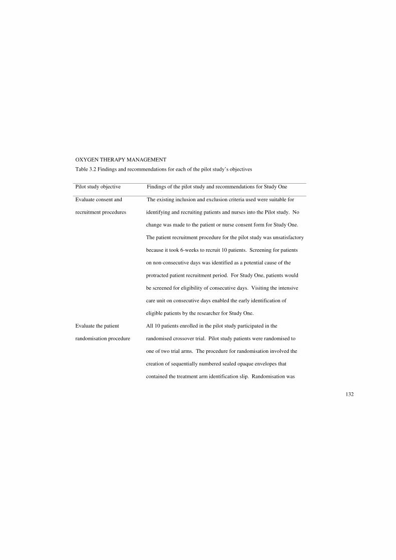
Pilot study .................................................................................................. 111�
Objectives ................................................................................................. 112�
Method ..................................................................................................... 112�
Results ...................................................................................................... 118�
Findings and recommendations of the pilot study objectives .................. 130�
Summary ....................................................................................................... 135�
Chapter 4: Randomised crossover trial & descriptive exploratory interviews .. 136�
Page 10
OXYGEN THERAPY MANAGEMENT
9
Introduction ................................................................................................... 136�
Study One (part A): randomised crossover trial ........................................ 136�
Introduction .............................................................................................. 136�
Method ..................................................................................................... 137�
Results ...................................................................................................... 144�
Study one (part B): descriptive exploratory interviews ............................. 147�
Introduction .............................................................................................. 147�
Method ..................................................................................................... 148�
Results ...................................................................................................... 152�
Summary of key findings ......................................................................... 162�
Chapter 5: A Medical Record Audit .................................................................. 164�
Introduction ................................................................................................... 164�
Method ........................................................................................................... 166�
Results ........................................................................................................... 170�
Summary of key findings .............................................................................. 178�
Chapter 6: An Observational Study ................................................................... 180�
Introduction ................................................................................................... 180�
Method ........................................................................................................... 183�
Results ........................................................................................................... 190�
Summary of key findings .............................................................................. 208�
Chapter 7: Discussion and conclusion ............................................................... 210�
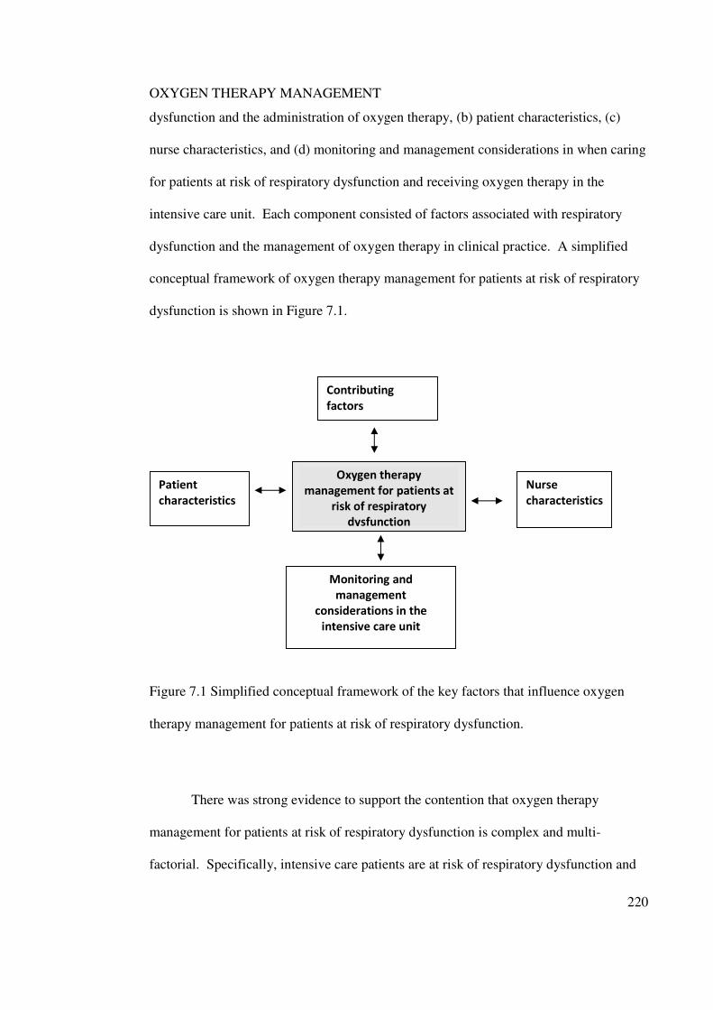
Outcomes of the research .............................................................................. 211�
Clinical efficacy and user-friendliness of oxygen delivery devices .......... 213�
Impact of patients’ and nurses’ perceptions on effective oxygen therapy 214�
Page 11
OXYGEN THERAPY MANAGEMENT
10
Nurses management and documentation oxygen therapy for patients at risk
of respiratory dysfunction ........................................................................ 216�
Comparison between the conceptual framework and the research findings . 219�
Strengths and limitations of the research ...................................................... 223�
Significance of the research findings ............................................................ 227�
Implications of research findings for future research .................................... 229�
Conclusion ..................................................................................................... 230�
References ......................................................................................................... 231�
APPENDICES ............................................................................................... 259�
Appendix A – Ethical approval documents from Deakin University ...... 259�
Appendix B – Ethical approval documents from Epworth Healthcare .... 262�
Appendix C – Patient information sheet and consent form ..................... 265�
Appendix D – Randomised crossover trial and participant interview data
.................................................................................................................. 269�
Appendix E – Nurse information sheet and consent form ....................... 273�
Appendix F – Medical record audit data collection form ........................ 277�
Appendix G – Clinical practice observation data collection form ........... 278�
Page 12
OXYGEN THERAPY MANAGEMENT
11
List of tables
Table 2.1 Published ranges for hypoxaemia, normoxaemia and hyperoxaemia ............. 35�
Table 2.2 Causes of hypoxaemia and responsiveness to oxygen therapy ....................... 38�
Table 2.3 Summary of key research relating to bradypnoea, tachypnoea and the presence
of hypoxaemia as a precursor to an adverse event .................................................. 47�
Table 2.4 Comparison of nasal prongs, face mask and nasopharyngeal oxygen catheter
oxygen delivery devices .......................................................................................... 61�
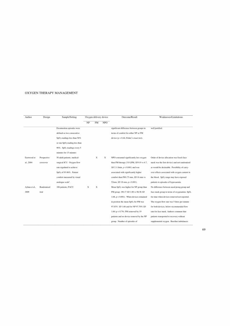
Table 2.5 Summary of studies that have compared the clinical effectiveness and comfort
of nasal prongs, face masks and nasopharyngeal oxygen catheters in adult
hospitalised patients ................................................................................................ 67�
Table 3.1 Comparison of nasal prongs, face mask and nasopharyngeal oxygen catheter
oxygen delivery devices (n = 8) ............................................................................ 120�
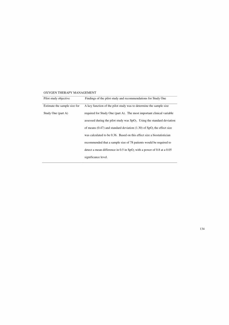
Table 3.2 Findings and recommendations for each of the pilot study’s objectives ...... 132�
Table 4.1 Comparison of nasal prongs, face mask and nasopharyngeal oxygen catheter
delivery devices ..................................................................................................... 146�
Table 5.1 Demographic, surgical type and outcomes for the cardiac surgical patients
included in this medical record audit (N = 210) .................................................... 171�
Table 5.2 Episodes of Hypoxaemia (SpO2 < 95%) while receiving oxygen therapy per
patient .................................................................................................................... 174�
Table 5.3 Episodes of tachypnoea (respiratory rate > 24 / minute) while receiving
oxygen therapy per patient .................................................................................... 176�
Table 6.1 Study time points for review of nursing observation charts and clinical
observation measurements .................................................................................... 184�
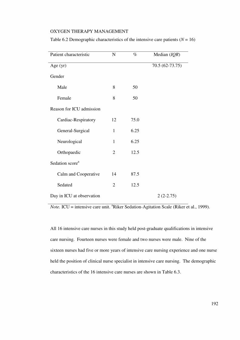
Table 6.2 Demographic characteristics of the intensive care patients (N = 16) ............ 192�
Page 13
OXYGEN THERAPY MANAGEMENT
12
Table 6.3 Demographic characteristics of the intensive care nurses (N = 16) .............. 193�
Table 6.4 Documented and observed oxygen saturation per patient ............................. 196�
Table 6.5 Documented and observed respiratory rates per patient ............................... 201�
List of figures
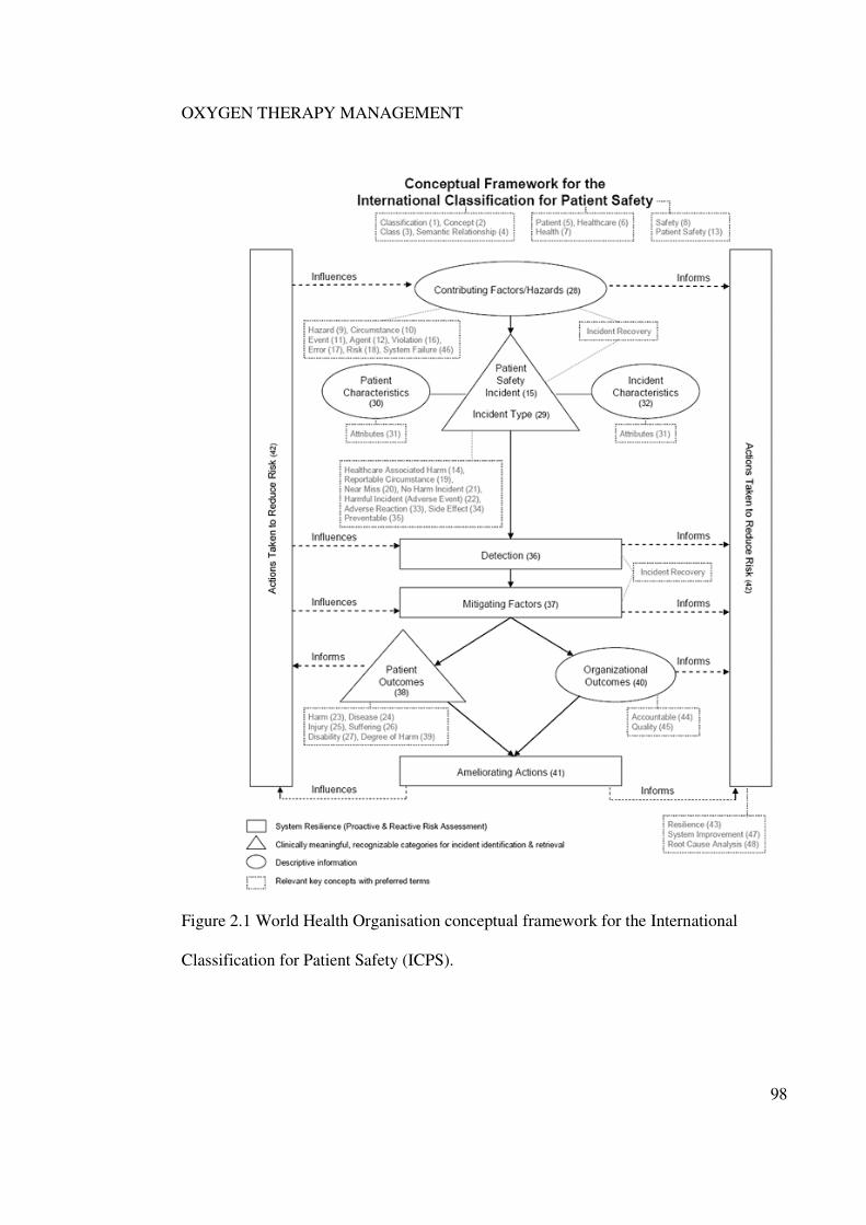
Figure 2.1 World Health Organisation conceptual framework for the International
Classification for Patient Safety (ICPS) .................................................................. 98�
Figure 2.2 Conceptual framework of the key factors that influenceoxygen therapy
management for patients at risk of respiratory dysfunction .................................. 100�
Figure 3.1 Summary diagram of the three linked studies reported in this thesis. ......... 108�
Figure 3.2 Pilot study patient randomisation and treatment allocation sequence for the
randomised crossover trial. .................................................................................... 114�
Figure 4.1 Prospective randomised crossover trial patient randomisation and treatment
allocation sequence.. .............................................................................................. 140�
Figure 7.1 Simplified conceptual framework of the key factors that influence oxygen
therapy management of patients at risk of respiratory dysfunction. ..................... 220�
Page 14
OXYGEN THERAPY MANAGEMENT
13
Abstract
Oxygen therapy is one of the major interventions used to manage respiratory
dysfunction. The failure to recognise and respond to respiratory dysfunction may result
in patients suffering respiratory related adverse events such as cardiac arrest, unplanned
intensive care unit admissions or unexpected death. It is recognised that management of
oxygen therapy for patients at risk of respiratory dysfunction is primarily undertaken by
nurses, is multi-factorial and often carried out in complex clinical settings. There is a
paucity of literature that takes account of the multiple factors that influence the
management of oxygen therapy in clinical practice. Therefore, to understand practice
and improve patient safety there is a need for a more comprehensive study that
investigates the management of oxygen therapy for patients at risk of respiratory
dysfunction.
This research examined, in detail, the oxygen therapy management for patients at
risk of respiratory dysfunction. Specifically, the research sought to (a) measure and
compare the oxygen flow rate required to maintain oxygen saturation equal to or greater
than 95% in adult patients using nasal prongs, face mask and nasopharyngeal oxygen
catheter devices, (b) explore intensive care patients’ and intensive care nurses’
perceptions of oxygen therapy, and (c) describe how intensive care nurses’ manage and
document oxygen therapy in clinical practice.
To appropriately examine the complexity of oxygen therapy management, this
thesis comprised three sequentially linked studies situated within a modified World
Page 15
OXYGEN THERAPY MANAGEMENT
14
Health Organisation patient safety conceptual framework. A mixed methods research
design was used. Data were collected by a randomised crossover trial involving 37
adults patients (Study One – part A) and participant interviews with 37 patients and 25
nurses (Study One – part B). In addition, a retrospective medical record audit (Study
Two) of 210 cardiac surgical patients first 24 hours of admission to the intensive care
unit was conducted. Finally, a prospective clinical practice observational study (Study
Three) involving 16 patients and 16 intensive care nurses was performed. These three
studies were conducted within a single healthcare organisation located in the Eastern
suburbs of Melbourne, Australia.
The findings demonstrated that nasal prongs, face mask and nasopharyngeal
oxygen catheter devices were effective at maintaining a pulse oximetry derived oxygen
saturation (SpO2) greater than 95% with no evidence of patients altering their respiratory
rate to compensate for a change in oxygen supply between devices. Face masks, which
use a higher oxygen flow compared to nasal devices, were deemed by patients to be the
least comfortable device. Importantly, patients wanted to receive oxygen via nasal
prongs or nasopharyngeal oxygen catheter as these devices were the most comfortable,
permitted ease of eating, drinking and talking. Conversely, nurses reported using
measures of a patient’s SpO2 and respiratory rate as drivers for their oxygen therapy
decisions. Nurses preferred to use the face mask as their first choice for oxygen
supplementation because of the ability to provide high oxygen flow rates. However,
differences in patients’ and nurses’ perspectives of oxygen therapy may compromise the
effectiveness of oxygen therapy with patients resistive to using a face mask.
Additionally, results showed that episodes of respiratory dysfunction were common
Page 16
OXYGEN THERAPY MANAGEMENT
15
among post-operative patients in the intensive care environment. However, despite the
occurrence of respiratory dysfunction the management of oxygen therapy did not change
in response to such events. The study also revealed that what nurses documented in the
intensive care environment including oxygen saturation and respiratory rate measures
did not reflect patient status and some nursing actions such as a failure to escalate
oxygen delivery in response to respiratory dysfunction and removal of oxygen therapy to
perform mouth care, hindered effective oxygen therapy. Collectively, the findings
revealed a need for health care professionals to review oxygen device selection in
specific clinical settings, the importance of involving patients in decisions about their
care and the need to appropriately document care that is provided.
These research findings fill gaps in the literature by providing overarching
information about perceptions and practice. Improvements are necessary in the selection
of oxygen delivery devices and the tailoring of the device choice to match the clinical
condition and activity of the patient. Similarly, documentation systems need to undergo
review in order to appropriately match the acuity, complexity and pace of contemporary
intensive care unit practice.
This research significantly contributes to understanding the interplay of the
multiple factors that impact on the effective management of oxygen therapy for patients
at risk of respiratory dysfunction. These findings can now inform future interventions
aimed at improving oxygen therapy management so that patient safety and outcomes can
be optimised.
Page 17
OXYGEN THERAPY MANAGEMENT
16
Terms and abbreviations
ABG Arterial blood gas
APACHE III score Acute physiology and chronic health evaluation III
score
Bradypnoea Respiratory rate less than eight breaths per minute
CO2 Carbon dioxide
ED Emergency department
FiO2 Fraction of inspired oxygen
FM Face mask
Flow rate The rate at which oxygen enters an oxygen
delivery device in litres per minute
HCO3 Bicarbonate
HREC Human research ethics committee
HVAS Horizontal visual analogue scale
Hypoxia A state of oxygen deficiency at the tissue or
cellular level
Hypoxaemia A state of oxygen deficiency in arterial blood
Hyperoxia A state of oxygen excess is a state of higher than
normal partial pressure of oxygen
Hyperoxaemia A state of above normal oxygen levels in the
arterial blood
ICPS International Classification for Patient Safety
Page 18
OXYGEN THERAPY MANAGEMENT
17
ICU Intensive Care Unit, a dedicated hospital ward
specialising in the care and management of patients
experiencing or likely to experience serious illness
Intensive care nurse A registered nurse employed in an intensive care
unit who is accountable and responsible for the
care of an intensive care patient
Intensive care patient A patient admitted to an intensive care unit
LPM Litres per minute
mmHg Millimetres of Mercury
MET Medical emergency team
Minute volume Volume in millilitres of gas inhaled over a one
minute period
NP Nasal prongs
NPO Nasopharyngeal oxygen catheter
O2 Oxygen
OFR Oxygen flow rate: The rate at which oxygen enters
an oxygen delivery device in litres per minute
PACU Post-anaesthetic care unit
PaO2 Partial pressure of oxygen in arterial blood
PaCO2 Partial pressure of carbon dioxide in arterial blood
PO2 Partial pressure of oxygen
RR Respiratory rate per minute
Respiratory dysfunction For the purposes of this thesis respiratory
Page 19
OXYGEN THERAPY MANAGEMENT
18
dysfunction is defined as bradypnoea (less than
eight breaths per minute), tachypnoea (greater than
24 breaths per minute) and / or hypoxaemia
(oxygen saturation of less than 95% measured by
pulse oximetry)
SaO2 Oxygen saturation of arterial blood measured by
arterial blood gas sampling
SpO2 Oxygen saturation measured by pulse oximetry
Tachypnoea Respiratory rate greater than 24 breaths per minute
Tidal volume Volume in milliliters of gas inhaled during one
breath
Page 20
OXYGEN THERAPY MANAGEMENT
19
Chapter 1: Introduction
Introduction
Oxygen therapy is one of the major interventions used to manage respiratory
dysfunction. Nurses play a vital role in the management of oxygen therapy in patients at
risk of respiratory dysfunction. The failure of nurses to recognise and respond to
respiratory dysfunction may result in patients suffering respiratory related adverse
events such as unexpected death, cardiac arrest, or unplanned intensive care unit
admissions. Although oxygen management is an important aspect of patient care, there
is a paucity of literature that takes account of the multiple factors that influence the
management of oxygen therapy. Therefore, to understand practice and improve patient
safety there is a need for a comprehensive study that investigate the management of
oxygen therapy for patients at risk of respiratory dysfunction.
Respiratory dysfunction is life threatening and well-recognised as a precursor to
in-hospital adverse events such as cardiac arrest, unplanned admission to the intensive
care unit and death (Cretikos et al., 2008; Harrison, Jacques, Kilborn, & McLaws, 2005).
In order to decrease mortality from adverse events, nurses must actively assess and treat
patients with respiratory dysfunction, rather than respond to respiratory related adverse
events (Considine, 2005a; Quach et al., 2008). The evidence base regarding the
recognition and response systems for respiratory dysfunction is developing. To provide
a safety buffer for patients whose respiratory function is deteriorating, there is a strong
need to explore the features and factors that are associated with the clinical management
of respiratory dysfunction.
Page 21
OXYGEN THERAPY MANAGEMENT
20
For the purposes of this thesis, respiratory dysfunction is defined as bradypnoea
(less than eight breaths per minute), tachypnoea (greater than 24 breaths per minute) and
/ or hypoxaemia (oxygen saturation of less than 95% measured by pulse oximetry).
Respiratory rate is the most important and sensitive indicator of serious illness in adults
but also the most poorly measured and documented clinical indicator (Cretikos et al.,
2008). Respiratory rate abnormalities, in particular tachypnoea, are indicative of
deteriorating respiratory function, or the manifestation of an abnormal physiological
state in another body system (Cretikos et al., 2008). Evidence from hospital based
studies of general ward patients suggests that an adult at rest with a respiratory rate
greater than 20 breaths per minute is likely to be unwell (Davey, McCance, & Budd,
1994; Kennedy, 2007) and that those with a respiratory rate greater than 24 breaths per
minute are likely to be critically ill (Cretikos et al., 2008; Grap, Glass, & Constantino,
1994; Harrison et al., 2005).
Hypoxaemia, a state of oxygen deficiency, is a clinical indicator of respiratory
dysfunction. Prolonged untreated hypoxaemia is life threatening because of the
imbalance between oxygen supply and oxygen consumption (Levy, 2008). The presence
of hypoxaemia prior to an adverse event increases the need for advanced respiratory
interventions (such as non-invasive ventilation), or transfer to the intensive care unit, to
avoid a fatal adverse event (Goldhill & McNarry, 2004; Skrifvars, Nurmi, Ikola,
Saarinen, & Castrén, 2006). In hospital settings nurses are responsible for the
assessment of patients’ overall physiological status and the detection of physiological
abnormalities, including hypoxaemia, bradypnoea and tachypnoea.
Page 22
OXYGEN THERAPY MANAGEMENT
21
Patients who are at high risk of respiratory dysfunction are admitted to the
intensive care unit for increased monitoring and specialised care. Australian intensive
care units are staffed on 1:1 or 1:2 nurse-patient ratios to ensure a high level of patient
monitoring, in particular monitoring of respiratory function and oxygen therapy
management (Australian College of Critical Care Nurses [ACCCN], 2003). This level
of staffing is required so that nurses can provide timely and appropriate interventions
amidst a number of competing physiological factors and patient care requirements.
Intensive care nurses are well placed to recognise and respond to the clinical signs of
respiratory dysfunction. Additionally, many intensive care nurses have post-graduate
specialist qualifications, with advanced skills in the assessment and management of
patients. Despite the importance of effective oxygen therapy management, there
remains minimal research on the oxygen delivery device, patient and nurse factors that
impact on the oxygen management practices used by intensive care nurses.
Oxygen therapy is commonly administered to intensive care patients to prevent
or relieve hypoxaemia. In order to manage oxygen therapy, intensive care nurses are
required to select an oxygen delivery device (such as nasal prongs and face mask) and an
oxygen flow rate to deliver oxygen to the patients. The majority of oxygen delivery
devices are suitable for use in patients with minimal respiratory distress and adequate
ventilatory patterns but who still require supplemental oxygen for therapeutic
(Eastwood, Gardner, & O’Connell, 2007). Few of the basic, commonly used, oxygen
delivery devices have been subjected to rigorous clinical review to establish their levels
of clinical comfort and effectiveness (Eastwood, Reeves, & Cowie, 2004). In order to
Page 23
OXYGEN THERAPY MANAGEMENT
22
better understand the processes involved in administering oxygen therapy it is essential
to further explore all stages of the oxygen therapy management process.
The choice nurses make about the oxygen delivery devices used and the
management of oxygen therapy is complex and influenced by a number of factors.
These factors are: nurses’ knowledge of oxygen therapy, patient preferences, and patient
care activities (Eastwood et al., 2007). According to Murphy et al. (2001) decisions
about oxygen therapy are often ad hoc, and other researchers highlight a lack of
empirical evidence to inform nursing practice (Considine, Botti, & Thomas, 2006;
O’Driscoll, Howard, & Davison, 2008). Studies in non-critical care settings have shown
that patients are at risk of respiratory dysfunction due to suboptimal monitoring,
management and documentation of oxygen therapy (Albin, Criner, Thomas, & Abou-
Jaoude, 1992; Attia, Nair, Mears, & Hitchcock, 2004; Boyle & Wong, 2006; Brokalaki
et al., 2004; Howell, 2001; Kor & Lim, 2000; Nolan, Winyard, & Goldhill, 1993; Small
et al., 1992; Stausholm, Rosenberg-Adamsen, Skriver, Kehlet, & Rosenberg, 1995).
These findings are alarming as suboptimal oxygen management can result in adverse
patient outcomes (Considine, Botti, & Thomas, 2005; Kernick & Magarey, 2010;
Murphy et al., 2001). Further work is necessary to provide information on how
intensive care nurses manage and document oxygen therapy for patients at risk of
respiratory dysfunction.
An important factor that requires attention and further exploration is the patients’
perspective including their experiences of oxygen therapy and their assessment of how
oxygen therapy impacts on their comfort and wellbeing. Patient perspectives are an
Page 24
OXYGEN THERAPY MANAGEMENT
23
important aspect of care as oxygen delivery device comfort has a direct impact on
patients’ acceptance and compliance with oxygen therapy (Nolan, Winyard, & Goldhill,
1993; Sasaki et al., 2003; Stausholm et al., l995). Patient anxiety and discomfort
wearing oxygen devices may lead to non-compliance and increased interruptions to
oxygen therapy (Eastwood et al., 2007). Interruptions to oxygen therapy place the
patient at risk of hypoxaemia that may result in death or cardiac arrest (Considine,
2005b: Eastwood et al., 2007). Therefore, identifying patients’ perceptions of oxygen
therapy is an important step in highlighting ‘real world’ factors that nurses could address
to optimise patient comfort and compliance with oxygen therapy.
The research agenda to date has largely ignored the interplay between these
multi-factorial clinical situations and the implications for patients at risk of respiratory
dysfunction. Gaps in the literature exist because most previous studies have tended to
investigate factors that impact on oxygen therapy in isolation, such as device
effectiveness, device comfort, or appropriateness of nursing practice without an
overarching in-practice approach. Consequently, the previous studies are unable to
provide strong evidence to inform intensive care nurses’ daily practice of oxygen
therapy.
There are two imperatives influencing the need for systematic research into how
oxygen therapy is managed for patients at risk of respiratory dysfunction: (a) the clinical
risk of respiratory dysfunction leading to increased morbidity and mortality, and (b) the
lack of evidence specifically supporting intensive care nurses’ management and
documentation of oxygen therapy. Due to the interplay of a number of factors that
Page 25
OXYGEN THERAPY MANAGEMENT
24
influence how nurses manage oxygen therapy, a multi-stage approach using linked
studies is necessary to increase understanding of the relationships between the numerous
factors that influence oxygen therapy.
Aim and objectives
The overall aim of this research was to provide a detailed analysis of oxygen
therapy management for patients at risk of respiratory dysfunction. The research
comprises a pilot study and three sequentially linked studies. The aims of the three
linked studies were to investigate:
• The clinical efficacy and user-friendliness of oxygen delivery devices
• Patients’ and nurses’ perception of oxygen therapy
• How intensive care nurses’ manage and document oxygen therapy for
patients at risk of respiratory dysfunction
The specific aims and objectives of the three studies are detailed below.
Study One
The aim of Study One was to evaluate the clinical efficacy and user-friendliness
of oxygen therapy devices from both the patient and nurse perspective. Study One was
divided into two parts – part A and part B. The objective of part A was to measure and
compare the oxygen flow rate required to maintain oxygen saturation equal to or greater
than 95% in adult patients using different oxygen delivery devices. A crossover trial
design was used to assess efficacy of nasal prongs, face mask, and nasopharyngeal
oxygen catheter. The objective of part B was to assess and compare patients’ and
Page 26
OXYGEN THERAPY MANAGEMENT
25
nurses’ perspectives of oxygen therapy. Patients and nurses participated in semi-
structured face-to-face interviews, within a descriptive exploratory design.
Study Two
The aim of Study Two was to describe how intensive care nurses administered
and managed oxygen therapy for adult cardiac surgical patients during the first 24 hours
of intensive care admission. Of particular interest to this study were the types of oxygen
delivery devices used, the frequency of documented hypoxaemia, the frequency of
documented respiratory rate abnormalities (tachypnoea and bradypnoea) and changes in
oxygen flow rate or oxygen delivery device in response to respiratory dysfunction
(hypoxaemia and / or tachypnoea).
Study Three
The aim of the third and final study was to prospectively observe how intensive
care nurses manage oxygen therapy. Of particular interest to this study was oxygen
delivery device fit, placement and flow rate, assessment of key indicators of oxygenation
(oxygen saturation and respiratory rate) and alterations to oxygen therapy in response to
hypoxaemia and / or tachypnoea. In Study Three a descriptive exploratory study was
used and data were collected using a structured observation tool, field notes and nursing
observation chart review.
Page 27
OXYGEN THERAPY MANAGEMENT
26
Significance
Intensive care nurses frequently manage oxygen therapy in patients with
respiratory dysfunction. Despite the common use of oxygen therapy by intensive care
nurses, the literature indicates that oxygen management practices vary and the body of
evidence on which to guide practice is lacking. Appropriate and timely management of
oxygen therapy is a key component to the prevention or treatment of respiratory
dysfunction. Oxygen therapy is a complex clinical activity and the results of the three
linked studies will contribute to the further knowledge of the factors that impact
intensive care nurses’ management of oxygen therapy. A greater understanding of
oxygen therapy management for patients at risk of respiratory dysfunction will aid
patient safety initiatives and identify other opportunities for practice improvement. In
addition, the findings from the research reported in this thesis will inform future
exploratory, observational and interventional studies to support the further development
of evidence based guidance for oxygen therapy management by intensive care nurses.
Thesis structure
This thesis is divided into seven chapters. In Chapter Two the literature related
to how intensive care nurses manage oxygen therapy is reviewed. This review of the
literature is set out in four sections. First, foundational concepts of oxygen physiology
and respiratory dysfunction and hypoxaemia as clinical risk and a threat to patient safety
are presented. Second, respiratory dysfunction and the role of oxygen therapy for the
treatment of respiratory dysfunction are described. Third, a review of the device, patient
and nurse related factors that influence the clinical management of oxygen therapy is
provided. Finally, a conceptual framework for this study of nursing management of
Page 28
OXYGEN THERAPY MANAGEMENT
27
oxygen therapy for patients at risk of respiratory dysfunction is detailed.
Fundamentally, the literature review identifies the gaps in the literature and
demonstrates an urgent clinical need for a better understanding of how oxygen therapy is
managed for patients at risk of respiratory dysfunction.
Chapter Three is the Methods Chapter. In Chapter Three the methods
undertaken to conduct the three linked studies are described and the aim, method,
results, and recommendations arising from the pilot study are detailed. The conduct of
the pilot study was a valuable and important methodological choice that enabled the
process and outcome measures of Study One to be validated.
The discussion in Chapter Four, Chapter Five and Chapter Six describe the
background, aim, method and a summary of key findings of the three linked studies,
respectively. In Chapter Seven, the research findings in relation to the overall research
aim are discussed. To conclude, the clinical implications of the findings are discussed
and recommendations for nursing practice, nursing research, and nursing education are
presented.
Page 29
OXYGEN THERAPY MANAGEMENT
28
Chapter 2: Literature Review
Introduction
Intensive care nurses play a major role in patient safety and risk management. In
the intensive care unit, it is nurses who monitor the patient 24-hours a day and who are
uniquely placed to recognise and respond to the early signs of respiratory dysfunction.
The purpose of Chapter Two is to define terms, provide background to the topic and to
review the literature related to interventions used to manage oxygen therapy for patients
experiencing, or at risk of, respiratory dysfunction. The five sections in Chapter Two
specifically explore or provide:
• The foundational concepts of respiratory physiology and oxygen delivery
• Respiratory dysfunction and hypoxaemia as clinical risk and the use of oxygen
therapy in the management of respiratory dysfunction
• Patient related factors that influence effective oxygen therapy
• Nurse related factors that influence the management and documentation of
oxygen therapy
• A description of, and rationale for, the conceptual framework supporting the
research presented in this thesis
Medical and nursing literature was accessed to inform the literature review of
oxygen therapy for patients at risk of respiratory dysfunction. In particular, the review
included referred healthcare journals, review articles, physiology textbooks as well as
specialist medical and nursing textbooks. Importantly, the literature review and
discussion presented in Chapter Two identifies gaps in the literature and demonstrates
Page 30
OXYGEN THERAPY MANAGEMENT
29
the clinical need for a better understanding of oxygen therapy management for patients
at risk of respiratory dysfunction.
Foundational concepts of respiratory physiology and oxygen delivery
Normal respiratory physiology
The human body needs to use oxygen from the atmosphere effectively to
maintain normal cell function. Aerobic metabolism is the normal process to produce
energy in the human body (Marieb, 2004). Under normal circumstances, every molecule
of oxygen generates 38 molecules of adenosine tri-phosphate as energy (West, 2008). If
a person cannot breathe in sufficient quantities of oxygen, or is in an atmosphere with
reduced oxygen, anaerobic metabolism occurs when glucose is used to generate the
energy required for the function of cells (Marieb, 2004). Anaerobic metabolism is very
inefficient, generating only two adenosine tri-phosphate molecules of energy and
produces a toxic by-product, lactic acid (West, 2008). Excessive anaerobic metabolism
causes a build-up of lactic acid that can damage intracellular function (Marieb, 2004)
and if untreated can cause lactic acidosis and hypoxia because of insufficient quantities
of glucose, fatty acids and oxygen which are needed for metabolic energy production
(Edwards, 2003). Left untreated, both excessive lactic acid build-up and prolonged
hypoxia can result in cell death leading to organ failure and the person dying (Considine,
2005a).
To understand how cells in our body use oxygen, it is necessary to understand
how oxygen moves from the atmosphere to the cells. The major stages in oxygen supply
Page 31
OXYGEN THERAPY MANAGEMENT
30
to the cells are: (a) ventilation, (b) external respiration (c), oxygen transport, and (d)
internal respiration (cellular metabolism).
Ventilation
Ventilation, or breathing, consists of inspiration and expiration. Ventilation is a
cyclic process that enables the exchange of oxygen and carbon dioxide between the
atmosphere and the lung (Marieb, 2004). During inspiration several muscle groups
work together to increase the volume of the thoracic cavity (Marieb, 2004; West 2008).
An increase in the volume of the thoracic cavity results in a decrease in intra-thoracic
pressure causing air, containing oxygen, to move from the atmosphere (an area of higher
pressure) and into the lung (an area of lower pressure) (Marieb, 2004). Expiration is a
passive process and occurs as inspiratory muscles relax. When the inspiratory muscles
relax pressure in the thoracic cavity increases and the volume of the thoracic cavity
decreases. The change in pressure (from an area of lower pressure to higher pressure)
within the lung enables carbon dioxide to leave the lung (Marieb, 2004; West, 2008)
External respiration
External respiration is the process of gas exchange between the pulmonary blood
and alveoli (Marieb, 2004). As air enters the lung alveoli, oxygen moves across the
alveolar membrane and into the pulmonary blood via diffusion and pressure gradients
(West, 2008). The major factors that influence the movement of oxygen and carbon
dioxide across the alveolar membrane are the partial pressure gradients of the gases,
structural characteristics of the alveolar membrane, and the matching of pulmonary
blood flow and alveolar ventilation (West, 2008).
Page 32
OXYGEN THERAPY MANAGEMENT
31
Oxygen transport
When oxygen has crossed from the alveoli into pulmonary capillary blood it is
transported in the bloodstream in two forms. Most of the oxygen (98.5%) is transported
bound to haemoglobin as oxyhaemoglobin (O2Hb) and a lesser amount of the oxygen
(1.5%) is transported dissolved in the blood plasma (Marieb, 2004). Haemoglobin is a
large protein molecule containing four subunits, each subunit contains a ferrous ion
within a haem group. A maximum of four oxygen molecules can bind reversibly to each
normal haemoglobin molecule (Berne & Levy, 1998). Two important physiological
properties of oxygen and haemoglobin are (a) that oxygen combines reversibly with
heamoglobin and (b) that molecular oxygen quickly and easily dissociates from
haemoglobin to enable a fast release at the site of tissues (Berne & Levy, 1998).
Haemoglobin is loaded with oxygen in the lungs where the partial pressure of oxygen is
highest (Marieb, 2004). Therefore, haemoglobin level is a major determinant of the
effectiveness of oxygen transport to the tissue (Levy, 2005). After transfer across the
alveolar-capillary membrane and becoming bound to haemoglobin, oxygen is efficiently
carried to the tissues for use in internal respiration by the cardiovascular system (Marieb,
2004). Cardiac output determined by heart rate and stroke volume is another major
determinant of the effectiveness of oxygen transport around the body (Marieb, 2004).
Clinically, the most common proxy measure for cardiac output is blood pressure,
making the assessment of a patient’s blood pressure an important surrogate indicator of
impaired cardiac output and therefore decreased oxygen delivery (Considine, 2005a).
Page 33
OXYGEN THERAPY MANAGEMENT
32
Internal respiration
Internal respiration occurs as oxygen and carbon dioxide are exchanged between
capillary blood and body cells via diffusion (West, 2008). Once inside the cell,
mitochondrial enzymes use oxygen to synthesise adenosine di-phosphate to the high-
energy adenosine tri-phosphate for the support of normal cellular function (West, 2008).
Normal and abnormal oxygen states
Normal oxygen states
The amount of oxygen contained in blood can be described and measured in two
ways, reflecting the ways oxygen is transported. One method is to describe the presence
of oxygen measured as millimetres of mercury (mmHg) to denote the measurement of
PaO2 measured in the arterial blood gas (ABG) sample. A second method is to measure
oxygen saturation, the percentage (%) of haemoglobin in the arterial circulation (SaO2)
or peripheral circulation (SpO2) saturated with oxygen (O’Driscoll et al., 2008). Oxygen
saturation can be obtained either by arterial blood gas sampling or using pulse oximetry
(O’Driscoll et al., 2008). There is a consensus in the literature that a normal oxygen
saturation reading for adults is SaO2 or SpO2 equal to or greater than 95% (Considine,
2005a; Crapo, Jensen, Hegewald, & Tashkin, 1999). These measurements are explained
later in the chapter, in the discussion on ways oxygen levels are monitored in the clinical
setting.
The relationship between SpO2 and PaO2 has important implications for
clinicians. When assessing the effectiveness of oxygen therapy both via pulse oximetry
and arterial blood gas analysis, clinicians must pay particular attention to correcting a
Page 34
OXYGEN THERAPY MANAGEMENT
33
low SpO2 due to the potential for a large decrease in PaO2. For example, a SpO2 drop
from 98% to 96% would shift the PaO2 from 104 mmHg to 82 mmHg, while a SaO2
drop from 95% to 87% would result in a PaO2 fall from 75 mmHg to 52 mmHg
(O’Driscoll et al., 2008). The ability of oxygen to bind to haemoglobin changes when
there are changes in the body, for example, changes in temperature, acidity (pH), carbon
dioxide and 2,3-diphophoglycerate (2,3 DPG) (O’Driscoll et al., 2008). Physiological
instances that give rise to a release of oxygen from haemoglobin include exercise or
elevated temperature due to infection or inflammation. The release of oxygen from
haemoglobin results in more oxygen being available for cells to use (Coggan, 2008a;
O’Driscoll et al., 2008).
In the lungs, oxygen more readily binds to haemoglobin as pH increases, PCO2
decreases, or as temperature falls, and the haemoglobin then become saturated with
oxygen. During times of rest, or normal body temperature, oxygen remains bound to
haemoglobin and ready for use by body tissues rather than being consumed by them.
Knowing the physiological factors associated with oxygen consumption in the body
aid’s clinician’s clinical decision regarding when and how to administer oxygen
(Coggan, 2008). Oxygen therapy management practices that ensure the patient is
provided with supplemental oxygen during times of physiological stress can optimise the
amount of oxygen available for use by the tissues (Coggan, 2008a, 2008b).
States of oxygen deficiency
Hypoxia and hypoxaemia are the terms used to describe the two states of oxygen
deficiency. Both may be the cause or the consequence of respiratory dysfunction and
Page 35
OXYGEN THERAPY MANAGEMENT
34
each are life-threatening (Considine, 2005a; Levy, 2005). Hypoxia and hypoxaemia are
detectable by SpO2 and SaO2 assessments (Berry & Pinard, 2002).
Hypoxia
Hypoxia is a state of oxygen deficiency at the tissue or cellular level (O’Driscoll
et al., 2008) and if prolonged it disrupts cellular function due to a lethal accumulation of
lactic metabolites, the by-product of anaerobic metabolism (West, 2008). There are four
causes of hypoxia: hypoxic hypoxia, anaemic hypoxia, stagnant hypoxia and histotoxic
hypoxia. Treatment and prevention of hypoxia, as assessed on pulse oximetry (SpO2),
arterial blood gas analysis (PaO2 and SaO2) or cardiac function (heart rate), is the
underlying rationale that supports the administration of supplemental oxygen
(Considine, 2005; Considine, 2005a). However, in the case of anaemic hypoxia,
stagnant hypoxia and histotoxic hypoxia a patient may present have a normal SpO2 or
PaO2 despite the use of supplemental oxygen being warranted. Consequently, clinician’s
knowledge of physiology, pathophysiology of oxygen states and other clinical
indicators, to assess the oxygenation status of patients together with oxygen saturation
should inform oxygen therapy management decisions (Considine, 2005a).
Hypoxaemia
Hypoxaemia is a state of oxygen deficiency in arterial blood and is defined as a
PaO2 value lower than 80 mmHg, or an SaO2 or as an SpO2 value less than 95% when
measured by pulse oximetry (O’Driscoll et al., 2008). Since the publication of studies in
the 1990s, it has been widely accepted that states of hypoxaemia are categorised into
mild, an SpO2 of 90%-94%, moderate hypoxaemic, an SpO2 less than 90% (Eastwood &
Page 36
OXYGEN THERAPY MANAGEMENT
35
Dennis, 2006; Leaver, Conway, & Hogate, 1994), and severe hypoxaemia, an SpO2 less
than 85% (Leaver, et al., 1994). Although it is widely acknowledged that a PaO2 less
than 60 mmHg clearly indicates hypoxaemia, a PaO2 between 60 and 80 mmHg with a
corresponding SaO2 or SpO2 between 90% and 95% remain clinically undefined
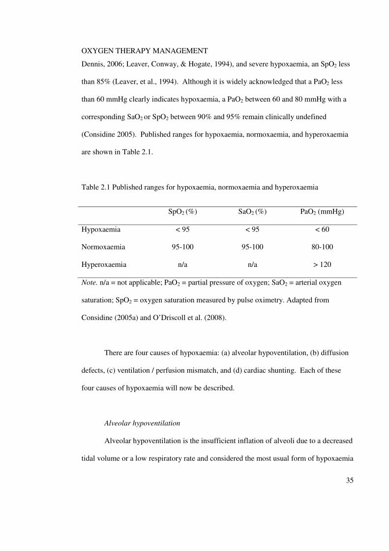
(Considine 2005). Published ranges for hypoxaemia, normoxaemia, and hyperoxaemia
are shown in Table 2.1.
Table 2.1 Published ranges for hypoxaemia, normoxaemia and hyperoxaemia
SpO2 (%) SaO2 (%) PaO2 (mmHg)
Hypoxaemia < 95 < 95 < 60
Normoxaemia 95-100 95-100 80-100
Hyperoxaemia n/a n/a > 120
Note. n/a = not applicable; PaO2 = partial pressure of oxygen; SaO2 = arterial oxygen
saturation; SpO2 = oxygen saturation measured by pulse oximetry. Adapted from
Considine (2005a) and O’Driscoll et al. (2008).
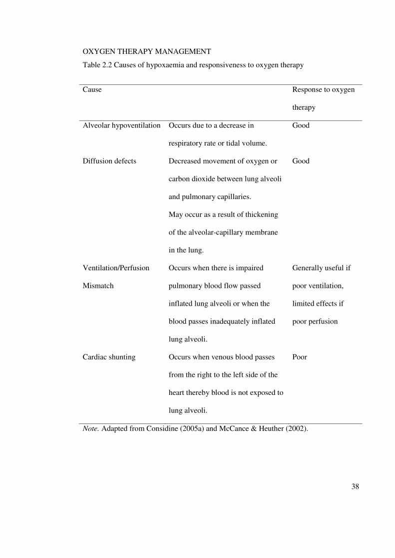
There are four causes of hypoxaemia: (a) alveolar hypoventilation, (b) diffusion
defects, (c) ventilation / perfusion mismatch, and (d) cardiac shunting. Each of these
four causes of hypoxaemia will now be described.
Alveolar hypoventilation
Alveolar hypoventilation is the insufficient inflation of alveoli due to a decreased
tidal volume or a low respiratory rate and considered the most usual form of hypoxaemia
Page 37
OXYGEN THERAPY MANAGEMENT
36
(Considine, 2005a). If the alveoli do not inflate sufficiently then the amount of oxygen
reduces, carbon dioxide accumulates and tissue oxygenation decreases due to ineffective
external respiration (Treacher & Leach, 1998). Oxygen therapy is used to manage
alveolar hypoventilation and is effective in increasing the amount of oxygen available
during inspiration (Strachan & Noble, 2001). Definitive management of alveolar
hypoventilation however, requires identification and correction of the underlying cause
(O’Driscoll et al., 2008).
Diffusion defects
Diffusion defects occur when there is poor oxygen exchange across the alveolar-
capillary membrane during external respiration (Leach & Treacher, 2002). Poor gas
exchange will result in insufficient oxygen availability at the cellular level (Leach &
Treacher, 1998; Treacher & Leach, 1998). Oxygen therapy in the management of
hypoxaemia associated with diffusion defects will increases PaO2, SaO2 and SpO2 by
making more oxygen available to bind with haemoglobin for use by the cells, however
definitive treatment of the underlying cause is necessary (Leach & Treacher, 2002).
Ventilation / perfusion mismatch
Ventilation / perfusion (V/Q) mismatch occurs when blood passes inadequately
ventilated alveoli, or the alveoli are adequately ventilated but blood flow is impaired or
there is a failure of haemoglobin to become saturated with oxygen in the lung
(O’Driscoll et al., 2008). Prolonged severe V/Q mismatch is a known clinical risk,
which can lead to death (Levy, 2005). Oxygen therapy will be of no value if the cause is
Page 38
OXYGEN THERAPY MANAGEMENT
37
due to a lack of blood perfusing the lungs, but will be of some benefit if the cause is
reduced ventilation (Considine, 2005a).
Cardiac shunting
Cardiac shunting, as the cause of hypoxaemia, arises from an anatomical
abnormality in which blood fails to reach the lungs due to venous blood passing from the
right to the left side of the heart (Considine, 2005a). Blood that bypasses the pulmonary
circulation cannot become saturated with oxygen, resulting in increasing amounts of
deoxygenated blood entering the systemic circulation (Considine, 2005a). Hypoxaemia
due to cardiac shunting is typically severe and the response to oxygen therapy is poor,
the main treatment option is surgical repair of this anatomical defect (Considine, 2005a).
The four causes of hypoxaemia and the responsiveness of each cause to oxygen therapy
are shown in Table 2.2.
Page 39
OXYGEN THERAPY MANAGEMENT
38
Table 2.2 Causes of hypoxaemia and responsiveness to oxygen therapy
Cause Response to oxygen
therapy
Alveolar hypoventilation Occurs due to a decrease in
respiratory rate or tidal volume.
Good
Diffusion defects Decreased movement of oxygen or
carbon dioxide between lung alveoli
and pulmonary capillaries.
May occur as a result of thickening
of the alveolar-capillary membrane
in the lung.
Good
Ventilation/Perfusion
Mismatch
Occurs when there is impaired
pulmonary blood flow passed
inflated lung alveoli or when the
blood passes inadequately inflated
lung alveoli.
Generally useful if
poor ventilation,
limited effects if
poor perfusion
Cardiac shunting Occurs when venous blood passes
from the right to the left side of the
heart thereby blood is not exposed to
lung alveoli.
Poor
Note. Adapted from Considine (2005a) and McCance & Heuther (2002).
Page 40
OXYGEN THERAPY MANAGEMENT
39
States of oxygen excess
States of oxygen deficiency carry significant risk and are typically avoided
(O’Driscoll et al., 2008). Similarly, states of oxygen excess, while providing a buffer of
safety in some patients, may also be injurious (de Jonge et al., 2008; Kilgannon et al.,
2010). For example, hyperoxia in the lungs causes histopathological injury,
atelelectasis, tracheobronchitis and alveolar protein leakage resulting in impaired oxygen
exchange (Altemeir & Sinclair, 2007; O'Driscoll et al., 2008). Systemically, oxygen
excess can generate free radicals in various organs and decrease cardiac output
(Altemeir & Sinclair, 2007; O’Driscoll et al., 2008).
Hyperoxia and hyperoxaemia are states of oxygen excess. Hyperoxia is a state
of higher than normal partial pressure of oxygen (PO2) and hyperoxaemia as a state of
elevated PaO2 (O'Driscoll et al., 2008). While it is widely acknowledged that
hyperoxaemia is reached at PaO2 values higher than >300 mmHg, a PaO2 of greater than
120 mmHg is also considered to be excessive (Kilgannon et al., 2010). Lack of a clear
definition of hyperoxaemia may cause confusion and lead to inconsistent practice
regimens among clinicians when deciding to reduce or cease oxygen therapy. While
hypoxaemia, with its physical signs and symptoms is relatively easily identified using
oxygen saturation and or PaO2, diagnosis of hyperoxaemia is only possible by measuring
PaO2 because SpO2 and SaO2 are only measured as a percentage and normal is 100%
(O’Driscoll et al, 2008).
Notwithstanding the risks associated with states of oxygen excess, there are
certain clinical situations in which raising PaO2 to values higher than normal is
Page 41
OXYGEN THERAPY MANAGEMENT
40
indicated. Carbon monoxide poisoning is one specific clinical condition in which high
concentrations of inspired oxygen are desirable (Harper & Croft-Baker, 2004). Carbon
monoxide has a 200 times greater affinity for haemoglobin than oxygen and remains
bound to haemoglobin for longer than oxygen (Weaver, 2009), causing anaemic hypoxia
and preventing oxygen from reaching the tissues (Harper & Croft-Baker, 2004; Weaver,
2009). Emergency management of the patient with carbon monoxide poisoning is to
remove the person from the carbon monoxide source and administer high concentrations
of inspired oxygen (Harper & Croft-Baker, 2004; Turner, Hamilton-Farrell, & Clark,
1999; Weaver, 2009) in an attempt to optimise oxygen uptake and delivery to displace
the carbon dioxide molecules from haemoglobin (Harper & Croft-Baker, 2004).
Oxygen monitoring methods
Oxygen therapy management is complex and clinicians must balance the
appropriate use of oxygen therapy to treat oxygen deficiency and avoid oxygen excess.
Although it is noted in the current literature that there are limitations to the assessment
methods used to measure oxygenation, the combination of monitoring and physical
assessment provides clinicians the tools to detect respiratory related abnormalities.
Monitoring PaO2, SaO2 and SpO2 using oxygen monitoring methods arterial
blood gases (ABGs) and pulse oximetry is an essential adjunct to patient assessment. It
is important to understand the advantages and disadvantages of these two methods of
monitoring oxygen because the majority of decisions made by clinicians to commence,
change or stop oxygen therapy are based on the findings of pulse oximetry or arterial
blood gas sampling. A full discussion of the clinical assessment of oxygenation,
Page 42
OXYGEN THERAPY MANAGEMENT
41
including respiratory rate, heart rate and blood pressure, skin pallor and conscious state
and how they relate to clinical assessment undertaken by nurses is presented later in the
chapter.
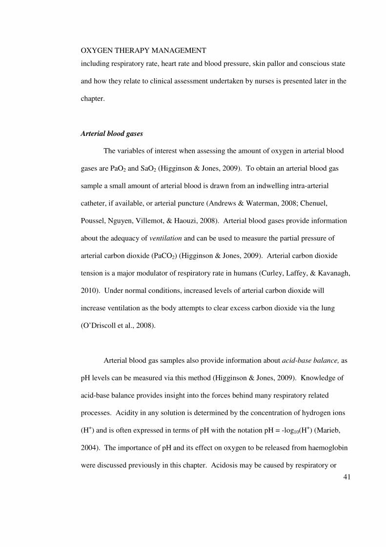
Arterial blood gases
The variables of interest when assessing the amount of oxygen in arterial blood
gases are PaO2 and SaO2 (Higginson & Jones, 2009). To obtain an arterial blood gas
sample a small amount of arterial blood is drawn from an indwelling intra-arterial
catheter, if available, or arterial puncture (Andrews & Waterman, 2008; Chenuel,
Poussel, Nguyen, Villemot, & Haouzi, 2008). Arterial blood gases provide information
about the adequacy of ventilation and can be used to measure the partial pressure of
arterial carbon dioxide (PaCO2) (Higginson & Jones, 2009). Arterial carbon dioxide
tension is a major modulator of respiratory rate in humans (Curley, Laffey, & Kavanagh,
2010). Under normal conditions, increased levels of arterial carbon dioxide will
increase ventilation as the body attempts to clear excess carbon dioxide via the lung
(O’Driscoll et al., 2008).
Arterial blood gas samples also provide information about acid-base balance, as
pH levels can be measured via this method (Higginson & Jones, 2009). Knowledge of
acid-base balance provides insight into the forces behind many respiratory related
processes. Acidity in any solution is determined by the concentration of hydrogen ions
(H+) and is often expressed in terms of pH with the notation pH = -log10(H+) (Marieb,
2004). The importance of pH and its effect on oxygen to be released from haemoglobin
were discussed previously in this chapter. Acidosis may be caused by respiratory or
Page 43
OXYGEN THERAPY MANAGEMENT
42
metabolic disorders (acid-base imbalances). Knowing the underlying cause of the
acidosis or alkalosis provides information with which nurses can use to make oxygen
therapy management decisions.
Pulse oximetry
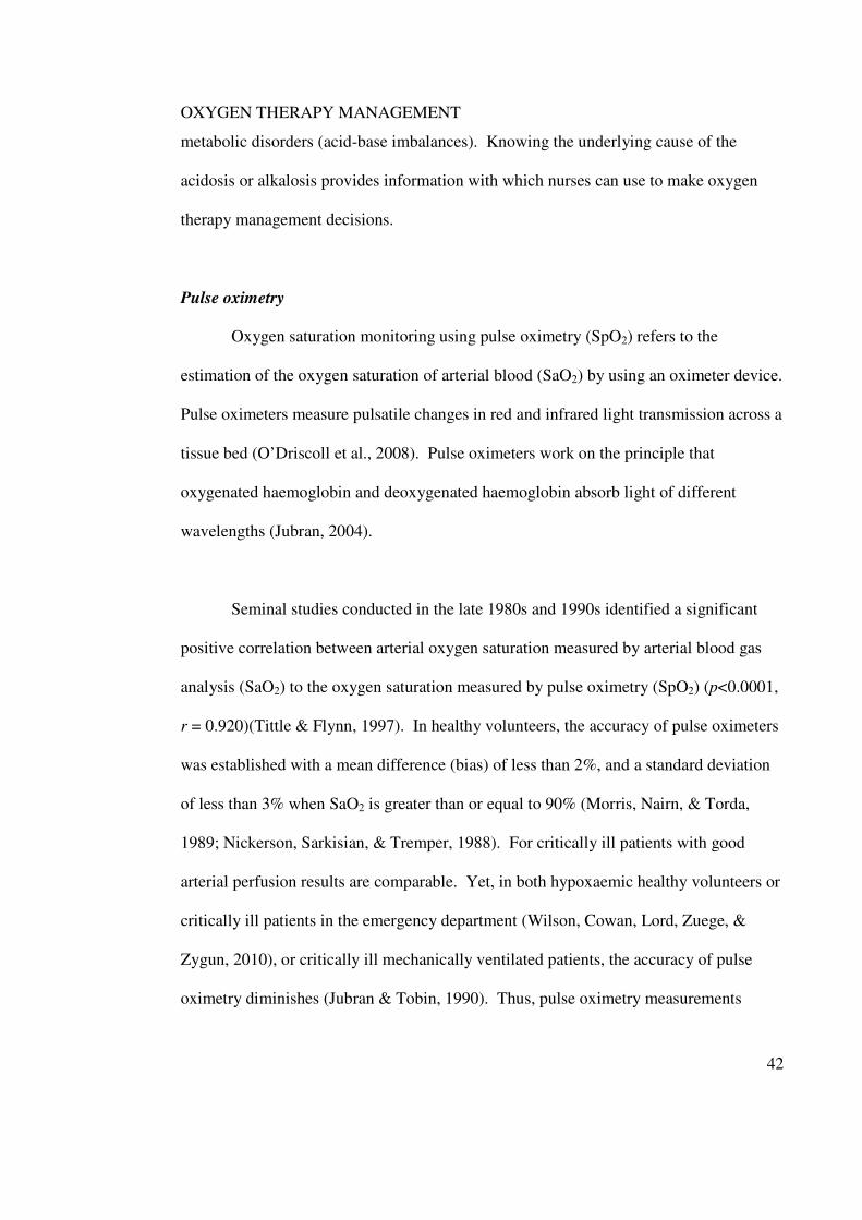
Oxygen saturation monitoring using pulse oximetry (SpO2) refers to the
estimation of the oxygen saturation of arterial blood (SaO2) by using an oximeter device.
Pulse oximeters measure pulsatile changes in red and infrared light transmission across a
tissue bed (O’Driscoll et al., 2008). Pulse oximeters work on the principle that
oxygenated haemoglobin and deoxygenated haemoglobin absorb light of different
wavelengths (Jubran, 2004).
Seminal studies conducted in the late 1980s and 1990s identified a significant
positive correlation between arterial oxygen saturation measured by arterial blood gas
analysis (SaO2) to the oxygen saturation measured by pulse oximetry (SpO2) (p<0.0001,
r = 0.920)(Tittle & Flynn, 1997). In healthy volunteers, the accuracy of pulse oximeters
was established with a mean difference (bias) of less than 2%, and a standard deviation
of less than 3% when SaO2 is greater than or equal to 90% (Morris, Nairn, & Torda,
1989; Nickerson, Sarkisian, & Tremper, 1988). For critically ill patients with good
arterial perfusion results are comparable. Yet, in both hypoxaemic healthy volunteers or
critically ill patients in the emergency department (Wilson, Cowan, Lord, Zuege, &
Zygun, 2010), or critically ill mechanically ventilated patients, the accuracy of pulse
oximetry diminishes (Jubran & Tobin, 1990). Thus, pulse oximetry measurements
Page 44
OXYGEN THERAPY MANAGEMENT
43
together with assessment of other clinical indicators of oxygenation are important when
assessing a patient’s respiratory state.
Pulse oximetry has some important limitations that need to be considered when
identifying the type of respiratory dysfunction experienced by the patient and to
determine if oxygen therapy is appropriate management. An important limitation of
pulse oximetry is that it does not provide information on ventilation (PaCO2 and pH)
(Clark, Giuliano, & Chen, 2006; Howell, 2002). Therefore, SpO2 readings should form
a part of a comprehensive clinical assessment of indicators of oxygenation and
respiratory function (Wong & Elliott, 2009) and play a key role in determining the need
for arterial blood gas sampling and when to seek further information about a patient’s
oxygen status (Miller, 1992; Van de Louw et al., 2001).
Potential sources of error in pulse oximetry measurements include intravenous
dyes (e.g. methylene blue), low-perfusion states (e.g. shock, sepsis and hypotension)
(Grace, 1994; Saito, Fukura, Shimada, & Fujita, 1995), dark skin pigmentation (Feiner,
Severinghaus, & Bickler, 2007) or the use presence of nail polish (Hinkelbein,
Genzwuerker, Sogl, & Fiedler, 2007) causing resulting in poor signal quality or strength.
Pulse oximeter probe placement is also known to affect the accuracy of readings (Kisiel
& Perkins, 2006) and poor placement may lead to errors related to oxygen management
(Wong & Elliott, 2008). Oximetry readings obtained from finger and earlobe probes
have been demonstrated to be more accurate than those obtained by placing the probe on
a toe (Jubran, 2004). However, the patient’s tolerance to having a probe placed on a
finger, earlobe or toe will also impact on the ability to obtain an accurate SpO2 reading
Page 45
OXYGEN THERAPY MANAGEMENT
44
(Jubran, 2004). Thus, site selection for the pulse oximetry probe is another important
consideration during the respiratory assessment of the patient.
So far, the physiological need for oxygen, normal and abnormal oxygen states,
and oxygen monitoring methods have been discussed. In the following section the
clinical risk of respiratory dysfunction and the evidence supporting the use of oxygen
therapy for the management of respiratory dysfunction is presented. The following
discussion includes information about nasal prongs, face mask and nasopharyngeal
oxygen catheter devices which are examined in the research studies discussed in
subsequent Chapters of this thesis.
Respiratory dysfunction and the role of oxygen therapy
Respiratory dysfunction and adverse events
Respiratory dysfunction is a clear clinical risk and indicator of serious illness and
precursor to adverse events such as cardiac arrest, unplanned intensive care unit
admission and death (Cretikos et al., 2008; Harrison et al., 2005; Quach et al., 2008).
When Considine (2004) reviewed the relationship between hypoxaemia and adverse
events, hypoxaemia was the most common reason for the activation of a Medical
Emergency Team. Respiratory dysfunction places patients at high risk of adverse events
and warrants timely and appropriate response from intensive care nurses.
Two of the strongest early warning signs of respiratory dysfunction are
hypoxaemia and abnormal respiratory rates. Unfortunately, many investigators have
identified that many patients in hospital have unrecognised or inappropriately treated
Page 46
OXYGEN THERAPY MANAGEMENT
45
hypoxaemia prior to an adverse event (Buist, Bernard, Nguyen, Moore, & Anderson,
2004; Cuthbertson, Boroujerdi, McKie, Aucott, & Prescrott, 2007; Goldhill, White, &
Sumner, 1999; McGloin, Adam, & Singer, 1999). In a review of physiological
observations prior to patient emergencies, Harrison et al. (2005) reported up to 37% of
ward patients had a SpO2 between 90% and 95% and 11% of patients had SpO2 of less
than 90%. Thus, early recognition and correction of physiological abnormalities, such
as hypoxaemia, is a fundamental step to reducing the personal cost of adverse events in
healthcare (Buist et al., 2002; Camarata, Weil, Hanashiro, & Shubin, 1971; Crispin &
Daffurn, 1998; Ehsani, Jackson, & Duckett, 2006).
Hypoxaemia is a significant risk factor for adverse events and a significant risk
factor for death following an adverse event (Goldhill & McNarry, 2004; Hillman et al.,
2001; Santiano et al., 2009; Schein, Hazday, Pena, Ruben, & Sprung, 1990; Skrifvars,
Nurmi, Ikola, Saarinen, & Castren, 2006; Suljaga-Pechtel, Goldberg, Strickon, Berger,
& Skovron, 1984). Buist et al. (2004) conducted a retrospective study to determine
whether abnormal clinical observations in a patient population could predict subsequent
in-hospital mortality. Over the study period, 6303 patients were admitted to five general
hospital wards at a suburban tertiary teaching hospital. A chart review of these patients
revealed that 564 (8.9%) experienced 1598 pre-determined clinical abnormal events and
146 (0.9%) patients died. The two most common clinical events were arterial oxygen
desaturation and hypotension. Buist et al., (2004) used a logistic regression model to
identify six abnormal clinical observations that could increase by almost seven-fold the
risk of onset of a medical emergency including cardiac arrest or mortality, with two of
those clinical observations related to respiratory dysfunction: respiratory rate less than
Page 47
OXYGEN THERAPY MANAGEMENT
46
six breaths per minute and an oxygenation saturation of less than 90%. Suljaga-Pechtel
et al. (1984) examined arterial blood gas samples of patients who experienced a cardiac
arrest and found that more than half of the patients had significant hypoxaemia (PaO2
less than 50 mmHg), but less than half of those patients were successfully resuscitated.
The findings of these two studies, although separated by 20 years are important, as the
presence of hypoxaemia remains a significant clinical indicator of a serious adverse
event.
Respiratory rate is a routine component of the physiological assessment of a
patient (Higginson & Jones, 2009; Simpson, 2006); however, greater awareness of the
importance of abnormal respiratory rates, as an indicator of serious illness, is advocated
(Cretikos et al., 2008). Crekitos et al. (2008) report that clinical studies conducted over
the past 20 years have shown that abnormal respiratory rates, in particular tachypnoea, is
an important predictor of serious adverse events such as cardiac arrest, or admission to
the intensive care unit. While respiratory rate, may be a non-specific indicator of
hypoxaemia, assessing and documenting respiratory rate is important because as noted
above tachypnoea is an early warning sign of a clinical adverse event. To help identify
and manage patients at risk of a respiratory related adverse event Crekitos et al. (2008)
concluded that improvements needed to be made to the frequency and recording of
respiratory rate in hospital observation charts. Table 2.3 provides a summary of key
studies that have specifically examined the association between derangements in the
clinical indicators of respiratory dysfunction (bradypnoea, tachypnoea and hypoxaemia)
and the occurrence of outcomes such as death, cardiac arrest, or admission to an
intensive care unit.
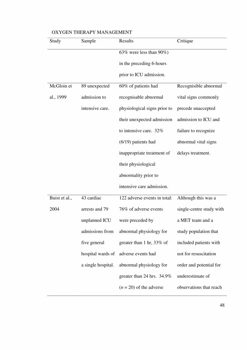
Page 48
OXYGEN THERAPY MANAGEMENT
47
Table 2.3 Summary of key research relating to bradypnoea, tachypnoea and the presence
of hypoxaemia as a precursor to an adverse event
Study Sample Results Critique
Camarata et
al., 1971
193 cardiac
arrests in 132
patients
40% (13/33) of patients
experienced a cardiac
arrest deemed to be
‘unexpected’.
Unexpected sudden
deterioration in
hospitalized patients
may result in cardiac
arrest.
Suljaga
Pechtel et al.,
1984
207 cardiac
arrests.
59% of patients had
significant hypoxaemia
(PaO2 less than 50 mmHg).
Patients with initial PaO2
greater than 50 mmHg are
more likely to be
resuscitated (58.3% vs.
42.3%, p < 0.05).
Significant respiratory
dysfunction at the onset
of resuscitation is a
strong indicative of a
poor patient outcome.
Goldhill et
al., 1999
79 admission to
intensive care
for 76 patients.
34% of patients
experienced cardiac arrest
prior to ICU admission.
75% of patients received
oxygen, 37% had an ABG
and 61% had SpO2
measurements (of which
Significant hypoxaemia
was commonly in the
hours preceding a
cardiac arrest and prior
to ICU admission.
Page 49
OXYGEN THERAPY MANAGEMENT
48
Study Sample Results Critique
63% were less than 90%)
in the preceding 6-hours
prior to ICU admission.
McGloin et
al., 1999
89 unexpected
admission to
intensive care.
60% of patients had
recognisable abnormal
physiological signs prior to
their unexpected admission
to intensive care. 32%
(6/19) patients had
inappropriate treatment of
their physiological
abnormality prior to
intensive care admission.
Recognisible abnormal
vital signs commonly
precede unaccepted
admission to ICU and
failure to recognize
abnormal vital signs
delays treatment.
Buist et al.,
2004
43 cardiac
arrests and 79
unplanned ICU
admissions from
five general
hospital wards of
a single hospital.
122 adverse events in total:
76% of adverse events
were preceded by
abnormal physiology for
greater than 1 hr, 33% of
adverse events had
abnormal physiology for
greater than 24 hrs. 34.9%
(n = 20) of the adverse
Although this was a
single-centre study with
a MET team and a
study population that
included patients with
not for resuscitation
order and potential for
underestimate of
observations that reach
Page 50
OXYGEN THERAPY MANAGEMENT
49
Study Sample Results Critique
events involved respiratory
related abnormalities.
MET criteria but were
not activated.
Quach et al.,
2008
Retrospective
medical record
audit of 200
patients who had
a MET call from
a single acute
care hospital.
Patients with respiratory
distress more likely to be
postoperative (40% vs
28%, p = 0.07). Hospital
mortality for MET calls
due to respiratory distress
was 38%. The median
duration of delay for MET
activation for patients with
respiratory distress was 12
hrs. A delay in activation a
MET was associated with
an increase in mortality
(OR, 2.10; 95% CI: 1.01-
4.34; p =0.045).
Single-centre
retrospective audit that
highlighted respiratory
distress was a
significant contributor
to in-hospital MET call
activation however
delay in MET call was
identified.
Study Sample Results Weaknesses/limitations
Considine et
al., 2009
Retrospective
case-control
study of
predictors of
Patients with respiratory
rate abnormalities at triage
(low RR less than 12 or
high RR greater than 22)
Single-centre study.
Reliance on medical
audit and the potential
for missing data,
Page 51
OXYGEN THERAPY MANAGEMENT
50
Study Sample Results Weaknesses/limitations
ICU admission
of 386 ED
patients.
had increased risk of
admission to intensive care
(OR 1.66, 95% CI: 1.05-
2.06).
delivered but
undocumented care.
Between-group
differences that failed to
reach statistical
significance may be
related to sample size.
Santino et al.,
2009
Descriptive
study of MET
calls in six
hospitals over a
12-month
period.
35.2% (n = 324) of
‘breathing’ related
problems results in the
calling of a MET with a
low SpO2 resulting in
29.2% (n = 249) of MET
activations. Breathing
problems accounted for
most (46%) of the
unexpected admissions to
ICU for ‘worried’ MET
calls.
Breathing abnormalities
resulting in low SpO2
readings contributed to
over a quarter of MET
calls. Se of subjective
MET call criteria can be
a useful adjunct to
standard MET call
criteria.
Note. ABG = arterial blood gas; ED = emergency department; ICU = intensive care unit;
MET = medical emergency team; PaO2 = partial pressure of arterial oxygen; RR =
respiratory rate; SpO2 = Oxygen saturation measured by pulse oximetry.
Page 52
OXYGEN THERAPY MANAGEMENT
51
Oxygen therapy
For the purpose of this thesis, oxygen therapy is defined as the therapeutic
administration of supplemental oxygen to patients using nasal prongs, face mask and
nasopharyngeal oxygen catheter. As the central tenant of this thesis is intensive care
nurses’ management of oxygen therapy, it is necessary to describe how oxygen therapy
is administered and the various factors that may make oxygen therapy difficult to
manage or ineffective.
In the following section three issues will be discussed. These are:
• Oxygen therapy, oxygen flow rate and mode of breathing
• Oxygen device size and fit
• Efficacy of the nasal prongs, face mask and nasopharyngeal oxygen catheter to
achieve and maintain adequate SpO2
Oxygen therapy and flow rates
All oxygen delivery devices assist patients to breathe air which has greater
concentrations of oxygen than room air (Stich & Cassella, 2009). The basic
requirements for the therapeutic use of oxygen delivery devices include: the ability to
control the percentage of oxygen in inspired gas, minimal accumulation of carbon
dioxide, minimal resistance to breathing (inspiration and expiration), efficiency and
economy in oxygen use and, adaptability to different respiratory rates (O’Driscoll et al.,
2008). Knowledge of the general characteristics of oxygen therapy is required to ensure
appropriate oxygen delivery device selection and use (O’Driscoll et al., 2008).
Page 53
OXYGEN THERAPY MANAGEMENT
52
Knowledge regarding oxygen management is vital for patient safety
interventions. Ensuring appropriate oxygen delivery device selection is a fundamental
step in response to and prevention of respiratory dysfunction. Commonly used oxygen
delivery devices include nasal prongs, face mask and nasopharyngeal oxygen catheter.
Each of these devices provide the patient with more than 21% FiO2 but less than 100%
due to the interrelationship of oxygen flow, device factors, such as functional apparatus
dead space, and patient factors such as peak inspiratory flow rate and expiratory pause
(Wagstaff & Soni, 2007; West, 2008). Differences in the oxygen flow, peak inspiratory
flow rate and the length of the expiratory pause will result in variations of the
effectiveness of oxygen delivery between patients and at times with an individual patient
on a breath-by-breath basis (Barnes, 2000; Calianno, Clifford, & Titano, 1995;
O'Connor & Vender, 1995).
The concentration of supplemental oxygen administered can be described either
as a concentration of oxygen expressed as a percentage (%), or as a fraction of inspired
oxygen expressed as a decimal (Stich & Cassella, 2009). For example, the concentration
of oxygen in room air is 21% oxygen or oxygen at a fraction of inspired oxygen of 0.21
(Pruitt & Jacobs, 2003; Stewart & Howard, 1990). Thus, an increase in the
concentration of supplemental oxygen can range from 22% to 100% or from 0.22 to 1.0
(Sim, Dean, Kinsella, Black, Carter, & Hughes, 2008; Stich & Cassella, 2009). For the
purpose of this thesis, the concentration of oxygen will be described as a percentage.
The performance of oxygen delivery devices is commonly described in terms of
‘delivered oxygen concentration’ and ‘inspired oxygen concentration’. ‘Delivered
Page 54
OXYGEN THERAPY MANAGEMENT
53
oxygen concentration’ refers to the concentration of oxygen at the site of the oxygen
delivery device (Goldstein, Young, & Rebuck, 1982; Stich & Cassella, 2009). Delivered
oxygen concentration is closely linked to the flow of oxygen into an oxygen delivery
device and has a direct impact on the concentration of oxygen that will be inspired by
the patient (Mulryan, 2009). For the purposes of this thesis the term ‘inspired oxygen
concentration’ refers to the concentration of oxygen actually inspired by the patient.
Normal inspiratory flow, in a healthy adult, ranges between 25 litres per minute and 53
litres per minute. Patients with respiratory dysfunction can increase their inspiratory
flow by 50 to 300 litres per minute (Rose & Hanlon, 2012). Because oxygen delivery
devices deliver oxygen at set flow rates, generally less than 30 litres per minute, the
additional volume of gas is obtained from the surrounding atmosphere (Rose & Hanlon,
2012). Thus, when room air is entrained the inspired oxygen concentration is decreased
as the room air dilute the amount of oxygen delivered to the patient when oxygen
delivery devices are used (Corley & Ringdal, 2012).
When respiratory rate or minute ventilation (the amount of oxygen drawn into
the lungs over a minute) increases, due to pain, anxiety or serious illness, the peak
inspiratory flow rate increases proportionately (Higginson & Jones, 2009). Conversely,
a decrease in respiratory rate or minute ventilation will decrease the volume of room air
entering the lungs and result in an increase in the inspired oxygen concentration
(O’Driscoll et al., 2008).
As discussed in the paragraphs above, oxygen flow rate is a key decision point in
the selection and use of oxygen delivery devices. Oxygen flow rate is the speed at
Page 55
OXYGEN THERAPY MANAGEMENT
54
which oxygen enters the oxygen delivery device and it is described as litres per minute
and equates to the value displayed by the oxygen flow meter (Barnes, 2000). Oxygen
flow meters, in use in Australian intensive care units, generally have a maximum flow
capacity of 15 litres per minute.
During normal breathing the respiratory pattern of air movement into and out of
the lung is essentially sinusoidal, with the peak inspiratory flow rate occurring during
the middle of inspiration (West, 2008). The term ‘inspiratory flow rate’ is the speed at
which air is drawn into the lungs and has a direct impact on the percentage of inspired
oxygen concentration (West, 2008).
Mode of breathing, via the nose or mouth, impacts on efficacy of oxygen
delivery devices to achieve satisfactory oxygen concentration (O’Driscoll et al., 2008).
Clinical studies have identified that mouth breathers receive higher concentration of
oxygen in the nasopharynx than nasal breathers when nasal devices are in use (Fairfield,
Goroszeniuk, Tully, & Adams, 1991; Waligora, 1970). To assess the impact of device
fit on a known FiO2, Boumphrey et al. (2003) tested 20 healthy adults to demonstrate
that tight fitting masks, opposed to lose fitting masks, resulted in higher expired oxygen
concentrations. They conclude that by using tight-fitting face masks it was possible to
achieve an expired concentration of oxygen of 85%, which equates with inspired oxygen
concentration of 97%. Thus, the potential implication of face mask fit should recognised
as delivered and inspired oxygen concentrations will vary depending on the fit of the
device (Boumphrey et al., 2003; Higginson & Jones, 2009).
Page 56
OXYGEN THERAPY MANAGEMENT
55
The difference between delivered and inspired oxygen concentration is a
physiological concept that nurses must understand when administering supplemental
oxygen. Respiratory rate and peak inspiratory flow rate directly influence the
effectiveness of oxygen delivery devices and the ability of the patient to maintain
adequate SpO2 or PaO2 levels (Eastwood et al., 2007). For example, when nasal prongs
or a simple face mask are used to deliver oxygen therapy, the inspired oxygen
concentration is less than the delivered oxygen concentration because during inspiration
room air is entrained (drawn in) to the lungs as well as the delivered oxygen (Sim et al.,
2008).
Whether oxygen therapy is effective or not depends on several patient-related
factors including: mode of breathing (e.g. nose breathing or mouth breathing); the shape
and fit of the oxygen delivery device and if it is comfortable for the patient to wear.
Oxygen device size, shape and fit
The oxygen delivery device influences effective oxygen therapy, especially the
device related factors of size, shape and fit. A device that is not the correct size or fits
poorly, either too small or too large may lead to variable amounts of oxygen being
delivered to the patient or device displacement resulting in periods that the patient does
not receive any supplemental oxygen. To demonstrate the importance of correct fit and
device placement, Nolan et al. (1992) investigated nocturnal activities and the impact of
oxygen delivery device removal by measuring the SpO2 on pulse oximetry of 30
postoperative patients, randomly allocated to face mask at four litres per minute or nasal
prongs at two litres per minute, and identified that in the face mask group the device
Page 57
OXYGEN THERAPY MANAGEMENT
56
remained in correct position in five patients but in ten patients the device was removed a
total of 28 times for up to 78 mins, 17 of those were for nursing duties. For the nasal
prongs group, one patient removed the device 18 times for a total period of 16 min 28
secs with an average SpO2 of 97% (range 90.8 – 99.3%), meaning that nasal prongs were
better tolerated. Consequently, nurses need to be aware of any activity that may
interrupt oxygen delivery and actively engage in strategies to prevent device removal.
To summarise, any change in oxygen flow rate and a patient’s peak inspiratory
flow rate on has an effect on the concentration of inspired oxygen. Mouth breathing
when wearing nasal prongs can decrease the concentration of inspired oxygen.
However, mouth breathers received higher oxygen concentration in the nasopharynx
than nasal breathers when nasal oxygen delivery devices were used (Dunlevy & Tyl,
1992; Nolan, Baxter, Winyard, Roulson, & Goldhill, 1992; Nolan et al., 1993; Ooi,
Joshi, & Soni, 1992). Factors influencing effective oxygen therapy included mode of
breathing and device size, shape and fit. As with oxygen flow rate and peak inspiratory
flow rate, mode of breathing, either by mouth or nose, has also been shown to impact on
FiO2 (Dunlevy & Tyl, 1992). Thus, it is important for nurses to assess the mode of
breathing in order to tailor the appropriate oxygen delivery device to optimise the
therapeutic effect of oxygen administration.
Oxygen delivery devices
Nasal prongs, face mask and nasopharyngeal oxygen catheter are often the first
choice selected by nurses for the management of respiratory dysfunction (Eastwood et
al., 2007). The discussion in the section to follow focuses on the general characteristics
Page 58
OXYGEN THERAPY MANAGEMENT
57
and care requirements for nasal prongs, face mask, and nasopharyngeal oxygen
catheters.
Nasal prongs
Nasal prongs consist of two short tapered prongs (about 1 cm in length)
(Eastwood et al., 2007), which when applied correctly to the patient’s face, each prong
lies approximately 1 cm within each nostril (Eastwood et al., 2007; Pruitt & Jacobs,
2003). To facilitate optimal positioning and functioning the tubing of the nasal prongs is
looped over the ears and secured under the patient’s chin (Barnes, 2000; Eastwood et al.,
2007). Oxygen flows for nasal prongs range from half a litre per minute to six litres per
minute and flow are adjusted to achieve a target oxygen saturation (Eastwood et al.,
2007).
The delivery of oxygen therapy via nasal prongs may be considered unsuitable in
any patient at risk of nasal damage, deformity or blockage, as adequate supplemental
cannot be assured (Eastwood & Dennis, 2006). Nasal prongs should be used with
caution in patients immediately following upper airway or nasal surgery, those with
impaired airway protection, or severe forms of hypoxaemia (Eastwood & Dennis, 2006).
Patients receiving supplemental oxygen via nasal prongs are monitored for signs
of ventilator adequacy including oxygen saturation, respiratory rate and respiratory
depth. The need to escalate therapy should be considered when oxygen saturation levels
are not being maintained. Patients receiving oxygen via nasal prongs need to be
Page 59
OXYGEN THERAPY MANAGEMENT
58
monitored for pressure sores at the base of the nose, ears or face, and that the nasal
prongs are positioned correctly (Kacmarek, Dimas, & Mack, 2005).
Face mask
The simple face mask is a plastic, ‘pear shaped’ device that covers the nose and
mouth. For optimal effect the face mask should fit firmly to the face and be secured in
place with a band around the back of the head (Barnes, 2000). Recommended oxygen
flow rates when using a face mask range from six litres per minute to 15 litres per
minute (Eastwood & Dennis, 2006). Typically, flow rates of five to 10 litres per minute
are used in the clinical setting to treat symptoms of moderate hypoxaemia (SpO2 90-
95%) (Eastwood & Denis, 2006). Oxygen flow rates of less than six litres per minute
when using a face mask are discouraged (O’Driscoll et al., 2008). Low oxygen flow
rates during face mask therapy may be insufficient to eliminate exhaled carbon dioxide
which may then be re-breathed by the patient (Jensen, Johnson, & Sandstedt, 1991;
O'Driscoll et al., 2008).
Some patients may experience claustrophobic sensations, drying of the mucous
membrane in the mouth and nose, or be at higher risk of aspiration if they vomit and it
collects in the mask. The patient receiving oxygen via a face mask requires ongoing
assessment of ventilation, pulmonary function and oxygenation and must be monitored
for compliance with therapy and assessed for the need to progress to more advanced or
simplified supports (Eastwood & Dennis, 2006). Additional monitoring should be
directed at detection of respiratory fatigue, non-compliance with therapy, device
removal or displacement, and device related complications such as undue mask pressure
Page 60
OXYGEN THERAPY MANAGEMENT
59
on the bridge of the nose, or soreness related to the mask’s strapping (Eastwood &
Dennis, 2006; Kacmarek et al., 2005; O’Driscoll et al., 2008; Sasaki et al, 2003).
Nasopharyngeal oxygen catheter
Nasopharyngeal oxygen catheters deliver of supplemental oxygen directly into the
nasopharynx (Eastwood et al., 2004; Frey, McQuillan, Shann, & Freezer, 2001).
Nasopharyngeal oxygen catheters are placed through the nose to the depth of the
nasopharynx and secured in position by using tape (Eastwood, et al., 2004).
Recommended oxygen flow rates for nasopharyngeal oxygen catheters is 1 litre per
minute to 6 litres per minute, to achieve approximate inspired oxygen concentration of
25-35%, with the oxygen flow rate adjusted to meet SpO2 targets (Eastwood & Dennis,
2006).
The nasopharyngeal oxygen catheter may be suitable when there are physical
limitations to the use of a face mask to deliver oxygen therapy, or if discomfort with face
masks or nasal prongs exist (Eastwood & Dennis, 2006). Contraindications to the use of
the nasopharyngeal oxygen catheter include epistaxis, nasal trauma or in patients
receiving anti-coagulant therapy (Eastwood & Dennis, 2006). Monitoring patients
receiving oxygen via the nasopharyngeal oxygen catheter include continuing evaluation
of pulmonary function, respiratory effort, and therapy-related complications such as
undue catheter pressure on the base of the nose or excessive nasopharyngeal drying.
Page 61
OXYGEN THERAPY MANAGEMENT
60
A comparison of general characteristics and relative advantages and
disadvantages of nasal prongs, face mask and nasopharyngeal oxygen catheter is shown
in Table 2.4.
Page 62
OXYGEN THERAPY MANAGEMENT
61
Table 2.4 Comparison of nasal prongs, face mask and nasopharyngeal oxygen catheter oxygen delivery devices
Device Oxygen
flow ratesa
Approximate
FiO2
Advantages Disadvantages
NPO 1 to 6
25-35% Low cost per unit, eating and
drinking not hindered, constant
oxygen delivery into upper airway,
difficult to dislodge
Catheter blockage, excessive mucus
accumulation in nasopharynx, invasiveness
of insertion procedure, risk of nasal trauma
and potential for bleeding
NP 0.5 to 6 22-40% Simple to apply and eating and
drinking not hindered
OFR greater than 4 LPM may cause
discomfort, device can be easily dislodged,
device can irritate skin around the ears and
nares
FM 6 to 15 30-90% Useful for short periods, able to
deliver higher FiO2 than nasal
prongs, or oxygen catheters
Recommended OFR greater than 6 LPM.
device removal to eat, cough or allow the
passage of vomitus, may induce feelings of
claustrophobia, CO2 re-breathing can occur
Note. CO2 = carbon dioxide; FiO2 = fraction of inspired oxygen; LPM = litres per minute; OFR = oxygen flow rate. aOxygen flow rate
reported in litres per minute. Adapted from Barnes (2000), Calianno et al. (1995), Eastwood and Dennis (2006) and O’Connor and
Vender (1995).
Page 63
OXYGEN THERAPY MANAGEMENT
62
Research comparing oxygen delivery devices
Oxygen therapy is a frequent intervention commonly applied to hospitalised
adult patients (Bateman & Leach, 1998; Shelly & Nightingale, 1999). Despite the
importance of oxygen therapy for prevention and treatment of respiratory dysfunction,
few of the commonly used oxygen delivery devices have been subject to critical
examination (Kernick & Magarey, 2010), and neither have the oxygen therapy
management decisions by intensive care nurses been explored (Eastwood et al., 2012).
The purpose of this section of the literature review is to describe and critique the
published research comparing nasal prongs, face mask and nasopharyngeal oxygen
catheters in adult hospitalised patients. Studies were included if they compared the three
aforementioned devices assessing effectiveness to achieve and maintain adequate
oxygen saturations and patient-related measures of comfort and compliance with
therapy. Studies were excluded if they:
• did not refer to oxygen saturation (SaO2 or SpO2) as an outcome measure
(Heller, Watson, & Imredy, 1965)
• did not report comfort outcome measures from bench studies (Bazuaye,
Stone, Corris, & Gibson, 1992; Fairfield et al., 1991; Goldstein et al.,
1982; Leigh, 1970; Ooi et al., 1992; Redding, McAfee, & Gross, 1978)
• were studies undertaken in healthy individuals (Boumphrey et al., 2003;
Hess, D'Agostino, Magrosky, Myers, & Shuman, 1984; Sasaki et al.,
2003; Waldau, Larsen, & Bonde, 1998)
• did not pertain to the population of interest: adult hospitalised patients
(Cogliano, Graham, & Clark, 2002; Frey et al., 2001; Muhe et al., 1998;
Page 64
OXYGEN THERAPY MANAGEMENT
63
Shann, Gatchalian, & Hutchinson, 1988; Weber, Palmer, Oparaugo, &
Mulholland, 1995; Wilson, Arnold, Connor, & Cusson, 1996)
• did not report primary research (Eastwood & Dennis, 2006; Kernick &
Magarey, 2010; Stewart & Howard, 1990), were not reported in English
(Lund, Holm-Knudsen, Nielsen, & Foge Jensen, 1996)
• did not include a comparison between two of the three oxygen delivery
devices under examination (Bambridge, 1993; Costello, Liston, &
McNicholas, 1995; English & Brown, 1994; Hudes, Marans, Hirano,
Scott, & Ho, 1989; Jacobsen, Neilsen, Brinklov, Stokke, & Hartmann-
Andersen, 1980; Williams, Jones, & Mapleson, 1988)
The literature review identified six key studies that had compared oxygen
therapy delivered via nasal prongs with face mask or oxygen therapy delivered via face
mask with nasopharyngeal oxygen catheter. The six identified studies were published
from 1993-2009 and had sample populations of drawn from the general ward, post-
anaesthetic care unit (PACU) or the ICU. Therapeutic innovations are frequently
introduced into clinical practice in an attempt to optimise patient outcomes. However,
oxygen delivery devices have not developed significantly for decades resulting in
therapy being administered with oxygen delivery devices, as such comparison of oxygen
delivery devices and their use over time is possible (Kernick & Magarey, 2010). Five of
the six studies were a comparison between nasal prongs and face mask (Ayhan, Iyigun,
Tastan, Emin, & Ozturk, 2009; Bolton & Russell, 2001; McBrien & Sellers, 1995;
Nolan, et al., 1993; Stausholm et al., 1995) and the sixth study compared the face mask
device with the nasopharyngeal oxygen catheter device (Eastwood et al., 2004). None
Page 65
OXYGEN THERAPY MANAGEMENT
64
of the studies identified had included a simultaneous comparative study of nasal prongs,
face mask and nasopharyngeal oxygen catheter devices. Oxygenation saturation (SaO2
or SpO2) was the primary outcome identified for the six studies selected.
In all studies SpO2 values were greater than or equal to 95% when the oxygen
delivery device remained in position. Eastwood, Revees & Cowie (2004) in a
prospective crossover study, identified a higher PaO2 for face mask therapy compared
with nasopharyngeal oxygen catheter use for 50 intensive care patients. The higher
PaO2 in this study could be attributed to the higher oxygen flow rate used with the face
mask device. Similarly, McBrien & Sellers (1995) in their crossover trial of nasal
prongs and face mask for 11 post-operative patients also showed that oxygen therapy via
face mask provided higher PaO2 values than that of the nasal prongs. In addition, using
a quasi-experimental design conducted in the post-anaesthetic care unit, Nolan et al.
(1993) identified patients randomly allocated to receive nasal prong oxygen therapy had
a higher incidence of hypoxaemia than that of patients who received face mask oxygen
therapy. The conclusion being drawn was that oxygen therapy via face mask would
reduced the incidence of hypoxaemia in patients requiring simple oxygen therapy during
periods of post-operative recovery.
The findings of the reviewed studies have important implications for the
selection of oxygen delivery devices by clinicians for the management of respiratory
dysfunction. However, the strength and level of evidence that can be derived from these
studies is not strong. As such, it is necessary to conduct further investigations into the
clinical effectiveness of nasal prongs, face mask and nasopharyngeal oxygen catheter
Page 66
OXYGEN THERAPY MANAGEMENT
65
devices, so that any future recommendations can be compared to a known quantity and
specific to the intensive care patient.
Patient comfort and compliance with therapy was the second outcome for the
review and a description of patient comfort or compliance with therapy was reported in
the identified studies (Ayhan et. al, 2009; Bolton & Russell, 2001; Eastwood et al.,
2004; McBrien & Sellers, 1995; Nolan et al., 2009; Stausholm et al., 1995). Patient
comfort is an influential factor on the clinical effectiveness of oxygen therapy because
an uncomfortable device is likely to be removed by the patient and expose the patient to
episodes of hypoxaemia. Findings from each of the reviewed studies showed that
patients preferred nasal devices compared with face mask. Importantly, findings
reported by Nolan et al., (1993) and Ayhan et al., (2009) identified that face mask
devices were more frequently removed than nasal prongs. A detailed description of the
various factors known to influence patient comfort and compliance with therapy is
presented later in this chapter. Table 2.5 presents a summary of six studies that have
compared the clinical effectiveness and comfort of nasal prongs, face mask and
nasopharyngeal oxygen catheter devices in adult hospitalised patients.
More information pertaining to the efficacy of oxygen therapy devices, the
potential complications of each device, how patients experience the use of the devices
and the experience of nurses in applying the devices to deliver oxygen therapy, would
provide knowledge on which nurses could base their practice. Gaps in the literature
exist because previous studies have failed to explore the multiplicity of factors that
Page 67
OXYGEN THERAPY MANAGEMENT
66
influence effective oxygen therapy practice. Consequently, the previous studies are
unable to provide strong evidence to inform clinicians’ oxygen management practices.
In summary, oxygen therapy enables clinicians to manage the risks associated
with respiratory dysfunction and the management of oxygen deficient states. The
section above has described oxygen therapy and focused on three specific oxygen
delivery devices. However, in clinical practice there is an interplay between oxygen
delivery device, patient, and nurse related factors. These factors each exert an influence
on the effectiveness of oxygen therapy. In the following sections of the literature review
the patient and nurse related factors known to influence oxygen therapy management are
discussed.
Page 68
OXYGEN THERAPY MANAGEMENT
67
Table 2.5 Summary of studies that have compared the clinical effectiveness and comfort of nasal prongs, face masks and nasopharyngeal
oxygen catheters in adult hospitalised patients
Author Design Sample/Setting Oxygen delivery device Outcome/Result Weaknesses/Limitations
NP FM NPO
Nolan et al.,
1993
Quasi-
experimental
30 patients, general
surgical ward: 1st
postoperative night.
Patients were allocated
randomly to receive
oxygen by FM at 4
litres/min or NP at 2
litres/minute. From 22:00
on the first night after
operation, the position of
the nasal cannula or face
mask was observed for 8h
using video and SpO2.
X X The mask for patients in the FM
group positioned correctly in five of
the 10 patients. FM removed a total of
28 times: (17 for nursing tasks) for a
median time of 2 min 39 sec (range
30 sec to 7 hr 40 min 40 sec). One
patient in the NP group removed the
NP for 16 min 38 sec and eight times
in another patient for a total of 1 hr 18
min 7 sec. The mean SpO2 with FM
on was 98% (range 96.1–99.9%), and
for the NP group 97% (range 90.8–
99.3%); with FM removed 95%
(range 89.8–98.8%).
Randomisation method not explicit.
Patients in the face mask group
commenced with a oxygen flow rate of 4
litres per minute. Primary outcome
measures based on compliance with
therapy and not satisfactory oxygenation.
Failure to intervene when patients became
hypoxaemic. Statistical analysis not
clearly presented and results
predominantly descriptive. No measure
of patient comfort or oxygen delivery
device preference.
Page 69
OXYGEN THERAPY MANAGEMENT
68
Author Design Sample/Setting Oxygen delivery device Outcome/Result Weaknesses/Limitations
NP FM NPO
Stausholm
et al, 1995
Prospective
crossover
25 surgical ward patients.
Patients rated oxygen
delivery device comfort
using a visual analogue
score (0 mm most
uncomfortable to 100 mm
most comfortable).
At inclusion, median SpO2 was 91% (76-
94%). During oxygen therapy SpO2
increased to 97% (93-100%) using the NP
(p < 0.0001) and 97% using the FM (p < 0
.001). In terms of comfort, NP scored 72
(19-100) mm and FM 42 (0-94) mm.
(Friedman’s analysis, p < 0.0001). The
main problem identified with the FM by
the patients was difficulty eating/drinking.
Small sample size. Randomisation method not
explicit. Potential time and order effects
associated with crossover trials.
McBrien &
Sellers, 1995
Prospective
crossover
11 adult patients in the
general ICU
X X SpO2 readings remained between 96 and
98% for each device. Greater PaO2 with
FM than NP (p < 0.05). Ten patients
ranked the devices with FM being more
comfortable than NP.
Small sample size. Rnadomisation procedure
poorly described. Presence of the naso-gastric
tube is a potential confounder influencing the
effectiveness of nasal oxygen delivery devices.
Bolton &
Russell, 2001
Quasi-
experimental
255 patients, PACU:
randomly allocated to receive
FM or NP oxygen therapy on
arrival. SpO2 was monitored
for 15 minutes immediately
following surgery.
X X Desaturation episodes were 7% (10/135
patients) for FM group and 14% (17/120
patients) of the NP group (p = 0.10).
Desaturation occurred more frequently in
male patients with NP undergoing
abdominal surgery. There was a non-
Randomisation procedure not free of bias as
each patient was allocated a device based on
their medical record number. Anaesthetic agent
a potential confounder for patient comfort
ratings. Statistical methods not clearly
describe and multiple subgroup analysis not
Page 70
OXYGEN THERAPY MANAGEMENT
69
Author Design Sample/Setting Oxygen delivery device Outcome/Result Weaknesses/Limitations
NP FM NPO
Desaturation episodes were
defined as two consecutive
SpO2 readings less than 94%
or one SpO2 reading less than
90%. SpO2 readings every 5
minutes for 15 minutes
significant difference between groups in
terms of comfort for either NP or FM
device (p = 0.44, Fisher’s exact test).
well justified.
Eastwood et
al., 2004
Prospective
crossover
50 adult patients, medical-
surgical ICU. Oxygen flow
rate regulated to achieve
SpO2 of 93-96%. Patient
comfort measured by visual
analogue scalea.
X X NPO consumed significantly less oxygen
than FM therapy (3.0 LPM, SD 0.9 vs 6.7,
SD 2.1 l/min, p < 0.001) and was
associated with significantly higher
comfort than FM (75 mm, SD 16 mm vs
52mm, SD 18 mm, p < 0.001).
Order of device allocation was fixed (face
mask was the first device) and not randomised
as would be desireable. Possibility of carry-
over effects associated with oxygen content in
the blood. SpO2 range may have exposed
patients to episodes of hypoxaemia.
Ayhan et al.,
2009
Randomised
trial
106 patients, PACU X X Mean SpO2 was higher for NP group than
FM group (98.17 SD 1.00 vs 96.56 SD
1.68, p < 0.001). When devices remained
in position the mean SpO2 for FM was
97.03% SD 1.66 and for NP 97.70% SD
1.66 (p = 0.79). FM removed by 19
patients and no device removal by the NP
group. Number of episodes of
No difference between nasal prong group and
face mask group in terms of oxygenation. SpO2
for time when devices removed not reported.
The oxygen flow rate was 5 litres per minute
for both devices, below recommended flow
rate for face mask. Authors comment that
patients transported to recovery without
supplemental oxygen. Baseline imbalances
Page 71
OXYGEN THERAPY MANAGEMENT
70
Author Design Sample/Setting Oxygen delivery device Outcome/Result Weaknesses/Limitations
NP FM NPO
desaturation (defined as two consecutive
SpO2 readings between 90 and 94% or one
SpO2 reading less than 90%) for the FM
group was 16 times and no episode of
desaturation by the NP group.
between groups.
Note. ICU = intensive care unit; LPM = litres per minute; NP = nasal prongs; FM = face mask; NPO = nasopharyngeal oxygen catheter; PACU = post-anaesthetic care unit; PaO2 = partial pressure of arterial
oxygen; SpO2 = Oxygen saturation measured by pulse oximetry. aHVAS: Horizontal visual analogue scale, measured in millimeters (0 mm = most uncomfortable to 100 mm = most comfortable).
Page 72
OXYGEN THERAPY MANAGEMENT
71
Patient related factors and their influence on oxygen therapy
It is hypothesised that patient compliance with oxygen therapy is a major factor
in the type of device use and level of patient comfort experienced (Eastwood & Dennis,
2006; Sasaki, et al., 2003). Indeed, for optimal health care and safe outcomes,
clinicians, including nurses, must be responsive to the needs, preferences and
involvement of patients (Australian Commission on Safety and Quality in Healthcare
[ACSQHC], 2011). While, nurses draw on multiple sources of knowledge in the course
of their clinical practice and interactions with patients, the nurse-patient interaction and
involvement is rarely acknowledge or explored (Rycroft-Malone, 2004). Studies have
demonstrated significant clinical quality and safety outcomes from involving patients in
decisions about their care (ACSQHC, 2011) and associated benefits that include
dramatically improved adherence to treatment regimens (Arbuthnott & Sharpe, 2009),
decreased rates of iatrogenic adverse events (Edgcumbe, 2009), decreased mortality
(Meterko, Wright, Lin, Lowy, & Cleary, 2010) and healthcare cost savings (Charmel &
Frampton, 2008).
There are three major determinants associated with oxygen therapy effective that
involve the patient. These three determinants of oxygen therapy effectiveness are:
• Device comfort
• Patient activity
• Patient compliance with therapy
Each of these factors will be described and discussed. It is important to
recognise the impact of patient related factors on the effectiveness of oxygen therapy
Page 73
OXYGEN THERAPY MANAGEMENT
72
because an ineffective device or the patient not accepting the application of the device
will directly impact on the nurse’s ability to manage oxygen therapy and prevent or treat
respiratory dysfunction.
The key patient related determinants that have a direct impact on the
effectiveness and efficacy of oxygen delivery devices include: device comfort and
compliance with therapy, and size, shape and fit of the oxygen delivery device. Each of
these factors will be described and discussed in the following sections. It is important to
recognise the impact on patient related factors and oxygen therapy effectiveness because
an ineffective device will directly impact on the ability to prevent or treat respiratory
dysfunction.
Patient comfort and compliance
Device comfort has a well-recognised impact on patient compliance with oxygen
therapy and may be a major factor in oxygen therapy effectiveness (Nolan et al., 1993;
Sasaki et al., 2003; Stausholm et al., 1995). Uncomfortable devices decrease efficacy
because the patient will remove an uncomfortable device. Australian investigators
Bolton & Russell (2001) compared comfort and performance of nasal prongs and face
mask in 255 patients in the post-anaesthetic care unit. Patients were randomly allocated
to receive either nasal prong oxygen therapy at 4 litres per minute or face mask oxygen
therapy at 6 to 8 litres per minute. Comfort data was recorded after the first 15 minutes
of oxygen therapy for each device. Although not reaching statistical significance,
findings identified a trend towards patients in the nasal prong group experiencing a
higher rate of desaturation (a SpO2 less than 94%)(14%) compared to the face mask
Page 74
OXYGEN THERAPY MANAGEMENT
73
group (7%)(p = 0.10) for face mask oxygen therapy. Comfort data was recorded after
the first 15 minute period of recovery and showed that 94% of the nasal prongs group
found nasal prongs comfortable compared to the face mask group where 91% of these
patients found the face mask comfortable (Bolton & Russell, 2001).
Seminal studies conducted in the early 1990s have highlighted the importance of
optimising patient compliance with oxygen therapy as a means of ensuring satisfactory
SpO2 and avoiding potential complications associated with hypoxaemia. Goldhill et al.
(1994) used a simplified education approach to investigate the effect of nurse and patient
education on oxygen mask placement after surgery, instructing nurses on the importance
of correct mask placement and teaching 15 patients the importance of keeping the mask
in place. Using video-surveillance during the night (22:00 – 06:00) the investigators
recorded the number of times the mask was removed, replaced and the associated events
the duration of time that the mask was on or off and any changes in SpO2 values
associated with mask position. Goldhill et al. (1994) compared the findings of this study
with the previous study reported by Nolan et al. (1992) in which the same surveillance
method – minus the education – was used. Findings of Goldhill et al. (1994) identified
that masks were removed 37 times and the median time the mask was off per patient was
4 mins and 3 secs and on four occasions masks were removed for greater than 10 mins,
but no masks were removed due to nursing interventions. Compared to the study by
Nolan et al. (1992) the duration of mask removal was decreased and nursing
interventions did not play a factor in mask removal.
Page 75
OXYGEN THERAPY MANAGEMENT
74
The frequency and duration of hypoxaemia can be associated with disruption to
oxygen therapy due to patient-related and nurse-related activities (Nolan et al., 1992). It
could be expected that disruption with oxygen therapy would be more consistent during
day-light hours as opposed to at night. Comparing the use of nasal prongs to Venturi
face mask, which is similar to the simple face mask, Costello et al. (1995) conducted a
crossover study involving 20 hypoxaemic patients. The investigators collected data a
half-hourly intervals between midnight and 08:00 noting whether the device was
positioned correctly and the device preference of the patient. Results showed that
Venturi face mask was more frequently dislodged (mean 2.0, SD 2.4 times per shift)
than nasal prongs (mean 0.7, SD 1.4 times per shift), and that nasal prongs were
preferred by patients. These results prompted the investigators to comment that,
although the benefit of controlling the inspired concentration of oxygen associated with
the Venturi face mask exists, this benefit is lost if the device is not being worn by the
patient. In addition, Costello et al. (1995) remarked that care must be taken to ensure
compliance with therapy so that hypoxaemic episodes are avoided. However, nasal
devices may be easily dislodged, which may result in hypoxaemia (Eastwood & Dennis,
2006; Hess et al., 1984; Stewart & Howard, 1990). Due to the paucity of information
specific related to the ‘patient and oxygen therapy in the hospital setting’, it is important
that studies address the issues patients’ consider important in the management of
respiratory dysfunction and the use of oxygen therapy. Therefore, it is important that
nurses monitor device comfort as an uncomfortable device may lead to increased
interruptions to the therapy and increased episodes of hypoxaemia (Eastwood et al.,
2007).
Page 76
OXYGEN THERAPY MANAGEMENT
75
Throughout the literature review there has been intermittent reference to the role
of the nurse in relation to oxygen therapy. The nurse is the key clinical decision maker
and has a large role to play in ensuring the effectiveness of oxygen therapy. In the
following section, nurse related factors impacting on the effectiveness of oxygen therapy
are discussed.
Nurse related factors and their influence on oxygen therapy
Irrespective of context, nurses as well as other key health personnel involved in
the management of oxygen therapy are responsible for the safe administration of oxygen
therapy and appropriate monitoring of patients receiving oxygen therapy. Almost all
intensive care patients receive some form of oxygen therapy during their admission to
the intensive care unit (Eastwood & Dennis, 2006). If intensive care nurses do not
manage oxygen therapy appropriately and safely the patient will be at risk of
hypoxaemia and worsening respiratory dysfunction (Considine et al., 2006; Eastwood &
Dennis 2006). To optimise the delivery of care and patient outcomes the factors that
impact on nurses’ management of oxygen therapy need to be explored. In this section of
the literature review, three key themes are discussed namely, monitoring respiratory
function and oxygen therapy effectiveness, exploring nurses’ management of oxygen
therapy in the clinical setting, and documentation of respiratory function and oxygen
therapy management interventions. Importantly, the discussion presented in this section
of the literature review identifies the gaps in the literature and demonstrates the urgent
clinical need for a better understanding of the how the intensive care nurse monitors,
manages and documents oxygen therapy.
Page 77
OXYGEN THERAPY MANAGEMENT
76
Monitoring respiratory function and oxygen therapy effectiveness
The discussion thus far has highlighted the importance of oxygen therapy for
patient safety and the strong deleterious relationship between respiratory dysfunction
and life-threatening adverse events. Accurate assessment and monitoring of respiratory
function is essential to the safety of intensive care patients, particularly in the prevention
of respiratory related adverse events. Despite the use of modern monitoring methods,
such as pulse oximetry, respiratory dysfunction remains a common precursor to
suffering an adverse event (Buist et al., 2002; Considine, Thomas, & Potter, 2009;
Harrison et al., 2006; Hodgetts et al., 2008, Kenward, Vlachonikolis, Payne, & Castle,
2002; Quach et al., 2008). To intervene in a timely fashion, it is important for clinicians
to continuously assess for, recognise, and respond to signs of respiratory dysfunction,
and if necessary, initiate oxygen therapy (Crekitos et al., 2008).
Nurses are the key personnel responsible for recognising the clinical signs of
respiratory dysfunction. Intensive care nurses assess respiratory function by observation
of respiratory rate and quality of respiration and use of clinical assessment tools, such as
pulse oximetry and arterial blood gas sample analysis. The latter two, pulse oximetry
and arterial blood gas sample analysis, have been discussed previously in this chapter.
In this section of the literature review the discussion centres on what intensive care
nurses actually do when performing and documenting a patient respiratory assessment
(ACSQHC, 2011). The following section examines the relationship between elements
of clinical assessment including observation of respiratory rate, heart rate, blood
Page 78
OXYGEN THERAPY MANAGEMENT
77
pressure, skin colour and conscious state as clinical indicators of respiratory dysfunction,
known precursors to adverse events.
Respiratory rate
Respiratory rate abnormalities, in particular tachypnoea, are a concern and a
clear indicator of worsening respiratory function, or the manifestation of the abnormal
physiological functioning in another body system (Cretikos et al., 2008). The way in
which nurses assess, document and interpret respiratory rate is influential in identifying
patients at risk of respiratory dysfunction. Abnormal respiratory rates have been
identified as having strong associations to the occurrence of respiratory related adverse
events (Crispin & Daffurn, 1998; Hourihan, Bishop, Hillman, Daffurn, & Lee, 1995;
Rubins & Moskowitz, 1988). In the majority of conditions an increase in respiratory
rate may indicate the presence of hypoxaemia and physiological derangements of other
body systems (Cretikos et al., 2008). As a consequence it is important for nurse to be
cognisant of the significance of an abnormal respiratory rate and relate this abnormality
to the overall clinical state of the patient.
Normal breathing is itself a rhythmic and largely effortless process (Kisiel &
Perkins, 2006). Breathing, as previously described, is a process of ventilation for the
exchange of oxygen between the atmosphere and the lungs. Alveolar ventilation, a
product of respiratory rate and tidal volume, is carefully controlled by the actions of
central and peripheral chemoreceptors and lung receptors (Cretikos et al., 2008). The
body attempts to correct hypoxaemia by increasing respiratory rate and tidal volume
(O’Driscoll et al, 2008) to increase the amount of oxygen available in the lung. Thus,
Page 79
OXYGEN THERAPY MANAGEMENT
78
changes in respiratory rate or tidal volume can be detected by counting, assessment and
documenting respiratory rate. For a healthy adult at rest, a respiratory rate of 12 to 18
breaths per minute is considered normal (Kisiel & Perkins, 2006).
Hospital based studies of general ward patients suggests that an adult at rest with
a respiratory rate greater than 20 is likely to be unwell (Davey, McCance, & Budd,
1994; Kennedy, 2007) and that a person whose respiratory rate is greater than 24
breaths/minute is likely to be critically ill (Cretikos et al., 2008; Grap et al., 1994;
Harrison et al., 2005). Bradypnoea is a late and insensitive sign of extreme hypoxaemia
(Considine & Botti, 2004) and may indicate severe respiratory system deterioration or
other underlying body system physiological derangement (Kennedy, 2007), or it may be
induced therapeutically via the use of an anaesthetic agent to intentionally depress the
respiratory drive, or be a consequence of narcotic analgesics (Strachan & Noble, 2001).
Heart rate and blood pressure
An abnormal heart rate or blood pressure may identify a patient at risk of
respiratory dysfunction and each must be viewed as a warning sign that requires further
investigation. Heart rate abnormalities, either bradycardia or tachycardia, have been
associated with respiratory related adverse events (Buist et al., 2002; Crispin & Daffurn,
1998; Jacques, Harrison, McLaws, & Kilborn, 2006). However, heart rate abnormalities
are not specific indicators of hypoxaemia or respiratory dysfunction (Considine et al.,
2005).
Page 80
OXYGEN THERAPY MANAGEMENT
79
Tachycardia may be a sign of early hypoxia and may be the way the body is
attempting to increase oxygen delivery to body cells during times of increase demand
(O’Driscoll et al., 2008). While respiratory dysfunction may be one of the causes of
tachycardia there are a variety of additional physiological and pharmacological reasons
for an increased heart rate. Medications such as inotropes (e.g. noradrenaline) or an
intravenous fluid bolus, are commonly used in the management of the intensive care
patients and increase the heart rate (Strachan & Noble, 2001). Thus, it is necessary to be
cognisant of the effect of medicines and other interventions on heart rate when assessing
the oxygenation status of their patients.
Blood pressure is a key component of tissue oxygenation as an adequate blood
pressure is necessary to ensure the circulation of blood through the body and oxygen
delivery. Fluctuations in blood pressure, first hypertension and then hypotension, is
physiologically plausible in the presence of respiratory dysfunction as such a trend
would indicate a physiological response to increase blood flow and hence blood supply
to effected areas prior to fatigue or overwhelming tissue hypoxia (Kallstrom, 2002;
Nerlich, 1997; O'Driscoll, et al., 2008; Thelan, Urden, Lough, & Stacy, 1998). Like
heart rate abnormalities, changes in blood pressure may not be specific indicators of
respiratory dysfunction (Considine, 2005). Therefore, the reliability of using blood
pressure alone as a clinical indicator to define the need for oxygen therapy is weak and
requires continued re-evaluation of the patient’s oxygen requirements (Considine, 2005).
Page 81
OXYGEN THERAPY MANAGEMENT
80
Skin colour
Although a patient’s skin colour is not routinely documented or recorded in the
same way as respiratory rate, heart rate or blood pressure (McGain et al., 2008),
alterations in skin colour may still influence a nurse’s decision about the need for
supplemental oxygen (Considine, 2005). Changes to skin colour is a late sign and may
not accurately indicate the presence of hypoxaemia. Cyanosis and pallor of the skin of
the face or limbs is a clinical observation indicating oxygen deficient states, identified
during a patient assessment.
Cyanosis itself is distinguished as a bluish appearance of the skin and occurs as a
late sign of significant hypoxaemia (Clark et al., 2006) however, some authors state
there are specific assessment parameters to confirm cyanosis on blood analysis: reduced
haemoglobin concentration or when the PaO2 is less than 45 mmHg (Considine 2005).
Pallor, the pale appearance, of the skin associated with a reduction in the amount of
oxyhaemoglobin circulating through the peripheral circulation as the body attempts to
concentrate oxygenated blood to vital organs (Berne & Levy, 1998). Pallor is most
typically identified on the face or palms of the hands and is evident by a pale appearance
of the lips, tongue and mouth and mucous membranes (Berne & Levy, 1998).
Unfortunately, pallor is a poor indicator of oxygen deficient states as there are many
causes of pallor, such as states associated with low haemoglobin, or vasoconstriction of
the extremities association with cold (Berne & Levy, 1998). Consequently, nursing
assessment should include other clinical indicators of respiratory dysfunction and nurses
should be alert to the possible presence of cyanosis.
Page 82
OXYGEN THERAPY MANAGEMENT
81
The areas of the body usually observed to detect cyanotic skin changes are the
mucus membranes, for example the lips, gums and inner eyelid, or nail bed and ear lobes
(Considine, 2005a; McCance & Heuther, 1994) however, detecting cyanotic states
through observation of skin is more difficult in patients with dark skin pigmentation
(Considine, 2005a) and for these patients it may be easier to detect cyanosis in the
mucus membranes and nail bed, rather than the ear lobes or on the broader surfaces of
the skin (Feiner, et al., 2007). Both ventilatory and circulatory problems can lead to
cyanosis; however, the patient who does not have signs of cyanosis may still be
hypoxaemic. It is therefore necessary for nurses for re-evaluation other indicators of
oxygen deficiency for their patients (O’Driscoll et al., 2008).
Conscious state
The brain is particularly susceptible to changes in oxygen supply and an
alteration in neurological status is an early sign of hypoxia because of inadequate
oxygen delivery to the tissue of the brain (Diringer, 2008), therefore, an altered
conscious state is a very reliable indicator of oxygen deficiency (Considine, 2005). This
‘sensitivity’ of brain function to hypoxemia or hypoxia can be explained by the brain’s
extremely high metabolic rate and reliance on oxygen as the primary source of cellular
energy (Diringer, 2008). Neurological signs associated with hypoxaemic states include
restlessness, agitation and anxiety (Nerlich, 1997; Pruitt & Jacobs, 2003), followed by
confusion and loss of consciousness (Bateman & Leach, 1998; Nerlich, 1997; Pruitt &
Jacobs, 2003). Prolonged cerebral hypoxia will result in neural death, brain failure and
body system failure (Considine, 2005a; Diringer, 2008). While neurological symptoms,
including altered conscious state, will manifest in the presence of hypercapnia and
Page 83
OXYGEN THERAPY MANAGEMENT
82
cerebral vasodilation (O’Driscoll et al., 2008) it is important for nurses to be vigilant in
their assessments oxygen deficiency.
In summary, nurses play a vital role in the assessment, recognition and
management of the signs of respiratory dysfunction and should be aware that respiratory
rate and conscious state are the most reliable clinical indicators of oxygenation but are
poorly assessed and documented (Considine, 2005a; Cretikos et al., 2008).
In the section to follow, the literature supporting nurses’ management of oxygen
therapy is reviewed. The discussion in the sections to follow focus on the role of the
nurse in making oxygen therapy management decisions and the importance of applying
knowledge in relation to oxygen therapy management.
Managing oxygen therapy in the clinical setting
Clinical decisions related to oxygen therapy management are vital to the
effective treatment or prevention of respiratory dysfunction. Intensive care nurses are
often the first members of the health care team to recognise and respond to the signs of
respiratory dysfunction (Eastwood & Dennis, 2006). Effective clinical decisions and
competent clinical care related to the management of oxygen therapy can result in safe
patient outcomes. Nurses frequently and independently make decisions about oxygen
therapy while planning and delivering care to patients (Eastwood & Dennis, 2006).
Therefore, an understanding of how nurses recognise and respond to the signs of
respiratory dysfunction is important in terms of the educational preparation of nurses, to
increase their knowledge and improve practice.
Page 84
OXYGEN THERAPY MANAGEMENT
83
Despite the importance of the nurse being able to recognise the signs and
symptoms of respiratory dysfunction, little is known about how intensive care nurses
respond to these signs and symptoms. A review of the current literature reveals a lack of
information to inform oxygen management practices and a poor understanding among
nurses of the legislative requirements governing the administration of oxygen in
Australia. However, there is a plethora of literature pertaining to the monitoring of
oxygen states and use oxygen delivery devices that is difficult to interpret, contradictory,
or based on opinion or physiological principles. The result is an inadequate knowledge
base and a lack of sound information that can guide nurses in making informed clinical
decisions about oxygen therapy (Considine, Botti & Thomas, 2007).
Legislative requirements regarding oxygen therapy in Australia
Nurses need to be aware of the legislative requirements that govern their
responsibilities when administering oxygen. In many non-Australian based texts,
oxygen is described as a medicine (or drug) with specific physiological and
pharmacological, dose related effects (Bateman & Leach, 1998; Bell, 1995; Dodd et al.,
2000; Oh & Duncan, 1988; Pruitt & Jacobs, 2003; Small et al., 1992). Such
recommendations may lead to incorrect assumptions being made by nurses in different
locations or jurisdictions about their legal responsibilities regarding supplemental
oxygen administration (Considine, 2009). For example, in two publications, one from
the United Kingdom (Dodd, et al., 2000) and one for the United States of America
(Small et al., 1992), recommendations are made for oxygen to be formally prescribed by
a doctor with specific requirements for oxygen flow rate, oxygen delivery device, and
Page 85
OXYGEN THERAPY MANAGEMENT
84
for oxygen delivery to be documented. Such recommendations may lead to nurses in
different jurisdictions making incorrect assumptions about their legal responsibilities
regarding supplemental oxygen administration.
In Australia, oxygen is not regarded as a medicine and there is no legal
requirement for oxygen to be prescribed in the hospital setting (Considine, 2009).
According to the material safety data sheet for compressed (medical) oxygen, under the
Standard Uniform Scheduling of Drugs and Poisons (SUSDP) criteria, oxygen has not
been allocated a poison schedule number and is classified as an unscheduled substance
(BOC gases, 2005 – MSDA #115). In the State of Victoria, Australia, oxygen is listed
as a unscheduled substance, it is not regulated by the Therapeutic Goods Act
(Victoria)(1994) or the Drugs, Poisons and Controlled Substances Act (Victoria) (1981),
and a prescription by a medical officer for oxygen is not required. However, in the
clinical setting doctors may make specific recommendations or provide assessment
parameter for when supplemental oxygen should be commenced or altered for specific
patients (Considine, 2009). As with the application all therapeutic interventions in the
intensive care setting, care is needed and a team approach to decisions that include
nurses, medical staff and allied health personnel responsible for the patient; and includes
decisions made in relation to supplemental oxygen administration (Considine, 2009).
Acute care hospitals now have an increasing proportion of patients with complex
clinical problems who are at risk of suffering an adverse event (Bellomo, Goldsmith,
Russell, & Uchino, 2002). An investigation of 21 hospitals in The Netherlands revealed
that 5.7% of 1.3 million hospital admissions in 2004 resulted in the unintentional harm
Page 86
OXYGEN THERAPY MANAGEMENT
85
of the patient (Sheldon, 2007). A similar a report published in 2002 into the 13 general
hospitals in New Zealand showed the proportion of hospital admissions associated with
an adverse event was 12.9% (Davis et al., 2002), compared with 16.6% in Australia
(Wilson et al., 1995) and 10.8% in the United Kingdom (Vincent, Neale, &
Woloshynowych, 2001). Ensuring that patients who are at risk of deterioration receive
appropriate and timely care has become, with increasing vigour, a key safety and quality
challenge for hospital managers.
Patient safety is an essential component of clinical practice and exceedingly so
for patients admitted to the intensive care unit (College of Intensive Care Medicine
[CICM], 2011). Intensive care units are dynamic environments characterised by
complex interactions among several healthcare specialties for patients who are
vulnerable to iatrogenic injury due to the acuity of their illness and the frequent
application of high-risk interventions (Dodek & Raboud, 2003; Rothschild et al., 2005).
Considering the tight coupling between the complexity of the intensive care setting and
the high risk of patient harm, the need to strengthen patient safety initiatives is striking.
However, several reports (Beckmann et al., 2003; Dodek & Raboud, 2003; Gillman et
al., 2006) have demonstrated an urgent need for those working within intensive care
units to improve the safety of common care processes, such as medication administration
and equipment use. Because oxygen therapy forms such an integral component of the
care of the intensive care patient, a greater focus on how nurses manage oxygen therapy
and critique of oxygen therapy management is desired.
Page 87
OXYGEN THERAPY MANAGEMENT
86
Knowledge and oxygen management decisions
A first step in the prevention of adverse events is acknowledgement that adverse
events can occur and importantly, to recognise that making improvements to prevent
such events, is possible (Valentin et al., 2006). Patient safety research uses information
gained by studying actual harmful incidents and investigating risky situations or
processes (Valentin et al., 2006). Indeed, it is fortunate that not every mistake leads to
patient harm or an adverse event. Although intensive care nurses are frequently
confronted with unforeseeable situations, it is obvious that many care activities are
routine and based on protocols. While routine activities may largely be innocuous with
respect to serious adverse events, variation to routine practice protocols between nurses
is undesirable and may precipitate an adverse event (Valentin et al., 2006). Given that
essentially all patients admitted to the intensive care unit receive oxygen therapy, the
practice of oxygen therapy management should be investigated as an attempt to improve
patient safety.
Once the decision to administered supplemental oxygen has been made, it is
usually the responsibility of the nurses to select the appropriate oxygen delivery device
and oxygen flow rate (Eastwood et al., 2007). Selection of oxygen delivery devices
enables efficient use of resources and appropriate tailoring of therapy to meet individual
patient needs. Selecting the right device can be difficult as there are a variety of devices
from which to choose from, and a lack of practical information on selecting the right
device (Eastwood et al., 2004). Variation in practice may reflect a lack of clinical
knowledge or evidence to support nurses’ practice of oxygen therapy.
Page 88
OXYGEN THERAPY MANAGEMENT
87
Providing education regarding the use of oxygen delivery devices to nurses is
important; because improved knowledge will improve decision-making and inform
oxygen therapy practice. Education about the therapeutic use of supplemental oxygen
could facilitate changes to practice to reduce inconsistencies in oxygen administration
practices (Considine et al., 2007). Investigators have reported on educational
interventions or the introduction of clinical practice guidelines and protocols to change
the oxygen administration practices of nurses and other healthcare professionals
(Considine et al., 2006; Cook, Reeve, Griffith, Mookadam, & Gibson, 1996; Goldhill et
al., 1994; Kor & Lim, 2000; Wong et al., 2000).
Considine et al., (2006) conclude that improving factual knowledge alone did not
appear to improve oxygen administration practices, and therefore a greater
understanding of the relationship between factual knowledge and clinical decision-
making is required. For example, using a pre-test/post-test, quasi-experimental design
Considine et al., (2007) aimed to examine the effect of educational preparation on 88
nurses in an emergency unit, a clinical specialty area that requires advance practice
interventions, and analysed the decisions they made regarding supplemental oxygen
administration. Fifty-one participated in the education program used in that study, a
written self-directed learning package and knowledge acquisition was evaluated using a
validated parallel multiple-choice questionnaire and 20 nurses were observed in
scenarios and ten were observed in clinical practice to evaluate clinical decision-making.
Findings demonstrated no change in the number or types of parameters used by nurses to
assess oxygenation, there was a significant decrease in device selection for specific
clinical scenarios, and an increase in the identification of respiratory distress as an
Page 89
OXYGEN THERAPY MANAGEMENT
88
outcome of the hypothetical respiratory distress scenario. The evaluation of education,
on evidence based information, and other ways that nurses attain knowledge and make
decisions is useful and further studies in the intensive care setting are recommended.
Only when nursing clinical practice, related to the management of oxygen therapy, is
observed and critiqued will it be possible for nursing practice to improve.
Previous studies have identified unwarranted variation in the oxygen therapy
practices of emergency department nurses (Considine et al., 2007). For example, in a
review of oxygen administration in general ward beds in a University-affiliated hospital
over twenty years ago, Albin et al. (1992) identified concerns over the use, prescription
and monitoring of oxygen therapy. A total of 274 ward patients had 507 SaO2
assessments performed via pulse oximetry. Using a oxygen saturation as greater than or
equal to 92%, Albin et al. (1992) identified that in 16% of assessments, all performed
while patients were receiving oxygen therapy, the prescribed oxygen flow rate was
insufficient to maintain a SaO2 greater than or equal to 92%. While in 46% of
assessments patients were classified as receiving more oxygen flow than required to
maintain a SaO2 greater than or equal to 92%. This finding demonstrates that there is
inconsistency in the administration of oxygen therapy to ward patients.
Recently, Australian investigators Eastwood et al. (2012) evaluated oxygen
administration and monitoring for adult ward patients in a teaching hospital of 400 beds.
These investigators, on a single day audited all patients to document the proportion of
patients receiving oxygen therapy and the method of its delivery. In addition, these
investigators sought to assess oxygen saturation and respiratory rate monitoring. Of all
Page 90
OXYGEN THERAPY MANAGEMENT
89
323 eligible patients included in the audited, seventy-seven patients (24%) were
receiving oxygen therapy, and of these 57 patients (75%) received oxygen by nasal
prongs. Oxygen saturation was documented in the medical record in 310 (93%) and
respiratory rate documented in 283 patients (88%) in the previous 12 hours.
Importantly, patients receiving oxygen had a lower median SpO2 (94% vs. 96%, p <
0.0001) and had a higher median respiratory rate (20 breaths per minute vs. 18 breaths
per minute, p = 0.0005). Crucially, the in-hospital mortality of patients receiving
oxygen therapy was 15.8% compared with 5.3% for those not on oxygen therapy. The
clinical implications of these findings as identified by the investigators were that oxygen
saturation and respiratory rate measures were not document in approximately 10% of
patients. Furthermore, these investigators identified that oxygen therapy in ward
patients identifies individuals with increased mortality. Consequently, nurses must
recognise that patients receiving oxygen therapy are high-risk and strategies to detect
patients at risk of hypoxaemia are advised.
Unwarranted variation in how intensive care nurses implement, manage and
document oxygen therapy is problematic, given that any decisions made by a nurse in
delivering or changing care interventions directly influences patient outcomes. To
establish if variability occurred in the reported oxygen therapy practices of intensive
care nurses, Eastwood et al. (2012) conducted an on-line questionnaire of intensive care
nurses working in Australia and New Zealand. A total of 542 nurses responded to the
survey and self-reported on their oxygen therapy practices. It was found that in response
to a falling SpO2, 8.9% of nurses would never escalate oxygen therapy without a request
from a medical doctor, and 51% of nurses would not routinely escalate oxygen therapy
Page 91
OXYGEN THERAPY MANAGEMENT
90
in the absence of a medical order, and more than 60% of nurses reported a tolerance for
a stable SpO2 of 90%. As described previously in this chapter a SpO2 of less than 95%
is considered indicative of hypoxaemia therefore, it is worrying that such a high
proportion of respondents reported a tolerance for an SpO2 of 90%. In the absence of
evidence to support maintaining a lower SpO2, the literature recommends it would
prudent to target higher SpO2 values (e.g. SpO2 95% or higher) as a buffer of safety
from hypoxaemia (O’Driscoll et al, 2008). A greater understanding of the clinical
assessment of indicators of oxygenation that influence intensive care nurses
management of oxygen therapy would be beneficial in helping to establish the nature of
current practice and aspects of practice amenable to change.
Several researchers have acknowledged that the challenge posed in the
variability of oxygen therapy management may be confounded by past published
protocols and reviews, that have often failed to provide practical information to assist
nurses with the selection and use of oxygen devices (Cunningham, 1997; Kallstrom,
2002; O'Connor & Vender, 1995; Treacher & Leach, 1998). As part of the clinical
decision making process, nurses may rely on information from their peers, or their own
previous clinical experience and limited knowledge gained elsewhere, to support their
practice. In 2008, the British Thoracic Society published, ‘Guideline for emergency
oxygen use in adults’ (O’Driscoll et al., 2008) a guideline for the emergency use of
oxygen for adult patients, which provides all clinicians with a detailed, comprehensive
and evidence-based supported information on a range of aspects of oxygen therapy to
aid clinical care of hospitalised patients. While the evidence amassed to inform this
guideline is comprehensive, the authors recognised gaps in the literature and still
Page 92
OXYGEN THERAPY MANAGEMENT
91
contend that more evidence is required in relation to oxygen administration for intensive
care or critically ill patients.
Clinical practice guidelines for oxygen therapy offer limited support to inform
and guide the clinical decisions of nurses (O’Driscoll et al., 2008) and other
investigators have evaluated the impact of such guidelines on the general administration
of oxygen in hospital. Kabar & Campbell (2006) used a pre-test/post-test method to
evaluate a guideline on the use of nasal prongs to deliver oxygen therapy, including a
pre-intervention hospital-wide audit of local oxygen management and the development
of the protocol, the intervention phase implementing the guideline and providing
didactic education to all nurses, and post-intervention phase a repeat audit. Findings
implied that increased knowledge gained via personal (verbal) education the oxygen
management of nurses can change. However, as some researchers note, the
sustainability of improvements to clinical practice produced because of educational
interventions remains under explored (Considine et al., 2007).
Documenting respiratory function and oxygen therapy
One of the main responsibilities of intensive care nurses is the comprehensive
and detailed documenting of physiological monitoring and assessments and the
assessment of interventions applied including the effectiveness of oxygen therapy. What
nurses document and how they use clinical indicators, for example of respiratory
dysfunction, is influential in identifying patients at risk of adverse event and preventing
adverse events related to respiratory dysfunction (Considine, 2005; Kisiel & Perkins,
2006). Current evidence to support intensive care nurses’ documentation practices is not
Page 93
OXYGEN THERAPY MANAGEMENT
92
strong and, little is known about how oxygen therapy is delivered on a daily basis
(Eastwood et al., 2012). A better understanding of how intensive care nurses document
respiratory function and oxygen therapy interventions is required. Information obtained
would inform the development of clinical practice recommendations to improve what
intensive care nurses document and how that information is used to support the
implementing or changes to interventions, for example to correct respiratory
dysfunction. This section of the literature reviews how, why and the importance of
intensive care nurses documenting respiratory function and oxygen therapy management
interventions.
Documentation of physiological monitoring assessment, measurements and
therapeutic interventions is of vital importance to patient safety. The Australian
intensive care setting offers a high level of patient monitoring and safety via:
recommended 1:1 nurse-patient ratios (ACCCN, 2003), continuous physiological
monitoring devices and, ease of rapid access to medical staff (CICM, 2011). Patient
admitted to an intensive care unit are at high risk of developing respiratory dysfunction
because of acute illness, chronic pathology, or requiring peri-operative care (Rodriguez-
Roisin & Roca, 2005). In an Australian intensive care setting it is customary to
document and perform on-the-hour physiological surveillance. Clinical information
systems may also be used in the intensive care unit to capture patient physiology, yet
their use in triggering interventions to mitigate potential adverse events has yet to be
fully explored (Adamson & Elliott, 2007; Buist et al., 2002). Importantly, the
documentation of physiology assessments and therapeutic interventions enable the nurse
Page 94
OXYGEN THERAPY MANAGEMENT
93
and other care staff to review a patient’s status, it forms part of the patient’s medical
record and is a legal requirement.
Three of the most important respiratory related variables that are documented by
intensive care nurses on an hourly basis are SpO2, respiratory rate and oxygen flow rate
for intensive care patients receiving oxygen therapy (Higginson & Jones, 2009; Simpson
2006). Accurate recording of SpO2, respiratory rate and oxygen flow rate values is of
particular importance because clinical decisions of other intensive care professional are
made in response to documented values. As an example, the importance of documenting
an accurate SpO2 can be emphasised by the non-linear relationship of PaO2 and SpO2 as
described by the oxyhaemoglobin dissociation curve (Marieb, 2004; O’Driscoll et al.,
2008). As previously described in this chapter, a small decrease in SpO2 can result in a
large decrease in PaO2 (O’Driscoll et al., 2008; Marieb, 2004).
Variability in clinical practice in relation to oxygen administration has been
identified previously (Considine et al., 2006; Gravil, O’Neill & Stevenson, 1997) and
relates to choice of oxygen device, oxygen flow rate and supportive care interventions to
match the acuity of the intensive care patient. In hospital settings, excluding intensive
care, previous studies have demonstrated suboptimal documentation of oxygen
management (Albin et al., 1992; Attia et al., 2004; Boyle & Wong, 2006; Brokalaki et
al., 2004; Gravil, et al., 1997; Hogan, 2006; Howell, 2001; Kor & Lim, 2000; Small et
al., 1992). There remain few published studies on how intensive care nurses document
the accuracy of SpO2, respiratory or oxygen flow rates for their patients. In response,
Page 95
OXYGEN THERAPY MANAGEMENT
94
exploration of how intensive care nurses document respiratory function and oxygen
therapy interventions is warranted (Hogan, 2006).
The documentation of respiratory function and oxygen therapy interventions has
important clinical implications for patient safety and clinical decision-making in the
intensive care unit. Accurate documentation is an important form of communication
between shifts and as a benchmark from which to compare the patient’s current
physiological state. Inaccurate recording of SpO2, respiratory rate or oxygen flow rate
may result in diagnostic errors, either under-diagnosing a potential or existing clinical
problem, or diagnosing a clinical problem that does not exist (Croskerry, 2002;
Croskerry 2003; Szaflarski, 1997). Educating intensive care nurses of the importance of
accurate documentation of respiratory function and oxygen therapy interventions may be
one step toward improving intensive care nurses’ documentation of oxygen therapy. As
Considine, et al. (2007) found in their study of emergency nurses, the acquirement of
specific knowledge related to oxygen therapy does not necessarily translate to clinical
competence or practice change. The clinical implication being that other additional
strategies to aid and improve oxygen management decisions made by nurses working in
advance practice environments may be required.
Overall, there have been few studies that have explored the routine
documentation practices of intensive care nurses in relation to respiratory function and
oxygen therapy interventions (Considine, 2005), nor have there been studies that
evaluate or provide a critical examination of how intensive care nurses document and
use clinical indicators of respiratory dysfunction to guide oxygen management. Only
Page 96
OXYGEN THERAPY MANAGEMENT
95
when further in-depth studies, exploring how intensive care nurses document and
respond to respiratory dysfunction, are undertaken will it be possible to identified areas
of clinical care that are sub-optimal. If such deficiencies in practice are identified and
respiratory related adverse events are to be prevented, then intensive care nurses must
take responsibility for documenting and appropriately responding to signs of respiratory
dysfunction.
Conceptual framework for nurses management of oxygen therapy
As demonstrated throughout the discussion in this chapter, the interplay of
factors on effective oxygen therapy included: the appropriate oxygen delivery device,
patient experience, nursing knowledge and practice and contextual characteristics, of
which the impact of each on the other remains poorly understood. The literature review
has highlighted the multi-factorial influences on the management of oxygen therapy and
the risk to patients exposed to long periods of untreated respiratory dysfunction.
Previous studies’ involving nurses and how nurses manage oxygen therapy, from both
the critical care and non-critical care settings, has shown that the interplay between
factors is complex. To guide the study reported in this thesis, it was necessary to
develop a conceptual framework that included the key components impacting on the
management of oxygen therapy for patients at risk of respiratory dysfunction.
The aim of the research reported in this thesis is to examine, in detail, the oxygen
therapy management for patients at risk of respiratory dysfunction. Given the strong
relationship between respiratory dysfunction, the intensive care context and adverse
events, the conceptual framework used in this research is patient safety and clinical risk
Page 97
OXYGEN THERAPY MANAGEMENT
96
management. The specific framework selected to underpin the research reported in this
thesis was the World Health Organisation International Classification for Patient Safety
(ICPS) (Runciman, Hibert, Thomson, Van Der Schaaf, Sherman & LeWalle, 2009).
The World Health Organisation World Alliance for Patient Safety developed the
ICPS conceptual framework to provide a common formal and uniform approach to
patient safety concepts (Runciman et al., 2009; Runciman et al., 2010). This conceptual
framework has 10 high level classes:
• Incident type
• Patient outcome
• Patient characteristics
• Incident characteristics
• Contributing factors/ hazards
• Organisation outcomes
• Detection
• Mitigating factors
• Ameliorating actions
• Actions taken to reduce risk
The ICPS conceptual framework provides foundational structure and can act as a
tool to guide investigations into aspects of clinical practice, such as respiratory
dysfunction and the management of oxygen therapy.
Page 98
OXYGEN THERAPY MANAGEMENT
97
The ICPS conceptual framework was specifically chosen because it incorporates
system, clinical, and descriptive information in relation to an actual or potential threat to
patient safety (Runciman et al., 2009; Runciman et al., 2010). The key elements of the
conceptual framework were:
• Incident type, a descriptive term for a category of incidents with common
features such as a clinical process or procedure
• Patient characteristics, which categorises patient demographics, original reasons
for seeking care and incorporates primary diagnosis
• Contributing factors / hazards, which encompasses the circumstances thought to
have played a part or contributed to the development of an incidence or to
increase the risk of an incident
• Detection, which is defined as the action or mechanism that leads to the
discovery of an incident, and
• Mitigating factors, which are the actions or circumstances that prevent or
moderate the progression of an incident toward harming the patient (Sherman et
al, 2009).
Figure 2.1 is the World Health Organisation conceptual framework for the
International Classification for Patient Safety (ICPS).
Page 99
OXYGEN THERAPY MANAGEMENT
98
Figure 2.1 World Health Organisation conceptual framework for the International
Classification for Patient Safety (ICPS).
Page 100
OXYGEN THERAPY MANAGEMENT
99
The use of the ICPS conceptual framework for the research conducted in this
thesis was informed by the findings of the literature review. The goals of the literature
review were to understand the impact of device related factors on compliance and
effectiveness of oxygen therapy, identifying patient and nurse perspectives on oxygen
therapy, and critically appraising the environmental characteristics associated with
monitoring and documentation of oxygen therapy. The outcome of the literature review
was the identification of key patient characteristics, nurse characteristics and
monitoring/documentation factors associated with how nurses manage oxygen therapy.
The conceptual framework developed for the research consisted of the central
component (the incident type) and four interconnecting key components. The central
component of the framework was the focus of this thesis: how nurses in the intensive
care unit manage oxygen therapy for patients at risk of respiratory dysfunction. Hence,
with the goal to critically review how nurses manage oxygen therapy for patients at risk
of respiratory dysfunction, the findings of the literature review and the ICPS conceptual
framework appropriately support the research reported in this thesis.
The four key components that bespoke this central component were:
• Contributing factors
• Patient characteristics
• Nurse characteristics
• Monitoring and management
The conceptual framework used in this thesis is shown in Figure 2.2.
Page 101
OXYGEN THERAPY MANAGEMENT
100
Figure 2.2 Conceptual framework of the key factors that influence oxygen therapy management for patients at risk of
respiratory dysfunction
Page 102
OXYGEN THERAPY MANAGEMENT
101
Summary
The management of oxygen therapy for patients at risk of respiratory dysfunction
often involves complex tasks which are performed in complex clinical settings. It is
essential for clinicians to promptly recognise and respond to the signs of respiratory
dysfunction to minimise the impact of respiratory related adverse events. Knowledge of
how intensive care nurses manage oxygen therapy and select the appropriate oxygen
delivery devices is largely absent from the current literature. The current evidence base
of intensive care nurses’ oxygen management practices has been limited by a paucity of
clinical studies and little detailed knowledge of the multi-factorial influences effective
oxygen therapy. Additionally, few studies have explored the patient experience of
oxygen therapy and the factors that either assist or hinder patient compliance with
oxygen therapy. Moreover, clinical studies have not explicitly addressed the interplay
between the management of oxygen therapy, patient factors, and nurse factors including
assessment, monitoring, documentation practices and decisions made to implement
therapy. Consequently, the management of oxygen therapy between nurses and the
impact of this variability of practice on safe patient outcomes remains unknown
(Cunningham 1997; Kallstrom 2002; O’Connor & Vender 1995; Treacher & Leach
1998).
In order to examine understand how oxygen therapy is managed oxygen therapy
for patients at risk of respiratory dysfunction, the World Health Organisation ICPS
conceptual framework was modified. The conceptual framework developed from an
analysis of the literature related to clinical risk and includes the goals of:
Page 103
OXYGEN THERAPY MANAGEMENT
102
• Understanding the impact of device related factors on compliance and effective
oxygen therapy
• Identifying patient and nurse perspectives on oxygen therapy
• Critically appraising the monitoring and management characteristics associated
with oxygen therapy
The developed framework was based on the clinical determinants of effective
oxygen therapy management as practiced by intensive care nurses and supported by the
literature.
The literature review has detailed the physiological need for oxygen. Evidence
has been presented that suggests the choice of oxygen delivery device, patient, nurse and
contextual characteristics impact individually and in combination on how effectively
nurses manage oxygen therapy. The first point is that oxygen delivery devices must
effectively maintain adequate SpO2 levels but are also required to be efficient in terms of
resource use and comfort. Yet, despite the universal use of oxygen delivery devices in
all areas of the hospital, including the intensive care unit, the current evidence to support
the selection and use of any particular delivery devices remains lacking.
The second point of the discussion centred on the complexity of the decisions
made by nurses when managing oxygen therapy and what informs those decision:
knowledge of physiology and pathophysiology, oxygen delivery device performance,
and, evaluating the appropriateness of oxygen management interventions. However,
decisions about oxygen therapy management made by health care professionals,
Page 104
OXYGEN THERAPY MANAGEMENT
103
including nurses, remain unsupported, due to a lack of sound evidence (Considine et al.,
2007). There is evidence that when patients have aspects of their care clearly explained
and are involved in their care, the risk of adverse events is reduced because the patient
feels engaged with and therefore complies with treatment decisions and interventions
(ACSQHC, 2011).
The third point of the discussion centres on the patient perspective of care and
current philosophies and practice in patient care that directly involved patients in
decisions about their care, and it is noted there are few recent studies that have explored
oxygen therapy from a patient’s perspective. To minimise patient-related interruptions
to oxygen therapy it is important to identify the factors the patient may consider has an
impact on the experience of the therapy. A fourth point focuses on the intensive care
environment, where critically ill patients who are at high risk of respiratory dysfunction
are admitted for continuous monitoring and rapid access to specialist medical staff and
care. Nonetheless, the impact of the intensive care setting on the ability of nurses to
appropriately monitor and document oxygen therapy in response to respiratory
dysfunction has not been adequately explored. Variability among individual nurses and
how they manage oxygen therapy is problematic, as the decisions made by nurses about
oxygen therapy may result in poor patient outcomes.
Chapter Two was devoted to reviewing the literature related to the complexity of
effective oxygen therapy management for patients at risk of respiratory dysfunction, and
the characteristics the impact on effective oxygen therapy management. The discussion
highlighted the deleterious impact of respiratory function and the key role nurses play in
Page 105
OXYGEN THERAPY MANAGEMENT
104
preventing or early recognition of respiratory related adverse events. Oxygen therapy is
an important aspect of patient care, however, there is a paucity of literature accounting
for the multiple factors that influence oxygen therapy management. To gain an
understanding of the interplay between the factors that impact the decisions made by
nurses when managing oxygen therapy, an innovative methodological approached was
required.
Chapter Three will expand on the methodological approach used to provide a
detailed analysis of oxygen therapy management for patients at risk of respiratory
dysfunction. The discussion is separated into two key sections: the research method of
the three sequentially linked studies; and the pilot study, that was conducted as the
prelude for Study One.
Page 106
OXYGEN THERAPY MANAGEMENT
105
Chapter 3: Method
Introduction
This thesis comprises a pilot study and three sequentially linked studies (see
Figure 3.1). The purpose of Chapter Three is to provide a detailed description of the
aims, methods, results and recommendations of the pilot study and an overview of the
other three linked studies. The pilot study was conducted prior to the commencement of
Study One of the three linked studies. A detailed description of the methods used in
each of the three linked studies will be presented in Chapter Four, Chapter Five and
Chapter Six respectively.
Oxygen therapy management for patients at risk of respiratory dysfunction is
complex and multifactorial. The literature on oxygen therapy management identifies a
number of key factors such as oxygen delivery devices used, patient factors, and nurse
factors that impact on the process. In addition, there are few studies that have addressed
patients’ experiences as recipients of oxygen therapy and the factors that enhance and
hinder their compliance with this therapy. Also, few studies have reviewed the nurses’
management of oxygen therapy or nurses’ perspective of the factors that assist or hinder
effective oxygen therapy. In the absence of sound research evidence on the management
of oxygen therapy there is a need for further research in order to address this gap.
Specifically, further research is necessary to determine the impact of the type of device
used, patient factors and nurse factors on oxygen therapy for patients at risk of
respiratory dysfunction.
Page 107
OXYGEN THERAPY MANAGEMENT
106
The purpose of this research was to provide a detailed analysis of oxygen therapy
management for patients at risk of respiratory dysfunction.
Overview of research aims
To address the research aims a pilot study and a series of three linked studies
were conducted. The pilot study was conducted to test the process and clinical outcome
measures of Study One.
The aim of Study One was to evaluate the effectiveness and user-friendliness of
oxygen delivery devices from both the patient and nurse perspective. Study One was
divided into two parts – part A and part B. The objective of part A was to measure and
compare the oxygen flow rate required to maintain oxygen saturation equal to or greater
than 95% in adult patients using different oxygen delivery devices (nasal prongs, face
mask and nasopharyngeal oxygen catheter). A crossover trial design was used to assess
efficacy of nasal prongs, face mask, and nasopharyngeal oxygen catheter. The objective
of part B was to assess and compare patients’ and nurses’ perspectives of oxygen
therapy. A complete description of the method of Study One is provided in Chapter
Four.
Study Two was undertaken to describe how intensive care nurses administered
and managed oxygen therapy for adult cardiac surgical patients during the first 24 hours
of intensive care admission. Of particular interest to this study were the types of oxygen
delivery devices used, the frequency of documented hypoxaemia, the frequency of
documented respiratory rate abnormalities (tachypnoea and bradypnoea) and changes in
Page 108
OXYGEN THERAPY MANAGEMENT
107
oxygen flow rate or oxygen delivery device in response to respiratory dysfunction
(hypoxaemia and / or tachypnoea). A complete description of the method of Study Two
is provided in Chapter Five.
The aim of Study Three was to prospectively observe how intensive care nurses
manage oxygen therapy. Of particular interest to this study was oxygen delivery device
fit, placement and flow rate, assessment of key indicators of oxygenation (oxygen
saturation and respiratory rate) and alterations to oxygen therapy in response to
hypoxaemia and / or tachypnoea. A complete description of the method of Study Three
is provided in Chapter Six.
A summary diagram of three linked studies used to provide a detailed analysis of
oxygen therapy management for patients at risk of respiratory dysfunction is shown in
Figure 3.1.
Page 109
OXYGEN THERAPY MANAGEMENT
108
Figure 3.1 Summary diagram of the three linked studies reported in this thesis.
Pilot study Randomised crossover trial
Patients N = 10 (convenience sample)
+ Descriptive exploratory interviews
Face-to-face interviews Patients N = 10 Nurses N = 5
(convenience sample)
Randomised crossover trial (part A) Patients N = 37
+ Descriptive exploratory interviews (part B)
Face-to-face interviews Patients N = 37 Nurses N = 25
Pilot Study
Study One
Study Two
Study Three
Retrospective medical record audit Adult cardiac surgical patients N = 245
(single centre study)
Prospective clinical practice observation Structured observation tool, field notes and chart review
Patients N = 16 Nurses N = 16
(single centre study)
Page 110
OXYGEN THERAPY MANAGEMENT
109
Design
Mixed methods research design was used to address the study objectives. Mixed
methods research combines quantitative and qualitative approaches to collecting,
analysing, interpreting and reporting data (Creswell, Shope, Plano-Clark, & Green,
2006; Whitehead & Elliott, 2007). Mixed methods design was deemed suitable as it
allows the participants to detail their own experience, and it allows the selection of
methods to answer complex research questions (Sandelowski, 2000).
Ethical considerations
The study commenced after Human Research and Ethics Committee approval
was granted by Deakin University (Burwood, Victoria, Australia)(Appendix A) and
Epworth Healthcare (Richmond, Victoria, Australia)(Appendix B). This research was
conducted in accordance with the National Statement on Ethical Conduct in Research
Involving Humans (National Health and Medical Research Council [NHMRC], 1999;
National Health and Medical Research Council [NHMRC], 2003), and the Australian
Code for the Responsible Conduct of Research (Australian Government, 2007). During
the conduct of the research, amendments were made to the study protocol and the two
Human Research and Ethics committees approved these changes prior to the
commencement of data collection.
As the consent procedures for the pilot study and each of the three linked studies
were different, specific issues related to consent are presented for each specific study in
Chapters Three, Four, Five and Six.
Page 111
OXYGEN THERAPY MANAGEMENT
110
Confidentiality, privacy and data storage
The main ethical issues raised by this research related to maintaining nurses’ and
patients’ anonymity, privacy and confidentiality, informed consent, and the secure
storage of data. Anonymity was maintained using a numerical coding system in which
each participant was allocated an identification number. All participant data were only
identifiable via their identification number. The identification numbers were kept
separate from identifying details including completed participant information sheets and
consent forms and were stored in locked filing cabinets separate to the data. The data
used in analyses was de-identified and did not contain any unique identifiers.
Privacy and confidentiality were maintained by ensuring that identifiable
information obtained was not disclosed to people not directly involved in the research.
Patients and nurses who participated in the study were informed that they could receive
a summary of the findings at the completion of the study if they wished, but none took
this option. All data pertaining to the study was stored in a password protected
computer database or on paper record that was only accessible to the researcher.
Reports and publications arising from the data presented only de-identified or
aggregated data.
During the study period, data were stored at the Austin Health Department of
Intensive Care Unit in a locked office. On completion of the study, the data were
archived at Deakin University in accordance with the Deakin University Human
research ethics guidelines for privacy and data storage. All data will be retained from a
period of 7 years from the date of publication in accordance with the Australian Code for
Page 112
OXYGEN THERAPY MANAGEMENT
111
the Responsible Conduct of Research (Australian Government, 2007). At the
completion of this requirement, the raw data sheets will be destroyed in accordance with
procedures for the destruction of confidential information. Electronic data will be
disposed of by erasure from portable USB drives and hard disks. Lists of names and
medical record numbers were destroyed at the completion of data collection and
analysis.
In general, informed consent was obtained by providing all participants with a
plain language statement that described the aims of the study, the requirements of the
participants and provisions for anonymity, privacy and confidentiality. For patient
participants, it also stated that participation in the study was voluntary and whether or
not they participated would not influence their care. Likewise for nurse participants, it
stated that participation in the study was voluntary and whether or not they participated
would not influence their employment. Both patient and nurse participants were offered
the opportunity to ask questions about the study.
Pilot study
A pilot study was conducted prior to the commencement of Study One to test the
research processes and clinical outcomes to be used. Conducting this pilot study
enabled reflection and refinement to the research design and clinical outcome measures
with the objective of improving the subsequent formal conduct of Study One (Gardner,
Gardner, MacLellan, & Osborne, 2003).
Page 113
OXYGEN THERAPY MANAGEMENT
112
Objectives
The four objectives of the pilot study were to:
• Evaluate recruitment and consent procedures
• Evaluate the patient randomisation procedure
• Validate data collection tools and data analysis methods
• Estimate the sample size for Study One (part A)
Method
Design
The objectives of the pilot study were addressed using a prospective randomized
crossover trial and a descriptive exploratory design. Randomised crossover trials are
experiments in which participants are randomly allocated to study arms where each arm
consists of two or more treatments administered consecutively (Mills et al., 2009;
Sibbald & Roberts, 1998). Descriptive exploratory studies enable researchers to form a
close relationship with participants in order to understand personal experience,
interpretations and constructs of chosen phenomena (Pope & Mays, 1995; Sim &
Wright, 2000).
Setting
The research setting for the pilot study was the Epworth Eastern ICU. Epworth
Eastern is a 220-bed, tertiary level, acute care hospital in the suburb of Box Hill,
Victoria, Australia. The Epworth Eastern ICU is an 8-bed general unit specialising in
cardio-thoracic and respiratory care.
Page 114
OXYGEN THERAPY MANAGEMENT
113
Sample
A convenience sample of 10 intensive care patients and five intensive care nurses
was used. Convenience sampling was chosen to efficiently and expediently obtain the
sample for this pilot study (Endacott & Botti, 2005). Patients were eligible to participate
in the study if they were aged 18 years or over, were receiving supplemental oxygen,
and were able to provide written informed consent. All intensive care nurses employed
on a full-time, part-time or casual basis in the Epworth Eastern ICU of the study setting
were eligible to participate. Intensive care nurses employed on a temporary basis from a
nursing agency were ineligible.
Procedure
The pilot study commenced after Human Research and Ethics Committee
approval from Deakin University (Burwood, Victoria, Australia)(Appendix A) and
Epworth Healthcare (Richmond, Victoria, Australia)(Appendix B).
There were two groups of participants in this study, these were: the patient group
and the nurse group. Initially, the researcher liaised closely with the Nurse Unit
Manager and scheduled times to come to the ICU for the purpose of patient and nurse
recruitment.
Patient group
Patients were recruited across all three shifts (morning, afternoon, evening) and
days of the week (weekdays and weekends). During these times, the researcher liaised
closely with ICU nursing staff to identify eligible patients. With the approval of the
Page 115
OXYGEN THERAPY MANAGEMENT
114
primary nurse, the researcher approached eligible patients and invited them to participate
in the study and provided them with the plain language statement and consent form
(Appendix C).
Consenting patients were randomised into one of two ‘treatment’ arms of the
prospective crossover trial (see Figure 3.2).
• Treatment arm A: nasal prongs, nasopharyngeal oxygen therapy, face mask
• Treatment arm B: nasal prongs, face mask, nasopharyngeal oxygen therapy
Figure 3.2 Pilot study patient randomisation and treatment allocation sequence for the
randomised crossover trial. NP = nasal prongs; NPO = nasopharyngeal oxygen catheter;
FM = face mask; R = randomisation.
Randomisation was stratified using a random permuted block method to ensure
balance of intervention allocations between the two treatment arms (Altman et al., 2001;
Sample ashout ashout
Measure
Outcomes
Measure
Outcomes
Measure
Outcomes
Page 116
OXYGEN THERAPY MANAGEMENT
115
Beller, Gebski, & Keech, 2002). An independent person not involved in the pilot study
performed the randomisation procedure and placed the treatment arm identification slip
within a sealed opaque envelope. After the patient provided written informed consent
the envelope was opened, the treatment arm identified, and the study procedures
commenced.
Patients in both treatment arms received oxygen by nasal prongs as their first
treatment to ensure that each patient could be adequately oxygenated. Oxygen was
administered via nasal prongs at half a litre to six litres per minute to ensure that each
patient had an adequate oxygen saturation (SpO2 greater than or equal to 95%) prior to
the change in device. Using the plethysmographic waveform on the pulse oximeter the
researcher determined accuracy of the oxygen saturation measurement. After a washout
period of 10 minutes, the patient’s oxygen saturation, respiratory rate and device oxygen
flow rate were recorded on a specifically-designed data collection tool (Appendix D).
The purpose of the washout period was used to reduce the likelihood of ‘carry-over’
effects between consecutive treatment periods (Sibbald & Roberts, 1998).
For patients in treatment arm A, the nasal prongs were removed and replaced
with nasopharyngeal oxygen catheter and for patients in treatment arm B nasal prongs
were changed to a face mask. For nasopharyngeal oxygen catheter, oxygen was
administered at one to six litres per minute and when face mask were used, the oxygen
flow rate ranged from six to 15 litres per minute in order to maintain SpO2 greater than
or equal to 95%. Again, after another washout period of 10 minutes the patient’s oxygen
saturation, respiratory rate and device oxygen flow rate were recorded. The final oxygen
Page 117
OXYGEN THERAPY MANAGEMENT
116
delivery device used for patients in treatment A was the face mask and the final device
for patients in treatment B arm was nasopharyngeal oxygen catheter. Oxygen was
administered using these devices as described above.
Following the third treatment period patients rated their level of comfort for each
device using a horizontal visual analogue scale (HVAS) (0 mm = most uncomfortable to
100 mm = most comfortable). Patients were asked to rate each device after the third
treatment period so that their comfort rating was informed by having experienced each
device under examination. Visual analogue scales are measurement tools used by
researchers to measure characteristics or attitudes that range across a continuum, such as
pain or comfort (Crichton, 2001). From the participant’s perspective a visual analogue
scale provides an opportunity to document, in simple form, their subjective level of
device comfort.
Immediately following the trial of nasal prongs, face mask and nasopharyngeal
oxygen catheter, patients were invited to participate in a face-to-face semi-structured
interview with the researcher. Patients were asked a few questions specifically about
their experiences of having oxygen therapy; which device made their breathing feel
easier, if they had experienced any difficulties associated with oxygen therapy, were
they able to eat, drink and perform basic personal activities while receiving oxygen
therapy, and how comfortable they perceived each oxygen device to be. At the end of
the interview patients were also asked if they could identify any additional factors that
may assist or hinder oxygen therapy. Patient interviews were audio-taped using a
portable voice recorder to facilitate transcription and data analysis.
Page 118
OXYGEN THERAPY MANAGEMENT
117
Nurse group
Nurses were approached in the intensive care unit and invited to participate in the
study during all three shifts (morning, afternoon, evening) and days of the week
(weekdays and weekends). Nurses who expressed an interest in the study were given an
explanation of the study by the researcher and provided with a plain language statement
and consent form (Appendix E). Consenting nurses then participated in a face-to-face
semi-structured interview lasting approximately 20 minutes, conducted at a mutually
convenient time and in a quiet place. Nurses were asked questions relating to their
management of oxygen therapy, how they helped patients to comply with oxygen
therapy and what difficulties were encountered when delivering oxygen therapy to
patients (Appendix D). At the end of the interview nurses were also asked if they could
identify any additional factors that may assist or hinder oxygen therapy. Nurse
interviews were audiotaped using a portable voice recorder. The nurse interview
recordings were then transcribed verbatim to facilitate the analysis of the data.
Data analysis
Quantitative data from the crossover trial was analysed using the Statistical
Package for the Social Sciences (SPSS) Version 11.5 for Windows (SPSS Inc, Chicago,
Ill.) software package. Data were analysed using descriptive statistics and repeated-
measures paired t-test. Qualitative data obtained from the interviews of patients and
nurses were managed using QSR Nvivo 2.0 (2002) computer software package.
Audiotaped interviews were transcribed verbatim. Interview data were analysed using
content analysis procedure. Common themes for patients and nurses were identified
Page 119
OXYGEN THERAPY MANAGEMENT
118
(Braun & Clarke, 2006). Initially data were analysed sentence by sentence in order to
capture major themes (Braun & Clarke, 2006). After this initial analysis, the researcher
and two research supervisors reviewed the information and coded the data into two
major categories for the patient group (issues associated with compliance with oxygen
therapy and ability to maintain activities of daily living) and three major categories for
the nurse group (factors that assist oxygen therapy compliance and effectiveness; factors
that hinder oxygen therapy compliance and effectiveness; and, strategies to optimise
oxygen therapy compliance and effectiveness). To achieve reliability in coding, coding
was undertaken by the researcher and two research supervisors until 100% agreement
was achieved.
Results
The findings of the pilot study are presented in three sections. The first section
details the findings of the randomised crossover trial involving the nasal prongs, face
mask and nasopharyngeal oxygen catheter. The second section describes the findings of
patients’ perspective of oxygen therapy. The third section describes the findings of
nurses’ perspective of oxygen therapy.
Details of oxygen delivery device effectiveness and comfort
Twenty-three patients were screened for eligibility over a 6-week period, of these
13 patients declined to participate in the study because they did not feel well enough to
participate or did not like the thought of the catheter being inserted into their nose. A
total of ten patients were recruited in the pilot study, four were male and six were
Page 120
OXYGEN THERAPY MANAGEMENT
119
female, with a mean age of 68 years (SD = 10.9 years). Enrolled patients were admitted
to the intensive care unit with cardio-vascular and respiratory related diagnoses.
Eight of the ten patient participants received oxygen by all three devices during
the study. Two of the ten patients received oxygen by nasal prongs and face mask only
because of failure of the nasopharyngeal oxygen catheter to be positioned correctly in
the nasopharynx and one patient declining to receive oxygen via the nasopharyngeal
oxygen catheter. The primary outcome measures associated with this randomised
crossover trial were SpO2, respiratory rate, oxygen flow rate and comfort with each
oxygen delivery device.
The normal pulse oximetry derived oxygen saturation is 95 to 100% (Considine
2005a). All patients (n = 8) that received oxygen by all three devices achieved SpO2
greater than or equal to 96%; the mean SpO2 for nasal prongs was 96.88%, the mean
SpO2 of the face mask was 97.88% and the mean SpO2 for nasopharyngeal oxygen
catheter was 96.88%. The respiratory rate amongst patients was also within normal
range (12-20 breaths per minute) demonstrating that no patient was in respiratory
distress or needed to alter their respiratory rate to compensate for a change in oxygen
supply. The mean respiratory rate for nasal prongs was 20.12 breaths per minute, for
face mask the mean respiratory rate was 20.50 breaths per minute and for
nasopharyngeal oxygen catheter the mean respiratory rate was 20.63 breaths per minute.
Oxygen flow rate describes the flow of oxygen delivered by each device. The
nasopharyngeal oxygen catheter required a lower oxygen flow rate than that of nasal
prongs and face mask to achieve equivalent SpO2. Therefore the rate of oxygen delivery
Page 121
OXYGEN THERAPY MANAGEMENT
120
was greater for the nasal prongs and face mask oxygen devices. In terms of comfort,
where 0 mm = most uncomfortable to 100 mm = most comfortable, the nasopharyngeal
oxygen catheter device rated higher in comfort (mean, 73.88 mm), than nasal prongs
(mean, 67.5 mm) and that of the face mask (mean, 47.38 mm). The results for patients
(n = 8) that received oxygen by all three devices are shown in Table 3.1.
Table 3.1 Comparison of nasal prongs, face mask and nasopharyngeal oxygen catheter
oxygen delivery devices (n = 8)
Variable M (SD)
NP NPO FM
Oxygen saturation (%)a 96.88 (1.55) 96.88 (1.45) 97.88 (1.12)
Respiratory rate (rate/min) 20.12 (2.64) 20.63 (7.97) 20.50 (2.39)
Oxygen flow (l/min) 3.63 (2.64) 2.75 (1.38) 5.31 (0.79)
Device comfortb 67.50 (16.44) 73.88 (15.74) 47.38 (30.10)
Note. NP = Nasal prongs; NPO = nasopharyngeal oxygen catheter; FM = face mask.
aSpO2: oxygen saturation measured by pulse oximetry. bHVAS: Horizontal visual
analogue scale, measured in millimeters (0 mm = most uncomfortable to 100 mm = most
comfortable).
Patients’ perspectives of oxygen therapy
A total of nine of the ten pilot study patients who were recruited, participated in
the interview process. The one patient who did not participate in the interview declined
as he was feeling too tired. Analysis of patient interview data on patients’ perceptions of
oxygen therapy revealed two themes, these were:
Page 122
OXYGEN THERAPY MANAGEMENT
121
• Issues associated with compliance with oxygen therapy
• Ability to maintain activities of daily living
A description of the two patient themes is presented below.
Theme 1: Issues associated with compliance with oxygen therapy
Patient compliance with keeping on an oxygen delivery device is a key
determinant of oxygen therapy effectiveness. Patients stated that how comfortable a
device was, having a well-fitted device, and feeling that they were breathing easily, were
important factors that assisted them to comply with oxygen therapy. One patient
described nasal prongs as the most user-friendly and comfortable oxygen device.
“Well I reckon it [nasal prongs] is easier to apply and manipulate and doesn’t
cause so much trouble when you’re moving around in the bed. It’s [oxygen flow] more
consistent, you don’t get much interruption to the flow.” (Patient 7: Pilot study)
Patients identified that having a well-fitted device was a contributing factor to
improving their compliance with oxygen therapy. Patients reported poor face mask fit
as contributing to feelings of discomfort because of poor fit over the nose and mouth.
One particular patient stated that the face mask was too large for her face and that
receiving face mask oxygen therapy was cumbersome.
“The face mask, on a hot day you can’t scratch your nose, it’s too big and
cumbersome.” (Patient 9: Pilot study)
Page 123
OXYGEN THERAPY MANAGEMENT
122
Patients highlighted that device comfort was a key factor that contributed to their
compliance with oxygen therapy. The patients described experiences with the oxygen
device both at the commencement of oxygen therapy, as in the case of nasopharyngeal
oxygen catheter or during therapy. One patient said that tubing of the nasal prongs that
looped over his ears was painful when he rested his head on the pillow.
“I’ve been wearing the nasal prongs for a long time and the only discomfort I
have is if I lie on my side and my ear squashes against the tube behind my ear but then
I’ve learnt to pull that out.” (Patient 1: Pilot study)
The insertion of the nasopharyngeal oxygen catheter was considered the most
uncomfortable aspect of oxygen therapy. One patient likened the insertion of the
nasopharyngeal oxygen catheter to that of a sharp instrument being inserted into their
nose.
“When it [nasopharyngeal oxygen catheter] was going in, yes it was sort of like
a sharp instrument cutting up the higher end of my cheek that’s what it felt like.”
(Patient 5: Pilot study)
However, the initial discomfort associated with the oxygen catheter insertion did
not persist during nasopharyngeal oxygen catheter use. Patients became accustomed to
receiving oxygen therapy via the nasopharyngeal oxygen catheter and did not report
Page 124
OXYGEN THERAPY MANAGEMENT
123
further discomfort during the treatment periods. On reflection one patient commented
that he found the nasopharyngeal oxygen catheter very comfortable.
“Yes I do think it’s [nasopharyngeal oxygen catheter] much more comfortable I
haven’t been moving about very much but it’s, you don’t really realise it’s there after a
while which is very good.” (Patient 2: Pilot study)
Patients perceived that the therapeutic benefit of receiving oxygen as
contributing positively to their compliance with oxygen therapy. None of the patients
reported experiencing breathing difficulties during the randomised crossover trial.
Patients did however differ in their device preference. Because the nasal prongs were
less invasive than the nasopharyngeal oxygen catheter and not as obstructive as the face
mask, one patient indicated her preference for nasal prongs.
“Probably the nasal prong, I suppose it probably felt less invasive, it seemed less
invasive perhaps and the access to the oxygen was all there when you needed it, things
like that.” (Patient 1: Pilot study)
In contrast, a different patient indicated that she preferred the nasopharyngeal
oxygen catheter because the position of the catheter in one nostril only was beneficial.
“Well I think the one I’ve got in now. The one prong [nasopharyngeal oxygen
catheter], I think it feels easier because it’s not rubbing on both sides of the
Page 125
OXYGEN THERAPY MANAGEMENT
124
nostrils and doesn’t make it feel so sore and it will stay in place better whereas
the other ones do slip out.” (Patient 8: Pilot study)
Theme 2: Ability to maintain activities of daily living
Patients’ ability to maintain activities of daily living impacted on their level of
compliance with oxygen therapy. Specifically, talking, eating, drinking, and changing
body position are common activities performed by patients during oxygen therapy.
Patients reported that nasal prongs and the nasopharyngeal oxygen catheter did not
hinder their ability to eat and drink, change body positions or perform basic
physiotherapy exercises. One patient described being able to perform basic activities
such as eating and drinking without a problem during oxygen therapy via the
nasopharyngeal oxygen catheter.
“…while it [nasopharyngeal oxygen catheter] has been there I’ve had my
breakfast and ate and had my drinks and things like that and got out of bed,… and I
really wouldn’t even know it [nasopharyngeal oxygen catheter] is there.” (Patient 8:
Pilot study)
For a different patient however, the ability to eat and drink was difficult during
face mask therapy and he went on to indicate a preference for the nasopharyngeal
oxygen catheter.
Page 126
OXYGEN THERAPY MANAGEMENT
125
“I reckon they’re [face mask] uncomfortable and they never ever fit properly
and now that I’m inclined to eating I flick them off the top of my head and then forget
about it.” (Patient 8:Pilot study)
Nurses’ perspectives of oxygen therapy
A total of five nurses who were recruited participated in the interview process.
Of these five nurses, one was male and four female. Three of the nurses were employed
as clinical nurse specialists in intensive care. The mean length of intensive care nursing
experience for the five nurses was 8.9 years (SD = 4.58 years). Two nurses were
employed on a full-time basis, two nurses were employed on a part-time basis and one
nurse was employed on a casual basis.
Analysis of nurse interview data on nurses’ perceptions of oxygen therapy
revealed three themes, these were:
• Factors that assisted oxygen therapy compliance and effectiveness
• Factors that hindered oxygen therapy compliance and effectiveness
• Strategies to optimise oxygen therapy compliance and effectiveness
A description of the three themes will now be presented.
Theme 1: Factors that assisted oxygen therapy compliance and effectiveness
Nurses’ ability to help patients comply with oxygen therapy is central to
achieving oxygen therapy effectiveness. Nurses reported that device comfort and a
patient’s previous experience with oxygen therapy as helping patients comply with
Page 127
OXYGEN THERAPY MANAGEMENT
126
oxygen therapy. In a succinct summary one nurse describes aspects of both device
comfort and causes of discomfort that will contribute to oxygen therapy compliance.
“I think nasal prongs seem to be more favoured by patients from degree of
flexibility and comfort, they can just put it in the nostrils and it doesn’t bother
them a lot, except for pressure point around the ear some times, then some
people who don’t tolerate them. But the mask [face mask] is generally well
tolerated too except for a few people who might have some fear of being
enclosed.” (Nurse 1: Pilot study)
One other nurse remarked that patients who had previously experienced and were
familiar with what is required were more likely to comply with oxygen therapy
treatment.
“I’d say previous experience. If the patient has had them before, they’re familiar
with them and they know what it feels like because for the first time a patient putting it
on they can find it uncomfortable.” (Nurse 3: Pilot study)
Theme 2: Factors that hindered oxygen therapy compliance and effectiveness
Nurses identified that an ill-fitting oxygen delivery device causes discomfort and
can lead to ineffective oxygen therapy if the patient removes the device. One particular
nurse stated that nasal prongs are often found displaced and oxygenating the forehead
rather than remaining in correct position.
Page 128
OXYGEN THERAPY MANAGEMENT
127
“To tell you the truth I think the most problematic ones are nasal prongs, I don’t
think the prongs are long enough, that’s just me I find quite often they’re up
people’s eyes, forehead and [on top of] nose, and more likely than not there’s
one prong in and the other one hanging out and quite often they shift around
people’s ears, they’re [nasal prongs] quite uncomfortable…” (Nurse 3: Pilot
study)
Similarly, a nurse highlighted a problem using the face mask as it is prone to
stick to the patient’s face because of the heat and moisture associated with breathing into
the face mask. A sticky mask is likely to be irritating and then be removed by the
patient.
“Not necessarily. I prefer to stay away from masks because they’re too drying,
they’re irritating, they get stuck on the patients face, they often don’t stay on the
face, they slide down or slide up, they oxygenate the eyes, ears or head where
something that’s, like I always tend towards the nasal prongs or even less
compliant patients the INT [nasopharyngeal oxygen catheter] because they’re
smaller, they don’t seem to get in the way, they’re not as irritating” (Nurse 2:
Pilot study)
The impact of a patient being confused and agitated during oxygen therapy was
another factor nurses identified as hindering oxygen therapy compliance. This is
because confused and agitated patients are likely to remove, though unintentionally, the
oxygen device they are wearing. One nurse stated that it was this ‘unintentional’
Page 129
OXYGEN THERAPY MANAGEMENT
128
removal of the mask that negatively impacted on oxygen delivery and the need to
change oxygen devices to improve compliance.
“Mainly I think that about whether the patient is confused or not. If they’re
confused they’re more likely to keep pulling the mask off or even sometimes the
nasal prongs, with the inner-cannula you can actually hide it a bit better, so they
will keep it on a bit better.” (Nurse 4: Pilot study)
Theme 3: Strategies to optimise oxygen therapy compliance and effectiveness
All nurses described a number of different strategies they used to help patients
comply with oxygen therapy. Analysis of the nurses’ interviews revealed educating the
patient and involving the patient in the management of oxygen therapy as key strategies
that optimise oxygen therapy compliance and effectiveness.
“I think it’s really important not to just plonk it [the oxygen delivery device] on
their face. You have to tell people or explain to people it’s really important you
do this because of these reasons and we’re doing this intervention for this
outcome and even though they might be people who are compliant I still think
still you have a responsibility to educate people and let them know why you’re
doing something.” (Nurse 3: Pilot study)
Nurses stated that patients tended to comply with oxygen therapy when they
were involved in the decision making process. One nurse described showing the patient
different devices and detailing the benefits and limitations of each device to the patient.
Page 130
OXYGEN THERAPY MANAGEMENT
129
“To gain patient compliance I try and put the decision into their hands so they
feel they’re in control of what type of oxygen flow they’ve got and they’ve got the
decision to make. By obviously giving them a choice, educating them about their
choices, giving them to option that they can trial if they don’t like they can
remove.” (Nurse 2: Pilot study)
Changing from one oxygen delivery device to another was an alternative strategy
that nurses used in an attempt to optimise patient compliance with oxygen therapy and
therefore oxygen therapy effectiveness. The main triggers for changing oxygen delivery
devices were a decrease in the patient’s oxygen saturation, increase in the patient’s
respiratory rate or effort, and the need for the patient to perform activities such as eating,
drinking or physiotherapy.
“Generally saturation, also respiratory rate, you know effort of breathing. If I
find that they seem fine and their saturations are great and you know like
everything is going fine. I always give them a trial on nasal [prongs], oh I’ll just
try you on some nasal prongs see how they go and if they’re dropping their
saturations or not maintaining them well generally you’ll do a, you know I’ll
have them on it for while … I generally fine that patients are more comfortable
on the intra nasal or the nasal prongs than the face mask.” (Nurse 5: Pilot study)
Page 131
OXYGEN THERAPY MANAGEMENT
130
Findings and recommendations of the pilot study objectives
The pilot study examined the feasibility of conducting Study One. The findings
and recommendations for each of the pilot study’s objectives are listed in Table 3.2.
Aspects of the pilot study that did not require change were the consent process for
patients and nurses, the content of the data collection tools and the method of data
analysis.
Pilot study data were used to estimate the sample size required for Study One
(part A). The most important clinical variable assessed during the pilot study was SpO2.
Using the standard deviation of means (0.47) and standard deviation (1.30) of SpO2 the
effect size was calculated to be 0.36. Based on this effect size a biostatistician
calculated that a sample size of 78 patients would be required to detect a mean
difference of 0.5 (medium effect size)(Cohen, 1988) in SpO2 with a power of 0.8 at a
0.05 significance level. The formula used to calculate the effect size was:
Standard Deviation of means / Standard Deviation = Effect size
The most significant finding of the pilot study and changes made in Study One
were related to the recruitment procedure for patients and the randomisation procedure.
Recruitment of sufficient numbers of patients in a timely fashion is a fundamental
component to the successful completion of clinical trials (Grap & Munro, 2003). The
patient recruitment procedure for the pilot study was unsatisfactory because it took 6-
weeks to recruit 10 patients. At that patient recruitment rate a total of 24 weeks would
be required to reach the sample of 39 patients. Screening for patients on non-
Page 132
OXYGEN THERAPY MANAGEMENT
131
consecutive days was identified as a potential cause of the protracted patient recruitment
period. To reduce the time to recruit patients, a change to the recruitment procedure was
required. For Study One, patients would be screened for eligibility of consecutive days.
Visiting the intensive care unit on consecutive days would enable the early identification
of eligible patients by the researcher. The changed patient recruitment procedure was
used in Study One.
Pilot study patients were randomised to one of two trial arms. The procedure for
randomisation involved the creation of sequentially numbered sealed opaque envelopes
that contained the treatment arm identification slip. Randomisation was stratified using
a random permuted block method and a balance of patients allocated to each treatment
arm achieved. Permuted block randomisation was repeated for Study One using three
treatment arms and not two treatment arms. The decision to use three treatment arms
was to reduce the likelihood of order effects by ensuring a balance in the number of
devices and the sequence in which they were trialled.
Page 133
OXYGEN THERAPY MANAGEMENT
132
Table 3.2 Findings and recommendations for each of the pilot study’s objectives
Pilot study objective Findings of the pilot study and recommendations for Study One
Evaluate consent and
recruitment procedures
The existing inclusion and exclusion criteria used were suitable for
identifying and recruiting patients and nurses into the Pilot study. No
change was made to the patient or nurse consent form for Study One.
The patient recruitment procedure for the pilot study was unsatisfactory
because it took 6-weeks to recruit 10 patients. Screening for patients
on non-consecutive days was identified as a potential cause of the
protracted patient recruitment period. For Study One, patients would
be screened for eligibility of consecutive days. Visiting the intensive
care unit on consecutive days enabled the early identification of
eligible patients by the researcher for Study One.
Evaluate the patient
randomisation procedure
All 10 patients enrolled in the pilot study participated in the
randomised crossover trial. Pilot study patients were randomised to
one of two trial arms. The procedure for randomisation involved the
creation of sequentially numbered sealed opaque envelopes that
contained the treatment arm identification slip. Randomisation was
Page 134
OXYGEN THERAPY MANAGEMENT
133
Pilot study objective Findings of the pilot study and recommendations for Study One
stratified using a random permuted block method and a balance of
patients allocated to each treatment arm achieved. Permuted block
randomisation was repeated for Study One using three treatment arms
to reduce the likelihood of order effects by ensuring a balance in the
number of devices and the sequence in which they were trialled.
Validate data collection tools
and data analysis methods
Pilot data focused on measures of device effectiveness (SpO2,
respiratory rate, oxygen flow rate), device comfort, and patients’ and
nurses’ perspectives of oxygen therapy. Specifically designed data
collection tools were used to record data. Reliability and validity of the
data collection tool was assured by subjecting the data collection tools
to expert review by two PhD prepared researchers and pilot testing in
the real clinical environment. The pilot testing included 10 patients
and 5 nurses. The interview questions of the pilot study for the nurse
group and the patient group were repeated in Study One. No changes
were made to the data collection tools or the method of data analysis
for Study One.
Page 135
OXYGEN THERAPY MANAGEMENT
134
Pilot study objective Findings of the pilot study and recommendations for Study One
Estimate the sample size for
Study One (part A)
A key function of the pilot study was to determine the sample size
required for Study One (part A). The most important clinical variable
assessed during the pilot study was SpO2. Using the standard deviation
of means (0.47) and standard deviation (1.30) of SpO2 the effect size
was calculated to be 0.36. Based on this effect size a biostatistician
recommended that a sample size of 78 patients would be required to
detect a mean difference in 0.5 in SpO2 with a power of 0.8 at a 0.05
significance level.
Page 136
OXYGEN THERAPY MANAGEMENT
135
Summary
Chapter Three has presented the pilot study and an overview of the three
sequentially linked studies of this thesis. Research processes and ethical considerations
of all studies were also provided. The objectives, methods and findings of the pilot
study were described. Importantly the recommendations of the pilot study that were
incorporated into Study One were identified. Chapter Four, Chapter Five and Chapter
Six present the aim, methods, results and a summary of key findings for each of the
three sequentially linked studies.
Page 137
OXYGEN THERAPY MANAGEMENT
136
Chapter 4: Randomised crossover trial & descriptive exploratory interviews
Introduction
The purpose of Chapter Four is to report the aim, method, result and a summary
of key findings from Study One. The aim of Study One was to evaluate the
effectiveness and user-friendliness of oxygen delivery devices from both the patient and
nurse perspectives. Study one was divided into two parts – part A and part B. The
objectives, method and results for part A and part B will be presented separately
followed by a summary of the key findings for Study One.
Study One (part A): randomised crossover trial
Introduction
Intensive care nurses frequently use nasal prongs, face mask and nasopharyngeal
oxygen catheter to administer oxygen therapy. Despite evidence of the efficacy of nasal
prongs, face mask and nasopharyngeal oxygen catheter, there are no published studies
comparing the efficacy and comfort of these three devices in the same patients
(Eastwood et al., 2004; O’Driscoll et al., 2007).
Objectives
The objective of part A was to measure and compare the oxygen flow rate
required to maintain oxygen saturation equal to or greater than 95% in adult patients
using different oxygen delivery devices (nasal prongs, face mask and nasopharyngeal
oxygen catheter). The primary outcome measures were:
• Patient oxygen saturation (SpO2)
Page 138
OXYGEN THERAPY MANAGEMENT
137
• Oxygen flow rate (litres per minute)
• Patient respiratory rate (per minute)
• Patient perceptions of comfort for each device using a horizontal visual analogue
scale (HVAS)
Method
Design
The objectives of Study One (part A) were addressed using a prospective
randomised crossover trial (Sibbald & Roberts, 1998; Mills et al., 2009). The use of the
crossover trial design was an important methodological choice for this study. A
particular strength of the crossover design is that treatments under investigation are
evaluated within the same patients, thereby eliminating between-subject variability
(Mills et al, 2009). Additionally, this design permits the opportunity for patients to
receive multiple consecutive treatments and express preferences for or against particular
treatments (Mills et al, 2009), such as oxygen delivery devices.
Setting
The research was conducted in the Epworth Eastern and Epworth Hospital
Intensive Care Units (ICUs). These two hospitals are part of the Epworth Healthcare
organisation. Epworth Eastern is a 220-bed, tertiary level, acute care hospital in Box
Hill, Victoria, Australia. The Epworth Eastern ICU is an 8-bed general unit specialising
in cardio-thoracic and respiratory care. Epworth Hospital is a 520-bed, tertiary level,
acute care hospital in Richmond, Victoria, Australia. The Epworth Hospital ICU is a 15-
bed adult general ICU that admits approximately 1,100 patients per year.
Page 139
OXYGEN THERAPY MANAGEMENT
138
Sample
A convenience sample of 37 intensive care patients were recruited into this
randomised crossover trial. Although 78 patients were recommended based on power
analysis, a convenience sample of 37 patients was deemed sufficient based on interim
analysis.
Patients were eligible to participate in the study if they were aged 18 years or
over, were receiving supplemental oxygen, and were able to provide written informed
consent. Patients were ineligible for recruitment into the study if they required high-
flow oxygen therapy, non-invasive oxygen therapy or mechanical ventilation, or had a
contraindication to the insertion of a nasopharyngeal oxygen catheter (e.g. nasal
deformity, facial trauma, epistaxis and nasal/sinus congestion).
Procedure
Study One (part A) commenced after approval from the Human Research and
Ethics Committee of Deakin University (Burwood, Victoria, Australia)(Appendix A) and
Epworth Healthcare (Richmond, Victoria, Australia)(Appendix B). The researcher
liaised closely with the Nurse Unit Managers of both Epworth Eastern and Epworth
Hospital ICUs to scheduled times to attend each ICU for the purpose of patient
recruitment.
Patients were recruited across all three shifts (morning, afternoon, evening) and
days of the week (weekdays and weekends). The researcher visited both ICUs on
Page 140
OXYGEN THERAPY MANAGEMENT
139
consecutive days to facilitate the early identification and recruitment of eligible patients.
With the approval of the primary nurse caring for the patient, the researcher approached
eligible patients, invited them to participate in the study, described the study procedures
and provided them with the plain language statement and consent form (Appendix C).
Patients were given time to read the plain language statement and consider their
participation in the study. Prior to obtaining written consent patients were offered the
opportunity to ask questions about the study.
Consenting patients were randomised into one of three ‘treatment’ arms of the
randomised crossover trial (see Figure 4.1).
• Treatment arm A: nasal prongs, nasopharyngeal oxygen catheter, face mask
• Treatment arm B: nasopharyngeal oxygen catheter, face mask, nasal prongs
• Treatment arm C: face mask, nasal prongs, nasopharyngeal oxygen catheter
Page 141
OXYGEN THERAPY MANAGEMENT
140
Figure 4.1 Prospective randomised crossover trial patient randomisation and treatment
allocation sequence. NP = nasal prongs. NPO = nasopharyngeal oxygen catheter. FM =
face mask. R = randomisation.
Randomisation was stratified using a random permuted block method to ensure
balance of the numbers of patients allocated to each of the three treatment arms (Beller,
et al., 2002). The order of the devices used in each treatment arm varied in order to
achieve a balance between the number and sequence of the type of device used. An
independent person not involved in Study One (part A) performed the randomisation
procedure and placed the treatment arm identification slip within an opaque enveloped
that was then sealed. Sequentially numbered envelopes were opened after informed
consent was obtained and the corresponding treatment arm followed.
Sample RR
NPNP
WashoutW
NPONPO FMFM
NPNPONPONP FMFM
WashoutW
Measure Outcomes
Measure Outcomes
Measure Outcomes
FM NP NPO
Washout Washout
A
B
C
Page 142
OXYGEN THERAPY MANAGEMENT
141
For patients in treatment arm A, nasal prongs were the first treatment. Oxygen
was administered via nasal prongs at half a litre to six litres per minute. For patients in
treatment arm B, the nasopharyngeal oxygen catheter was the first treatment. Oxygen
was administered via nasopharyngeal oxygen catheter at one to six litres per minute. For
patients in treatment arm C, the face mask was the first treatment. Oxygen was
administered via face mask at six to 15 litres per minute. Oxygen flow rates for all three
devices were administered to achieve a patient SpO2 of greater than or equal to 95% at
the lowest achievable oxygen flow rate. The researcher, using the plethysmographic
waveform on the pulse oximeter, determined accuracy of the SpO2 measurement
(O’Driscoll, et al, 2008; Jubran, 2004). After a period of 10 minutes with a stable
plethysmographic waveform, the patient’s SpO2, respiratory rate and device oxygen
flow rate were recorded on a specifically designed data collection tool (Appendix D).
The period of 10 minutes was used as a washout period. Washout periods are an
important aspect of randomised crossover trials. A washout period is performed to
reduce the potential for the effects of one treatment impacting on subsequent treatments,
known as ‘carry-over’ effects (Sibbald & Roberts, 1998). The period of 10 minutes has
been shown to be adequate for the saturation of oxygen in the blood to stablise after a
change in inspired oxygen concentration (Cakar et al., 2001).
For patients in treatment arm A, the nasal prongs were removed and replaced
with nasopharyngeal oxygen catheter, for patients in treatment arm B the
nasopharyngeal oxygen catheter was changed to face mask, and for patients in treatment
arm C, face mask was changed to the nasopharyngeal oxygen catheter. The oxygen flow
rate was administered to maintain SpO2 greater than or equal to 95%. After a washout
Page 143
OXYGEN THERAPY MANAGEMENT
142
period of 10 minutes the patient’s SpO2, respiratory rate and device oxygen flow rate
were recorded.
The final oxygen delivery device used for patients in treatment arm A was the
face mask, for patients in treatment arm B the final device was nasal prongs, and for
patients in treatment arm C the final device was nasopharyngeal oxygen catheter.
Oxygen was administered using these devices as described above. After a final washout
period of 10 minutes the patient’s SpO2, respiratory rate and device oxygen flow rate
were recorded.
Following the third treatment period patients were asked to rate their level of
comfort for each oxygen delivery device using a horizontal visual analogue scale
(HVAS) (0 mm = most uncomfortable to 100 mm = most comfortable). Patients were
asked to rate each device after the third treatment period so that their comfort rating was
informed by having experienced each device under examination. All data for Study One
(part A) were collected between February and September 2007.
Data analysis
Quantitative data from the crossover trial were analysed using the Statistical
Package for the Social Sciences (SPSS) Version 14 for Windows (2005)(SPSS Inc,
Chicago, Ill.) software package. Descriptive statistical tests (frequency, mean and
standard deviation) were used to examine patients’ demographic characteristics (age,
gender, reason for admission to intensive care). One-way repeated analysis of variance
(ANOVA) was used to compare the mean scores of each dependent variable (SpO2,
Page 144
OXYGEN THERAPY MANAGEMENT
143
respiratory rate, oxygen flow rate and device comfort) between and within patients
(Pallant, 2005; Polit & Hungler, 1997). One-way ANOVA was appropriate for use in
this study because ANOVA is a parametric procedure used to test the significance of
mean group differences between two or more groups (Polit & Hungler, 1997). A series
of multiple comparisons (post hoc tests) were performed to examine the statistical
significance of the difference between group means for each dependent variable (Pallant,
2005; Polit & Hungler, 1997). The findings of multiple comparisons identify where the
differences amongst groups exist and are reported at a significance level of p < 0.05
(Pallant, 2005; Polit & Hungler, 1997).
Ethical considerations
Participation in this study was voluntary and patients were informed that whether
they participated or not would not influence their care. Patients were informed of the
requirements of participation and the provisions made for anonymity, privacy and
confidentiality verbally by the researcher and in writing by the participant information
sheet and consent form. Patient anonymity was maintained by the use of a numerical
coding system in which each patient was allocated a unique identification number.
Patient identification numbers were kept separate from identifying details (including
completed plain language statement and consent forms) and were stored in locked filing
cabinets separate to the data.
All data pertaining to the study was stored in a password protected computer
database or on paper record that was only accessible to the researcher. The data used in
analyses was de-identified and did not contain any identification numbers. Publications
Page 145
OXYGEN THERAPY MANAGEMENT
144
and presentations arising from the study presented only de-identified or aggregated data.
Patients who participated in the study were informed that they could receive a summary
of the findings at the completion of the study if they wished, but none took this option.
On completion of the study, the data were archived at Deakin University in
accordance with the Deakin University Human research ethics guidelines for privacy
and data storage. All data will be retained from a period of seven years from the date of
publication in accordance with the Australian Code for the Responsible Conduct of
Research (Australian Government, 2007). A broader discussion of these ethical
considerations was provided in Chapter Three.
Agreement was reached between the researcher and the Human Research and
Ethics Committees that if the researcher were to witness patient circumstances that were
life threatening or potentially life threatening, the researcher would notify the intensive
care nurse caring for the patient or the intensive care nurse in charge of the intensive
care unit at the time. If a time critical life-threatening event, such as a cardiac arrest,
was to be un-witnessed by other staff, the researcher would activate the emergency
response alarm. Aside from conducting study-related procedures the researcher did not
actively participate in patient care.
Results
Of 70 eligible patients, 37 patients participated in the study (53%). Reasons for
why eligible patients did not participate were: did not wish to participate without
providing a reason (n = 21); did not wish to participate because they did not like the
Page 146
OXYGEN THERAPY MANAGEMENT
145
thought of the nasopharyngeal oxygen catheter being inserted through the nose (n = 7);
and, the patient stating that they did not feel well enough to participate (n = 5). Of the
37 patients that consented to participate in the study, 24 were male and 13 were female.
Their mean age was 68 years (SD 10.18). Twenty patients (54%) were admitted to the
ICU with a medical/surgical diagnosis while the remaining 17 patients (46%) were
cardio-thoracic admissions.
The primary outcome measures for Study One (part A) were patient oxygen
saturation (SpO2), patient respiratory rate, oxygen flow rate, and patient perception of
comfort with each oxygen delivery device. The normal SpO2 for adults is 95 to 100%
(Considine, 2005a; Crapo, et al, 1999). All three devices were effective at maintaining
SpO2 at or greater than 95%: the mean SpO2 for nasal prongs was 97.0%, for face mask
the mean SpO2 was 98.8%, and the mean SpO2 for nasopharyngeal oxygen catheter was
97.7%, as shown in Table 4.1. Although there was a statistically significant difference
between these SpO2 for the different devices (p < 0.001), the difference was not
clinically significant as all measurements were within normal limits (Considine, 2005a;
Crapo, et al, 1999).
Oxygen flow rate indicates the amount of oxygen being delivered by an oxygen
delivery device when providing oxygen therapy. Nasopharyngeal oxygen required a
lower oxygen flow rate (2.1 litres per minute) than nasal prongs (2.2 litres per minute)
and face mask (6.1 litres per minute) (p < 0.001) to achieve an equivalent SpO2 during
the treatment period, as shown in Table 4.1. The mean respiratory rate for nasal prongs
was 19.9 breaths per minute, for the face mask the mean respiratory rate was 19.8
Page 147
OXYGEN THERAPY MANAGEMENT
146
breaths per minute and for the nasopharyngeal oxygen catheter the mean respiratory rate
was 19.9 breaths per minute (p = 0.491). There were no significant differences in
patients’ respiratory rates between devices, as shown in Table 4.1.
In terms of oxygen delivery device comfort, where 0 mm = most uncomfortable
to 100 mm = most comfortable, patients rated the nasal prongs (mean 65.5 mm) the most
comfortable device followed by the nasopharyngeal oxygen catheter (mean 62.8 mm)
and then the face mask (mean 49.4 mm) (p < 0.001), as shown in Table 4.1.
Table 4.1 Comparison of nasal prongs, face mask and nasopharyngeal oxygen catheter
delivery devices (N = 37)
M (SD) Multiple
Comparison
Variable (1) NP (2) NPO (3) FM p
Oxygen saturationa (%)a 97.0 (1.9) 97.7 (1.7) 98.8 (1.3) <0.001 (1) (2) (3)
Oxygen flow (l/min) 2.6 (1.0) 2.2 (0.9) 6.1 (0.4) <0.001 (1) (2) (3)
Respiratory (rate/min) 19.9 (3.2) 19.9 (3.0) 19.8 (3.1) 0.491
Device comfortb 65.5 (14.3) 62.8 (19.4) 49.4 (21.4) <0.001 [(1) = (2)] (3)
Note. NP = Nasal prongs; NPO = nasopharyngeal oxygen catheter; FM = face mask.
aOxygen saturation measured by pulse oximetry. bHVAS: Horizontal visual analogue scale, measured in
millimeters (0 mm = most uncomfortable to 100 mm = most comfortable).
The findings of this study have shown that nasal prongs, face mask and the
nasopharyngeal oxygen catheter were able to maintain an SpO2 greater than 95%. In
addition, oxygen delivery by nasal prongs and nasopharyngeal oxygen catheter allowed
for less oxygen rate of oxygen delivery and greater device comfort compared to the face
Page 148
OXYGEN THERAPY MANAGEMENT
147
mask in adult patients. All three oxygen delivery devices achieved an SpO2 greater than
95% but there were clear differences in patient comfort so it was important to gain a
deeper understanding of patient’s perspectives of oxygen therapy and to explore nurse’s
perspectives of how they optimise oxygen therapy effectiveness.
Study one (part B): descriptive exploratory interviews
Introduction
Patients and nurses play an equally important role in optimising compliance
with, and effectiveness of, oxygen therapy. Despite the frequent use of oxygen therapy
as an intervention in hospital settings, few studies have explicitly reviewed patients’ and
nurses’ perspectives of oxygen therapy (Considine et al, 2006; Eastwood et al., 2007;
Stausholm et al., 1995). Understanding patients’ and nurses’ perceptions of oxygen
therapy is important in order to identify the factors that enhance or hinder effectiveness
of, and compliance with, oxygen therapy. A better understanding of oxygen therapy
from patients’ and nurses’ perspectives will make a significant contribution to the
evidence-base related to oxygen therapy.
Objectives
The objectives of Study One (part B) were to assess and compare patients’ and
nurses’ perspectives of oxygen therapy. Of particular interest were patients’ preferences
and experiences of receiving oxygen therapy using nasal prongs, face mask and
nasopharyngeal oxygen catheter and nurses’ perspectives of the factors that influence
their management of oxygen therapy.
Page 149
OXYGEN THERAPY MANAGEMENT
148
Method
Design
The objectives of Study One (part B) were addressed by using a descriptive
exploratory design that included face-to-face interviews with participants. Descriptive
exploratory studies enable researchers to form close relationships with participants in an
attempt to understand personal experience, interpretations and perspectives of chosen
phenomena, such as oxygen therapy (Pope & Mays, 1995; Sim & Wright, 2000).
Setting
Study One (part B) was conducted in the Epworth Eastern and Epworth Hospital
ICUs. A detailed description of the study setting is presented earlier in this chapter (part
A: Setting).
Sample
There were two groups of participants in this study: patients and nurses. A
convenience sample of 37 intensive care patients (the same as those from Study One,
part A) was recruited for this study and participated in the interview process. Eligible
patients were those aged 18 years or over, were receiving supplemental oxygen, and
were able to provide written informed consent. Patients ineligible to participate were
those who required high-flow oxygen therapy, non-invasive oxygen therapy or
mechanical ventilation, or had a contraindication to the insertion of a nasopharyngeal
oxygen catheter (e.g. nasal deformity, facial trauma, epistaxis and nasal/sinus
congestion). All intensive care nurses employed on a full-time, part-time or casual basis
in the Epworth Eastern and Epworth Hospital ICUs were eligible to participate.
Page 150
OXYGEN THERAPY MANAGEMENT
149
Intensive care nurses employed on a temporary basis from a nursing agency were
ineligible. A convenience sample of 25 intensive care nurses was recruited into this
study and participated in the interview process.
Procedure
The study commenced after Human Research and Ethics Committee approval
from Deakin University (Burwood, Victoria, Australia)(Appendix A) and Epworth
Healthcare (Richmond, Victoria, Australia)(Appendix B).
For the purpose of patient and nurse recruitment the researcher liaised with the
Nurse Unit Managers of both ICUs to arrange times to visit each ICU. Recruitment of
participants and the conduct of all interviews occurred between February 2007 and
September 2007.
Procedure: Patient group
Patients were recruited into the study during all three shifts (morning, afternoon,
evening) and days of the week (weekdays and weekends). During these times, the
researcher visited the Epworth Eastern and Epworth Hospital ICUs to identify eligible
patients. The researcher approached eligible patients, invited them to participate in the
study, described the study procedures and provided them with the plain language
statement and consent form (Appendix C). Patients were given time to read the plain
language statement and consider their participation in the study. In addition, prior to
obtaining written consent patients were offered the opportunity to ask questions about
the study.
Page 151
OXYGEN THERAPY MANAGEMENT
150
Consenting patients then participated in a face-to-face semi-structured interview
lasting approximately 15 minutes with the researcher. Interviews were conducted at the
patient’s bedside located within the intensive care unit. Patients were asked a series of
questions about their experiences of having oxygen therapy; which device made their
breathing feel easier; if they had experienced any difficulties associated with oxygen
therapy; if they were able to eat, drink and perform basic personal activities; and, how
comfortable they perceived each device to be. At the conclusion of the interview
patients were asked if they could identify any additional factors that may assist or hinder
oxygen therapy. Patient interviews were audio-taped using a portable voice recorder.
Patient interview recordings were then transcribed verbatim to facilitate data analysis.
Procedure: Nurse group
Nurses were approached in the ICU and invited to participate in the study during
all three shifts (morning, afternoon, evening) and days of the week (weekdays and
weekends). Nurses who expressed an interest in the study were provided with a verbal
explanation of the study and provided with a plain language statement and consent form
(Appendix E). Nurses were given time to read the plain language statement and consider
their participation in the study. Prior to obtaining written consent nurses were offered
the opportunity to ask questions about the study.
Consenting nurses then participated in a face-to-face semi-structured interview
lasting approximately 20 minutes, conducted at a mutually convenient time and in a
quiet place. Nurses were asked questions relating to their management of oxygen
Page 152
OXYGEN THERAPY MANAGEMENT
151
therapy; how they helped patients to comply with oxygen therapy; and, what difficulties
were encountered when delivering oxygen therapy to patients. At the end of the
interview nurses were also asked if they could identify any additional factors that may
assist or hinder oxygen therapy (Appendix D). Nurse interviews were audio-taped using
a portable voice recorder. The nurse interview recordings were then transcribed
verbatim to facilitate the analysis of the data.
Data analysis
Qualitative data obtained from patient and nurse interviews were managed using
QSR Nvivo 2.0 (2002) computer software package. Interview transcripts were analysed
using content analysis procedure (Braun & Clarke, 2006). Content analysis enabled the
researchers to extract the keywords and themes recorded in the interview transcripts
(Braun & Clarke, 2006). Initially, interview transcripts (raw data) were analysed
sentence by sentence in order to capture major themes (Bruan & Clarke, 2006). The
researcher and supervisors then reviewed the collated information and coded the data
into three major categories for the patient group (level of comfort with oxygen delivery
devices, ability to maintain activities of daily living and therapeutic effect) and five
major categories for the nurse group (therapeutic effect, issues associated with oxygen
therapy compliance, strategies to optimise oxygen therapy compliance, familiarity with
oxygen delivery devices, and triggers for changing oxygen delivery devices). The
process of theme development was emergent and all subthemes werer compared and
unified around a core theme central to the patient/nurse experience of oxygen therapy.
To achieve reliability in coding of patient and nurse interview data, coding was
Page 153
OXYGEN THERAPY MANAGEMENT
152
undertaken by the researcher and two research supervisors until 100% agreement was
achieved.
Ethical considerations
Participation in this study was voluntary and patients and nurses were informed
they could withdraw at any time, yet none took this option. In particular, patients were
informed that whether they participated or not would not influence their care. Nurses
were informed that whether they participated or not would not influence their
employment status. Patients and nurses were informed of provisions for anonymity,
privacy and confidentiality both verbally by the researcher and in writing via the plain
language statement and consent form. To maintain anonymity, patient data and nurse
data were allocated a numerical code and only aggregated data or re-identified
quotations were used in publications and conference presentations.
Results
The findings of Study One (part B) are presented in two sections. The first
section describes patients’ perspectives of oxygen therapy. The second section describes
nurses’ perspectives of oxygen therapy.
Patients’ perspectives of oxygen therapy
Seventy patients were approached to participate in the study, of whom 37 (53%)
agreed to participate in the study. Reasons why eligible patients did not participate in
the study were: did not wish to participate without providing a reason (n = 21); did not
wish to participate because they did not like the thought of the nasopharyngeal oxygen
Page 154
OXYGEN THERAPY MANAGEMENT
153
catheter being inserted through the nose (associated with participation in Study One,
part A)(n = 7); and, the patient was not feeling well enough to participate (n = 5). Of the
37 consenting patients, 24 were male and 13 were female and their mean age was 68
years (SD 10.18). Twenty patients (54%) were admitted to the ICU with a
medical/surgical diagnosis while the remaining 17 patients (46%) were cardio-thoracic
admissions.
Analysis of patient interview data revealed three themes impacted on patients’
perceptions of oxygen therapy:
• Level of comfort with oxygen delivery devices
• Ability to maintain activities of daily living
• Therapeutic effect
A description of each theme on the patients’ perceptions of oxygen therapy will
now be presented.
Theme 1: Level of comfort with oxygen delivery devices
Patients perceived that the level of device comfort contributed positively to their
compliance with oxygen therapy. Patients described negative aspects of the oxygen
devices as being the tubing of the nasal prongs causing discomfort around the ears or
that an ill-fitting face mask made it difficult to eat and talk. One patient described the
face mask as making him feel miserable because the face mask had become sticky,
smelly and that the flow of oxygen through the device was noisy.
Page 155
OXYGEN THERAPY MANAGEMENT
154
“I can tell you which one made it miserable and that was the mask... it was hot,
sticky, uncomfortable, smelly, made a shocking noise.” (Patient 22)
Patients who had experienced nasopharyngeal oxygen catheter described the
insertion of the nasopharyngeal oxygen catheter as being unpleasant. Patients however
reported that the nasopharyngeal oxygen catheter did not become displaced or mal-
positioned as often as nasal prongs or face mask devices. One patient described the pros
and cons of oxygen therapy associated with the face mask, nasal prongs and the
nasopharyngeal oxygen catheter.
“Well, with the [face] mask you can’t see beyond the mask…I don’t think I would
be able to read fine print. With the [nasal] prongs…I can see everything, vision
is retained, and with the [nasopharyngeal oxygen] catheter it is the best of the
three - you can read and it’s off your face, except for the intrusion. (Patient 16)
Theme 2: Ability to maintain activities of daily living
Patients’ ability to maintain activities of daily living impacted on their level of
compliance with oxygen therapy. Patients had a desire to talk, drink and change body
positions without interruption to oxygen therapy or having the device make such
activities difficult. Of the devices trialled, patients reported that nasal prongs and the
nasopharyngeal oxygen catheter did not hinder their ability to eat, drink and talk
compared with the face mask.
Page 156
OXYGEN THERAPY MANAGEMENT
155
“The mask was good for delivery of the oxygen, but I found it inconvenient
because you can’t eat and [it was] difficult to communicate. People don’t really
understand what you’re saying, so you have to remove it to converse.” (Patient 18)
For one patient, the physical structure of his face resulted in poor face mask fit
that in turn contributed negatively to his ability to talk effectively.
“My jaw was, even though the straps were quite tight talking to you, the bottom
jaw kept knocking the mask down.” (Patient 19)
Theme 3: Therapeutic effect
Patients considered the nasal prongs, face mask and the nasopharyngeal oxygen
catheter to be safe and effective oxygen delivery devices. Overall, analysis of patient
interview data indicated that they preferred using nasal prongs and the nasopharyngeal
oxygen catheter to using the face mask. Patients felt nasal prongs to be the least
cumbersome of the three devices. However, the feeling of receiving oxygen associated
with the rate of flow of oxygen through the mask gave a sense of comfort to one patient.
“Well, the mask made you realise you were getting air - you were conscious of
the flow of air, you felt your face was getting air. Whereas with the catheter
[nasopharyngeal] you are unaware you have air coming into your system.”
(Patient 16)
Page 157
OXYGEN THERAPY MANAGEMENT
156
Nurses’ perspectives of oxygen therapy
A total of 25 intensive care nurses participated in the interview process. Of these
3 were male and 22 were female. Three nurses were employed as Clinical Nurse
Specialists in intensive care and three nurses held Associate Nurse Unit Manager
positions. Nine nurses (36%) were employed on a full-time basis, twelve nurses (48%)
were employed on a part-time basis and four nurses (16%) were employed on a casual
basis. Eleven nurses (44%) reported having nine or greater years of critical care nursing
experience.
Analysis of nurse interview data revealed six themes impacted on nurses’
perceptions of oxygen therapy:
• Therapeutic effect
• Issues associated with oxygen therapy compliance
• Strategies to optimise oxygen therapy compliance
• Familiarity with oxygen delivery devices
• Triggers for changing oxygen delivery devices
A description of each theme on the nurses’ perception on oxygen therapy will
now be presented.
Theme 1: Therapeutic effect
Nurses play a key role in recognising the clinical signs of respiratory
dysfunction. Accurate assessment and monitoring of respiratory dysfunction is an
essential step in the process of minimising respiratory related adverse events. Nurses
Page 158
OXYGEN THERAPY MANAGEMENT
157
reported monitoring SpO2 and PaO2 as essential determinants of oxygen therapy
effectiveness. In particular, nurses specified that maintaining oxygen saturation at
greater than 95% was the typical SpO2 target for patients receiving oxygen therapy.
“Normally we try and keep them above 95%, especially because most of our
patients are cardiac patients, and, yeah, 99% is ideal but, yeah, 95%.” (Nurse 9)
Other indicators of respiratory function and oxygen therapy effectiveness that the
nurses reported using included respiratory rate, heart rate and blood pressure.
“Well, clinically looking at the patient and taking into account the parameters,
but [also] their oxygen saturation, their respiratory rate and the workload of
respiration with breathing - are they getting adequate flow? And then the other
parameters, of course, being the heart rate, blood pressure.” (Nurse 10)
One nurse mentioned the importance of being aware of a patient’s past medical
history as a means to identify conditions that may impact on respiratory function.
“Obviously you’ll be looking at the patient as a whole - you’ll be looking at if
they’ve got an arterial line and their oxygenation and their gases. It’s not just
their oxygen saturation - you’ll be looking at their past history, so looking at
their chest X-ray, underlying conditions, smoker or anything like that.” (Nurse
20)
Page 159
OXYGEN THERAPY MANAGEMENT
158
Theme 2: Issues associated with compliance with oxygen therapy
All nurses described various factors they perceived as impacting on a patient
compliance with oxygen therapy. Nurses regarded a patient’s conscious state as being a
key determinant of whether or not a patient would leave an oxygen delivery device in
the correct position. An altered conscious state impacts on the ability of the patient to
follow commands or retain information. Nurses identified that agitated confused
patients were particularly difficult to achieve effective oxygen therapy because of the
increased incidence of device displacement or removal. Nurses remarked that in the
management of oxygen therapy for a confused agitated patient they were continually re-
positioning the oxygen device and frequently encouraging the patient to comply with
oxygen therapy.
“It’s repetitive and ongoing education, not once-off. But people who are
oriented, probably once or twice would be enough for them to understand.” (Nurse 14)
Nurses identified that an ill-fitting oxygen delivery device causes discomfort,
leading to irritation, which can lead to device removal by the patient. Nurses
commented that if the patient experienced device discomfort then the patient was more
likely to move the device partially or entirely.
“I think compliance is directly related to patient comfort, so I think if they’re not
comfortable they probably won’t be compliant.” (Nurse 20)
Page 160
OXYGEN THERAPY MANAGEMENT
159
Theme 3: Strategies to optimise oxygen therapy compliance
All nurses described, in varying detail, strategies that can be employed to
optimise patient compliance with oxygen therapy. Analysis of the nurses’ interview data
revealed that educating the patient and providing reassurance were key strategies they
used to optimise oxygen therapy compliance and effectiveness.
“Letting them [to] know why they’ve got the oxygen on and that it is needed and
[for] how long. If you can sort of give them an idea of how long they’re going to
be having it on for, and if they are a confused patient you might have to restrain
them or maybe stick it down to their face so they can’t take if off so easily.”
(Nurse 1)
Another strategy employed by nurses to optimise device comfort and increase
compliance with oxygen therapy was comfort measures. For the nasal prongs the nurses
stated that they would place gauze behind the ears to act as padding to limit the pressure
of the tubing. For the face mask, nurses would loosen the strapping to avoid an overly
tight fit and to minimise the amount of pressure the strapping would exert on the
patient’s cheeks.
“Well, again I think just ensuring that they’re comfortable and making sure that
the elastic is not digging into their ears and making sure the nasal prong isn’t
going into their cheeks or anything like that - make sure it’s in the correct
position and it’s not too tight around their necks or anything like that.” (Nurse
20)
Page 161
OXYGEN THERAPY MANAGEMENT
160
Manufacturers do not recommend modifying oxygen delivery devices, however
nurses reported that at times cutting a face mask or tips of the nasal prongs in an attempt
to improve patient comfort.
“Making them as comfortable as you can, like cutting off the tips of the nasal
prongs or just putting tape on their nose.” (Nurse 5)
This willingness to modify the oxygen therapy device was echoed by another
nurse:
“If they don’t like the face mask, then sometimes I’ll just cut the face mask into
half so that it doesn’t really irritate their nose.” (Nurse 21)
Theme 4: Familiarity with oxygen delivery devices
The type of oxygen delivery device nurses preferred to use differed. At times
nurses indicated a preference for face mask, while other nurses preferred to use nasal
prongs where possible. A majority of nurses were aware of the nasopharyngeal oxygen
catheter but few nurses described using this device in their routine practice of oxygen
therapy.
“I find them all fairly user-friendly. The nasal prongs often fall off so [they are]
not so user- friendly, and I’ve used the nasal catheter oxygen [nasopharyngeal
oxygen catheter] therapy before and I’ve found that quite good. We just haven’t
Page 162
OXYGEN THERAPY MANAGEMENT
161
been in the practice of using it [nasopharyngeal oxygen catheter] in this unit.”
(Nurse 6)
“I haven’t had a lot of experience using just the nasal catheter device
[nasopharyngeal oxygen catheter], but I think they seem to be quite comfortable for the
patients and probably more secure in staying in.” (Nurse10)
Nurses preferred using face masks as they perceived that they deliver a higher
oxygen flow rate when compared to nasal prongs or nasopharyngeal oxygen catheter
devices. By contrast, nurses also identified that the face mask itself may induce
claustrophobic sensations in some patients so that the face mask needed to be removed
to enable the patient to eat and drink, and that the face mask made it difficult for patients
to talk at times.
“…the way it [face mask] makes them feel claustrophobic, it feels tight on their
face… I find that patients feel they’re obstructed by a mask, sometimes they lift it
up to be able to talk …I guess they just don’t feel comfortable being able to talk
through it or something.” (Nurse 7)
Nurses identified that, where a patient had nasal prongs in situ, mouth care could
be attended without disrupting therapy. However, nurses also mentioned that because
nasal prongs may be easily dislodged or can cause nasal discomfort with higher oxygen
flow rates there are disadvantages associated with nasal devices.
Page 163
OXYGEN THERAPY MANAGEMENT
162
“…some people have a thing about the [face] mask and prefer nasal prongs and
vice versa sometimes. The nasal pharyngeal [nasopharyngeal oxygen catheter] seems
to be less discomfort I guess and you get more compliance with it [nasopharyngeal
oxygen catheter].” (Nurse 25)
Theme 5: Triggers for changing oxygen delivery devices
Changing from one oxygen delivery device to another is an important strategy
that nurses used in maintaining patient compliance with and effectiveness of oxygen
therapy. Nurses would use different triggers to escalate oxygen therapy or as a means to
reduce / remove oxygen therapy when the patient no longer required supplemental
oxygen. Triggers associated with escalation of oxygen therapy were hypoxaemia or an
increase in a patient’s respiratory rate or respiratory effort. While triggers to remove or
cease oxygen therapy involved preparing the patient for transfer to the ward setting or
assessing the patient’s respiratory function without oxygen therapy.
“Mainly I start with face mask and you can give a higher percentage of oxygen,
and then it depends on changing it to nasal prongs with results of their arterial blood
gases.” (Nurse 6)
Summary of key findings
The key findings of Study One identified that nasal prongs, face mask and
nasopharyngeal oxygen catheter were able to maintain an oxygen saturation (SpO2)
greater than 95%. Importantly, oxygen therapy by nasal prongs and nasopharyngeal
Page 164
OXYGEN THERAPY MANAGEMENT
163
oxygen catheter provided greater device comfort for patients and required less oxygen
use compared to face mask in adult patients. Analysis of interview data revealed
patients and nurses had both similar and different perspectives of oxygen therapy.
Patients wanted to receive oxygen via the most comfortable device that also permitted
ease of eating, drinking and talking whereas nurses used physiological measures of
effectiveness (SpO2 and respiratory rate) as drivers for oxygen therapy decisions.
Differences in patients’ and nurses’ perspectives of oxygen therapy may compromise the
effectiveness of oxygen therapy. Therefore, shared decision making, goal setting, and
partnering with patients to integrate patient preferences into nurses’ oxygen therapy
decisions may increase the effectiveness and comfort of oxygen therapy.
Having established the patient experience as a previously under-reported factor
in oxygen therapy effectiveness, the contextual issues that have an impact on nurses’
oxygen management practices warrant further examination in order to describe how
nurses managed oxygen therapy in response to documented evidence of respiratory
dysfunction.
In Chapter Five, the aim, objective and results of Study Two is presented. Study
Two is a retrospective descriptive exploratory study that undertaken to was to describe
how intensive care nurses administered and managed oxygen therapy for adult cardiac
surgical patients during the first 24 hours of intensive care admission.
Page 165
OXYGEN THERAPY MANAGEMENT
164
Chapter 5: A Medical Record Audit
The purpose of Chapter Five is to present the aim, method, findings and a
summary of key findings of Study Two.
Study Two was designed to enable the researcher to retrospectively evaluate and
describe how intensive care nurses administered and managed oxygen therapy for adult
cardiac surgical patients during the first 24 hours of intensive care admission.
Introduction
Patients admitted to intensive care units following cardiac surgery receive
oxygen therapy to minimise the risk of experiencing hypoxaemia and to assist
respiratory function (Wynne & Botti, 2004). Failure to maintain adequate oxygen
saturation and respiratory function can compromise a patient’s cardiac function, and if
prolonged, can result in death (Considine, 2005a). Intensive care nurses are responsible
for the monitoring, management and documentation of oxygen therapy for patients in the
intensive care unit following cardiac surgery.
Variability in the oxygen management practices of nurses has been identified
previously (Considine et al., 2006; Eastwood et al., 2009; Gravil et al., 1997) and relates
to the selection of oxygen delivery devices, oxygen flow rate and response to clinical
indicators of respiratory dysfunction. Current evidence to support intensive care nurses
management of oxygen therapy is not strong, and little is known about how intensive
care nurses manage oxygen therapy for patients during the first 24 hours after cardiac
Page 166
OXYGEN THERAPY MANAGEMENT
165
surgery. A greater understanding of how intensive care nurses manage oxygen therapy
will aid patient safety initiatives and identify opportunities for practice improvement
aimed at correcting or preventing respiratory dysfunction.
Objectives
The objectives of Study Two were to describe how intensive care nurses
administered and managed oxygen therapy for adult cardiac surgical patients during the
first 24 hours of intensive care admission. Key outcome measures were:
• Details of oxygen delivery devices (nasal prong, face mask and nasopharyngeal
oxygen catheter) including frequency of device use and oxygen flow rate used
for each device
• Frequency of documented hypoxaemia
• Frequency of documented respiratory rate abnormalities (tachypnoea and
bradypnoea)
• Changes in oxygen flow rate or oxygen delivery device in response to respiratory
dysfunction (hypoxaemia, tachypnoea or bradypnoea)
For the purposes of this study hypoxaemia was defined as an SpO2 less than
95%, tachypnoea was defined as a respiratory rate greater than 24 breaths per minute
(Cretikos et al., 2008) and bradypnoea was defined as a respiratory rate less than eight
breaths per minute (Davey et al., 1994). These physiological parameters were selected,
as they are key indicators of respiratory dysfunction (Considine, 2005a; Fieselmann,
Hendryx, Helms, & Wakefield, 1993; Goldhill & McNarry, 2004; Goldhill et al., 2005;
Hodgetts et al., 2002).
Page 167
OXYGEN THERAPY MANAGEMENT
166
Method
Design
Retrospective studies focus on reviewing events that have occurred enable
researchers to describe the phenomena under investigation and to quantify the
relationship between factors associated with study variables (Hess, 2004; Sim & Wright,
2000). A retrospective descriptive design was used to address the objectives of Study
Two. Descriptive studies are non-experimental studies that use existing data that have
been recorded for non-research related reasons (Hess, 2004). A descriptive design was
chosen because it assisted the researcher to examine management approaches and
patterns for oxygen therapy in the complex clinical setting of intensive care (Elliott &
Thompson, 2007).
Setting
This study was conducted in the Epworth Eastern hospital in Box Hill, Victoria,
Australia. At the time of the audit Epworth Eastern had an 8-bed level-2 ICU that
admitted approximately 100 adult cardiac surgical patients each year.
Sample
The medical records of adult patients who underwent a cardiac surgical
procedure between 1 January 2005 and 31 May 2008 were examined. Patients were
included if they met one of the following Australian refined diagnostic-related group
(DRG) codes (Australian Government, 2006): cardiac valve procedure [F03Z, F04A,
F04B]; coronary bypass [F05A, F05B, F06A, F06B]; or cardiothoracic/vascular
Page 168
OXYGEN THERAPY MANAGEMENT
167
procedures [F07Z]. Patients less than 18 years of age, who remained mechanically
ventilated for greater than 24 hours or who had received non-invasive ventilation during
the first 24 hours of intensive care admission were excluded from the audit.
Procedure
Data were collected using a retrospective medical record audit. Retrospective
medical record audits are used to examine variables of interest (e.g. oxygen delivery
device use, clinical indicators of respiratory dysfunction) and to explore relationships
between those variables with outcomes or management processes (e.g. nursing
management of oxygen therapy) (Elliott & Thompson, 2007; Gearing, Mian, Barber, &
Ickowicz, 2006). This data collection method also enables the researcher to access a
large number of records, evaluate practice over a long period of time and to collect data
in a standardised fashion without disrupting clinical practice (Gearing et al., 2006;
Worster & Haines, 2004).
The medical records of 247 patients were retrieved from the computerised
document management system of Epworth Eastern. A single researcher extracted data
from each record using a purposefully designed data extraction tool (Appendix F). The
following variables were extracted from each medical record:
• Demographic data: age, gender, Acute Physiology And Chronic Health
Evaluation (APACHE) II score (Knaus, Draper, Wagner, & Zimmerman,
1985)
• System data: ICU and hospital length of stay and hospital discharge
destination
Page 169
OXYGEN THERAPY MANAGEMENT
168
• Physiological data: patient oxygen saturation (SpO2) and patient respiratory
rate
• ICU management data: oxygen delivery device used, oxygen flow rate and
duration of mechanical ventilation
Physiological and ICU management data were extracted from the hourly entries
from the nursing observation charts. Data was collected for the first 24 hours of the
patient’s admission to the ICU to capture the first device used after extubation and any
subsequent changes in oxygen therapy during the immediate post-operative period. All
data extraction was completed between June 2008 and October 2008.
Data analysis
Quantitative data from the medical record audit were analysed using the IBM
Statistical Package for the Social Sciences (SPSS) Statistics (Version 20) software
package. Descriptive statistical tests were used to examine patients’ demographic
characteristics, frequency of the oxygen delivery devices used, and frequency of
documented hypoxaemia and respiratory dysfunction. When data were not normally
distributed, median and inter-quartile range (IQR) are reported. No imputation was
performed, as the proportion of missing values was so low: <3% of available data points
had missing data.
Ethical consideration
Study Two was approved by the Human Research and Ethics Committees of
Deakin University (Burwood, Victoria, Australia)(Appendix A) and Epworth Healthcare
Page 170
OXYGEN THERAPY MANAGEMENT
169
(Richmond, Victoria Australia)(Appendix B) as ‘Low & Negligible Risk Research’
(NHMRC, 2007). Patients were not informed of the research and no additional consent
was required as the research involved secondary use of previously collected data. A
waiver for the need for participant consent was granted in accordance with Section 2.3
of the National Statement on Ethical Conduct in Human Research 2007 (NHMRC,
2007). The research qualified for a waiver of consent because the benefit from the
research justified any risks of harm associated with not seeking consent; it was
impracticable to obtain consent, and there was adequate protection of patient
confidentiality.
Patient anonymity was maintained by the use of a numerical coding system in
which each medical record was allocated a unique identification number. Identification
numbers were kept separate from identifying details (patient log) and were stored in
locked filing cabinets separate to the data. The data used in analyses was de-identified.
All data pertaining to the study was stored in a password protected computer database or
on paper records that were only accessible to the researcher.
On completion of the study, the data were archived at Deakin University in
accordance with the Deakin University Human research ethics guidelines for privacy
and data storage. All data will be retained for a period of seven years from the date of
publication in accordance with the Australian Code for the Responsible Conduct of
Research (Australian Government, 2007). Publications and presentations arising from
the study presented only de-identified aggregated data. A broader discussion of these
ethical considerations was provided in Chapter Three.
Page 171
OXYGEN THERAPY MANAGEMENT
170
Results
The findings of Study Three are presented in two sections. The first section
describes the patient characteristics and details of the oxygen delivery devices used. The
second section describes the frequency of respiratory dysfunction (hypoxaemia,
bradypnoea and tachypnoea) and the changes in oxygen flow rate or delivery device in
response to hypoxaemia and respiratory dysfunction.
Patient characteristics
Of 247 eligible patients, a total of 210 patients met the inclusion criteria and
were included in this audit. The medical records of 37 patients were excluded because
12 patients required mechanical ventilation for greater than 24 hours, 23 patients
received non-invasive ventilation during the first 24 hours of intensive care admission,
and the medical records of two patients were missing.
There were 164 males (78%) and the median age was 73 years (IQR 63-78
years). A total of 142 patients (68%) had a coronary bypass procedure, while 63 patients
(30%) had a cardiac valve procedure, and the remaining five patients (2%) had a
cardiothoracic/vascular procedure. The median APACHE II score was 17 (IQR 14-20)
indicating an overall low severity of illness in this cohort of patients (Knaus et al.,
1985). A full description of the demographic, surgical type and outcomes for the 210
cardiac surgical patients is shown in Table 5.1.
Page 172
OXYGEN THERAPY MANAGEMENT
171
Table 5.1 Demographic, surgical type and outcomes for the cardiac surgical patients
included in this medical record audit (N = 210)
Patient characteristics
n (%) Median (IQR)
Age (yr) 73 (63-78)
Male 64 (78.1)
Surgical procedure
Cardiac valve
Coronary bypass
Cardiothoracic/vascular
63 (30.0)
142 (67.6)
5 (2.4)
Severity of illness scores
APACHE IIa
17 (14-20)
Length of stay
Intensive care unit (days) 2 (2-2)
Hospital (days) 10 (8-13)
Discharge destination
Home
Other healthcare institution
Deceased
163 (77.6)
45(21.4)
2 (1)
Note. ICU = intensive care unit. aAPACHE: Acute Physiological and Chronic Health
Evaluation.
Page 173
OXYGEN THERAPY MANAGEMENT
172
Oxygen delivery devices
All 210 patients were mechanically ventilated when admitted to the intensive
care unit following their cardiac surgical procedure. The median time from intensive
care unit admission to extubation was eight hours (IQR 5-12 hours). All patients
received oxygen via one or more of the oxygen delivery devices (nasal prongs, face
mask and nasopharyngeal oxygen catheter) examined in this thesis. Following
extubation until the end of the first 24 hours of intensive care unit admission, 172
patients (82%) received oxygen via nasal prongs, 197 patients (94%) received oxygen
via face mask, and 56 patients (27%) received oxygen via the nasopharyngeal oxygen
catheter. These results include patients who received more than one oxygen delivery
device.
During the audit period, 22 patients (10%) received supplemental oxygen by one
oxygen delivery device. Of these, four patients received oxygen via nasal prongs, 14
patients received oxygen via face mask and four patients received oxygen via
nasopharyngeal oxygen catheter. Two oxygen delivery devices were used for 165
patients (79%). Of these, 146 patients received oxygen via nasal prongs and face mask,
14 patients received oxygen via face mask and nasopharyngeal oxygen catheter and five
patients received oxygen via nasal prongs and nasopharyngeal oxygen catheter. All
three oxygen delivery devices (nasal prongs, face mask or the nasopharyngeal oxygen
catheters) were used in 23 patients (11%).
The median oxygen flow rates used during the audit period were three litres per
minute for nasal prongs (IQR 3-4 litres per minute), six litres per minute for face mask
Page 174
OXYGEN THERAPY MANAGEMENT
173
(IQR 6-7.25 litres per minute), and three litres per minute for the nasopharyngeal oxygen
catheter (IQR 3-4 litres per minute).
Respiratory dysfunction
During the audit one or more episodes of respiratory dysfunction were
documented for 55(26%) patients. This result includes six patients who experienced
episodes of both hypoxaemia and tachypnoea. There are two sections to follow. The
first section describes the patients who experienced one or more episodes of
hypoxeamia. The second section describes the patients who experienced one or more
episodes of tachypnoea or bradypnoea.
Hypoxaemia
Sixty-six episodes of hypoxemia were documented in 42 patients (20%) while
receiving supplemental oxygen, as shown in Table 5.2. The median number of
hypoxaemic episodes per patient was 2 (IQR 1-3 episodes). The severity of hypoxaemic
episodes varied (Mdn = SpO2 94%, IQR = SpO2 92-94%) and the lowest level of
hypoxaemia documented was 90% for seven episodes in six patients; one patient had
two episodes of SpO2 of 90% and five patients had one episode of SpO2 of 90%. Hourly
entries from the nursing observation chart were examined to determine the duration of
hypoxaemic episodes. Forty-nine episodes of hypoxaemia were documented for one
hour, eight episodes for two hours, four episodes for three hours, three episodes for four
hours and two episodes for five hours. These results show that hourly observations
recorded hypoxaemia for 14 patients (21%) for two or more consecutive hours.
Page 175
OXYGEN THERAPY MANAGEMENT
174
Table 5.2 Episodes of hypoxaemia (SpO2 < 95%) while receiving oxygen therapy per
patient
Patient number
Time SpO2 Time SpO2 Time SpO2 Time SpO2 Time SpO2
2 13:00 94 14 24:00 94 28 21:00 94 41 17:00 94 43 20:00 94 49 13:00 94 53 19:00 93 57 18:00 94 59 17:00 91 88 22:00 93 145 12:00 93 153 18:00 94 160 20:00 94 165 21:00 94 167 19:00 94 207 21:00 90 229 06:00 92 215 15:00 94 8 15:00 91 16:00 94 24 19:00 94 24:00 90 30 16:00 93 21:00 94 44 13:00 94 20:00 94 68 19:00 94 20:00 91 99 18:00 94 19:00 94 100 23:00 94 24:00 94 105 06:00 94 18:00 90 113 15:00 90 18:00 90 116 18:00 94 24:00 93 182 19:00 92 24:00 94 12 17:00 92 22:00 94 24:00 92 70 18:00 92 19:00 93 22:00 93 111 14:00 94 17:00 93 23:00 94 138 17:00 94 21:00 91 24:00 92 197 14:00 90 15:00 92 18:00 94 232 19:00 94 20:00 94 21:00 94 22 20:00 94 21:00 92 22:00 94 23:00 94 52 07:00 94 08:00 94 09:00 92 12:00 94 17:00 94 102 09:00 94 18:00 92 19:00 93 20:00 93 21:00 93 144 15:00 94 17:00 94 20:00 92 21:00 93 22:00 94 239 19:00 94 20:00 93 21:00 92 23:00 93 24:00 93 137 18:00 94 20:00 93 21:00 91 22:00 93 23:00 93 137 cont.
24:00 90
218 09:00 94 11:00 94 13:00 93 14:00 93 15:00 94 218 16:00 92 20:00 92 21:00 92 22:00 93 23:00 94 218 cont. 23:00 93
Page 176
OXYGEN THERAPY MANAGEMENT
175
Respiratory rate abnormalities
Twenty-five episodes of tachypnoea were documented in 19 patients (9%) while
receiving supplemental oxygen, as shown in Table 5.3. The median number of
tachypnoeic episodes per patient was 1 (IQR 1-2 episodes). The severity of tachypnoeic
episodes varied (Mdn = respiratory rate 28 breaths per minute, IQR = respiratory rate 26-
28 breaths per minute) and the highest respiratory rate documented was 32 breaths per
minute documented once in two different patients. Hourly entries from the nursing
observation chart were examined to determine the duration of tachypnoeic episodes.
Twenty episodes of tachypnoea were documented for one hour, three episodes for two
hours, one episode for three hours and one episode for four hours. These results show
that hourly observations recorded tachypnoea for five patients (20%) for two or more
consecutive hours. Two episodes of bradypnoea occurred in two patients, both with a
respiratory rate of seven breaths per minute for a duration of one hour.
Page 177
OXYGEN THERAPY MANAGEMENT
176
Table 5.3 Episodes of tachypnoea (respiratory rate > 24 / minute) while receiving
oxygen therapy per patient
Patient Time RR Time RR Time RR Time RR Time RR
46 09:00 28
63 19:00 28
111 23:00 28
116 18:00 29
138 24:00 28
179 20:00 26
200 15:00 26
205 19:00 32
221 18:00 26
227 24:00 28
245 17:00 26
14 21:00 28 24:00 26
51 21:00 29 23:00 25
143 04:00 28 05:00 26
160 21:00 28 22:00 32
210 13:00 26 14:00 28
41 14:00 26 15:00 28 16:00 30
232 18:00 26 19:00 29 20:00 25 21:00 28
106 05:00 26 10:00 28 13:00 25 15:00 26 18:00 28
Note. RR = respiratory rate.
Page 178
OXYGEN THERAPY MANAGEMENT
177
Changes in oxygen flow rate or oxygen delivery device in response to
respiratory dysfunction
Hypoxaemia
Hourly entries from the nursing observation charts were examined to determine
if, in the following hour, the oxygen flow rate or the oxygen delivery device had been
altered in response to episodes of hypoxaemia. For 51 of the 66 episodes (77%) of
hypoxaemia in 34 patients there was no change to the oxygen flow rate or oxygen
delivery device. For 11 episodes (17%) of hypoxaemia in eight patients, the oxygen
delivery device changed from nasal prongs to face mask hence there was an increase in
oxygen flow rate as a result of the change in oxygen delivery device. In four episodes
(6%) of hypoxaemia in three patients, the oxygen delivery device was changed from
face mask to nasal prongs decreasing the oxygen flow rate as a consequence of the
change in oxygen delivery device. These results include three patients where there was
both no change and a change in the oxygen delivery device and flow rate in response to
more than one episode of hypoxaemia.
Respiratory rate abnormalities
Hourly entries from the nursing observation charts were examined to determine
if, in the following hour, the oxygen flow rate or the oxygen delivery device had been
altered in response to episodes of respiratory rate abnormalities. There were 23 episodes
(92%) of tachypnoea in 18 patients where the oxygen flow rate and the oxygen delivery
device were not changed. For the remaining two episodes (8%) of tachypnoea in two
patients, the oxygen delivery device changed from nasal prongs to face mask increasing
the oxygen flow rate as a consequence of the change in oxygen delivery device. These
Page 179
OXYGEN THERAPY MANAGEMENT
178
results include one patient who had both no change and a change in the oxygen delivery
device and flow rate in response to more than one episode of tachypnoea. There were
two episodes of bradypnoea in two patients, where neither patient had the oxygen flow
rate or oxygen delivery device changed.
Summary of key findings
The key findings of Study Two were that nasal prongs, face masks and
nasopharyngeal oxygen catheters were used to administer oxygen to all cardiac surgical
patients during the first 24 hours of intensive care admission. Respiratory dysfunction
(hypoxaemia and / or respiratory rate abnormalities) affected a quarter of the patients in
this study. Of major concern is that all episodes of hypoxaemia and respiratory rate
abnormalities occurred when the patient was receiving supplemental oxygen. In
addition, few changes to oxygen delivery devices and / or oxygen flow rates were
documented following episodes of respiratory dysfunction. These findings suggest that
despite being in the intensive care unit for close monitoring by nurses, there were many
episodes of respiratory dysfunction that remained untreated according to the nursing
observation charts. These findings also raise the question of whether or not
documentation on the nursing observation charts accurately reflects clinical practice, as
it is possible that hypoxaemic, tachypnoeic and bradypnoeic episodes were treated but
not documented.
In the following chapter, the third and final of the three linked studies is
described. Study Three was a clinical practice observation study that sought to
prospectively observe how intensive care nurses’ managed oxygen therapy and compare
Page 180
OXYGEN THERAPY MANAGEMENT
179
observed practice with nurses’ documented measures of patients’ oxygen saturation and
respiratory rate. Study Three was an important study for the researcher to undertake as
it provided the ability to assess and critically examine intensive care nurses’
management and documentation of oxygen therapy in actual clinical practice thus
overcoming the limitations of medical record audit.
Page 181
OXYGEN THERAPY MANAGEMENT
180
Chapter 6: An Observational Study
The purpose of Chapter Six is to present the aim, method, findings and a
summary of key findings for Study Three.
Study Three was the final of the three sequentially linked studies reported in this
thesis exploring oxygen therapy management for patients at risk of respiratory
dysfunction. The findings of Study One and Study Two have provided valuable
information and insights into the management of oxygen therapy in intensive care
including the clinical efficacy of three devices (nasal prongs, face mask and
nasopharyngeal oxygen catheter) and insights into the patient and nurse experience of
oxygen therapy. However, the findings of Study One and Study Two were unable to
describe how nurses managed oxygen therapy in daily practice and whether or not the
nurses treated hypoxaemia and tachypnoea despite not documenting the intervention. In
response, Study Three was designed to enable the researcher to prospectively observe
how intensive care nurses’ manage and document oxygen therapy in the actual clinical
setting.
Introduction
Australian intensive care units are staffed on a 1:1 or 1:2 nurse-patient ratio to
ensure a high level of patient monitoring with rapid access to specialist medical staff and
care (ACCCN, 2003; CICM, 2012). Intensive care patients receive oxygen therapy for
the treatment or prevention of hypoxaemia due to acute illness, chronic pathology, or
peri-operative care (Eastwood et al., 2004, Rodriquez-Roisin & Roca, 2005). The
Page 182
OXYGEN THERAPY MANAGEMENT
181
findings of Study Two demonstrated that all patients in Study Two received oxygen by
nasal prongs, face mask and nasopharyngeal oxygen catheters in the first 24 hours of
intensive care following cardiac surgery. However, the findings of Study Two also
showed that despite being in the intensive care unit and receiving oxygen therapy, one in
five patients (20%) had documented hypoxaemia and one in ten patients (9%) had
documented tachypnoea. Prolonged exposure to hypoxaemia and tachypnoea place
patients at risk of worsening respiratory dysfunction and potential death (Considine et
al., 2006; Eastwood & Dennis, 2006). However, the congruence between documented
practice and real clinical practice is yet to be determined and warrants further
investigation.
Studies conducted in non-critical care settings have shown that patients are at
risk of respiratory dysfunction due to suboptimal monitoring, management and
documentation of oxygen therapy (Albin et al., 1992; Attia et al., 2004; Boyle & Wong,
2006; Brokalaki et al., 2004b; Howell, 2001; Kor & Lim, 2000; Nolan et al., 1993;
Small et al., 1992; Stausholm et al., 1995). Gaps in the literature on how intensive care
nurses manage oxygen therapy exist because previous studies have tended to investigate
factors related to device effectiveness or influences on device comfort in isolation,
without examining how intensive care nurses manage oxygen therapy. Given the
clinical consequences of hypoxaemia, inconsistencies in the management of oxygen
therapy by intensive care nurses may compromise patient safety. Thus, in light of the
findings from Study One and Study Two, it is important to understand how intensive
care nurses manage and document oxygen therapy in actual clinical practice in order to
Page 183
OXYGEN THERAPY MANAGEMENT
182
identify opportunities for practice improvement, specifically related to nurses’
management of oxygen therapy during episodes of respiratory dysfunction.
Objectives
The objectives of Study Three were to: (a) observe how intensive care nurses
manage and document oxygen therapy and, (b) compare observed and documented
oxygen therapy. Outcome measures were:
• Details of oxygen delivery devices (nasal prongs, face mask and nasopharyngeal
oxygen catheter) including frequency of device use and oxygen flow rate used
for each device
• Frequency of documented and observed hypoxaemia
• Frequency of documented and observed tachypnoea
• Frequency of observed episodes of respiratory dysfunction (hypoxaemia and
tachypnoea) that were, and were not, documented
• Nursing activities that promote or hinder effective oxygen therapy
For the purposes of this study hypoxaemia was defined as a SpO2 less than 95%
and tachypnoea was defined as a respiratory rate greater than twenty-four breaths per
minute (Cretikos et al., 2008; Davey et al., 1994). These physiological parameters were
selected because they are key clinical indicators of respiratory dysfunction (Considine,
2005a; Fieselmann et al., 1993; Goldhill et al, 2004; Goldhill et al., 2005; Hodgetts et
al., 2002).
Page 184
OXYGEN THERAPY MANAGEMENT
183
Method
Design
A prospective observational design was used to address the objectives of Study
Three. Observational designs are non-experimental studies that aim to better understand
phenomena that are insufficiently described in the literature or poorly understood (Elliott
& Thompson, 2007). The prospective direction of the design was chosen because it
enabled the researcher to examine the management approaches and factors impacting
oxygen therapy management in the complex setting of intensive care as they occurred
(Elliott & Thompson, 2007). The collection and analysis of observational data was an
important methodological choice for this study as it allowed for a differentiation
between what participants say they do and what participants actually do in clinical
practice (Beanland, Schneider, LoBiondo-Wood, & Haber, 1999; Wolf, 2007).
Setting
This study was conducted in the Epworth Hospital in Richmond, Victoria,
Australia. At the time of the study, Epworth Hospital had a 15-bed level-3 ICU that
admitted approximately 1,100 adult patients per year.
Sample
A convenience sample of 16 intensive care patients and 16 intensive care nurses
were observed in this study. Intensive care patients were eligible to participate in the
study if they were aged 18 years or older, were receiving oxygen therapy by nasal
prongs, face mask or nasopharyngeal oxygen delivery devices, and had continuous SpO2
Page 185
OXYGEN THERAPY MANAGEMENT
184
and respiratory rate monitoring. Intensive care nurses included in the study were those
caring for the intensive care patients that met the patient eligibility criteria.
Procedure
Study Three commenced after Human Research and Ethics Committee approval
from Deakin University (Burwood, Victoria, Australia)(Appendix A) and Epworth
Healthcare (Richmond, Victoria, Australia)(Appendix B). To establish a schedule of
days to visit the ICU to conduct the observation periods and extract data from the
nursing observation charts, the researcher liaised with the Epworth Hospital ICU Nurse
Unit Manager. During each observation period data was extracted from the nursing
observation charts on three occasions and observation measurements were made on six
occasions. A description of the time point measurements for the collection of
documented data and observational data is shown in Table 6.1.
Table 6.1 Study time points for review of nursing observation charts and clinical
observation measurements
Action Time point
T1 T2 T3 T4 T5 T6
Nursing observation chart review X X X
Clinical observation X X X X X X
Study data were collected using a purposefully developed structured data
collection tool that incorporated structured observation, field notes and nursing
Page 186
OXYGEN THERAPY MANAGEMENT
185
observation chart review (Appendix G). Structured observation is a quantitative
approach that enables the researcher to quantify behaviour and events through the use of
purposefully developed data collection tools (Whitehead & Annells, 2007; Mulhall,
2003; Robson, 2002). Structured observation was deemed an important methodological
choice for this study as it allowed for the researcher to prospectively observe and capture
the activities of intensive care nurses and patients related to oxygen therapy as they
occurred (Mulhall, 2003; Polit & Hungler, 1997; Wolf, 2007). Field notes augmented
the structured observation data. The structured data collection tool recorded events and
effects of oxygen therapy monitoring and management while the field notes captured the
contextual influences on oxygen therapy. Nursing observation charts were reviewed to
examine documented variables of interest (SpO2, respiratory rate, oxygen flow rate).
The structured data collection tool was based on prior research findings of the
researchers (Eastwood et al., 2007), key clinical indicators of respiratory dysfunction
(hypoxaemia and tachypnoea) as identified in the literature review (Buist et al., 2004;
Considine et al., 2009; Cretikos et al, 2008; Quach et al., 2008) and known influences on
oxygen therapy effectiveness (Eastwood et al., 2009; Goldhill et al., 1994; Nolan et al.,
1992).
Reliability and validity of the data collection tool was assured by subjecting the
tool to expert review by two PhD prepared researchers and pilot testing in the real
clinical environment. The pilot testing period included six participants (three couples
consisting of an intensive care nurse and an intensive care patient). Following analysis
Page 187
OXYGEN THERAPY MANAGEMENT
186
of the pilot study data no changes were made to the observation procedure or data
collection tools. The data collected were objective in nature and included:
• Situational data: nurse to patient ratio, time period of observation; day of the
week the observation was performed
• Device data: oxygen delivery device in use, device fit and correct placement,
oxygen delivery device displacement, removal or change
• Patient specific data: demographic data, oxygen saturation (SpO2) and
respiratory rate, Riker sedation agitation score (Riker, Picard, & Fraser, 1999)
activities of daily living (e.g. eating, transferring from bed to chair, visitors in
attendance, medical or allied health professional review)
• Nurse specific data: demographic data, performance of a respiratory
assessment, oxygen delivery device check, oxygen flow meter check, any
changes to oxygen flow rate, oxygen delivery device placement or
manipulation
Eight two-hour observation periods were conducted. The observation periods
were: 08:00-10:00 (n = 2), 12:00-14:00 (n = 2), 14:00-16:00 (n = 2), and 16:00-18:00 (n
= 2) hours. Observation period times were purposefully selected to coincide with key
periods of patient/nurse activities, for example, nursing/medical handover, meal times,
visitation times, and afternoon/evening rest periods. The study included 32 participants
(sixteen intensive care nurses and sixteen intensive care patients).
During each observation period, documented data and observed data were
collected concurrently. Documented data was extracted from the hourly entries made on
Page 188
OXYGEN THERAPY MANAGEMENT
187
the nursing observation chart and included the patient’s SpO2 and respiratory rate, the
oxygen flow rate and the oxygen delivery device in use.
During each two-hour observation period, data were collected every 20 minutes
giving six points of measurement per patient and a total of 96 points of measurement for
16 patients. Observed data collected included oxygen delivery device in use, oxygen
delivery device removal or change, SpO2, respiratory rate, Riker sedation agitation
score, and patient activities of daily living (eating, drinking, transferring from bed to
chair). During each observation period, field notes were written by the researcher to
describe the interplay of nurse and patient activities on oxygen therapy; for example
respiratory assessment, repositioning of an oxygen delivery device and changes to the
oxygen flow rate.
One researcher performed all observations to prevent observer bias and optimise
consistency in the use of the data collection tools. For each observation period, the
researcher adopted a ‘pure observer’ role to minimise any disruption to patient care
(Mulhall, 2003; Polit & Hungler, 1997). During each observation period the researcher
sat at a desk located in close proximity to the study participants and so was able to easily
observe the interactions between intensive care patients and intensive care nurses, see
the displayed physiological monitoring (for SpO2 and respiratory rate), and hear bedside
conversations between the intensive care patient and intensive care nurse. All
observation periods were conducted between May and June 2009.
Page 189
OXYGEN THERAPY MANAGEMENT
188
Data analysis
Quantitative data from the data collection tool were analysed using the IBM
Statistical Package for the Social Sciences (SPSS) Statistics (Version 20) software
package. Descriptive statistical tests were used to examine patients’ documented and
observed measures of SpO2 and respiratory rate. When data were not normally
distributed, median and inter-quartile range (IQR) are reported. Data were also analysed
to identify changes in oxygen delivery device, changes in oxygen flow rate in response
to hypoxaemia and tachypnoea and differences between documented and observed
SpO2, respiratory rate and oxygen flow rate.
Content analysis was used to analyse the qualitative data obtained from the field
note transcripts. Content analysis enabled the researcher to objectively and
systematically extract the keywords and themes recorded in the field notes (Bryman,
2008; Neuendorf, 2002). Each set of field notes were transcribed verbatim and
reviewed. Transcripts (raw data) were then coded using keyword/phrase descriptions to
describe key activities, for example ‘sitting out of bed’ or ‘deep breathing and coughing
exercises’. Next, the keywords/phrases were grouped into behavioural concepts, for
example behaviours that either assisted or hindered oxygen therapy effectiveness.
Reliability in coding was achieved as the researcher and research supervisors undertook
coding until 100% agreement was reached.
Ethical considerations
Study Three was approved by the Human Research and Ethics Committees of
Deakin University (Burwood, Victoria, Australia)(Appendix A) and Epworth Healthcare
Page 190
OXYGEN THERAPY MANAGEMENT
189
(Richmond, Victoria Australia)(Appendix B) as Low & Negligible Risk Research
involving limited disclosure (NHMRC, 2007). A waiver for the need for participant
consent was granted in accordance with Section 2.3 of the National Statement on Ethical
Conduct in Human Research 2007 (NHMRC, 2007). The research qualified for a waiver
of consent because it did not involve active concealment or planned description; there
was no suitable alternative involving fuller disclosure by which the objectives of the
research could be achieved; the benefit from the research would justify any risks of harm
and there was adequate protection of participant confidentiality. Limited disclosure for
consent was an important methodological choice to facilitate the aims of the research
without unduly influencing participant behaviour.
During the conduct of Study Three, patients and nurses were aware that they
were being observed due to the presence of the researcher. Patients and nurses were
informed of the broad aims of the study and were told that the researcher was seeking to
explore how patient care was being delivered in the ICU. No patient or nurse declared
that they did not wish to participate in the study.
Anonymity was maintained by providing each patient and nurse with a unique
identifier and data for analysis was de-identified. Patient and nurse anonymity was
maintained by the use of a numerical coding system in which each patient and nurse was
allocated a unique identification number. Identification numbers were kept separate
from participant identifying details and were stored in locked filing cabinets separate to
the data. Only de-identified data was used in analyses. All data pertaining to the study
was stored in a password protected computer database or on paper record that was only
Page 191
OXYGEN THERAPY MANAGEMENT
190
accessible to the researcher. Publications and presentations arising from the study
presented only de-identified or aggregated data.
On completion of the study, the data were archived at Deakin University in
accordance with the Deakin University Human research ethics guidelines for privacy
and data storage. All data will be retained for a period of seven years from the date of
publication in accordance with the Australian Code for the Responsible Conduct of
Research (Australian Government, 2007). A broader discussion of these ethical
considerations was provided in Chapter Three.
The conduct of Study Three, like that of Study One, required that the researcher
be present in the intensive care unit and be witness to patient care activities. As
described previously (Study One, part A, ethical considerations), agreement was
reached between the researcher and the Human Research and Ethics Committees that if
the researcher were to witness patient circumstances that were life threatening or
potentially life threatening, the researcher would notify the intensive care nurse caring
for the patient or the intensive care nurse in charge of the intensive care unit at the time.
If a time critical life-threatening event, such as a cardiac arrest, was to be un-witnessed
by other staff, the researcher would activate the emergency response alarm. The
researcher did not actively participate in patient care.
Results
The findings of Study Three are presented in three sections. The first section
describes the patient and nurse characteristics and oxygen delivery device use. The
Page 192
OXYGEN THERAPY MANAGEMENT
191
second section describes the frequency of episodes of hypoxaemia and tachypnoea that
were, and were not, documented and changes in oxygen flow rate or delivery device in
response to hypoxaemia or tachypnoea. The final section describes nurses’ activities
that assisted or hindered oxygen therapy effectiveness.
Patient and nurse characteristics and oxygen delivery device use
Patient and nurse characteristics
Over the 32 hours of recorded observations, there were 96 points of observed
data and 48 points of documented data involving 16 patients and 16 intensive care
nurses. There were eight female and eight male patients observed during this study.
Twelve patients (75%) were admitted to the intensive care unit with a cardio-respiratory
diagnosis, two patients (6.25%) were orthopeadic admissions, one patient was a
neurological admission and the final patient was a general-surgical admission. The
nurse-to-patient ratio during each observation period was 1:1. The demographic
characteristics of the 16 intensive care patients are shown in Table 6.2.
Page 193
OXYGEN THERAPY MANAGEMENT
192
Table 6.2 Demographic characteristics of the intensive care patients (N = 16)
Patient characteristic N % Median (IQR)
Age (yr) 70.5 (62-73.75)
Gender
Male
Female
8
8
50
50
Reason for ICU admission
Cardiac-Respiratory
General-Surgical
Neurological
Orthopaedic
12
1
1
2
75.0
6.25
6.25
12.5
Sedation scorea
Calm and Cooperative
Sedated
14
2
87.5
12.5
Day in ICU at observation 2 (2-2.75)
Note. ICU = intensive care unit. aRiker Sedation-Agitation Scale (Riker et al., 1999).
All 16 intensive care nurses in this study held post-graduate qualifications in intensive
care nursing. Fourteen nurses were female and two nurses were male. Nine of the
sixteen nurses had five or more years of intensive care nursing experience and one nurse
held the position of clinical nurse specialist in intensive care nursing. The demographic
characteristics of the 16 intensive care nurses are shown in Table 6.3.
Page 194
OXYGEN THERAPY MANAGEMENT
193
Table 6.3 Demographic characteristics of the intensive care nurses (N = 16)
Nurse characteristic n %
Gender
Male
Female
2
14
12.5
87.5
Current clinical position
Intensive care nursea
CNS (ICU)b
15
1
93.75
6.25
Intensive care experience (in years)
0 – 4
5 – 8
9 – 12
13 – 16
17+
7
6
1
1
1
43.75
37.5
6.25
6.25
6.25
Note. aIntensive care nurse, a registered nurse employed in an intensive care unit who is
accountable and responsible for the care of an intensive care patient. bCNS (ICU), an
intensive care nurse who is employed as a clinical nurse specialist in the intensive care
unit setting.
Oxygen delivery device use
All 16 patients received oxygen during the observation periods: 13 patients
received oxygen solely via nasal prongs and two patients received oxygen solely via face
mask. The remaining patient first received oxygen via a face mask and then nasal
prongs. No patient received supplemental oxygen by a nasopharyngeal oxygen catheter.
Page 195
OXYGEN THERAPY MANAGEMENT
194
The median oxygen flow rates used during the audit period were 4 litres per
minute for nasal prongs (IQR 2.75 – 4 litres per minute) and 10 litres per minute for face
mask (IQR 10 – 10 litres per minute). One patient had one documented oxygen flow
rate of 10 litres per minute via the nasal prongs, which is higher than the recommended
oxygen flow rate for this device.
Analysis of the 96 points of observed data revealed 11 episodes in eight patients
when the patient did not wear the oxygen delivery device and was therefore not
receiving supplemental oxygen. Entries from the field notes were examined to
determine the reasons why the oxygen delivery devices were not being worn. Of the 11
episodes, four episodes occurred with one patient (Patient 9) during a trial of room air
prior to ICU discharge and three single episodes occurred with three different patients
(Patients 1, 6 and 10) to facilitate eating. Single episodes for two patients (Patients 5
and 7) coincided with the transfer of the patient from the bed to a chair. Another single
episode for Patient 4 occurred during a respiratory function assessment performed by a
physiotherapist. Finally, there was one episode where Patient 12 removed her face mask
because the face mask was too large and uncomfortable.
Documented and observed hypoxaemia and tachypnoea
Documented and observed SpO2 and respiratory rate were examined to identify
episodes of hypoxaemia and tachypnoea. Separate comparisons of the documented and
observed hypoxaemia and tachypnoea will now be presented.
Page 196
OXYGEN THERAPY MANAGEMENT
195
Documented and observed hypoxaemia
Documented SpO2 measurements from the hourly entries of the nursing
observation chart were examined to identify episodes of hypoxaemia (defined as an
SpO2 less than 95%). All SpO2 measurements were documented on the nursing
observation charts hourly: all entries were made on the hour and there was no
documentation of SpO2 at any other time during the observation periods. There were a
total of 46 documented SpO2 measurements recorded; pulse oximetry was not in use for
the two missing SpO2 measurements. The median documented SpO2 was 98% (IQR 97
– 100%). There were four documented hypoxaemic episodes in three patients, as shown
in Table 6.4: Patient 10 had two hypoxaemic episodes and Patients 5 and 9 had one
hypoxaemic episode each. During all of these hypoxaemic episodes the documented
SpO2 was 94% for all patients.
Page 197
OXYGEN THERAPY MANAGEMENT
196
Table 6.4 Documented and observed oxygen saturation per patient
Patient SpO2 20 mins 40 mins 60 mins 80 mins 100 mins 120 mins P1 Documented 100 100 100 Observed 100 100 98 100 100 99 P2 Documented 99 99 97 Observed 100 100 100 93* 92* 98 P3 Documented 100 98 99 Observed 96 100 96 94* 96 92* P4 Documented 99 99 100 Observed 97 97 -- -- -- 98 P5 Documented 99 94* 97 Observed 96 95 94* 96 97 97 P6 Documented 97 97 98 Observed 98 93* 98 97 98 98 P7 Documented 96 97 97 Observed 97 97 93* 96 97 97 P8 Documented 99 98 99 Observed 98 90* 91* 91* 93* 94* P9 Documented 98 94* 96 Observed 95 95 92* 96 96 95 P10 Documented 98 94* 94* Observed 98 93* 94* 90 96 86* P11 Documented 98 98 98 Observed 99 98 94* 97 96 98 P12 Documented 95 98 96 Observed 92* 98 95 97 96 96 P13 Documented 97 100 100 Observed 98 96 95 95 95 97 P14 Documented 100 100 100 Observed 100 100 100 99 97 99 P15 Documented -- -- 96 Observed -- -- -- 97 97 95 P16 Documented 100 100 100 Observed 98 96 100 99 99 100 Note. *hypoxaemia = SpO2 less than or equal to 94%.
Page 198
OXYGEN THERAPY MANAGEMENT
197
The nursing observation charts were also examined to determine if the oxygen
flow rate or oxygen delivery device was changed in response to a documented
hypoxaemic episode. For the three patients (Patients 5, 9 and 10) who had documented
hypoxaemic episodes, Patient 10 was receiving oxygen by nasal prongs and had the
oxygen flow rate increased from 2 litres per minute to 3 litres per minute in response to a
SpO2 of 94%. Hypoxaemia was documented in Patient 10 at 01:00 hours, the change in
oxygen flow rate was observed to have occurred at 01:40 hours however the change in
oxygen flow rate was not documented until 02:00 hours. There was no change to the
oxygen flow rate for Patients 5 and 9 and there were no changes to oxygen delivery
devices for Patients 5, 9 or 10 following documentation of hypoxaemia.
Observed SpO2 measurements were examined to identify episodes of
hypoxaemia and to enable comparison of documented and observed hypoxemia. There
were 90 observed SpO2 measurements; pulse oximetry was not in use for the missing six
SpO2 measurements. The median observed SpO2 was 97% (IQR 95 – 98%). There were
20 observed hypoxaemic episodes in 10 patients (Table 6.4). Of these, Patient 8 had
five hypoxaemic episodes (SpO2 values of 90%, 91%, 91%, 93% and 94%). Patient 10
had four hypoxaemic episodes (SpO2 values of 93%, 94%, 90% and 86%), Patients 2
and 3 each had two hypoxaemic episodes (SpO2 values of 93% and 92% for Patient 2
and 94% and 92% for Patient 3) and five patients (Patients 5, 6, 7, 11, and 12) each had
one hypoxaemic episode. The lowest observed SpO2 was 86% for one episode in Patient
10. All patients experienced hypoxaemic episodes while they were receiving
supplemental oxygen.
Page 199
OXYGEN THERAPY MANAGEMENT
198
The nursing observation charts were also examined to determine if the oxygen
flow rate or oxygen delivery device had been changed in response to an observed
hypoxaemic episode. Of the ten patients in whom a hypoxaemic episode was observed,
only Patient 10 had their oxygen flow rate increased from 2 litres per minute via nasal
prongs to three litres per minute via nasal prongs: this was the same patient referred to in
the above discussion about documented hypoxaemia. There were no changes to the
oxygen flow rates for the other nine patients who had observed episodes of hypoxaemia.
Further, there were no changes to oxygen delivery device for any of the ten patients in
whom an episode of hypoxaemia was observed.
There are 45 sets of documented and observed SpO2 data and yet, 32 sets of data
(71%) had different measurements. On 24 occasions the documented SpO2 value was
higher than the observed SpO2 (documented SpO2 range of 1-8% higher), on 13
occasions the documented and observed SpO2 were the same, and on eight occasions the
documented SpO2 value was lower than observed SpO2 (documented SpO2 was 1%
lower for each instance), as shown in Table 6.4. The largest discrepancy between
documented and observed SpO2 was for Patient 10 where the documented SpO2 was
94% and the observed SpO2 was 86%. There were six episodes where observed
hypoxaemia was documented as a normal SpO2 in five patients. Patients 3, 7, 10 and 12
each had had one episode of observed hypoxaemia that was documented as a normal
SpO2 (Patient 3 had a documented of SpO2 of 99% and an observed SpO2 of 92%,
Patient 7 had a documented SpO2 of 97% and an observed of SpO2 of 93%, Patient 11
had a documented SpO2 of 98% and an observed SpO2 of 94% and Patient 12 had a
documented SpO2 of 95% and an observed SpO2 of 92%. Patient 8 had two episodes of
Page 200
OXYGEN THERAPY MANAGEMENT
199
observed hypoxaemia where the documented SpO2 was normal: (a) documented SpO2
was 98% and the observed SpO2 was 91% and (b) documented SpO2 was 99% and the
observed SpO2 was 94% (Table 6.4).
Documented and observed SpO2 measurements were examined to identify and
compare episodes of hypoxaemia that were, and were not, documented. As discussed
above, all SpO2 measurements were documented on the nursing observation charts
hourly with no documentation of SpO2 at any other time during the observation periods.
There were four hypoxaemic episodes documented in three patients however there were
20 observed episodes of hypoxaemia in ten patients. Four of these episodes of
hypoxaemia were both documented and observed in three patients (Patients 5, 9, and
10). However, nine hypoxaemic episodes observed in five patients (Patient 2, 3, 6, 8,
and 10) were not documented. Of the hypoxaemic episodes that were observed but not
documented, three occurred in Patient 8 (observed SpO2 of 90%, 91% and 93%), and
two occurred in Patient 2 and Patient 10 (observed SpO2 of 92% and 93% for Patient 2
and 90% and 93% for Patient 10). Patients 3 and 6 both had one hypoxaemic episode
(observed SpO2 of 94% for Patient 3 and 93% for Patient 6) that was not documented.
The lowest observed SpO2 that was not documented was 90%, occurring once in
Patients 8 and 10.
Documented and observed tachypnoea
Documented respiratory rate measurements from the hourly entries of the
nursing observation chart were examined to identify episodes of tachypnoea (defined as
a respiratory rate greater than 24 breaths per minute). All respiratory rate measurements
Page 201
OXYGEN THERAPY MANAGEMENT
200
were documented on the nursing observation charts hourly: all entries were made on the
hour and there was no documentation of respiratory rate at any other time during the
observation periods. There were 48 documented respiratory rate measurements
recorded. The median documented respiratory rate was 19.5 breaths per minute (IQR 18
– 22 breaths per minute). There were six documented tachypnoeic episodes in four
patients, as shown in Table 6.5. Patient 10 had three documented tachypnoeic episodes
(respiratory rate measurement of 26 breaths per minute for each episode) and Patients 1,
6 and 15 each had one documented tachypnoeic episode in which the respiratory rate
was 26 breaths per minute.
Page 202
OXYGEN THERAPY MANAGEMENT
201
Table 6.5 Documented and observed respiratory rates per patient
Patient Respiratory rate
20 mins 40 mins 60 mins 80 mins 100 mins 120 mins
P1 Documented 26* 18 18 Observed 26* 24 24 20 22 18 P2 Documented 14 18 16 Observed 20 22 22 22 18 20 P3 Documented 18 12 18 Observed 18 18 18 18 16 18 P4 Documented 14 19 18 Observed 21 20 21 20 20 21 P5 Documented 22 22 22 Observed 22 22 22 22 22 22 P6 Documented 18 20 26* Observed 22 22 20 26* 24 22 P7 Documented 24 20 18 Observed 24 24 20 22 22 22 P8 Documented 18 18 18 Observed 20 20 18 20 18 18 P9 Documented 20 16 16 Observed 19 19 19 18 19 19 P10 Documented 26* 26* 26* Observed 33* 26* 25* 28* 26* 28* P11 Documented 22 22 22 Observed 21 22 25* 24 22 21 P12 Documented 18 20 18 Observed 21 22 21 22 18 18 P13 Documented 12 12 12 Observed 12 14 14 12 14 12 P14 Documented 22 22 22 Observed 22 22 24 24 22 20 P15 Documented 24 24 26* Observed 26* 22 26* 26* 24 24 P16 Documented 18 22 22 Observed 19 20 20 22 22 20 * tachypnoea (respiratory rate greater than 24 breaths per minute).
Page 203
OXYGEN THERAPY MANAGEMENT
202
The nursing observation charts were also examined to determine if the oxygen
flow rate or oxygen delivery device was changed in response to a documented
tachypnoeic episode. Of the four patients (Patients 1, 6, 10 and 15) who had a
documented tachypnoeic episode, Patient 10 had their oxygen flow rate increased from 2
litres per minute via nasal prongs to 3 litres per minute via nasal prongs. There were no
changes to the oxygen flow rates for Patients 1, 6 and 15 and there were no changes to
oxygen delivery devices for Patients 1, 6, 10 and 15 following documentation of
tachypnoea.
Observed respiratory rate measurements were examined to identify episodes of
tachypnoea and to enable comparison of documented and observed tachypnoea. There
were 96 observed respiratory rate measurements recorded. The median observed
respiratory rate was 21 breaths per minute (IQR 19 – 22 breaths per minute). There were
12 observed tachypnoeic episodes in five patients (Patients 1,6, 10, 11 and 15) (Table
6.5). Of these, Patient 10 had six tachypnoeic episodes (respiratory rate values of 33,
26, 25, 28, 26 and 28 breaths per minute). Patient 15 had three tachypnoeic episodes
(respiratory rate values of 26, 26 and 26 breaths per minute). Patients 1, 6 and 11 each
had one tachypnoeic episode with respiratory rate values of 26, 26 and 25 respectively.
The highest observed respiratory rate was 33 breaths per minute for one episode in
Patient 10. All patients experienced observed tachypnoeic episodes while they were
receiving supplemental oxygen.
The nursing observation charts were also examined to determine if the oxygen
flow rate or oxygen delivery device had been changed in response to an observed
Page 204
OXYGEN THERAPY MANAGEMENT
203
tachypnoeic episode. For the five patients (Patients 1, 6, 10, 11 and 15) who had an
observed tachypnoeic episode, only Patient 10 had their oxygen flow rate increased from
2 litres per minute using nasal prongs to 3 litres per minute. Patient 10 was the same
patient referred to in the above discussion about documented tachypnoea. There was no
change to the oxygen flow rate for Patients 1, 6, 11 and 15 and there were no changes to
oxygen delivery devices for Patients 1, 6, 10, 11 and 15 following documentation of
tachypnoea.
There are 48 sets of documented and observed respiratory rate data and yet, 32
sets of data (67%) had different measurements. On eight occasions the documented
respiratory rate was higher than the observed respiratory rate (documented respiratory
rate ranged from 1-4 breaths per minute higher than observed), on 16 occasions the
documented and observed respiratory rate measurements were the same, and on 24
occasions the documented respiratory rate was lower than the observed respiratory rate
(documented respiratory rate ranged from 1-7 breaths per minute lower than observed
respiratory rate), as shown in Table 6.5. The largest discrepancy between documented
and observed respiratory rate was for Patient 4 where the documented respiratory rate
was 14 breaths per minute and the observed respiratory rate was 21 breaths per minute.
There were three episodes of observed tachypnoea that were documented as normal
respiratory rate in two patients (Patient 11 had a documented respiratory rate of 22
breaths per minute and an observed respiratory rate of 25 breaths per minute, Patient 15
had a documented respiratory rate of 24 breaths per minute and observed respiratory rate
of 26 breaths per minute) on two occasions, as shown in Table 6.5.
Page 205
OXYGEN THERAPY MANAGEMENT
204
Documented and observed respiratory rate measurements were examined to
identify and compare episodes of tachypnoea that were, and were not, documented. As
discussed above, all documented respiratory rate measurements were documented on the
nursing observation charts hourly with no documentation of respiratory rate at any other
time during the observation periods. There were four episodes of tachypnoea that were
both documented and observed in two patients (Patients 1 and 10). However, there were
five observed tachypnoeic episodes in three patients (Patients 6, 10 and 15) that were not
documented. Patient 10 had three tachypnoeic episodes (observed respiratory rate
measurements of 26, 26 and 26 breaths per minute) and Patients 6 and 15 had one
tachypnoeic episode each (observed respiratory rate value of 26 breaths per minute) that
was not documented. The highest observed but not documented respiratory rate was 28
breaths per minute for one episode for Patient 10. All three patients (Patients 6, 10 and
15) that had an episode of observed but not documented tachypnoea had at least one
observed and documented episode of tachypnoea made on their nursing observation
chart.
During each observation period the researcher kept field notes to help to describe
the interactions between intensive care patients and intensive care nurses. In the section
to follow, a description of the observed nurses’ activities that assisted and hindered
oxygen therapy effectiveness is provided.
Page 206
OXYGEN THERAPY MANAGEMENT
205
Nurses’ activities that assisted or hindered oxygen therapy effectiveness
The findings presented in the following sections describe nurses’ observed
oxygen therapy practice. Analysis of the textual data obtained from the field notes
identified two broad themes related to impact of nurses’ activities on the effectiveness of
oxygen therapy:
• Activities that promoted effective oxygen therapy
• Activities that hindered effective oxygen therapy
A description of each theme will now be presented.
Theme 1: Activities that promoted effective oxygen therapy
Over the 16 hours of observation, the researcher observed the intensive care
nurses performing therapeutic activities that promoted effective oxygen therapy. The
activities included positioning the patient to optimise lung expansion and assisting the
patients to complete deep breathing and coughing exercises. Positioning of patient
allows for the abdominal contents to fall away from the diaphragm and permits full
expansion of the chest wall during inspiration (Stiller, 2000). Nurses 5, 7, 9, 11 and 12
were observed to re-position the patient for the purposes of improved oxygenation and
gas exchange. Deep breathing and coughing exercises are taught to patients to improve
oxygenation and maintain respiratory function (Stiller, 2000). Twelve intensive care
nurses (Nurses 1, 2, 4, 5, 6, 7, 8, 9, 10, 11, 13, and 16) were observed to help their
patient perform chest physiotherapy maneuvers e.g. encouraging and assisting the
patient to perform deep breathing and coughing exercises. During deep breathing and
coughing exercises the nurse instructs the patient to take slow breaths, and then to hold
Page 207
OXYGEN THERAPY MANAGEMENT
206
the breath for a few seconds at the end of inspiration. Holding the breath in at end of
inspiration increases intra-pleural pressure and reopens collapsed alveoli, thereby
increasing the number of alveoli that can participate in gas exchange (Stiller, 2000).
Nurse 7 was observed providing help to his patient for the purposes of deep breathing
and coughing exercises, the patient was resting in bed during this time.
“The nurse asks the patient to take some deep breaths. The patient complies but
appears to be taking shallow breaths initially. The patient is helping and pulls on the
bed rails to help sit herself forward.” (Nurse 7)
Additionally, the analysis of the field notes identified that seven intensive care
nurses (Nurses 2, 4, 6, 7, 11, 13 and 16) provided verbal reassurance and encouragement
to the patient to foster compliance with oxygen therapy. Nurse 2 was overheard to
explicitly instruct the patient to take a couple of deep breaths and was observed to
provide a rolled-up towel to act as a brace while the patient complied with the request.
“Shortly after movement the patient’s SpO2 drops. The nurse is heard to
instruct the patient to deep breathe and cough and to also take a couple of
deep breaths. Comment by patient to nurse is that it hurts to breathe deep.
Nurse provides patient with rolled-up towel. Patient places towel to chest.”
(Nurse 2)
Page 208
OXYGEN THERAPY MANAGEMENT
207
Theme 2: Activities that hindered effective oxygen therapy
In contrast to the activities that promoted effective oxygen therapy, the
researcher observed nursing actions that were deemed to hinder effective oxygen
therapy. These activities included: a failure to increase the oxygen flow rate despite the
patient becoming hypoxaemic; a decrease in the oxygen flow rate despite the patient’s
SpO2 decreasing from 100% to 93% when the patient was transferred from a chair to the
bed; and the removal of the face mask in order to provide mouth care resulting in a fall
of SpO2 from 100% to 92%. The provision of mouth care to patient participant lasted
two minutes and after replacing the face mask three minutes passed before the observed
SpO2 returned to 100%.
Episodes of suboptimal monitoring and documentation of SpO2 and respiratory
rate were observed in this study. For example, one nurse had documented a SpO2
reading for the 01:00 hour mark of the observation period, despite the patient not having
a pulse oximetry device in place for the preceding 20 minutes. The following is an
extract of the event involving Nurse 4.
“To assist with eating the pulse oximetry device is removed by the patient. In
response to the monitors alarm sounding to indicate a failed SpO2 reading,
the nurse silences the alarm. The pulse oximetry probe was off for a total of 1
hour and 25 minutes.” (Nurse 4)
During the documentation period involving Patient 10, despite tachypnoea and
hypoxaemia being documented on the nursing observation chart, Nurse 10 did not
Page 209
OXYGEN THERAPY MANAGEMENT
208
change the patient’s oxygen delivery device despite the availability of oxygen delivery
devices in the patient’s bed space. The additional oxygen delivery devices that were
available for use for this patient were capable of providing higher oxygen flow rates than
the nasal prongs the patient was wearing throughout the observation period.
“Patient currently tachypnoeic with a respiratory rate >30/minute for periods
of 1-2 minutes. Although currently on nasal prongs, there is a high-flow
oxygen therapy device set-up in the patient’s bed space. There is also a
previously used face mask oxygen delivery device in the bed bay. This would
suggest that previously the respiratory requirements or oxygen demands of
this patient have required high-flow therapy.” (Nurse 10)
Summary of key findings
The key findings of Study Three were that observed respiratory dysfunction
(hypoxaemia and / or tachypnoea) affected the majority of patients in this study, yet few
patients had their respiratory dysfunction documented on the nursing observation chart.
Documented SpO2 (median SpO2 98%) tended to be higher than observed SpO2 (median
SpO2 97%). Documented respiratory rate measurements (median 19.5 breaths per
minute) tended to be lower than observed respiratory rate measurements (median 21
breaths per minute). All episodes of respiratory dysfunction occurred while patients
were receiving supplemental oxygen and few changes to oxygen flow rates or oxygen
delivery devices by intensive care nurses were made. Additionally, no hypoxaemic or
tachypnoeic episodes occurring during the hour (therefore not coinciding with the
routine hourly documentation practices) were documented.
Page 210
OXYGEN THERAPY MANAGEMENT
209
The following chapter is Chapter Seven. The purpose of Chapter Seven is to
draw together and discuss the research findings in relation to the overall research aim.
In addition, the clinical implications of the findings are discussed and recommendations
for nursing practice, policy development and nursing education are presented.
Page 211
OXYGEN THERAPY MANAGEMENT
210
Chapter 7: Discussion and conclusion
It is recognised that management of oxygen therapy for patients at risk of
respiratory dysfunction is primarily undertaken by nurses, is multi-factorial and often
carried out in complex clinical settings. The aim of the three studies presented in this
thesis was to investigate, in detail, the management of oxygen therapy for patients at risk
of respiratory dysfunction. Those three studies identified three key factors known to
impact on effective oxygen therapy: (a) the selection and use of an oxygen delivery
device, (b) the experience, including patient’s experience of receiving oxygen therapy
and nurses’ experience managing oxygen therapy, and (c) how oxygen therapy is
managed and documented in clinical practice.
In order to appropriately investigate the complexity of oxygen therapy
management, three sequentially linked studies, situated within a modified World Health
Organisation patient safety conceptual framework, were completed (Runciman et al.,
2009; Runciman et al., 2010). The three major objectives of this study were to
investigate:
• The clinical efficacy and user-friendliness of oxygen delivery devices
• Patients’ and nurses’ perceptions of oxygen therapy
• How intensive care nurses’ manage and document oxygen therapy for patients at
risk of respiratory dysfunction
The findings of the three studies reported in this thesis fill gaps in the literature
by providing overarching information about perceptions and practice. Most previous
Page 212
OXYGEN THERAPY MANAGEMENT
211
studies have tended to investigate factors that impact on oxygen therapy in isolation,
without an overarching in-practice approach. In this chapter, the implications of these
findings in relation to how intensive care nurses manage oxygen therapy and the
conceptual framework that underpinned the research are considered. The significance of
these research findings in relation to nursing practice, policy development, nursing
education and avenues for future research are discussed.
Outcomes of the research
Four major findings arising from the research are reported in this thesis. Study
One (Part A) confirmed that nasal prongs, face mask and nasopharyngeal oxygen
catheter devices were effective at maintaining an SpO2 greater than 95% with no
evidence of patients altering their respiratory rate to compensate for a change in oxygen
supply between devices. Face masks, which use a higher oxygen flow compared to
nasal devices, were deemed by patients to be the least comfortable device. In part B of
that study, it was shown that patients’ and nurses’ have clear perspectives on oxygen
therapy. Importantly, patients wanted to receive oxygen via nasal prongs or
nasopharyngeal oxygen catheter as these devices were the most comfortable, permitted
ease of eating, drinking and talking. Conversely, nurses reported using measures of a
patient’s SpO2 and respiratory rate as drivers for their oxygen therapy decisions. Nurses
preferred to use the face mask as their first choice for oxygen supplementation because
of the ability to provide high oxygen flow rates. However, differences in patients’ and
nurses’ perspectives of oxygen therapy may compromise the effectiveness of oxygen
therapy with patients resistive to using a face mask. In the second study, results showed
that episodes of respiratory dysfunction were common among post-operative patients in
Page 213
OXYGEN THERAPY MANAGEMENT
212
the intensive care environment. This study showed there were few alterations made to
the type of oxygen delivery device used, or changes to oxygen flow rates in response
respiratory dysfunction. The third study also revealed that what nurses documented in
the intensive care environment including oxygen saturation and respiratory rate
measures did not reflect patient status and some nursing actions hindered effective
oxygen therapy. For example, in one observation period, despite the patient
experiencing tachypnoea and hypoxaemia, the intensive care nurse did not change
oxygen therapy despite the availability of a range of oxygen delivery devices in the
patient’s bed space. In addition, although for only a short period of time, a drop in
oxygen saturation was observed when the intensive care nurse removed the patient’s
face mask to provide mouth care. These findings suggested that the intensive care
environment did not protect the patient from suboptimal oxygen therapy management.
Collectively, the findings of the three studies revealed a need for health care
professionals to review oxygen device selection in specific clinical settings, the
importance of involving patients in decisions about their care and the need to
appropriately document patient status and response to oxygen therapy. Additionally,
there is a need to further understand and implement strategies to assist in the selection of
oxygen delivery devices, engagement of patients and accurate documentation of all
aspects of oxygen therapy to optimise patient safety. Further, it may be useful to
develop patient-centred practice guidelines or local protocols related to oxygen therapy.
Page 214
OXYGEN THERAPY MANAGEMENT
213
Clinical efficacy and user-friendliness of oxygen delivery devices
The effectiveness of nasal prongs, face mask and nasopharyngeal oxygen
catheter to maintain a normal SpO2 together with the absence of indicators of respiratory
dysfunction (hypoxaemia, tachypnoea and bradypnoea) confirmed in Study One, concur
with what other studies have shown. Study One showed that these devices were safe
and effective at providing low-level oxygen supplementation for the assessment period.
Other studies comparing nasal prongs and face mask (Ayhan et al., 2009; Nolan et al.,
1993; McBrien & Sellers, 1995; Stausholm et al., 1995) or face mask and
nasopharyngeal oxygen catheters (Eastwood et al., 2004) have shown equivalence in
maintaining a normal SpO2 when the device was correctly positioned. These previous
studies compared devices in isolation or they only compared two devices. The
significance of the current study is that it is the first study to simultaneously compare
three devices (nasal prongs, nasopharyngeal oxygen catheter and face mask) to
determine the implications of device selection in adult patients.
The findings of Study One, and those of others (Ayhan et al., 2009; Bolton &
Russell, 2001; McBrien & Sellers, 1995; Stausholm et al., 1995), support the use of
nasal devices in preference to face mask. Ensuring that oxygen is administered in a
timely and appropriate way using the right device is an important aspect of patient care.
By selecting the appropriate oxygen delivery device a number of outcomes can be
predicted, including: more efficient use of resources (i.e. oxygen, oxygen therapy
equipment and nursing time), providing treatment tailored to better meet patient needs,
healthcare cost savings, and increased patient satisfaction and compliance with oxygen
therapy (Ayhan et al., 2009; Bolton & Russell, 2001; Eastwood et al., 2007). The use of
Page 215
OXYGEN THERAPY MANAGEMENT
214
nasal devices can overcome the disadvantages associated with the face mask, which are
reported to include claustrophobic sensations in some patients, frequent device removal
and impaired eating and drinking (Barnes, 2000; Eastwood & Dennis, 2006; Macmarek
et al., 2005).
Impact of patients’ and nurses’ perceptions on effective oxygen therapy
Part B of Study One is one of only a few studies that have explored the patient
and nurse experience of oxygen therapy application and management and the factors that
either assist or hinder patient compliance with oxygen therapy (Ayhan et al., 2009;
Bolton & Russell, 2001; Nolan et al 1993; Sasaki et al., 2003; Stausholm et al 1993).
This study showed that nurses’ need to listen to and understand the patient experience of
oxygen therapy and any past experiences. When nurses involve the patient in their own
care and selection of oxygen delivery device, interruptions to oxygen therapy may be
minimised and adverse events related to hypoxaemia could be avoided.
In Study Two it was found that a majority of intensive care nurses would choose
the face mask as their delivery device of choice but patients preferred nasal prongs. A
difference in preferred delivery device has implications for oxygen therapy effectiveness
and highlights a deficiency in the inclusion of the patient in care decisions. Nurses’
preference for face masks was due to perceptions that masks are safer as they deliver
higher oxygen flow rates than nasal devices (Nerlich 1997; McConnell 1997).
Continued unnecessary face mask use may pose an unnecessary clinical risk by (a)
impairing communication leading to misunderstanding between nursing staff and
patient, and (b) increased risk of device removal due to discomfort; both instances may
Page 216
OXYGEN THERAPY MANAGEMENT
215
increase the risk of a respiratory related adverse event. When managing oxygen therapy,
nurses should be mindful of the advantages and disadvantages associated with device
selection and use and the implications for patient compliance with therapy.
It was hypothesised that device comfort would be the main factor for patient
compliance with oxygen therapy. Previous researchers have also identified the device
comfort has a direct impact on a patient’s acceptance of a device and compliance with
therapy (Nolan et al., 1993; Sasaki et al., 2003; Stausholm et al., 1995). It is also
acknowledged that patient anxiety and discomfort wearing oxygen devices may lead to
non-compliance and increased interruptions to oxygen therapy (Eastwood, Gardner,
O’Connell, 2007). There is evidence that when patients are involved in their own care,
the risk of adverse events is reduced because the patient feels engaged with, and
therefore complies with, treatment interventions (Arbuthnott & Sharpe, 2009; ACSQHC,
2011).
In terms of patient functional status, for example being able to eat and drink
during oxygen therapy, patients preferred the nasal devices. Reasons indicated by
patients for their preference for nasal devices to the face mask were: comfort, nasal
prongs were more likely to stay correctly positioned compared with the face mask, and
the oxygen flow associated with the nasal devices caused less drying of the mouth and
nose than the higher oxygen flow of the face mask. Because individual patients have
particular oxygen therapy requirements, further exploration as to how nurses can best
individualise patient care and tailor therapy to achieve satisfactory blood oxygen levels
and to optimise patient comfort and compliance is warranted. Other researchers have
Page 217
OXYGEN THERAPY MANAGEMENT
216
demonstrated that nasal prongs are more likely to remain in situ than face mask and are
therefore more likely to maintain adequate saturation (Nolan et al., 1993). The removal
of oxygen therapy devices interrupts oxygen delivery, and places the patient at risk of
hypoxaemia. Consequently, nurses need to be aware of any activity that may interrupt
oxygen delivery and actively engage in strategies to prevent hypoxaemia; these may
involve including the patient in conversations about oxygen therapy device selection and
the need for compliance with therapy.
Nurses management and documentation oxygen therapy for patients at risk
of respiratory dysfunction
The findings of studies Two and Three are consistent with other research on the
management of oxygen therapy for patients at risk of respiratory dysfunction, evaluation
of the oxygen management practices among nurses (Considine et al., 2006; Cook et al.,
1996; Wong et al., 2000); and literature on patient compliance and comfort with oxygen
therapy (Bolton & Russell, 2001; Stausholm, et al., 1995). The findings concur with
previous research that shows deficiencies in perceptions of the clinical significance of an
elevated respiratory rate (Cretikos et al., 2008; Hogan, 2006; Quach et al., 2008), and the
documentation of vital signs for hospitalised patients (Helliwell et al., 2002; McGain et
al., 2008).
The studies reported in this thesis showed a majority of the episodes of
hypoxaemia and tachypnoea were not documented on the nursing observation chart.
Failing to document abnormal vital signs occurring within-the-hour may miss subtle
trends and variations that trigger early interventions to address worsening physiology.
Page 218
OXYGEN THERAPY MANAGEMENT
217
This calls into question the value of the nursing observation chart and how to best
document the care and clinical status of intensive care patients. Evidence from
published studies arising from the adverse events literature has clearly identified that
documented abnormal vital signs almost always precede adverse events, such as cardiac
arrest (Buist et al., 1999; Harrison et al., 2006). It is foreseeable that technological
advancements in physiological patient monitoring will provide access to real time data
to optimise the ability of nurses to respond to the early signs of clinical deterioration.
The discrepancy between observed and documented SpO2 and respiratory rate is
of particular concern. This raises questions about the actual function of the nursing
observation chart and its role as a mode of communication. For example, documented
SpO2 was higher than observed and documented respiratory rate was lower than
observed. Discrepancies in the documentation of SpO2 have important implications
related to the content of oxygen in arterial blood. The non-linear relationship between
the partial pressure of arterial oxygen (PaO2) and SpO2 means that a small decrease in
SpO2 can result in a large decrease in PaO2 (O’Driscoll et al., 2008). Likewise,
recording a lower than actual respiratory rate is problematic, as an elevated respiratory
rate is a clinically significant indicator of physiological deterioration (Cretikos et al.,
2008; Hogan, 2006; Quach et al., 2008).
Inaccurate or incorrect documentation of vital signs on nursing observation
charts raise issues of clinical risk. It is well known that an elevated respiratory rate is
linked to worsening respiratory function or is representative of derangement in another
body system (Cretikos et al., 2008). Inaccurate documentation of vital signs may be
Page 219
OXYGEN THERAPY MANAGEMENT
218
reflective of a cognitive disposition of nurses to ‘ascertainment bias’ (Croskerry, 2002;
Croskerry, 2003). ‘Ascertainment bias’ occurs when the nurse’s thinking is pre-shaped
by what the nurse specifically hopes to find (Croskerry, 2002). For example, the nurse
may record SpO2 and respiratory rate that are suggestive of an improving patient, when
in actuality the patient’s physiological state may be deteriorating. Consequently, there is
risk of other nurses, doctors, or physiotherapists making a diagnostic error by either
failing to appreciate the true extent of a clinical problem or not identifying a clinical
problem that exists (Szaflarksi, 1997). Appropriate monitoring and accurate
documentation of measures of SpO2 and respiratory rate should limit the risk of
diagnostic errors (under-diagnosing a potential or existing clinical problem; diagnosing a
clinical problem that does not exist) being made by other intensive care healthcare
professionals.
Alarmingly, all episodes of respiratory dysfunction identified in the current
research occurred while the patient was receiving supplemental oxygen. Few patients
had their episode of respiratory dysfunction treated, either by an increase in the oxygen
flow rate or a change in the oxygen delivery device despite being situated in the
intensive care unit and having close physiological monitoring in place. A failure to
intervene appropriately to signs of clinical deterioration may reflect a lack of clinical
knowledge or evidence to support nurses’ practice of oxygen therapy. Several
researchers have identified that current oxygen therapy practices are suboptimal, with
oxygen being administered incorrectly at times (Cook et al., 1996; Wong et al., 2000).
These investigators have attributed suboptimal oxygen therapy management to a variety
of causes, including failure to administer prescribed treatment (Kor & Lim, 2000;
Page 220
OXYGEN THERAPY MANAGEMENT
219
Howell, 2001), failure to monitor blood oxygen levels appropriately (Cook et al., 1996),
and lack of knowledge about the physiological and pharmacological principles of
oxygen therapy (Brokalaki et al., 2004a; Cooper, 2002). While the objective of the
research reported in this study was not to assess the appropriateness of nursing practice,
the findings presented are consistent with findings from earlier research about
suboptimal nursing practices (for example, the nurse who described cutting the mask in
two). Therefore, there is a need to critically examine the traditional practices of hourly
documentation and enhance physiological monitoring systems in order to facilitate
responsiveness to clinical deterioration and optimise patient safety.
Comparison between the conceptual framework and the research findings
The World Health Organisation International Classification for Patient Safety
(ICPS) underpinned the conceptual framework used to support the research reported in
this thesis (Runciman, et al., 2009). Given the strong relationship between respiratory
dysfunction, the intensive care context and respiratory related adverse events, selecting a
conceptual framework situated in patient safety and clinical risk was appropriate.
The conceptual framework used for this thesis (Runciman, et al, 2009) identified
key components known to influence oxygen therapy management for patients at risk of
respiratory dysfunction. The conceptual framework developed consisted of a central
component and four key components. The central component was ‘oxygen therapy
management of oxygen therapy for patients at risk of respiratory dysfunction’. Informed
by the findings of the literature review, the four key components that bespoke the central
component were: (a) contributing factors to the pathophysiology of respiratory
Page 221
OXYGEN THERAPY MANAGEMENT
220
dysfunction and the administration of oxygen therapy, (b) patient characteristics, (c)
nurse characteristics, and (d) monitoring and management considerations in when caring
for patients at risk of respiratory dysfunction and receiving oxygen therapy in the
intensive care unit. Each component consisted of factors associated with respiratory
dysfunction and the management of oxygen therapy in clinical practice. A simplified
conceptual framework of oxygen therapy management for patients at risk of respiratory
dysfunction is shown in Figure 7.1.
Figure 7.1 Simplified conceptual framework of the key factors that influence oxygen
therapy management for patients at risk of respiratory dysfunction.
There was strong evidence to support the contention that oxygen therapy
management for patients at risk of respiratory dysfunction is complex and multi-
factorial. Specifically, intensive care patients are at risk of respiratory dysfunction and
Page 222
OXYGEN THERAPY MANAGEMENT
221
remain at risk even when receiving supplemental oxygen. When positioned correctly,
nasal prongs, face mask and nasopharyngeal oxygen catheter devices were all shown to
maintain an oxygen saturation at or greater than 95%, thus suitable for use in clinical
practice. Further, differences between patients’ and nurses’ perceptions of oxygen do
influence the effectiveness of oxygen therapy.
‘Nurse characteristics’ was clearly supported by the research findings as an
important component of the conceptual framework. Specifically, nurses used
physiological measures (e.g. oxygen saturation and respiratory rate) as determinants of
therapeutic effect of oxygen therapy and incorporated clinical knowledge of oxygen
therapy and respiratory dysfunction into their practice. Nurses were observed to manage
other care activities to minimise interruption to oxygen therapy and therefore optimise
the effectiveness of oxygen therapy. Additionally, nurses were also observed to educate
patients about their oxygen therapy as an additional means to optimise oxygen therapy
effectiveness and patient compliance with therapy. Detailed information about the type
and timing of education nurses use to educate patients about oxygen therapy is however
lacking and therefore remains a research gap that warrants investigation. Crucially, the
ritualistic approach to documentation (e.g. the on-the-hour recording of vitals signs) did
not accurately reflect the patient’s overall clinical status. There was failure to document
important abnormalities of RR and oxygen saturation, so the vital sign observation chart
often gave a false impression of physiological stability. Evaluation of how intensive
care nurses’ can best use current documentation systems associated with the recording of
oxygen related variables warrants detailed investigation.
Page 223
OXYGEN THERAPY MANAGEMENT
222
‘Patient characteristics’ was also supported as a critical component of the
conceptual framework. Patient’s experiences of oxygen therapy do affect their
compliance with therapy and compliance remains a major factor in oxygen therapy
effectiveness. Patients linked compliance with oxygen therapy to comfort of the oxygen
delivery device and the ability to perform normal daily activities such as talking and
eating, while receiving oxygen therapy. The new knowledge of patients’ experiences of
oxygen therapy generated in this study has provided additional evidence to support
nurses’ management of oxygen therapy. Importantly, patient’s experiences of oxygen
therapy influence its effectiveness and the results of this study highlight that
incorporating the patient and their preferences into oxygen management decisions will
decrease clinical risk of respiratory dysfunction by optimizing compliance with oxygen
therapy.
‘Monitoring and management considerations in the intensive care’ was
confirmed by the research findings as an essential component of the conceptual
framework. Nurses provided physiological monitoring, respiratory assessment and
implemented therapeutic interventions related to oxygen therapy for intensive care
patients. Nonetheless, the intensive care environment did not always protect patients
from experiencing episodes of respiratory dysfunction. Thus, two factors associated
with the ‘monitoring and management considerations in the intensive care’ remain
underexplored. Firstly, there were episodes where documentation of oxygenation and
respiratory rate was inaccurate, the reasons for which remain unclear. Secondly, reasons
behind why intensive care nurses failed to appropriately alter oxygen flow rates or
change oxygen delivery devices in response to signs of respiratory dysfunction also
Page 224
OXYGEN THERAPY MANAGEMENT
223
remains undetermined. Extensive exploration of intensive care nurses’ make clinical
decision-making associated with the management and documentation of oxygen therapy
now warrants investigation.
Comparisons between the conceptual framework and the research findings
confirm the strong interplay between key components associated with oxygen therapy,
and the effectiveness of oxygen therapy cannot be strongly aligned to one dominant
component . Furthermore, comparisons between the conceptual framework and the
research findings have identified three new gaps in the literature. Firstly, knowledge of
how nurses recognise and respond to the early signs of respiratory dysfunction remains
unclear. Secondly, knowledge of how nurses select and use of oxygen delivery devices
remains poorly understood. Thirdly, knowledge of to best monitoring and document
oxygen saturation and respiratory rate to facilitate the early recognition of respiratory
dysfunction is lacking. Collectively, the research findings together with the identified
gaps in the literature can now inform the development of other conceptual frameworks
and support future investigations into oxygen therapy management for patients at risk of
respiratory dysfunction.
Strengths and limitations of the research
This study makes an important contribution to the understanding of the interplay
of factors that impact on nurses’ management of oxygen therapy for patients at risk of
respiratory dysfunction. The major strengths of the research included the capture of
robust and verifiable outcomes (SpO2, respiratory rate, oxygen flow rate), the self-
reported data from patients and nurses about their perceptions of oxygen therapy and the
Page 225
OXYGEN THERAPY MANAGEMENT
224
examination of what oxygen therapy management and documentation as it occurred in
clinical practice. The above sections have discussed the research findings and illustrated
the relationship between the research findings and those of other investigators. Like all
research designs there are a number of limitations that should be acknowledged and
considered when interpreting the research findings and designing future studies.
Sampling limitations
For the research findings to be applicable to a broader range of intensive care
patients and nurses, the sample should accurately represent the larger study population
(Endacott & Botti, 2007). The three studies were conducted within a single healthcare
organisation, located in the Eastern suburbs of Melbourne, Australia. Inherent to this
setting are cultural and socio-economic characteristics’ at the societal level and nursing
culture and educational norms at the unit level. Nonetheless, the reported participant
characteristics are nonetheless likely to be consistent with the characteristics of other
intensive care patients and intensive care nurses in other Australian settings.
Furthermore, given the frequent use of oxygen therapy, the common use of nasal prongs,
face mask and nasopharyngeal oxygen catheter, applicability of the research findings to
other intensive care units is likely.
Limitations of the data collection methods
Limitations of randomised crossover trials
The main limitations associated with the conduct of randomised crossover trials
are the possibility of carry-over and order related effects (Sibbald & Roberts, 1998). By
convention a ‘washout’ period lessens the impact of any carry-over effect, yet for Study
Page 226
OXYGEN THERAPY MANAGEMENT
225
One (Part A) interrupting the delivery of oxygen to patients would have been a threat to
patient safety. In lieu of a washout period of ‘no oxygen’ the study procedure included a
10-minute period between oxygen administration and outcome measurement for each
device. To minimise order related effects three trial arms were used and patients were
randomly allocated to each trial arm using a permuted block randomisation procedure.
While the potential of carry-over and order and effects was recognised a priori, a parallel
trial would have trebled the number of patient participants, prolonged the duration of the
study, and prevented patients from trialing all three devices.
Limitations of face-to-face interviews
A major limitation of conducting face-to-face interviews is interviewer bias, in
which the interview technique, verbal and non-verbal behaviours of the interviewer may
affect participant’s responses (Polit & Hungler, 1997; Sim & Wright, 2000). To
minimise the effect of interviewer bias, a single researcher using a purposefully
developed interview schedule conducted all interviews. Additional strategies used by
the researcher to reduce the effect of interviewer bias included establishing rapport with
the participants, defining the topic areas at the commencement of the interview and
following the interview schedule (Ivey, 1994). To facilitate a friendly, polite and
productive interview, patient interviews were conducted at the patient’s bedside and
nurse interviews were conducted in a quiet location and at time convenient to nurse
participants.
Page 227
OXYGEN THERAPY MANAGEMENT
226
Limitations of medical record audits
In this research, the medical records of 210 cardiac surgical patients spanning a
period of three years were reviewed and assessed using a pre-established data collection
tool. However, like other retrospective studies, retrospective medical record audits are
limited by the nature of the data available and that no causal relationship can be made, as
documentation does not thoroughly reflect patient care (Gearing et al., 2006; Hess,
2004). In particular, perceived inaccuracies in documentation could be explained by
other factors, which were not addressed in this thesis. For example, it may have been
possible that the nurse-to-patient ratio for some cardiac surgical patients during the
immediate post-operative was 1:2. Thus, it is likely that ongoing respiratory assessment
of the cardiac surgical patients was occurring but without documentation. Nonetheless,
by evaluating oxygen delivery device use, clinical indicators of respiratory dysfunction
and exploring relationships between those variables with outcomes, valuable insights
into oxygen therapy management in the intensive care unit has been achieved. Other
limitations of data extracted from medical records include incomplete documentation,
missing records, unrecoverable information and variation in use of medical/nursing
abbreviations (Gearing et al., 2006). Thus, the documented values obtained in this
research may have underestimated what is likely to be happening and may not be
reflective of the lowest SpO2 or the highest respiratory rate experienced by cardiac
surgical patients.
Limitations of clinical practice observation
The major limitation of observational data collection is observational bias such
as the ‘Hawthorne effect’ (Whitehead & Annells, 2007), in which the behaviours of
Page 228
OXYGEN THERAPY MANAGEMENT
227
those being observed may have changed. Participants in this study knew that they were
being observed due to the presence of the researcher. To minimise the ‘Hawthorne
effect’, participants were informed of the broad aims of the study and were told that the
researcher was seeking to observe how patient care was being delivered in the intensive
care unit. The researcher adopted a ‘pure observer’ role to avoid disrupting patient care
(Mulhall, 2003; Polit & Hungler, 1997). In addition, the researcher sat at a desk located
close to the patient’s bed location. By being close to participants it was possible for the
researcher to observe and record nurse-patient interactions without impacting on
participant behaviour.
In summary, there were some limitations inherent to research design that related
to participant sampling and the individual data collection methods that were used.
However, by using different data collection methods a detailed evaluation of how
nurses’ managed oxygen therapy for patients at risk of respiratory dysfunction was
achieved. Therefore, while the limitations impact on the generalisability of the research
findings to other settings and patient populations, they do not prevent the value of the
data and the contribution to the understanding of the interplay between factors that
influence the management of oxygen therapy in the intensive care setting.
Significance of the research findings
In response to identified complexities associated with the management of oxygen
therapy for intensive care patients, intensive care nurses must remain diligent and
engaged with their practice of oxygen therapy. Improvements in the selection of oxygen
delivery devices and the tailoring of the device to match the clinical condition and
Page 229
OXYGEN THERAPY MANAGEMENT
228
activity of the patient are needed. Documentation systems need to be reviewed in order
to appropriately match the acuity, complexity and pace of contemporary intensive care
unit practice. Educational interventions to improve nurses’ knowledge and competence
in oxygen management and documentation are indicated. In addition, specific
interventions to promote and facilitate the use of evidence-based practice by intensive
care nurses, development of a framework that includes intensive care nurses in
educational interventions, the development of integrated educational pathways and the
writing of unit-based protocols is warranted (Eastwood et al, 2008; Thomson, Angus, &
Scott, 2000). Importantly, to contribute to effective and efficient patient care, changing
clinical practice to incorporate the best available evidence will required a multi-factorial
approach that combines education, local champions, decision support tools and clinical
reminders (Bhattacharyya, Reeves, Garfinkel, & Zwarenstein, 2006; Eastwood et al.,
2008; Titler & Everett, 2001).
Constructive feedback to intensive care nurses, while emphasising the positive
aspects (promoting patient comfort with oxygen therapy, correct fitting of oxygen
delivery devices, and the appropriate timing on nursing care interventions) of current
oxygen management practices, should stress the importance of diligently documenting
oxygen saturation and respiratory rate. Significantly, the research findings presented in
the these studies provides evidence from which future observational and interventional
studies aimed at improving nurses’ management of oxygen therapy and the rapid,
accurate detection and management of respiratory dysfunction, in particular hypoxaemia
and tachypnoea, in the intensive care unit, can be conducted.
Page 230
OXYGEN THERAPY MANAGEMENT
229
While the aim of the current research was not to investigate how intensive care
nurses make oxygen management decisions, variability in the documentation,
monitoring and delivery of low-flow oxygen therapy suggests disparity in clinical
decision making. Variability in oxygen therapy practice among intensive care nurses is
likely to continue until there is evidence from clinical trials to support development of
clinical practice guidelines with clear recommendations for practice. Future research
should address this issue by developing and trialing an ‘evidence-based guideline’ on the
management of oxygen therapy.
Implications of research findings for future research
Throughout the discussion in this chapter, the implications of the research
findings for future research have been illustrated. They highlight the need for strategies
to assist healthcare professional to judiciously select the most appropriate oxygen
device, involve patients in their care decisions, and the need to align care documented
with care that is provided. Significantly, the research findings provide evidence from
which future observational and interventional studies aimed at improving patient safety
and outcomes associated with oxygen therapy management can be conducted. It is
important that future studies be conducted in order to:
• Better understand how intensive care nurses manage oxygen therapy for a wider
variety of intensive care patients
• Identify the decision-making processes of intensive care nurses so that quality
improvement initiatives target specific and modifiable areas of practice
• Inform the development and integration into practice of clinical practice
guidelines for the management oxygen therapy in the intensive care setting
Page 231
OXYGEN THERAPY MANAGEMENT
230
• Improve methods of monitoring and documenting vital signs while intensive care
patients receive oxygen therapy
Conclusion
Nurses play a vital role in the management of oxygen therapy for patients at risk
of respiratory dysfunction. How nurses recognise and respond to signs of respiratory
dysfunction has the potential to reduce the incidence of respiratory related adverse
events. Using a series of linked studies situated within a clinical risk and patient safety
framework, it was possible to explore the complexity of oxygen therapy management for
patients at risk of respiratory dysfunction. Overall, the research findings revealed a need
for health care professionals to review the way in which oxygen devices are selected, the
importance of involving patients in decisions about their care, and the need to
appropriately document care that is provided. Importantly, the research findings provide
a better understanding of the factors that impact intensive care nurses’ management of
oxygen therapy in daily practice. These findings can be used to inform future
interventions to improve oxygen therapy management aimed at optimising patient safety
and outcomes.
Page 232
OXYGEN THERAPY MANAGEMENT
231
References
Australian College of Critical Care Nurses. (2003). Intensive Care Nursing Staffing
Position Statement (2003). Retrieved from
http://www.acccn.com.au/content/view/34/59/
Adamson, H., & Elliott, D. (2007). Clinical Information. In D. Elliott, L. Aitken & W.
Chaboyer (Eds.), ACCCN's Critical Care Nursing (pp. 33-56.). Sydney: Elsevier.
Albin, R. J., Criner, G. J., Thomas, S., & Abou-Jaoude, S. (1992). Pattern of non-ICU
inpatient supplemental oxygen utilization in a university hospital. Chest, 102(6),
1672-1675. doi: 10.1378/chest.102.6.1672
Altemeir, W. A., & Sinclair, S. E. (2007). Hyperoxia in intensive care: why is more not
always better. Current Opinion In Critical Care, 13(1), 73-78. doi:
10.1097/MCC.0b013e32801162.cb
Altman, D. G., Schulz, K. F., Moher, D., Egger, M., Davidoff, F., Elbourne, D., . . .
Lang, T. (2001). The revised CONSORT statement for reporting randomized
trials: explanation and elaboration. Annals of Internal Medicine, 134(8), 663-694.
Andrews, T., & Waterman, H. (2008). What factors influence arterial blood gas
sampling patterns? Nursing in Critical Care, 13(3), 132-137. doi: 10.111/j.1478-
5153.2008.00274.x
Arbuthnott, A., & Sharpe, D. (2009). The effect of physician-patient collaboration on
patient adherence in non-psychiatric medicine. Patient Education and
Counseling, 77(1), 60-67. doi: 10.1016/j.pec.2009.03.022
Attia, J. R., Nair, B. R., Mears, S. R., & Hitchcock, K. I. (2004). Patient–oxygen
dissociation curves: surveying the spectrum of oxygen-delivery methods.
Medical Journal of Australia, 181(11/12), 677-678.
Page 233
OXYGEN THERAPY MANAGEMENT
232
Australian Commission on Safety and Quality in Healthcare (ASCQHC)(2011),
National Safety and Quality Health Service Standards, ACSQHC, Sydney.
Australian Government (2007) Australian Code for the Responsible Conduct of
Research. Canberra. Retrieved from
http://www.nhmrc.gov.au/guidelines/publications/r39
Australian Government (2006) Department of Health and Ageing, Australian refined
diagnosis related groups, version 5.2. Canberra. Retrieved from
http://www.nhmrc.gov.au/guidelines/publications/r39
Ayhan, H., Iyigun, E., Tastan, S., Emin, O., & Ozturk, E. (2009). Comparison of two
different oxygen delivery methods in the early postoperative period: a
randomized trial. Journal of Advanced Nursing, 65(5), 1237-1247. doi:
10.1111/j.1365.2648.2009.04984.x
Bambridge, A. D. (1993). Nasal catheters for oxygen administration: an audit of safety
and patient comfort. British Journal of Theatre Nursing, 2(10), S11-16.
Barnes, T. A. (2000). Equipment for mixed gas and oxygen therapy. Respiratory Care
Clinics Of North America, 6(4), 545-595.
Bateman, N. T., & Leach, R. M. (1998). ABC of oxygen. Acute oxygen therapy. British
Medical Journal, 317(7161), 798-801. doi: 10/1136/bmj.317.7161.798
Bazuaye, E. A., Stone, T. N., Corris, P. A., & Gibson, J. C. (1992). Variability of
inspired oxygen concentration with nasal cannulas. Thorax, 47(8), 609-611. doi:
10.1136/thx.47.8.609.
Beanland, C., Schneider, Z., LoBiondo-Wood, G., & Haber, I. (1999). Nursing
Research: Methods, Critical Appraisal and Utilisation. Sydney: Mosby.
Page 234
OXYGEN THERAPY MANAGEMENT
233
Beckmann, U., Bohringer, C., Carless, R., Gillies, D. M., Runciman, W. B., Wu, A., &
Pronovost, P. J. (2003). Evaluation of two methods for quality improvement in
intensive care: Facilitated incident monitoring and retrospective medical chart
review. Critical Care Medicine, 31(4), 1006-1011. doi:
10.1097/01.CCM.0000060016.21525.3C
Bell, C. (1995). Is this what the doctor ordered? Accuracy of oxygen therapy prescribed
and delivered in hospital. Professional Nurse, 10(5), 297-300.
Beller, E. M., Gebski, V., & Keech, A. C. (2002). Randomisation in clinical trials.
Medical Journal Of Australia, 177(10), 565-567.
Bellomo, R., Goldsmith, D., Russell, S., & Uchino, S. (2002). Postoperative serious
adverse events in a teaching hospital: a prospective study. Medical Journal Of
Australia, 176(5), 216-218.
Berne, R. M., & Levy, M. N. (Eds.). (1998). Physiology (4th ed.). Philadelphia: Mosby.
Berry, B. E., & Pinard, A. E. (2002). Assessing tissue oxygenation. Critical Care Nurse,
22(3), 22.
Bhattacharyya, O., Reeves, S., Garfinkel, S., & Zwarenstein, M. (2006). Designing
theoretically-informed implementation interventions: fine in theory, but evidence
of effectiveness in practice is needed. Implementation science, 1(5). doi:
10.1186/1748-5908-1-5
BOC Gases. (2000) Material Safety Data Sheet #115: Medical Oyxgen, compressed.
North Ryde: BOC Gases. http/www.boc.com.au/msds_internet/msdspdf/115.pdf
Bolton, J., & Russell, W. J. (2001). Are nasal spectacles adequate for supplementary
oxygen in patients after anaesthesia? International Journal of Nursing Practice,
7(5), 329-335. doi: 10.1046/j.1440-172X.2001.00297.x
Page 235
OXYGEN THERAPY MANAGEMENT
234
Boumphrey, S. M., Morris, E. A., & Kinsella, S. M. (2003). 100% inspired oxygen from
a Hudson mask-a realistic goal? Resuscitation, 57(1), 69-72. doi: 10.1016/S0300-
9572(02)00436-7
Boyle, M., & Wong, J. (2006). Prescribing oxygen therapy. An audit of oxygen
prescribing practices on medical wards at North Shore Hospital, Auckland, New
Zealand. The New Zealand Medical Journal, 119(1238), U2080.
Braun, V., & Clarke, V. (2006). Using thematic analysis in psychology. Psychology,
3(2), 77-101. doi: 10.1191/1478088706qp063oa
Brokalaki, H., Matziou, V., Zyga, S., Kapella, M., Tsaras, K., Brokalaki, E., &
Myrianthefs, P. (2004). Omissions and errors during oxygen therapy of
hospitalized patients in a large city of Greece. Intensive & Critical Care Nursing,
20(6), 352-357. doi: 10.1016/j.iccn.2004.07.003
Bryman, A. (2008). Social research methods (3rd ed.). Oxford: Oxford University Press.
Buist, M., Bernard, S., Nguyen, T. V., Moore, G., & Anderson, J. (2004). Association
between clinically abnormal observations and subsequent in-hospital mortality: a
prospective study. Resuscitation, 62(2), 137-141. doi:
10.1016/.resuscitation.2004.03.005
Buist, M. D., Moore, G. E., Bernard, S. A., Waxman, B. P., Anderson, J. N., & Nguyen,
T. V. (2002). Effects of a medical emergency team on reduction of incidence of
and mortality from unexpected cardiac arrests in hospital: preliminary study.
British medical Journal, 324(7334), 387-390.
Cakar, N., Tugrul., M., Demirarslan, A., Nahum, A., Adams, A., Akyncy, O., & Telci,
L. (2001). Time required for partial pressure of arterial oxygen equilibration
Page 236
OXYGEN THERAPY MANAGEMENT
235
during mechanical ventilation after a step change in fractional inspired oxygen
concentration. Intensive Care Medicine, 27(4), 655-659.
Calianno, C., Clifford, D. W., & Titano, K. (1995). Oxygen therapy: giving your patient
breathing room. Nursing, 25(12), 33-39.
Camarata, S. J., Weil, M. H., Hanashiro, P. K., & Shubin, H. (1971). Cardiac arrest in
the critically ill. I. A study of predisposing causes in 132 patients. Circulation,
44(4), 688-695. doi: 10.1161/01.CIR.44.4.688
Charmel, P. A., & Frampton, S. B. (2008). Building the business case for patient-
centered care. Healthcare Financial Management, 62(3), 80-85.
Chenuel, B., Poussel, M., Nguyen, T. P., Villemot, J. P., & Haouzi, P. (2008). Arterial
oxygen partial pressure and cardiovascular surgery in elderly patients. Interactive
Cardiovascular and Thoracic Surgery, 7(5), 819-824. doi:
10.1510/icvts.2008.176560
Clark, A. J., Giuliano, K., & Chen, H. (2006). Pulse oximetry revisited: "But his O2 sat
was normal!". Clinical Nurse Specialist, 20(6), 268-272. doi: 10.1097/
00002800-200611000-00004
Coggon, J. M. (2008a). Arterial blood gas analysis 1: understanding ABG reports.
Nursing Times, 104(18), 28-29.
Coggon, J. M. (2008b). Arterial blood gas analysis 2: compensatory mechanisms.
Nursing Times, 104(19), 24-25.
Cogliano, M. S., Graham, A. C., & Clark, V. A. (2002). Supplementary oxygen
administration for elective Caesarean section under spinal anaesthesia.
Anaesthesia, 57(1), 66-69. doi: 10.1046/j.1365-2044.2002.023727.x
Page 237
OXYGEN THERAPY MANAGEMENT
236
Cohen J. (1988) Statistical power analysis for the behavioral sciences, (2nd ed.). New
Jersey: Lawrence Erlbaum Associates.
College of Intensive Care Medicine (CICM).(2011) Policy statement IC-1. Minimum
standards for intensive care units. Retrieved from
http://www.cicm.org.au/cms_files/IC-
01%20Minimum%20Standards%20For%20Intensive%20Care%20Units%20-
%20Current%20September%202011.pdf
Considine, J. (2005a). The reliability of clinical indicators of oxygenation: a literature
review. Contemporary Nurse, 18(3), 258-267. doi: 10.5172/conu.18.3.258
Considine, J. (2005b). The role of nurses in preventing adverse events related to
respiratory dysfunction: literature review. Journal of Advanced Nursing, 49(6),
624-633. doi: 10.1111/j.1365-2648.2004.033337.x
Considine, J. (2009). Oxygenation. In J. Dempsey, J. French, S. Hillege & V. Wilson
(Eds.), Fundamentals of Nursing and Midwifery: A person centred approach to
care (pp. 1197-1254). Sydney: Lippincott Williams & Wilkins.
Considine, J., & Botti, M. (2004). Who, when and where? Identification of patients at
risk of an in-hospital adverse event: implications for nursing practice.
International Journal of Nursing Practice, 10(1), 21-31. doi: 10.1111/j.1440-
172X.2003.00452.x
Considine, J., Botti, M., & Thomas, S. (2005). Effect of a self-directed learning package
on emergency nurses' knowledge of assessment of oxygenation and use of
supplemental oxygen. Nursing & Health Sciences, 7(3), 199-208. doi:
10.1111/j.1442-2018.2005.00236.x
Page 238
OXYGEN THERAPY MANAGEMENT
237
Considine, J., Botti, M., & Thomas, S. (2006). The effects of specific educational
preparation on emergency nurses' clinical decisions regarding supplemental
oxygen administration. Nursing & Health Sciences, 8(2), 73-80. doi:
10.1111/j.1442-2018.2006.00252.x
Considine, J., Botti, M., & Thomas, S. (2007). The effect of education on hypothetical
and actual oxygen administration decisions. Nurse Education Today, 27(6), 651-
660. doi: 10.1016/j.nedt.2006.10.001
Considine, J., Thomas, S., & Potter, R. (2009). Predictors of critical care admission in
emergency department patients triaged as low to moderate urgency. Journal of
Advanced Nursing, 65(4), 818-827. doi. 10.1111/j.1365-2648.2008.0438.x
Cook, D. J., Reeve, B. K., Griffith, L. E., Mookadam, F., & Gibson, J. C. (1996).
Multidisciplinary education for oxygen prescription. A continuous quality
improvement study. Archives Of Internal Medicine, 156(16), 1797-1801. doi.
10.1001/archinte.1996.00440150043004.
Cooper, N. (2002). Oxygen therapy - myths and misconceptions. Care of the Critically
Ill, 18(3), 74-77.
Corley, A., & Ringdal, M. (2012). Respiratory assessment and monitoring. In D. Elliott,
L. Aitken & W. Chaboyer (Eds.), ACCCN's critical care nursing (2nd ed., pp.
325-351.). Sydney: Elsevier Australia.
Costello, R. W., Liston, R., & McNicholas, W. T. (1995). Compliance at night with low
flow oxygen therapy: a comparison of nasal cannulae and Venturi face masks.
Thorax, 50(4), 405-406. doi:10.1136/thx.50.4.405
Page 239
OXYGEN THERAPY MANAGEMENT
238
Crapo, R. O., Jensen, R. L., Hegewald, M., & Tashkin, D. P. (1999). Arterial blood gas
reference values for sea level and an altitude of 1,400 meters. American Journal
Of Respiratory And Critical Care Medicine, 160(5), 1525-1531.
Creswell, J. W., Shope, R., Plano-Clark, V. L., & Green, D., O. (2006). How interpretive
qualitative research extends mixed methods research. Research in the Schools,
13(11), 1-11.
Cretikos, M. A., Bellomo, R., Hillman, K., Chen, J., Finfer, S., & Flabouris, A. (2008).
Respiratory rate: the neglected vital sign. Medical Journal Of Australia, 188(11),
657-659.
Crichton, N. (2001) Visual Analogue Scale (VAS). Journal of Clinical Nursing, 10 (s
706).
Crispin, C., & Daffurn, K. (1998). Nurses' response to acute severe illness. Australian
Critical Care, 11(4), 131-133. doi: 10.1016/S1036-7314(98)70500-4
Croskerry, P. (2002). Achieving quality in clinical decision making: cognitive strategies
and detection of bias. Academic Emergency Medicine, 9, 1184-1204. doi:
10.1197/aemj.9.11.1184
Croskerry, P. (2003). The importance of cognitive errors in diagnosis and strategies to
minimize them. Academic Medicine, 78(8), 775-780.
Cunningham, B. (1997). Oxygen protocols for the prevention of hypoxemia. A review
for case managers. Journal of Case Management, 6(1), 3-7.
Curley, G., Laffey, J. G., & Kavanagh, B. P. (2010). Bench-to-bedside review: carbon
dioxide. Critical Care, 14(2), 220. doi:10.1186/cc8926
Cuthbertson, B. H., Boroujerdi, M., McKie, L., Aucott, L., & Prescrott, G. (2007). Can
physiological variables and early warning scoring systems allow early
Page 240
OXYGEN THERAPY MANAGEMENT
239
recognition of the deteriorating surgical patient? Critical Care Medicine, 35(2),
402-409.
Davey, S. S., McCance, K. L., & Budd, M. C. (1994). Alterations of Pulmonary
Function. In K. L. McCance & S. E. Huether (Eds.), Pathophysiology: the
biologic basis for diseases in adults and children (2nd ed., pp. 1148-1190). St.
Louis: Mosby.
Davis, P., Lay-Yee, R., Briant, R., Ali, W., Scott, A., & Schug, S. (2002). Adverse
events in New Zealand public hospitals I: Occurrence and impact. New Zealand
Medical Journal, 115(1167), U271.
de Jonge, E., Peelen, L., P.J, K., Joore, H., de Lange, D., van deer Voort, P. H. J., . . . de
Keizer, N. C. (2008). Association between administered oxygen, arterial partial
oxygen pressure and mortality in mechanically ventilated intensive care unit
patients. Critical Care, 12(R156). doi:10.1186/cc7150
Diringer, M., N. (2008). Hyperoxia: good or bad for the injured brain? Current Opinion
In Critical Care, 14, 167-171. doi: 10.1097/MCC.0b013e3282f57552
Dodd, M. E., Kellet, F., Davis, A., Simpson, J. C., Webb, A. K., Haworth, C. S., &
Niven, R. M. (2000). Audit of oxygen prescribing before and after the
introduction of a prescription chart. British Medical Journal (Clinical Research
Ed.), 321(7265), 864-865. doi: 10.1136/bmj.321.7265.864
Dodek, P. M., & Raboud, J. (2003). Explicit approach to rounds in an ICU improves
communication and satisfaction of providers. Intensive Care Medicine, 29(9),
1584-1588.
Dunlevy, C. L., & Tyl, S. E. (1992). The effect of oral versus nasal breathing on oxygen
concentrations received from nasal cannulas. Respiratory Care, 37(4), 357-360.
Page 241
OXYGEN THERAPY MANAGEMENT
240
Eastwood, G., Gardner, A., & O'Connell, B. (2007). Low-flow oxygen therapy: selecting
the right device. Australian Nursing Journal, 15(4), 27-30.
Eastwood, G. M., & Dennis, M. J. (2006). Nasopharyngeal oxygen (NPO) as a safe and
comfortable alternative to face mask oxygen therapy. Australian Critical Care,
19(1), 22-24. doi: 10.1016/S1036-7314(06)80019-6
Eastwood, G. M., O'Connell, B., & Gardner, A. (2008). Selecting the right integration of
research into practice strategy. Journal of Nursing Care Quality, 23(3), 258-265.
doi: 10.1097/01.NCQ.0000324591.76590.e9
Eastwood, G.M, O'Connell B, Gardner A., & Considine J. (2009) Patients' and nurses'
perspectives on oxygen therapy: a qualitative study. Journal of Advanced
Nursing, 65(3):534-641. doi: 10.1111/j.1365-2648.2008.04933
Eastwood, G. M., Reade, M. C., Peck, L., Baldwin, I., Considine, J., & Bellomo, R.
(2012). Critical care nurses' opinion and self-reported practice of oxygen therapy:
a survey. Australian Critical Care, 25(1), 23-30. doi: 10.1016/j.aucc.2011.05.001
Eastwood, G. M., Reade, M. C., Peck, L., Jones, D., & Bellomo, R. (2011) Intensivists'
opinion and self-reported practice of oxygen therapy. Anaesthesia and Intensive
Care, 39(1), 122-126.
Eastwood, G. M., Reeves, J. H., & Cowie, B. S. (2004). Nasopharyngeal oxygen in adult
intensive care--lower flows and increased comfort. Anaesthesia and Intensive
Care, 32(5), 670-671.
Edgcumbe, D. P. (2009). Patients' perceptions of hospital cleanliness are correlated with
rates of meticillin-resistant Staphylococcus aureus bacteraemia. The Journal of
hospital infection, 71(1), 99-101. doi: 10.1016/k.jhin.2008.09.009
Page 242
OXYGEN THERAPY MANAGEMENT
241
Edwards, S. (2003). Cellular pathophysiology. Part 2: Responses following hypoxia.
Professional Nurse, 18(11),636-639.
Ehsani, J. P., Jackson, T., & Duckett, S. J. (2006). The incidence and cost of adverse
events in Victorian hospitals 2003-04. Medical Journal of Australia, 184(11),
551-555.
Elliott, D., & Thompson, D. Common quantitative methods. In Z. Schneider, D.
Whitehead, D. Elliott, G. LoBiondo-Wood & J. Haber (Eds.), Nursing &
Midwifery research: Methods and approaisal for evidence-based practice (3rd
ed., pp. 166-174). Sydney: Mosby-Elsevier.
Endacott, R., & Botti, M. (2005). Clinical research 3: sample selection. Intensive &
Critical Care Nursing, 21(1), 51-55.
English, I., & Brown, C. (1994). Recovery nursing: oxygen mask or nasal catheter? An
analysis. Nursing Standard, 8(26), 27-31.
Fairfield, J. E., Goroszeniuk, T., Tully, A. M., & Adams, A. P. (1991). Oxygen delivery
systems--a comparison of two devices. Anaesthesia, 46(2), 135-138. doi:
10.1111/j.1365-2044.1991.tb09360.x
Feiner, J. R., Severinghaus, J. W., & Bickler, P. E. (2007). Dark skin decreases the
accuracy of pulse oximeters at low oxygen saturations: The effects of oximeter
probe type and gender. Anesthesia Analgesia, 105(6), S18-23. doi:
10.1213/01.ane.0000285988.35174.d9
Fieselmann, J. F., Hendryx, M. S., Helms, C. M., & Wakefield, D. S. (1993).
Respiratory rate predicts cardiopulmonary arrest for internal medicine inpatients.
Journal of General Internal Medicine, 8(7), 354-360. doi: 10.1007/BF02600071
Page 243
OXYGEN THERAPY MANAGEMENT
242
Frey, B., McQuillan, P. J., Shann, F., & Freezer, N. (2001). Nasopharyngeal oxygen
therapy produces positive end-expiratory pressure in infants. European Journal
of Pediatrics, 160(9), 556-560. 10.1007/s004310100798
Gardner, G., Gardner, A., MacLellan, L., & Osborne, S. (2003). Reconceptualising the
objectives of a pilot study for clinical research. International Journal of Nursing
Studies, 40(7), 719-724. doi:10.1016/S0020-7489(03)00011-7
Gearing, R. E., Mian, I. A., Barber, J., & Ickowicz, A. (2006). A methodology for
conducting retrospective chart review research in child and adolescent
psychiatry. Journal of the Canadian Academy of Child and Adolescent
Psychiatry, 15(3), 126-134.
Gillman, L., Leslie, G., Williams, T., Fawcett, K., Bell, R., & McGibbon, V. (2006).
Adverse events experienced while transferring the critically ill patient from the
emergency department to the intensive care unit. Emergency Medicine Journal,
23, 858-861. doi:10.1136/emj.2006.037697
Goldhill, D. R., Baxter, M. K., & Nolan, K. M. (1994). Effect of education on oxygen
mask placement. British Journal Of Anaesthesia, 72(2), 234-236. doi:
10.1093/bja/72.2.234
Goldhill, D. R., & McNarry, A. F. (2004). Physiological abnormalities in early warning
scores are related to mortality in adult inpatients. British Journal Of Anaesthesia,
92(6), 882-884. doi: 10.1093/bja/aeh113
Goldhill, D. R., McNarry, A. F., Mandersloot, G., & McGinley, A. (2005). A
physiologically-based early warning score for ward patients: the association
between score and outcome. Anaesthesia, 60(6), 547-553.doi: 10.1111/j.1365-
2044.2005.04186.x
Page 244
OXYGEN THERAPY MANAGEMENT
243
Goldhill, D. R., White, S. A., & Sumner, A. (1999). Physiological values and procedures
in the 24 h before ICU admission from the ward. Anaesthesia, 54(6), 529-534.
doi: 10.1046/j.1365-2044.1999.00837.x
Goldstein, R. S., Young, J., & Rebuck, A. S. (1982). Effect of breathing pattern on
oxygen concentration received from standard face masks. Lancet, 2(8309), 1188-
1190. doi: 10.1016/S0140-6736(82)91204-1
Grace, R. F. (1994). Pulse oximetry. Gold standard or false sense of security? Medical
Journal Of Australia, 160(10), 638-644.
Grap, M. J., Glass, C., & Constantino, S. (1994). Accurate assessment of ventilation and
oxygenation. MEDSURG Nursing, 3(6), 435-444.
Gravil, J. H., O'Neill, V. J., & Stevenson, R. D. (1997). Audit of oxygen therapy.
International Journal Of Clinical Practice, 51(4), 217-218.
Harper, A., & Croft-Baker, J. (2004). Carbon monoxide poisoning: undetected by both
patients and their doctors. Age Ageing, 33(2), 105-109. doi:
10.1093/ageing/afh038
Harrison, G. A., Jacques, T. C., Kilborn, G., & McLaws, M. L. (2005). The prevalence
of recordings of the signs of critical conditions and emergency responses in
hospital wards—the SOCCER study. Resuscitation, 65(2), 149-157. doi:
10.1016/j.resuscitation.2004.11.017
Harrison, G. A., Jacques, T. C., McLaws, M. L., & Kilborn, G. (2006). Combinations of
early signs of critical illness predict in-hospital death - The SOCCER Study
(signs of critical conditions and emergency responses). Resuscitation, 71, 327-
324. doi: 10.1016/j.resuscitation.2006.05.008
Page 245
OXYGEN THERAPY MANAGEMENT
244
Heller, M. L., Watson, T. R., Jr., & Imredy, D. S. (1965). Postoperative hypoxemia and
its treatment with nasal oxygen: polarographic study. Surgery, 58(5), 819-823.
Helliwell, V. C., Hadfield, J. H., & Gould, T. (2002). Documentation of respiratory rate
for acutely sick hospital inpatients - an observational study. Intensive Care
Medicine, 28(Suppl 1), S21.
Hess, D., D'Agostino, D., Magrosky, S., Myers, C., & Shuman, J. (1984). Effect of nasal
cannula displacement on arterial oxygen tension. Respiratory Care, 29(1), 21-24.
Hess, D. R. (2004). Retrospective studies and chart reviews. Respiratory Care, 49(10),
1171-1174.
Higginson, R., & Jones, B. (2009). Respiratory assessment in critically ill patients:
airway and breathing. British Journal of Nursing, 18(8), 456, 458-461.
Hillman, K., Bristow, P. J., Chey, T., Daffurn, K., Jacques, T., Norman, S. L., . . .
Simmons, G. (2001). Antecedents to hospital deaths. Internal Medicine Journal,
31(6), 343-348. doi: 10.1046/j.1445-5994.2001.00077.x
Hinkelbein, J., Genzwuerker, H. V., Sogl, R., & Fiedler, F. (2007). Effect of nail polish
on oxygen saturation determined by pulse oximetry in critically ill patients.
Resuscitation, 72(1), 82-91. doi: 10.1016/j.resuscitation.2006.06.024
Hodgetts, T. J., Kenward, G., Vlachonikolis, I. G., Payne, S., & Castle, N. (2002). The
identification of risk factors for cardiac arrest and formulation of activation
criteria to alert a medical emergency team. Resuscitation, 54(2), 125-131.
doi:10.1016/S0300-9572(02)00100-4
Hogan, J. (2006). Why don't nurses monitor the respiratory rates of patients? British
Journal of Nursing, 15(9),489-492.
Page 246
OXYGEN THERAPY MANAGEMENT
245
Hourihan, F., Bishop, G. F., Hillman, K., Daffurn, K., & Lee, A. (1995). The Medical
Emergency Team: a new strategy to identify and intervene in high risk patients.
Clinical Intensive Care, 6(6), 292-272. doi: 10.3109/tcic.6.6.269.272
Howell, M. (2001). An audit of oxygen prescribing in acute general medical wards.
Professional Nurse, 17(4), 221-224.
Howell, M. (2002). Pulse oximetry: an audit of nursing and medical staff understanding.
British Journal of Nursing, 11(3), 191-197.
Hudes, E. T., Marans, H. J., Hirano, G. M., Scott, A. C., & Ho, K. (1989). Recovery
room oxygenation: a comparison of nasal catheters and 40 per cent oxygen
masks. Canadian Journal Of Anaesthesia, 36(1), 20-24. doi:
10.1007/BF03010881
Ivey, A. E. (1994). Intentional interviewing and counseling: facilitating client
development in a multicultural society (3rd edition ed.). California: Brooks/Cole
Publishing Company.
Jacobsen, J. B., Neilsen, H., Brinklov, M. M., Stokke, D. B., & Hartmann-Andersen, J.
F. (1980). Efficiency of two variable performance techniques of oxygen therapy
in relieving postoperative hypoxaemia. British Journal Of Anaesthesia, 52(9),
925-930. doi: 10.1093/bja/52.9.925
Jacques, T., Harrison, G. A., McLaws, M. L., & Kilborn, G. (2006). Signs of critical
conditions and emergency responses (SOCCER): a model for predicting adverse
events in the inpatient setting. Resuscitation, 69(2), 175-183. doi:
10.1016/j.resuscitation.2005.08.015
Jensen, A. G., Johnson, A., & Sandstedt, S. (1991). Rebreathing during oxygen
treatment with face mask. The effect of oxygen flow rates on ventilation. Acta
Page 247
OXYGEN THERAPY MANAGEMENT
246
Anaesthesiologica Scandinavica, 35(4), 289-292. doi: 10.1111/j.1399-
6576.1991.tb03291.x
Jubran, A. (2004). Pulse oximetry. Intensive Care Medicine, 30(11), 2017-2020.
Jubran, A., & Tobin, M. J. (1990). Reliability of pulse oximetry in titrating supplemental
oxygen therapy in ventilator-dependent patients. Chest, 97(6), 1420-1425.
Kacmarek, R. M., Dimas, S., & Mack, C. W. (2005). The essentials of respiratory care
(4th ed.). St Louis: Elsevier Mosby.
Kallstrom, T. J. (2002). AARC Clinical Practice Guideline: oxygen therapy for adults in
the acute care facility--2002 revision & update. Respiratory Care, 47(6), 717-
720.
Kennedy, S. (2007). Detecting changes in the respiratory status of ward patients.
Nursing Standard, 21(49), 42-46.
Kernick, J., & Magarey, J. (2010). What is the evidence for the use of high flow nasal
cannula oxygen in adult patients admitted to critical care units? A systematic
review. Australian Critical Care, 23(2), 53-70. doi: 10.1016/j.aucc.2010.01.001
Kilgannon, J. H., Jones, A. E., Shapiro, N. I., Angelos, M. G., Milcarek, B., Hunter, K., .
. . for the Emergency Medicine Shock Research Network, I. (2010). Association
Between Arterial Hyperoxia Following Resuscitation From Cardiac Arrest and
In-Hospital Mortality. Journal of the American Medical Association, 303(21),
2165-2171. doi: 10.1001/jama.2010.707
Kisiel, M., & Perkins, C. (2006). Nursing observations: knowledge to help prevent
critical illness. British Journal of Nursing, 15(19), 1052-1056.
Page 248
OXYGEN THERAPY MANAGEMENT
247
Knaus, W. A., Draper, E. A., Wagner, D. P., & Zimmerman, J. E. (1985). APACHE II: a
severity of disease classification system. Critical Care Medicine, 13(10), 818-
829.
Kor, A. C., & Lim, T. K. (2000). Audit of oxygen therapy in acute general medical
wards following an educational programme. Annals Of The Academy Of
Medicine, Singapore, 29(2), 177-181.
Leach, R. M., & Treacher, D. F. (1998). Oxygen transport-2. Tissue hypoxia. British
Medical Journal), 317(7169), 1370-1373. doi: 10.1136/bmj.317.7169.1370
Leach, R. M., & Treacher, D. F. (2002). The pulmonary physician in critical care 2:
Oxygen delivery and consumption in the critically ill. Thorax, 57, 170-177. doi:
10.1136/thorax.57.2.170
Leaver, H., Conway, J. H., & Hogate, S. T. (1994). The incidence of post-operative
hypoxaemia following lobectomy and pneumonectomy: a pilot study.
Physiotherapy, 80(8), 521-527. doi: 10.1016/S0031-9406(10)60843-7
Leigh, J. M. (1970). Variation in performance of oxygen therapy devices. Anaesthesia,
25(2), 210-222.
Levy, M. M. (2005). Pathophysiology of oxygen delivery in respiratory failure. Chest,
128(5 Suppl 2), 547S-553S.
Lund, J., Holm-Knudsen, R. J., Nielsen, J., & Foge Jensen, P. B. (1996). [Nasal
catheters versus Hudson face mask in oxygen therapy. Ugeskrift for laeger,
158(28), 4077-4079.
Marieb, E. (2004). Human Anatomy & Physiology. San Francisco: Pearson Benjamin
Cummings.
Page 249
OXYGEN THERAPY MANAGEMENT
248
McBrien, M. E., & Sellers, W. F. S. (1995). A comparison of three variable performance
devices for postoperative oxygen therapy. Anaesthesia, 50(2), 136-138. doi:
10.1111/j.1365-2044.1995.tb15096.x
McCance, K. L., & Heuther, S. E. (1994). Pathophysiology: the biologic basis for
disease in adults and children (2nd ed.). St. Louis: Mosby-Year Book, Inc.
McGain, F., Cretikos, M., Jones, D., Van Dyk, S., Buist, M., Opdam, H., . . . Bellomo,
R. (2008). Documentation of clinical review and vital signs after major surgery.
Medical Journal of Australia, 189(7), 380-383.
McGloin, H., Adam, S. K., & Singer, M. (1999). Unexpected deaths and referrals to
intensive care of patients on general wards. Are some cases potentially
avoidable? Journal of the Royal College of Physicians of London, 33(3), 255-
259.
Meterko, M., Wright, S., Lin, H., Lowy, E., & Cleary, P. D. (2010). Mortality among
patients with acute myocardial infarction: the influences of patient-centered care
and evidence-based medicine. Health Services Research, 45(5 Pt 1), 1188-1204.
doi: 10.1111/j.1475-6773.2010.01138.x
Miller, P. (1992). Using pulse oximetry to make clinical nursing decisions. Orthopaedic
Nursing, 11(4), 39-42.
Mills, J. E., Chan, A., Wu, P., Vail, P., Guyatt, G. H., & Altman, D. G. (2009). Design,
analysis, and presentation of crossover trials. Trials, 10, 27-33. doi:
10.1186/1745-6215-10-27
Morris, E. A., Nairn, M., & Torda, T. A. (1989). A comparison of fifteen pulse
oximeters. Part I: a clinical comparison; Part II: a test of performance under
conditions of poor perfusion. Anesthesia & Intensive Care, 17(1),62-82.
Page 250
OXYGEN THERAPY MANAGEMENT
249
Muhe, L., Degefu, H., Worku, B., Oljira, B., & Mulholland, E. K. (1998). Comparison
of nasal prongs with nasal catheters in the delivery of oxygen to children with
hypoxia. Journal of Tropical Pediatrics, 44(6), 365-368. doi:
10.1093/tropej/44.6.365
Mulhall, A. (2003). In the field: notes on observation in qualitative research. Journal of
Advanced Nursing, 41(3), 306-313. doi: 10.1046/j.1365-2648.2003.02514.x
Mulryan, C. (2009). A new approach to oxygen therapy in adults. British Journal of
Healthcare Assistants, 3(3),129-132.
Murphy, R., Mackway-Jones, K., Sammy, I., Driscoll, P., Gray, A., O'Driscoll, R., . . .
Kishen, R. (2001). Emergency oxygen therapy for the breathless patient.
Guidelines prepared by North West Oxygen Group. Emergency Medicine
Journal, 18(6), 421-423. doi: 10.1136/emj.18.6.142
National Health and Medical Research Council. (1999) National Statement on Ethical
Conduct in Research Involving Humans. Canberra. Retrieved from
http://www.nhmrc.gov.au/guidelines/publications/e72
National Health and Medical Research Council. (2003) National Statement on Ethical
Conduct in Research Involving Humans. Canberra. Retrieved from
http://www.nhmrc.gov.au/guidelines/publications/e72
Nerlich, S. (1997). Clinical update. Oxygen therapy. Australian Nursing Journal, 5(3),
1.
Neuendorf, K. A. (2002). The content analysis guidebook, California: Sage Publications.
Nickerson, B. G., Sarkisian, C., & Tremper, K. (1988). Bias and precision of pulse
oximeters and arterial oximeters. Chest, 93(3), 515-517. doi:
doi:10.1378/chest.93.3.515
Page 251
OXYGEN THERAPY MANAGEMENT
250
Nolan, K. M., Baxter, M. K., Winyard, J. A., Roulson, C. J., & Goldhill, D. R. (1992).
Video surveillance of oxygen administration by mask in postoperative patients.
British Journal Of Anaesthesia, 69(2), 194-196. doi: 10.1093/bja/69.2.194
Nolan, K. M., Winyard, J. A., & Goldhill, D. R. (1993). Comparison of nasal cannulae
with face mask for oxygen administration to postoperative patients. British
Journal Of Anaesthesia, 70(4), 440-442. doi: 10.1093/bja/70.4.440
O'Connor, B. S., & Vender, J. S. (1995). Oxygen therapy. Critical Care Clinics, 11(1),
67-78.
O'Driscoll, B., Howard, L., & Davison, A. (2008). British Thoracic Society guideline for
emergency oxygen use in adults. Thorax, 63(suppl 6), vi1-68. doi:
10.1136/thx.2008.102947
Oh, T. E., & Duncan, A. W. (1988). Oxygen therapy. Medical Journal Of Australia,
149(3), 141-146.
Ooi, R., Joshi, P., & Soni, N. (1992). An evaluation of oxygen delivery using nasal
prongs. Anaesthesia, 47(7), 591-593. doi: 10.1111/j.1365-2044.1992.tb02331.x
Pallant, J. (2005). SPSS survival manual : a step by step guide to data analysis using
SPSS. (2nd ed.). Sydney: Allen & Unwin.
Polit, D. F., & Hungler, B. P. (1997). Essentials of nursing research : methods,
appraisal, and utilization (4th ed.). Philadelphia: Lippincott.
Pope, C., & Mays, N. (1995). Reaching the parts other methods cannot reach: an
introduction to qualitative methods in health and health services research. British
Medical Journal, 311(6996), 42-45.
Pruitt, W. C., & Jacobs, M. (2003). Breathing lessons: basics of oxygen therapy.
Nursing, 33(10), 43-45.
Page 252
OXYGEN THERAPY MANAGEMENT
251
Quach, J. L., Downey, A. W., Hasse, M., Hasse-Fielitz, A., Jones, D., & Bellomo, R.
(2008). Characteristics and outcomes of patients receiving a medical emergency
team review for respiratory distress or hypotension. Journal Of Critical Care,
23(3), 325-331. doi: 10.1097/01.CCM.0000300277.41113.46
Redding, J. S., McAfee, D. D., & Gross, C. W. (1978). Oxygen concentrations received
from commonly used delivery systems. Southern Medical Journal, 71(2), 169-
172.
Riker, R. R., Picard, J. T., & Fraser, G. L. (1999). Prospective evaluation of the
Sedation-Agitation Scale for adult critically ill patients. Critical Care Medicine,
27(7), 1325-1329.
Robson, C. (2002). Real world research : a resource for social scientists and
practitioner-researchers. Oxford: Blackwell Publishers.
Rodriguez-Roisin, R., & Roca, J. (2005). Mechanisms of hypoxemia. Intensive Care
Medicine, 31(8), 1017-1919.
Rose, L., & Hanlon, G. (2012). Ventilation and oxygenation management. In D. Elliott,
L. Aitken & W. Chaboyer (Eds.), ACCCN's critical care nursing (2nd ed., pp.
281-413). Sydney: Elsevier Australia.
Rothschild, J. M., Landrigan, C. P., Cronin, J. W., Kaushal, R., Lockley, S. W., Burdick,
E., . . . Bates, D. W. (2005). The Critical Care Safety Study: The incidence and
nature of adverse events and serious medical errors in intensive care. Critical
Care Medicine, 33(8), 1694-1700. doi: 10.1097/01.CCM.0000171609.91035.BD
Rubins, H. B., & Moskowitz, M. A. (1988). Discharge decision-making in a medical
intensive care unit. Identifying patients at high risk of unexpected death or unit
Page 253
OXYGEN THERAPY MANAGEMENT
252
readmission. American Journal of Medicine, 84(5), 863-869. doi: 10.1016/0002-
9343(88)90064-2
Runciman, W., Hibbert, P., Thomson, R., Van Der Schaaf, T., Sherman, H., & Lewalle,
P. (2009). Towards an International Classification for Patient Safety: key
concepts and terms. International Society for Quality in Health Care, 21(1), 18-
26. doi: 10.1093/intqhc/mzn057
Runciman, W. B., Baker, G. R., Michel, P., Dovey, S., Lilford, R. J., Jensen, N., . . .
Bates, D. (2010). Tracing the foundations of a conceptual framework for a
patient safety ontology. Quality & Safety in Health Care, 19(6), e56. doi:
10.1136/qshc.2009.035147
Rycroft-Malone, J. (2004). The PARIHS framework--a framework for guiding the
implementation of evidence-based practice. Journal of Nursing Care Quality,
19(4), 297-304.
Saito, S., Fukura, H., Shimada, H., & Fujita, T. (1995). Prolonged interference of blue
dye "patent blue" with puse oximetry readings. Acta Anaesthesiologica
Scandinavica, 39(2), 268-269. doi: 10.1111/j.1399-6576.1995.tb04055.x
Sandelowski, M. (2000). Combining qualitative and quantitative sampling, data
collection, and analysis techniques in mixed-methods studies. Research in
Nursing & Health, 23, 246-255. doi: 10.1002/1098-240X(200006)246::AID-
NUR9>3.0.CO;2-H
Santiano, N., Young, L., Hillman, K., Parr, M., Jayasinghe, S., Baramy, L., . . . Hanger,
G. (2009). Analysis of Medical Emergency Team calls comparing subjective to
"objective" call criteria. Resuscitation, 80,(1) 44-49. doi:
10.1016/j.resuscitation.2008.08.010
Page 254
OXYGEN THERAPY MANAGEMENT
253
Sasaki, H., Yamakage, M., Iwasaki, S., Mizuuchi, M., & Namiki, A. (2003). Design of
oxygen delivery systems influences both effectiveness and comfort in adult
volunteers. Canadian Journal of Anaesthesia, 50(10), 1052-1055. doi:
10.1007/BF03018373
Schein, R. M., Hazday, N., Pena, M., Ruben, B. H., & Sprung, C. L. (1990). Clinical
antecedents to in-hospital cardiopulmonary arrest. Chest, 98(6), 1388-1392.
Shann, F., Gatchalian, S., & Hutchinson, R. (1988). Nasopharyngeal oxygen in children.
Lancet, 2(8622), 1238-1240. doi: 10.1016/S0140-6736(88)90821-5
Sheldon, T. (2007). Dutch study shows that 40% of adverse incidents in hospital are
avoidable. British Medical Journal, 334(7600), 925. doi:
10.1135/bmj/39199.498137.DB
Shelly, M. P., & Nightingale, P. (1999). ABC of intensive care: respiratory support.
British Medical Journal, 318(7199), 1674-1677. doi:
10.1136/bmj.318.7199.1674
Sibbald, B., & Roberts, C. (1998). Understanding controlled trials. Crossover trials.
British Medical Journal, 316(7146), 1719-1719. doi:
10.1136/bmj.316.7146.1719
Sim, J., & Wright, C. (2000). Research in health care : concepts, designs and methods.
Cheltenham: Stanley Thrones Ltd.
Sim, M. A. B., Dean, P., Kinsella, J., Black, R., Carter, R., & Hughes, M. (2008).
Performance of oxygen delivery devices when the breathing pattern of
respiratory failure is simulated. Anaesthesia, 63(9), 938-940. doi:
10.1111/j.1365-2044.2008.05536.x
Simpson, H. (2006). Respiratory assessment. British Journal of Nursing, 15(9), 484-488.
Page 255
OXYGEN THERAPY MANAGEMENT
254
Skrifvars, M. B., Nurmi, J., Ikola, K., Saarinen, K., & Castrén, M. (2006). Reduced
survival following resuscitation in patients with documented clinically abnormal
observations prior to in-hospital cardiac arrest. Resuscitation, 70(2), 215-222.
doi: 10.1016/j.resuscitation.2006.01.002
Small, D., Duha, A., Wieskopf, B., Dajczman, E., Laporta, D., Kreisman, H., . . . Frank,
H. (1992). Uses and misuses of oxygen in hospitalized patients. The American
Journal Of Medicine, 92(6), 591-595. doi: 10.1016/0002-9343(92)90775-7
Stausholm, K., Rosenberg-Adamsen, S., Skriver, M., Kehlet, H., & Rosenberg, J.
(1995). Comparison of three devices for oxygen administration in the late
postoperative period. British Journal Of Anaesthesia, 74(5), 607-609. doi:
10.1093/bja/74.5.607
Stewart, A. G., & Howard, P. (1990). Devices for low flow O2 administration. The
European Respiratory Journal, 3(7), 812-817.
Stich, J. C., & Cassella, D. M. (2009). Oxygen delivery devices. Nursing, 51, 51-54.
Stiller, K. (2000). Physiotherapy in intensive care: towards an evidence-based practice.
Chest, 118(6), 1801-1813.
Strachan, L., & Noble, D. W. (2001). Hypoxia and surgical patients--prevention and
treatment of an unnecessary cause of morbidity and mortality. Journal Of The
Royal College Of Surgeons Of Edinburgh, 46(5), 297-302.
Suljaga-Pechtel, K., Goldberg, E., Strickon, P., Berger, M., & Skovron, M. L. (1984).
Cardiopulmonary resuscitation in a hospitalized population: prospective study of
factors associated with outcome. Resuscitation, 12(2), 77-95. doi: 10.1016/0300-
9572(84)90061-3
Page 256
OXYGEN THERAPY MANAGEMENT
255
Szaflarski, N. L. (1997). Diagnostic reasoning in acute and critical care. AACN Clinical
Issues, 8(3), 291-302. doi: 10.1097/00044067-199708000-00002
Thelan, L. A., Urden, L. D., Lough, M. E., & Stacy, K. M. (Eds.). (1998). Critical care
nursing: diagnosis and management (3rd ed.). St. Louis: Mosby..
Thomson, P., Angus, N. J., & Scott, J. (2000). Building a framework for getting
evidence into critical care education and practice. Intensive & Critical Care
Nursing, 16(3), 164-174. doi: 10.1054/iccn.2000.1483
Titler, M. G., & Everett, L. Q. (2001). Translating research into practice: considerations
for critical care investigators. Critical Care Nursing Clinics of North America,
13(4), 587-604.
Tittle, M., & Flynn, M. B. (1997). Correlation of pulse oximetry and co-oximetry.
Dimensions in Critical Care Nursing, 16, 88-95.
Treacher, D. F., & Leach, R. M. (1998). Oxygen transport-1. Basic principles. British
Medical Journal, 317(7168), 1302-1306. doi: 10.1136/bmj.317.7168.1302
Turner, M., Hamilton-Farrell, M. R., & Clark, R. J. (1999). Carbon monoxide poisoning:
an update. Journal of Accident and Emergency Medicine, 16(2), 92-96.
Valentin, A., Capuzzo, M., Guidet, B., Moreno, R. P., Dolanski, L., Bauer, P., &
Metnitz, P. G. (2006). Patient safety in intensive care: results from the
multinational Sentinel Events Evaluation (SEE) study. Intensive Care Medicine,
32(10), 1591-1598. doi: 10.1007/s00134-006-0290-7
Van de Louw, A., Cracco, C., Cerf, C., Harf, A., Duvaldestin, P., Lemaire, F., &
Brochard, L. (2001). Accuracy of pulse oximetry in the intensive care unit.
Intensive Care Medicine, 27(10), 1606-1613. doi: 10.1007/s001340101064
Page 257
OXYGEN THERAPY MANAGEMENT
256
Vincent, C., Neale, G., & Woloshynowych, M. (2001). Adverse events in British
hospital: preliminary retrospective record review. British Medical Journal,
322(7285), 517-519. doi: 10.1136/bmj.322.7285.517
Wagstaff, T. A. J., & Soni, N. (2007). Performance of six types of oxygen delivery
devices at varying respiratory rates. Anaesthesia, 62(5), 492-503. doi:
10.1111/j.1365-2044.2007.05026.x
Waldau, T., Larsen, V. H., & Bonde, J. (1998). Evaluation of five oxygen delivery
devices in spontaneously breathing subjects by oxygraphy. Anaesthesia, 53(3),
256-263. doi: 10.1046/j.1365-2044.1998.00318.x
Waligora, B. M. (1970). The effect of nasal and oral breathing upon nasopharyngeal
oxygen concentrations. Nursing research, 19(1), 75-78.
Weaver, L. K. (2009). Carbon Monoxide Poisoning. New England Journal of Medicine,
360(12), 1217-1225. doi:10.1056/NEJMcp0808891
Weber, M. W., Palmer, A., Oparaugo, A., & Mulholland, E. K. (1995). Comparison of
nasal prongs and nasopharyngeal catheter for the delivery of oxygen in children
with hypoxemia because of a lower respiratory tract infection. Journal of
Pediatrics, 127(3), 378-383. doi: 10.1016/S0022-3476(95)70067-6
West, J. B. (2008). Pulmonary physiology: The essentials (8th ed.). Philadephia:
Lippincott Williams & Wilkins.
Whitehead, D., & Annells, M. (2007). Sampling data and data collection in qualitative
research. In Z. Schneider, D. Whitehead, D. Elliott, G. LoBiondo-Wood & J.
Haber (Eds.), Nursing & midwifery research: methods and appraisal for
evidence-based practice (3rd ed., pp. 122-137). Sydney: Elsevier Australia.
Page 258
OXYGEN THERAPY MANAGEMENT
257
Whitehead, D., & Elliott, D. (2007). Mmixed-methods research. In Z. Schneider, D.
Whitehead, D. Elliott, G. LoBiondo-Wood & J. Haber (Eds.), Nursing &
midwifery research: methods and appraisal for evidence-based practice (2nd
edition ed., pp. 248-267). Sydney: Elsevier Australia.
Williams, A. B., Jones, P. L., & Mapleson, W. W. (1988). A comparison of oxygen
therapy devices used in the postoperative recovery period. Anaesthesia, 43(2),
131-135. doi: 10.1111/j.1365-2044.1988.tb05483.x
Wilson, B. J., Cowan, H. J., Lord, J. A., Zuege, D. J., & Zygun, D. A. (2010). The
accuracy of pulse oximetry in emergency department patients with severe sepsis
and septic shock: a retrospective cohort study. BMC Emergency Medicine, 5(10),
9.
Wilson, J., Arnold, C., Connor, R., & Cusson, R. (1996). Evaluation of oxygen delivery
with the use of nasopharyngeal catheters and nasal cannulas. Neonatal Network,
15(4), 15-22.
Wilson, R. M., Runciman, W. B., Gibberd, R. W., Harrison, B. T., Newby, L., &
Hamilton, J. D. (1995). The Quality in Australian Health Care Study. Medical
Journal Of Australia, 163(9), 458-471.
Wolf, Z. R. (2007). Ethnography: The method. In P. Mulhall (Ed.), Nursing Research: A
qualitative perspective (4th ed ed., pp. 293-303). Massachusetts: Jones and
Bartlett Publishers.
Wong, C., Visram, F., Cook, D., Griffith, L., Randall, J., O'Brien, B., & Higgins, D.
(2000). Development, dissemination, implementation and evaluation of a clinical
pathway for oxygen therapy. Canadian Medical Association Journal, 162(1), 29-
33.
Page 259
OXYGEN THERAPY MANAGEMENT
258
Wong, M., & Elliott, M. (2009). The use of medical orders in acute care oxygen therapy.
British Journal of Nursing, 18(8), 462-464.
Worster, A., & Haines, T. (2004). Advanced statistics: understanding medical record
review (MRR) studies. Academic Emergency Medicine, 11(2), 187-192. doi:
10.1111/j.1553-2712.2004.tb01433.x
Wynne, R., & Botti, M. (2004). Postoperative pulmonary dysfunction in adults after
cardiac surgery with cardiopulmonary bypass: clinical significance and
implications for practice. American Journal of Critical Care, 13(5), 384-393.
Page 260
OXYGEN THERAPY MANAGEMENT
259
APPENDICES
Appendix A – Ethical approval documents from Deakin University
Page 261
OXYGEN THERAPY MANAGEMENT
260
Page 262
OXYGEN THERAPY MANAGEMENT
261
Page 263
OXYGEN THERAPY MANAGEMENT
262
Appendix B – Ethical approval documents from Epworth Healthcare
Page 264
OXYGEN THERAPY MANAGEMENT
263
Page 265
OXYGEN THERAPY MANAGEMENT
264
Page 266
OXYGEN THERAPY MANAGEMENT
265
Appendix C – Patient information sheet and consent form
Dear Patient,
My name is Glenn Eastwood. I am a student enrolled in the Doctor of Philosophy course in the School of
Nursing, Deakin University. With Professor Bev O’Connell as the Principal Investigator, I will be
working with a team of researchers from the university and the hospital. The other team members are
Associate Professor Anne Gardner (Deakin University), and Dr Benno Ihle (Epworth Hospital).
You are invited to participate in a research project about oxygen therapy in the intensive care unit (ICU).
This study investigates how we administer oxygen to patients and whether we can give it in a more
comfortable and efficient manner. We also wish to identify factors that assist or hinder the use of oxygen
therapy devices from both the patients’ and nurses’ perspective.
We need to know more about low-flow oxygen therapy devices in the ICU as comfort with the device,
compliance with therapy and satisfactory blood oxygen levels are all vital aspects of oxygen therapy.
Although this research may not benefit you directly, your participation in this study is likely to lead to
improved decisions about modes of oxygen therapy, provide greater choice of device and increase patient
comfort.
If you choose to participate, you will be asked to use and give us your opinion on three (3) low-flow
oxygen therapy devices. The devices are: a soft nasopharyngeal oxygen catheter, nasal prongs, and an
oxygen face mask. The nasopharyngeal oxygen, involves the insertion of an oxygen catheter through a
nostril and into the back of your nose. Nasal prongs are short tubes placed at the base of the nose. Face
masks are semi-rigid plastic masks that cover the nose and mouth. These three devices will be randomly
allocated to you. We will measure the amount of oxygen required by each device to provide you with a
safe level of oxygen.
After receiving oxygen by each device you will be asked to indicate how comfortable you found each
device to be. Examples of the questions you will be asked are: Which device do you feel made your
Page 267
OXYGEN THERAPY MANAGEMENT
266
breathing easier and why? Did you experience any difficulties with receiving oxygen by the three
devices? After a period of two hours I will return to re-assess comfort of your current oxygen therapy
device and ask you similar questions as I did earlier. At a separate time I will ask the nursing staff similar
questions concerning low-flow oxygen therapy and low-flow oxygen therapy devices.
To help us describe the patients that participate in this study we will retrieve information about you from
your medical record and audiotape the interview for analysis purposes. It is anticipated that the first part
of the study will last approximately 30 minutes. The second part is conducted two hours after the first
part. It is anticipated that the second part of the study will last approximately 5 minutes.
Risks associated with this study include a failure to achieve adequate oxygen saturations, bleeding from
the nose following insertion of the oxygen catheter, and drying of the mouth and nose associated with
oxygen flow. These risks will be minimized by continuous monitoring of your blood oxygen levels,
insertion of the oxygen catheter by an experienced nurse, and asking you to tell us of any discomfort
experienced in relation to the oxygen flow.
Your participation in this main study is voluntary and you are free to withdraw at anytime. Where
possible, we will provide information about this study to your relative or significant other so that they can
assist you with the consent process and provide us with any information where necessary on your behalf.
In addition, the doctor in charge of this ICU is aware of the study and has agreed to you being approached
to participate. Should you decide to withdraw from the study your present and future care will not be
affected. If you choose to withdraw we will only retain minimum demographic data about you.
To ensure confidentiality you will be assigned a code and this code kept in a code book accessed by the
Principal Investigator. These codes will be used to identify you. All data will be recorded in a coded
manner. When the study is complete all written material will be kept in a secure location at Deakin
University for a period of 7 years. Results will be published in nursing journals and presented at
professional conferences. Although no published information will identify you directly, at your request
we will be happy to provide you with a summary of the overall results.
This study has been approved by Epworth Hospital Ethics Committee and the Deakin University Ethics
Committee. If you would like to discuss the study further, please ring the Principal Investigator, Professor
Bev O’Connell on (03) 9594 4240 or Mr Glenn Eastwood on (03) 9508 1905.
Should you have any concerns about the conduct of this research study, please contact Ms Louise Grey,
Epworth Hospital Human Research Ethics Committee Coordinator, Tel. (03) 9426 6218.
Alternatively, you may contact the Executive Officer, Human Research Ethics, Deakin University, on Tel:
(03) 9251 7123 or E-mail: [email protected] Please quote project no. EC 122-2005.
Page 268
OXYGEN THERAPY MANAGEMENT
267
1. INVESTIGATOR:
I, …………………………………………………… have fully explained the aims, risks and
procedures of the research study to ….…….…….……………………………………
Signed:………………………………………………………………..Date: ……………………………..
2. THE PERSON GIVING CONSENT:
I, ………………………………………………………………………
(print name)
of ………………………………………………………………………
agree to take part in the research study described in the Patient Information Statement, being
conducted by ……………………………..…
and who has fully explained the research study to me and given me a copy of the Patient Information
Statement.
I understand that
- I am free to withdraw from the study at any time and any information obtained from me will not be used other than demographic data and the reason why I withdrew from the study. - my clinical status is paramount, any deterioration in my oxygenation will be immediately detected, measures to correct the deterioration implemented, and I will be withdrawn from the study. - as a participant I will be allocated a code, that my name and address will be kept separately from it and, any information that I provide will not be made public in any form that could reveal my identity to an outside party i.e. that I will remain fully anonymous. - aggregated results will be used for research purposes and may be reported in scientific and academic journals
In this study I wish to
(please tick)
Fully participate Only provide demographic data Not provide any data at all
Signed:………………………………………………………………….Date: …………………………..
Signature of Witness: …………………………………………………Date: …………………………..
Name and Address of Witness: …………………………………………………………………………
Page 269
OXYGEN THERAPY MANAGEMENT
268
3. INDEPENDENT CONTACT PERSON
Should you have any concerns about the conduct of this research study, please contact Ms Louise Grey, Epworth
Hospital Human Research Ethics Committee Coordinator, Tel. (03) 9426 6218.
Alternatively, you may contact the Executive Officer, Human Research Ethics, Deakin University, on Tel: (03) 9251
7123 or E-mail: [email protected] Please quote project no. EC 122-2005.
Page 270
OXYGEN THERAPY MANAGEMENT
269
Appendix D – Randomised crossover trial and participant interview data
NURSE PARTICIPANT
Nurse Code Identification Number: __________ Study Date: ___/___/___
Age (in years): 20-30 31-40 41-50 51-60 61-70 70+
Gender: male female
Current clinical position: RN CCRN CNS (ICU) ANUM Other ______
Employment status: Full-time Part-time Casual
General nursing qualification: Hosp. cert. Nursing Dip. Nursing Deg.
Critical care qualification: No qualification Certificate Graduate Cert.
Postgraduate Dip. Postgraduate Deg.
Critical care experience (in years): 0-4 5-8 9-12 13-16 17+
Q.1 – What patient factors do you feel influence the compliance with low-flow oxygen therapy devices such as nasal prongs and face masks? Response: Q.2 – How do you promote patient compliance with low-flow oxygen therapy? Response: Q.3 – What criteria do you use to select a low-flow oxygen device? Response: Q.4 –What criteria do you use to change a patient from one low-flow oxygen therapy device to another? Response: Q.5 – What low-flow oxygen therapy device do you find user friendly and why? Response:
Thank-you for your participation in this study.
Page 271
OXYGEN THERAPY MANAGEMENT
270
PATIENT PARTICIPANT
Patient Code Identification Number: ____________
Patient Unit Record (UR) number: ____________
Study Date: ___/___/___ Date of ICU admission: ___/___/___
D.O.B ___/___/___ Gender: male female
Reason for admission to ICU:
Cardiovascular Respiratory Renal General-Surgical
Neurological Orthopaedic Sepsis Other __________
Apache II Score: _____ SAPS II Score: ____ Hb level____
Assessment of Orientation in Person, Time and Place
Ask patient: Correct Incorrect
What is your name?
What is the year?
What is the month?
Where are you?
Anatomical position of patient during study:
Sitting-out-of-bed
In-bed ( lateral supine semi-recumbent)
Page 272
OXYGEN THERAPY MANAGEMENT
271
Low-flow Oxygen Therapy Devices Patient randomized to: Arm 1 Arm 2 Arm 3
Low-Flow Oxygen Therapy Device
NP NPO FM
Time commenced
oxygen therapy
Time to achieve
target SpO2 (mins)
Oxygen saturation
95% reached
Oxygen flow rate to
achieve SpO2
Respiration rate
once target SpO2
reached
Note. NP Nasal prongs; NPO Nasopharyngeal Oxygen; FM Semi-rigid plastic face mask Comfort with oxygen therapy device: (Patient to complete) 1. How comfortable did you find the nasal prongs?
________________________________________________ Most uncomfortable Most comfortable 2. How comfortable did you find the nasopharyngeal oxygen catheter?
_________________________________________________ Most uncomfortable Most comfortable
3. How comfortable did you find the face mask? _________________________________________________ Most uncomfortable Most comfortable
Page 273
OXYGEN THERAPY MANAGEMENT
272
Semi-Structured Questions: Q.1 – Which device did you feel made you breathing easier and why? Response: Q.2 – Did you experience any difficulties when receiving oxygen by these three devices? Response: Q.3 – In what ways did the oxygen devices impact on your ability to perform the activities you wanted to do? Response Q.4 – How long do you think you need to assess the comfort of the device you are wearing? Response 2 hour follow-up: Mode of oxygen therapy NPO NP FM Other: ________
How comfortable did you find – (the current oxygen therapy device)? _________________________________________________________
Most uncomfortable Most comfortable
Q. Over the last two hours, do you think the current oxygen therapy device has enabled you to do the activities that you wanted to?
Thank-you for your participation in this study.
Page 274
OXYGEN THERAPY MANAGEMENT
273
Appendix E – Nurse information sheet and consent form
Dear Colleague,
My name is Glenn Eastwood. I am a student enrolled in the Doctor of Philosophy course in the School of
Nursing, Deakin University. With Professor Bev O’Connell as the Principal Investigator, I will be
working with a team of researchers from the university and the hospital. The other team members are
Associate Professor Anne Gardner (Deakin University), and Dr Benno Ihle (Epworth Hospital).
You are invited to participate in a research project about oxygen therapy in the intensive care unit (ICU).
This study investigates how we administer oxygen to patients and whether we can give it in a more
comfortable and efficient manner. We also wish to identify factors that assist or hinder the use of oxygen
therapy devices from both the patients’ and nurses’ perspective.
The purpose of the research study is to examine the usefulness of three oxygen therapy devices (e.g. face
masks) used in the Intensive Care Unit (ICU). We also wish to identify factors that assist or hinder the use
of oxygen therapy devices from both the patients’ and nurses’ perspective. We need to know more about
low-flow oxygen therapy devices in the ICU as comfort with the device, compliance with therapy and
satisfactory blood oxygen levels are all vital aspects of oxygen therapy. Although this research may not
benefit you directly, your participation in this research is likely to lead to improved decisions about modes
of oxygen therapy, provide greater choice of device, and increase patient comfort.
If you agree to participate, you will be asked a series of open-ended questions that focus on low-flow
oxygen therapy in the ICU setting. Examples of the questions you will be asked are: Which device do you
find user friendly and why? What criteria do you use to select a low-flow oxygen therapy device? It is
anticipated that the time required to complete the interview schedule will be approximately 30 minutes.
Your participation in this research study is voluntary and you are free to withdraw at anytime. Should you
withdraw from the study we will retain only minimum demographic data and the reason why you
withdrew. To ensure confidentiality you will be assigned a code and this code kept in a code book
accessed by the Principal Investigator. These codes will be used to identify you. All data will be recorded
Page 275
OXYGEN THERAPY MANAGEMENT
274
in a coded manner. When the study is complete all written material will be kept in a secure location at
Deakin University for a period of 7 years. Results will be published in nursing journals and presented at
professional conferences. Although no published information will identify you directly, at your request
we will be happy to provide you with a summary of the overall results.
This research study has been approved by Epworth Hospital Ethics Committee and the Deakin University
Ethics Committee. If you would like to discuss the study further, please ring the Principal Investigator,
Professor Bev O’Connell on (03) 9594 4240 or Mr Glenn Eastwood on (03) 9508 1905.
Should you have any concerns about the conduct of this research study, please contact Ms Louise Grey,
Epworth Hospital Human Research Ethics Committee Coordinator, Tel. (03) 9426 6218.
Alternatively, you may contact the Executive Officer, Human Research Ethics, Deakin University, on Tel:
(03) 9251 7123 or E-mail: [email protected] Please quote project no. EC 122-2005.
Page 276
OXYGEN THERAPY MANAGEMENT
275
1. INVESTIGATOR:
I, …………………………………………………… have fully explained the
aims, risks and procedures of the research study to ………………………………………..…………
Signed:………………………………………………………………..Date:……………………………..
2. THE PERSON GIVING CONSENT:
I, ………………………………………………………………………
(print name)
of ………………………………………………………………………
agree to take part in the research study described in the Nurse Information Statement, being
conducted by ………………………..………
and who has fully explained the research study to me and given me a copy of the Nurse Information
Statement.
I understand that
- I am free to withdraw my consent at any time during the study in which event my participation in the study will immediately cease and any information obtained from me will not be used other than demographic data and the reason why I withdrew from the study. - any information that I provide will not be made public in any form that could reveal my identity to an outside party i.e. that I will remain fully anonymous. - as a participant I will be allocated a code and that my name and address will be kept separately from it. - aggregated results will be used for research purposes and may be reported in scientific and academic journals.
In this study I wish to
(please tick)
Fully participate Only provide demographic data Not provide any data at all
Signed: ………………………………………………………………….Date: …………………………..
Signature of Witness: …………………………………………………Date: ……………….…………..
Name and Address of Witness: ……………………………………………………………….…………
Page 277
OXYGEN THERAPY MANAGEMENT
276
INDEPENDENT CONTACT PERSON
Should you have any concerns about the conduct of this research study, please contact Ms Louise Grey, Epworth
Hospital Human Research Ethics Committee Coordinator, Tel. (03) 9426 6218.
Alternatively, you may contact the Executive Officer, Human Research Ethics, Deakin University, on Tel: (03) 9251
7123 or E-mail: [email protected] Please quote project no. EC 122-2005.
Page 278
OXYGEN THERAPY MANAGEMENT
277
Appendix F – Medical record audit data collection form
Patient Code Identification Number: __________ (Review Date: ___/___/___ )
Age: _______ years Gender: male female
DRG: F07Z F03Z F04A F04B F05A F05B F06A F06B
Apache II score: ________ Apache III score: ________
Time 00 01 02 03 04 05 06 07 08 09 10 11 12
SpO2
HR
O2 device
O2 flow
Pt position
Pt location
Time 13 14 15 16 17 18 19 20 21 22 23 24
SpO2
HR
O2 device
O2 flow
Pt position
Pt location
Page 279
OXYGEN THERAPY MANAGEMENT
278
Appendix G – Clinical practice observation data collection form
Data collection tool Date of observation: __ / __ / __ Code Number: ________
Patient gender: Male Female Nurse:Patient ratio: 1:1 1:2 1:3
Time of observation: 08:00 – 10:00 12:00 – 14:00 14:00 – 16:00
16:00 – 18:00
Time (mins) 0 -10 11 – 20 21 – 30 31 – 40 41 – 50 51 – 60
Device
O2 device
Correct position
Displaced
Removed
Device changed
Patient
SpO2
Patient position (RIB/SOOB)
Riker Score
Activity:
Meal
Nursing care
(mouthcare/Temp.)
Visitor
Physiotherapy
Medical review
Nurse
Respiratory assessment
Device check
Oxygen flow meter check
Change in oxygen flow rate
Device repositioned
Page 280
OXYGEN THERAPY MANAGEMENT
279
Time (mins) 61 -70 71 –80 81 – 90 91 – 100 101 – 110 111 –
120
Device
O2 device
Correct position
Displaced
Removed
Device changed
Patient
SpO2
Patient position (RIB/SOOB)
Riker Score
Activity:
Meal
Nursing care
(mouthcare/Temp.)
Visitor
Physiotherapy
Medical review
Nurse
Respiratory assessment
Device check
Oxygen flow meter check
Change in oxygen flow rate
Device repositioned
Chart Review
T1 (0 minutes) T3 (60 minutes) T6 (120 minutes)
SpO2
Respiratory rate
Oxygen flow rate
Page 281
OXYGEN THERAPY MANAGEMENT
280
Nurse demographic data
Age (in years): 20-30 31-40 41-50 51-60 61-70 70+
Gender: male female
Current clinical position: RN CCRN CNS (ICU) ANUM Other ______
Employment status: Full-time Part-time Casual
General nursing qualification: Hosp. cert. Nursing Dip. Nursing Deg.
Critical care qualification: No qualification Certificate Graduate Cert.
Postgraduate Dip. Postgraduate Deg.
Critical care experience (in years): 0-4 5-8 9-12 13-16 17+
Patient demographic data
DOB: ____ / ____ / ____ Gender: male female Days in ICU: _______
Cardiovascular Respiratory Renal General-Surgical
Neurological Orthopaedic Sepsis Other __________
Concurrent nacrotic analgesic infusion: No Yes type: _____________
Patient demographic data
DOB: ____ / ____ / ____ Gender: male female Days in ICU: _______
Cardiovascular Respiratory Renal General-Surgical
Neurological Orthopaedic Sepsis Other __________
Concurrent nacrotic analgesic infusion: No Yes type: _____________