40
University of Cyprus Biomedical Imaging and Applied Optics ECE 370 Introduction to Biomedical Engineering Musculoskeletal Physiology

University of Cyprus
Biomedical Imaging and Applied Optics
ECE 370
Introduction to Biomedical Engineering
Musculoskeletal Physiology
2 2
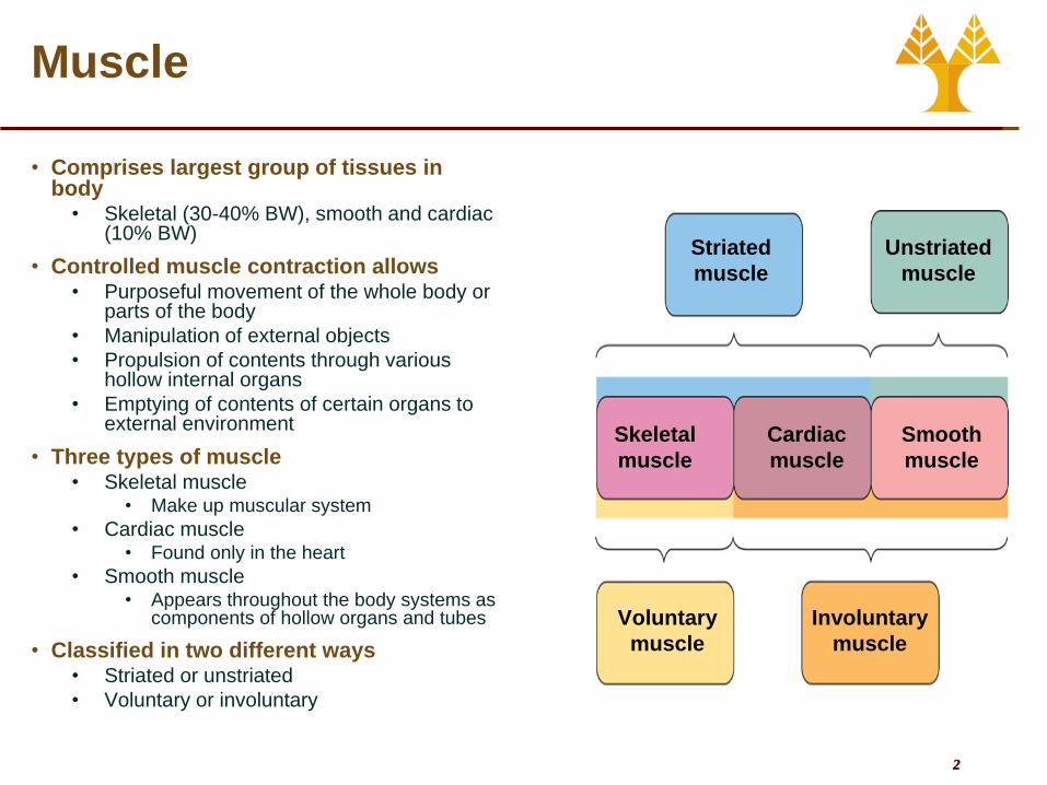
Muscle
• Comprises largest group of tissues in body
• Skeletal (30-40% BW), smooth and cardiac (10% BW)
• Controlled muscle contraction allows • Purposeful movement of the whole body or
parts of the body
• Manipulation of external objects
• Propulsion of contents through various hollow internal organs
• Emptying of contents of certain organs to external environment
• Three types of muscle • Skeletal muscle
• Make up muscular system
• Cardiac muscle • Found only in the heart
• Smooth muscle • Appears throughout the body systems as
components of hollow organs and tubes
• Classified in two different ways • Striated or unstriated
• Voluntary or involuntary
Striated
muscle
Unstriated
muscle
Skeletal
muscle
Cardiac
muscle
Smooth
muscle
Voluntary
muscle
Involuntary
muscle

3 3
Structure of Skeletal Muscle
• Muscle consists a number of
muscle fibers lying parallel
to one another and held
together by connective
tissue
• Single skeletal muscle cell is
known as a muscle fiber
• Multinucleated
• Large, elongated, and
cylindrically shaped
• Fibers usually extend entire
length of muscle
Muscle
Tendon
Muscle fiber
(a single
muscle cell)
Connective
tissue
Muscle fiber Dark A band Light I band
Myofibril

4 4
Neuromuscular Junction
• Axon terminal of motor
neuron forms neuromuscular
junction with a single muscle
cell
• Terminal button (of neuron)
• Motor End Plate (of muscle
cell)
• One neuron may send axons
to one or more muscle fibers

5 5
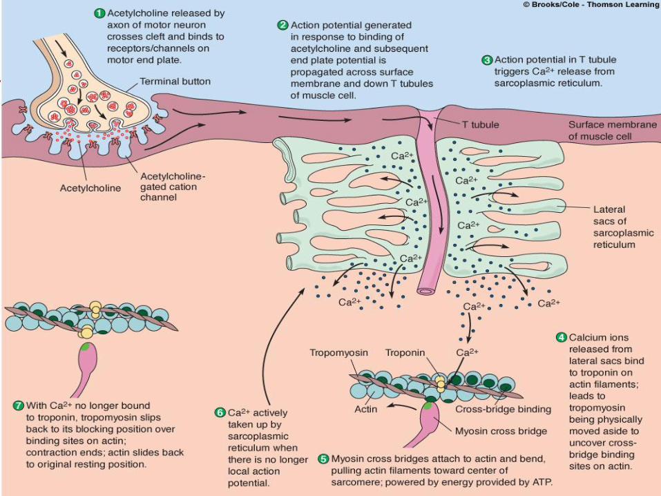
Neuromuscular Junction
• Signals are passed between nerve terminal and muscle fiber by means of neurotransmitter ACh
• AP in motor neuron reaches terminal
• Voltage-gated Ca2+ channels open
• ACh is released by exocytosis
• ACh diffuses across the space and binds to receptor sites on motor end plate of muscle cell membrane
• Binding triggers opening of cation channels in motor end plate
• Na+ movements (larger than K+ movements) depolarize motor end plate, producing end-plate potential
• Local current flow between depolarized end plate and adjacent muscle cell membrane brings adjacent areas to threshold
• Action potential is initiated and propagated throughout muscle fiber
Myelin sheath
Axon
terminal
Terminal button
Vesicle of acetylcholine
Acetylcholine
receptor site
Acetycholinesterase
Voltage-gated
Na+ channel
Chemically gated
cation channel
Motor end plate
Contractile elements
within muscle fiber
Voltage-gated
calcium channel
Action potential
propagation
in motor neuron
Action potential
propagation
in muscle fiber

6 6
Neuromuscular Junction
• Acetylcholinesterase
• On the chemically-gated
cation channels of the end
plate
• Inactivates ACh (as ACh
molecules attaches and
detaches from the receptors)
• Ends end-plate potential and
the action potential
• Ensures prompt termination of
contraction

7 7
Neuromuscular Junction
• Neuromuscular junction is vulnerable to chemical agents and diseases • Black widow spider venom
• Causes explosive release of ACh
• Prolonged depolarization keeps Na+ channels at inactive state
• Respiratory failure from diaphragm paralysis
• Botulism toxin • From food infected with Clostridium Botulinum Botulism
• Blocks release of ACh
• Respiratory failure from inability to contract diaphragm
• Curare • Poisonous arrowheads
• Binds at ACh receptor sites but has no activity and is not degrated
• Organophosphates • Pesticide and military nerve gases
• Prevent inactivation of Ach by inhibiting AChE
• Effect similar to Black widow spider venom
• Myasthenia gravis inactivates ACh receptor sites • Autoimmune condition (Antibodies against ACh receptors)
• ACh is degraded before it can act.
• Antidote is neostigmine (inhibits AChE and prolongs ACh action)

8 8
Excitation-Contraction Coupling
• Muscle fiber (cell)
• Many myofibrils
• Myofibrils
• Contractile elements of muscle fiber
• Viewed microscopically they display
alternating dark (the A bands) and
light bands (the I bands) giving
appearance of striations
• Regular arrangement of thick and thin
filaments
• Thick filaments – myosin (protein)
• Thin filaments – actin (protein)
• Sarcomere
• Functional unit of skeletal muscle
• Found between two Z lines
• Z lines connect thin filaments of two
adjoining sarcomeres
Muscle fiber Dark A band Light I band
Myofibril

9 9
Excitation-Contraction Coupling
• Thick filaments
• Myosin
• Several hundred of them
• Heads form cross bridges
between thick and thin
filaments
• Cross bridge has two
important sites critical to
contractile process
• An actin-binding site
• A myosin ATPase (ATP-
splitting) site
Actin binding site
Myosin ATPase site
Heads
Tail
Myosin molecule
Myosin molecule
Cross bridge
Thick filament

10 10
Excitation-Contraction Coupling
• Thin filaments
• Actin
• Primary structural component of thin filaments
• Each actin molecule has special binding site for attachment with myosin cross bridge
• Tropomyosin
• Thread-like molecules that covers actin sites blocking interaction with thick filaments
• Troponin
• Made of three polypeptide units
• One binds to tropomyosin
• One binds to actin
• One can bind with Ca2+
• When Ca2+ binds to troponin
• Tropomyosin moves away from blocking position
• Cross bridges can form
Actin molecules
Binding site for
attachment
with myosin
cross bridge
Actin helix
+
Tropomyosin Troponin
Thin filament
Thin filament
TropomyosinCross-bridge binding sites
Myosin cross bridge
Actin binding site
Relaxed
Excited
Actin
Tropomyosin
Cross-bridge binding site
Troponin

11 11
Excitation-Contraction Coupling
• Power Stroke
• Ca2+ released into
sarcoplasm
• Myosin heads bind to actin,
binding sides exposed
• Myosin heads swivel toward
center of sarcomere (power
stroke)
• ADP released
• ATP binds to myosin head
and detaches it from actin
• Hydrolysis of ATP transfers
energy to myosin head and
reorients it

12 12
Excitation-Contraction Coupling
• Contraction continues if
ATP is available and
Ca2+ level in
sarcoplasm is high
• Myosin head remains
attached until ATP
binds rigor mortis
• Ca2+ released
• ATP quickly depleted
• Onset in a few hours
• Can last 1-2 days
• Ca2+ stores are in the
sarcoplasmic reticulum

13 13
Excitation-Contraction Coupling
• Sarcoplasmic Reticulum (SR)
• Modified endoplasmic reticulum
• A fine network of interconnected compartments that surround each myofibril
• Not continuous but encircles myofibril throughout its length
• T tubules
• Run perpendicularly from surface of muscle cell membrane into central portions of the muscle fiber
• T tubule membrane is continuous with surface membrane action potential on surface membrane spreads down into T-tubule
• Spread of action potential triggers release of Ca2+ from SR into cytosol
Surface membrane of muscle fiber
Myofibrils
Lateral
sacs
Segments of
sarcoplasmic
reticulum
Transverse
(T) tubule
I band A band I band
14 14
Excitation-Contraction Coupling

15 15
Excitation-Contraction Coupling
• Contractile activity • AP is very short (1-2 msec)
• Contraction does not start until enough Ca2+ is released
• Latent period
• Contraction process requires time to complete
• Contraction time (~50 msec)
• Relaxation also requires time to complete
• Relaxation time (~50 msec)
• Twitch – Contraction of single muscle fiber from single AP
• Brief, weak contraction
• Produced from single action potential
• Too short and too weak to be useful
• Normally does not take place in body
Latent
period
Contraction
time
Relaxation
time
Muscle
twitch
Contractile
response
Action
potential
Stimulation

16 16
Contractile activity
Action potentials
Single
twitch
Twitch
summation
Tetanus
Stimulation
ceases or
fatigue
begins
Muscle fiber restimulated after it has completely relaxed
Muscle fiber is restimulated before it has completely relaxed
Muscle fiber is stimulated
so rapidly that it does not
have an opportunity to relax
at all between stimuli
Frequency of Stimulation
• Twitch summation • Individual twitches are
summed • AP much sorter in time than
contraction Multiple APs can be delivered
• Results from sustained elevation of cytosolic calcium
• Tetanus • Occurs if muscle fiber is
stimulated so rapidly that it does not have a chance to relax between stimuli
• Contraction is usually three to four times stronger than a single twitch
• Do not confuse with the disease of the same name!

17 17
Skeletal Muscle Mechanics
• Muscle consists of groups of muscle fibers bundled together and attached to bones
• Connective tissue covering muscle divides muscle internally into bundles
• Connective tissue extends beyond ends of muscle to form tendons
• Tendons attach muscle to bone
• Muscle Contraction • Contractions of whole muscle can
be of varying strength
• Two primary factors which can be adjusted to accomplish gradation of whole-muscle tension
• Tension developed by each contracting fiber
• Number of muscle fibers contracting within a muscle

18 18
Motor Unit Recruitment
• Motor unit • One motor neuron and the muscle
fibers it innervates
• Number of muscle fibers varies among different motor units
• Number of muscle fibers per motor unit and number of motor units per muscle vary widely
• Muscles that produce precise, delicate movements contain fewer fibers per motor unit
• Muscles performing powerful, coarsely controlled movement have larger number of fibers per motor unit
• Asynchronous recruitment of motor units helps delay or prevent fatigue
• Muscle fibers which fatigue easily are recruited later
• Can engage in endurance activities for a long time but can only deliver full force for brief periods of time
Spinal cord
= Motor unit 1
= Motor unit 2
= Motor unit 3
19 19
Lever Systems
• Bones, muscles, and joints
interact to form lever
systems
• Bones function as levers
• Joints function as fulcrums
• Skeletal muscles provide force
to move bones
• Muscles usually exert more
force than actual weight of
load!
• Advantages: higher speed,
more distance
Fulcrum
BicepsInsertion
of biceps
Fulcrum
for lever

20 20
Skeletal Muscle Metabolism
• Contraction-Relaxation Steps
Requiring ATP
• Splitting of ATP by myosin
ATPase provides energy for
power stroke of cross bridge
• Binding of fresh molecule of
ATP to myosin lets bridge
detach from actin filament at
end of power stroke so cycle
can be repeated
• Active transport of Ca2+ back
into sarcoplasmic reticulum
during relaxation depends on
energy derived from
breakdown of ATP

21 21
Skeletal Muscle Metabolism
• Energy Sources for Contraction • Transfer of high-energy phosphate from creatine
phosphate to ADP • First energy storehouse tapped at onset of contractile
activity
• Short duration or bursts of exercise • ~160g or 12.5 kcal
• E.g. 100 m running
• Glycolysis in anaerobic conditions • Supports anaerobic or high-intensity exercise
• Less efficient but much faster than oxidative phosphorylation
• Quickly depletes glycogen supplies
• Lactic acid is produced • Soreness that occurs during the time (not after) intense
exercise
• Energy depletion and ↓ pH contribute to muscle fatigue
• Oxidative phosphorylation (citric acid cycle and electron transport system)
• Moderate exercise (Aerobic or endurance-type exercise)
• Takes place within muscle mitochondria if sufficient O2 is present
• Deeper & faster breathing, ↑ Heart rate and contraction, Dilation of blood vessels
• Myoglobin (Similar to hemoglobin) Increase the transfer and store of O2 in muscle cells
• Uses glucose or fatty acids • Glucose derived from muscle glycogen (chains of
glucose) stores • Limited (~ 150g or 600 kcal)
• Athletes can store more (2000 kcal for marathon runners)
• Glucose derived from liver glycogen stores • Limited (~80-200g or 320-800 kcal)
• Fatty acids derived from lipolisis • Plenty of these! (~15kg or 135.000 kcal)
creatinine phosphate metabolism
Mitochondria
22 22
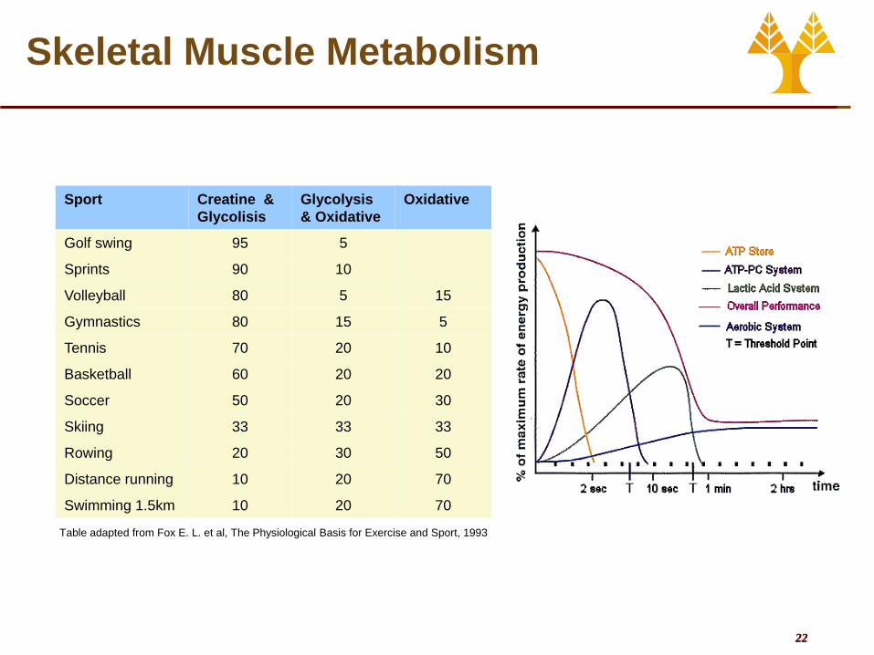
Skeletal Muscle Metabolism
Sport Creatine &
Glycolisis
Glycolysis
& Oxidative
Oxidative
Golf swing 95 5
Sprints 90 10
Volleyball 80 5 15
Gymnastics 80 15 5
Tennis 70 20 10
Basketball 60 20 20
Soccer 50 20 30
Skiing 33 33 33
Rowing 20 30 50
Distance running 10 20 70
Swimming 1.5km 10 20 70
Table adapted from Fox E. L. et al, The Physiological Basis for Exercise and Sport, 1993

23 23
Fatigue
• Contractile activity can not be sustained indefinitely Fatigue
• Muscle Fatigue • Occurs when exercising muscle can no longer respond to
stimulation with same degree of contractile activity
• Defense mechanism that protects muscle from reaching point at which it can no longer produce ATP
• Underlying causes of muscle fatigue are unclear. Implicated • ADP increase (interferes with cross-bridges and Ca2+ uptake in the SR)
• Lactic acid accumulation (may interfere with key enzymes in energy-producing pathways)
• Accumulation of extracellular K+ (decrease in membrane potential)
• Depletion of glycogen
• Central Fatigue • Occurs when CNS no longer adequately activates motor neurons
supplying working muscles
• Often psychologically based • Discomfort, boredom or tiredness
• Mechanisms involved in central fatigue are poorly understood
• Recovery • Excess postexercise O2 consumption (EPOC) helps
• Restore Creatine Phosphate (few minutes)
• Replenish ATP
• Convert Lactic acid to pyruvate for oxidative ATP generation
• Cover increased general O2 demand because of higher temperature
• Nutrient replenishment (1-2 days after a marathon)

24 24
Types of Muscle
• Types of Motor Units • Red muscle fibers
• Oxidative fibers contain more mitochondria and myoglobin and have a richer blood supply red meat
• Slow to contract, can sustain contraction
• White muscle fibers • Few mitochondria, anaerobic
metabolism
• Contract and fatigue rapidly
• Fast motor units • Larger diameter, faster
conducting neurons
• Rapidly fatiguing white fibers
• Slow motor units • Smaller diameter, slower
conducting neurons
• Slowly fatiguing red fibers

25 25
Muscle Adaptation & Repair
• Muscle has a high degree of plasticity • Improvement of oxidative capacity
• From regular aerobic exercise
• Capillaries and mitochondria increase
• Hypertrophy • From anaerobic high intensity exercise
• Muscle fiber diameter increases (more actin and myosin)
• Mainly fast-glycolytic fibers
• Testosterone and other steroids increase the synthesis of actin and myosin
• Steroid abuse
• Fast muscle fibers are interconvertible • Oxidative ↔ glycolytic
• But NOT fast ↔ slow
• Muscle atrophy • Disuse atrophy (e.g. space exploration)
• Denervation atrophy (e.g. paralysis)
• Muscle has limited repair cababilities • Satellite cells can create a few myoblasts
which fuse and create a few muscle fibers

26 26
Control of Motor Movement
• Frontal lobe – Primary Motor Cortex
• Voluntary control for muscle movement
• Motor cortex on each side controls muscles on the opposite side of the body
• Tracts originating in the cortex cross (at level of pyramids) before continuing down spinal cord to terminate muscle
• Body regions are topographically mapped
• Different parts of the body are not equally represented
• Motor Homonculus
• Proportional to precision and complexity of motor skills
• Controls the opposite side of the body
• Damage on right side results in motor deficit on left side

27 27
Control of Motor Movement
• Efferent pathway
• Primary motor cortex upper motor neurons Brain stem (cross to the other side) spinal cord Lower motor neurons
• Input to lower motor-neurons
• From upper motor neurons (primary motor cortex)
• Responsible for fine voluntary movement
• From afferent neurons
• Usually through intervening interneurons
• Responsible for spinal reflexes (e.g. withdrawal)
Efferent
pathway
Afferent
pathway
Muscles
Stimulus
ResponseReceptor
Interneurons
Primary
motor
cortex
Spinal cord
Corticospinal tracts
Muscle

28 28
Control of Motor Movement
• Movement
• The motor cortex itself does
not initiate movement
• Frontal lobe Strategy
• with inputs from the parietal
lode (body and world map)
• Premotor and Supplementary
motor cortex Planning
• Motor Cortex Execution
• Cerebellum Coordination
• Basal Ganglia Initiation and
Correction
Supplementary
motor area (programming of complex
movement)
Primary motor cortex (Voluntary movement)
Posterior parietal
cortex (integration of
somatosensory and
visual input)
Premotor cortex (coordination of complex
movements)
Cerebellum (coordination, muscle
tone, posture)
29 29
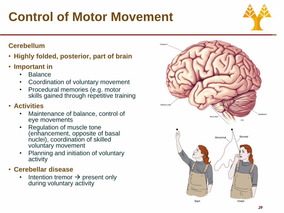
Control of Motor Movement
Cerebellum
• Highly folded, posterior, part of brain
• Important in • Balance
• Coordination of voluntary movement
• Procedural memories (e.g. motor skills gained through repetitive training
• Activities • Maintenance of balance, control of
eye movements
• Regulation of muscle tone (enhancement, opposite of basal nuclei), coordination of skilled voluntary movement
• Planning and initiation of voluntary activity
• Cerebellar disease • Intention tremor present only
during voluntary activity
30 30
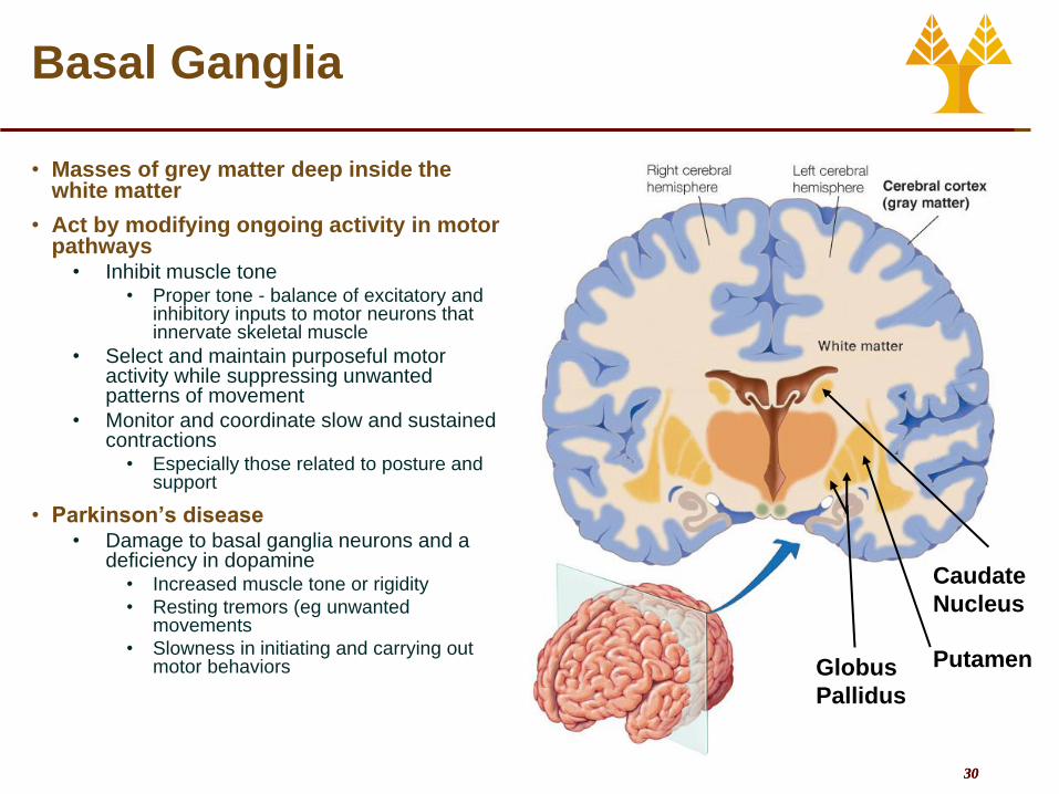
Basal Ganglia
• Masses of grey matter deep inside the white matter
• Act by modifying ongoing activity in motor pathways
• Inhibit muscle tone • Proper tone - balance of excitatory and
inhibitory inputs to motor neurons that innervate skeletal muscle
• Select and maintain purposeful motor activity while suppressing unwanted patterns of movement
• Monitor and coordinate slow and sustained contractions
• Especially those related to posture and support
• Parkinson’s disease • Damage to basal ganglia neurons and a
deficiency in dopamine • Increased muscle tone or rigidity
• Resting tremors (eg unwanted movements
• Slowness in initiating and carrying out motor behaviors
Caudate
Nucleus
Putamen Globus
Pallidus

31 31
Muscle Receptors
• Receptors are necessary to plan and control complicated movement and balance
• The brain receives information from all muscles and joints in the body proprioception
• Two types of muscle receptors
• Muscle spindles
• Monitor muscle length and tension
• Golgi tendon organs
• Monitor whole muscle tension

32 32
Muscle Receptors
• Muscle Spindles • Consist of collections of
specialized muscle fibers known as intrafusal fibers
• Lie within spindle-shaped connective tissue capsules parallel to extrafusal fibers
• Have contractile ends and a non-contractile central portion
• Each spindle has its own private nerve supply
• Plays key role in stretch reflex
• Efferent • Gamma motor neurons*
• Afferent • Primary (annulospiral) endings
(in the central portion)
• Secondary (flower-spray) endings (at the end segments)
* Efferent neurons to extrafusal fibers are
called alpha motor neurons
Alpha motor
neuron axon
Gamma motor
neuron axon
Secondary (flower-spray)
endings of afferent
fibers
Extrafusal (“ordinary”)
muscle fibers
Capsule
Intrafusal (spindle)
muscle fibers
Contractile end portions
of intrafusal fiber
Noncontractile
central portion
of intrafusal
fiber
Primary (annulospiral)
endings of afferent fibers

33 33
Muscle Receptors
• Coactivation of alpha
and gamma motor
neurons
• Spindle coactivation
during muscle
contraction
• Spindle contracted to
reduce length
• With no coactivation
• Slackened spindle
• Not sensitive to
stretch
• Adjustment to keep
muscle spindles
sensitive to stretch
Extrafusal
skeletal
muscle fiber
Intrafusal
muscle
spindle fiber
Spinal
cordγ
α
34 34
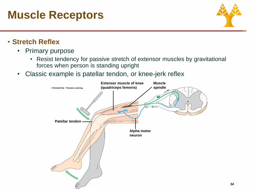
Muscle Receptors
• Stretch Reflex
• Primary purpose
• Resist tendency for passive stretch of extensor muscles by gravitational forces when person is standing upright
• Classic example is patellar tendon, or knee-jerk reflex
Patellar tendon
Extensor muscle of knee
(quadriceps femoris)
Muscle
spindle
Alpha motor
neuron

35 35
Muscle Receptors
• More complicated reflexes
Κάμψη
Έκταση
Κάμψη
Έκταση

36 36
Muscle Receptors
• Gogli Tendon Organs
• Provide necessary feedback
for overall muscle tension
• Integrates all factors which
influence tension
• Specialized nerve fibers
embedded in the tendons
• Stretch of tendons exerts force
on nerve endings
• Increase firing rate
• Part of this information
reaches conscious awareness
• We are aware of tension (but
not of length) of muscles

37 37
Skeletal System
• Bones are made of several tissues • Primarily made of collagen and
hydroxyapatite - Ca10(PO4)6(OH)2
• About 206 bones in the human body
• Functions of Skeletal System • SUPPORT:
• Hard framework that supports and anchors the soft organs of the body.
• PROTECTION: • Surrounds organs such as the brain and
spinal cord.
• MOVEMENT: • Allows for muscle attachment therefore the
bones are used as levers.
• STORAGE: • Minerals and lipids are stored within bone
material.
• BLOOD CELL FORMATION: • The bone marrow is responsible for blood
cell production.

38 38
Structure of Bone and Joints
• Features of a long bone: • Epiphysis:
• Ends of the bone.
• Diaphysis: • The shaft of the bone which surrounds the medullary
cavity.
• Articular Cartilage: • Cushions the ends of the bones and allows for smooth
movement.
• Bone Structure • Periosteum – hard outer covering
• Cells for growth and repair
• Compact bone – hard strong layer • Bone cells, blood vessels, protein with Ca and P
• Spongy bone – at ends of long bones • Has small open spaces to lighten weight
• Marrow cavity – hollow in middle of long bones
• Bone Marrow • Red marrow – produces blood cells and clotting factors
• Found in humerus, femur, sternum, ribs, vertebrae, pelvis
• Produces RBC 2 million per second
• Yellow marrow – stores fat • Found in many bones

39 39
Structure of Bone and Joints
• Osteoporosis • Decline in Bone Density
• Bone Resorption > Bone Deposition
• Increase Risk for Fracture • compression fractures of vertebrae
• hip fractures
• Role of calcium, vitamin D, estrogen, exercise
• Broken Bones • Fracture is a break of the bone
• Simple or Complex fracture
• Regrowth of bone: • Spongy bone forms in first few days
• Blood vessels regrow and spongy bone hardens
• Full healing takes 1-2 months
• Healing capacity diminishes with age

40 40
Structure of Bone and Joints
• Joints • Cartilage covers ends of
movable bones • Reduces friction
• Lubricated by fluid from capillaries
• Arthritis: • Osteoarthritis- 90% of pop. By
age 40
• chronic inflammation of articular cartilage
• can be normal age-dependent change
• can also be pathology due to • Age-related changes
• decrease blood supply
• trauma


