Accepted by A. Kroh: 27 June 2011; published 12 Dec. 2012
Echinoderms don’t suck: evidence against the involvement of suction in tube foot attachment*
ELISE HENNEBERT1, ROMANA SANTOS2 & PATRICK FLAMMANG1,3
1 Université de Mons—UMONS, Biology of Marine Organisms and Biomimetics, Mons, Belgium 2 Unidade de Investigação em Ciências Orais e Biomédicas (UICOB), Faculdade de Medicina Dentária, Universidade de Lisboa, Portugal 3 Corresponding author, E-mail: [email protected]
*In: Kroh, A. & Reich, M. (Eds.) Echinoderm Research 2010: Proceedings of the Seventh European Conference on Echinoderms, Göttingen, Germany, 2–9 October 2010. Zoosymposia, 7, xii + 316 pp.
Abstract
Suction has usually been regarded as the primary functional mean for attachment in sea star and sea urchin tube feet and this hypothesis has become widespread in the public knowledge. Yet, a few studies have suggested that adhesive secre-tions may also play a prominent role in tube foot attachment. Here we use a morphological and biomechanical approach to investigate the role of suction in asteroid and echinoid tube foot attachment. Microscopic observations of tube feet rapidly fixed while they were attached to a smooth substratum show that their distal surfaces are totally flat and lack a suction cavity. Detachment force and tenacity of a single tube foot appear to be independent of the pulling angle: i.e., the introduction of a shear component in the pulling force does not decrease attachment strength as would be expected for a sucker. Moreover, sea urchin tube feet attach as strongly to perforated surfaces, which preclude pressure reduction, as to their unperforated counterparts. Taken together, these results clearly show that echinoderm tube feet rely on adhesive secretions and not on suction.
Animal structures designed for suction attachment are usually discs or hemispheres with elastic edges which can conform closely to surfaces. Their attachment depends upon the creation of a reduced pressure in the space between the suction cup and the substratum. This is generally achieved by mus-cular contraction arranged so as to draw the centre of the cup away from the surface, thus resulting in an increase of the volume of the suction cavity (Nachtigall 1974). In Echinoderms of the classes Asteroidea, Echinoidea and Holothuroidea, locomotion and attachment to surfaces are mediated by disc-ending tube feet. Since the first scientific echinoderm descriptions (e.g., Forbes 1841; Hamann 1884; Romanes 1885; Cuénot 1891), these tube feet have been described as miniature suckers. This idea, imparted by most zoology textbooks, has pervaded popular knowledge. A typical illustration can be found in Pixar’s animated movie “Finding Nemo” in which Peach the sea star makes the character-istic popping sound of a rubber sucker with its tube feet. This assumption that disc-ending tube feet function as suckers presumably originated from their distal end which looks like a cup with a central
depression (Fig. 1A, B). In sea stars and sea urchins, histological studies later corroborated this view by showing the occurrence of a specialized muscle, the so-called levator muscle, acting as a retractor of the central part of the disc (Smith 1937, 1947; Nichols 1966; Fig. 1B). In sea cucumbers, on the other hand, the levator muscle is lacking and the presence of a large ossicle underlying the whole disc of the tube feet precludes suction attachment (Nichols 1966; Flammang 1996).
Like the tube feet of all other echinoderms, asteroid and echinoid tube feet also rely on chemi-cal adhesion and their adhesive secretions are left on the substratum as footprints after tube foot detachment (Flammang et al. 1998; Santos & Flammang 2006; Hennebert et al. 2008). In these two echinoderm classes, numerous studies reported that tube foot attachment is partly due to suction and partly to adhesives (see Flammang 1996 for review). Among them, only Paine’s (1926) study on sea stars attempted to estimate the relative proportions of suction and adhesion strengths (56 % and 44 %, respectively). However, there is no agreement on this ratio. Some authors argue that the proportion allotted to adhesive secretions has been underestimated (Thomas & Hermans 1985; Flammang 1996) while others believe that suction has been overlooked (Smith 1991). Recently, it was also proposed that, under short pulses of wave-generated forces, a slight passive suction effect might contribute to the overall detachment force of disc-ending tube feet because of their flared, mushroom-shaped geometry (Gorb & Varenberg 2007; Heepe et al. 2011).
Thomas & Hermans (1985) reported that the sea star Leptasterias hexactis can attach to a stainless steel plankton screen whose meshes should preclude sucker-like attachment. Although they claimed that adhesion to this screen was strong, no comparison was made with a smooth unperforated surface of the same material and no quantitative data were provided. In this work, we used both morphologi-cal and biomechanical experiments to investigate the involvement of suction in tube foot attachment in the asteroid Asterias rubens and in the echinoid Paracentrotus lividus, two species in which the sucker-like functioning of tube feet was originally described (Forbes 1841; Romanes 1885; Cuénot 1891; Smith 1947).
Materials and Methods
Morphology of attached tube feet. Tube feet from individuals of A. rubens and P. lividus were allowed to adhere to clean pieces of glass and of Spurr’s epoxy resin. After a tube foot became firmly attached to the substratum it was quickly amputated, fixed and processed for scanning electron microscopy (SEM) or for light microscopy (LM). For SEM, tube feet attached to glass were frozen in liquid nitrogen for a few seconds and freeze-substituted with a 1 % solution of osmium tetroxide in absolute acetone at -80°C for 3 days. They were then washed in absolute acetone, gradually trans-ferred to room temperature conditions, transferred to 100 % ethanol, critical-point dried, mounted on metal stubs and, after sputter-coating with gold, viewed in a JEOL JSM-6100 SEM at 10 kV. Controls were performed by fixing unattached tube feet in Bouin’s fluid (see Santos et al. 2005). For LM, tube feet attached to resin blocks were fixed in a solution containing 3 % glutaraldehyde and 1 % osmium tetroxide in cacodylate buffer (0.1 M, pH 7.8; adjusted to 1030 mOsm with NaCl) for 30 min at room temperature. They were rinsed in cacodylate buffer, post-fixed in 3 % glutaraldehyde in the same buffer, dehydrated in graded ethanol series and embedded in Spurr’s resin. Semi-thin sections (1 µm) were cut with a Reichert OmU2 ultramicrotome equipped with a glass knife, stained with an equivolumic mixture of 1 % Azur II and 1 % methylene blue solutions, observed and photographed
with a Zeiss Axioscope A1 microscope equipped with an AxioCam ICc 3 camera. The same protocol was applied to unattached tube feet for control.
Attachment strength of individual tube feet. Attachment force measurements of individual tube feet were performed as described in Santos et al. (2005). Each experiment was performed with a minimum of three sea urchins and three sea stars totally immersed in containers filled with seawater.
FIGURE 1. Morphology of unattached (A, B) and attached tube feet (C–F) of the sea star Asterias rubens (B, E, F) and of the sea urchin Paracentrotus lividus (A, C, D). A and B: Unattached tube feet from both species consist of a proximal cy-linder, the stem (S), connected to an apical disc (D), which presents a central depression (arrow). In B, the levator muscle (LM) can be observed at the junction between the stem and the disc. C and E: SEM images of tube feet cryofixed while attached to a smooth glass surface. In C, some adhesive material (AM) is visible on the surface of the disc. D and F: LM views of longitudinal sections through tube feet attached to a smooth epoxy resin substratum (RS). The soft adhesive pad (AP), supported by the connective tissue terminal plate (TP), lies flat against the substratum.
Specimens were put upside-down (to induce tube foot attachment) and a 1 cm2 piece of substratum, connected to an electronic dynamometer (Mecmesin AFG 10 N), was presented to the tube feet (Fig. 3A). When a single tube foot remained attached to the substratum for at least 10 s, the dynamometer was moved upwards at a constant speed of 15 mm/min until it detached, and the detachment force (Fd [N]) was recorded. The footprint left by the tube foot after detachment was stained for 1 min with a 0.05 % aqueous solution of the cationic dye crystal violet, photographed (Fig. 2A), and the digitized image analyzed with Semaphore® software to calculate the surface area of the footprint (S [m2]). The tenacity (T [Nm-2 or Pa]) was then calculated as:
T = Fd / S. (1)To test the influence of the angle of pull on tube foot detachment force and tenacity, square pieces
of smooth glass were connected to the dynamometer by a surgical thread glued at the level of their centre of gravity (Fig. 3A, B). With this arrangement, when the tube foot attaches at the centre of the glass square, the substratum remains horizontal and the pull is exerted perpendicular to its surface (α = 90°). On the other hand, if the tube foot attaches at the periphery of the square, the substratum tilts and the pulling angle is reduced (α<90°), introducing a shear component parallel to the surface of the substratum (Fig. 3B). Calibration of the device with threads glued to the substratum at different dis-tances from its centre and ballasted with a 10 g weight (corresponding to the mean detachment force of one tube foot) indicated a linear relationship (n = 8; r2 = 0.919; p < 0.001) between distance from the center (D [mm]) and angle of pull (α [°]) according to the following equation:
α = -4.116 * D + 82.075. (2)Attachment strength (pull exerted perpendicular to the surface) was also measured on a perforated
substratum consisting of a thin sheet of polystyrene that had been homogeneously perforated with fine entomological needles at a density of about 1 hole per mm2. SEM observations revealed that the holes measured 0.41 ± 0.08 mm in diameter (mean ± SD, n = 15), and that the surface between them remains flat and smooth. The holes are smaller than the tube foot discs: in terms of surface area, each hole corresponds to about 26 % of the mean disc adhesive surface area calculated from footprint diameters (Fig. 2A).
Abbreviations: LM: light microscopy, SEM: scanning electron microscopy
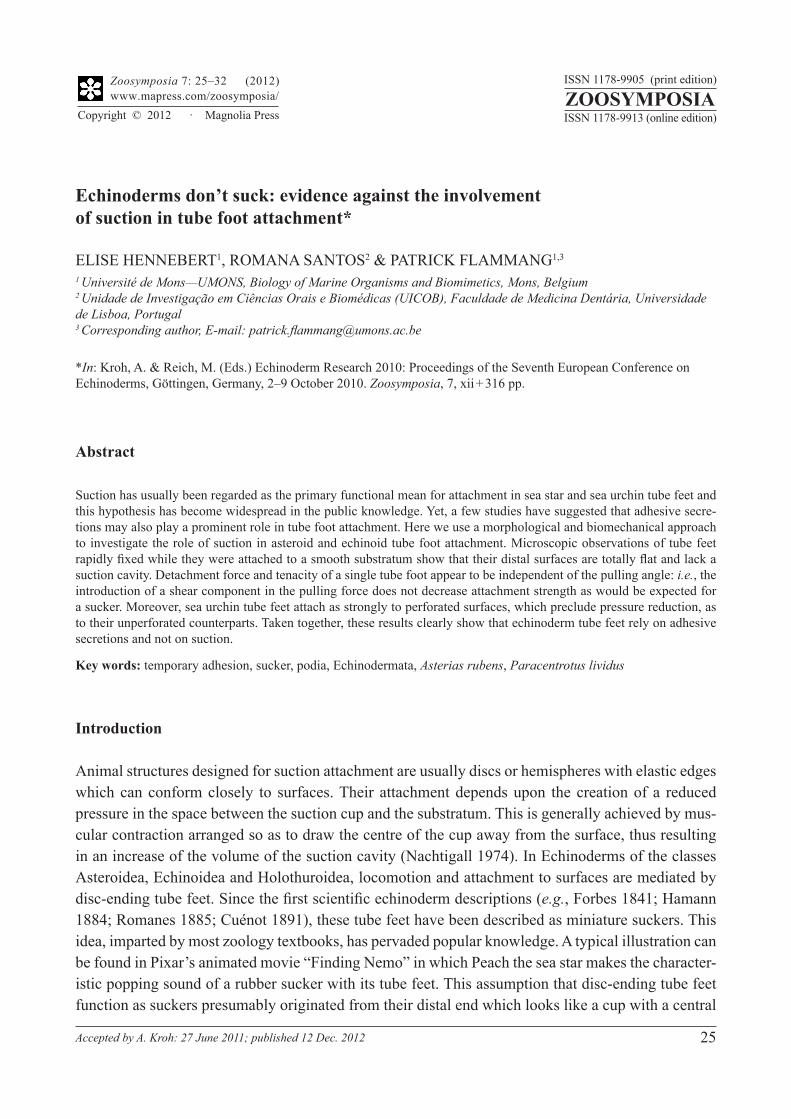
FIGURE 2. Photographs of a complete footprint of Paracentrotus lividus (A) and of a footprint of Asterias rubens (B) presenting radial channels devoid of adhesive material (arrows). Both footprints were stained with a 0.05 % aqueous so-lution of the cationic dye crystal violet.
Morphological arguments. Previous models describing the functioning of the sucker in echinoderm tube feet postulate the formation of a large suction cavity between the attached foot and substratum when the levator muscle contracts during disc attachment (see illustrations in Smith 1947; Nichols 1966; Mooi 1986). In these models, adhesive secretions appear to seal the rim of the foot to the substratum and, after tube foot detachment, form a ring-shaped footprint. None of our morphologi-cal observations corroborates these models. In both A. rubens and P. lividus, tube feet cryofixed and freeze-substituted while attached to smooth glass present a totally flat distal surface in SEM (Fig. 1C, E). In no case has the small central depression visible when tube feet are fixed by classical chemical fixation (e.g., in Bouin’s fluid; Santos et al. 2005; Fig. 1A) been observed when tube feet are quickly frozen. Longitudinal sections through tube feet from both species attached to smooth resin blocks also show that the disc surface is completely flat and very close to the substratum surface (Fig. 1D, F), the two surfaces being separated only by a very thin layer of adhesive secretions. Occasionally, however, some shallow grooves were observed at the level of the epidermis. As far as footprints are concerned, although some are indeed ring-shaped, most of them are disc-shaped, being made up of a complete, homogeneous layer of adhesive material (Fig. 2A; see also Flammang et al. 1998; Santos & Flam-mang 2006; Hennebert et al. 2008). In A. rubens, many footprints also present radial channels devoid of adhesive material (Fig. 2B) and, in consequence, cannot seal the rim of the foot to the substratum.
FIGURE 3. Variation of tube foot attachment strength with angle of pull. A: Photo showing a glass substratum (S) pre-sented to the tube feet (TF) of an individual of Paracentrotus lividus. B: Diagram illustrating the experimental procedure used to vary the angle of pull in function of the place where the tube foot (in black) attaches to a glass surface (in grey, see text for details). C and D: Relationships between tube foot detachment force or tenacity and angle of pull for the sea star Asterias rubens (crosses) and the sea urchin P. lividus (circles).
Therefore, the morphological evidence suggests that sea star and sea urchin tube feet do not function as suckers. Suction, however, cannot be ruled out altogether because, in animals like limpets, it has been shown that muscular contraction can decrease the pressure within the inexpansible mucus layer under the foot, creating effective suction without the formation of a large suction cavity (Denny 1988; Smith 1991).
Mechanical arguments. In order to investigate further whether suction plays a role in tube foot attachment, we measured attachment strengths of the tube feet of A. rubens and P. lividus at different pulling angles on glass, and at a constant angle on perforated polystyrene (Figs 3 and 4). These condi-tions are known to influence suction, as in limpets which slide easily on a smooth surface but detach immediately when they slide over a hole (Smith 1991).
The effect of the pulling angle on the attachment strength of tube feet was analysed by linear regres-sion showing that, in asteroids as well as in echinoids, there is no significant relationship between pulling angle and detachment force (p = 0.228, r2 = 0.063, n = 25 for A. rubens; p = 0.521, r2 = 0.015, n = 29 for P. lividus; Fig. 3C), or tenacity (p = 0.625, r2 = 0.011, n = 25 for A. rubens, p = 0.526, r2 = 0.015, n = 29 for P. lividus; Fig. 3D). The calculated values for r2, ranging from 0.01 to 0.06, suggest that only 1 to 6 % of the variation in attachment strength can be explained by a change in the pulling angle. In other words, there is no decrease in the attachment strength when a large shear component is intro-duced in the pulling direction. By contrast, in a true sucker the force due to pressure difference acts in a direction normal to the surface of the substratum and does not provide any inherent resistance to shear forces attempting to slide the sucker along a smooth substratum (Denny 1988).
Attachment strength measurements on a perforated film of polystyrene (pulling angle 90°) could only be performed on tube feet of P. lividus. Indeed, sea urchin tube feet attach readily to a large variety of substrata (Santos & Flammang 2006). On the contrary, despite our numerous attempts, sea
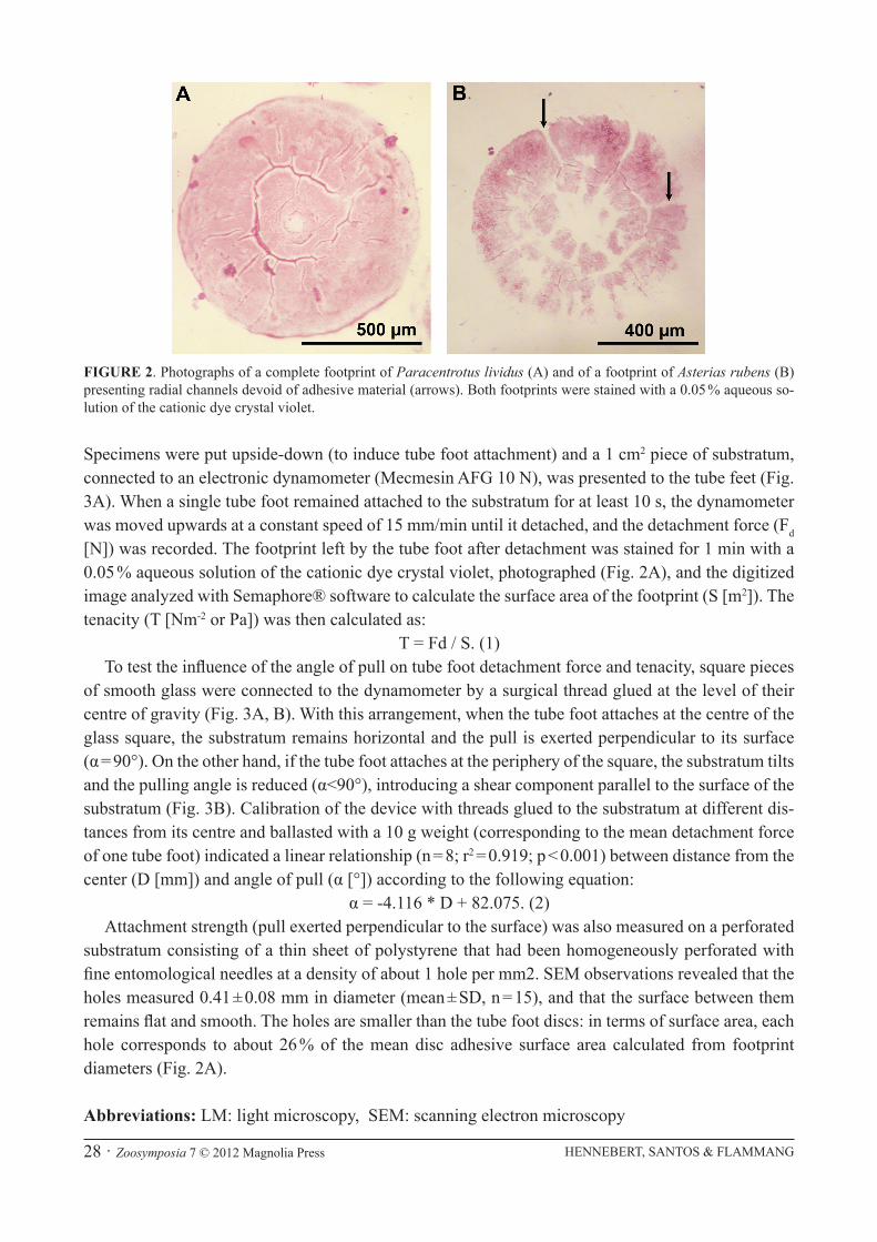
FIGURE 4. Effect of leaks on the attachment strength of tube feet. A: Illustration of the grouping of adhesion measure-ments on the perforated polystyrene film according to whether the footprint covers a hole, totally (group I) or partially (group II), or not (group III). B and C: Mean values (+SD) of detachment force and tenacity measured on this perforated substratum for the tube feet of the sea urchin Paracentrotus lividus. Scales bars in A = 0.5 mm.
star tube feet did not adhere firmly to low surface energy substrata (i.e., polypropylene, polystyrene and teflon), even non-perforated, therefore precluding adhesion measurements (see also Santos et al. 2005; Hennebert et al. 2008). The measurements have been divided into three groups (Fig. 4A): those in which the tube foot covered one hole completely (group I, n = 28) or partially (group II, n = 13), and those in which it attached to a flat smooth area between holes (group III, n = 26). One-way ANOVAs demonstrate that there is no significant difference between the three groups in terms of detachment force (df = 2.65; F = 0.267; p = 0.767; Fig. 4B) or tenacity (df = 2.65; F = 0.715; p = 0.493; Fig. 4C). This experiment clearly negates the involvement of a suction mechanism in echinoid tube foot attach-ment. Indeed, in the case of tube feet completely covering holes, no pressure difference could have formed and still no significant change in the attachment strength was observed.
Conclusions
Taken together with the qualitative observations made by Thomas & Hermans (1985) that tube feet of L. hexactis adhere strongly to a fine-meshed, stainless steel plankton screen; and the measurements by Santos et al. (2005) in which tube feet of P. lividus attached as strongly to a rough porous poly-propylene substratum as to a plain smooth one, the results of our morphological and biomechanical experiments unambiguously demonstrate that, contrary to the generally accepted notion, sea star and sea urchin tube feet are not functioning as active suckers. Instead, the high adhesion strength of tube feet in these echinoderms is exclusively attributable to gluing through specialized adhesive secretions.
Acknowledgements
Thanks to Dr. J.H. Waite for many useful comments on the manuscript. E.H. and P.F. are respec-tively Postdoctoral Researcher and Research Director of the Fund for Scientific Research of Belgium (F.R.S.-FNRS). R.S. is an Associate Researcher of Ciência 2008 Program supported by Fundação para a Ciência e Tecnologia. Work supported in part by a FRFC Grant n° 2.4532.07 and by COST Action TD0906 (http://www.cost-bioadhesives.org/). This study is a contribution from the “Centre Interuni-versitaire de Biologie Marine” (CIBIM) and from the Research Institute for Biosciences (UMONS).
References
Denny, M.W. (1988) Biology and the Mechanics of the Wave-Swept Environment. Princeton University Press, Princeton, 329 pp.
Flammang, P. (1996) Adhesion in echinoderms. In: Jangoux, M. & Lawrence, J.M. (Eds.), Echinoderm Studies 5. A.A. Balkema, Rotterdam, pp. 1–60.
Flammang, P., Van Cauwenberge, A., Alexandre, H. & Jangoux, M. (1998) A study of the temporary adhesion of the podia in the sea star Asterias rubens (Echinodermata, Asteroidea) through their footprints. Journal of Experimental Biology, 201, 2383–2395.
Gorb, S. & Varenberg, M. (2007) Mushroom-shaped geometry of contact elements in biological adhesive systems. Jour-nal of Adhesion Science and Technology, 21, 1175–1183.
Hamann, O. (1884) Beiträge zur Histologie der Echinodermen. Vol. I. Fischer Verlag, Jena, 100 pp.Heepe, L., Varenberg, M., Itovich, Y. & Gorb, S. (2011) Suction component in adhesion of mushroom-shaped microstruc-
ture. Journal of the Royal Society Interface, 8, 585–589.
Hennebert, E., Viville, P., Lazzaroni, R. & Flammang, P. (2008) Micro- and nanostructure of the adhesive material secreted by the tube feet of the sea star Asterias rubens. Journal of Structural Biology, 164, 108–118.
Mooi, R. (1986) Non-respiratory podia of clypeasteroids (Echinodermata, Echinoides): I. Functional anatomy. Zoomor-phology, 106, 21–30.
Nachtigall, V. (1974) Biological Mechanisms of Attachment. The Comparative Morphology and Bioengineering of Organs for Linkage, Suction, and Adhesion. Springer-Verlag, Berlin, 194 pp.
Nichols, D. (1966) Functional morphology of the water-vascular system. In: Boolootian, R.A. (Ed.), Physiology of Echi-nodermata. Interscience Publishers, New York, pp. 219–240.
Paine, V.L. (1926) Adhesion of the tube feet in starfishes. Journal of Experimental Zoology, 45, 361–366.Romanes, G.J. (1885) Jelly-Fish, Star-Fish and Sea-Urchins. Kegan Paul, Trench & Co., London, 323 pp.Santos, R. & Flammang, P. (2006) Morphology and tenacity of the tube foot disc of three common European sea urchin
species: a comparative study. Biofouling, 22, 187–200. Santos, R., Gorb, S., Jamar, V. & Flammang, P. (2005) Adhesion of echinoderm tube feet to rough surfaces. Journal of
Experimental Biology, 208, 2555–2567.Smith, A.M. (1991) The role of suction in the adhesion of limpets. Journal of Experimental Biology, 161, 151–169.Smith, J.E. (1937) The structure and function of the tube feet in certain echinoderms. Journal of the Marine Biological
Association of the United Kingdom, 22, 345–357.Smith, J.E. (1947) The activities of the tube feet of Asterias rubens L. I. The mechanics of movement and of posture.
Quarterly Journal of Microscopical Science, 88, 1–14.Thomas, L.A. & Hermans, C.O. (1985) Adhesive interactions between the tube feet of a starfish, Leptasterias hexactis,