Imaging features of small bowel and colorectal cancer in inflammatory bowel disease L. Hristova, V. Laurent, O. Bruot, P. Ganne, J. Mathias, D. Regent; Vandoeuvre-les-Nancy/FR ECR 2010 Vienna
Transcript
Imaging features of small bowel and colorectal cancer in inflammatory bowel disease
L. Hristova, V. Laurent, O. Bruot, P. Ganne, J. Mathias, D. Regent; Vandoeuvre-les-Nancy/FR
ECR 2010 Vienna
Learning Objectives
•To know the risk factors for developing small bowel cancer (SBC) and colorectal cancer (CRC) in patients with inflammatory bowel disease (IBD)
•To illustrate the imaging features of small bowel and colorectal malignancy in IBD
•To emphasize the difficulty in establishing a diagnosis
•To know the clinical and pathological features of CRC and SBC in patients with IBD
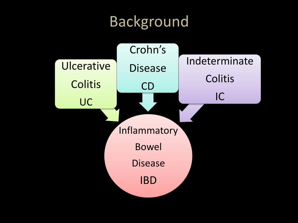
Background
Inflammatory
Bowel
Disease
IBD
Ulcerative
Colitis
UC
Crohn’s
Disease
CD
Indeterminate
Colitis
IC
Patients with IBD have increased risk for developing colorectal and small bowel cancers.
The prevalence of CRC in patients with UC is approximately 3.7% overall and 5.4% for those with pancolitis (1).
The prevalence of CRC in Crohn’s colitis is similar(2).
Patients with CD have relative risk 28 for developing small-bowel cancer compared to the background population (3).
No increased risk was found in stomach and anal cancer in patients with IBD (4,5).
Risk factors for CRC in patients with IBD
Duration of Disease
The risk of CRC becomes greater than that of the general population after 8 to 10 years from the onset of disease (1).
The cumulative incidence of CRC is 2.5% after 20 years of IBD, 7.6% after 30 years and 10.8% after 40 years (7).
Anatomic extent
UC: the standardized incidence ratio (SIR) for CRC increase gradually from 1.7-fold in proctitis, 2.8-fold in left-sided colitis to 14.8-fold in pancolitis, compared with age-matched population without UC (8).
CD: the risk of CRC is increased when the extent of the colic involvement is greater then one third(7).
Primary Sclerosing Cholangitis
The concomitant presence of PSC in IBD patients confers a high risk for developing colorectal cancer(9,10).
The cumulative incidence of CRC in UC patients was 33% at 20 years (10).
When liver transplantation is necessary, prophylactic colectomy should be considered(11).
• Young age at onset,when younger then 25, increases the risk of developing CRC(1)
• When onset is after 30 there is no insreased risk of CRC
Young age at onset
• Family history of sporadic CRC increases twice the risk of CRC(13)
Familiy history of CRC
• The increased severity of inflammation correlates with increased frequency of dysplasia(7)
• Patients with longstanding quiescent colitis remain at risk for developing CRC
Degree of endoscopic and
histologic activity
Screening colonoscopy Consensus Conference 2004 by Crohn’s and Colitis
Fondation of America CCFA (6)
Screening colonoscopy should begin in patients with IBD:
8-10 years after the onset of IBD symptoms
+ 1. UC pancolitis or left-sided colitis
2. Crohn’s colitis involving at least one third of the colon
3. At onset of PSC if associated
Clinical and pathological features of colorectal cancer in IBD
• 10 year earlier then in sporadic CRC
• UC: The mean age is 52 years (14)
• CD: the mean age is 54 years(15)
Age at diagnosis
• Tumor occurs in area of macroscopic disease
• CD: tumors occurs in ileocaecal and rectosigmoid regions
• UC: from the rectum to the right-sided colon (15)
Anatomical Location
• Frequency of mucinous and signet ring cell tumor is higher then in general population(15)
• Synchronous tumor locations Histology
Risk factors for SBC in patients with Crohn’s Disease
The relative risk of small bowel carcinoma in CD seems to be 28.4 times higher compared to general population (3).
• Essential factor
• The mean duration of CD is 19 years (3)
Duration of disease
• Distal jejunal and ileal location (16) Anatomic
extent
• When younger then 25 years at onset of CD Young age at
onset
• Strictures
• Chronic fistulous disease
• Small bowel bypass loops Complications
Clinical and pathological features of SBC in CD
• The median age of diagnosis is 48 years versus 65 in general population(16)
Age at diagnosis
• The highest incidence in the distal jejunum and ileum: area of macroscopic disease
Anatomic location
• Adenocarcinoma with signet ring cell is frequent: up to one third (17) Histology
Patients and materials
The computerized medical record system Explore in the PACS at the Radiology
department of the University Hospital of Nancy was used to identify patients with
IBD and concomitant SBC or CRC.
The diagnosis of IBD, CRC and SBC were confirmed by clinical, imaging,
endoscopic and histological criteria.
Only patients who had a scanner or magnetic resonance were accepted.
There were 15 patients with both, IBD and CRC-12 or SBC-3, between 2001 and
2009.
There were 12 patients with IBD and concomitant colorectal cancer.
8 of them had Crohn’s disease and only 4 had Ulcerative Colitis.
8
4 4
75% of the patients with CRC had severe pancolitis.
40% of the CRC were located in the left colon.
42% of CRC were with Signet Ring Cell component. CRC with signet ring cell are only 1% of CRC in general population.
Histological aspect of colic adenocarcinoma with
signet ring cell component. This is a signet ring
cell pattern of adenocarcinoma in which the cells
are filled with mucin vacuoles that push the
nucleus to one side, as shown at the arrow.
Only 3 (25%) of CRC were discovered by screening colonoscopy. Five (42%) of CRC presented an occlusion. Three (25%) had worsening of the IBD and one had anemia.
In these 5 cases of CRC presenting as occlusion the pre operative diagnosis of neoplasia was not suspected. The imaging findings indicated an inflammatory benign stenosis.
3
5
3
1
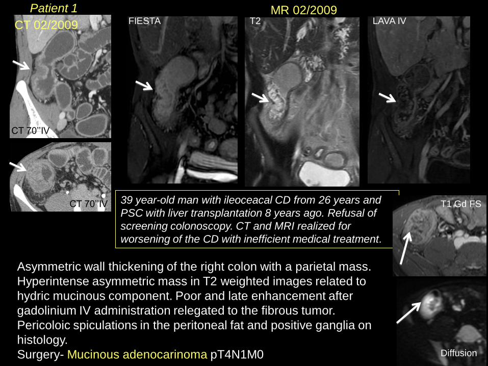
39 year-old man with ileoceacal CD from 26 years and
PSC with liver transplantation 8 years ago. Refusal of
screening colonoscopy. CT and MRI realized for
worsening of the CD with inefficient medical treatment.
Asymmetric wall thickening of the right colon with a parietal mass.
Hyperintense asymmetric mass in T2 weighted images related to
hydric mucinous component. Poor and late enhancement after
gadolinium IV administration relegated to the fibrous tumor.
Pericoloic spiculations in the peritoneal fat and positive ganglia on
histology.
Surgery- Mucinous adenocarinoma pT4N1M0
CT 70’’IV
CT 70’’IV
FIESTA T2 LAVA IV
T1 Gd FS
CT 02/2009
MR 02/2009
Diffusion
Patient 1
CT 08/2009 CT 12/2007
58 years old female with CD from 2 years
and long history of digestive disorder.
Pancolitis and difficult medical treatment,
right-sided colon stenosis.
2 years later. Stenosis and right-sided colitis responsible for small bowel
occlusion. The stenosis was present in 2007.
Ileocaecal surgical resection: adenocarcinoma with signet ring cell: T4N2M0.
CT IV 70’’
CT IV 70’’ CT IV 70’’
CT IV 70’’
CT IV 70’’
CT IV 70’’
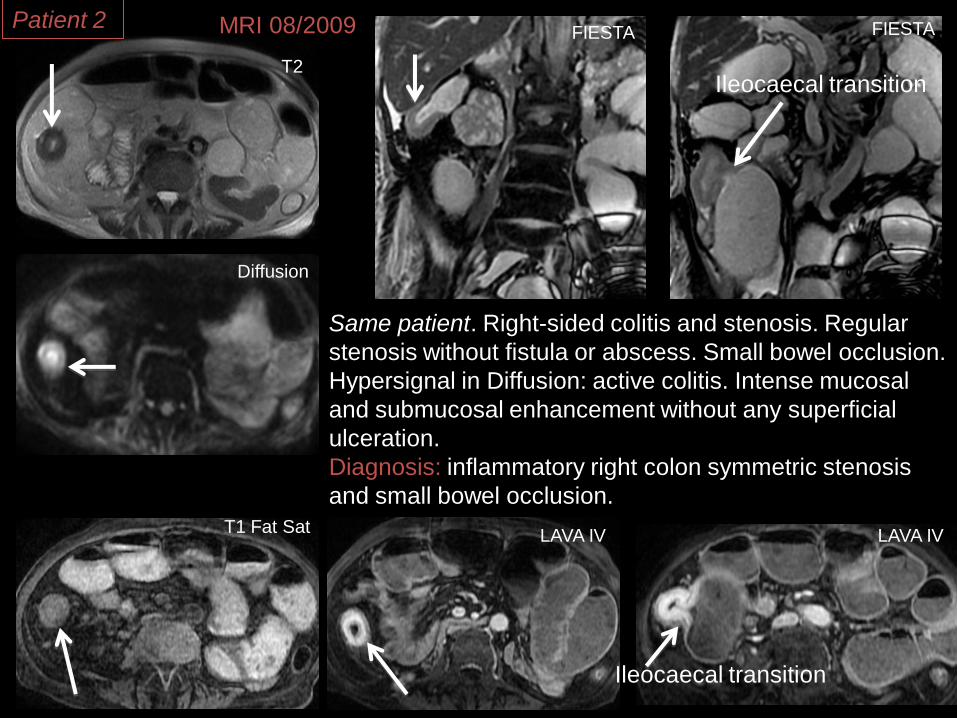
Same patient 2 years later: 60 years old. Small
bowel occlusion resistant to medical treatment
since 2 months and lost of 2 kg.
Patient 2
T2
Diffusion
T1 Fat Sat LAVA IV LAVA IV
FIESTA FIESTA MRI 08/2009
Same patient. Right-sided colitis and stenosis. Regular
stenosis without fistula or abscess. Small bowel occlusion.
Hypersignal in Diffusion: active colitis. Intense mucosal
and submucosal enhancement without any superficial
ulceration.
Diagnosis: inflammatory right colon symmetric stenosis
and small bowel occlusion.
Ileocaecal transition
Ileocaecal transition
Patient 2
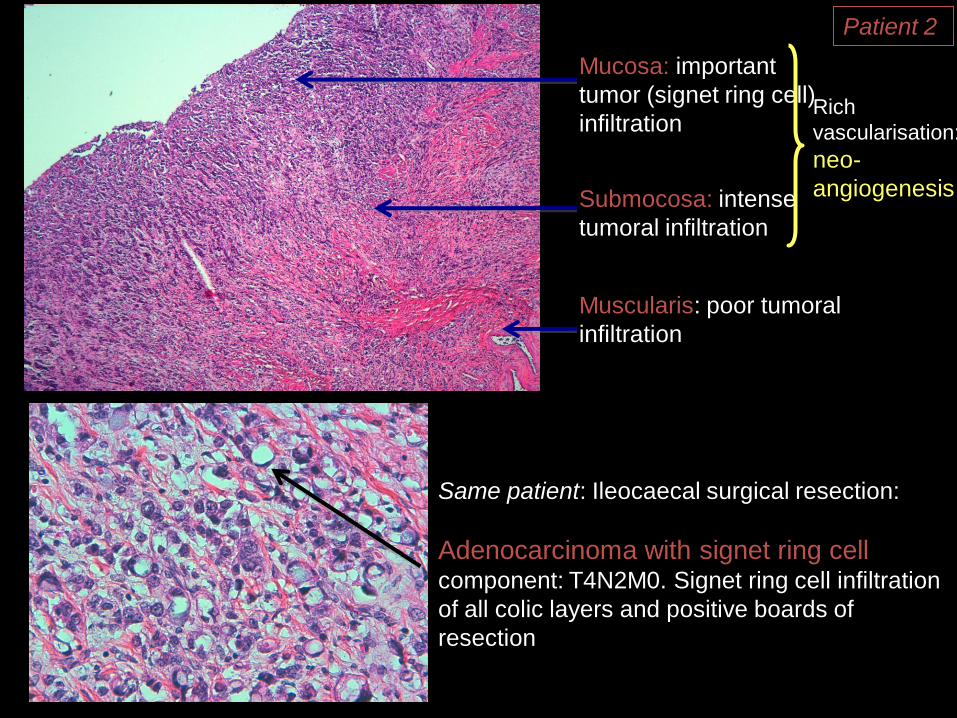
Same patient: Ileocaecal surgical resection:
Adenocarcinoma with signet ring cell component: T4N2M0. Signet ring cell infiltration
of all colic layers and positive boards of
resection
Mucosa: important
tumor (signet ring cell)
infiltration
Submocosa: intense
tumoral infiltration
Muscularis: poor tumoral
infiltration
Rich
vascularisation:
neo-
angiogenesis
Patient 2
Same patient: Adenocarcinoma with signet ring cell T4: the signet ring
cells infiltrate all layer of the colic wall and disorganize its structure. The
enhancement of the inner layer corresponds of the tumor and its vessels.
Mucosa: important
tumor (signet ring
cells in blue)
infiltration
Submocosa: intense
tumor infiltration
Muscularis: poor tumor
infiltration (blue) and
disorganization
Pericolic fat: tumor
infiltration
Right colon stenosis Right colon stenosis
Patient 2
T2
T2
Diffusion
Diffusion
T1 Gd fat sat
T1 Gd fat sat
T1 Gd fat sat
42 years-old man with CD from 15 years.
Rectosigmoitis and anal fistula, difficult
medical treatment. Transverse colostomy
of discharge.
Same patient 5 months later.
Severe rectosigmoiditis and
anal lesion. Small bowel
occlusion though the
transverse colostomy.
CT IV 70’’
Severe rectosigmoiditis with submucosal
edema in hypersignal T2, important
enhancement of all layers of
rectosigmoid and hypersignal in Diffusion.
Complex anal fistula at 10h.
MRI &CT 03/2009
T2
T1 Gd fat sat
CT IV 70’’
fistula
Patient 3 MRI 10/2008
Same patient. Small bowel occlusion
though the transvers colostomy. MRI 10/2008
The aspect of the ileitis in
the beginning of the
rectosigmoiditis.
T2
FIESTA
Diffusion
FIESTA FIESTA
LAVA LAVA arterial LAVA portal
LAVA 3
Small bowel occlusion due to an ileum inflammatory
symmetric stenosis with progressive beginning, infiltration of
adjoining fat.
MRI 03/2009 Patient 3
CT 03/2009
Same patient.
CT IV 70’’
CT IV 70’’
CT IV 70’’ CT IV 70’’
Abscess
Small bowel occlusion du to an ileum inflammatory
circumferential stenosis. Important infiltration in the
periileal fat. Little abscess. Ileitis with intense
enhancement of the inner layer.
Ileo-colic total resection: adenocarcionma with signet
ring cell component spread out from ileum to the
rectosigmoid and the anus, except the transverse
colon.
T4 N2 M1
Patient 3
Same patient: Adenocarcinoma with signet ring cell T4 spread out from ileum to the
rectosigmoid and the anus, except the transverse colon. The signet ring cells
infiltrate all the layers, the inner layer is thin, the muscularis is very infiltrated and
thick.
Mucosa: important
tumor (signet ring
cells) infiltration
Submocosa: intense
tumor infiltration
Muscularis: intense
tumoral infiltration (in
blue) and disorganization
Pericolic fat: tumor
infiltration
Rectitis
Malignant rectitis
Patient 3
CT 04/2007
31 years old female, CD from 14 years.
Adenocarcinoma with signet ring cell right-
sided colon 3 years ago diagnosed with
small bowel occlusion and perforation.
Recidivism of the colic adenocarcinoma with signet ring cell in the
transverse colon. Symmetric stenosis without mass. Intense enhancement
of the inner layer without ulceration or abscess.
Patient 4
Mucosa: important
tumor (signet ring
cells) infiltration
Submocosa: intense
tumor infiltration
Muscularis: poor
tumor infiltration
Pericolic fat: tumor
infiltration
Same patient: Recidivism of the colic adenocarcinoma with signet ring cell
in the transverse colon: intense tumor infiltration of the mucosa and
submucosa corresponding of the enhancement.
Transverse colon stenosis
Transverse colon stenosis
Patient 4
MR 29/09/2006
CT 17/10/2006 25 years old man with CD from 11 years, PSC and liver
transplantation 3 years ago. Colic adenocarcinoma with
signet ring cell component one year ago (2005).
Colectomy. Occlusion and rectitis.
T2
T2
T2
T1 Gd Fat Sat
T1 Gd Fat Sat
T1 Gd Fat Sat
MRI: rectitis responsible for small bowel occlusion, resistant to medical treatment. The
submucosa is in hypersignal on T2 and with important enhancement after Gadolinium
IV. Important local fat infiltration and perirectal ganglia
Rectoscopy showed
local recidive, and a
stent was positioned.
The occlusion was
not resolved and the
tumor progressed.
Occlusion
Submucosal
edema
Patient 5
Mucosa: important
tumor (signet ring
cells) infiltration
Submocosa: intense
tumor infiltration
Muscularis: poor
tumor infiltration
Pericolic fat: tumor
infiltration
Same patient: Adenocarcinoma with signet ring cells of the right colon T4 (scanner
not available), the tumor infiltration is intense in the muscosa and submucosa.
Right colon stenosis
Patient 5
01/2001 CT
38 years-old man,
UC from 14 years.
Occlusion and
difficult medical
treatment.
CT IV 70’’
Rectocolitis, enhancement of the inner colic layer without ulceration. Spiculation
in the pericolic fat. Ascites.
Diagnosis: severe rectitis and left-sided colitis
Surgery: tumor infiltration of colon, aorta, pelvis, peritoneal carcinomatosis.
Histology: Adenocarcinoma with signet ring cell component T4N2M1
CT IV 70’’ CT IV 70’’
CT IV 70’’
Patient 6
Small bowel cancer
There were 3 patients with CD and concomitant small bowel cancer.
Two patients had ileocaecal affect and one had only small bowel affect.
Two SBC were in the ileum and one was in the duodenum.
2 adenocarcinomas and 1 adenocarcinoma with signet ring cell
component.
All SBC presented with occlusion during a disease flare.
CT 10/01/2005
43 years old man with CD
from 4 years. Small bowel
chronic occlusion from 3
months resistant to all
medical treatment.
Entero CT at dignosis, CD with
ileitis and inflammatory stenosis
of the ileum.
CT /2008
CT IV 70’
CT IV 70’
CT IV 70’
CT IV 70’
CT IV 70’
CT IV 70’
CT IV 70’
CT: Small bowel occlusion with ileitis and long
inflammatory stenosis. Ascites and peritoneal fat
infiltration.
Patient 7
Occlusion was not resolved with medical treatment and the patient was
operated: ileocaecal resection.
Diagnosis: Focal Adenocarcinoma with signet ring cell component of
the ileum on 4 cm of length: T3 N1 M0. The tumor is not
macroscopically visible. Corresponds probably to a focal thickening of
the ileum stenosis.
Signet ring cell
Patient 7
Malignant stenosis ileum
Mucosa: intense
tumoral infiltration
Submucosa: tumoral
infiltration
Muscularis: very poor
tumoral infiltration Ileitis
The tumor is impossible
to locate in the long
inflammatory stenosis
The colorectal and small bowel malignancy in IBD are well known.
We confirmed in our series the higher percentage of mucinous and signet ring cell
types, the younger age at diagnosis and the relationship with the anatomic location
of IBD and cancer.
Most of our patients had Crohn’s Disease.
All adenocarcinomas with signet ring cells presented with occlusion and the pre
operative imaging diagnosis was benign inflammatory stenosis.
We could not identify the population of IBD followed in our department: the key
words « IBD », « UC » and « Crohn » given too much results.
The prevalence and the incidence were not calculated.
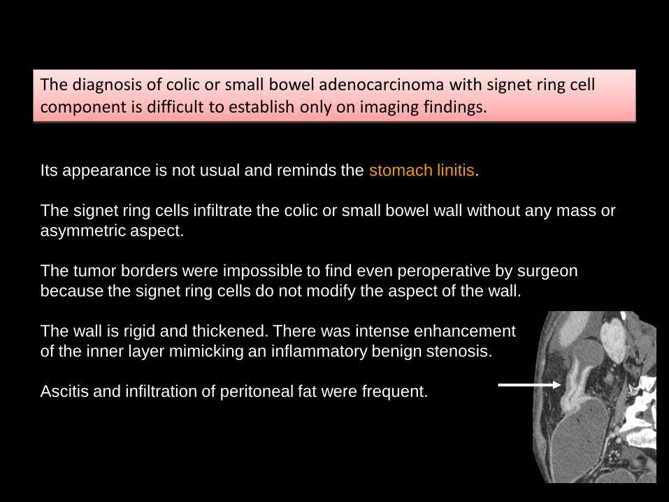
The diagnosis of colic or small bowel adenocarcinoma with signet ring cell component is difficult to establish only on imaging findings.
Its appearance is not usual and reminds the stomach linitis.
The signet ring cells infiltrate the colic or small bowel wall without any mass or
asymmetric aspect.
The tumor borders were impossible to find even peroperative by surgeon
because the signet ring cells do not modify the aspect of the wall.
The wall is rigid and thickened. There was intense enhancement
of the inner layer mimicking an inflammatory benign stenosis.
Ascitis and infiltration of peritoneal fat were frequent.
We tried to explain these similarity by radio-pathologic correlation in Crohn’s ileitis and small bowel adenocarcioma with signet ring cell. We used one patient with CD and malignant stenosis and one patient with CD and inflammatory stenosis, both with small bowel occlusion.
The colic or small bowel linitis in IBD are the differential diagnosis of colitis or ileitis.
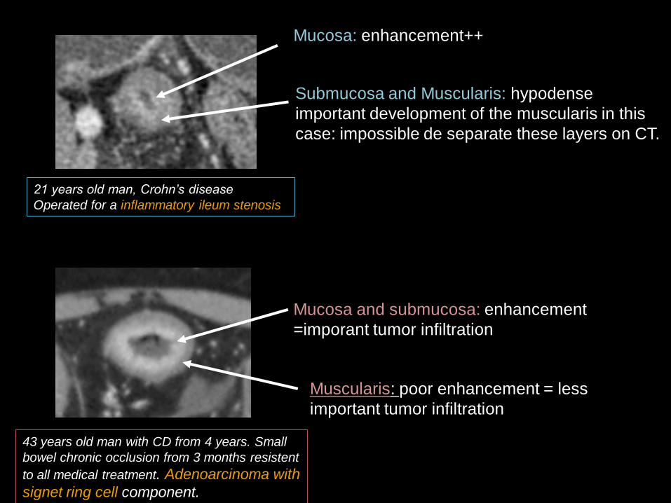
21 years old man, Crohn’s disease
Operated for a inflammatory ileum stenosis
Submucosa and Muscularis: hypodense
important development of the muscularis in this
case: impossible de separate these layers on CT.
Mucosa: enhancement++
Mucosa and submucosa: enhancement
=imporant tumor infiltration
Muscularis: poor enhancement = less
important tumor infiltration
43 years old man with CD from 4 years. Small
bowel chronic occlusion from 3 months resistent
to all medical treatment. Adenoarcinoma with
signet ring cell component.
Muscularis
Submucosa: imporant thickening of
muscularis mucosa
Mucosa
Mucosa: intense tumor infiltration
Muscularis: poor tumor infiltration
Submucosa: tumor infiltration
Inflammatory ileum stenosis, CD
Malignant stenosis, CD
Malignant stenosis: signet ring cell, CD
Mucosa
Submucosa
Muscularis Less important tumor infiltration
Tumor infiltration of mucosa and
submucosa are accompanied with
intense inflammatory infiltration and
edema.
Richer vascularisation than an
inflammatory Crohn’s stenosis: neo
angiogenesis
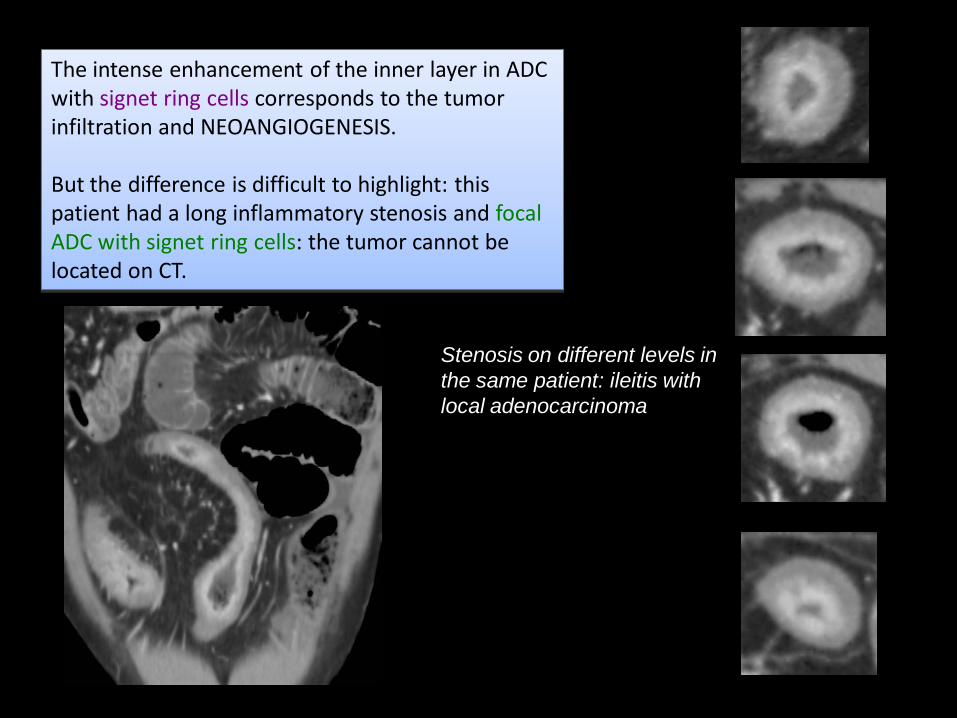
The intense enhancement of the inner layer in ADC with signet ring cells corresponds to the tumor infiltration and NEOANGIOGENESIS. But the difference is difficult to highlight: this patient had a long inflammatory stenosis and focal ADC with signet ring cells: the tumor cannot be located on CT.
Stenosis on different levels in
the same patient: ileitis with
local adenocarcinoma
When the tumor infiltratates totaly the colic wall (advanced T4) the enhancement
and edema were intense without stratification.
CT IV 70’’
T4
CT IV 70’’
T4
Conclusion
The intestinal tract malignancy is a cause of death in longstanding and severe IBD. Risk factors are well known. Most important are duration of IBD and anatomic extent. Most of these cancers have similar imaging presentation to usual small bowel and colorectal cancers.
Large percentage (about 30%) of intestinal tract malignancy in IBD are mucinous adenocarcinoma or adenocarcinoma with signet ring component. The adenocarcinoma with signet ring component presents as a circular symmetric stenosis and mimics a disease flare with inflammatory stenosis. Malignant stenosis must be suspected when a patient with IBD and risk factors presents an occlusion by stenosis resistant to medical treatment.
Imaging features of ADC with signet ring cells in IBD versus inflammatory stenosis
The intense enhancement of the inner colic/small bowel layer > 5mm : tumor infiltration and neo angiogenesis of the mucosa and submucosa, visible on 70’’ after contrast IV injection on CT.
The intense enhancement of all the colic/small bowel without stratification wall when the tumor infiltrates the muscularis: advanced T4.
Abrievations
• IBD: inflammatory bowel disease
• CCR: colorectal cancer
• SBC: small bowel cancer
• CD: Crohn’s disease
• UC: ulcerative colitis
• PSC: primary sclerosing cholangitis
• ADC: adenocarcinoma
References 1Bowel Dis, 2005. 11(9): p. 828-32. . Eaden, J.A., K.R. Abrams, and J.F. Mayberry, The risk of colorectal cancer in ulcerative colitis: a meta-analysis. Gut, 2001. 48(4):
p. 526-35.
2. Itzkowitz, S.H. and X. Yio, Inflammation and cancer IV. Colorectal cancer in inflammatory bowel disease: the role of inflammation. Am J Physiol Gastrointest Liver
Physiol, 2004. 287(1): p. G7-17.
3. von Roon, A.C., et al., The risk of cancer in patients with Crohn's disease. Dis Colon Rectum, 2007. 50(6): p. 839-55.
4. Bernstein, C.N., et al., Cancer risk in patients with inflammatory bowel disease: a population-based study. Cancer, 2001. 91(4): p. 854-62.
5. Jess, T., et al., Intestinal and extra-intestinal cancer in Crohn's disease: follow-up of a population-based cohort in Copenhagen County, Denmark. Aliment Pharmacol
Ther, 2004. 19(3): p. 287-93.
6. Itzkowitz, S.H. and D.H. Present, Consensus conference: Colorectal cancer screening and surveillance in inflammatory bowel disease. Inflamm Bowel Dis, 2005.
11(3): p. 314-21.
7. Rutter, M., et al., Severity of inflammation is a risk factor for colorectal neoplasia in ulcerative colitis. Gastroenterology, 2004. 126(2): p. 451-9.
8. Ekbom, A., et al., Ulcerative colitis and colorectal cancer. A population-based study. N Engl J Med, 1990. 323(18): p. 1228-33.
9. Jayaram, H., J. Satsangi, and R.W. Chapman, Increased colorectal neoplasia in chronic ulcerative colitis complicated by primary sclerosing cholangitis: fact or fiction?
Gut, 2001. 48(3): p. 430-4.
10. Kornfeld, D., A. Ekbom, and T. Ihre, Is there an excess risk for colorectal cancer in patients with ulcerative colitis and concomitant primary sclerosing cholangitis? A
population based study. Gut, 1997. 41(4): p. 522-5.
11. Xie, J. and S.H. Itzkowitz, Cancer in inflammatory bowel disease. World J Gastroenterol, 2008. 14(3): p. 378-89.
12. Ekbom, A., et al., Increased risk of large-bowel cancer in Crohn's disease with colonic involvement. Lancet, 1990. 336(8711): p. 357-9.
13. Askling, J., et al., Family history as a risk factor for colorectal cancer in inflammatory bowel disease. Gastroenterology, 2001. 120(6): p. 1356-62.
14. Pinczowski, D., et al., Risk factors for colorectal cancer in patients with ulcerative colitis: a case-control study. Gastroenterology, 1994. 107(1): p. 117-20.
15. Choi, P.M. and M.P. Zelig, Similarity of colorectal cancer in Crohn's disease and ulcerative colitis: implications for carcinogenesis and prevention. Gut, 1994. 35(7):
p. 950-4.
16. Feldstein, R.C., S. Sood, and S. Katz, Small bowel adenocarcinoma in Crohn's disease. Inflamm Bowel Dis, 2008. 14(8): p. 1154-7.
17. Palascak-Juif, V., et al., Small bowel adenocarcinoma in patients with Crohn's disease compared with small bowel adenocarcinoma de novo. Inflamm
This digital poster was realised with the support of the
Society of Abdominal and Digestive Imaging of France.