cooled sCMOS camera edge 4.2 bi up to 95% quantum efficiency deep cooled down to -25 °C input windows selectable back illuminated sCMOS sensor bi back illuminated compact design resolution 2048 x 2048 pixel with 6.5 µm pixel size
Transcript
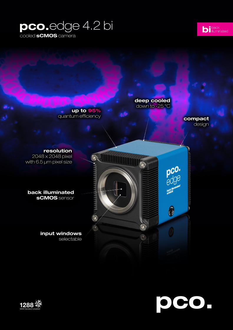
cooled sCMOS camera
edge 4.2 bi
up to 95%quantum efficiency
deep cooleddown to -25 °C
input windowsselectable
back illuminated sCMOS sensor
bibackilluminated
compactdesign
resolution2048 x 2048 pixel
with 6.5 µm pixel size
2
sCMOS image sensor
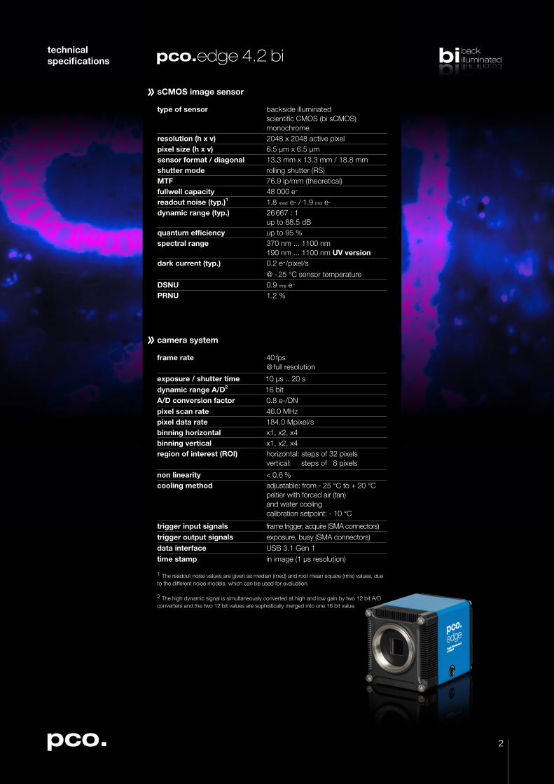
type of sensor backside illuminated scientific CMOS (bi sCMOS)monochrome
resolution (h x v) 2048 x 2048 active pixelpixel size (h x v) 6.5 µm x 6.5 µmsensor format / diagonal 13.3 mm x 13.3 mm / 18.8 mmshutter mode rolling shutter (RS)MTF 76.9 lp/mm (theoretical)fullwell capacity 48 000 e-
readout noise (typ.)1 1.8 med e- / 1.9 rms e-dynamic range (typ.) 26 667 : 1
up to 88.5 dBquantum efficiency up to 95 % spectral range 370 nm ... 1100 nm
exposure / shutter time 10 µs .. 20 sdynamic range A/D2 16 bitA/D conversion factor 0.8 e-/DNpixel scan rate 46.0 MHzpixel data rate 184.0 Mpixel/sbinning horizontal x1, x2, x4binning vertical x1, x2, x4region of interest (ROI) horizontal: steps of 32 pixels
vertical: steps of 8 pixels
non linearity < 0.6 % cooling method adjustable: from - 25 °C to + 20 °C
peltier with forced air (fan) and water coolingcalibration setpoint: - 10 °C
trigger input signals frame trigger, acquire (SMA connectors)trigger output signals exposure, busy (SMA connectors)data interface USB 3.1 Gen 1time stamp in image (1 µs resolution)
1 The readout noise values are given as median (med) and root mean square (rms) values, due to the different noise models, which can be used for evaluation. 2 The high dynamic signal is simultaneously converted at high and low gain by two 12 bit A/D converters and the two 12 bit values are sophistically merged into one 16 bit value.
3
bibackilluminatedbibackilluminated
technical specifications edge 4.2 bi
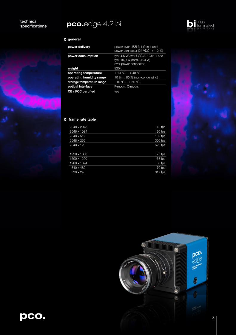
frame rate table
2048 x 2048 40 fps2048 x 1024 80 fps2048 x 512 159 fps2048 x 256 300 fps2048 x 128 520 fps
1920 x 1080 76 fps1600 x 1200 68 fps1280 x 1024 80 fps 640 x 480 170 fps 320 x 240 317 fps
general
power delivery power over USB 3.1 Gen 1 and power connector (24 VDC +/- 10 %)
power consumption typ. 4.5 W over USB 3.1 Gen 1 and typ. 10.0 W (max. 22.0 W) over power connector
weight 920 goperating temperature + 10 °C ... + 40 °Coperating humidity range 10 % ... 80 % (non-condensing)storage temperature range - 10 °C ... + 60 °Coptical interface F-mount, C-mount
CE / FCC certified yes
4
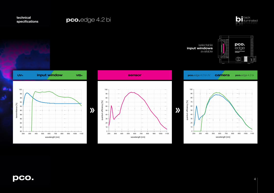
selectableinput windows
available
bibackilluminatedbibackilluminated
technical specifications edge 4.2 bi
wavelength [nm]
200 300 400 500 600 700 800 900 1000 1100
quan
tum
effi
cien
cy [%
]
0
10
20
30
40
50
60
70
80
90
100
edge 4.2 bi UV edge 4.2 bicameraVIS+UV+ input window
wavelength [nm]
200 300 400 500 600 700 800 900 1000 1100
trans
mitt
ance
[%]
80
82
84
86
88
90
92
94
96
98
100
sensor
wavelength [nm]
200 300 400 500 600 700 800 900 1000 1100
quan
tum
effi
cien
cy [%
]
0
10
20
30
40
50
60
70
80
90
100
5
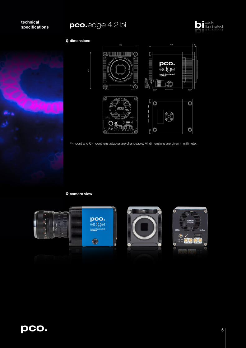
camera view
dimensions
made in germany
USB 3.1 Gen 1
armrec
status
1 in 2 3 out 4
on
off
0 99 10
85
80
bibackilluminatedbibackilluminated
F-mount and C-mount lens adapter are changeable. All dimensions are given in millimeter.
technical specifications edge 4.2 bi
6
bibackilluminatedbibackilluminated
third party integrations
software
With pco.camware you control all camera settings, the image acquisition and the storage of your image data. The pco.sdk is the complementary software development kit. It includes dynamic link libraries for user customization and integration on Windows-PC platforms. Drivers for popular third party software packages are also available for you.
All this items like pco.camware, pco.sdk and third party drivers, are free-to-download at www.pco.de.
technical specifications edge 4.2 bi
applications
brightfield microscopy microscopy | fluorescence microscopy | digital pathology | single molecule localization microscopy | lightsheet fluorescence microscopy (LSFM) | calcium imaging | FRET | FRAP | structured illumination microscopy (SIM) | high-speed bright field ratio imaging | high throughput screening | high content screening | biochip reading | TIRF microscopy | spinning disk confocal microscopy | 3D metrology | ophthalmology | photovoltaic inspection | industrial quality inspection | lucky astronomy | bio luminescence | chemo luminescence