Edinburgh Research Explorer Neonatal Colonisation Expands a Specific Intestinal Antigen- Presenting Cell Subset Prior to CD4 T-Cell Expansion, without Altering T-Cell Repertoire Citation for published version: Inman, CF, Laycock, GM, Mitchard, L, Harley, R, Warwick, J, Burt, R, van Diemen, PM, Stevens, M & Bailey, M 2012, 'Neonatal Colonisation Expands a Specific Intestinal Antigen-Presenting Cell Subset Prior to CD4 T-Cell Expansion, without Altering T-Cell Repertoire' PLoS One, vol. 7, no. 3, pp. e33707. DOI: 10.1371/journal.pone.0033707 Digital Object Identifier (DOI): 10.1371/journal.pone.0033707 Link: Link to publication record in Edinburgh Research Explorer Document Version: Publisher's PDF, also known as Version of record Published In: PLoS One Publisher Rights Statement: This is an open-access article distributed under the terms of the Creative Commons Attribution License, which permits unrestricted use, distribution, and reproduction in any medium, provided the original author and source are credited. General rights Copyright for the publications made accessible via the Edinburgh Research Explorer is retained by the author(s) and / or other copyright owners and it is a condition of accessing these publications that users recognise and abide by the legal requirements associated with these rights. Take down policy The University of Edinburgh has made every reasonable effort to ensure that Edinburgh Research Explorer content complies with UK legislation. If you believe that the public display of this file breaches copyright please contact [email protected] providing details, and we will remove access to the work immediately and investigate your claim. Download date: 16. Sep. 2018
Transcript
Edinburgh Research Explorer
Neonatal Colonisation Expands a Specific Intestinal Antigen-Presenting Cell Subset Prior to CD4 T-Cell Expansion, withoutAltering T-Cell Repertoire
Citation for published version:Inman, CF, Laycock, GM, Mitchard, L, Harley, R, Warwick, J, Burt, R, van Diemen, PM, Stevens, M &Bailey, M 2012, 'Neonatal Colonisation Expands a Specific Intestinal Antigen-Presenting Cell Subset Priorto CD4 T-Cell Expansion, without Altering T-Cell Repertoire' PLoS One, vol. 7, no. 3, pp. e33707. DOI:10.1371/journal.pone.0033707
Digital Object Identifier (DOI):10.1371/journal.pone.0033707
Link:Link to publication record in Edinburgh Research Explorer
Document Version:Publisher's PDF, also known as Version of record
Published In:PLoS One
Publisher Rights Statement:This is an open-access article distributed under the terms of the Creative Commons Attribution License, whichpermitsunrestricted use, distribution, and reproduction in any medium, provided the original author and source arecredited.
General rightsCopyright for the publications made accessible via the Edinburgh Research Explorer is retained by the author(s)and / or other copyright owners and it is a condition of accessing these publications that users recognise andabide by the legal requirements associated with these rights.
Take down policyThe University of Edinburgh has made every reasonable effort to ensure that Edinburgh Research Explorercontent complies with UK legislation. If you believe that the public display of this file breaches copyright pleasecontact [email protected] providing details, and we will remove access to the work immediately andinvestigate your claim.
Neonatal Colonisation Expands a Specific IntestinalAntigen-Presenting Cell Subset Prior to CD4 T-CellExpansion, without Altering T-Cell RepertoireCharlotte F. Inman1*.¤a, Georgina M. Laycock1., Louisa Mitchard1, Ross Harley1, James Warwick1,
Rachel Burt1, Pauline M. van Diemen2¤b, Mark Stevens2¤c, Mick Bailey1
1 School of Clinical Veterinary Science, University of Bristol, Langford, Bristol, United Kingdom, 2 Institute for Animal Health, Compton, Newbury, Berkshire, United
Kingdom
Abstract
Interactions between the early-life colonising intestinal microbiota and the developing immune system are critical indetermining the nature of immune responses in later life. Studies in neonatal animals in which this interaction can beexamined are central to understanding the mechanisms by which the microbiota impacts on immune development and todeveloping therapies based on manipulation of the microbiome. The inbred piglet model represents a system that iscomparable to human neonates and allows for control of the impact of maternal factors. Here we show that colonisationwith a defined microbiota produces expansion of mucosal plasma cells and of T-lymphocytes without altering the repertoireof alpha beta T-cells in the intestine. Importantly, this is preceded by microbially-induced expansion of a signal regulatoryprotein a-positive (SIRPa+) antigen-presenting cell subset, whilst SIRPa2CD11R1+ antigen-presenting cells (APCs) areunaffected by colonisation. The central role of intestinal APCs in the induction and maintenance of mucosal immunityimplicates SIRPa+ antigen-presenting cells as orchestrators of early-life mucosal immune development.
Citation: Inman CF, Laycock GM, Mitchard L, Harley R, Warwick J, et al. (2012) Neonatal Colonisation Expands a Specific Intestinal Antigen-Presenting Cell SubsetPrior to CD4 T-Cell Expansion, without Altering T-Cell Repertoire. PLoS ONE 7(3): e33707. doi:10.1371/journal.pone.0033707
Editor: Jorg Hermann Fritz, McGill University, Canada
Received November 14, 2011; Accepted February 15, 2012; Published March 19, 2012
Copyright: � 2012 Inman et al. This is an open-access article distributed under the terms of the Creative Commons Attribution License, which permitsunrestricted use, distribution, and reproduction in any medium, provided the original author and source are credited.
Funding: The work was funded by the Department for Environment, Food, and Rural Affairs and the Higher Education Funding Council for England through theVeterinary Training in Research Initiative grant VT0104. The funders had no role in study design, data collection and analysis, decision to publish, or preparation ofthe manuscript.
Competing Interests: The authors have declared that no competing interests exist.
¤a Current address: School of Cancer Studies, University of Birmingham, Edgbaston, Birmingham, United Kingdom¤b Current address: The Jenner Institute, Nuffield Department of Medicine, Oxford University, Oxford, United Kingdom¤c Current address: The Roslin Institute and Royal (Dick) School of Veterinary Studies, University of Edinburgh, Midlothian, United Kingdom
Introduction
The importance of microbial colonisation of the gut in immune
development is well established. Early studies in germ-free (GF)
mice demonstrated that colonisation was critical for the induction
of IgA- producing plasma cells in the intestine [1] and drove
CD4+ cell expansion [2]. Studies in rabbits have demonstrated a
similar picture: follicular development was arrested in rabbit
appendices that were ligated to prevent colonisation [3] and the
appendices of GF rabbits contain reduced numbers of lymphocytes
[4]. More recent studies have shown that this development is
qualitative as well as quantitative: colonisation produces a wider B-
cell repertoire in pigs and results in differentiation of CD4 T
helper (Th) cell subsets in mice [5–7]. However, it appears that
simple ‘colonisation’ is not enough to generate a fully effective
immune system capable of recognising and eliminating pathogen
whilst not responding to self, food and commensal bacterial
antigen. In fact, epidemiological studies in humans (linking
microbiota and the incidence of allergy) have indicated that the
nature of colonisation in the post-natal period is important in
determining the character of the immune system that subsequently
emerges [8,9]. In order to fully investigate these observations, it is
necessary to use a reductionist rather than holistic approach to
experimental design, colonising GF animals with a specifically
defined microbiota and measuring the effect on immune
development. These studies have been elegantly performed in
rodent models and have identified specific species of bacteria that
appear to be responsible for the development of specific arms of
the immune response. For example, Bacteroides fragilis has been
shown to be important in the development of FoxP3+ T cells in the
intestine and segmented filamentous bacteria (SFB) have been
shown to drive the appearance of Th17 cells in the intestinal
lamina propria [10,11]. This data confirms that the type of
colonisation may be as important as the colonisation event itself.
The antigen-presenting cell (APC) is central to the induction
and maintenance of immune responses. This is particularly clear
in the intestine where APCs can be observed directly interacting
with the microbiota [12,13] and with T cells, not only in the lymph
nodes but also in the mucosa [14]. Work in mice has identified
functionally distinct populations of dendritic cells (DCs) in the
intestinal mucosa, distinguished by their expression of CD103,
CX3CR1 and CD11b [15,16] and it appears that these different
intestinal APC subsets are important in generating different types
of immune response [17,18]. There are also distinct populations of
PLoS ONE | www.plosone.org 1 March 2012 | Volume 7 | Issue 3 | e33707
intestinal APC in the pig (based on expression of CD16, CD11R1
and SIRPa [14,19,20]) and it appears that these may also be
functionally different [20]. Whilst this suggests that the presence of
different APC subsets may be important across species, it remains
to be seen whether it is possible to identify direct functional
equivalents for all of the recognised mouse intestinal APC subsets
in other animals. For example, although the same monoclonal
antibody recognizes human CD11b and pig CD11R1 [21], there
are significant differences in the distribution of the target
molecules [22], indicating that they are not necessarily functionally
equivalent in mouse, human, and pig. Nevertheless, it appears that
expression of SIRPa may, in fact, identify APCs with a similar
phenotype between species. For example, SIRPa expression has
been shown to be correlated with the ability of APCs to migrate to
the lymph nodes in pigs and mice [20,23]. In humans, sheep, cattle
and rats, SIRPa expression distinguishes functionally different
APC subsets [24–28]. Thus, relative expression of SIRPa by
intestinal APCs is likely to be important in determining immune
function in the gut. In relation to the effects of the microbiota on
intestinal APCs, SIRPa has not been examined. However, work in
adult mice has demonstrated that colonisation preferentially
expands the CX3CR1+ APC subset in the colon [17]. We have
previously demonstrated a selective effect of different microbiota
(associated with high hygiene and low hygiene rearing environ-
ments) on myeloid APCs in the neonatal pig intestine but not on
endothelial APCs [29]. However, generally, data on the effects of
colonisation on APC subsets in the small intestine, and on
neonates, is sparse. This is despite the well documented
importance of early life colonisation and subsequent immune
development on the ability to mount effective responses later in
life.
Whilst mucosal APCs are a core part of immunity in the
intestine, T cells are one of the ‘workhorses’ of the adaptive
immune response that, as a population, are capable of responding
to a massive range of antigens due to the variability and
promiscuity of the T cell receptors expressed by different clones.
The hypervariable complementarity determining region 3 (CDR3)
of the T cell receptor (TCR) interacts with the major histocom-
patibility complex (MHC) plus peptide on the surface of APCs.
Activation of T cells with a specific antigen may lead to expansion
of a few specific T cell clones. By using spectratyping to analyse
CDR3 lengths of T cells within the intestinal mucosal population,
it is possible to perceive whether one or more dominant CDR3
lengths are present and, thus, whether there is likely to have been
antigen-specific recruitment of T-cells to the mucosa. For example,
spectratype studies in rats have shown that T-cells in the intestine
have a restricted, oligoclonal repertoire [30]. By comparing the
distributions of CDR3 lengths in animals colonised with a defined
flora to the distributions in GF animals, it is possible to determine
whether colonisation alters T cell repertoire in the gut. This is an
important matter for investigation, as changes in repertoire
associated with colonisation may have a major impact on gut
immune function. For example, although induction of a broad
repertoire of T-cell clones following colonisation of the gut may
subsequently promote host defence against an extremely wide
range of pathogens, competition for resources between ‘useful’ T
cell clones and T cells with irrelevant TCRs may mean that an
alternative strategy, in which only a limited cohort of ‘useful’ T cell
clones is induced, proves to be more effective in defending against
a (restricted) range of pathogens. Notably, in relation to the effects
of intestinal colonisation on B-cells, previous studies in GF pigs
and mice have, in the majority, indicated that B cell expansion and
IgA production after colonisation is primarily antigen non-specific
[31–34]. Whilst spectratyping does not allow for identification of
expansion of specific T cell clones in response to specific antigens,
it has the advantage of generating a general overview of the effects
of colonisation on the diversity of the TCR repertoire.
In order to control for maternal effects, it is necessary to
examine immune development in neonates reared away from the
mother, and without transferred antigen or antibody. As a
consequence of this, these types of studies are technically difficult
in rodents, where significant transfer occurs in utero [35]. As a
solution to this problem, we have used a piglet GF model where
highly inbred Babraham piglets are derived by Caesarean section
and housed individually under sterile conditions. The use of the
pig model reduces variation associated with maternal factors and
allows for controlled colonisation with a defined microbiota. The
pig model is particularly appropriate for the study of the impact of
intestinal colonisation since no placental transfer of macromole-
cules takes place [35,36], and since intra-individual stability and
inter-individual variability of the microbiota are more comparable
between humans and pigs than between humans and mice [37].
Pigs have comparable digestive physiology to humans and full
genome studies have demonstrated fewer differences between
humans and pigs that between humans and mice [38]. In addition,
as in human infants, the neonatal piglet has a poorly developed
mucosal immune system [39]. Whilst the GF pig model has been
well utilised over a number of years for these reasons, a further
advantage of the model described in this study is in the use of
inbred, Babraham pigs, which reduces the variation associated
with genetic factors.
We set out to investigate the effects of colonisation with a
defined microbiota on development of the adaptive mucosal
immune system in a neonatal pig model. To this end, we have
developed a defined microbiota and used this to colonise neonatal
piglets in the first 24 hours of life, to reflect the situation in human
infants. We used quantitative immunohistology to examine the
effects of colonisation on B and T lymphocyte cell numbers. On
finding, in concordance with previous studies, that colonisation
increased the numbers of these cells in the mucosa, we used
spectratype analysis of 21 TRbV groups/subgroups (see Table S1)
to examine whether it also altered T cell repertoire diversity.
Interestingly, colonisation had no apparent effect on TCR Vbrepertoire, diversity suggesting that the expansion, whilst driven by
colonisation, did not solely involve expansion of T-cells specific for
the microbiota. Given our previous studies on intestinal APC
subsets and, more importantly, the observation that in adult mice,
a specific subset is affected by colonisation, we were keen to
examine the effects of colonisation on APC subsets in the neonate.
Strikingly, quantitative immunohistology showed that colonisation
in neonates specifically increased the SIRPa+ APC subset.
Materials and Methods
Ethics statementAll animal experiments were conducted in accordance with the
Animal (Scientific Procedures) Act 1986 (licence 30/2485) with the
approval of the Institute for Animal Health Ethical Review
Committee.
Animals and housingFour separate experiments (see Table 1) were performed in
which inbred Babraham piglets were derived by Caesarean section
into a sterile chamber. Within two hours, the animals were
transferred to individual sterile housing (Bell Isolation Systems,
UK), or killed for day 0 time point sampling as described below.
Piglets were fed on tinned, autoclaved evaporated milk (Tesco
PLC, UK) and had free access to sterile water.
Neonatal Colonisation and Immune Development
PLoS ONE | www.plosone.org 2 March 2012 | Volume 7 | Issue 3 | e33707
Microbial floraThe colonisation microbiota consisted of a cocktail of bacteria
Image analysisTen images/tissue of each stain were captured using a Leica
DMR-A fluorescence microscope fitted with appropriate single
colour filters, a Hamamatsu Orca-ER camera (Hamamatsu, UK)
and Q-fluoro software (Leica, Germany). Images were analysed as
described previously using ImageJ software [47]. Briefly, levels of
background staining (threshold levels) in all colour channels were
obtained from negative control slides (prepared in conjunction
with each sample) using the ImageJ macro ‘multiplecolourbackgrounds’.
These were then applied to each positively stained image so that
all pixels above threshold were set to their appropriate colour and
all those below were set to black. This technique has been
previously optimised to minimise variation between samples [47].
Table 1. Numbers and ages of GF and colonised pigs in eachexperiment.
Experiment Age of pigs (days) No. GF pigs No. colonised pigs
1 0 5 N/A
2 5 3 3
3 21 3 4
4 21 1 2
Four experiments were performed with between five and seven pigs in each.Pigs were euthanised and samples taken either at 0 (1 experiment), 5 (1experiment) or 21 (2 experiments) days of age.doi:10.1371/journal.pone.0033707.t001
Neonatal Colonisation and Immune Development
PLoS ONE | www.plosone.org 3 March 2012 | Volume 7 | Issue 3 | e33707
The number of pixels positive for each colour were then analysed
using the ImageJ macro ‘multiplecolouranalysis’. This data was then
expressed as the proportion of pixels positive for each colour in the
whole image.
RNA extraction, analysis and reverse transcriptionSections of spleen, proximal jejunum and distal jejunum tissue
(without Peyer’s patches) were collected into RNALater (Ambion
Ltd., UK) according to manufacturer’s instructions and stored at -
70uC. Tissues were homogenized with a stainless steel bead using a
TissueLyser (Qiagen, UK). RNA was extracted from tissue
samples using the SV RNA Total Isolation System (Promega,
UK) according to manufacturer’s instructions, and concentration
and quality was assessed using the Experion Automated
Electrophoresis system (Bio-Rad, UK). Reverse transcription was
performed using random hexamers and the Improm II Reverse
Transcription system (Promega). cDNA was diluted to a final
working volume of 150 ml and stored at 220uC.
Porcine T cell receptor V beta (pTRbV) Spectratype PCRPhylogenetic analysis of 363 TRbV nucleotide sequences
(unpublished data) allowed the identification of 20 TRbV groups.
Nineteen of these contained clones corresponding to the 19 pig
TRbV groups named by Butler et al [48], using the IMGT
designation. The final group contained a sequence (GenBank
AB079527) with high homology to human TRbV24 transcripts:
this sequence was recently confirmed as porcine TRbV24 [49].
Within TRbV12S, one sequence (GenBank AY690854) had a
significantly lower identity with other TRbV sequences in the
group. This sequence was, therefore, designated TRbV12-AS: a
subgroup of TRbV12S.
For each cDNA sample, TRbV group/subgroup-specific PCR
products were generated using sense primers specific for one of 20
TRbV groups, or 1 TRbV subgroup, and a universal Cb-region
antisense primer (Table S1) labelled with 59 FAM, HEX or TET.
PCR was performed using a PTC-200 DNA Engine (MJ
Research, Massachusetts). Each PCR mixture comprised 2 ml
template in 25 ml 16 HotStartaq mastermix (Qiagen, UK)
containing 200 nM each of sense TRbV-specific primer and
labelled antisense Cb-region primer and 3.0 mM MgCl2. PCR
conditions were 95uC for 15 minutes, then 40 cycles of 15 s at
95uC, 15 s at 60uC, and 30 s at 72uC and 5 minutes at 72uC. PCR
products from positive and negative control samples were
visualised on a 2% agarose gel stained with ethidium bromide
prior to sample purification. PCR product purification was
performed using the Nucleospin Multi-96 Extract PCR clean-up
kit (Macherey-Nagel, Germany). Fluorimetry of the purified PCR
products was performed using the quantitative plate read function
on the MX3005P QPCR system (Stratagene, UK). A standard
curve was constructed for the fluorochromes FAM, HEX and
TET from a two-fold serial dilution from 10 nM to 0.15625 nM
using the universal Cb-region antisense primer labelled with each
fluorochrome diluted in AE buffer, and a buffer only negative
control. Approximate concentrations of fluorescently-labelled
PCR products were derived and used to determine the amount
of purified PCR product for subsequent use.
Spectratype analysisFor generation of spectratype electropherograms, purified PCR
products were appropriately mixed (different sizes and fluoro-
chromes) and processed using a MegaBACE 1000 DNA analysis
system (Amersham, UK) at the University of Bristol Transcrip-
tomics Facility. Electropherograms generated for each well were
analysed using the MegaBACE Genetic Profiler v 1.5 (Amersham).
Each electropherogram peak analysed was verified as within the
anticipated bp range. Peaks which were not differentiable from
background fluorescence (,70 relative fluorescence units (rfu)) or
which were .1 bp away from the expected size were rejected.
Peak rfu (heights) were standardised by conversion to a proportion
of the total peak rfu for a given spectratype electropherogram [30].
StatisticsData were analysed using SPSS 18.0 software (SPSS Inc.,
Chicago). Immunohistology images were analysed using analysis of
variance ANOVA with post-hoc testing using least significant
difference and with proportion positive area as the outcome
variable and treatment (colonisation status) and age as explanatory
variables. Pig was included in the model as a covariate. Where
appropriate, a Bonferroni correction was applied for multiple tests.
For spectratyping, analysis was performed using principle
component analysis (PCA) followed by a univariate general linear
model (GLM) with post-hoc testing using least significant
difference and hierarchical cluster analysis. PCA was performed
on all TRbV groups/subgroup at once for a maximum of 25
iterations for convergence using an unrotated factor solution. All
components analysed had Eigenvalues greater than 1. Hierarchi-
cal cluster analysis was performed using Wards method with
Whilst other studies in pigs have demonstrated an increase in
mucosal plasma cells and lymphocytes on colonisation [31,32,39],
we wanted to study the effects of colonisation with our microbiota
on these cells in more detail. Therefore, we examined the effects of
colonisation on lymphoid cells at 5 and 21 days of age.
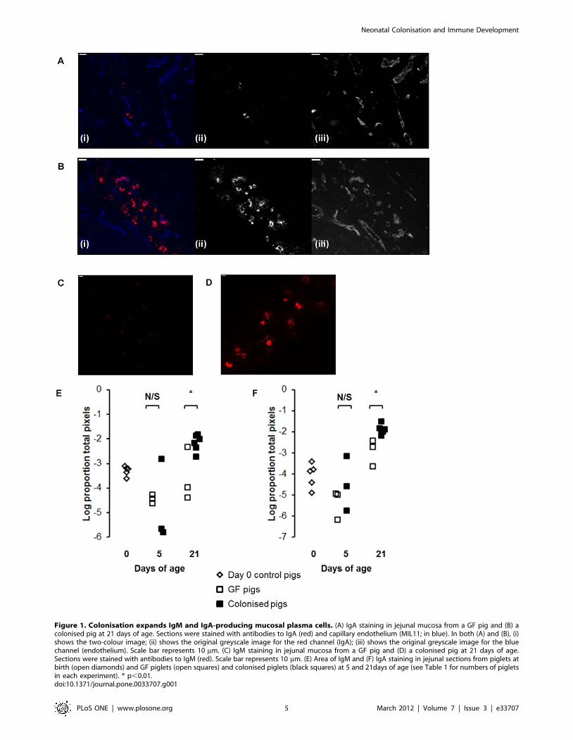
Since Ig-producing cells reside in the intestinal crypts in the pig
small intestine, we examined the crypt regions of jejunal sections
from GF and colonised pigs using immunofluorescent labelling to
look for the effects of colonisation on Ig-producing cells. This
showed a clear increase in both IgM and IgA-producing plasma
cells associated with colonisation (Figure 1A and B). Quantitative
analysis of the images showed that this effect was statistically
significant by 21 days of age and not at 5 days (Figure 1C and D;
p,0.01).
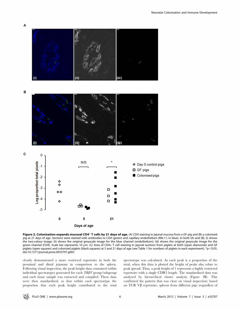
A similar picture could be seen in the CD4 T-cell compartment,
colonisation producing an increase in CD4 T-cells (figure 2A and
B). Similar to plasma cells, this increase was only significant by 21
days (figure 2C; p,0.05). As demonstrated previously, CD4 T
cells could be seen co-localising with MHCII+ cells in the lamina
propria (data not shown) [14].
TCR V beta is unaffected by colonisation-induced T-cellexpansion
Having demonstrated that our novel microbiota expanded the
CD4 T-cell compartment, we investigated whether this expansion
altered TCR Vb repertoire by examining complementarity
determining region 3 (CDR3 length) within 20 TRbV groups
and 1 TRbV subgroup (Table S1). Preliminary visual inspection of
electropherograms indicated that spectratypes generated from
splenic tissues typically demonstrated a Gaussian distribution
pattern, whereas spectratypes generated from the proximal and
distal jejunum clearly showed skewed distribution patterns
(Figure 3A). These findings are in concordance with our previous
studies in rats [30], where examination of CDR3 length plots
Neonatal Colonisation and Immune Development
PLoS ONE | www.plosone.org 4 March 2012 | Volume 7 | Issue 3 | e33707
Figure 1. Colonisation expands IgM and IgA-producing mucosal plasma cells. (A) IgA staining in jejunal mucosa from a GF pig and (B) acolonised pig at 21 days of age. Sections were stained with antibodies to IgA (red) and capillary endothelium (MIL11; in blue). In both (A) and (B), (i)shows the two-colour image; (ii) shows the original greyscale image for the red channel (IgA); (iii) shows the original greyscale image for the bluechannel (endothelium). Scale bar represents 10 mm. (C) IgM staining in jejunal mucosa from a GF pig and (D) a colonised pig at 21 days of age.Sections were stained with antibodies to IgM (red). Scale bar represents 10 mm. (E) Area of IgM and (F) IgA staining in jejunal sections from piglets atbirth (open diamonds) and GF piglets (open squares) and colonised piglets (black squares) at 5 and 21days of age (see Table 1 for numbers of pigletsin each experiment). * p,0.01.doi:10.1371/journal.pone.0033707.g001
Neonatal Colonisation and Immune Development
PLoS ONE | www.plosone.org 5 March 2012 | Volume 7 | Issue 3 | e33707
clearly demonstrated a more restricted repertoire in both the
proximal and distal jejunum in comparison to the spleen.
Following visual inspection, the peak height data contained within
individual spectratypes generated for each TRbV group/subgroup
and each tissue sample was extracted and compiled. These data
were then standardised, so that within each spectratype the
proportion that each peak height contributed to the total
spectratype was calculated. As each peak is a proportion of the
total, when this data is plotted the height of peaks also relate to
peak spread. Thus, a peak height of 1 represents a highly restricted
repertoire with a single CDR3 length. The standardised data was
analysed by hierarchical cluster analysis (Figure 3B). This
confirmed the pattern that was clear on visual inspection: based
on TCR Vb repertoire, spleens from different pigs (regardless of
Figure 2. Colonisation expands mucosal CD4+ T cells by 21 days of age. (A) CD4 staining in jejunal mucosa from a GF pig and (B) a colonisedpig at 21 days of age. Sections were stained with antibodies to CD4 (green) and capillary endothelium (MIL11; in blue). In both (A) and (B), (i) showsthe two-colour image; (ii) shows the original greyscale image for the blue channel (endothelium); (iii) shows the original greyscale image for thegreen channel (CD4). Scale bar represents 10 mm. (C) Area of CD4+ T cell staining in jejunal sections from piglets at birth (open diamonds) and GFpiglets (open squares) and colonised piglets (black squares) at 5 and 21 days of age (see Table 1 for numbers of piglets in each experiment). *p,0.05.doi:10.1371/journal.pone.0033707.g002
Neonatal Colonisation and Immune Development
PLoS ONE | www.plosone.org 6 March 2012 | Volume 7 | Issue 3 | e33707
Figure 3. Repertoire is not altered on expansion of mucosal CD4 T-cells. (A) Representative spectratype electropherograms of TRbV29 for agerm-free pig, and a colonised pig from the same litter of piglets. In general, samples from the spleen demonstrate a more Gaussianelectropherogram distribution than intestinal samples. RFU: relative fluorescence units, x axis: CDR3 length (base pair size). (B) Hierarchical clusteranalysis of CDR3 lengths from 20 TRbV groups and 1 subgroup from the proximal jejunum, distal jejunum and spleen from pigs at 21 days of age.doi:10.1371/journal.pone.0033707.g003
Neonatal Colonisation and Immune Development
PLoS ONE | www.plosone.org 7 March 2012 | Volume 7 | Issue 3 | e33707
treatment) clustered together, and a more restricted T-cell
repertoire was recruited to both the proximal and distal jejunum
in comparison to the solid lymphoid tissue from the spleen.
However, the cluster analysis also demonstrated a random
distribution of GF and colonised pigs through the hierarchy, and
provided no evidence that the diversity of the TCR Vb repertoire
in the intestine was affected by colonisation (Figure 3B). If
colonisation had influenced repertoire, the expectation would be
that the GF pigs would cluster together based on colonisation
status: this was clearly not the case. This was confirmed by PCA of
CDR3 lengths followed by GLM for all TRbV groups/subgroup.
PCA allows a reduction in the number of dimensions of a dataset,
transforming a set of possible correlated variable into a group of
uncorrelated variables (principle components). This is a useful
technique for the analysis of spectratype data where large numbers
of different possible CDR3 lengths may exist within each TRbV
group/subgroup. Having generated the uncorrelated principle
components, it is then possible to use GLM to examine the effects
of factors such as colonisation status, and tissue on each
component. Whilst there was a significant effect of tissue (spleen
or jejunum) on principal components 1 and 4 (p,0.01), there was
no effect of status (GF or colonised) on any of the principle
components (Table 2), indicating that colonisation status did not
affect TCR Vb repertoire in the intestine.
Colonisation alters mucosal APC subsetsGiven the previous studies indicating that microflora may affect
APC subsets in different ways in mice and pigs [17,29], and the
observation that DCs are able to make direct contact with the
intestinal microbiota in the mouse intestine [12,13], we hypothe-
sised that colonisation would result in early recruitment of specific
subsets of APC rather than act on all subsets.
Since the majority of myeloid APCs reside in the villi of the pig
small intestine, we used immunofluorescence to examine MHCII
expression on the two main groups of APC in pig intestinal villi
(myeloid (professional, potentially migratory) APCs and endothe-
lial APCs). Several subsets of myeloid APC have been described in
the pig [14,20], and MHCII is constitutively expressed, as in
humans, on porcine intestinal capillary endothelium. Examination
of the villi but not the crypts meant that B-cell associated MHCII
was excluded from the analysis. The expression of MHCII in the
intestinal mucosa increased with age in both colonised and GF
pigs. Strikingly, there was no effect of colonisation on total MHCII
expression (Figure 4A). However, analysis of immunohistology
images indicated that, whilst colonisation did not affect total
MHCII expression, it did alter partitioning of the MHCII between
different APC subsets (Figure 4B and C). Quantitative image
analysis showed no difference in MHCII expression on endothe-
lium (data not shown). However, examination of the migratory
APC subsets demonstrated a clear effect on partitioning of MHCII
between SIRPa+ and SIRPa2 APCs (Figure 4D). At birth,
MHCII was split roughly equally between SIRPa+ and SIRPa2
APC subsets. At 5 days, this situation persisted in GF pigs, whereas
colonisation increased the amount of MHCII associated with
SIRPa+ APCs. Further analysis showed that this was due to an
increase in the SIRPa+CD11R12 population but not the
SIRPa+CD11R1+ population (p,0.003; Figure 5 A–D). However,
by 21 days, colonisation increased all subsets of SIRPa+ APCs
(SIRPa+CD11R12 and SIRPa+CD11R1+; p,0.003; Figure 5 A–
D). Conversely, in the GF controls at this time point, MHCII was
now preferentially associated with SIRPa2 APC subsets
(Figure 4D) due to an increase in SIRPa2CD11R1+ APCs and
a decrease in SIRPa+CD11R12 APCs (p,0.003; Figure 5 E–G).
Discussion
The importance of the interaction between the intestinal
microbiota and the immune system is becoming increasingly clear
[50]. It is widely postulated that the intestinal microbiota may not
only impact on active immune responses within the gut, but that
changes in the microbiome may also contribute to the increasing
incidence of allergic disease [9,51]. Therefore, development of
models in which this interaction can be studied in neonates are
central to understanding the mechanisms by which the microbiota
impacts on immune development and, in the long term, to
developing therapies based on manipulation of the microbiota. In
this study, we describe a highly inbred GF pig model that prevents
contact of the piglets with the sow, controlling for the impact of
maternal factors on immune development. It should be noted that
the piglets received no vitamin supplementation, potentially
impacting on the development of the immune system. For
example, the vitamin K-dependent protein Gas6 has been shown
to be important for immune regulation: incubation of dendritic
cells with Gas6 decreases their proinflammatory cytokine produc-
tion [52]. In addition, the influence of vitamin A on intestinal
homeostasis is well documented [53,54]. Nevertheless, we are able
to show that the microbiota used in these experiments expands the
immune system in neonates over a time course comparable to that
observed in conventionally reared piglets [55] demonstrating that,
whilst this is a reductionist model, it is also applicable to the
colonisation process in conventional, neonatal animals.
effects of colonisation on different APC subsets in neonatal
animals. The data suggest that the expression of SIRPa on
mucosal APCs was critically regulated by colonisation, whereas the
expression of CD11R1 increased with age, independent of
colonisation. Previous studies in pigs and rodents have correlated
the expression of SIRPa by intestinal APCs with specific functions.
In pigs, the expression of this molecule has been associated with
migration from the mucosa to the lymph nodes [20], although we
have not examined expression of SIRPa in the mesenteric lymph
nodes in the studies described here. In mice, studies have
demonstrated that the SIRPa+CD1032 APC subset may be
involved in the generation of inflammatory bowel disease,
inducing a Th17 response both in vitro and in vivo [23]. In the
work presented here, there was a clear increase in SIRPa+ APCs
alone as early as 5 days after colonisation. Notably, MHCII
expression in the intestinal villi (and hence APC number) appeared
Table 2. T-cell repertoire is different between tissues but notbetween GF and colonised pigs.
Principal components
1 2 3 4 5
Corrected model 0.006 N/S N/S 0.001 N/S
Status N/S N/S N/S N/S N/S
Tissue 0.004 N/S N/S 0.0003 N/S
Status*tissue N/S N/S N/S N/S N/S
Principle component analysis of CDR3 lengths for all V beta families wasperformed for a maximum of 25 iterations for convergence using an un-rotatedfactor solution. Table 2 shows the p-values from a fully factorial general linearmodel of the first five principal components with colonisation status (GF orcolonised) and tissue as fixed factors. A p-value of ,0.01 was consideredsignificant using a Bonferroni correction for 5 tests.doi:10.1371/journal.pone.0033707.t002
Neonatal Colonisation and Immune Development
PLoS ONE | www.plosone.org 8 March 2012 | Volume 7 | Issue 3 | e33707
Neonatal Colonisation and Immune Development
PLoS ONE | www.plosone.org 9 March 2012 | Volume 7 | Issue 3 | e33707
unaffected by colonisation, indicating that the increase in SIRPa+
APCs was at the expense of SIRPa2 APCs. Since immunohistol-
ogy measures areas of cells as a proportion of total area, rather
than of total cells (as in flow cytometry), the most likely explanation
for the change is an upregulation of the molecule on previously
SIRPa2 APCs. However, whilst our preferred hypothesis is that
APC subsets within the mucosa are highly plastic at this age, it is
also possible that SIRPa+ and SIRPa2 APCs are generated from
separate precursors and that the increase in SIRPa+ APCs reflects
a combination of two independent effects: an increased retention/
recruitment of these cells combined with decreased retention/
recruitment of SIRPa2 APCs. This would be supported by studies
in mice demonstrating generation of intestinal DC subsets from
different precursors [16].
Rodent studies have indicated that the SIRPa2CD47 interac-
tion is required for migration to the lymph nodes [23]. Therefore,
the upregulation of SIRPa demonstrated here in response to
colonisation may reflect an increased need for antigen sampling
and subsequent migration by APCs in the colonised pigs. The
increased ability of APCs from the colonised pigs to migrate to
organised lymphoid tissue and prime naıve CD4+ T-cells would
result in a later increase in CD4+ T-cells in the mucosa as observed
in this study: while the impact of colonisation on APC subsets was
clear by 5 days after birth, the effect on CD4+ T-cells was not
apparent until 21 days. However, previous studies by our group
and others have demonstrated that antigen presentation may also
occur directly in the intestinal mucosa in pigs and humans [14,56].
Therefore, a role for SIRPa+ APCs in directing immune responses
in situ in the mucosa cannot be ruled out.
Expansion of the lymphoid component of the intestinal mucosa
has previously been demonstrated in mice and pigs [33,34,39] and
we have confirmed this observation in the study described here.
Interestingly, our results also indicate that both plasma cells and
CD4 T-cells in the intestine increased between pigs at day 0 and
day 5. The day 0 pigs were caesarean derived, 48 hrs before a
natural birth was due. As such, they represent fetal pigs in terms of
their development rather than neonatal animals. We have
previously observed a population of lymphocytes in the intestines
of foetal piglets that is not present in neonatal animals
(unpublished data). This observation is reinforced by the results
presented here. We hypothesise that the fetal cortisol ‘spike’
associated with the process of parturition may result in the death of
these cells or of their movement from the mucosa to the periphery.
Clearly, this observation requires further investigation.
We have extended the previous studies in pigs to show that
TCR Vb repertoire diversity is unaffected by expansion of the T-
cell compartment, although we cannot exclude the possibility that
this expansion may have masked more subtle repertoire skewing.
Due to the limitations of the spectratyping technique, relatively
rare clones (that may, nevertheless, be important for microbiome-
host homeostasis) may be below the level of detection or obscured
by other more dominant clones within a TRbV group. In addition,
it should be noted that if two T-cells share the same CDR3 length,
this does not necessarily mean that they have the same TCR
sequence: i.e. the same length of CDR3 does not necessarily
equate to the same sequence and therefore spectratyping does not
discriminate between different clones that have the same CDR3
length. Moreover, analysis of TCR Vb chain alone represents only
a portion of the total TCR repertoire diversity present within the
T-cell population.
Similar, non-specific expansion of B-cell repertoire has been
proposed from studies in SFB monocolonised mice in which only a
small proportion of the IgA produced was SFB-specific [33,34],
and with studies in pigs indicating that production of immuno-
globulin in response to colonisation is antigen non-specific [31,32].
However, more recent experiments carried out in a reversible GF
colonisation system in mice have suggested that IgA production
was specific for the colonising microbiota (specifically E. coli
HA107) [57]. It is possible that this is a consequence of the effects
of different colonising bacteria on stroma as well as on immune
cells. For example, the preferential induction of classical mitogenic
cytokines (such as IL-7 and IL-15) by SFB but not by E. coli
HA107 may lead to an antigen non-specific expansion in the
former system but not in the latter. However, a recent paper has
also indicated that commensal antigen-specific peripherally-
induced regulatory T cells are generated in response to
colonisation [58]. Clearly, further work is required to elucidate
the reasons for these apparently opposing sets of data, since
polyclonal expansion of T- and B-cell repertoire by microbial
colonisation would be likely to affect responses to unrelated
antigens (pathogens or food) to a much greater extent than caused
by antigen specific responses to microbiota. Whilst the data
presented here demonstrates that colonisation does not influence
repertoire in the gut, the intestinal repertoire was nevertheless
skewed in comparison to the repertoire in the spleen. Previous
work by our group has indicated that this is also the case in the rat
[30] and the data is also supported by mouse studies demonstrat-
ing an oligoclonal repertoire in the gut [59]. Thus, the intestinal
environment clearly promotes either the recruitment or retention
of specific T cell clones. Since there was no apparent difference
between germ-free and colonised piglets in recruited repertoire,
this selection may be largely driven by food-derived, non-microbial
antigens: a comparison of the effects of diets containing different
antigens in a GF system would go some way to addressing this
question.
In this study we have used a piglet model to study the effects of
defined microbial colonisation on development of the immune
system in early neonates. Our results suggest a defined sequence of
events in immune development after colonisation. Firstly,
microbial colonisation drives the recruitment of SIRPa+ APCs to
the intestine, while SIRPa2CD11R1+ APCs expand with age
regardless of colonisation status; secondly, colonisation recruits
CD4+ T cells to the mucosa without altering the repertoire of the
T-cell population. We propose that this T-cell recruitment is
directly driven by the early recruitment of SIRPa+ APC subsets,
and that the T-cells then co-operate with APCs and stroma to
promote class-switching and plasma cell differentiation. Thus, we
propose that the critical interaction in neonates is between SIRPa+
APCs and factors in the microbiota, and that this occurs very
quickly after colonisation begins. Thus, any manipulation of this
Figure 4. Colonisation affects mucosal MHCII+ APC subsets. (A) Area of MHCII+ cell staining in jejunal sections from piglets at birth (opendiamonds) and GF piglets (open squares) and colonised piglets (black squares) at 5 and 21 days of age (see Table 1 for numbers of piglets in eachexperiment). (B) SIRPa staining in jejunal mucosa from a GF pig and (C) a colonised pig at 21 days of age. (B) (i) and (C) (i) show staining withantibodies to CD16 (red), SIRPa (green) and CD11R1 (blue). (B) (ii) and (C) (ii) show the same sections with the same cells in red and blue but withMHCII DR in green. In both (B) and (C), (iii) shows the red channel (SIRPa); (iv) shows the green channel (CD16), (v) shows the blue channel (CD11R1);(vi) shows the infra-red channel (MHC class II). Scale bar represents 10 mm. (D) Partitioning of MHCII between SIRPa+ (green shading) and SIRPa2
(purple shading) APC subsets in the intestines of pigs at birth and from both treatment groups at 5 days and 21 days of age (see Table 1 for numbersof piglets in each experiment).doi:10.1371/journal.pone.0033707.g004
Neonatal Colonisation and Immune Development
PLoS ONE | www.plosone.org 10 March 2012 | Volume 7 | Issue 3 | e33707
Figure 5. Colonisation selectively increases SIRPa+ APC subsets within the first five days of life. Intestinal APC (MHCII+) subsets weredefined by their expression of SIRPa, CD11R1 and CD16. (A–D) show SIRPa+ subsets (E–G) show SIRPa2 subsets from piglets at birth (n = 5; light greybars) and GF piglets (white bars) and colonised piglets (dark grey bars) at 5 (n = 3 GF and n = 3 colonised) and 21 (n = 4 GF and n = 6 colonised) days ofage (see Table 1 for numbers of piglets in each experiment). Error bars represent standard errors of the mean. After a Bonferroni correction wasapplied for multiple tests (ANOVA), a p-value of ,0.003 was considered to be significant.*p,0.003. The MHCII+ SIRPa2 CD11R12CD162 subset is notshown as this represents endothelial MHCII, which was unaffected by colonisation or age.doi:10.1371/journal.pone.0033707.g005
Neonatal Colonisation and Immune Development
PLoS ONE | www.plosone.org 11 March 2012 | Volume 7 | Issue 3 | e33707
interaction would have to occur in the immediate post natal period
in conventional animals where the colonisation process starts at
birth.
Supporting Information
Table S1 TRbV group/subgroup specific primer sequences
used for spectratyping. All PCR reactions had the same reverse
primer, Universal Cb. Twenty TRbV group, and 1 TRbV
subgroup-specific forwards primers are listed. ‘S’ added after the
Vb group indicates designation according to the ImMunoGeneTic
(IMGT) system [39].
(DOC)
Acknowledgments
We would like to thank Jane Coghill of the University of Bristol
Transcriptomics Facility for performing capillary electrophoresis of
spectratype PCR products.
Author Contributions
Conceived and designed the experiments: CFI GL RH MS MB.
Performed the experiments: CFI GL LM RH JW RB PvD MS MB.
Analyzed the data: CFI GL JW MB. Contributed reagents/materials/
analysis tools: CFI GL RH PvD MS MB. Wrote the paper: CFI GL RH
MB.
References
1. Crabbe PA, Bazin H, Eyssen H, Heremans JF (1968) The normal microbial flora
as a major stimulus for proliferation of plasma cells synthesizing IgA in the gut.
The germ-free intestinal tract. International Archives of Allergy and Applied
Immunology 34: 362–375.
2. Dobber R, Hertogh-Huijbregts A, Rozing J, Bottomly K, Nagelkerken L (1992)
The involvement of the intestinal microflora in the expansion of CD4+ T cells
with a naive phenotype in the periphery. Developmental Immunology 2:
141–150.
3. Perey DY, Good RA (1968) Experimental arrest and induction of lymphoid
development in intestinal lymphoepithelial tissues of rabbits. Laboratory
investigation; a journal of technical methods and pathology 18: 15–26.
4. Stpankova R, Kovaru F, Kruml J (1980) Lymphatic tissue of the intestinal tract
of germfree and conventional rabbits. Folia Microbiologica 25: 491–495.
5. Strauch UG, Obermeier F, Grunwald N, Gurster S, Dunger N, et al. (2005)
Influence of intestinal bacteria on induction of regulatory T cells: lessons from a
transfer model of colitis. Gut 54: 1546–1552.
6. Geuking MB, Cahenzli J, Lawson MAE, Ng DCK, Slack E, et al. (2011)
Intestinal bacterial colonization induces mutualistic regulatory T cell responses.
Immunity 34: 794–806.
7. Butler JE, Sun J, Weber P, Navarro P, Francis D (2000) Antibody repertoire
development in fetal and newborn piglets, III. Colonization of the gastrointes-
tinal tract selectively diversifies the preimmune repertoire in mucosal lymphoid
tissues. Immunology 100: 119–130.
8. Bjorksten B, Naaber P, Sepp E, Mikelsaar M (1999) The intestinal microflora in
allergic Estonian and Swedish 2-year-old children. Clinical and Experimental
allergy: Journal of the British Society for Allergy and Clinical Immunology 29:
342–346.
9. Bjorksten B, Sepp E, Julge K, Voor T, Mikelsaar M (2001) Allergy development
and the intestinal microflora during the first year of life. The Journal of Allergy
and Clinical Immunology 108: 516–520.
10. Round JL, Mazmanian SK (2010) Inducible Foxp3+ regulatory T-cell
development by a commensal bacterium of the intestinal microbiota.
Proceedings of the National Academy of Sciences of the United States of
America 107: 12204–12209.
11. Ivanov II, Atarashi K, Manel N, Brodie EL, Karaoz U, et al. (2010) Induction of
intestinal Th17 cells by segmented filamentous bacteria. Cell 139: 485–498.
12. Rescigno M, Urbano M, Valzasina B, Francolini M, Rotta G, et al. (2001)
Dendritic cells express tight junction proteins and penetrate gut epithelial
monolayers to sample bacteria. Nature Immunology 2: 361–367.
13. Chieppa M, Rescigno M, Huang AYC, Germain RN (2006) Dynamic imaging
of dendritic cell extension into the small bowel lumen in response to epithelial
cell TLR engagement. The Journal of Experimental Medicine 203: 2841–2852.
14. Inman CF, Singha S, Lewis M, Bradley B, Stokes C, et al. (2010) Dendritic cells
interact with CD4 T cells in intestinal mucosa. Journal of Leukocyte Biology 88:
571–578.
15. Bogunovic M, Ginhoux F, Helft J, Shang L, Hashimoto D, et al. (2009) Origin
of the lamina propria dendritic cell network. Immunity 31: 513–525.
16. Varol C, Vallon-Eberhard A, Elinav E, Aychek T, Shapira Y, et al. (2009)
Intestinal lamina propria dendritic cell subsets have different origin and
functions. Immunity 31: 502–512.
17. Niess JH, Adler G (2010) Enteric flora expands gut lamina propria CX3CR1+dendritic cells supporting inflammatory immune responses under normal and
inflammatory conditions. Journal of Immunology 184: 2026–2037.
18. Persson EK, Jaensson E, Agace WW (2010) The diverse ontogeny and function
of murine small intestinal dendritic cell/macrophage subsets. Immunobiology
215: 692–697.
19. Haverson K, Singha S, Stokes CR, Bailey M (2000) Professional and non-
professional antigen-presenting cells in the porcine small intestine. Immunology
Transfer of maternal cytokines to suckling piglets: in vivo and in vitro models
with implications for immunomodulation of neonatal immunity. Veterinary
Immunology and Immunopathology 117: 236–248.
37. Thompson CL, Hofer MJ, Campbell IL, Holmes AJ (2010) Community
dynamics in the mouse gut microbiota: a possible role for IRF9-regulated genes
in community homeostasis. PLoS One 5: e10335.
38. Jørgensen FG, Hobolth A, Hornshøj H, Bendixen C, Fredholm M, et al. (2005)
Comparative analysis of protein coding sequences from human, mouse and the
domesticated pig. BMC Biology 3: 2.
39. Rothkotter HJ, Ulbrich H, Pabst R (1991) The postnatal development of gut
lamina propria lymphocytes: number, proliferation, and T and B cell subsets in
conventional and germ-free pigs. Pediatric Research 29: 237–242.
Neonatal Colonisation and Immune Development
PLoS ONE | www.plosone.org 12 March 2012 | Volume 7 | Issue 3 | e33707
40. Leser TD, Amenuvor JZ, Jensen TK, Lindecrona RH, Boye M, et al. (2002)
Culture-independent analysis of gut bacteria: the pig gastrointestinal tractmicrobiota revisited. Applied and Environmental Microbiology 68: 673–690.
41. Janczyk P, Pieper R, Smidt H, Souffrant WB (2007) Changes in the diversity of
pig ileal lactobacilli around weaning determined by means of 16S rRNA geneamplification and denaturing gradient gel electrophoresis. FEMS Microbiology
Ecology 61: 132–140.42. Duncan SH, Aminov RI, Scott KP, Louis P, Stanton TB, et al. (2006) Proposal
of Roseburia faecis sp. nov., Roseburia hominis sp. nov. and Roseburia
inulinivorans sp. nov., based on isolates from human faeces. Internationaljournal of systematic and evolutionary microbiology 56: 2437–2441.
43. Konstantinov SR, Smidt H, de Vos WM (2005) Representational differenceanalysis and real-time PCR for strain-specific quantification of Lactobacillus
sobrius sp. nov. Applied and Environmental Microbiology 71: 7578–7581.44. Konstantinov SR, Poznanski E, Fuentes S, Akkermans ADL, Smidt H, et al.
(2006) Lactobacillus sobrius sp. nov., abundant in the intestine of weaning
piglets. International Journal of Systematic and Evolutionary Microbiology 56:29–32.
45. Konstantinov SR, Smidt H, Akkermans ADL, Casini L, Trevisi P, et al. (2008)Feeding of Lactobacillus sobrius reduces Escherichia coli F4 levels in the gut and
promotes growth of infected piglets. FEMS Microbiology Ecology 66: 599–607.
46. Orcutt RP, FJ Gianni RJ (1987) Development of an altered Schaedler flora forNCI gnotobiotic rodents. Microecology and Therapy. 59 p.
47. Inman CF, Rees LEN, Barker E, Haverson K, Stokes CR, et al. (2005)Validation of computer-assisted, pixel-based analysis of multiple-colour
immunofluorescence histology. Journal of Immunological Methods 302:156–167.
48. Butler JE, Wertz N, Sun J, Sacco RE (2005) Comparison of the expressed
porcine Vbeta and Jbeta repertoire of thymocytes and peripheral T cells.Immunology 114: 184–193.
49. Eguchi-Ogawa T, Toki D, Uenishi H (2009) Genomic structure of the whole D-J-C clusters and the upstream region coding V segments of the TRB locus in pig.
Developmental and Comparative Immunology 33: 1111–1119.
50. Garrett WS, Gordon JI, Glimcher LH (2010) Homeostasis and inflammation in
the intestine. Cell 140: 859–870.
51. Noverr MC, Huffnagle GB (2004) Does the microbiota regulate immune
responses outside the gut? Trends in Microbiology 12: 562–568.
52. Rothlin CV, Ghosh S, Zuniga EI, Oldstone MBA, Lemke G (2007) TAM
receptors are pleiotropic inhibitors of the innate immune response. Cell 131:
1124–1136.
53. Coombes JL, Siddiqui KRR, Arancibia-Carcamo CV, Hall J, Sun C-M, et al.
(2007) A functionally specialized population of mucosal CD103+ DCs induces
Foxp3+ regulatory T cells via a TGF-beta and retinoic acid-dependent
mechanism. The Journal of Experimental Medicine 204: 1757–1764.
54. Mora JR, Iwata M, Eksteen B, Song S-Y, Junt T, et al. (2006) Generation of gut-
homing IgA-secreting B cells by intestinal dendritic cells. Science 314:
1157–1160.
55. Bailey M, Haverson K, Inman C, Harris C, Jones P, et al. (2005) The
development of the mucosal immune system pre- and post-weaning: balancing
regulatory and effector function. The Proceedings of the Nutrition Society 64:
451–457.
56. Saada JI, Barrera CA, Reyes VE, Adegboyega PA, Suarez G, et al. (2004)
Intestinal myofibroblasts and immune tolerance. Annals of the New York
Academy of Sciences 1029: 379–381.
57. Hapfelmeier S, Lawson MAE, Slack E, Kirundi JK, Stoel M, et al. (2010)
Reversible microbial colonization of germ-free mice reveals the dynamics of IgA