Compared to other surgical fields, there are relatively few Urological Emergencies,
however, they require rapid diagnosis and immediate treatment.
Urological emergencies are classified into:Urological emergencies are classified into:
Traumatic (From the kidney to the external genitalia)
Non traumatic
Renal Trauma
Haematuria The most important symptom that need
immediate medical help.
Ureteral Injury
Renal Colic (pain) The most important symptom that brings the
patients to the emergency.
Bladder Trauma Urinary Retention Inability to empty the bladder completely.
Urethral Injury Acute Scrotum
External Genital Injury
Priapism An involuntary, prolonged erection unrelated to
sexual stimulation and unrelieved by ejaculation.
HematuriaHematuria 11
● What are the types of hematuria?What are the types of hematuria?
Gross (Macroscopic, Visible, Clinically visible)
Microscopic (non visible, not clinical)
1 ml of blood in 1 liter of urine visible for the patients.
3 or more RBCs/High power, in 2 out of 3 properly collected samples (AUA).
Here the patient is told that he has Hematuria.
Emergency or urgency Not an emergency or urgency
Susceptibility to develop a cancer is >40% Susceptibility to develop a cancer is 1-3%
● Causes vary according to:Causes vary according to:
1. Patient’s Age
People >40 have more susceptibility to develop painless hematuria that is caused by transitional cell carcinoma of bladder.
2. Symptomatic or Asymptomatic: Painful or painless changes your list of differentials.
Painful Benign usually. Most common differential (top of the list): Renal stones, UTI, Trauma, Renal Vein thrombosis; and the last in your list will be malignancy.
Painless Chronic and malignant usually.
3. The existence of risk factors for malignancy , Smoking is the most important risk factor in the saudi arabia especially in immunocompromised patient.
4. The type: Gross or Microscopic.
1 Blood in the urine البول الدموي.
● Causes:Causes:
Prerenal (Systemic)
Usually caused by hematological disorder (e.g. sickle cell anemia, hemophilia, thalassemia), developed by some medication (e.g. anticoagulants drugs) or some other diseases like liver failure, SLE and malaria. It can also be related to Chemotherapy.
Caused by obstruction of urinary tract downstream from the kidneys. Examples: urethral stricture, BPH, UTIs, schistosomiasis “بلهارسیا”, trauma or tumor (bladder or ureter) e.g. transitional cell carcinoma of bladder, urethral polyp/tumor.
● Management:Management:
● Gross Haematuria mandates full work up. ○ Work up:
● History. More important than P/E in this case. ● P/E (Physical Examination): usually no much signs, (Sometimes if the patient
is a Sickler (sickle cell anemia) you can see signs of anemia).
● Investigations :Investigations :
● The single most important imaging method is CTU (CT Urography). And this is the gold standard method.
● Insert 3 way foley urethral catheter and bladder wash out for heavy bleeding. ● Treat according to the cause.
● History of Haematuria:History of Haematuria:
Age Important to consider because each age group has a specific cause of hematuria.
Residency Very important question in this case. Why? Because schistosomiasis which is common only in some regions is one of the most common causes.
Duration You should know how many days, episodes.
Occupation Increased risk in those working in specific industries (e.g. people who work in paint and tire factories have a high risk of TCC because of aromatic material inhalation).
Pain Painless or Painful?
Which can give you a clinical clue when you are making your diagnosis.
How dark is urine? Used to assess the severity.
Bleeding other site Or trauma e.g. per-rectal bleeding.
Clots and shape of clots
If it’s sausage like → came from the kidney and passed through the ureter.
Timing of hematuria
Help in justifying the location of the bleeding:
● At the beginning of voiding : (mostly due to the anterior urethra).
● At the end : (Mostly due to the bladder neck, trigone and posterior urethra.).
● Total and midstream → at or above the level of bladder.
Associated Symptoms Urinary symptoms and other systemic symptoms.
Chronic History Bleeding disorders like Sickle Cell anemia , infections TB (usually renal TB associated with Hx of pulmonary TB), bilharziasis and stone disease.
Family History Malignancies that can cause hematuria (prostatic cancer) or hematological disorders.
Drugs ● Anticoagulants :(Warfarin). ● Rifampicin: can cause red or orange discoloration of the urine.
Colored food ● Beetroot “الشمندر” ● Some candies especially cotton candies.
Smoking Why is it important? It is the risk factor number one for TCC (Transitional cell carcinoma) in Saudi Arabia and renal tumors.
Renal Colic Renal Colic Helpful Video 7:24
● The most common urological emergency (in Saudi Arabia cases are seen daily). ● One of the most common causes of differentials associated with “Acute Abdomen”
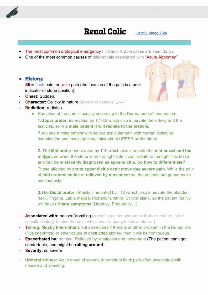
● History:History: - Site: flank pain, or groin pain (the location of the pain is a poor
indicator of stone position). - Onset: Sudden. - Character: Colicky in nature (goes and comes) “مغص” - Radiation: radiates.
● Radiation of the pain is usually according to the Dermatome of innervation: 1.Upper ureter: innervated by T7,8,9 which also innervate the kidney and the testicles, so in a male patient it will radiate to the testicle. if you see a male patient with severe testicular pain with normal testicular examination and investigations, think about UPPER ureter stone.
2. The Mid ureter: innervated by T10 which also innervate the mid bowel and the midgut, so when the stone is on the right side it can radiate to the right iliac fossa and can be mistakenly diagnosed as appendicitis. So how to differentiate? Those affected by acute appendicitis can’t move due severe pain. While the pain of mid ureteral colic are relieved by movement so, the patients are gonna move continuously.
3.The Distal ureter : Mainly innervated by T12 (which also innervate the bladder neck, Trigone, Labia majora, Posterior urethra, Scrotal skin) , so the patient mainly will have urinary symptoms (Urgency, Frequency…).
- Associated with: nausea/Vomiting (as well as other symptoms that are related to the specific etiology behind the pain, which we are going to know later on).
- Timing: Mostly Intermittent, but sometimes if there is another problem in the kidney like (Pyelonephritis) or other cause of obstructed kidney, then it will be continuous.
- Exacerbated by: nothing; Relieved by: analgesia and movement (The patient can’t get comfortable, and might be rolling around.
- Severity: so severe.
- Ureteral stones: Acute onset of severe, intermittent flank pain often associated with nausea and vomiting.
● Work Up:Work Up: - History - Examination: patient tends to move around, in an attempt to find a comfortable position. - +/- Fever ( when the renal colic is associated with pyelonephritis).
● Investigation:Investigation: - Pregnancy test. The benefit is to exclude ectopic pregnancy, and also we need it for
radiological consideration. - MSU (Mid stream urine) (you will find RBCs in the urine). 2
- U&E (Urea and Electrolyte) (if you the patient has renal impairment it indicates : either bilateral pathology or a solitary kidney blocked by a stone).
2 A mid-stream urine sample means you don't collect the first or last part of urine that comes out. This reduces the risk of the sample being contaminated with bacteria from: your hands, the skin around the urethra, or the tube that carries urine out of the body.
- This is an abdominal X Ray. - We do it to determine the
location of Radiopaque stones.
-The Hyperechogenic shadow indicate the stones. -we can see the the stone but not that clear (you can’t see the ureter with it).
- Note that: the patient come to you having renal problem and this test needs contrast to be done which contains iodine that may cause rigidity and renal failure.
Helical CTU (Helical CT without contrast) MRI
● The gold standard (it is a plain CT). ● Suitable for pregnant ladies (No radiation).
● greater specificity (95%) and sensitivity (97%). ● Can identify other non-stone causes of flank
pain. ● No need for contrast administration. ● Faster, taking just a few minutes. ● The cost of CTU is almost equivalent to that of
IVU. ● Note that: there is no need to contrast because
the patient come to you complain of most likely renal problem and the only thing that we can see in CT without contrast is a stone.
● very accurate way of determining whether or not a stone is present in the ureter.
● Time consuming. ● Expensive.
● Management:Management: ● Medical Management: Medical Management: - Pain relief:
- NSAIDs. E.g. Brufen, voltaren - Intramuscular or intravenous injection, by mouth, or per rectum. - +/- Opiate analgesics (pethidine or morphine).
- Hyper hydration (IV fluids and drinking water). - 5mm Stone or less: ‘watchful waiting’ with analgesic supplements; 95% of 5mm or less
stones pass spontaneously.
● Surgical Management Surgical Management ○ Indications for Intervention: (و مثل أي حالة ما نلجأ للجراحة إال في حاالت معینة و هي كاآلتي)
1. To Relieve Obstruction and/or Remove the stone. 2. Pain that fails to respond to analgesics. 3. Association with fever. (fever highly suggests pyelonephritis which requires
drainage because it may cause septicemia). 4. Renal function impairment caused by stone, which may cause uremia. And
how do we assess renal functioning? = increase creatinine. 5. Obstruction unrelieved (not to exceed 4 weeks! (because after 4 weeks the
obstruction will cause necrosis) 4ممكن ننتظر الحصوة تنزل بنفسها لكن لو استمر الموضوع ل .أسابیع البد من تدخل جراحي
6. Personal or occupational reasons: doctors or pilots.
● Types of Surgical intervention:
- Temporary relief of the obstruction.
Insertion of a double coil
or JJ stent (stent means tubular
support=دعامة)
A thin tube is inserted into the ureter to prevent or treat obstruction of the urine flow from the kidney also known as urinary stent.
percutaneous nephrostomy tube
We Insert a catheter through your skin into the kidney to drain the urine.
Painful inability to void, with relief of pain following drainage of the bladder by catheterization. More common in Men than in Women.
● Causes:Causes: Men Women (rare)
➔ Benign prostatic enlargement (BPE) due to BPH, The most common.
➔ Carcinoma of the prostate ➔ Prostatic abscess ➔ Urethral stricture ➔ Stones ➔ Constipation
➔ Pelvic prolapse (cystocele, rectocele, uterine) ➔ pelvic masses (e.g., ovarian masses) ➔ Urethral stenosis ➔ Urethral diverticulum or stricture ➔ Post surgery for ‘stress’ incontinence ➔ Transvaginal tape (sling) in those with stress
incontinence(لو كان مشدود أكثر من الالزم) is very common.
Initial ManagementInitial Management: 1. Give the patient analgesic to prevent spasm. 2. Urethral catheterization if you can’t enter it (stricture in urethra), use the Suprapubic catheter
(SPC) which passes directly to the bladder through skin.
Late Management Late Management definitive treatmendefinitive treatmentt:: ● Treating the underlying cause.
● Chronic Urinary Retention:Chronic Urinary Retention: - Obstruction develops slowly, the bladder is distended (stretched) very gradually over weeks/months, Pain is not a feature. (mostly related to diabetes). Note: many patient with chronic retention present to ER with renal failure.
● Presentation:Presentation: - Urinary dribbling. - Overflow incontinence. - Palpable Bladder with no pain. - Symptoms of renal failure. (nausea and malaise) - Pyelonephritis and even renal failure
● Management:Management: - Treatment is directed to renal support. - Bladder drainage. - Late Treatment of underlying cause.
- Hydrocele (Examination will reveal positive transillumination test and big scrotum): 4
This is a common condition, especially in older men, in which fluid collects in the tunica vaginalis,
resulting in an enlarged but painless scrotum.The cause of most hydrocoeles is unknown
(idiopathic). Hydrocoele is a common abnormality in children. It is due to failure of closure of the
processus vaginalis after descent of the testis.
- Idiopathic scrotal edema
- Tumor
- Spermatocele
- Non-urogenital pathology e.g. adductor tendinitis
3 Also known as scrotal pain or testicular pain 4 A test that is used to illuminate a body cavity by transmission of light through the cavity.
● Torsion of the Spermatic cord:Torsion of the Spermatic cord: Most serious
● General consideration:General consideration: - Common among teenagers (12-18) years (always think of torsion until proven otherwise). - Possible in children and neonates. - Unlikely after the age of 25 years. - True surgical emergency of the highest order. - Irreversible ischemic injury to the testicular parenchyma may begin as soon as 4 hours.
As the duration of torsion increases the possibility of testicular salvage Decreases. - The twisting will lead to occlusion of venous return→ swelling and blockage of arterial
supply → the testis will be dead.
● Anatomical variations:Anatomical variations: A. Normal. B. Bell clapper deformity: tunica vaginalis surrounds the
whole testicle so it is very loose (larger than normal). C. Loose epididymal attachment to the testis. D. Torsed testis with horizontal lie.
- Types of torsion: - Extra-vaginal (tunica vaginalis is involved) - Intra-vaginal
● PresentationPresentation::
- Acute onset of scrotal pain. (severe and intermittent due to attacks of torsion and detorsion).
- Majority with history of prior episodes of severe, self-limited scrotal pain and swelling - Nausea/Vomiting due to pain (important sign to focus on). - Referred to the ipsilateral lower quadrant of the abdomen. - Children may present with abdominal pain and might not complain of testicular pain.
Any child comes to you complain of with nausea and vomiting or complains of severe abdominal pain needs to have genital examination. Simple scenario: a mother brought her child to the clinic and said she “my child went to school and ate bad food and now he complains of abdominal pain, nausea and vomiting” after making further inspection, the child had a Torsion of the cord.
- Dysuria and other bladder symptoms are usually absent. (unlike Epididymitis) - Spontaneous
● Physical examinationPhysical examination: : importantimportant - The affected testis is high riding transverse orientation. - Acute swelling and scrotal edema or secondary hydrocele. - Absent Cremasteric reflex. - Testis is tender and larger than other side - Elevation of the scrotum causes more pain (negative Prehn's sign).
● investigations:investigations: - this is an emergency case (requires immediate scrotal exploration), with a high degree
of suspicion is enough to send the patient to the OR immediately “ما نجلس نأخر المریض عشان فحوصات” ● Adjunctive tests: نستخدمها عادًة لما نكون متأكدین إنه شيء غیر التورشن - To aid in differential diagnosis of the acute scrotum. - To confirm the absence of torsion of the cord. - Tests used:
- Sound Doppler examination of the cord and testis: High false-positive and false-negatives. - Color Doppler ultrasound:
- Investigation of choice - Done in OR - Assessment of anatomy and
determining the presence or absence of blood flow. 5
- Sensitivity: 88.9% specificity of 98.8%
- Operator dependent.
● Radionuclide imaging: - Assessment of testicular blood flow. - Shows a photopenic area in cases of torsion. - A sensitivity of 90%, and specificity of 89%. - False impression from hyperemia of scrotal wall. - Not helpful to determine a Hydrocele or Hematoma (does not assess anatomy) - Difficult to do in emergency.
● Surgical exploration: - Diagnostic and therapeutic. - A scrotal incision is done and the affected site should be examined first: - The cord should be detorsed. - Testes with marginal viability should be placed in warm and re-examined after several minutes. - A necrotic testis should be removed. - If the testis is to be preserved, it should be fixed. - The contralateral testis must be fixed to prevent subsequent torsion.
5 In the pictures: in the left there is absence of blood supply, secondary hydrocele without arterial flow.
Surgical Recall (EXTRA):
What is it Testicular torsion? Torsion (twist) o the spermatic cord, resulting in venous outflow obstruction, and subsequent arterial occlusion >> infarction of the testicle. What is the classic history? Acute onset of scrotal pain usually after vigorous activity or minor trauma What is a “bell clapper” deformity? Bilateral non attachment of the testicles by the gubernaculum to the scrotum (free like the clappers of a bell) What are the symptoms? Pain in the scrotum, suprapubic pain What are the signs? Very tender, swollen, elevated testicle; non illumination; absence of cremasteric reflex. What is the differential diagnosis? Testicular trauma, inguinal hernia, epididymitis, appendage torsion How is the diagnosis made? Surgical exploration, U/S (solid mass) and Doppler ow study, cold Tc-99m scan (nuclear study) What is the treatment? Surgical detorsion and bilateral orchiopexy to the scrotum.
● Epididymo-orchitis:Epididymo-orchitis:
● Presentation:Presentation: - Common in KSA (can be manifestation of brucella) - Indolent process (gradual presentation). - Scrotal swelling, erythema, and pain - Usually gradual and not sudden. - Dysuria and fever is more common in patients with history of STD like gonorrhea or UTI. - Can be related to BRUCELLA.
● Physical examination (P/E) :Physical examination (P/E) : - Localized swollen and tender epididymis, or a massively swollen hemiscrotum with absence of
landmarks. - Cremasteric reflex should be present ( if it’s present → Epididymo-orchitis, and if not → Torsion of
the Spermatic cord). - Positive Prehn's sign indicates there is pain relief with lifting the affected testicle
● UrinalysisUrinalysis: pyuria, bacteriuria and/or a positive urine culture and WBC.
● Management:Management: - Bed rest for 1 to 3 days then relative restriction. - Scrotal elevation, the use of an athletic supporter > - Parenteral or oral antibiotic therapy should be instituted when UTI is
documented or suspected. - Urethral instrumentation should be avoided (because it may lead to
septicemia).
PriapismPriapism
- Persistent erection of the penis for more than 4 hours that is NOT related or accompanied by sexual desire. associated with intracavernosal self-injection for impotence (the most common cause).
● CausesCauses:: - Primary (Idiopathic) : 30% - 50% of the cases. - Secondary: Drugs, Trauma, Neurological, Hematological disease (very common), And tumors:
pelvic malignancies.
● TypesTypes::
Ischemic (most common) Nonischemic
(veno-occlusive, low flow) (worse) (arterial, high flow)
Due to hematological disease e.g.Sickle cell, Leukemia, malignant infiltration of the corpora cavernosa with malignant disease, pelvic Tumor, or drugs such as (prostaglandin injection; prazosin).
Due to perineal trauma, which creates an arteriovenous fistula. (pudendal artery fistula)
Painful Painless
● The diagnosisThe diagnosis: (Usually obvious from the history) - Duration of erection: >4 hours? - Is it painful or not?. - Previous history and treatment of priapism ? - Identify any predisposing factors and underlying cause.
● Examination :Examination : - Erect, tender penis (in low- flow) or not. - Characteristically the corpora cavernosa are rigid and the glans is flaccid. - Abdominal examination for evidence of malignant disease - Digital rectal exam (DRE): to examine the prostate and check anal tone.(neurological assessment).
● Investigation:Investigation: - CBC (white cell count and differential, reticulocyte count). - Hemoglobin electrophoresis for sickle cell disease. - Urinalysis including urine toxicology.
- Blood gases taken from either corpora: this table is very important.
Low flow (ischemic/occlusive) High flow (non ischemic/ fistula)
Blood color dark blood bright red blood similar to arterial blood at room temperature
- Color flow duplex ultrasonography in cavernosal arteries:
○ Ischemic (inflow is low or nonexistent). ○ Non-ischemic (inflow is normal to high).
- Penile pudendal arteriography (VERY IMPORTANT for patients with nonischemic type and sometimes you can manage them with it).
● Treatment:Treatment: - Depends on the type of priapism. - Conservative treatment should first be tried; ask the patient to climb the stairs to open venous
channels. - Medical treatment. - Surgical treatment. - Treatment of underlying cause.
Recall (EXTRA): What is priapism? Persistent penile erection. What are its causes? Low flow: leukemia, drugs (e.g.,prazosin), sickle-cell disease, erectile dysfunction treatment gone wrong High flow: pudendal artery fistula, usually from trauma What is the first-line treatment?
1. Aspiration of blood from corporus cavernosum 2. α - Adrenergic agent
Renal TraumaRenal Trauma 66
● The kidneys relatively protected (by the thoracic cage) from
traumatic injuries so a considerable degree of force is usually required to injure a kidney.
● Mechanism and causes:Mechanism and causes: - Blunt trauma:
- Direct blow “ضربة مباشرة” or acceleration/ deceleration injuries. - Road traffic accidents, falls from a height, fall on flank. Very Common
● Renal imaging:Renal imaging: - Indications for renal imaging:
- Penetrating chest, flank, and abdominal wounds. - A history of a rapid acceleration or deceleration. fall from a height or road traffic accidents. - Macroscopic (Gross) haematuria. - Microscopic [>5 red blood cells (RBCs) per high powered field] or dipstick. - Hematuria in hypotensive patient (SBP <90 mmHg ). (you give the patient a lot of fluids and
he didn’t respond). - Any child with microscopic or dipstick haematuria who has sustained trauma, even <5 RBCs.
● What imaging study?What imaging study?
● Available Modalities:Available Modalities:
IVU - Replaced by the contrast enhanced CT.ما صاروا یستخدموها كثیر هاألیام فاستبدلوها - On-table IVU (intraoperative) if patient is transferred immediately to the operating theater
without having a CT scan and retroperitoneal hematoma is found. - Done to see if other (non-injured) kidney is functioning and/or exists because the
injured kidney might have to be removed.
CT scan - Spiral non contrast: does not allow accurate staging. Good only with stones. - With contrast (CT urography OR CT of abdomen OR CT angio):
- imaging study of choice: accurate, rapid, can assess other abdominal injuries and structures.
6 Usually renal injury leads to retroperitoneal hematoma.Retroperitoneal hematoma: refers to an accumulation of blood found in the retroperitoneal space. 7 During operations
Renal US - Advantages: (mainly to follow up after CT) – can certainly establish the presence of two kidneys. – the presence of a retroperitoneal hematoma. – power Doppler can identify the presence of blood flow in the renal vessels.
- Disadvantages: – cannot accurately identify parenchymal tears, collecting system injuries, or extravasations of urine until a later stage when a urine collection has had time to accumulate.
● Grades and StagesGrades and Stages :: done by CT with contrast 88
I Flank pain + Hematuria with or without pericapsular Hematoma, but no evident kidney damage, So Only perinephric (subcapsular) hematoma without kidney tearing.
II Injury to the cortex (tearing) with hematoma only of 1 cm or less.
III Injury to the cortex and medulla without reaching the collecting system with hematoma more than 1 cm.
IV Injury reaching the collecting system OR thrombosis to the renal vessels. - On IVU there will be extravasations of contrast (outside the kidney) and decreased filling.
V Bleedly shattered kidney completely (Completely injured) or avulsion (tearing) of renal pedicle (Hilum).
● Management:Management: ● Those who are Conservatively treated:
- Over 95% of blunt injuries. - 50% of renal stab injuries and 25% of renal gunshot wounds With the help of CT scan
(needs a specialized center). ● Conservative treatment Include:
1. Wide Bore IV line to transfuse fluids.(big cannula) 2. IV antibiotics. Because hematoma is most likely to cause an infection. 3. Bed rest. Advise the patient to rest on the bed especially during first 3 days because if he
moves the clot will dislodge and he will bleed. 4. Vital signs monitoring. 5. Serial CBC and HCT 9
6. Follow up US &/or CT.
8 Most of cases of massive renal injury lead to removal of other kidney. 9 Hematocrit
● Surgical exploration. - Persistent bleeding (persistent tachycardia and/or hypotension failing to respond to
appropriate fluid and blood replacement). - Retroperitoneal hematoma: still held by peritoneum so, it’s better not to interfere, we mark
the hematoma (if it is not expanding quickly then don’t open the peritoneum). - Expanding perirenal hematoma (again the patient will show signs of continued bleeding)
(needs Surgical intervention). - Pulsatile perirenal hematoma (indicate large blood vessels injury and needs Surgical
intervention).
Ureteral InjuriesUreteral Injuries
The ureters are protected from external trauma by surrounding bony structures , muscles and other organs therefore injury is rare.
● Mechanism and causes:Mechanism and causes: External trauma (rare) Internal trauma (iatrogenic)
- Severe force is required.
- Blunt or penetrating:
1. Blunt external trauma severe enough to injure the ureters will usually be associated with multiple other injuries. 2. Penetrating knife or bullets to the abdomen or chest may damage the ureter, as well as other organs.
- Uncommon, but is more common than external trauma.
- Iatrogenic::
1. Hysterectomy (because the ureter is really close to the uterine artery), oophorectomy, and sigmoid colectomy. 2. Caesarean section. 3. Ureteroscopy. 4. Aortoiliac vascular graft replacement. 5. Laparoscopies. 6. Orthopedic operations.
● Diagnosis:Diagnosis: - Requires a high index of suspicion, usually diagnosed Intra-operatively.
- Late diagnosis:
1. An ileus: the presence of urine within the peritoneal cavity. 2. Prolonged postoperative fever or overt urinary sepsis. 3. Persistent drainage of fluid (urine w/ high Creatinine) from abdominal or pelvic drains, from
the abdominal wound, or from the vagina. 4. Flank pain if the ureter has been ligated. 5. An abdominal mass, representing a urinoma (urinoma:A urinoma is a mass formed by
encapsulated extravasated urine. It may follow closed renal injury, surgical operation or arise spontaneously in the presence of obstruction).
6. Vague abdominal pain (make us suspicious).
● Treatment options:Treatment options: - JJ stenting (if the injury is partial). - Primary closure of partial transection of the ureter. - Direct ureter to ureter anastomosis. - Re-implantation of the ureter into the bladder using a psoas hitch or a Boari flap (most 10 11
used). - Trans uretero-ureterostomy (we transfer one ureter to the other). - Auto-transplantation of the kidney into the pelvis. - Replacement of the ureter with ileum. - Permanent cutaneous ureterostomy. - Nephrectomy.
Bladder InjuriesBladder Injuries 1212
● Causes:Causes: ○ Iatrogenic injury: of or relating to illness caused by medical examination or treatment like
the following: 1. Transurethral resection of bladder tumor (TURBT): is a procedure in which bladder tumors
can be removed from the bladder wall. 2. Cystoscopic bladder biopsy: A bladder biopsy can be done as part of a cystoscopy. 3. Transurethral resection of prostate (TURP): is a surgery used to treat urinary problems due
to an enlarged prostate. A combined visual and surgical instrument (resectoscope) is inserted through the tip of the penis and into urethra.
4. Cystolitholapaxy: is a procedure to break up bladder stones into smaller pieces and remove them.
5. Caesarean section or Hysterectomy especially as an emergency (most common). 6. Total hip replacement (very rare).
10 The psoas hitch is a useful technique for bridging a defect involving the lower third of the ureter, With the psoas hitch, the bladder is pulled up and secured to the psoas muscle, to reduce the distance between the distal ureter and the bladder. 11 A Boari bladder flap is one of the options for ureteral reimplantation when the diseased ureteric segment is long (e.g. more than 5 cm). It involves tubularization of a flap of bladder to extend from the bladder to the ureteral orifice. 12 the good thing about bladder most of the time it’s a forgivable organ you injure it and suture it and leave a catheter until it heals
● Cont. Causes:Cont. Causes: ● Penetrating trauma to the lower abdomen or back. ● Blunt pelvic trauma in association with pelvic fracture or ‘minor’ trauma in a drunkard patient. ● Rapid deceleration injury seat belt injury with full bladder in the absence of a pelvic fracture
(which causes an injury to the breast and the bladder). ● Spontaneous rupture after bladder augmentation we can augment the bladder with the 13
intestine patch (نرقعها).
● Types of perforation:Types of perforation: (mostly extra-peritoneal)
● Male External genitalia injuries:Male External genitalia injuries: - Penile Fracture, eggplant deformity sign (the injury is mainly in the Corpora spongiosum) picture. - Glans Injury (especially with circumcision). - Penile amputation and injuries (Call the security and the psychiatrist) - Scrotal Injuries.
● Female External genitalia injuries:Female External genitalia injuries: - In sports, crime or during vaginal labour. - Managed by Gynecologists unless the urethra or the bladder is
involved. - In Road traffic accidents it’s extremely rare in female while its very
Non traumatic urological emergenciesNon traumatic urological emergencies 1-Hematuria1-Hematuria : is the most important symptom that needs immediate medical help. Cause vary according to : 1-Age. 2-painful or painless. 3-risk factors for malignancy like smoking 4-types gross of microscopic. Work up : 1-History more important than PE 2 -Physical examination . Investigation: CT urography which is the gold standard. 2-renal colic 2-renal colic : the most urological emergency. Investigation: 1-pregnancy test. 2-MSU. 3-U&E. Radiological investigations: 1-KUB 2-RUS 3-IVU 4-MRI 5-helical CT without contrast which is the gold standard. 3-Acute Urinary Retention3-Acute Urinary Retention Painful inability to void یجي المریض یصیح بالطوارئ. Initial management: 1-give the patient analgesic. 2-urethral catheterization. Late management : treating the underlying cause. Chronic Urinary RetentionChronic Urinary Retention :pain is not a feature and many patient come to ER with renal failure. Management: treatment : 1- renal support 2-bladder drainage. 3-late treatment of the underlying cause. 4-Acute Scrotum 4-Acute Scrotum : requires prompt evaluation, differential diagnosis and potentially immediate surgical exploration. Differential diagnosis: 1-Torsion of the spermatic cord the most serious 2-epididymitis the most common. 3-epididymo-orchitis. 4-hydrocele. Torsion of the spermatic cord : common among teenagers 12-18 years Presentation: 1-Acute onset of scrotal pain. 2-nausea and vomiting. 3-children may present with abdomen pain not testicular pain . PE of Torsion of the spermatic cord: 1-Acute swelling and scrotal edema. 2-absent of Cremasteric reflex. 3-testis is the larger than other side. Epididymo-orchitis- Cremasteric reflex should be present. 5-Priapism:5-Priapism: persistent erection of the penis more than 4 hours not related to sexual desire . Cause: primary: idiopathic 30-50% of the cases. Secondary: drugs , trauma, neurological and hematological . Types : 1-ischemic, painful is the most common. 2-non ischemic painless. The diagnosis usually obvious from the history, duration of erection more than 4 hours.
Traumatic urological emergenciesTraumatic urological emergencies 1- Renal Trauma1- Renal Trauma : Mechanism and causes: 1-blunt trauma, direct blow and road traffic accidents . 2 penetrating trauma, knives , gunshot and iatrogenic . Radiological image : 1-IVU only indication is intraoperative without having CT scan in case of retroperitoneal hematoma . 2-CT scan imaging study of choice, accurate, rapid. 3-Renal US Management: 1-Conservative, includes: wide Bore IV line, IV antibiotics, Bed rest , vital signs, serial CBC and HCT and follow up US and CT . 2 Surgical exploration. 2-Urethral injuries2-Urethral injuries : Mechanism and causes : 1-external trauma: severe force is required, blunted or penetrating. 2-internal trauma : more common than external, iatrogenic causes: hysterectomy, Caesarean section. 3-Bladder injuries3-Bladder injuries : Causes :1 -iatrogenic injury: TURBT , cyctoscopic bladder biopsy, TURP, cystolitholapaxy, Caesarean section (most common), total hip replacement. 2-penetrating trauma 3-blunt pelvic trauma 4-Rapid deceleration injury. Types of perforation : 1-intraperitoneal perforation. 2-extra-peritoneal perforation. Presentation: classic triad of symptoms 1-suprapubic pain and tenderness 2-difficulty or inability to pass urine. 3-haematuria. Extraperitoneal management : 1-bladder drainage by Foley catheter 2-Open repair (Surgery). 4- Anterior Urethral injury:4- Anterior Urethral injury: Mechanism: 1-Majority are a result of straddle injury in boys or men. 2-penile fractures. 3-inflating a catheter balloon on the anterior Urethral. Sing and symptom: meatal bleeding, difficulty in passing urine, frank haematuria. Diagnosis: diagnostic tool is retrograde urethrography. Management: 1-contusion: small gauge Urethral catheter for one week 2 - partial rupture of anterior urethra: majority managed by suprapubic urinary diversion 3-complete rupture of anterior urethra: A unstable patient : use a suprapubic catheter B stable patient urethra may be immediate repaired or suprapubic catheter 4-penetrating anterior urethral injuries are managed by surgical debridement and repair 5-Posterior urethral injuries:5-Posterior urethral injuries: Mechanism: mostly are associated with pelvic fracture, 10-20% associated with bladder rupture. Sing: high riding prostate when Examining by digital rectal exam . Management: type 1: stretch injury with intact urethra and stenting with a Urethral catheter. Type 2 treatment with stenting a Urethral catheter. Type 3 : initial management with suprapubic cystostomy and attempting primary repair at 7 to 10 days after injury.
QuestionsQuestions 1- A 13-year old boy presented to the Emergency Room with painful right scrotal swelling. It was gradual in onset over the last 5 days. He gave history of dysuria and suprapubic pain for the last 2 weeks. The most common cause of his symptoms is:
a. Epididymitis b. Hydrocele c. Testicular Torsion d. Testicular Trauma
2- 15-year-old boy presented to emergency department with 4 hours history of sudden onset scrotal pain which excruciating. On local examination he has significant tenderness of the left hemiscrotum with high lying left testis. What will be the most appropriate next step?
a. Scrotal US with color Doppler study b. Radionuclide for the scrotum c. Urine analysis d. Immediate scrotal exploration
3- 12 years old boy presented to the emergency room with severe sudden testicular pain for 3 hours, with no history of trauma, what is the most likely the diagnosis?
a. Hydrocele b. Testicular Torsion c. Tuberculosis epididymitis d. Varicocele
4- which one of these is a common cause of ischemic priapism?
a. sickle cell disease b. idiopathic c. Trauma d. SLE
5- If the diagnosis is testicular torsion how would you further proceed with your work up?
a. Take the patient to CT scan b. Give the patient analgesia and ask him to return to you in 3 days c. Take the patient to OR immediately for surgical exploration d. Administer antibiotics as testicular torsion is an infectious emergency
6- Which of the following is an indication for a surgical Intervention in ureteric stones?
a. Gross hematuria b. If the stones is 6 millimeter in diameter c. Impaired renal function test due to obstruction d. Stone in distal ureter