Stress-only myocardial perfusion imaging .. . it IS time for a change!
Sanjeev P. Bhavnani, MD, and Gary V. Heller, MD, PhD
See re lated article, doi: lO.l007/ sl2350· 0 ll -940 5-9.
Stress-only SPECT imaging has emerged as an important and viable alternative to conventional reststress SPECT protocols in selected patients. Advantages of stress-only imaging include improvements in patient convenience, reducing radiation exposure, increasing laboratory efficiency and cost containment. These important advances provided by stress-only acquisition protocols represent a more efficient use of SPECT technology and begs the question is it time for a change? 1
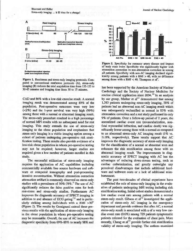
Several SPECT imaging protocols are recommended by the· American Society of Nuclear Cardiology which include dual and single isotope imaging protocols.2 Technetium (Tc-99m) radiopharmaceutical based protocols can be performed as one-day protocols as an initial stress-rest or rest-stress acquisition and are widely adopted in clinical practice. The radiation exposure from these protocols is significant at 11-18 mSv for Tc-99m tracers and 27-30 mSv for dual isotope rest thallium-201/stress Tc-99m procedures.1 Stress-only acquisition protocols utilize a single dose of Tc-99m based radiotracer injected at peak stress (3-7 mSv) with avoidance of the rest · injection if the stress acquisition is normal. Stress-only imaging reduces SPECT acquisition time by more than 50% and markedly reduces total patient time in the laboratory (Figure 1 ).
The concept of stress-only imaging is not new as the literature extends back to the mid 1990s and early 2000s. Previous studies suggest that a normal stress result does not benefit from a rest study. However, in the era prior to attenuation correction (AC), the need for rest imaging was 50%-89% and unacceptably high. 3 Attenuation
From the Hartford Hospital, University of Connecticut School of Medicine, Hartford, CT.
Reprint requests: Gary V. Heller, MD, PhD, Hartford Hospital, University of Connecticut School of Medicine, 80 Seymour Street, Hartford, CT 06102-5037; [email protected].
correction procedures have been shown to substantially reduce auenuation artifacr;and-asa tesun,-sigiiificaiffiy - ·improve diagnostic specificity.3 One of the first appli-cations of attenuation correction to stress-only imaging was reported by Heller et al. 4 In this study, 10 inde-pendent readers interpreted 90 stress-only studies in a blinded and sequential fashion: MPI first, MPI plus gated SPECT data and finally AC-MPI plus gated SPECT data. Interpreters were asked to provide diag-nostic confidence (definitely normal, probably normal, equivocal, probably abnormal or definitely abnormal) as well as the perceived need for a rest study. Important findings were the following: attenuation corrected studies resulted in fewer non-definitive interpretations and significantly reduced the recommended need for a rest study. The authors also found that the need for a rest study was primarily in those patients with CAD and especially among those with a history of a prior myo-cardial infarction. Thus this and other studies suggest that the ideal candidates for stress-only imaging are those patients with no known CAD and if CAD is present, no prior history of a myocardial infarction or coronary revascularization procedure.
In this issue of the Journal, Gemignani et al5 report on another application of stress-only SPECT imaging in the obese population. This important study provides evidence that the prevalence of a normal SPECT result is high among obese individuals undergoing pre-operative risk assessment prior to bariatric surgery. This conclusion is drawn from a cohort evaluation of 383 obese patients referred for preoperative risk stratification. The majority, 81% underwent an exercise MPI and 67% completed a stress-only protocol. Stress-only imaging was obtained with a single injection of 25-40 mCi of Tc-99m sestamibi or tetrofosmin with rest imaging only upon abnormal or equivocal results. Gated SPECT images were obtained with line source attenuation correction (A C), and applied in all studies, a method that has been shown to improve the diagnostic accuracy and specificity of imaging results.6
Overall, the referred population had an average age of 42 ± 10 years, BMI of 49 ± 8 and 83% were women. A clustering of coronary risk factors was observed among 23%-44%. It was anticipated that the cohort would demonstr.at.e a high pr.evalence of CAD, when in fact the opposite was true, with only 1% with known
Bhavnani and Heller Stress-only imaging ... it IS time for a change!
Figure 1. Rest/stress and stress-only imaging protocols. Compared to conventional rest/stress protocols (A), stress-only imaging (B) reduces the total acquisition time from 135-155 to 30-45 minutes and imaging time from 30 to 15 minutes.
CAD and 86% with a low-risk exercise result. A normal imaging result was demonstrated among 89% of the population. Post-operative outcomes were very low (<2%) and the 1-year survival was very high (99%) among those with a normal or abnormal imaging result. The stress-only procedure resulted in a high percentage of normal MPI results with no subsequent need for rest imaging. This study extends the use of stress-only imaging to the obese population and emphasizes that stress-only imaging is a viable imaging option among a cohort of patients undergoing pre-operative risk stratification testing. These results also provide evidence of a low-risk obese population in whom pre-operative testing may not be required; however, larger studies are required given a low number of patients enrolled in this study.
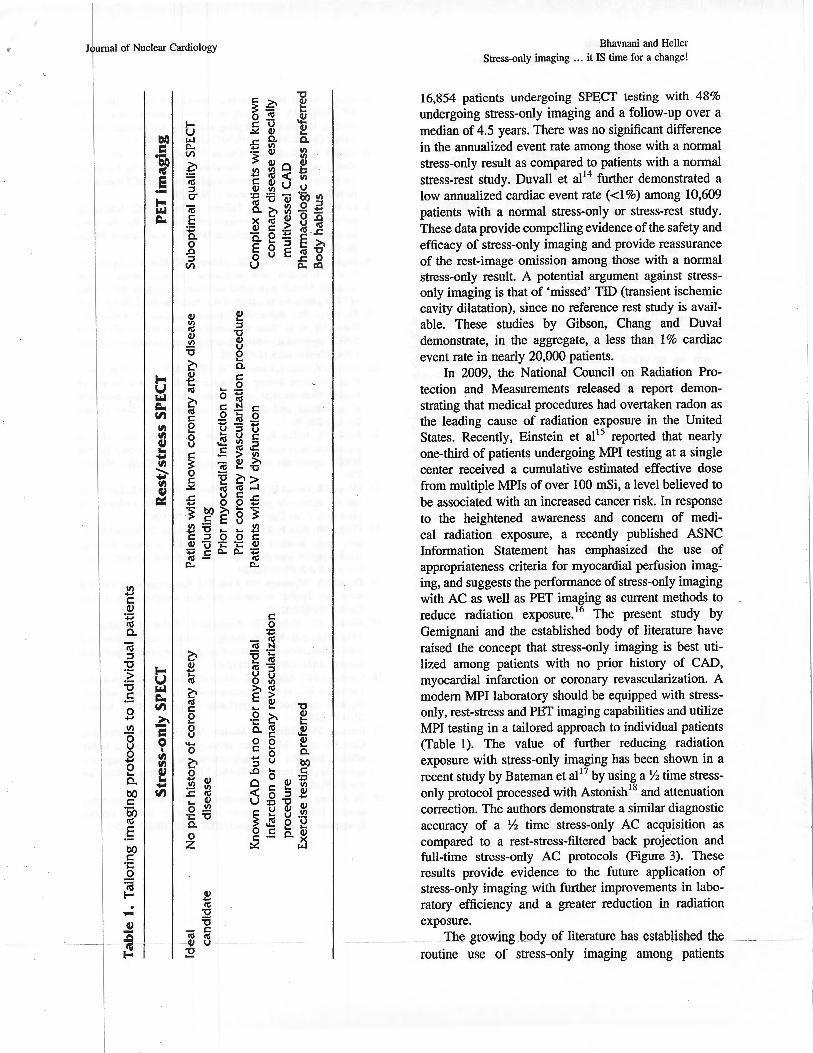
The successful utilization of stress-only imaging requires the application of AC capabilities including commercially available line source hardware and software or computed tomography and post-processing iterative reconstruction. Without attenuation correction attenuation artifact is commonly observed on stress-only studies with a prevalence of 50%-78%.3 The use of AC significantly reduces the false positive rates for both rest-stress and stress-only studies. Furthermore AC improves the diagnostic accuracy of SPECT imaging in addition to and absence of ECG gating7
•8 and is partic
ularly striking among individuals with a BMI >309
(Figure 2). The results by Gemignani extend these previous results with the application of stress-only imaging in the obese population in whom pre-operative testing may be reasonable. Overall, the use of AC increases the diagnostic specificity from 69%-88% to nearly 98% and.
Journal of Nuclear Cardiology
0 Nori-AC • AC 100 90 80 70 60
"'o 50 40 30 20 10
0 All Pts BMI < 30 BM 30-40 BMI > 40
Figure 2. Specificity for coronary artery disease and impact of body mass index Specificity was significantly higher with attenuation correction vs non-attenuation correction in among all patients. Specificity with non-AC imaging declined significantly among patients with a BMI > 40, with no difference among those with a BMI < 40. Thompson et al9•
has been supported by the American Society of Nuclear Cardiology and the Society of Nuclear Medicine for routine clinical application since 2004. 10 In an analysis by our group, Mathur et al 11 demonstrated that among 1,383 patients undergoing stress-only imaging, 58% of patients had an abnormal non-AC imaging result which was subsequently reclassified as normal in 83% with attenuation correction and a rest study performed in only 9% of patients. Over a follow-up period of 2 years, the annualized cardiac event rate (revascularization, nonfatal myocardial infraction, and cardiac death) was significantly lower among those with a normal as compared to an abnormal stress-only AC imaging result (1% vs. 11.8%, respectively). These results support that AC improves the diagnostic accuracy of stress-only imaging for the classification of a normal or abnormal scan and enhances the risk stratification among those with an abnormal imaging result. The improvements in diagnostic accuracy of SPECT imaging with AC has the advantages of reducing down-stream testing, such as cardiac catheterization, and greatly outweighs any perceived disadvantages that include additional hardware and software costs or a lack of additional reimbursement.
The past two-decades of clinical experience have established the role of stress-only imaging in the evaluation of patients undergoing MPI testing including risk stratification testing. Initial cohort studies demonstrated a low cardiac event rate among patients with a normal stress-only result. Gibson et al12 investigated the application of stress-only AC imaging in the emergency department and provide evidence that after AC, a normal stress-only result is associated with a of a very low cardiac event rate (0.6%) among 729 patients symptomatic patients referred for the evaluation of chest pain. More recently, Chang etal 13 provide further risk stratification validity of stress-only imaging. The authors examined
•,
Journal of Nuclear Cardiology
.:1 c (1)
~ 0.
<i1 :::l -c :~ -c .5 .8 II)
8 .8 0 ... a. bl) c
~ .5 bO c
·;::: 0
~ . -" ---~ -....
~ ·~ E ·-
~ ~ <U
~ <U c: e 8
...... 0
~ 0 ~ ~ ..c: <U .... 4.1 0 .!!!
·;:: "0 0. 0 z
Bhavnani and Heller Stress-only imaging ... it IS time for a change!
16,854 patients undergoing SPECT testing with 48% undergoing stress-only imaging and a follow-up over a median of 4.5 years. There was no significant difference in the annualized event rate among those with a normal stress-only result as compared to patients with a normal stress-rest study. Duvall et al14 further demonstrated a low annualized cardiac event rate ( <1%) among 10,609 patients with a normal stress-only or stress-rest study. These data provide compelling evidence of the safety and efficacy of stress-only imaging and provide reassurance of the rest-image omission among those with a normal stress-only result. A potential argument against stressonly imaging is that of 'missed' TID (transient ischemic cavity dilatation), since no reference rest study is available. These studies by Gibson, Chang and Duval demonstrate, in the aggregate, a less than I% cardiac event rate in nearly 20,000 patients.
In 2009, the National Council on Radiation Protection and Measurements released a report demonstrating that medical procedures had overtaken radon as the leading cause of radiation exposure in the United States. Recently, Einstein et al15 reported that nearly one-third of patients undergoing MPI testing at a single center received a cumulative estimated effective dose from multiple MPis of over 100 mSi, a level believed to be associated with an increased cancer risk. In response to the heightened awareness and concern of medical radiation exposure, a recently published ASNC Information Statement has emphasized the use of appropriateness criteria for myocardial perfusion imaging, and suggests the performance of stress-only imaging with AC as well as PET imaging as current methods to reduce radiation exposure. 16 The present study by Gemignani and the established body of literature have raised the concept that stress-only imaging is best utilized among patients with no prior history of CAD, myocardial infarction or coronary revascularization. A modern MPI laboratory should be equipped with stressonly, rest-stress and PET imaging capabilities and utilize MPI testing in a tailored approach to individual patients (Table 1). The value of further reducing radiation exposure with stress-only imaging has been shown in a recent study by Bateman et al17 by using a Y2 time stressonly protocol processed with Astonish 18 and attenuation correction. The authors demonstrate a similar diagnostic accuracy of a Y2 time stress-only AC acquisition as compared to a rest-stress-filtered back projection and full-time stress-only AC protocols (Figure 3). These results provide evidence to the future application of stress-only imaging with further improvements in laboratory efficiency and a greater reduction in radiation exposure.
TbJ! gr:owing body_ of literature has established the ~-~- ~
routine use of stress-only imaging among patients
Bhavnani and Heller Stress-only imaging ... it IS time for a change!
~ FT·LSAC [J HT·LSAC
80
60 0/o
40
20
0 Normalcy Sensltlvny Speclllcny
Figure 3. Diagnostic accuracy of rest-stress filtered back projection (open bars), half-time stress-only with AC (hatched bars), and full-time stress-only with AC (dotted bars). There was no statistical difference between half-time stress-only vs rest/stress or half-time stress-only vs full-time stress-only results. Bateman et a1 17
•
without CAD, which now includes the obese population. Stress-only imaging continues to fulfill its promise of limiting radiation exposure, incr-easing laboratory throughput and optimizing resource and cost utilization and thus should be utilized among all appropriate patients undergoing myocardial perfusion imaging. It IS time for a change!
Conflict of interest
Dr Heller receives research support from Phillips Medical Systems, General Electric Healthcare, and Bracco Diagnostics. Dr Bhavnani reports no disclosures.
References
l. Mahmarian JJ. Stress only myocaridal perfusion imaging: Is it time for a change? J Nucl Cardiol 2010;17:529-35.
2. Hansen CL, Goldstein RA, Akinboboye 00, Berman DS, Botvinick EH, Churchwell KB, et al. Myocardial perfusion and function: Single photon emission computed tomography. J Nucl Cardiol 2007;14:e39-60.
3. Singh B, Bateman TM, Case JA, Heller GV. Attenuation artifact, attenuation correction, and the future of myocardial perfusion SPECT. J Nucl Cardiol 2007;2:153-64.
4. Heller GV, Bateman TM, Johnson LL, Cullom SJ, Case JA, Galt JR, et al. Clinical value of attenuation correction in stress-only Tc-99m sestamibi SPECT imaging. J Nucl Cardiol 2004;11: 273-81.
Journal of Nuclear Cardiology
5. Gemignani AS, Muhlebach SO, Abbott BG, Roye GD, Harrington DT, Arrighi JA. Stress-only or stress-rest myocardial perfusion imaging in patients undergoing evaluation for bariatric surgery. J Nucl Cardiol201l. doi:I0.1007/s i2350-0II-9405-9.
6. Hendel RC, Berman DS, Cullom SJ, Follansbee W, Heller GV, Kiat H, et al. Multicenter clinical trail to evaluate the efficacy of correction for photon attenuation and scatter in SPECT myocardial perfusion imaging. Circulation 1999;99:2742-9.
7. Links JM, DePuey EO, Taillefer R, Becker LC. Attenuation correction and gating synergistically improve the diagnostic accuracy of myocardial perfusion SPECT. J Nucl Cardiol 2002;9: 183-7.
8. Baghdasarian SB, Noble GL, Ahlberg AW, Katten D, Heller GV. Risk stratification with attenuation corrected stress Tc-99m sestamibi SPECT myocardial perfusion imaging in the absence of ECG-gating due to arrhythmias. J Nucl Cardiol2009;16:533-9.
9. Thompson RC, Heller GV, Johnson LL, Case JA, Cullom SJ, Garcia EV, et al. Vaiue of attenuation correction on ECG-gated SPECT myocardial perfusion imaging related to body mass index. J Nucl Cardiol 2005;12:195-202.
10. Heller GV, Links J, Bateman TM, Ziffer JA, Ficaro E, Cohen MC, et al. American Society of Nuclear Cardiology and Society of Nuclear Medicine joint position statement: Attenuation correction of myocardial perfusion SPECT scintigraphy. J Nucl Cardiol 2004; 11 :229-30.
11. Mathur S, Ruffin R, Ahlberg AW, Heller GV. The value of attenuation correction in patients undergoing stress-only T~-99m SPECT myocardial perfusion imaging. J Nucl Cardiol 2010;17: 750.
12. Gibson PB, Demus D, Noto R, Hudson W, Johnson LL. Low event rate for stress-only perfusion imaging in patients evaluated for chest pain. JAm Coli Cardiol 2002;39:999-1004.
13. Chang SM, Nabi F, Xu J, Raza U, Mahmarian JJ. Normal stressonly versus standard stress/rest myocardial perfusion imaging: Similar patient mortality with reduced radiation exposure. J Am Coli Cardiol 2009;55:221-30.
14. Duvall WL, Wijetunga MN, Klein TM. The prognosis of a normal stress-only Tc-99m myocardial perfusion imaging study. J Nucl Cardiol 2010;17:370-7.
15. Einstein AJ, Weiner SD, Bernheim A, Kulon M, Bokhari S, Johnson LL, et al. Multiple testing, cumulative radiation dose, and clinical indications in patients undergoing myocardial perfusion imaging. JAMA 2010;304:2137-44.
16. Cerqueira MD, Allman KC, Ficaro EP, Hansen CL, Nichols KJ, Thompson RC, et al. Recommendations for reducing radiation exposure in myocardial perfusion imaging. J Nucl Cardiol 20 10; 17:709-18.
17. Bateman TM, Heller GV, McGhie AI, Courter SA, Golub RA, Case JA, et al. Multicenter investigation comparing a highly efficient half-time stress-only attenuation correction approach against standard rest-stress Tc-99m SPECT imaging. J Nucl Cardiol 2009;16:726-35.
18. Ye J, Song X, Zhao Z. Iterative SPECT reconstruction using matched filtering for improved image quality. Nuclear Science Symposium Conference Record, 2006. IEEE 2006;4:2285-7.