NEWSLETTER summer 2012 Newsletter Summer 2012 CONTENT Scientific Highlights p. 2 –6 • Identification of a New Key Player for KSHV Infection of Endothelial Cells • Discovery of a Novel Protein involved in the Protection against Colon Cancer People p. 7 • Dr. med. Jonathan Jantsch Förderpreis der Deutschen Gesellschaft für Hygiene und Mikrobiologie • Prof. Dr. rer. nat. Nikolaus Fiebiger 90 th Birthday Celebration Upcoming Events p. 8 EDITORIAL Prior to the summer break I wanted to update you on some recent developments within the medical Immunology Campus erlangen. In accordance with the timetable of the DFG, the full application for the final funding period of the Collaborative research Center 643 “strategies of Cellular Immune Intervention” (2013 – 2016) was handed in just a few weeks ago. In the name of all project leaders I would like to specifically thank Gerold schuler, Alex- ander steinkasserer and Brigitte Wölfel for their tremendous efforts to put together these 400 pages, which involved a huge amount of scientific writing, editing, and administrative work. The applica- tion looks very convincing and we are all optimistic with respect to the forthcoming on-site evaluation on september 18 and 19. The preparations for the 27 th Annual Conference of the european macrophage and Dendritic Cell society (emDs), which will take place in erlangen from October 10 to 12, 2013, are going ahead very well. The conference will focus on the effects of microenvi- ronmental factors, microorganisms and metabolites on myeloid cells. All 18 invited speakers from europe and the usA have agreed to come to erlangen. many thanks go to sonja Pötzsch, our scientific Coordinator, who worked really hard to have a preliminary program ready one year prior to the meeting, and to all members of the local scientific organizing committee for their input and support. A first announcement flyer of the meeting can be downloaded from the homepage of the emDs (http://www.macrophage.de) and of the conference which will open shortly (http://www.emds2013.eu). And for those of you, who are working on myeloid cells are not yet members of the emDs, please consider to sign up as a member (the annual membership fee is € 25), which has various advantages. The executive committee of the medical Immunology Campus erlangen has recently updated the long-existing outline for an application for a Leibniz-Institute in the field of immunology. Based on the recruitment of a number of new immunologists to erlangen during the past 5 years, the thematic focus of the proposed initia- tive has been adjusted and now has a strong focus on translatio- nal immunology and the implementation of new technologies for the diagnosis and treatment of immunological diseases. The President of the FAu fully supports this initiative and has already started the necessary fresh conversations with the ministry for science, education and Culture and the Leibniz-Gemeinschaft. I wish you all a relaxing summer break and as usual invite you for new ideas and suggestions to keep the medical Immunology Campus erlangen thriving. Prof. Christian Bogdan Chairman of the Medical Immunology Campus Erlangen Dear colleagues and friends, An Interdisciplinary Center of the Friedrich-Alexander-Universität Erlangen-Nürnberg
Transcript
newsLetter summer 2012
NewsletterSummer 2012
C O n t e n t
Scientific Highlights p. 2 – 6
• Identification of a New Key Player for KSHV Infection of Endothelial Cells
• Discovery of a Novel Protein involved in the Protection against Colon Cancer
People p. 7
• Dr. med. Jonathan Jantsch Förderpreis der Deutschen Gesellschaft für Hygiene und Mikrobiologie
• Prof. Dr. rer. nat. Nikolaus Fiebiger 90th Birthday Celebration
Upcoming Events p. 8
Editorial
Prior to the summer break I wanted to update you on some recent developments within the medical Immunology Campus erlangen. In accordance with the timetable of the DFG, the full application for the final funding period of the Collaborative research Center 643 “strategies of Cellular Immune Intervention” (2013 – 2016) was handed in just a few weeks ago. In the name of all project leaders I would like to specifically thank Gerold schuler, Alex-
ander steinkasserer and Brigitte Wölfel for their tremendous efforts to put together these 400 pages, which involved a huge amount of scientific writing, editing, and administrative work. The applica-tion looks very convincing and we are all optimistic with respect to the forthcoming on-site evaluation on september 18 and 19.
The preparations for the 27th Annual Conference of the european macrophage and Dendritic Cell society (emDs), which will take place in erlangen from October 10 to 12, 2013, are going ahead very well. The conference will focus on the effects of microenvi-ronmental factors, microorganisms and metabolites on myeloid cells. All 18 invited speakers from europe and the usA have agreed to come to erlangen. many thanks go to sonja Pötzsch, our scientific Coordinator, who worked really hard to have a preliminary program ready one year prior to the meeting, and to all members of the local scientific organizing committee for their input and support. A first announcement flyer of the meeting can be downloaded from the homepage of the emDs (http://www.macrophage.de) and of the conference which will open shortly (http://www.emds2013.eu). And for those of you, who are working on myeloid cells are not yet members of the emDs, please consider to sign up as a member (the annual membership fee is € 25), which has various advantages.
The executive committee of the medical Immunology Campus erlangen has recently updated the long-existing outline for an application for a Leibniz-Institute in the field of immunology. Based on the recruitment of a number of new immunologists to erlangen during the past 5 years, the thematic focus of the proposed initia-tive has been adjusted and now has a strong focus on translatio-nal immunology and the implementation of new technologies for the diagnosis and treatment of immunological diseases. The President of the FAu fully supports this initiative and has already started the necessary fresh conversations with the ministry for science, education and Culture and the Leibniz-Gemeinschaft.
I wish you all a relaxing summer break and as usual invite you for new ideas and suggestions to keep the medical Immunology Campus erlangen thriving.
Prof. Christian BogdanChairman of the Medical Immunology Campus Erlangen
Dear colleagues and friends,
An Interdiscipl inary Center of the Friedrich-Alexander-Universität Erlangen-Nürnberg
summer 2012 2
… entry of KSHV into host cells
is a complex multi-step process
involving several partially unidentified
agents …
… we identified EphA2 as
a receptor for the KSHV glyco-
proteins H and L …
… EphA2 could be a possible
new target to combat Kaposi’s
sarcoma …
Kaposi’s sarcoma–associated herpesvirus (KsHV), also termed human herpesvirus-8 (HHV-8) is the causative agent of Kaposi’s sarcoma, a highly vascularized tumor originating from lymphatic endothelial cells, and of at least two different B cell malignancies. entry of herpesviruses into host cells is seen as a multistep process. It involves at least four viral glycoproteins and several cellular membrane-associated proteins which mediate attachment of the virus followed by endocytosis and fusion of virion envelope with endosomal membranes. For KsHV, several host factors have been identified to facilitate this process. These include heparan sulfate, various integrins and the fusion mediator xCT. However, these almost ubiquitously expressed molecules do not explain the specific tropism of KsHV for cells of the endo-thelial lineage. using mass spectrometry, we were now able to identify the ephrin receptor tyrosin kinase A2 (ephA2) as a receptor for the KsHV glycoproteins H and L (gH/gL). We could show that ephA2 does not only bind with high specificity and affinity to KsHV virions and gH/gL, but also ephA2 expression was closely correlated with the susceptibility of primary cells and cell lines of endo-thelial origin to KsHV infection. KsHV infection was enhanced upon ephA2 overexpression and inhibited by sirNA mediated silencing of ephA2. Pretreatment of KsHV with soluble ephA2 resulted in inhibition of KsHV infection by up to 90%. simi-larly, pretreating epithelial or endothelial cells with the soluble ephA2 ligand ephrinA4 impaired KsHV infection and deletion of the gene encoding ephA2 essentially abolished KsHV infection of mouse endothelial cells. In situ histochemistry revealed a close correlation between KsHV infection and ephA2 expression in skin biopsy specimens from Kaposi’s sarcoma patients.
SciEntific HigHligHtS
Identification of a new Key Player for KsHV Infection of endothelial Cells
The Ephrin Receptor Tyrosine Kinase A2 is a Cellular Receptor for Kaposi’s Sarcoma-associated Herpesvirus
FrANK NeIPeL
INsTITuT FÜr KLINIsCHe uND mOLeKuLAre VIrOLOGIe, uNIVersITÄTsKLINIKum erLANGeN
ephA2 is overexpressed in numerous malignancies and deregulated ephA2 signaling is a feature of many solid tumors. ephA2 expression is also associated with tumor progression and metastasis. most notably, ephA2 has been shown to be a key player in both VeGF-dependent and –independent angiogenesis. Ks is characterized by chronic, ongoing KsHV infection and neo-vascularisation. Thus, a dual role of ephA2 in Ks pathogenesis could be hypothesized. In summary, our results identify ephA2 as KsHV receptor and possible new target to combat Ks, as ephA2 is highly expressed in Ks tissues, plays a critical role in KsHV infection of endothelial cells and is known to be involved in angiogenesis.
A.S. Hahn · J.K. Kaufmann · E. Wies · E. Naschberger · J. Panteleev-Ivlev K. Schmidt · A. Holzer · M. Schmidt · J. Chen · S. Konig · A. Ensser J. Myoung · N.H. Brockmeyer · M. Sturzl · B. Fleckenstein · F. Neipel – 2012. The ephrin receptor tyrosine kinase A2 is a cellular receptor for Kaposi‘s sarcoma-associated herpesvirus. Nature medicine [Epub ahead of print] PMID: 22635007
newsLetter 3
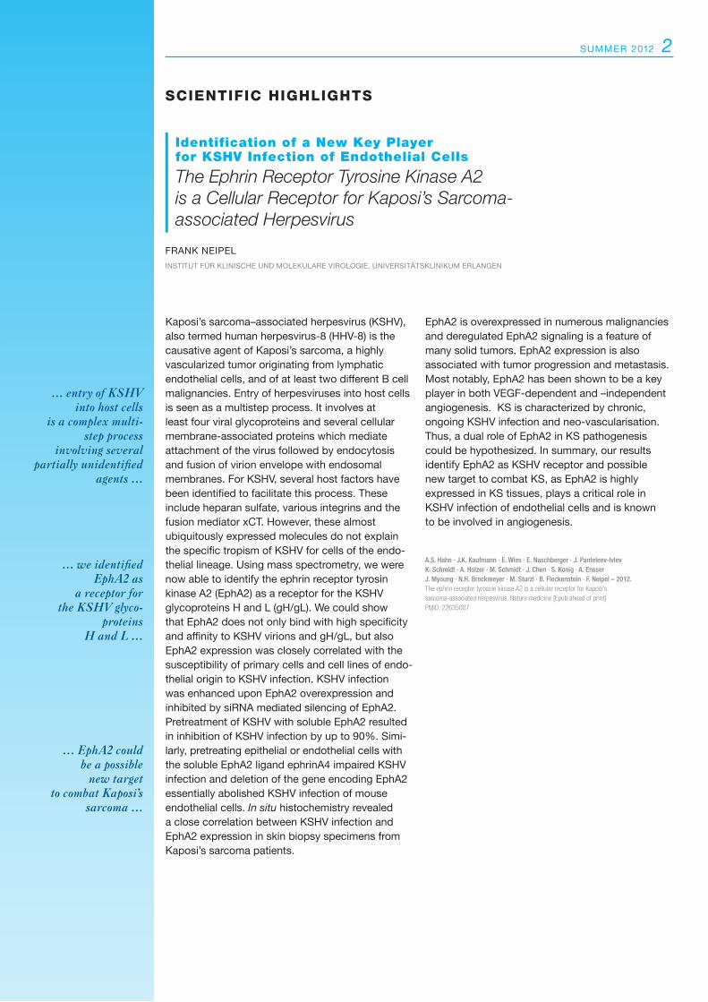
FIgure 2 EphA2 and LANA-1 expression in KS.
a–f) Immunohistochemical detection of LANA-1 and ephA2 in two different Kaposi’s sarcoma skin lesions (Ks1, Ks2) and one lung Kaposi’s sarcoma (Ks3).
g) ephA2 expression in breast cancer, used as a positive control.
h, i) ephA2 and LANA-1 expression in unin-volved tissue sections of the skin of a subject with Kaposi’s sarcoma. similar tissue areas of consecutive sections are indicated by asterisks. Areas with Kaposi’s sarcoma spindle cells, showing prominent nuclear LANA-1 staining and cytoplasmic ephA2 staining in consecutive sections, are indicated by black arrows. Teal arrows indicate ephA2 expression in the epidermis overlaying Kaposi’s sarcoma
b, d) in the tumor cells of breast cancer
g) or the epidermal layer of un involved skin
h) The scale bar in
i) is 50 µm and applies to all other panels.
A) Dose-dependent inhibition of KsHV infection by soluble ephA2. Infection of LeCs, sLK cells, KsImm cells and m7/2 cells. rKsHV.219 was pre incu-bated with ephA2-Fc or Fc. Infection rate without protein was set to 100% (sLK and KsImm n = 6, LeC and m7/2 n = 3, error bars represent s.d.).
B) Infection of LeCs after ephA2 knockdown. A non-sense sirNA (siNon) was set to 100% relative infection, a sirNA targeted at the ephrin recep-tor tyrosin kinase B4 ephB4) was used as additional negative control (n = 4, error bars represent s.e.m.; *P < 0.001).
C) Infection of primary murine pulmonary microvascular endothelial cells (mPmeC) from either WT or an EphA2-knockout (KO) mouse or EphA2-knockout cells transduced with a mouse ephA2 cDNA as a control. Where indicated, KsHV was preincubated with ephA2-Fc or control protein (2 µg ml-1, n = 3, error bars represent s.d.)
D) Correlation of ephA2 mrNA expression as determined by qrT-PCr and KsHV infection in human primary cells and cell lines (R2 = square correlation coefficient; sLK and KsImm n = 6, otherwise n = 3).
B
D
A
C
FIgure 1 Analysis of KSHV susceptibility and EphA2 expression in cell culture.
ΔE2Smurf2 WTSmurf2 control GFP GFP
untransfected
WTS
mur
f2-‐G
FP
hydr
ophi
lic Fra
c=on
WTS
mur
f2-‐G
FP
hydr
opho
bic Fr
ac=o
n
ΔE2S
mur
f2-‐G
FP
hydr
opho
bic Fr
ac=o
n
ΔE2S
mur
f2-‐G
FP
hydr
ophi
lic Fra
c=on
α-‐GFP
ΔE2Smurf2-‐GFP
ΔE2Smurf2-‐GFP LysoTracker-‐Red Overlay Overlay
ΔE2Smurf2-‐GFP EEA1 Overlay
TGFβRII Overlay
GFP
Overlay
Overlay
A
FIgure 1
summer 2012 4
Colorectal cancer is among the most frequent malignancies in the world, with an incidence of more than 1 million cases per year. The molecular mechanisms and pathways that contribute to colon carcinogenesis are not completely understood. TGF-beta is a pleiotropic cytokine, which plays a central role in driving tumorigenesis. It is involved in several processes like differentiation, apoptosis, proliferation and migration. In tumor development, it has a dual role, serving first as a tumor suppres-sor and then switching to a tumor promotor. In T lymphocytes, it has immunosuppressive effects and suppresses proliferation of cells and produc-tion of proinflammatory cytokines. smad ubiquitin regulatory factor (smurf) 2 is an ubiquitin ligase that regulates the TGF-beta pathway in a negative way by polyubiquitination of the TGF-beta receptor II for proteasomal degradation. In the current manus cript, we could identify a novel splice form of smurf 2, which we denoted delta e2 smurf2. Compared to the wild-type form (WT) delta e2 smurf2 possesses a spliced exon2 in its C2 domain, which is necessary for membrane binding
and receptor degradation. Interestingly, the novel variant of smurf2 exhibits a contrary effect of its WT form and serves as a positive regulator of the TGF-beta signaling via lysosomal degradation of the WTsmurf2. Overexpression of delta e2 smurf2 in T lymphocytes revealed an upregulation of the TGF-beta receptor II and the downstream canonical signaling. Furthermore, mice were completely pro-tected in tumor development in an AOm/Dss driven tumor model. This effect was due to loss of proliferation and suppression of proinflammatory cytokines of tumor infiltrating T lymphocytes con-cerning to higher sensitivity towards TGF-beta. Thus, delta e2 smurf2 appears to play an important functional role in colitis-associated colon cancer by controlling smurf2 expression and TGF-beta signaling in T lymphocytes and thereby regulates tumor growth in vivo.
H. Dornhoff · C. Becker · S. Wirtz · D. Strand · S. Tenzer S. Rosfa · C. Neufert · J. Mudter · J. Markl · J. Siebler M.F. Neurath – 2012. A variant of Smurf2 protects mice against colitis- associated colon cancer by inducing trans- forming growth factor beta signaling. Gastroenterology 142: 1183 -1194 e1184.
SciEntific HigHligHtS
Discovery of a novel Protein involved in the Protection against Colon Cancer
A Variant of Smurf2 protects Mice against Colitis-associated Colon Cancer by Inducing Transforming Growth Factor beta Signaling
HeIKe DOrNHOFF · mArKus NeurATHDePArTmeNT OF INTerNAL meDICINe I, uNIVersITÄTsKLINIKum erLANGeN
… TGF-beta, which is negatively regulated
by Smurf2, plays an immunosuppressive
role in T lymphocytes …
… we identified a novel splice form of Smurf2,
which we called delta E2 Smurf2 …
… delta E2 Smurf 2 exhibits contrary effects to
wild-type form and seems to be important for
the protection against tumor development
in the colon ...
ΔE2Smurf2 WTSmurf2 control GFP GFP
untransfected
WTS
mur
f2-‐G
FP
hydr
ophi
lic Fra
c=on
WTS
mur
f2-‐G
FP
hydr
opho
bic Fr
ac=o
n
ΔE2S
mur
f2-‐G
FP
hydr
opho
bic Fr
ac=o
n
ΔE2S
mur
f2-‐G
FP
hydr
ophi
lic Fra
c=on
α-‐GFP
ΔE2Smurf2-‐GFP
ΔE2Smurf2-‐GFP LysoTracker-‐Red Overlay Overlay
ΔE2Smurf2-‐GFP EEA1 Overlay
TGFβRII Overlay
GFP
Overlay
Overlay
B
C
E F
D
-‐TGFβ
+TGFβ
WTSmurf2 ΔE2Smurf2 14,4%
15,3%
29,6%
52,0%
Figure 1
β-‐Ac=n
WTSmurf2 control ΔE2Smurf2
+TGFβ
α-‐TGFβRII
CD4
TGFβ
RII
α-‐Smad2
control WTSmurf2 ΔE2Smurf2 TGFβ
α-‐pSmad3
α-‐Smad3
+ + + cDNA
α-‐pSmad2
0
2
4
6
8
10
12
control ΔE2Smurf2 WTSmurf2
Lucife
rase
ac7
vity
x-‐fold
without TGFβ
with TGFβ
Smad3 promoter assay *
**
Isotype -‐TGFβ
+TGFβ
WTSmurf2 ΔE2Smurf2 14,4%
15,3%
29,6%
52,0%
Figure 1
β-‐Ac=n
WTSmurf2 control ΔE2Smurf2
+TGFβ
α-‐TGFβRII
CD4
TGFβ
RII
α-‐Smad2
control WTSmurf2 ΔE2Smurf2 TGFβ
α-‐pSmad3
α-‐Smad3
+ + + cDNA
α-‐pSmad2
0
2
4
6
8
10
12
control ΔE2Smurf2 WTSmurf2
Lucife
rase
ac7
vity
x-‐fold
without TGFβ
with TGFβ
Smad3 promoter assay *
**
Isotype
5 newsLetter
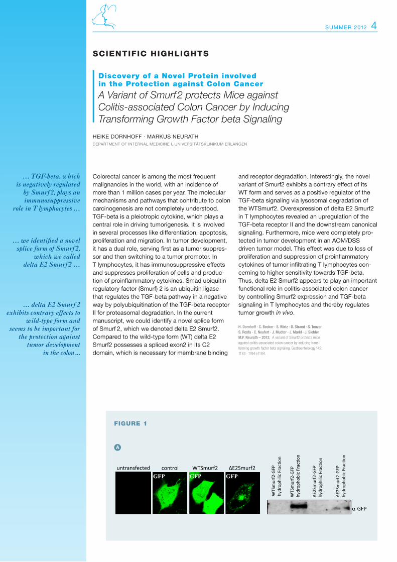
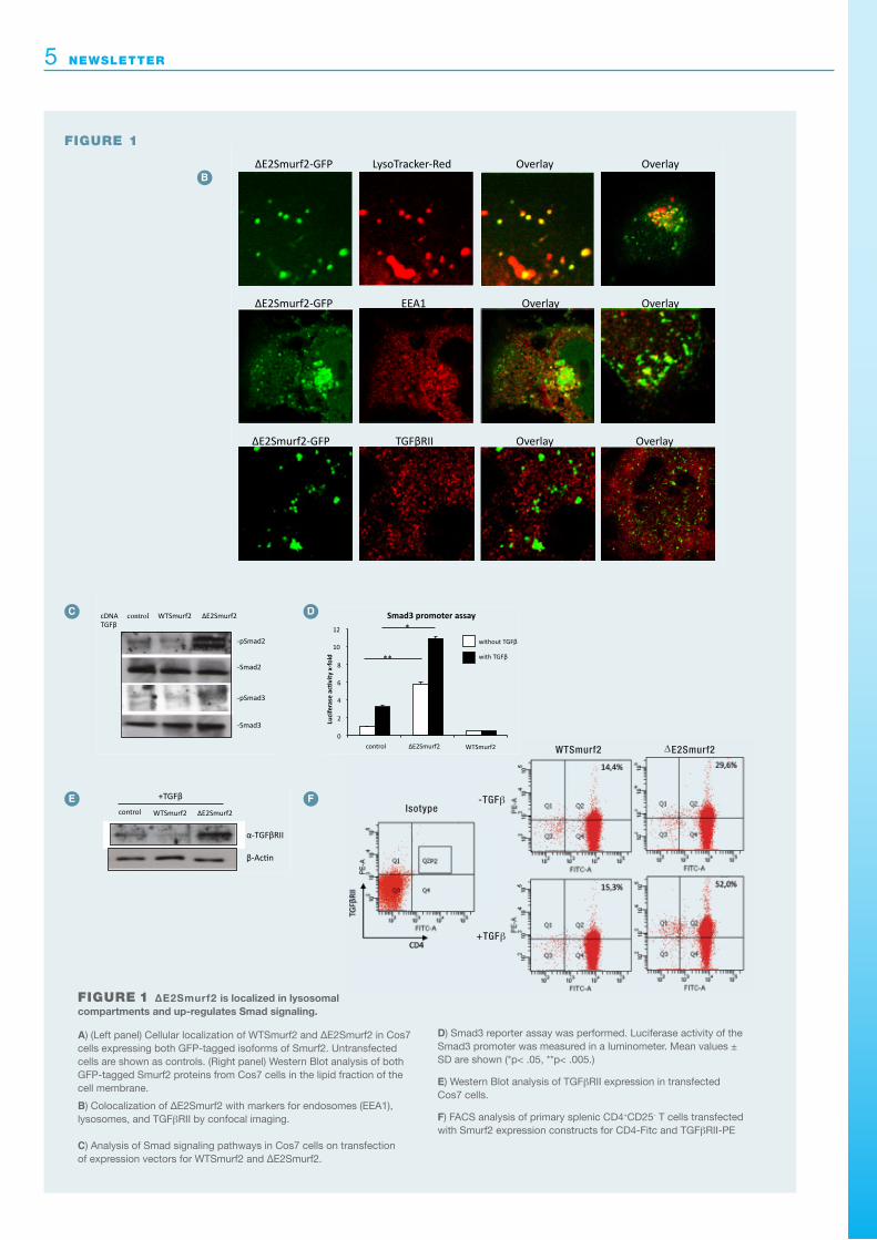
FIgure 1 ΔE2Smurf2 is localized in lysosomal compartments and up-regulates Smad signaling.
A) (Left panel) Cellular localization of WTsmurf2 and Δe2smurf2 in Cos7 cells expressing both GFP-tagged isoforms of smurf2. untransfected cells are shown as controls. (right panel) Western Blot analysis of both GFP-tagged smurf2 proteins from Cos7 cells in the lipid fraction of the cell membrane.
B) Colocalization of Δe2smurf2 with markers for endosomes (eeA1), lysosomes, and TGFβrII by confocal imaging.
C) Analysis of smad signaling pathways in Cos7 cells on transfection of expression vectors for WTsmurf2 and Δe2smurf2.
D) smad3 reporter assay was performed. Luciferase activity of the smad3 promoter was measured in a luminometer. mean values ± sD are shown (*p< .05, **p< .005.)
E) Western Blot analysis of TGFβrII expression in transfected Cos7 cells.
F) FACs analysis of primary splenic CD4+CD25- T cells transfected with smurf2 expression constructs for CD4-Fitc and TGFβrII-Pe
FIgure 1
Isotype-TGF β
WTSmurf2 ΔE2Smurf2
+TGF β
summer 2012 6
FIgure 2
ΔE2Smurf2 protects mice from tumor development in
the AOM/DSS model of colitis-associated colon cancer.
A) Analysis of Δe2smurf2 transgenic and WT control mice in the AOm/Dss model of colitis-associated colon cancer. representative endoscopic images at day 63 in both groups are shown. Data are representative of 5 independent experi-ments with 5 animals per group (*p < .05).
B) Colonic cryosections from AOm/Dss-treated WT and transgenic mice and immunohistochemistry for CD4, CD11c, myeloperoxidase, TGFβrII, and IL-6.
FIgure 2
WT
TG
Das Bild kann nicht angezeigt werden. Dieser Computer verfügt möglicherweise über zu wenig Arbeitsspeicher, um das Bild zu öffnen, oder das Bild ist beschädigt. Starten Sie den Computer neu, und öffnen Sie dann erneut die Datei. Wenn weiterhin das rote x angezeigt wird, müssen Sie das Bild möglicherweise löschen und dann erneut einfügen.
Das Bild kann nicht angezeigt werden. Dieser Computer verfügt möglicherweise über zu wenig Arbeitsspeicher, um das Bild zu öffnen, oder das Bild ist beschädigt. Starten Sie den Computer neu, und öffnen Sie dann erneut die Datei. Wenn weiterhin das rote x angezeigt wird, müssen Sie das Bild möglicherweise löschen und dann erneut einfügen.
TG WT
tum
or size sc
ore
WT
TG
0
5
10
15
20
25
day21 day 42 day 63
*
0 0,5 1
1,5 2
2,5 3
3,5 4
day 21 day 42 day 63
* *
WT
TG
aver
age tu
mor
size
CD4
MPO
TG WT
H&E 100x
H&E 200x
0
5
10
15
20
25
day 21 day 42 day 63
tota
l no.
of t
umor
s
*
*
n.d. WT
TG
Das Bild kann nicht angezeigt werden. Dieser Computer verfügt möglicherweise über zu wenig Arbeitsspeicher, um das Bild zu öffnen, oder das Bild ist beschädigt. Starten Sie den Computer neu, und öffnen Sie dann erneut die Datei. Wenn weiterhin das rote x angezeigt wird, müssen Sie das Bild möglicherweise löschen und dann erneut einfügen.
Das Bild kann nicht angezeigt werden. Dieser Computer verfügt möglicherweise über zu wenig Arbeitsspeicher, um das Bild zu öffnen, oder das Bild ist beschädigt. Starten Sie den Computer neu, und öffnen Sie dann erneut die Datei. Wenn weiterhin das rote x angezeigt wird, müssen Sie das Bild möglicherweise löschen und dann erneut einfügen.
TG WT
tum
or size sc
ore
WT
TG
0
5
10
15
20
25
day21 day 42 day 63
*
0 0,5 1
1,5 2
2,5 3
3,5 4
day 21 day 42 day 63
* *
WT
TG
aver
age tu
mor
size
CD4
MPO
TG WT
H&E 100x
H&E 200x
0
5
10
15
20
25
day 21 day 42 day 63
tota
l no.
of t
umor
s
*
*
n.d.
0 100
200
300
400
500
600
700
an=-‐CD3/28 an=-‐CD3/28 1ngTGFβ
IL-‐1
7 [p
g/m
l]
*
* 0
5000
10000
15000
20000
25000
30000
35000
an=-‐CD3/28 an=-‐CD3/28 1ngTGFβ
IL-‐2
[pg/
ml]
* 0
2000
4000
6000
8000
10000
12000
14000
an=-‐CD3/28 an=-‐CD3/28 1ngTGFβ
IFN-‐γ [p
g/m
l]
0 1000 2000 3000 4000 5000 6000 7000 8000 9000
10000
an=-‐CD3/28 an=-‐CD3/28 1ngTGFβ
IL-‐4
[pg/
ml] WT
TG
* 0
100
200
300
400
500 600
700
an=-‐CD3/CD28 an=-‐CD3/28 1ngTGFβ
IL-‐6
[pg/
ml]
TGFβRII
IL-6
TG WT
CD11c
WT WTTG TG
H&
E 100xH
&E 200x
CD
4M
PO
A
B
7 newsLetter
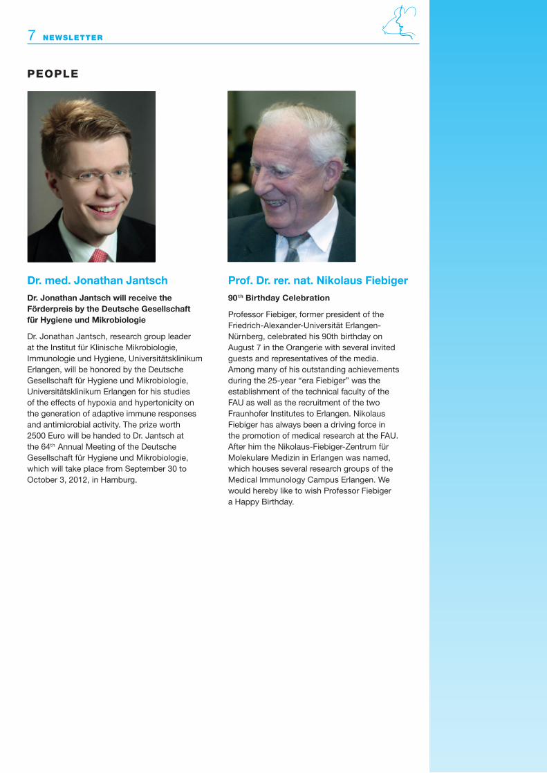
Dr. med. Jonathan Jantsch
Dr. Jonathan Jantsch will receive the Förderpreis by the Deutsche Gesellschaft für Hygiene und Mikrobiologie
Dr. Jonathan Jantsch, research group leader at the Institut für Klinische mikrobiologie, Immunologie und Hygiene, universitätsklinikum erlangen, will be honored by the Deutsche Gesellschaft für Hygiene und mikrobiologie, universitätsklinikum erlangen for his studies of the effects of hypoxia and hypertonicity on the generation of adaptive immune responses and antimicrobial activity. The prize worth 2500 euro will be handed to Dr. Jantsch at the 64th Annual meeting of the Deutsche Gesellschaft für Hygiene und mikrobiologie, which will take place from september 30 to October 3, 2012, in Hamburg.
Prof. Dr. rer. nat. Nikolaus Fiebiger
90th Birthday Celebration
Professor Fiebiger, former president of the Friedrich-Alexander-universität erlangen- Nürnberg, celebrated his 90th birthday on August 7 in the Orangerie with several invited guests and representatives of the media. Among many of his outstanding achievements during the 25-year “era Fiebiger” was the establishment of the technical faculty of the FAu as well as the recruitment of the two Fraunhofer Institutes to erlangen. Nikolaus Fiebiger has always been a driving force in the promotion of medical research at the FAu. After him the Nikolaus-Fiebiger-Zentrum für molekulare medizin in erlangen was named, which houses several research groups of the medical Immunology Campus erlangen. We would hereby like to wish Professor Fiebiger a Happy Birthday.
pEoplE
newsLetter 8
Medical Immunology Campus Erlangen Executive BoardProf. Dr. med. Christian Bogdan (Chairman) Prof. Dr. rer. nat. Diana Dudziak Prof. Dr. med. Kai-uwe eckardt Prof. Dr. med. Bernhard Fleckenstein Prof. Dr. rer. nat. Hans-martin Jäck Prof. Dr. med. Andreas mackensen Prof. Dr. med. markus Neurath (Deputy Chairman) Prof. Dr. med. Georg schett Prof. Dr. med. Gerold schuler (Deputy Chairman) Prof. Dr. rer. nat. Alexander steinkasserer Prof. Dr. rer. nat. Thomas Winkler Dr. rer. nat. sonja Pötzsch (scientific Coordinator)