+ All Categories
Home > Documents > Effect-Directed Analysis of Ah-Receptor Mediated Toxicants ... · Effect-Directed Analysis of...

Effect-Directed Analysis of Ah-Receptor Mediated Toxicants ... · Effect-Directed Analysis of...

Date post: 13-Jun-2020
Category:
Upload: others
View: 8 times
Download: 0 times
Share this document with a friend
29
Effect-Directed Analysis of Ah-Receptor Mediated Toxicants, Mutagens, and Endocrine Disruptors in Sediments and Biota Markus Hecker and John P. Giesy Abstract Sediments and associated biota represent important sources for the exposure of aquatic organisms to environmental toxicants including dioxin-like compounds, genotoxic chemicals, and endocrine disruptors. One of the key chal- lenges that environmental toxicologists and risk assessors are facing is the charac- terization and assessment of toxicological risks associated with such complex matrices such as sediments. Therefore, approaches have been developed supple- menting chemical analysis with bioanalytical techniques that make use of the specific properties of certain groups of chemicals to interfere with specific biological processes. This type of analysis has been coined effect-directed analysis (EDA), and is based on a combination of fractionation procedures, biotesting, and subsequent chemical analyses. In this chapter, we review the current state of the art of EDA regarding the assessment of sediment and biota samples for dioxin-like, genotoxic, and endocrine disrupting potentials. We discuss in vivo and in vitro screening concepts that are used in combination with fractionation and chemical analytical techniques to aid in the risk assessment of these chemical groups in sediments and biota. Advantages and disadvantages of current EDA strategies are considered, and recommendations for more realistic and relevant EDA approaches are given. Specifically, these include the use of optimized biotest- batteries covering a broad range of different endpoints as well as the inclusion of in vivo tests, and the parallel assessment of ecologically relevant parameters such M. Hecker (*) School of the Environment & Sustainability, University of Saskatchewan, 44 Campus Drive, Saskatoon, SK S7N 5B3, Canada e-mail: [email protected] J.P. Giesy Department of Veterinary Biomedical Sciences and Toxicology Centre, University of Saskatchewan, Saskatoon, SK, Canada and Department of Biology and Chemistry, City University of Hong Kong, Hong Kong, SAR, China and Zoology Department, College of Science, King Saud University, P. O. Box 2455, Riyadh 11451, Saudi Arabia W. Brack (ed.), Effect-Directed Analysis of Complex Environmental Contamination, Hdb Env Chem (2011) 15: 285–314, DOI 10.1007/978-3-642-18384-3_12, # Springer-Verlag Berlin Heidelberg 2011 285
Transcript

Effect-Directed Analysis of Ah-Receptor

Mediated Toxicants, Mutagens, and Endocrine

Disruptors in Sediments and Biota

Markus Hecker and John P. Giesy

Abstract Sediments and associated biota represent important sources for the

exposure of aquatic organisms to environmental toxicants including dioxin-like

compounds, genotoxic chemicals, and endocrine disruptors. One of the key chal-

lenges that environmental toxicologists and risk assessors are facing is the charac-

terization and assessment of toxicological risks associated with such complex

matrices such as sediments. Therefore, approaches have been developed supple-

menting chemical analysis with bioanalytical techniques that make use of the

specific properties of certain groups of chemicals to interfere with specific

biological processes. This type of analysis has been coined effect-directed analysis

(EDA), and is based on a combination of fractionation procedures, biotesting, and

subsequent chemical analyses. In this chapter, we review the current state of the art

of EDA regarding the assessment of sediment and biota samples for dioxin-like,

genotoxic, and endocrine disrupting potentials. We discuss in vivo and in vitro

screening concepts that are used in combination with fractionation and chemical

analytical techniques to aid in the risk assessment of these chemical groups in

sediments and biota. Advantages and disadvantages of current EDA strategies

are considered, and recommendations for more realistic and relevant EDA

approaches are given. Specifically, these include the use of optimized biotest-

batteries covering a broad range of different endpoints as well as the inclusion

of in vivo tests, and the parallel assessment of ecologically relevant parameters such

M. Hecker (*)

School of the Environment & Sustainability, University of Saskatchewan, 44 Campus Drive,

Saskatoon, SK S7N 5B3, Canada

e-mail: [email protected]

J.P. Giesy

Department of Veterinary Biomedical Sciences and Toxicology Centre, University of

Saskatchewan, Saskatoon, SK, Canada

and

Department of Biology and Chemistry, City University of Hong Kong, Hong Kong, SAR, China

and

Zoology Department, College of Science, King Saud University, P. O. Box 2455, Riyadh 11451,

Saudi Arabia

W. Brack (ed.), Effect-Directed Analysis of Complex Environmental Contamination,Hdb Env Chem (2011) 15: 285–314, DOI 10.1007/978-3-642-18384-3_12,# Springer-Verlag Berlin Heidelberg 2011

285

as benthic community structure. Furthermore, the need for refinement and standar-

dization of current sediment EDA approaches that allow capturing and assessing

exposures to unknown or emerging chemicals such as endocrine disruptors, per-

fluorinated compounds, or polybrominated and mixed halogenated dibenzo-

p-dioxins and -furans is discussed.

Keywords Bioassay, Complex mixtures, Dioxin-like chemicals, Endocrine

activity, Fractionation, Genotoxicity, In vitro, Toxicity identification and evaluation

Contents

1 Background . . . . . . . . . . . . . . . . . . . . . . . . . . . . . . . . . . . . . . . . . . . . . . . . . . . . . . . . . . . . . . . . . . . . . . . . . . . . . . . 286

1.1 Toxicity Identification and Evaluation vs. Effect-Directed Analysis . . . . . . . . . . . . . . . 287

1.2 Dioxin-Like Activities . . . . . . . . . . . . . . . . . . . . . . . . . . . . . . . . . . . . . . . . . . . . . . . . . . . . . . . . . . . . . . 289

1.3 Genotoxicity . . . . . . . . . . . . . . . . . . . . . . . . . . . . . . . . . . . . . . . . . . . . . . . . . . . . . . . . . . . . . . . . . . . . . . . . 291

1.4 Endocrine Disruption . . . . . . . . . . . . . . . . . . . . . . . . . . . . . . . . . . . . . . . . . . . . . . . . . . . . . . . . . . . . . . . 292

2 Effect-Directed Analysis in Sediments and Biota . . . . . . . . . . . . . . . . . . . . . . . . . . . . . . . . . . . . . . . . . 293

2.1 In Vivo Bioassays . . . . . . . . . . . . . . . . . . . . . . . . . . . . . . . . . . . . . . . . . . . . . . . . . . . . . . . . . . . . . . . . . . . 294

2.2 In Vitro Bioassays . . . . . . . . . . . . . . . . . . . . . . . . . . . . . . . . . . . . . . . . . . . . . . . . . . . . . . . . . . . . . . . . . . 295

2.3 Bioassay-Directed Fractionation . . . . . . . . . . . . . . . . . . . . . . . . . . . . . . . . . . . . . . . . . . . . . . . . . . . . 296

2.4 Relative Potency and Potency Balance Analysis . . . . . . . . . . . . . . . . . . . . . . . . . . . . . . . . . . . 299

3 Limitations and Perspectives of Bioassay-Directed EDA . . . . . . . . . . . . . . . . . . . . . . . . . . . . . . . . . 302

4 Conclusions . . . . . . . . . . . . . . . . . . . . . . . . . . . . . . . . . . . . . . . . . . . . . . . . . . . . . . . . . . . . . . . . . . . . . . . . . . . . . . . 305

References . . . . . . . . . . . . . . . . . . . . . . . . . . . . . . . . . . . . . . . . . . . . . . . . . . . . . . . . . . . . . . . . . . . . . . . . . . . . . . . . . . . . 306

1 Background

Since the middle of the twentieth century there has been increasing concern over the

potential adverse effects of xenobiotics present in the environment on human and

wildlife populations. Three groups of toxicants that are of particular interest relative

to potential environmental health effects are dioxin-like, genotoxic, and endocrine

active chemicals. Some of these ubiquitous compounds are hydrophobic, lipophilic,

and resistant to biological and chemical degradation. These properties impart persis-

tency and a propensity to bioaccumulate and biomagnify to concentrations that can

cause deleterious effects. Sediments, especially, represent long-term sinks for some

contaminants [1–8], which can become bioavailable through remobilization events

such as floods or dredging, or through benthic or bottom-dwelling organisms [9–13].

Severely contaminated sediments have been reported to occur in rivers and lakes, and

have been shown to be acutely and chronically toxic to fish and benthic invertebrate

species [8, 14–16]. Under certain conditions, toxicity of sediments has been reported

to contribute to decreases in reproductive success, recruitment, and alterations in

community structure, effects which could potentially cause changes in population

structure. For example, amphipod abundance in San Francisco Bay was found to

be inversely proportional to contamination and measured toxicity of sediments

[14]. Studies have shown that toxic sediments can affect aquatic species. Zebrafish

286 M. Hecker and J.P. Giesy

embryos exposed to sediments from theDanubeRiver showed impaired development

and decreased hatching rates compared to control fish [8]. Sediments have

been classified as genotoxic, mutagenic, endocrine disrupting, or recognized for

dioxin-like effects [8, 15, 17, 18]. In addition to their acute or chronic toxicity to

benthic invertebrates, accumulated residues in sediments can be a pool that can be

accumulated through food webs and have subsequent toxic effects on the predators.

Chemicals typically occur as mixtures in environmental matrices such as sedi-

ments and biota, and can include different congeners and isomers of both natural

and anthropogenic origin. Concentrations and toxic potencies of compounds pres-

ent in mixtures can range over several orders of magnitude. In addition, interactions

among different classes of compounds, such as estrogenic vs. anti-estrogenic, can

modulate the toxic potential. Exposure to mixtures and the potential for greater and

less than additive effects of mixtures complicates hazard evaluation and risk

assessment of complex mixtures of xenobiotics. So, even if complete information

is available about the concentrations of all inorganic and organic residues, it is

difficult to predict the effects of the mixture. However, this complete knowledge is

seldom the case. In fact there might be residues present that have not yet been

described in the literature. There are contaminants and/or their degradation pro-

ducts that can cause toxicity for which there are no analytical methods or authentic

standards. Furthermore, toxic effects of some contaminants, even those, which are

analytically determined, are not well characterized. There are potentially significant

classes of contaminants for which little or no information on their effects on

organisms is available. In other words, chemical analysis has been used to identify

and quantify only those chemicals for which analytical techniques and standards are

available. In environmental monitoring, chemists find what they look for. If they do

not know to look for a chemical, then it will not be quantified. Finally, instrumental

analyses do not account for interactions among the chemicals in mixtures and

provide little information on their biological availability and provide no informa-

tion on their effects. Thus, relying on quantification of individual residues by

instrumental analyses, while useful for source identification, can underestimate

the potential risks posed by these chemicals and some toxicologically important

compounds could be overlooked. In summary, analysis of the vast number of

chemicals typically present in an environmental sample would not only be prohibi-

tively expensive but simply impossible due to limits in the available analytical

methodologies for many chemicals, especially since often no a priori knowledge of

the chemicals present in the sample exists.

1.1 Toxicity Identification and Evaluation vs. Effect-DirectedAnalysis

To overcome some of the above discussed limitations, bioanalytical approaches

have been developed to supplement instrumental chemical analysis. These bioana-

lytical techniques make use of the specific properties of certain groups of chemicals

Effect-Directed Analysis of Ah-Receptor Mediated Toxicants 287

to affect biological systems. Such bioassays are often based on in vitro responses of

cells or even cell-free responses of biomolecules but can also utilize whole organ-

ism in vivo systems. The use of bioassays in chemical identification has several

advantages relative to instrumental analyses. First, the assays “read out” directly in

terms of a biological response. This could be a molecular change or even something

as simple as lethality. The bioassay responds to all of the chemicals in the mixture

so even unidentified chemicals that cause a particular endpoint are measured even if

they cannot be identified. These assays also respond proportionally to the aggregate

effects of mixtures and account for all of the interactions among chemicals. In fact,

bioassays can be used in conjunction with instrumental analyses in a “potency

balance” approach that helps determine if there are unidentified active compounds.

In particular, bioassays can be used with a fractionation scheme to direct instru-

mental analyses to identify unidentified active chemicals in a mixture.

In the late 1980s, one of the first standardized effect-directed analysis (EDA)

procedures, the toxicity identification and evaluation (TIE) approach, had been

established by the US-EPA. This approach focuses primarily on the identification

and evaluation of organic or inorganic contaminants in aqueous samples using a

combination of in vivo tests, fractionation, and chemical analysis, and is character-

ized by the following three steps (reviewed in [19]):

1. Toxicity characterization by assignment of toxicity to general groups of tox-

icants (typically bioassay directed analysis)

2. Identification of suspected toxicants (chemical analytical determination)

3. Confirmation of the suspected cause of toxicity

The basic concept of the TIE approach is the removal of compound groups from

the tested matrix until the toxicity of the sample disappears. Suspected chemicals

are then identified by analytical chemistry, and their toxicity is confirmed by means

of the same bioassay used in the initial characterization phase. Approaches like TIE

have been an important step towards improving environmental risk assessment

(ERA) focusing on surface waters. It has been increasingly recognized, however,

that particle-bound contaminants such as suspended matter or sediments, or those

accumulating through the food chain might be of greater ecotoxicological relevance

with respect to moderately or strongly lipophilic compounds. While there have been

some recent developments of standardized EPA techniques and procedures for

sediment TIE [20, 21], there are still a number of uncertainties associated with

this approach. For example, it is often difficult to construct an artificial mixture of

the suspect compounds identified in the characterization phase, because these may

not always be commercially available. Furthermore, exposure of tests organisms as

part of the confirmation step is problematic since it requires spiking of clean or

artificial sediments with the suspected chemicals, and which often alters the toxi-

cological properties of the tests matrix because of differing adsorption and/or

bioavailability properties. Finally, TIE primarily relies on whole organism tests

that often do not provide information on the specific mode of toxicity of a sample,

and which can be helpful in identifying the compound groups responsible for the

measured effect.

288 M. Hecker and J.P. Giesy

A recent approach that aims to overcome some of the shortcomings of TIE is the

EDA procedure as reviewed by Brack [19, 22]. Like TIE, the principle of EDA is to

use a biological response to direct the identification of causative agents in a

complex matrix such as sediment. During the conduct of an EDA, however, the

causal substances are identified by analyzing different extracts that were prepared

from the original sample, e.g., by separation based on lipophilicity, polarity, size,

etc., and not by a retrospective step-by-step exclusion of certain compound groups

as done in TIE. Furthermore, during EDA the biological analysis phase is typically

dominated by in vitro or in vivo mechanistic assays that enable identification of the

properties of a sample to interact with specific biological pathways or targets, and

thus, can narrow the group of suspected chemicals (e.g., dioxin-like chemical that

can be detected by means of an AhR assay as described in Sect. 2 in [23]). Thus,

EDA has some distinct advantages over sediment TIE by better intertwining the

specific biological (toxicity) and analytical pathways. However, the TIE approach

is considered to be of greater ecological relevance because it does not require

alteration of the sample through extraction, and utilizes whole organisms that are

tested in direct contact with the matrix of concern.

1.2 Dioxin-Like Activities

Chemicals that elicit toxic effects similar to that of 2,3,7,8-tetrachlorodibenzo-

p-dioxin (TCDD), known as dioxin-like chemicals, are of concern due to their

ability to cause hepatotoxicity, embryotoxicity, teratogenicity, immunotoxicity,

dermal toxicity, lethality, carcinogenesis, wasting syndrome, and tumor promotion

in many different species at relatively small concentrations [24, 25]. A number of

studies have demonstrated that several toxic and biochemical effects caused by

dioxin-like chemicals are mediated through the aryl hydrocarbon receptor (AhR)

[26, 27] and associated dioxin responsive elements (DREs) on DNA. The AhR,

which belongs to the basic helix-loop-helix protein family [28], is a ligand-

dependent transcription factor, complexed with heat shock proteins located in the

cytosol. The strength with which congeners bind to the AhR is directly proportional

to the toxicity, enhanced gene transcription, and enzyme activities mediated by the

AhR mechanism [29]. The role of the AhR in mediating toxic and biological effects

of dioxin-like chemicals has been well documented in a number of studies, even

though the exact biochemical mechanism leading to the pleotrophic toxic responses

is yet to be elucidated [22]. After binding of ligands to cytosolic AhR, heat shock

proteins dissociate from the complex, the receptor ligand complex is activated and

translocated to the nucleus, where it forms a dimer with the Ah receptor nuclear

translocator (ARNT) protein and possibly other factors. The heteromeric ligand:

AhR:ARNT complex binds with high affinity to specific DREs on DNA. Binding of

the transformed ligand-AhR-ARNT complex to DREs results in DNA bending,

disruption of chromatin and nucleosome, and thus increased promoter accessibility

and transcriptional activation of adjacent responsive genes (Fig. 1) [31, 32].

Effect-Directed Analysis of Ah-Receptor Mediated Toxicants 289

The traditional, well-known ligands for AhR have been described as hydropho-

bic aromatic compounds with planar structure of a particular size of the molecule or

a part of the molecule, which fits the binding sites [33, 34]. Thus, the ability of these

ligands to bind to the AhR and to cause toxic effects greatly depends on their

structure and substitution pattern. These include planar congeners of polychlori-

nated dibenzo-p-dioxins and polychlorinated dibenzofurans (PCDDs and PCDFs),

chlorinated azobenzenes and azoxybenzenes, polychlorinated biphenyls (PCBs),

several polycyclicaromatic hydrocarbons (PAHs) and polychlorinated napthalenes

(PCNs) [35]. Other chemicals that have been suggested as potential AhR agonists

due to their stereochemical configuration, but not yet experimentally confirmed,

include polybrominated and chloro-/bromo- analogs of the previously listed

classes of compounds [36], alkylated-chlorinated dioxins and furans, chlorinated

Light

Luciferase

ER-Responsive Genes

Xenoestrogenor estrogen

“Estrogenic Effects”

CYTOSOL

ERE

ER

Nuclear Factors

ER

P

mRNADNA Binding

P

NUCLEUS

ER+ HSP

HSP

AhR ligand

Light

Luciferase

+ AhRHSP90HSP90

HSP HSP90

HSP90HSP

ARNT

NuclearFactorsP

DREModulation of Gene

Expression

NUCLEUSmRNA

DNA Binding

AhR-Responsive GenesCYP1A1 etc.

CYTOSOL

ARNTAhR

Fig. 1 Mechanism of AhR-

or ER-receptor-mediated

response in cells (adapted

from [30]). For description

see text. HSP heat shock

proteins, P ¼ phosphates:

phosphorylation is an

important regulatory factor

for receptor function

290 M. Hecker and J.P. Giesy

dibenzothiophenes, chlorinated xanthenes and xanthones [37], polychlorinated

diphenylthiophenes (PCDTs), anisols (PCAs), anthracenes (PCAN), fluorenes

(PCFL), and others [38]. Recently, new types of relatively weak AhR ligands

or inducers (compared to TCDD) have been identified, which include both natural

and synthetic compounds [31]. These compounds deviate from the traditional

criteria of planarity, aromaticity, and hydrophobicity. The natural compounds

that bind to the AhR include, among others, indoles, tryptophan-derived products,

oxidized carotinoids, and heterocyclic amines. Some pesticides or drugs with

various structures, such as imidazoles and pyridines, also possess the AhR binding

ability. These ligands act as transient inducers and bind to the AhR with weak

affinity and are rapidly degraded by the induced detoxification enzymes.

1.3 Genotoxicity

Chemicals in the environment can cause overt toxicity, but they can also cause

subtle changes that may not result in immediate toxicity. One such effect is

genotoxicity. Chemicals, such as biotransformation products of some polycyclic

aromatic hydrocarbons (PAH), which are common contaminants in sediments, can

bind to DNA where they cause a number of types of damage. The resulting DNA

adducts can result in point mutations or strand breaks or other types of reorganiza-

tion of the DNA that can result in adverse effects in germ cells and can result in

decreased fitness of individuals in subsequent generations [39].

A number of in vitro and in vivo techniques have been developed to screen for

these effects. Here we provide three examples of tests that have been found to be

useful in screening of sediments for genotoxic potentials. The first measures point

mutations or mutagenicity in vitro, while the second is an in vivo test that measures

the occurrence of DNA strand breaks. The Ames assay uses the TA 98 Salmonellatyphimurium bacteria strain to measure frame-shift mutations and the TA 100

S. typhimurium bacteria strain to measure base pair substitutions [4]. These strains

are mutants that cannot produce histidine. A colorimetric measure of the number of

back-mutated cells that are able to produce histidine is used as a measure of the

mutagenic potency of a pure chemical or sediment extract.

Some chemicals need to be metabolically activated to cause mutagenicity.

Because Salmonella do not possess the metabolic machinery to bioactivate mole-

cules such as certain PAHs, S9 microsomal preparation can be added to samples.

The Ames assay can be conducted using different bacteria strains, the most used of

which are the mutated strains (TA 98 and TA 100) and with and without pre-

incubation of the extract with the S9 microsomes to enable a comprehensive

assessment of the mutagenic potential of a sample.

Another method to determine genotoxic effects measure DNA fragmentation

[40]. This assay, which is variously called the alkaline DNA unwinding assay or the

comet assay, measures small fragments of DNA that occur due to breaks in the

DNA. Under alkaline conditions double-stranded DNAwill unwind, such that when

Effect-Directed Analysis of Ah-Receptor Mediated Toxicants 291

separated by polyacrylamide gel electrophoresis (PAGE), the smaller fragments of

DNA migrate more quickly than the larger strands of DNA. The more fragmenta-

tion, the more bands that can be identified. Because of the pattern formed on a two-

dimensional PAGE that represents a comet, this assay is often referred to as the

“Comet” assay [41]. DNA strand breaks can be studied in either in vitro or in vivo

assays.

A third type of genotoxicity assay that is commonly used to assess genotoxicity

of chemicals or complex matrices such as sediments is the micronucleus test. The

assay measures the formation of a micronucleus during the metaphase/anaphase

transition of mitosis, e.g., as the result of an acentric chromosome fragment

detaching from a chromosome after breakage, and which coalesce into bodies of

chromatin material referred to as micronuclei. The number of micronuclei present

in cells is directly proportional to DNA damage [42].

1.4 Endocrine Disruption

Over the past 2 decades, there has been increasing concern about the possible

effects of chemicals in the environment on the endocrine and reproductive systems

in humans and wildlife [43, 44]. Such compounds have the potential to disrupt

normal reproduction or developmental processes which can lead to adverse health

effects such as compromised reproductive capacity, breast and testicular cancer,

reproductive dysfunction such as feminization or demasculinization of males, and

other adverse effects. A range of classes of compounds including natural products,

pharmaceuticals, pesticides, and other industrial chemicals have been shown to

affect endocrine systems of wildlife and humans. The manner by which these

chemicals can interact with the endocrine system is manifold, and in general it is

distinguished between compounds that elicit their response through binding to

hormone receptors, and those that act through other mechanisms such as interfer-

ence with steroidogenesis. While any chemical that causes an organism to be unable

to maintain homeostatic regulation could be classified as an endocrine disrupting

chemical (EDC), chemicals classified as EDCs have typically been those that are

either able to bind to hormone receptors or can modulate the expression of steroid

hormones such as estrogens or androgens or thyroid hormones. There are both

natural and artificial chemicals that can modulate the endocrine system. These

chemicals can be direct-acting and cause effects as receptor agonists or antagonists

or can have indirect effects that ultimately modulate expression of genes that lead to

effects that are similar to those caused by direct-acting effects. For example, a

chemical that can induce the activity of CYP19 (aromatase) can result in the

production of more endogenous estradiol (E2), and subsequently would cause an

estrogenic effect even though it might not bind to the estrogen receptor (ER).

Much of the research in the area of environmental endocrine disruption has been

focused on chemicals that can bind to hormone receptors including the estrogen

receptor (ER) and androgen receptor (AR) as either agonists or antagonists.

292 M. Hecker and J.P. Giesy

However, various modes of actions have been reported, which include binding of

chemical to other nuclear receptors, which then interact with an estrogen responsive

element; acting through other receptors and/or signal transduction pathways; mod-

ulations of steroidogenesis and catabolism of active steroid hormones [45–48].

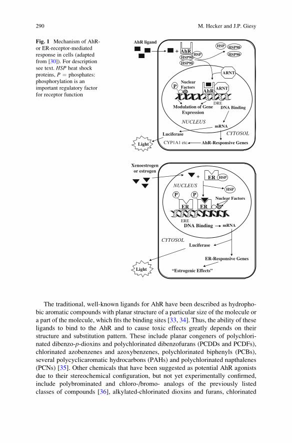
Estrogenic compounds are characterized by their ability to bind to and activate the

estrogen receptor, which is a transcription factor belonging to the steroid receptor

family. While there are structural similarities among some compounds that are ER

agonists, other ER-active compounds do not share similar structures. Upon binding

of an estrogenic compound to the ligand binding domain of the ER (located

predominantly in the nucleus), the associated heat shock protein complex, which

masks the DNA binding domain, dissociates and subsequently the ligand occupied

receptor dimerizes. The homodimer complex interacts with specific DNA sequences

referred to as estrogen response elements (EREs) located in the regulatory regions of

estrogen-inducible genes. ER complexes bound to an ERE recruit additional tran-

scription factors, leading to increased gene transcription and synthesis of proteins

required for expression of hormonal action (Fig. 1) [49]. A series of natural and

synthetic endocrine disrupting compounds have been identified by different in vivo

and/or in vitro methods. Unlike chemicals that can directly interact with the nuclear

hormone receptors, there is a multitude of different ways by which chemicals can

interact with other endocrine processes such as steroidogenesis. For instance, sub-

stances such as some imidazole-like fungicides and phyto-flavonoids have been

shown to modulate hormone production by affecting activities of the steroidogenic

enzymes aromatase (CYP19) and 17b-hydroxysteroid-degydrogenase (17b-HSD),respectively [45, 46, 50]. Other chemicals such as naphthenic acids can inhibit

estradiol metabolism, and thereby increase estradiol concentrations in vitro [51].

EDCs such as pesticides, plasticizers, plant sterols, PAHs, etc., have all been

measured in sediments and have been shown to disrupt the endocrine system in

in vitro and in vivo assays [46, 52, 53]. For example, known estrogen receptor

agonists, such as 17a-ethinylestradiol (EE2), 17b-estradiol (E2), and bisphenol A,

have been measured in sediments in several ecosystems [54–56]. Sediment extracts

from the Upper Danube River produced estrogenic-like responses in a transcrip-

tional ER assay [8]. It has also been reported that the same sediments caused

embryo toxicity, disruptions in hatching rates and time to hatch in Danio rerioembryos [8]. Other endocrine effects that were caused by sediment-associated

contaminants were changes in the expression of key genes involved in steroidogen-

esis [57], and alteration in the production of the sex steroids testosterone (T) and E2

[18] using the H295R cell line.

2 Effect-Directed Analysis in Sediments and Biota

Specific testing systems have been developed for the detection of dioxin-like,

genotoxic, and endocrine active potential in environmental samples. These systems

can be separated into two general categories: (1) in vivo assays using whole

Effect-Directed Analysis of Ah-Receptor Mediated Toxicants 293

organisms, or (2) in vitro tests utilizing cellular or sub-cellular systems that detect

interactions with specific biological functions. These bioassays are used to assess

the net effects of a complex sample to an animal or in vitro system. Organisms are

predominantly used to identify effects of sediment-bound pollutants in direct contact

assays with the unaltered sample on apical endpoints such as growth, reproduction,

and survival. They are typically utilized to assess the biological risk of a given

exposure but often do not allow pinpointing the effect to specific contaminants in a

sample. Therefore, whole organism assays are often paired with a parallel exposure

assessment by means of a combination of in vitro assays and analytical chemistry. In

vitro bioassays are based on the responses of either wild type or genetically altered

eukaryotic or prokaryotic cells that enable the assessment of potencies of individual

chemicals or complex mixtures of environmental contaminants in extracts to cause

effects specific to the exposure with certain chemical groups. Specifically, either

endogenous responses or specific exogenous induced alterations incorporated into

a cell are used for the measurements. The induction of specific responses following

the exposure of cells to specific chemicals or mixtures of compounds can be assessed

by measuring endogenous or engineered responses such as mutations, DNA strand

breaks, protein expression, enzyme activity, etc., depending on the test system and

endpoint.

2.1 In Vivo Bioassays

There are a number of organisms that are amenable to determine the toxicity of

sediments [58]. Here we will focus on those that are most useful for studying the

three classes of chemicals discussed in this chapter. While there are a number of

hypotheses and recommendations for the use of invertebrate systems to assess the

endocrine-disrupting potential of sediments to date, only very few studies have used

this approach relative to EDA [59, 60]. One of the key issues associated with the use

of different species is uncertainties regarding the predictability of endocrine effects

to vertebrates because they have different hormone systems. Also, since most

invertebrates do not express the AhR, they are essentially unresponsive to dioxin-

like compounds. Finally, while benthic invertebrates might represent useful senti-

nels for the assessment of genotoxic potentials of sediments [61], they are rarely

used in this context.

For application in the EDA process described here, therefore, we will focus on

vertebrate assays because they have been successfully used in EDA of sediments.

There have been numerous efforts to use fish species such as sanddab (Cithar-ichthys stigmaeus), California halibut (Paralichthys californicus), flounder (Pla-tichthys flesus), or trout (Oncorhynchus mykiss) to assess exposure to dioxin like orEDCs in sediments [54, 62–64]. However, most of these studies utilized large

organisms in time- and cost-intensive experiments. One promising test is the

zebrafish (D. rerio) embryo sediment contact test [15, 65].

294 M. Hecker and J.P. Giesy

The zebrafish is a small, easily cultured freshwater fish that reaches sexual

maturity in approximately 3 months. They produce between 50 and 200 eggs

every 2–3 days and the embryos develop rapidly. The eggs have a transparent

chorion, which makes it relatively easy to monitor development of the embryo. The

size of the zebrafish allows exposures to be performed in 24-, 48-, or 96-well culture

plates. The protocol for using these fish is fairly simple. Zebrafish embryos 1–2 h

postfertilization are exposed directly to sediments or to diluted sediment extracts in

96-well plates in 100 mL ISO water containing 20 mM CaCl22H2O, 5 mM

MgSO47H2O, 7.5 mM NaHCO3, and 0.037 mM KCl. Embryos are covered with

an oxygen permeable cover and incubated at 27�C for 48 h. The embryos can be

monitored for various endpoints, including lethality and deformities, but can also be

used in subsequent measures of gene expression and enzyme activities such as

CYP1A, which is under the control of the DRE, and genotoxicity, e.g., by means of

the comet assay [15, 66]. The only shortcomings of embryo-based assays, such as

the zebrafish egg contact test, are its limitation to measure effects on the endocrine

system due to the lack of sexual differentiation at this life stage.

2.2 In Vitro Bioassays

In vitro bioassays have been used to assess dioxin-like, genotoxic, and endocrine

activities in a variety of environmental matrices, including sediments and biota.

Various environmental samples, such as sediments [4, 18, 52, 53, 67, 68] or particu-

late matter [69, 70], sludge [71, 72], and animal tissues, have been assessed regarding

their potential to cause toxicity in vivo or in vitro. Significant dioxin-like activity has

been observed in egg extracts of birds such as herring gull, cormorant, and great blue

heron [73, 74] as well as in birds at different stages of development [75]. Among

other animals, extracts of fish (white sucker, juvenile whitefish) [37, 72], bivalves

[76], and otter [77] have also been tested. Different than in tests with live organisms,

in vitro assays typically require clean up and extraction of the original sediment or

tissue samples prior to testing. This is usually done through extraction by organic

solvents. The solvent of choice needs to be compatible with the cell system, not

causing any effect by itself, but enabling distribution of the extracted material to the

cells. Extracts can be cytotoxic, which is caused by some compounds present in

complex mixtures. For example, sulfur is a major cytotoxic constituent in sediment

extracts, which should be eliminated prior to performing dioxin-like or estrogenic

activities. The measurement of cell viability/cytotoxicity is essential in all bioassays

dealing with complex mixtures as well as single compounds. Cell bioassays with

multi-well plate formats enable themeasurement of several samples at the same time.

In addition, current procedures allow subsequent measurement of viability index,

enzyme activity, and protein content in the same multi-well plates [35].

A number of different measurement endpoints are used to assess the exposure to

dioxin-like, genotoxic, and endocrine active chemicals. Exposure to chemicals that

exhibit dioxin-like properties can be measured by increased expression and induced

Effect-Directed Analysis of Ah-Receptor Mediated Toxicants 295

activity of cytochrome P4501A1 and its monooxygenase activities, such as 7-

ethoxyresorufin O-deethylase (EROD) or aryl hydrocarbon hydroxylase (AHH)

[63, 78]. Genotoxicity can be assessed by measuring a variety of endpoints,

including DNA strand breaks using, e.g., the comet assay, micronucleus formation,

and mutations [40, 79–81]. The potential of chemicals or environmental samples to

interact with the endocrine system is typically assessed by means of three end-

points: (1) binding to the estrogen receptor (ER), (2) binding to the AR, and (3)

alteration of sex steroid production through interaction with steroidogenic path-

ways. Determination of the potential of a chemical to interact with the ER or AR is

either conducted by means of direct receptor binding assays or by transcriptional

assays using genetically modified cells [49]. The latter are typically obtained by

transfection of the so-called wild type cells with recombinant expression vectors,

which contain selective responsive elements upstream of a reporter gene. The most

common reporter genes are firefly luciferase (luc), alkaline phosphatase (PAP),

chloramphenicol acetyl transferase (CAT), or ~b galactosidase (LacZ) [82, 83].

Effects on steroidogenesis can be measured at the gene, protein, or end-product

level. Common assays include the quantification of changes in steroidogenic or

metabolic genes [84], CYP19 aromatase enzyme activity (transforms androgens

into estrogens) [46], or quantification of steroid hormones [47, 85]. A list of some

bacterial, yeast, animal, and human cell lines used for the detection of in vitro

TCDD-like, genotoxic, or endocrine activity is shown (Table 1).

2.3 Bioassay-Directed Fractionation

As discussed previously, in vitro bioassays as well as certain in vivo tests such as the

D. rerio embryo sediment toxicity assay can be used in support of the characterization

of complex mixtures in sediments or biota. Specifically, bioassays can be used in

combination with specific analytical techniques in a tiered approach, which is termed

bioassay-directed fractionation methodology. This approach provides information

needed for monitoring and risk assessment of the compounds with specific modes

of action and may lead to identification of novel classes of environmental toxicants

[70]. Specifically, a complex sample (e.g., sediment or tissue extract) is first analyzed

using one (for specific questions such as the characterization of dioxin-like chemi-

cals) or a combination of multiple (nontarget analysis) bioassays representing differ-

ent modes of biological action. Parallel to the assessment of effects on specific

biological processes, the general toxicity of a sample (e.g., cytotoxicity for a cell-

based bioassay) that could mask a specific response is evaluated. If a sample has been

identified as having a significantly altered biological activity it is then subjected to

fractionation, separating the chemicals contained in it by chemical–physical proper-

ties, such as polarity, molecular weight, or any other physical–chemical properties or

combinations thereof [19]. These fractions are then again analyzed for their potential

to interfere with biological processes using both acute and mechanism specific

bioassays. This procedure is repeated until the activity can be pinpointed to one or

296 M. Hecker and J.P. Giesy

multiple specific fractions that contain chemicals of certain properties. These frac-

tions are then subjected to chemical analysis for suspected compounds. One possible

strategy for toxicants identification and evaluation (TIE) in complex mixtures is

shown (Fig. 2). The general steps are as follows: (1) Screening of the whole extract –

to determine the samples containing significant toxic potencies, which require further

chemical analysis. If no significant response is observed, there is no need to conduct

expensive, time- and material-consuming chemical analysis. Since the method detec-

tion limit is known for the bioassay, an upper limit of concentration of toxic

equivalents in the sample can be defined. (2) Fractionation of the samples that were

active in bioassays and chromatographic analysis can be used to determine the most

Table 1 Examples of cell lines used in effect directed analyses of sediments and biota

Effect type Cell line Source Toxicity endpoints References

AhR receptor

ligands

H4IIE-Luc Rat hepatoma cell

line

Ah receptor-mediated

luciferase reporter gene

assay

[30, 86]

RTL-W1 Rainbow trout liver

cell line

7-ethoxyresorufin O-

deethylase (EROD)

activity (CYP1A1)

[68, 87]

Genotoxic

chemicals

DNA-

repair-

deficient

DT40

Chicken DT40

B-lymphocyte

line

Screening and

characterizing the

genotoxicity

[88]

Ames test Salmonella TA98

strain

Prokaryote cell assay to

assess the potential of

chemical compounds to

cause point mutations

[80]

Salmonella TA100

strain

Prokaryote cell assay to

assess the potential of

chemical compounds

to cause frame shift

mutations

[80]

Endocrine

disrupting

chemicals

H295R Human adrenal

cancer cells

Endocrine disrupting

activities: modulation of

steroidogenesis

[84, 85, 89]

MVLN-

assay

Transformed

MCF-7 human

breast cancer

cell line

Estrogen receptor-mediated

luciferase reporter gene

assay

[90]

T47D-

KBluc

Transformed T-

47D human

breast cancer

cell line

Estrogen receptor-mediated

luciferase reporter gene

assay

[91]

MDA-KB Transformed MDA

human breast

cancer cell line

Androgen receptor-

mediated luciferase

reporter gene assay

[92]

YES Transformed yeast

cell

Estrogen Lac-Z reporter

gene assay

[93]

YAS Transformed yeast

cell

Androgen Lac-Z reporter

gene assay

[94]

Effect-Directed Analysis of Ah-Receptor Mediated Toxicants 297

Fig. 2 Screening system: Toxicant identification and evaluation strategy (adapted with permis-

sion from [30])

298 M. Hecker and J.P. Giesy

probable contributors to the total activity. (3) Generating the full dose–response

relationship of the unfractionated sample or fractions thereof, so that the total activity

of the sample can be determined as response equivalents. It should be noted, however,

that such a mass-balance approach is only feasible for chemicals for which potencies

relative to a positive control, such as 2,3,7,8-TCDD for AhR-mediated responses or

E2 for ER binding assays, can be calculated. For more complex assays such as the

H295R Steroidogenesis assay that enables the determination of multiple parallel

endpoints such as effects on androgen and estrogen production as well as changes

in enzyme activity, relative potency approaches have yet to be developed. In such

cases, data is simply expressed in terms of changes relative to the applicable solvent

control [85].

Where applicable, calculation of the potency balance is accomplished by com-

paring the potency of a mixture observed in a bioassay with the potency calculated

as the sum of the products of concentrations of individual, compounds quantified by

instrumental analyses and their RP vales. If the values derived and fractionation

does not indicate that there were antagonistic effects in the whole extract, it can be

concluded that all of the significant contributors to the total complex mixture have

been identified. However, if the total activity determined from the bioassay is

significantly greater than those predicted from the instrumental analysis it can be

inferred that there are unidentified compounds or that there is synergism. By

comparing the activity of the whole extract to that of the various fractions, it is

possible to determine if the difference is due to the presence of unidentified

compounds or synergism. Also, depending on the fraction where an unknown

compound occurs gives insight into the most appropriate instrumental analysis to

apply for identification and quantification of active chemicals in a fraction. Antag-

onisms can occur, particularly between non-AhR-active and AhR-active PCB

congeners [38]. These antagonisms will affect the potency balance and need to be

considered.

Finally, some nonactive parent compounds in complex mixtures contained in

environmental matrices can be metabolically activated to potent inducers of spe-

cific biological responses; alternatively the active compound can be biotransformed

to nonactive metabolites. This phenomenon can also influence mass-potency bal-

ance analysis, metabolic activation or deactivation of chemicals would result in an

apparent synergism or antagonism of the chemicals detected in the mixture. For

most compounds, the activity of their metabolites is unknown. Some of the cell

lines possess a number of metabolic capabilities and upon prolonged duration of

exposure they can partly simulate in vivo biotransformation of some compounds.

This fact can be used analytically by use of selective inhibitors.

2.4 Relative Potency and Potency Balance Analysis

To apply the potency balance approach with complex mixtures, species- and

endpoint-specific relative potencies (RPs) or toxicity equivalency factors (TEFs)

Effect-Directed Analysis of Ah-Receptor Mediated Toxicants 299

need to be determined relative to validated standard chemicals previously shown to

exert the desired effect.

2.4.1 Estimation of Relative Potencies of Complex Mixtures in Sediments

and Biota

The RPs of samples are usually calculated as the amount of standard (reference

toxicant) giving the same response as the sample, commonly based on the amount

of sample needed to produce 50% of the maximal standard response (EC50). The

exogenous compound with the greatest known affinity as well as toxicity, 2,3,7,8-

tetrachlodibenzo-p-dioxin, is used as a standard for AhR-mediated responses. The

endogenous substrate E2 serves as a standard for ER-mediated activity, and

the endogenous nonaromatizable androgen dihydrotestosterone (DHT) is used for

determining RPs in AR assays. Activities of samples are then expressed as bioas-

say-derived equivalents: dioxin equivalents (TCDD-EQ), estradiol equivalents

(E2–EQ), or DHT equivalents (DHT-EQ) per specified amount of sample. For

calculating and comparing the equivalents, complete dose–response curves from

step-wise diluted extracts and standards should be obtained. This is rather difficult

with mixture extracts obtained from environmental matrices such as sediments or

biota. Common problems encountered when determining the relative potencies of

complex mixtures include different efficacy (maximal induced response), nonpar-

allel slopes, cytotoxicity at greater concentrations or insufficient mass of agonists to

reach full efficacy or the occurrence of partial agonists that do not attain the

maximum possible response. These limitations must be taken into account when

calculating the relative potency of the sample. There is always variation in the EC50

in replicates measured on different days due to differences in plating density.

For some cell lines, the normalization for protein content can solve this problem.

For endogenous enzyme activities, the normalization to protein content is neces-

sary. In some transgenic cell lines, normalization to the amount of protein present

has been inadvisable because of increased variability of the normalized results.

Protein normalization is not recommended in cell lines used for estrogen-receptor

mediated activity, where response induction correlates with estrogen-induced

protein synthesis [95].

2.4.2 Potency Balance Calculations

In the potency-balance approach, the potency of a mixture to elicit a particular

measurement endpoint determined by a bioassay is compared with the sum of

potencies of individual compounds determined by instrumental analysis and cor-

rected by application of a RP. This strategy has been widely used for dioxin-like

[24, 96] and estrogenic compounds [29]. Toxic equivalents (TEQs) are calculated

by multiplying the relative potency (RP) for the specific assay (if available) or

the international toxic equivalency factor (TEF) by concentration of the specific

300 M. Hecker and J.P. Giesy

congener giving total sum toxic equivalents per mass unit. For calculating the TEQs

from chemical data, effects are assumed to be additive (Equation 1).

TEQ ¼XN

I¼1

CONC:OF COMPOUNDi � TEFi (1)

TEFs are species-, endpoint-, and assay-specific determination of potency

expressed relative to the standard, they can vary widely depending on the species

and endpoint. The relative potencies (RPs) should be used for bioassay-directed

potency-balance calculation for complex mixtures that are specific for studied

endpoint and assay [96]. The international TEFs are consensus values, based on

many different types of assays [97, 98] including multiple in vitro and in vivo

endpoints for multiple species. TEF values are order-of-magnitude estimates suit-

able for risk assessment purposes. Because of the differences in RPs among species,

specific sets of international TEFs have been established for mammals, fish, and

birds [97, 98]. Currently, TEFs and RPs are available for dioxins, furans, some

PCBs and PAHs from a number of assays. There are many compounds with

potential AhR-mediated activity for which RPs are unavailable and TEFs have

not been established. Therefore, those compounds cannot be included in the mass-

balance calculations.

Limited RP values are available for estrogenic compounds. RPs have been

established only by use of in vitro bioassays for a few alkylphenolic compounds,

phytoestrogens, some pesticides, PCBs, and PAHs [95, 99]. In this case, by

calculating the E2-EQs based on analytical results, one can estimate the proportion

of the total activity determined by bioassay that is represented by the compounds

which have been quantified and have known relative potencies. There are several

limitations of calculating TEQs from analytical results: (1) RPs or TEFs are

available for only a limited number of chemicals; for some compounds there are

no endpoint-specific nor consensus values for TEFs available; (2) TEFs derived for

other species, usually from mammals, where the most research has been conducted,

may not be suitable for use with nonmammalian species due to the interspecies

differences in sensitivity; (3) there may be some compounds not routinely detected

whose contribution to the activity would be overlooked; (4) application of the

additive approach is routinely used in the total activity calculation; potential

interactions among compounds in a mixture, such as synergism or antagonism are

neglected; and (5) detailed analysis of trace contaminants require specialized

equipment such as HRGC/HRMS (high resolution gas chromatograph/mass spec-

trophotometry), which is not available in all laboratories and may be prohibitively

expensive. Toxic equivalents estimated based on analytical data are correlated with

the bioassay results in some situations, depending on the composition of the

complex mixture of compounds in the samples. For biota, highly significant corre-

lations have been found between bioassay derived EROD activity and instrumen-

tally measured TEQs in extracts of fish or bird samples [37, 73]. However, toxic

activities determined in the bioassays and concentrations of known dioxin-like or

Effect-Directed Analysis of Ah-Receptor Mediated Toxicants 301

xenoestrogenic compounds are sometimes not correlated. For instance, data

obtained from bioassays may be an independent parameter that is predictive of

ecotoxicological effects. Besides nonadditive (synergistic or antagonistic) interac-

tions among individual ligands, differences between TEQs derived in bioassays and

those calculated from concentrations of individual compounds may be caused by

the following events: (1) there are some other active compounds present, which

were not identified by the chemical analysis [100]; (2) noncomplete dose responses

or cytotoxicity disabling accurate estimations of toxic equivalents; (3) the RPs or

TEFs used may not be appropriate. Potencies of mixtures determined by use of

bioassays have ecotoxicological relevance because they represent an integrated

biological response, but are limited in that the TEQ concentration cannot be

“moved” among trophic positions. While the REP can correct for the relative

potency of a chemical it does not embody any information on partitioning behavior

or degradation or rates dissipation, which are factors that influence disposition of

chemicals in the environment.

3 Limitations and Perspectives of Bioassay-Directed EDA

It is necessary to point out disadvantages and limitations of bioassays to be used in

support of EDA of sediments and biota. While they provide a greater degree of

realism and enable assessment of biological relevant exposures, the main limita-

tions associated with benthic in vivo tests are the lack of specificity of the response

and the often resource and time intensive nature of studies including whole organ-

isms, especially vertebrates. The former is particularly of concern in the context of

EDA because it limits the characterization of specific types of exposure, which is

the core component of bioassay-directed EDA. However, there are currently efforts

under way such as the DanTox project that aims to overcome these limitations by

utilizing high throughput and mechanism-specific in vitro bioassay formats, includ-

ing exposure of eggs or embryos [66]. In contrast, in vitro bioassays do not account

for the pharmacokinetics, tissue distribution, and biotransformation that may occur

in vivo. If cell lines possess only limited metabolic activities, substances active

after bioactivation may not be detected by in vitro system [101]. Bioassays do not

identify the individual compounds causing the response and are often limited in

scope because they focus on specific compounds, e.g. those who act through

a specific receptor-mediated mechanism of action. This is especially true in

context with the assessment of endocrine disrupting potentials of environmental

samples. The majority of studies that were conducted to characterize exposure with

endocrine-active chemicals in sediments, effluents, and surface waters focus on the

presence of compounds that can interact with the nuclear hormone receptors,

namely the ER or AR. Recent studies have demonstrated that relying on a few

“popular” endpoints such as ER-mediated effects can provide a false assessment of

the actual exposure and are prone to miss relevant effects [18, 102]. In this context,

it seems to be commonly misunderstood that the phenomenon of endocrine

302 M. Hecker and J.P. Giesy

disruption is not limited to chemicals that can mimic hormones such as estrogens or

androgens. Endocrine disruption appears to be a much broader issue that cannot be

seen separated from other classic toxicological issues. This is especially true with

regard to impacts on the synthesis or metabolization of sex steroids, processes that

primarily depend on P450 enzymes and can be affected by many different chemi-

cals including PCBs, pesticides, etc. [46, 103, 104]. In fact, induction or inhibition

of the production of estradiol or testosterone can have much greater impacts than

exposure to estrogen or androgen mimics because they are among the most potent

receptor agonists. This indicates the need for more comprehensive and integrative

approaches in support of EDA of environmental samples such as sediments or biota.

Potentials of residues in sediments from the Danube River, Germany would not

have been identified if the sediments had been examined solely for ER-modulating

potential [18]. To address these issues it is becoming increasingly popular to

include a variety of biossays detecting multiple different types of effects in EDA

of environmental samples [18, 102]. Furthermore, incorporation of high throughput

microwell format, in vivo tests, such as the D. rerio embryo assay into such test

batteries has the potential to overcome the above discussed concerns regarding the

lack of realism of in vitro systems [66, 102]. We strongly encourage expansion of

on these recent trends by further broadening the spectrum of endpoints utilized in

biotest batteries as well as to integrate in vivo tests such the above discussed

D. rerio embryo assay used in support of EDA of sediment or biota samples.

Similarly, it was recently suggested that EDA could be used as an additional line

of evidence in comprehensive weight-of-evidence studies [105], aiming at the

identification of the unknown substances responsible for the biological effects in

the bioassay paired with parallel assessment of the ecological relevant endpoints

such as benthic invertebrate community structure (Fig. 3).

Chemicalanalyses Bioassays

Communitystructure

+

Effect directedAnalyses

Identification of the contaminantsresponsible for the effects

Fig. 3 Recommended combination of the triad approach with EDA (adapted from [105])

Effect-Directed Analysis of Ah-Receptor Mediated Toxicants 303

There are increasing concerns about emerging contaminants including perfluori-

nated compound [106, 107] as well as polybrominated and mixed halogenated

dibenzo-p-dioxins and -furans [108]. For these compounds, no, or only a limited

number of sufficiently specific bioassays are available, and thus, such exposures

often cannot be appropriately addressed. As a consequence, there is still a great need

for refinement and standardization of current sediment EDA approaches that allow

capturing and assessing exposures to these chemicals. Furthermore, while well

established for its use in ERA of contaminants such as dioxin-like, genotoxic/

mutagenic, or estrogenic substances, the continuing discovery of new contaminant

groups of concern in the environment or new effect types pose new challenges to

classical EDA approaches. These challenges include establishing bioassays that are

specific to the biological activity of chemical groups of concern, as well as the

identification and description of relative potencies to model compounds characteris-

tic for these types of effects to enable the utilization of mass-balance approaches.

Another issue is the lack of correlation between the chemical analytical data and

the biological response measured by the bioassay. For example, as discussed in one

of the previous sections, the chemically detected TEQs can often not explain the

biological activity measured with the bioassay (bio-TEQ) (e.g., [109, 110]). Simi-

larly, attempts to correlate genotoxicity of complex environmental samples

measured by tests such as the Ames, the micronucleus, or the comet assay with

PAH concentrations often fail [111–115], suggesting a contribution of other non-

regulated mutagens to the observed biological effect. In a number of studies

assessing sediments by EDA it could be demonstrated, for example, that in addition

to priority pollutants several nonregulated PAHs, including perylene and benzo[a]

fluoranthene, 11H-indeno[2,1,7-cde]pyrene, a methylbenzo[e]pyrene and a methyl

perylene, were present at significant concentrations in the analyzed samples [116].

Furthermore, Fernandez et al. [117] showed that more polar compounds, including

several polycyclic quinones and nitroquinones, as well as nitro-PAHs, contributed

significantly to the mutagenic effects of marine sediments. Furthermore, one of the

key challenges for the assessment of the contribution of individual chemicals to the

bioassay derived estrogenic potential (estradiol equivalents; EEQs) of a sample is

the sensitivity of the utilized analytical method, as has been demonstrated by a

study assessing estrogenic compounds in complex environmental samples in the

catchment area of the River Neckar, Germany [118]. Those estrogenic chemicals

that were detected at concentrations above the method detection limits, including

nonyl- and octylphenol, phthalates, PCBs, bisphenol A, and DDT, were only able to

explain 9–14% of the total Bio-EEQs. In contrast, when the method detection limits

of chemicals that could not be detected by the utilized analytical methods, specifi-

cally 17b-estradiol and ethinylestradiol, were taken as a basis of estimation for

the Bio-EEQs, 95% of the Chem-EEQ could be explained. Advanced analytical

methods for natural and synthetic hormones with lower detection limits such as

HRLC/HRMS (high resolution liquid chromatography/high resolution mass spec-

troscopy) are one way to reduce this problem [119–121].

EDA has been shown to have the potential as a powerful tool in support of

ERA, and already is routinely utilized in environmental monitoring programs

304 M. Hecker and J.P. Giesy

[13, 109, 120, 122–125]. However, to date EDA is almost exclusively based on

measurable effects in in vitro and in vivo biotests. To address current needs of

regulators and risk assessors, therefore, an increasing focus should be on the

integration of EDA into ERA. Specifically, there is need for the development

of tools to confirm EDA-determined key toxicants as stressors in populations,

communities, and ecosystems [120].

4 Conclusions

In summary, there have been increasing efforts over the past decade to improve

current strategies in the assessment of ecological risks associated with the exposure

to environmental contaminants of concern through sediments or biota. It has been

recognized that classic approaches relying on a few endpoints or analysis types

(e.g., analytical chemistry) alone are not only impractical but also tend to wrongly

or incompletely assess a given exposure. This is particularly true with respect to

exposure to complex mixtures of different types of chemicals that are typical for

many aquatic environments. One example is the focus on chemicals that interact

with the estrogen receptor (ER) as a representative of endocrine effects. As dis-

cussed in the previous section, identification of ER binding potentials represents

only one facet of a much more complex issue and can lead to a wrong assessment of

the true endocrine disrupting potential of a sample. To address these shortcomings,

therefore, studies increasingly rely on advanced EDA concepts by using multiple-

endpoint bioassay batteries in combination with fractionation techniques followed

by confirmatory targeted chemical analysis.

Also, there are increasing concerns about emerging contaminants including

EDCs, perfluorinated compounds, as well as polybrominated and mixed haloge-

nated dibenzo-p-dioxins and -furans, for which no or only a limited number of

sufficiently specific bioassays are available. As a consequence, such exposures

often cannot be appropriately addressed, and there is still a great need for refine-

ment and standardization of current sediment EDA approaches that allow capturing

and assessing exposures to these chemicals. Furthermore, while well established

for its use in ERA of contaminants such as dioxin-like, genotoxic/mutagenic, or

estrogenic substances, the continuing discovery of new contaminant groups of

concern in the environment or new effect types pose new challenges to classical

EDA approaches. These challenges include establishing bioassays that are specific

to the biological activity of chemical groups of concern, as well as the identification

and description of relative potencies of model compounds characteristic for these

types of effects to enable the utilization of mass-balance approaches.

There is need to increase the realism of environmental effect analysis to enable

linking exposure to biological relevant outcomes that can ultimately predict effects

at the population and/or community level. As discussed in this chapter, it is

therefore recommended to integrate standard EDA practices based on mecha-

nism-specific in vitro bioassays, high throughput in vivo tests, and analytical

Effect-Directed Analysis of Ah-Receptor Mediated Toxicants 305

chemistry into more comprehensive assessments of ecologically relevant endpoints

such as benthic invertebrate community structure, e.g., as part of weight-of-

evidence studies.

Acknowledgements We wish to acknowledge Drs. Klara Hilscherova and Henner Hollert and all

the other people with whom the authors have cooperated over the years while developing the basis

for this book chapter. Finally, we wish to thank Dr. Xiaowei Zhang for his help with providing

information on cell based in vitro assays. Prof. Giesy was supported by the Canada Research Chair

program, an at large Chair Professorship at the Department of Biology and Chemistry and State

Key Laboratory in Marine Pollution, City University of Hong Kong, The Einstein Professor

Program of the Chinese Academy of Sciences and the Visiting Professor Program of King Saud

University.

References

1. Apitz SE (2008) Adaptive management principles and sediment management. J Soils Sedi-

ments 8:359–362

2. Brils J et al (2007) Sediment management: an essential element of river basin management

plans. J Soils Sediments 7:117–132

3. Bunge M, Kahkonen MA, Ramisch W, Opel M, Vogler S, Walkow F, Salkinoia-Salonen M,

Lechner U (2007) Biological activity in a heavily organohalogen-contaminated river sedi-

ment. Environ Sci Pollut Res 14:3–10

4. Chen GS, White PA (2004) The mutagenic hazards of aquatic sediments: a review. Mutat

Res 567:151–225

5. Forstner U, Salomons W (2008) Trends and challenges in sediment research 2008: the role of

sediments in river basin management. J Soils Sediments 8:281–283

6. Karlsson J, Sundberg H, Akerman G, Grunder K, Eklund B, Breitholtz M (2008) Hazard

identification of contaminated sites – ranking potential toxicity of organic sediment extracts

in crustacean and fish. J Soils Sediments 8:263–274

7. Kase R, Hansen PD, Fischer B, Manz W, Heininger P, Reifferscheid G (2008)

Integral assessment of estrogenic potentials of sediment-associated samples – Part 1: The

influence of salinity on the in vitro tests ELRA, E-Screen and YES. Environ Sci Pol Res

15:75–83

8. Keiter S, Rastall A, Kosmehl T, Wurm K, Erdinger L, Braunbeck T, Hollert H (2006)

Ecotoxicological assessment of sediment, suspended matter and water samples in the upper

Danube River. A pilot study in search for the causes for the decline of fish catches Environ

Sci Pollut Res 13:308–19

9. Babek O, Hilscherova K, Nehyba S, Zeman J, Famera M, Francu J, Holoubek I, Machat J,

Klanova J (2008) Contamination history of suspended river sediments accumulated in oxbow

lakes over the last 25 years. Journal of Soils and Sediments 8:165–176

10. Gerbersdorf SU, Jancke T, Westrich B (2007) Sediment properties for assessing the erosion

risk of contaminated riverine sites. Journal of Soils and Sediments 7:25–35

11. Hilscherova K, Dusek L, Kubik V, Cupr P, Hofman J, Klanova J, Holoubek I (2007)

Redistribution of organic pollutants in river sediments and alluvial soils related to major

floods. Journal of Soils and Sediments 7:167–177

12. Schulze T, RickingM, Schroter-Kermani C, Korner A, Denner HD,Weinfurtner K,Winkler A,

Pekdeger A (2007) The German Environmental Specimen Bank – sampling, processing, and

archiving sediment and suspended particulatematter. Journal of Soils and Sediments 7:361–367

13. W€olz J, Engwall M, Maletz S, Olsman Takner H, van Bavel B, Kammann U, Klempt M,

Weber R, Braunbeck T, Hollert H (2008) Changes in toxicity and Ah receptor agonist

306 M. Hecker and J.P. Giesy

activity of suspended particulate matter during flood events at the rivers Neckar and Rhine –

a mass balance approach using in vitro methods and chemical analysis. Environ Sci Pollut

Res 15:536–53

14. Swartz R, Cole F, Lamberson J, Ferraro S, Schults D, Deben W, Lee H, Ozretich JR (1994)

Sediment toxicity, contamination and amphipod abundance at a DDT- and Dieldrin- con-

taminated site in San Francisco Bay. Environ Toxicol Chem 13:949–962

15. Kosmehl T, Krebs F, Manz W, Braunbeck T, Hollert H (2007) Differentiation between

bioavailable and total hazard potential of sediment-induced DNA fragmentation as measured

by the comet assay with zebrafish embryos. J Soils Sediments 7:377–387

16. Anderson B, Hunt J, Phillips B, Thompson B, Lowe S, Taberski K, Carr S (2007) Patterns

and trends in sediment toxicity in the San Francisco Estuary. Environ Res 105:145–155

17. Keiter S, Grund S, van Bavel B, Hagberg J, Engwall M, Kammann U, Klempt M, Manz W,

Olsman H, Braunbeck T, Hollert H (2008) Activities and identification of aryl hydro-

carbon receptor agonists in sediments from the Danube river. Anal Bioanal Chem

390:2009–2019

18. Grund S, Higley E, Sch€onberger R, Suter M, Giesy JP, Braunbeck T, Hecker M, Hollert H

(2009) Assessment of the endocrine disrupting potential of sediments from the Upper

Danube River (Germany) using a battery of in vitro bioassays and chemical analysis. Ecotox

Environ Saf. Published online (DOI 10.1007/s11356-010-0390-3)

19. Brack W (2003) Effect-directed analysis: a promising tool for the identification of organic

toxicants in complex mixtures? Anal Bioanal Chem 377:397–407

20. US-EPA (2007) Sediment toxicity identification evaluation (TIE) phases I, II and III

Guidance Document EPA/600/R-07/080. United States Environmental Protection Agency,

Office of Research and Development. Washington, DC, USA

21. Phillips BM, Anderson BS, Hunt JW, Clark SL, Voorhees JP, Tjeerdema RS, Casteline J,

Stewart M (2009) Evaluation of phase II toxicity identification evaluation methods for

freshwater whole sediment and interstitial water. Chemosphere 74:648–653

22. Brack W, Bandowa N, Schwab K, Schulze T, Streck G (2009) Bioavailability in effect-

directed analysis of organic toxicants in sediments. Trends Anal Chem 28:543–549

23. Burgess RM, Ho AD KT, Biales WB (2011) Recent developments in whole sediment

toxicity identification evaluations (TIEs): innovations in manipulations and endpoints. In:

Brack W (ed) Effect-directed analysis of complex environmental contamination. Springer,

Heidelberg

24. Ahlborg UG, Brouwer A, Fingerhut MA, Jacobson JL, Jacobson SW, Kennedy SW, Kettrup

AAF, Koeman JH, Poiger H, Rapper C, Safe S, Tuomisto SRF, OJ V, den Berg M (1992)

Impact of Polychlorinated Dibenzo-p-dioxins, dibenzofurans, and biphenyls on human and

environmental health, with special emphasis on application of the toxic equivalency factor

concept. Eur J Pharmacol 228:179–199

25. Peterson RE, Theobald HM, Kimmerl GL (1993) Developmental and reproductive toxicity

of dioxins and related compounds: cross-species comparisons. Crit Rev Toxicol 23:283–335

26. Nebert D. W., Puga A., Vasiliou V. (1993) Role of the Ah Receptor and the Dioxin-Inducible

[Ah] Gene Battery in Toxicity, Cancer, and Signal Transduction. Ann NY Acad Sci

685:625–641

27. Lucier GW, Portier CH J, Gallo MA (1993) Receptor mechanism and dose-response models

for the effects of dioxins. Environ Health Perspect 1:36–44

28. Zacharewski TR, Berhane K, Gillesby BE, Burnison BK (1995) Detection of estrogen- and

dioxin-like activity in pulp and paper mill black liquor and effluent using in vitro recombi-

nant receptor/reporter gene assays. Environ Sci Technol 29:2140–2146

29. Safe SH (1995) Modulation of gene expression and endocrine response pathways by 2, 3, 7,

8-tetrachloridobenzo-p-dioxin and related compounds. Pharmacol Ther 2:247–281

30. Hilscherova K, Machala M, Kannan K, Blankenship AL, Giesy JP (2000) Cell bioassays for

detection of aryl hydrocarbon (AhR) and estrogen receptor (ER) mediated activity in

environmental samples. Environ Sci Pollut Res 7:159–171

Effect-Directed Analysis of Ah-Receptor Mediated Toxicants 307

31. Denison MS, Heath-Pagliuso S (1998) The Ah receptor: a regulator of the biochemical and

toxicological actions of structurally diverse chemicals. Bull Environ Contam Toxicol

61:557–568

32. Hankinson O (1995) The aryl hydrocarbon receptor complex. Annu Rev Pharmacol Toxicol

35:307–340

33. Poland A, Knutson JC (1982) 2, 3, 7, 8-tetrachlorodibenzo-p-dioxin and related halogenated

aromatic hydrocarbons: examination of the mechanism of toxicity. Annu Rev Pharmacol

Toxicol 22:517–554

34. Lewis DFV, Ioannides C, Parke DV (1986) Molecular dimensions of the substrate binding

site of cytochrome P-448. Biochem Pharmacol 35:2179–2185

35. Blankenship A, Kannan K, Villalobos S, Villeneuve D, Faladysz J, Imagawa T, Jacobsson E,

Giesy J (2000) Relative potencies of halowax mixtures and individual polychlorinated

napthalenes (PCNs) to induce Ah receptor mediated responses in the rat hepatoma H4IIE-

luc cell bioassay. Environ Sci Technol 34:3153–3158

36. Till M, Behnisch P, Hagenmaier H, Bock KW, Schrenk D (1997) Dioxin-like components in

incinerator fly ash: a comparison between chemical analysis data and results from a cell

culture bioassay. Environ Health Perspect 105:1326–1332

37. Van den Heuvel MR, Munkittrick KR, Van der Kraak GJ, McMaaster ME, Portt CB, Servos

MR, Dixon DJ (1994) Survey of receiving-water environmental impacts associated with

discharges from pulp mills. 4. Bioassay-derived 2, 3, 7, 8-tetrachlorodibenzo-p-dioxin toxic

equivalent concentration in white sucker in relation to biochemical indicators of impact.

Environ Toxicol Chem 13:1117–1126

38. Sanderson JT, Giesy JP (1998) Wildlife toxicology, functional response assays. In: Meyers

RA (ed) Encyclopedia of environmental analysis and remediation. Wiley, New York,

pp 5272–5297

39. Diekmann M, Hultsch V, Nagel R (2004) On the relevance of genotoxicity for fish popula-

tions I: effects of a model genotoxicant on zebrafish (Danio rerio) in a complete life-cycle

test. Aquat Toxicol 68:13–26

40. Dusinska M, Slamenova D (1992) Application of alkaline unwinding assay for detection of

mutagen-induced DNA strand breaks. Cell Biol Toxicol 8:207–216

41. Fairbairn DW, Olive PL, O’Neill KL (1995) The comet assay: a comprehensive review.

Mutat Res 339:37–59

42. Almassy Z, Krepinsky AB, Bianco A, K€oteles GJ (1987) The present state and perspectives ofmicronucleus assay in radiation protection. A review. Int J Rad Appl Instrum A 38:241–249

43. Kavlock RJ, Daston GP, De Rosa C, Fenner-Crisp P, Gray LE, Kaattari S, Lucier G, Luster

M, Mac MJ, Maczka C, Miller R, Moore J, Rolland R, Scott G, Sheehan DM, Sinks T, Tilson

HA (1996) Environ Health Perspect 104:715–740

44. Ankley G, Mihaich E, Stahl R, Tillitt D, Colborn T, McMaster S, Miller R, Bantle J,

Campbell P, Denslow N, Dickerson R, Folmar L, Fry M, Giesy J, Gray LE, Guiney P,

Hutchinson T, Kennedy S, Kramer V, LeBlanc G, Mayes M, Nimrod A, Patino R, Peterson

R, Purdy R, Ringer R, Thomas P, Touart L, Van Der Kraak G, Zacharewski T (1998)

Overview of a workshop on screening methods for detecting potential (anti-) estrogenic/

androgenic chemicals in wildlife. Environ Toxicol Chem 17:68–87

45. Machala M, Vondracek J (1998) Estrogenic activity of xenobiotics. Vet Med Czech

10:311–317

46. Higley EB, Newsted JL, Zhang X, Giesy JP, Hecker M (2010) Differential assessment of

chemical effects on aromatase activity, and E2 and T production using the H295R cell line.

Environ Sci Pollut Res 17:1137–1148

47. Hecker M, Giesy JP (2008) Novel trends in endocrine disruptor testing: the H295R Steroido-

genesis Assay to identify inducers and inhibitors of hormone production. Anal Biochem

Chem 390:287–291

48. Brooks J, Thompson L (2005) Mammalian ligands and genistein decrease the activities of

aromatase and 17b-hydroysteroid dehydrogenase in MCF-7 cells. J Steroid Biochem Molec

Bio 94:461–467

308 M. Hecker and J.P. Giesy

49. Sonnenschein C, Soto A (1998) An updated review of environmental estrogen and androgen

mimics and antagonists. J Steroid Biochem Molec Biol 65:143–150

50. Sanderson JT, Slobbe L, Lansbergen GWA, Safe S, van den Berg M (2001) 2, 3, 7, 8-

tetrachlorodibenzo-p-dioxin and diindolylmethanes differentially induce cytochrome p450

1a1, 1b1, and 19 in h295r human adrenocortical carcinoma cells. Toxicol Sci 61:40–48

51. He Y, Wiseman SB, Zhang X, Hecker M, Jones PD, Gamel El-Din M, Martin JW, Giesy JP

(2010) Ozonation attenuates the endocrine disruptive effects of sediment-free oil sands

process water in the H295R cell line. Chemosphere 80:578–584

52. Tremblay L, Van Der Kraak G (1998) Use of a series of homologous in vitro and in vivo

assays to evaluate the endocrine modulating actions of b-sitosterol in rainbow trout. Aquat

Toxicol 43:149–162

53. Arcaro KF, O’Keefe PW, Yang Y, Clayton W, Gierthy JF (1999) Antiestrogenicity of

environmental polycyclic aromatic hydrocarbons in human breast cancer cells. Toxicology

133:115–127

54. Vigano L, Arillo A, Falugi C, Melodia F, Polesello S (2001) Biomarkers of exposure and

effect in flounder (Platichthys flesus) exposed to sediments of the Adriatic Sea. Marine Pol

Bul 42:887–894

55. Robinson BJ, Hui JPM, Soo EC, Hellou J (2009) Estrogenic compounds in seawater and

sediment from Halifax Harbour, Nova Scotia, Canada. Environ Toxicol Chem 28:18–25

56. Rempel MA, Wanga Y, Armstrong J, Schlenk D (2008) Uptake of estradiol from sediment

by hornyhead turbot (Pleuronichthys verticalis) and effects on oxidative DNA damage in

male gonads. Marine Environ Res 66:111–112

57. Blaha L, Hilscherova K, Mazurova E, Hecker M, Jones PD, Bradley P, Gracia TR, Duris Z,

Holoubek I, Giesy JP (2006) Alteration of steroidogenesis in H295R cells by organic

sediment contaminants and relationships to other endocrine disrupting effects. Environ Int

32:749–759

58. Giesy JP, Hoke RA (1989) Freshwater sediment toxicity bioassessment: rationale for species

selection and test design. J Great Lakes Res 15:539–569

59. Duft M, Schulte-Oehlmann U,Weltje L, TillmannM, Oehlmann J (2003) Stimulated embryo

production as a parameter of estrogenic exposure via sediments in the freshwater mudsnail

Potamopyrgus antipodarum. Aquat Toxicol 64:437–449

60. Langston WJ, Burt GR, Chesman BS (2007) Feminisation of male clams Scrobicularia plana

from estuaries in Southwest UK and its induction by endocrine-disrupting chemicals. Mar

Ecol Prog Ser 333:173–184

61. Palmqvist A, Selck H, Rasmussen LJ, Forbes VE (2003) Biotransformation and genotoxicity

of fluoranthene in the deposit-feeding polychaete Capitella sp I. Environ Toxicol Chem

22:2977–2985

62. Gunther AJ, Spies RB, Stegeman J, Woodin B, Carney D, Oakden J, Hain L (1998) EROD

activity in fish as an independent measure of contaminant-induced mortality of invertebrates

in sediment bioassays. Marine Environ Res 44:41–49

63. Schlenk D, Sapozhnikova Y, Irwin MA, Xie L, Hwang W, Reddy S, Brownawell BJ,

Armstrong J, Kelly M, Montagne DE, Kolodziej EP, Sedlak D, Snyder S (2005) In vivo

bioassay-guided fractionation of marine sediment extracts from the Southern California

Bight, USA, for estrogenic activity. Environ Toxicol Chem 24:2820–2826

64. Munkittrick KR, Blunt B, Leggett M, Huestis S, McCarthy LH (1995) Development of a

sediment bioassay for determining the bioavailability and effects of PAHs on fish. J Aquatic

Ecosystem Health 4:169–181

65. Hollert H, Keiter S, K€onig N, Rudolf M, Ulrich M, Braunbeck T (2003) A new sediment

contact assay to assess particle-bound pollutants using zebrafish Danio rerio embryos. J Soils

Sediments 3:197–207

66. Keiter S, Peddinghaus S, Feiler U, von der Goltz B, Hafner C, Ho NY, Rastegar S, Otte JC,

Ottermanns R, Reifferscheid G, Straehle U, Braunbeck T, Hammers-Wirtz M, Hollert H

(2010) DanTox-a novel joint research project using zebrafish (Danio rerio) to identify

Effect-Directed Analysis of Ah-Receptor Mediated Toxicants 309

specific toxicity and molecular modes of action of sediment-bound pollutants. J Soils

Sediments 10:714–717

67. Murk AJ, Legler J, Denison MS, Giesy JP, Van De Guchte C, Brouwer A (1996) Chemical-

activated luciferase gene expression (CALUX): a novel in vitro bioassay for ah receptor

active compounds in sediments and pore water. Fundam Appl Toxicol 33:149–60

68. Brack W, Schirmer K (2003) Effect-directed identification of oxygen and sulphur hetero-

cycles as major polycyclic aromatic cytochrome P4501A-inducers in a contaminated sedi-

ment. Environ Sci Technol 37:3062–3070

69. Engwall M, Broman D, Dencker L, Naf C, Zehbur Y, Brunstrom B (1997) Toxic potencies of

extracts from sediments and settling particulate matter collected in the recipient of a

bleached pulp mill effluent before and after abandoning chlorine bleaching. Environ Toxicol

Chem 16:1187–1194

70. Brunstrom B, Broman D, Dencker L, Naf C, Vejlens E, Zehbur Y (1992) Extracts from

settling particulate matter collected in the Stockholm archipelago waters: embryolethality,

immunotoxicity and EROD, inducing potency of fractions containing aliphatics/monoaro-

matics, diaromatics or polyaromatics. Environ Toxicol Chem 11:1441–1449

71. Legler J, Van Den Brink C, Brower A, Vethaak D, Van Der Saag P, Murk T, Van Der Burg B

(1998) Assessment of (Anti)estrogenic compounds using a stably transfected luciferase

reporter gene assay in the human T47-D breast cancer cell line. Organohalogen Compd

37:265–268

72. Koistinen J, Soimasuo M, Tukia K, Oikari A, Blankenship A, Giesy JP (1998) Induction of

EROD activity in hepa-1 mouse hepatoma cells and estrogenicity in MCF-7 human breast

cancer cells by extracts of pulp mill effluents, sludge, and sediments exposed to effluents.

Environ Toxicol Chem 17:1499–1507

73. Tillit DE, Ankley GT, Verbrugge DA, Giesy JP, Ludwig JP, Kubiak JT (1991) H4IIE rat

hepatoma cell bioassay-derived 2, 3, 7, 8-tetrachlorodibenzo-p-dioxin equivalents in colonial

fish-eating waterbird eggs from the Great Lakes. Arch Environ Contam Toxicol 21:91–101

74. Kennedy SW, Lorenzen A, Norstrom RJ (1996) Chicken embryo hepatocyte bioassay for

measuring cytochrome P4501A-Based 2, 3, 7, 8-tetrachlorodibenzo-p-dioxin equivalent

concentrations in environmental samples. Environ Sci Technol 30:706–715

75. Jones PD, Giesy JP, Newsted JL, Verbrugge DA, Ludwig JP, Ludwig ME, Auman HJ,

Crawford A, Tiillit DE, Kubiak TJ, Best DA (1994) Accumulation of 2, 3, 7, 8-tetrachlor-

odibenzo-p-dioxin equivalents by double-Crested cormorant (phalacrocorax auritus, pelica-

niformes) chicks in the North American Great Lakes. Ecotoxicol Environ Saf 27:192–209

76. White PA, Blaise C, Rasmussen JB (1997) Detection of genotoxic substances in bivalve

molluscs from the Saguenay Fjord (Canada), using the SOS chromotest. Mutat Res

392:277–300

77. Murk AJ, Leonard BEG, Van Hattum B, Luit R, Van Der Weiden MEJ, Smit M (1998)

Application of biomarkers for exposure and effect of polyhalogenated aromatic hydrocar-

bons in naturally exposed European otters (Lutra lutra). Environ Toxicol Pharmacol

6:91–102

78. Hahn ME, Woodward BL, Stegeman JJ, Kennedy SW (1996) Rapid assessment of induced

cytochrome P4501A protein and catalytic activity in fish hepatoma cells grown in multiwell

plates: response to TCDD, TCDF, and two planar PCBs. Environ Toxicol Chem 4:582–591

79. Ames B, Lee F, Durston W (1973) An improved bacterial test system for the detection and

classification of mutagens and carcinogens. Proc Natl Acad Sci USA 70:782–786

80. Mortelmans K, Zeiger E (2000) The Ames Salmonella/microsome mutagenicity assay.

Mutat Res 455:29–60

81. Da Rocha CAM, Dos Santos RA, De Oliveira BM, Da Cunha LA, Ribeiro HF, Rodriguez

Burbano RM (2009) The micronucleus assay in fish species as an important tool for

xenobiotic exposure risk assessment – a brief review and an example using neotropical

fish exposed to methylmercury. Rev Fish Sci 17:478–484

310 M. Hecker and J.P. Giesy

82. Joyeux A, Balaguer P, Germain P, Boussioux AM, Pons M, Nicolas JC (1997) Engineered

cell lines as a tool for monitoring biological activity of hormone analogs. Anal Biochem

249:119–130

83. Zacharewski TR (1997) In vitro bioassays for assessing estrogenic substances. Environ Sci

Technol 31:613–623

84. Zhang X, Yu RM, Jones PD, Lam GK, Newsted JL, Gracia T, Hecker M, Hilscherova K,

Sanderson T, Wu RS, Giesy JP (2005) Quantitative RT-PCR methods for evaluating

toxicant-induced effects on steroidogenesis using the H295R cell line. Environ Sci Technol

39:2777–2785

85. Hecker M, Newsted JL, Murphy MB, Higley EB, Jones PD, Wu R, Giesy JP (2006) Human

adrenocarcinoma (H295R) cells for rapid in vitro determination of effects on steroidogene-

sis: hormone production. Toxicol Appl Pharmacol 217:114–124

86. Sanderson JT, Aarts JMMJ, Brouwer A, Froese KL, Denison MS, Giesy JP (1996) Compari-

son of Ah receptor-mediated luciferase and ethoxyresorufin-O-deethylase induction in H4IIE

cells: Implications for their use as bioanalytical tools for the detection of polyhalogenated

aromatic hydrocarbons. Toxicol Appl Pharmacol 137:316–325

87. Lee LEJ, Clemons JH, Bechtel DG, Caldwell SJ, Han K-B, Pasitschniak-Arts M, Mosser DD,

Bols NC (1993) Development and characterization of a rainbow trout liver cell line expressing

cytochrome P450-dependent monooxygenase activity. Cell Biol Toxicol 9:279–294

88. Ji K, Kogame T, Choi K, Wang X, Lee J, Taniguchi Y, Takeda S (2009) A novel approach

using DNA-repair-deficient chicken DT40 cell lines for screening and characterizing the

genotoxicity of environmental contaminants. Environ Health Perspect 117:1737–1744

89. Zhang X, Yu RM, Jones PD, Lam GK, Newsted JL, Gracia T, Hecker M, Hilscherova K,

Sanderson T, Wu RS, Giesy JP (2005) Quantitative RT-PCR methods for evaluating

toxicant-induced effects on steroidogenesis using the H295R cell line. Environ Sci Technol

39:2777–2785

90. Demirpence E, Duchesne M-J, Badia E, Gagne D, Pons M (1993) MVLN Cells: a biolumi-

nescent MCF-7-derived cell line to study the modulation of estrogenic activity. J Steroid

Biochem Mol Biol 46:355–364

91. Wilson VS, Bobseine K, Gray LE Jr (2004) Development and characterization of a cell line

that stably expresses an estrogen-responsive luciferase reporter for the detection of estrogen

receptor agonist and antagonists. Toxicol Sci 81:69–77

92. Wilson VS, Bobseine K, Lambright CR, Gray LE Jr (2002) A novel cell line, MDA-kb2, that

stably expresses an androgen- and glucocorticoid-responsive reporter for the detection of

hormone receptor agonists and antagonists. Toxicol Sci 66:69–81

93. Routledge EJ, Sumpter JP (1996) Estrogenic activity of surfactants and some of their

degradation products assessed using a recombinant yeast screen. Environ Toxicol Chem

15:241–248

94. Purvis IJ, Chotai D, Dykes CW, Lubahn DB, French FS, Wilson EM, Hobden AN (1991) An

androgen-inducible expression system for Saccharomyces cerevisiae. Gene 106:35–4295. Villeneuve D, Blankenship AL, Giesy JP (1998) Interactions between environmental xeno-

biotics and estrogen receptor-mediated responses. In: Denison MS, Helferich WG (eds)

Toxicant-receptor interactions. Taylor and Francis, Philadelphia, PA, pp 69–99

96. Villeneuve D, Richter CA, Giesy JP (1999) Rainbow trout cell bioassay derived TEFs for

halogenated aromatic hydrocarbons: a comparison and sensitivity analysis. Environ Toxicol

Chem 18:879–888

97. Van den Berg M, Birnbaum L, Bosveld BTC, Brunstrom B, Cook P, Feley M, Giesy JP,

Hanberg A, Hasegawa R, Kennedy S, Kubiak T, Larsen JC, Van Leeuwen FXR, Djien Liem

AK, Nolt C, Peterson RE, Poellinger L, Safe S, Schrenk D, Tillit D, Tysklind M, Younes M,

Waern F, Zacharewski T (1998) Toxic equivalency factors (TEFs) for PCBs, PCDDs, PCDFs

for humans and wildlife. Environ Health Perspect 106:775–790

98. Van den Berg M, Birnbaum LS, Denison M, De Vito M, Farland W, Feeley M, Fiedler H,

Hakansson H, Hanberg A, Haws L, Rose M, Safe S, Schrenk D, Tohyama C, Tritscher A,

Effect-Directed Analysis of Ah-Receptor Mediated Toxicants 311

Tuomisto J, Tysklind M, Walker N, Peterson RE (2006) The 2005 World Health Organiza-

tion reevaluation of human and mammalian toxic equivalency factors for dioxins and dioxin-

like compounds. Toxicol Sci 93:223–241

99. Kuiper GGJM, Lemmen JG, Carlsson B, Corton JC, Safe SH, van der Saag PT, van der Burg

B, Gustafsson J-A (1998) Interaction of estrogenic chemicals and phytoestrogens with

estrogen receptor ß. Endocrinology 139:4252–4263

100. Willett KL, Gardinali PR, Sericano JL, Wade TL, Safe SH (1997) Characterization of

the H411E rat hepatoma cell bioassay for evaluation of environmental samples con-

taining polynuclear aromatic hydrocarbons (PAHs). Arch Environ Contam Toxicol

32:442–448

101. Villeneuve D, Crunkilton RL, Devita WM (1997) Aryl hydrocarbon receptor-mediated toxic

potency of dissolved lipophilic organic contaminants collected from Lincoln Creek,

Milwaukee, Wisconsin, USA, to PLHC-1 (Poeciliopsis Lucida) Fish Hepatoma Cells.

Environ Toxicol Chem 16:977–984

102. Higley EB, Grund S, Seiler TB, Lubcke-von Varel U, Brack W, Schulz T, Giesy JP, Hollert

H, Hecker M (2011) Endocrine disrupting, genoxic and teratogenic effects of upper Danube

river sediments using effect directed analysis and chemical fractionation. Enviro Toxicol

Chem. Submitted for Publication

103. Xu Y, Yu RMK, Zhang XW, Murphy MB, Giesy JP, Lam MHW, Lam PKS, Wu RSS, Yu

HX (2006) Effects of PCBs and MeSO2-PCBs on adrenocortical steroidogenesis in H295R

human adrenocortical carcinoma cells. Chemosphere 63:772–784

104. Wojtowicz AK, Goch M, Gregoraszczuk EL (2005) Polychlorinated biphenyls (PCB 153 and

PCB 126) action on conversion of 20-hydroxylated cholesterol to progesterone, androstenedi-

one to testosterone, and testosterone to estradiol 17 beta. Exp Clin Endocrinol Diabetes

113:464–470

105. Chapman PM, Hollert H (2006) Should the sediment quality triad become a tetrad, a pentad,

or possibly even a hexad? J Soil Sed 6:4–8

106. Giesy JP, Kannan K (2002) Perfluorochemical surfactants in the environment. Environ Sci

Technol 36:146A–152A

107. Jernbro S, Rocha PS, Keiter S, Skutlarek D, Farber H, Jones PD, Giesy JP, Hollert H,

Engwall M (2007) Perfluorooctane sulfonate increases the genotoxicity of cyclophospha-

mide in the micronucleus assay with V79 cells. Further proof of alterations in cell membrane

properties caused by PFOS. Environ Sci Pollut Res 14:85–7

108. Olsman H, Schnurer A, Bjornfoth H, van Bavel B, Engwall M (2007) Fractionation and

determination of Ah receptor (AhR) agonists in organic waste after anaerobic biodegradation

and in batch experiments with PCB and decaBDE. Environ Sci Pollut Res 14:36–43

109. Brack W et al (2005) MODELKEY. Models for assessing and forecasting the impact of

environmental key pollutants on freshwater and marine ecosystems and biodiversity. Envi-

ron Sci Pollut Res 12:252–6

110. Brack W, Blaha L, Giesy JP, Grote M, Moeder M, Schrader S, Hecker M (2008) Poly-

chlorinated naphthalenes and other dioxin-like compounds in Elbe River sediments. Environ

Toxicol Chem 27:519–28

111. Broman D, N€af C, Rannug U (1994) Mutagenic effect of extracts from particular matter

collected with sediment traps in the archipelago of Stockholm and the open Northern Baltic.

Bull Environ Contam Toxicol 53:669–674

112. Gustavsson L, Hollert H, Jonsson S, van Bavel B, Engwall M (2007) Reed beds receiving

industrial sludge containing nitroaromatic compounds. Effects of outgoing water and bed

material extracts in the umu-c genotoxicity assay, DR-CALUX assay and on early life stage

development in zebrafish (Danio rerio). Environ Sci Pollut Res Int 14:202–11

113. Kosmehl T, Krebs F, Manz W, Erdinger L, Braunbeck T, Hollert H (2004) Comparative

genotoxicity testing of Rhine river sediment extracts using the permanent cell lines RTG-

2 and RTL-W1 in the comet assay and Ames assay. J Soil Sed 4:84–94

312 M. Hecker and J.P. Giesy

114. Reifferscheid G, Ziemann C, Fieblinger D, Dill F, Gminski R, Grummt HJ, Hafner C, Hollert

H, Kunz S, Rodrigo G, Stopper H, Selke D (2008) Measurement of genotoxicity in waste-

water samples with the in vitro micronucleus test: results of a round-robin study in the

context of standardisation according to ISO. Mutat Res 649:15–27

115. Vahl HH, Karbe L, Westendorf J (1997) Genotoxicity assessment of suspended particulate

matter in the Elbe river: comparison of Salmonellamicrosome test, arabinose resistance test,

and umu-test. Mutat Res 394:81–93

116. BrackW, Erdinger L, Schirmer K, Hollert H (2005) Effect-directed analysis of mutagens and

ethoxyresorufin-o-deethylase inducers in aquatic sediments. Environ Toxicol Chem

24:2445–2458

117. Fernandez R, Grifoll PM, Sonas AM, Bayona JM, Albaiges J (1992) Bioassay-directed

chemical analysis of genotoxic components in coastal sediments. Environ Sci Technol

26:817–829

118. Hollert H, Durr M, Holtey-Weber R, Islinger M, Brack W, Farber H, Erdinger L, Braunbeck

T (2005) Endocrine disruption of water and sediment extracts in a non-radioactive dot blot/

RNAse protection-assay using isolated hepatocytes of rainbow trout. Environ Sci Pollut Res

12:347–360

119. Aerni HR, Kobler B, Rutishauser BV, Wettstein FE, Fischer R, Giger W, Hungerbuhler A,

Marazuela MD, Peter A, Schonenberger R, Vogeli AC, Suter MJ, Eggen RI (2004) Com-

bined biological and chemical assessment of estrogenic activities in wastewater treatment

plant effluents. Anal Bioanal Chem 378:688–696

120. Brack W, Klamer HJ, Lopez de Alda M, Barcelo D (2007) Effect-directed analysis of key

toxicants in European river basins a review. Environ Sci Pollut Res 14:30–38

121. Chang H, Wan Y, Naile J, Zhang X, Wiseman S, Hecker M, Hu J, Lam MHW, Giesy JP,

Jones PD (2010) Simultaneous quantification of multiple classes of phenolic compounds in

blood plasma by liquid chromatography-electrospray tandem mass spectrometry. J Chrom A

1217:506–513

122. Biselli S, Reineke N, Heinzel N, Kammann U, Franke S, Huhnerfuss H, Theobald N (2005)

Bioassay-directed fractionation of organic extracts of marine surface sediments from the

North and Baltic Sea – Part I: determination and identification of organic pollutants. J Soil

Sediments 5:171–181

123. Phillips BM, Anderson BS, Hunt JW, Clark SL, Voorhees JP, Tjeerdema RS, Casteline J,

Stewart M (2009) Evaluation of phase II toxicity identification evaluation methods for

freshwater whole sediment and interstitial water. Chemosphere 74:648–653

124. Von der Ohe PC, De Deckere E, Pruss A, Munoz I, Wolfram G, Villagrasa M, Ginebreda A,

Hein M, Brack W (2009) Toward an integrated assessment of the ecological and chemical

status of European river basins. Integr Environ Assess Manag 5:50–61

125. Weiss JM, Hamers T, Thomas KV, van der Linden S, Leonards PE, Lamoree MH (2009)

Masking effect of anti-androgens on androgenic activity in European river sediment unveiled

by effect-directed analysis. Anal Bioanal Chem 394:1385–1397

Effect-Directed Analysis of Ah-Receptor Mediated Toxicants 313


Recommended