JOURNAL OF PLANT PROTECTION RESEARCH Vol. 54, No. 2 (2014) *Corresponding address: [email protected]Effect of amino acid application on induced resistance against citrus canker disease in lime plants Vahideh Hasabi 1, 3 *, Hossein Askari 2, 3 , Seyed Mehdi Alavi 3 , Hamidreza Zamanizadeh 1 1 Department of Plant Protection, College of Agriculture and Natural Resources, Science and Research Branch, Islamic Azad University, P.O. Box 14515-775, Tehran, Iran 2 Department of Biotechnology, Faculty of New Technologies and Energy Engineering, Shahid Beheshti University G.C., 1194968319, Evin, Tehran, Iran 3 Department of Plant Biotechnology, National Institute for Genetic Engineering and Biotechnology, P.O. Box 14155-6343, Tehran, Iran Received: December 11, 2013 Accepted: May 7, 2014 Abstract: Citrus bacterial canker, caused by Xanthomonas citri subsp. citri (Xcc), is a destructive disease. So far, the chemicals used to control this pathogen are either ineffective or harmful to the environment. To improve control of this disease, lime (Citrus aurantifolia) were treated with L-arginine, L-methionine, L-ornithine, and distilled water. Plants were inoculated with Xcc, 48 hours post treat- ment. Lesion diameters of inoculated leaves were evaluated four weeks after inoculation with a bacterial suspension. Changes in β-1,3-glucanase transcript levels and activity of antioxidant enzymes, catalase, peroxidase, and phenylalanine ammonia-lyase were investigated at 48 hours post treatment and 24, 48, and 72 hours post inoculation. Based on the results of phenotypic, antioxidant enzyme activity and a molecular study of the stressed plants, it was found that those plants treated with the amino acid methionine significantly increased the plant induced resistance as well as decreased the severity of disease by reducing necrotic lesion size. Key words: amino acids, bacterial citrus canker disease, induced resistance Introduction Citrus bacterial canker is one of the most feared of cit- rus diseases. It affects all the important citrus crops and causes extensive damage to citrus. Severity of infection varies with different species and varieties, and prevail- ing climatic conditions. The disease is caused by Xan- thomonas citri subsp. citri (Xcc, syn. X. citri pv. citri, Ga- briel et al. 1989), and is characterised by the occurrence of conspicuous raised necrotic lesions that develop on the leaves, twigs, and fruits. Severe infections can cause a range of symptoms from defoliation, blemished fruit, premature fruit drop, and twig dieback, to general tree decline (Francis et al. 2009; Dewdney et al. 2012). Non- marketable quality due to lesions and premature fruit drop are the most economically important damages. There are no highly effective canker disease suppression strategies for the most susceptible cultivars of citrus. Cop- per reduces bacterial populations on leaf surfaces, but the long-term uses of copper bactericides induce copper resistance in xanthomonad populations. Also, accumula- tion of copper in citrus soils has potential phytotoxic and adverse environmental effects. However, other contact bactericides are not as effective as copper because they lack sufficient residual activity to protect leaf and fruit surfaces for extended periods (Graham et al. 2000). The development of more effective alternative compounds to control citrus canker is crucial. Systemic acquired re- sistance (SAR) is a mechanism of induced defense that may confer long-lasting protection against a broad spec- trum of microorganisms (van Loon et al. 2006). Induced resistance requires the signal molecule salicylic acid and is associated with the accumulation of pathogenicity-re- lated (PR) proteins, and induction of defense enzymes. The latter two are thought to contribute to resistance. For example, soil application of salicylic acid (SA) and the biocontrol agent, Trichoderma harzianum (TH), increased the activities of both peroxidase and polyphenol oxidase enzymes in tomato plants infected with Fusarium oxyspo- rum f. sp. lycopersici (Ojha and Chandra Chaterjee 2012). Foliar application of acibenzolar-S-methyl (ASM), neo- nicotinoid imidacloprid (IMID) and 2,6-dichloroiso nico- tinic acid (INA) induces resistance against a broad range of disease-causing organisms including fungi, bacteria, viruses, and nematodes, and induces resistance in citrus plants against canker disease (Jakab et al. 2001; Dekkers et al. 2004; Graham and Leite 2007). β-Aminobutyric acid (BABA) is a non-protein amino acid which induces resis- tance against a broad range of disease-causing organisms including fungi, bacteria, viruses, and nematodes (Jaka- bet et al. 2001; Francis et al. 2009). β-Aminobutyric acid was shown to induce resistance in tomato plants against fungal pathogens like Phytophthora infestans (Cohen 1994),
Transcript
JOURNAL OF PLANT PROTECTION RESEARCH Vol. 54, No. 2 (2014)
1 Department of Plant Protection, College of Agriculture and Natural Resources, Science and Research Branch, Islamic Azad University, P.O. Box 14515-775, Tehran, Iran2 Department of Biotechnology, Faculty of New Technologies and Energy Engineering, Shahid Beheshti University G.C., 1194968319, Evin, Tehran, Iran3 Department of Plant Biotechnology, National Institute for Genetic Engineering and Biotechnology, P.O. Box 14155-6343, Tehran, Iran
Received: December 11, 2013 Accepted: May 7, 2014
Abstract: Citrus bacterial canker, caused by Xanthomonas citri subsp. citri (Xcc), is a destructive disease. So far, the chemicals used to control this pathogen are either ineffective or harmful to the environment. To improve control of this disease, lime (Citrus aurantifolia) were treated with L-arginine, L-methionine, L-ornithine, and distilled water. Plants were inoculated with Xcc, 48 hours post treat-ment. Lesion diameters of inoculated leaves were evaluated four weeks after inoculation with a bacterial suspension. Changes in β-1,3-glucanase transcript levels and activity of antioxidant enzymes, catalase, peroxidase, and phenylalanine ammonia-lyase were investigated at 48 hours post treatment and 24, 48, and 72 hours post inoculation. Based on the results of phenotypic, antioxidant enzyme activity and a molecular study of the stressed plants, it was found that those plants treated with the amino acid methionine significantly increased the plant induced resistance as well as decreased the severity of disease by reducing necrotic lesion size.
IntroductionCitrus bacterial canker is one of the most feared of cit-rus diseases. It affects all the important citrus crops and causes extensive damage to citrus. Severity of infection varies with different species and varieties, and prevail-ing climatic conditions. The disease is caused by Xan-thomonas citri subsp. citri (Xcc, syn. X. citri pv. citri, Ga-briel et al. 1989), and is characterised by the occurrence of conspicuous raised necrotic lesions that develop on the leaves, twigs, and fruits. Severe infections can cause a range of symptoms from defoliation, blemished fruit, premature fruit drop, and twig dieback, to general tree decline (Francis et al. 2009; Dewdney et al. 2012). Non-marketable quality due to lesions and premature fruit drop are the most economically important damages. There are no highly effective canker disease suppression strategies for the most susceptible cultivars of citrus. Cop-per reduces bacterial populations on leaf surfaces, but the long-term uses of copper bactericides induce copper resistance in xanthomonad populations. Also, accumula-tion of copper in citrus soils has potential phytotoxic and adverse environmental effects. However, other contact bactericides are not as effective as copper because they lack sufficient residual activity to protect leaf and fruit surfaces for extended periods (Graham et al. 2000). The development of more effective alternative compounds
to control citrus canker is crucial. Systemic acquired re-sistance (SAR) is a mechanism of induced defense that may confer long-lasting protection against a broad spec-trum of microorganisms (van Loon et al. 2006). Induced resistance requires the signal molecule salicylic acid and is associated with the accumulation of pathogenicity-re-lated (PR) proteins, and induction of defense enzymes. The latter two are thought to contribute to resistance. For example, soil application of salicylic acid (SA) and the biocontrol agent, Trichoderma harzianum (TH), increased the activities of both peroxidase and polyphenol oxidase enzymes in tomato plants infected with Fusarium oxyspo-rum f. sp. lycopersici (Ojha and Chandra Chaterjee 2012). Foliar application of acibenzolar-S-methyl (ASM), neo-nicotinoid imidacloprid (IMID) and 2,6-dichloroiso nico-tinic acid (INA) induces resistance against a broad range of disease-causing organisms including fungi, bacteria, viruses, and nematodes, and induces resistance in citrus plants against canker disease (Jakab et al. 2001; Dekkers et al. 2004; Graham and Leite 2007). β-Aminobutyric acid (BABA) is a non-protein amino acid which induces resis-tance against a broad range of disease-causing organisms including fungi, bacteria, viruses, and nematodes (Jaka-bet et al. 2001; Francis et al. 2009). β-Aminobutyric acid was shown to induce resistance in tomato plants against fungal pathogens like Phytophthora infestans (Cohen 1994),
Effect of amino acid application on induced resistance against citrus canker disease in lime plants 145
F. oxysporum f. sp. lycopersici (Li et al. 1996), Botrytis cine-rea (Cohen 2002), and the nematode Meloidogyne javanica (Oka et al. 1999). Pretreatment with BABA can also be ef-fective against bacterial diseases by inducing systemic re-sistance against Xanthomonas vesicatoria in tomato (Cohen 2002) and X. citri pv. citri (Graham and Leite 2007; Francis et al. 2009). Resistance to diseases in plants was induced by BABA, either through the activation of a signaling pathway that depends on SA or through the activation of a novel signaling cascade not dependent on SA but on jasmonic acid or ethylene (Zimmerli et al. 2000). Also, enhanced disease resistance induced by the amino acids L-methionine and riboflavin against powdery mildew infection has been demonstrated on melon, cantaloupe, squash, pea, and strawberry (Tzeng et al. 1996; Sarosh et al. 2005). However, the research on the effect of amino ac-ids on the induction of resistance in plants is very limited. So, we tested the effect of the amino acids: L-arginine, L-methionine, and L-ornithine on induced resistance against citrus canker disease in lime plants.
Materials and Methods
Plant material and treatments
For the greenhouse experiments, two-year-old lime juve-niles were prepared and they were maintained at 25–30°C and 60% relative humidity, in a greenhouse. Lime plants were foliar spray-treated with different concentrations viz., 0, 5, 10, 25, and 50 mM of amino acids L-arginine, L-methionine, L-ornithine (Merck Millipore, Germany). Distilled water was also used in the preparation of the plants (Sarosh et al. 2005).
Bacterial culture and inoculation
Bacterial inoculum was prepared with Xcc strain 88 ob-tained from the National Institute of Genetic Engineer-ing and Biotechnology (NIGEB), and was stored at –80°C. For the preparation of the bacterial suspension, the bac-terial strain was cultured in Nutrient Agar (NA) (Merck Millipore, Germany) and grown at 28°C for 24 h. Then, a single colony was transferred to Yeast Extract Peptone (YP) medium and grown at 28°C for 24 h to log phase. The final bacterial suspension was pelleted at 10,000 g for 20 min and re-suspended in distilled water to reach to an OD600 of 0.4, equivalent to a 7 × 10⁸ colony forming unit (CFU/ ml) (Graham and Leite 2007).
Immature leaves were inoculated using a needleless tuberculin syringe to produce a zone of water-soaked tis-sue, 2 mm beyond the diameter of the syringe opening. The infiltrated area of the leaf was approximately 6 mm diameter and contained 5 μl of bacterial suspension. Two injection infiltrations were performed on each side of the mid-vein and eight leaves were inoculated per plant.
Disease severity
For an estimation of disease severity, the diameter of the necrotic lesion was evaluated four weeks after treatment. The optimum concentration of L-methionine, which does
not affect toxicity and reduced the necrotic lesion diameter significantly (25 mM), was analysed and compared with L- -arginine and L-ornithine at the same concentration.
Reverse transcription PCR (RT-PCR)
Treated and inoculated leaves were collected 48 hours post treatment (hpt) and 24, 48, and 72 hours post inocu-lation (hpi) to monitoring the expression of the PR-2 gene and frozen in liquid nitrogen. The leaves were then stored at −80°C until processed.
Total RNA was extracted by using the RNX-Plus kit (Cinnagen Co.). One μg total RNA was DNase treated and used to make cDNA by OligodT primer and M-MulV Reverse Transcriptase kit (Vivantis Co.), according to the manufacturer’s instructions.
The expression of β-1,3-glucanase was monitored by the relative transcription levels (mRNA) of PR-2 gene us-ing primers designed for the β-1,3-glucanase gene from Citrus sinensis (NCBI data base accession AJ000081, PR-2) (http://www.ncbi.nlm.nih.gov). Analysis was done by comparing the PCR band signals. Each PCR reaction con-tained 0.8 μl of each primer, 1 μl of cDNA template, 0.75 μl of MgCl2, 0.5 μl of dNTPs 10 mM, 0.3 μl Taq DNA poly-merase, and 2.5 μl of 10× PCR Master Mix (Cinnagen Co.); a total volume of 25 μl. The cycles were programme: the initial denaturing cycle at 94°C for 2 min, followed by 40 cycles of denaturing at 94°C for 15 sec, annealing at 59°C for 40 sec and elongation at 72°C for 20 sec, followed by a final cycle at 72°C for 2 min. Expression was analysed before and after inoculation, and after treatment.
Enzyme extraction
Extraction of the enzymes from the frozen leaf was carried out using the method originated by Gapinska et al. (2008). Samples were powdered with liquid nitrogen. A sample that was 0.5 g was homogenized in 2 ml of 50 mM potas-sium phosphate buffer (pH 7) containing 1% (w/v) polyvi-nyl-polypyrrolidone (PVPP) and 1 m MEDTA. The homog-enate was centrifuged at 10,000 rpm for 20 min at 4°C. The supernatants were used as crude enzyme extracts to assay the enzyme activities and protein contents. The soluble pro-tein content was assayed according to the Bradford method (1976) using bovine serum albumin as a standard protein.
Catalase activity assay
Catalatic activity was measured spectrophotometrically according to Dihindsa et al. (1991). The reaction mixture consisted of 50 mM phosphate buffer (pH 7), 15 mM of H2O2, and 100 μl diluted enzyme extract in a total volume of 2 ml. The decomposition of H2O2 was followed by a de-cline in absorbance at 415 nm (Dhindsa et al. 1981).
Peroxidase activity assay
The peroxidase activity was determined spectrophotomet-rically according to the method described by Reuveni et al. (1995). The assay mixture contained 700 μl of 50 mM potas-sium phosphate buffer (pH 7), 100 μl of 4% guaiacol, 100 μl
146 Journal of Plant Protection Research 54 (2), 2014
diluted enzyme extract, and 100 μl of 1% H2O2. Changes in the absorbance at 470 nm were measured for 3 min. Peroxi-dase activity was expressed as ΔA470/min/mg protein.
Phenylalanine ammonia-lyase activity assay
Enzyme activity was determined based on the production of trans-cinnamic acid following the method of Yao and Tian (2005). A reaction mixture of 100 μl of enzyme ex-tract, 1 ml of 50 mM potassium phosphate buffer (pH 7), and 500 μl of 1 mM L-phenylalanine (Merck Millipore, Ger-many) was incubated for 1 h at 37°C. The reaction was ter-minated by keeping the samples on ice and the absorbance of the supernatant was measured at 290 nm. The amount of trans-cinnamic acid produced, was determined based on its standard curve (Merck Millipore, Germany). The specific activity of phenylalanine ammonia-lyase was defined as the amount of enzyme extract that produces an increase in the trans-cinnamic acid per mg of total protein per hour.
Statistical analyses
All treatments were arranged in a randomised complete design. All statistical analyses were done using the Statis-tic Analysis System program (SAS/IML, version 9.1) and Duncan’s multiple range tests. Mean differences were considered to be significant at p < 0.05.
Results
Disease severity
Disease severity of treated plants inoculated with a X. citri subsp. citri suspension, was evaluated by calculating the necrotic lesion diameter, four weeks after inoculation. The results showed that a spray treatment with L-methi-onine, reduced lesion size by about 78.5% as compared to the water control. Thus while water-treated leaves showed lesion sizes of about 1.4 cm, methionin-treated plants had lesions of only about 0.3 cm. No significant
reduction in lesion size was observed following treatment with L-arginine or L-ornithine (Fig. 1–2).
RT-PCR
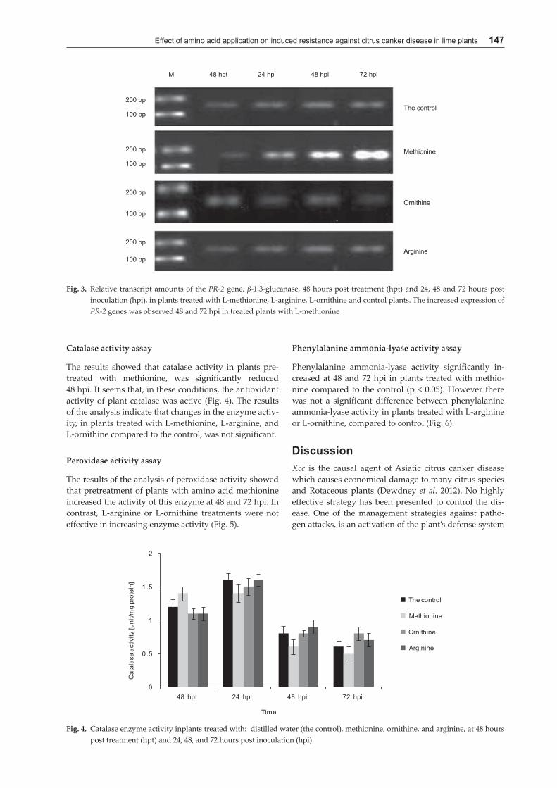
Semi-quantitative reverse transcription PCR was used to compare the relative transcript amounts of the PR-2 gene, β-1,3-glucanase, 48 hpt, and 24, 48, and 72 hpi in plants treated with L-methionine, L-arginine, L-ornithine, and in the control plants.
The results of RT-PCR showed that relative expres-sion of PR-2 genes increased in treated plants with amino acid methionine as compared with the control plants. The increased expression was observed 48 and 72 hpi. It should be noted that the maximum increase in the sampling time was at 72 hpi. The amount of gene expression in the control plants was fixed at all times, which indicates this gene is expressed uniformly at all times (Fig. 3).
Fig. 1. Effect of foliar applied amino acids L-methionine (A) and L-arginine and L-ornithine (B) compared to water control on disease severity of citrus canker on lime leaves, four weeks after inoculation. Spray treatment with L-methionine, reduced lesion size by about 78.5% as compared to the water control. No significant reduction in lesion size was observed following treatment with L-arginine or L-ornithine
Fig. 2. The diameter of necrotic lesions in plants treated with: distilled water (the control), methionine, ornithine, and arginine four weeks after inoculation with bacterial sus-pension. Different letters showed significant difference based on Duncan’s test in p < 0.05
Effect of amino acid application on induced resistance against citrus canker disease in lime plants 147
Catalase activity assay
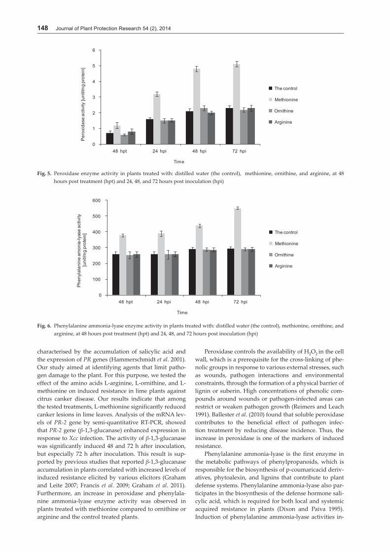
The results showed that catalase activity in plants pre-treated with methionine, was significantly reduced 48 hpi. It seems that, in these conditions, the antioxidant activity of plant catalase was active (Fig. 4). The results of the analysis indicate that changes in the enzyme activ-ity, in plants treated with L-methionine, L-arginine, and L-ornithine compared to the control, was not significant.
Peroxidase activity assay
The results of the analysis of peroxidase activity showed that pretreatment of plants with amino acid methionine increased the activity of this enzyme at 48 and 72 hpi. In contrast, L-arginine or L-ornithine treatments were not effective in increasing enzyme activity (Fig. 5).
Phenylalanine ammonia-lyase activity assay
Phenylalanine ammonia-lyase activity significantly in-creased at 48 and 72 hpi in plants treated with methio-nine compared to the control (p < 0.05). However there was not a significant difference between phenylalanine ammonia-lyase activity in plants treated with L-arginine or L-ornithine, compared to control (Fig. 6).
DiscussionXcc is the causal agent of Asiatic citrus canker disease which causes economical damage to many citrus species and Rotaceous plants (Dewdney et al. 2012). No highly effective strategy has been presented to control the dis-ease. One of the management strategies against patho-gen attacks, is an activation of the plant’s defense system
Fig. 3. Relative transcript amounts of the PR-2 gene, β-1,3-glucanase, 48 hours post treatment (hpt) and 24, 48 and 72 hours post inoculation (hpi), in plants treated with L-methionine, L-arginine, L-ornithine and control plants. The increased expression of PR-2 genes was observed 48 and 72 hpi in treated plants with L-methionine
Fig. 4. Catalase enzyme activity inplants treated with: distilled water (the control), methionine, ornithine, and arginine, at 48 hours post treatment (hpt) and 24, 48, and 72 hours post inoculation (hpi)
148 Journal of Plant Protection Research 54 (2), 2014
characterised by the accumulation of salicylic acid and the expression of PR genes (Hammerschmidt et al. 2001). Our study aimed at identifying agents that limit patho-gen damage to the plant. For this purpose, we tested the effect of the amino acids L-arginine, L-ornithine, and L-methionine on induced resistance in lime plants against citrus canker disease. Our results indicate that among the tested treatments, L-methionine significantly reduced canker lesions in lime leaves. Analysis of the mRNA lev-els of PR-2 gene by semi-quantitative RT-PCR, showed that PR-2 gene (β-1,3-glucanase) enhanced expression in response to Xcc infection. The activity of β-1,3-glucanase was significantly induced 48 and 72 h after inoculation, but especially 72 h after inoculation. This result is sup-ported by previous studies that reported β-1,3-glucanase accumulation in plants correlated with increased levels of induced resistance elicited by various elicitors (Graham and Leite 2007; Francis et al. 2009; Graham et al. 2011). Furthermore, an increase in peroxidase and phenylala-nine ammonia-lyase enzyme activity was observed in plants treated with methionine compared to ornithine or arginine and the control treated plants.
Peroxidase controls the availability of H2O2 in the cell wall, which is a prerequisite for the cross-linking of phe-nolic groups in response to various external stresses, such as wounds, pathogen interactions and environmental constraints, through the formation of a physical barrier of lignin or suberin. High concentrations of phenolic com-pounds around wounds or pathogen-infected areas can restrict or weaken pathogen growth (Reimers and Leach 1991). Ballester et al. (2010) found that soluble peroxidase contributes to the beneficial effect of pathogen infec-tion treatment by reducing disease incidence. Thus, the increase in peroxidase is one of the markers of induced resistance.
Phenylalanine ammonia-lyase is the first enzyme in the metabolic pathways of phenylpropanoids, which is responsible for the biosynthesis of p-coumaricacid deriv-atives, phytoalexin, and lignins that contribute to plant defense systems. Phenylalanine ammonia-lyase also par-ticipates in the biosynthesis of the defense hormone sali-cylic acid, which is required for both local and systemic acquired resistance in plants (Dixon and Paiva 1995). Induction of phenylalanine ammonia-lyase activities in-
Fig. 6. Phenylalanine ammonia-lyase enzyme activity in plants treated with: distilled water (the control), methionine, ornithine, and arginine, at 48 hours post treatment (hpt) and 24, 48, and 72 hours post inoculation (hpi)
Fig. 5. Peroxidase enzyme activity in plants treated with: distilled water (the control), methionine, ornithine, and arginine, at 48 hours post treatment (hpt) and 24, 48, and 72 hours post inoculation (hpi)
Effect of amino acid application on induced resistance against citrus canker disease in lime plants 149
creased significantly in plants treated with L-methionine. This result is in agreement with previous findings that phenylalanine ammonia-lyase is involved in increas-ing resistance and significantly increases in response to the stimulation of different resistance elicitors in citrus fruit (Droby et al. 2009; Ballester et al. 2010). The present study assessed the effect of the L-methionine on disease development. Enhanced disease resistance induced by L-methionine and riboflavin against powdery mildew infection has been demonstrated for melon, cantaloupe, squash, pea, and strawberry (Tzeng et al. 1996; Sarosh et al. 2005). The results from this study indicated that pre-treatment of lime plants with L-methionine induced the levels of disease resistance against Xcc, and significantly reduced the severity of disease.
ReferencesBallester A.R., Izquierdo A., Lafuente M.T., González-Candelas
L. 2010. Biochemical and molecular characterization of induced resistance against Penicillium digitatum in citrus fruit. Postharvest Biol. Technol. 56 (1): 31–38.
Bradford M.M. 1976. A rapid and sensitive method for quantiza-tion of microgram quantities of protein utilizing the prin-ciple of protein-dye binding. Anal. Biochem. 72 (1): 248–254.
Cohen Y., Niderman T., Mosinger E., Fluhr R. 1994. β-Amino-butyric acid induces the accumulation of pathogenesis-re-lated proteins in tomato (Lycopersicon esculentum Mill.) and resistance to late blight infections caused by Phytophthora infestans. Plant Physiol. 104 (1): 59–66.
Dekkers M.G.H., Graham J.H., Burns J.K., Cubero J., Colburn G.C. 2004. Evaluation of chemical inducers and PR protein reporters for induced systemic resistance to citrus bacterial diseases. Phytopathology 94 (6): S25.
Dewdney M.M., Graham J.H. 2012. Florida Citrus Pest Manage-ment Guide: Citrus Canker. Institute of Food and Agricul-tural Sciences, University of Florida, 4 pp.
Dhindsa R.S., Dhindsa P., Thorpe A.T. 1981. Leaf senescence cor-related with increased levels of membrane permeability and lipid peroxidation and decrease levels of superoxide dismutase and catalase. J. Exp. Bot. 32 (1): 93–101.
Droby S., Wisniewski M., Macarisin D., Wilson C. 2009. Twen-ty years of posthar-vest biocontrol research: is it time for a new paradigm? Postharvest Biol. Technol. 52 (2): 137–145.
Francis M.I., Redondo A., Burns J.K., Graham J.H. 2009. Soil ap-plication of imidacloprid and related SAR-inducing com-pounds produces effective and persistent control of citrus canker. Eur. J. Plant Pathol. 124 (2): 283–292.
Gabriel D.W., Kingsley M.T., Hunter J.E., Gottwald T.R. 1989. Re-instatement of Xanthomonas citri (ex Hasse) and X. phaseoli (ex Smith) and reclassification of all X. campestris pv. citri strains. Int. J. Syst. Bacteriol. 39 (1): 14–22.
Gapinska M., Sklodowska M., Gabara B. 2008. Effect of short- and long-term salinity on the activities of antioxidative en-
zymes and lipid peroxidation in tomato roots. Acta Physi-ol. Plant 30 (1): 11–18.
Graham J.H., Myers M.E. 2011. Soil application of SAR inducers imidacloprid, thiamethoxam, and acibenzolar-S-methyl for citrus canker control in young grapefruit trees. Plant Dis. 95 (6): 725–728.
Graham J.H., Leite R.P., Jr. 2007. Soil applied neonicotinoids for control of bacterial diseases on young citrus trees. p. 107. In: Proc. of International Workshops: “PR-Proteins” and “Induced Resistance Against Insects and Diseases” Doorn, The Netherlands, 10–14 May 2007.
Hammerschmidt R., Métraux J.-P., van Loon L.C. 2001. Induc-ing resistance: a summary of papers presented at the First International Symposium on Induced Resistance to Plant Diseases, Corfu, May 2000. Eur. J. Plant Pathol. 107 (1): 1–6.
Jakab G., Cottier V., Toquin V., Rigoli G., Zimmerli L. 2001. β-Aminobutyric acidinduced resistance in plants. Eur. J. Plant Pathol. 107 (1): 29–37.
Li J., Zingen-Sell I., Buchenauer H. 1996. Induction of resistan-ceof cotton plants to Verticillium and of tomato plants to Fu-sarium wilt by 3-aminobutyric acid and methyl jasmonate. J. Plant Dis. Prot. 103 (3): 288–299.
Ojha S., Chandra Chatterjee N. 2012. Induction of resistance in tomato plants against Fusarium oxysporum f. sp. lycopersici mediated through salicylic acid and Trichoderma harzianum. J. Plant Prot. Res. 52 (2): 220–225.
Oka Y., Cohen Y., Spiegel Y. 1999. Local and systemic induced resistance to the root-knot nematode in tomato by DL-β-amino-n-butyric acid. Phytopathology 89 (12): 1138–1143.
Reimers P.J., Leach J.E. 1991. Race-specific resistance to Xan-thomonas oryzae pv. oryzae conferred by bacterial blight resistance gene Xa-10 in rice (Oryza sativa) involves accu-mulation of a lignin-like substance in host tissues. Physiol. Mol. Plant Pathol. 38 (1): 39–55.
Reuveni R. 1995. Biochemical marker of disease resistance. p. 99–114. In: “Molecular Methods in Plant Pathology” (R.P. Singh, U.S. Singh, eds.). Boca Raton, CRC Press, Florida, USA, 507 pp.
Sarosh B.R., Sivaramakrishnan S., Shetty H.S. 2005. Elicitation of defense related enzymes and resistance by L-methionine in pearl millet against downy mildew disease caused by Scle-rospora graminicola. Plant Physiol. Biochem. 43 (8): 808–815.
Tzeng D.D., Tzeng H.C., Chen R., Cheng A., Tsai C.C., Chen C., Hwang T., Yeh Y., DeVay J.E. 1996. The use of MR formu-lation as a novel and environmentally safe photodynamic fungicide for the control of powdery mildews. Crop Prot. 15 (4): 341–347.
Van Loon L.C., Rep M., Pieterse C.M.J. 2006. Significance of in-ducible defense-related proteins in infected plants. Annu. Rev. Phytopathol. 44 (1): 135–162.
Yao H.J., Tian S.P. 2005. Effects of a biocontrol agent and methyl jasmonate on postharvest diseases of peach fruit and the possible mechanisms involved. J. Appl. Microbiol. 98 (4): 941–950.
Zimmerli L., Jakab G., Matraux J.P., Mauch-Mani B. 2000. Poten-tiation of pathogen-specific defense mechanisms in Arabi-dopsis by β-aminobutyric acid. Proc. Natl. Acad. Sci. USA 97 (23): 12920–12925.