This study compared the microtensile bondstrength (MTBS) of three all-in-one adhesive sys-tems and a two-step system using two types ofburs to prepare the dentin surfaces. Flat coronal
surfaces of 24 extracted human molars were pro-duced using either regular-grit or superfine-gritdiamond burs. Resin composite was then bondedto equal numbers of these surfaces using one ofthe four adhesives: Clearfil SE Bond (CSE), G-Bond (GB), SSB-200 (SSB) or Prompt L-Pop(PLP). After storage for 24 hours in 37°C distilledwater, the bonded teeth were sectioned intoslices (0.7-mm thick) perpendicular to the bondedsurface. The specimens were then subjected tomicrotensile testing and the bond strengths werecalculated at failure. Bond strength data wereanalyzed by two-way ANOVA and the Games-Howell test for interaction between adhesive andtype of cut dentin. The fractured surfaces wereobserved by SEM to determine the failure mode.In addition, to observe the effect of conditioning,equal numbers of the two bur-cut dentin surfacesof eight additional teeth were conditioned withthe adhesives and observed by SEM. Based on theresults, when CSE and SSB were bonded todentin cut with a regular-grit diamond bur, theMTBS values were significantly lower than thatof superfine bur-cut dentin; whereas, GB andPLP showed no significant differences in MTBSbetween the two differently cut surfaces. SEMobservation of the fractured surfaces revealed amixed mode (adhesive in some areas and cohe-sive in others in the same sample) of failure in allspecimens except PLP, which showed cohesivefailure within the adhesive for both types of bur
Effect of Different Bur Grindingon the Bond Strength ofSelf–etching Adhesives
S Semeraro • D Mezzanzanica • D Spreafico • M GaglianiD Re • T Tanaka • SK Sidhu • H Sano
Clinical Relevance
In some self-etching systems, selecting the proper bur type for cutting dentin is impor-tant for improving bond strength.
Stefano Semeraro, DDS, Department of Prosthodontics,School of Dentistry, University of Milan, Milan, Italy
Dario Mezzanzanica, DDS, Department of Restorative andEndodontics, School of Dentistry, University of Milan, Milan, Italy
Diego Spreafico, DDS, Department of Prosthodontics, Schoolof Dentistry, University of Milan, Milan, Italy
Massimo Gagliani, MD, DDS, PhD, associate professor,Department of Restorative and Endodontics, School ofDentistry, University of Milan, Milan, Italy
Dino Re, MD, DDS, PhD, researcher, Department ofProsthodontics, School of Dentistry, University of Milan,Milan, Italy
Toru Tanaka, DDS, PhD, assistant professor, Cariology,Operative Dentistry and Endodontology, Department of OralHealth Science, Hokkaido University Graduate School ofDental Medicine, Sapporo, Japan
Sharanbir K Sidhu, BDS, MSc, PhD, FICD, MFDSRCS,FADM, lecturer, Restorative Dentistry, School of DentalSciences, University of Newcastle, Newcastle upon Tyne, UK
*Hidehiko Sano, DDS, PhD, professor, Cariology, OperativeDentistry and Endodontology, Department of Oral HealthScience, Hokkaido University Graduate School of DentalMedicine, Sapporo, Japan
*Reprint request: Kita 13, Nishi 7, Kita-ku, Sapporo 060-8586,Japan; e-mail: [email protected]
DOI: 10.2341/04-171
Laboratory Research
318 Operative Dentistry
preparation. Generally, SEMs of the conditionedsurfaces using both types of burs showed partialremoval of the smear layer for CSE, minimal forGB and SSB and complete removal for PLP.
In conclusion, when cutting dentin, selectingthe proper bur type is important for improvingthe bond strength of some self-etching adhesivesystems.
INTRODUCTION
The smear layer has been defined as a layer of debris onthe surface of dental tissues created by cutting a tooth(Eick & others, 1970). It varies in thickness, roughness,density and degree of attachment to the underlyingtooth structure according to the surface preparation(Charbeneau, Peyton & Anthony, 1957; Gilboe & others,1980). As part of restorative procedures in adhesive den-tistry, the smear layer must be removed, modified orimpregnated by the resin to allow for bonding betweenthe tooth and the restorative material (Swift Jr,Perdigão & Heymann, 1995; Pashley & Carvalho, 1997).
The poor performance of early dentin adhesive sys-tems was thought to occur because the smear layer wasnot removed, resulting in the adhesive bonding to thesurface of the smeared debris (Watanabe, Nakabayashi& Pashley, 1994a) and not to the underlying dentin(Eick & others, 1970). The potential of the smear layerto create an adverse effect on dentin bonding has beenreported by Prati and others (1990). The smear layeradheres weakly to dentin, and its removal by an aciddemineralizing agent prior to the application of a bondingsystem has been reported to result in stronger bonds(Pashley, 1991). However, others have reported that thetreatment of dentin with acids can cause the collapse ofexposed collagen fibers due to the removal of the sup-porting hydroxyapatite and the denaturation ofcollagen (Nakabayashi, 1992; Pashley & others, 1993).The remaining matted collagen surface becomes moredifficult to impregnate with adhesive monomers. Toovercome this problem, the application of a primer aimsto restore the permeability of acid-treated dentin andfacilitates the penetration of applied monomers.Therefore, the use of an acidic conditioner is necessaryto dissolve and remove the smear layer to expose theintertubular and peritubular dentin, remove the debrisfrom the dentin surface and demineralize the superfi-cial dentin matrix, thus allowing the subsequent infil-tration of the resin into the dentin surface.
Clinically, after carious dentin has been removed orany other kind of dentin instrumentation has beenperformed, a smear layer is formed over the dentinsurface (Pashley & Carvalho, 1997; Ogata & others,2001). The nature of the smear layer depends on thetype of bur used. In addition, different speeds of thebur and the pressure applied may influence the kind ofsmear layer.
Coarse and superfine diamond burs each create a dif-ferent smear layer. Coarse diamond burs create a thicksmear layer containing cut collagen fibers and hydrox-yapatite crystallites (Ayad, Rosenstiel & Hassan, 1996;Gwinnett, 1984). This can interfere with bonding of theadhesive agents, as it is not easy for some adhesivemonomers to permeate dentin smears and impregnatethe underlying dentin (Nakabayashi & Saimi, 1996).Differences in the smear layer generated by burs andabrasive papers have been reported to affect the bondstrengths of resins to dentin (Tagami & others, 1991;Watanabe, Saimi & Nakabayashi, 1994b).
The “all-in-one” adhesive systems are simple to use,as the steps of etching, priming and bonding occur inone single application step. A previous study of thesesystems has demonstrated the influence of the type ofsmear layer generated on their bond strength (Inoue &others, 2001). However, that study found that use of acoarse diamond bur may reduce the possibility of pene-tration of bonding monomers into dentinal substrate inthese systems. It is not known whether these differ-ences are also true for the newer, all-in-one adhesives.The reasons for using self-etching primers include easyhandling by the operator (Sano & others, 1998;Miyazaki, Onose & Moore, 2000) and high clinical per-formance (Latta & others, 1997). These primers weremarketed in the last decade and since then, there havebeen many self-etching primers available. Currentadvances in these systems are toward one-bottle, all-in-one adhesives. Older versions of self-etching primerswere believed to be susceptible to the presence of thesmear layer in terms of bond strengths (Toida,Watanabe & Nakabayashi, 1995). When self-etchingprimers are applied to the smear layer situated on thetooth surface, the acid primer can simultaneously modifyor dissolve the smear layer and decalcify the dentin(Watanabe & others, 1994a); this procedure producesgood adhesion both to enamel and dentin (Kanemura,Sano & Tagami, 1999). Recent reports have demon-strated that some all-in-one systems bond well to thinnersmear layer covered dentin (Inoue & others, 2001;Koase & others, 2004).
This study compared the microtensile bond strengthof four adhesive systems (including three all-in-one sys-tems) after preparing the dentinal surface with either acoarse or superfine diamond bur. The null hypothesiswas that there are no differences in microtensile bondstrengths to dentin prepared with either a coarse dia-mond bur or a superfine bur.
METHODS AND MATERIALS
Adhesives and Bonding Procedures
Twenty-four caries-free extracted human molars,stored at 37°C in distilled water, were used for thisstudy, in accordance with local institutional guidelines.The coronal surfaces of the teeth were trimmed using a
319
model trimmer (MT-7 Morita Corp, Kyoto, Japan) inorder to form a long, flat dentin surface at the mid-crown level. A smear layer was then created by remov-ing a thin layer of the surface with a high-speed dia-mond bur under water-cooling. Twelve teeth were pre-pared with a regular-grit diamond bur (Diamond PointFG, #106RD, Shofu, Kyoto, Japan), while theremainder were prepared using a superfine-grit dia-mond bur (Diamond Point FG, #SF 106RD, Shofu Inc).These two types of dentin substrates were randomlyassigned to one of the four bonding treatments carriedout according to the respective manufacturers’ instruc-tions.
After applying the adhesive in each tooth, resin com-posite (Clearfil AP-X, Kuraray, Okayama, Japan) wasbuilt-up incrementally (in three increments) to a heightof 5 mm. Each increment was light cured for 40 sec-onds, and the specimens were then stored in distilledwater for 24 hours at 37°C.
Microtensile Bond Strength Testing
The specimens were sectioned into six slabs, approxi-mately 0.7-mm thick, perpendicular to the bonded sur-face using a low-speed diamond saw (Isomet, Buehler,Lake Bluff, IL, USA) under water. These slabs weretrimmed to an hourglass shape to form a gentle curvealong the adhesive interface from both sides, using asuperfine-grit diamond bur as described by Sano andothers (1994). The width at the narrowest portion wasapproximately 1.4 mm, and the thickness of the bondedarea of each specimen was verified by a digital microm-eter. The specimens were then attached to a Ciucchi’sjig (Paul & others, 1999) with cyanoacrylate adhesive(Model Repair II Blue, Dentsply-Sankin, Otahara,Japan) connected to a desktop testing apparatus (EZtest, Shimadzu, Kyoto, Japan).
The specimens were then subjected to a microtensilestrength test at a crosshead speed of 1 mm/minute untilfailure occurred. The tensile bond strength was calcu-lated as the load at failure divided by the bonded area.Bond strength data were analyzed by two-way ANOVAand the Games-Howell test for interaction between theadhesive and type of cut dentin. The surfaces of thefractured specimens were visually inspected under alight microscope (20x),then observed micro-scopically with SEMto determine the fail-ure mode.
Failure ModeAnalysis
For determining themodes of fracture,both the dentin andcomposite halves ofthe fractured
specimens were observed with a FE-SEM microscope(Hitachi S4000, Tokyo, Japan). The failure modes wereclassified as interfacial (fracture between the dentin orthe hybrid layer and the overlying adhesive in thesame sample), mixed (interfacial and partial cohesivefailure in dentin or composite in the same sample) orcohesive (failure within dentin only, cohesive in adhe-sive only or cohesive in resin composite only), wher-ever relevant.
SEM Observation of Dentin Surface Treated withthe Adhesives
In order to understand the effect of conditioning on thedentinal surfaces treated with the two types of burs, afurther evaluation was conducted. The coronal surfacesof eight additional teeth were trimmed at the mid-crown level; the flat dentin surfaces of four teeth werethen treated using the two burs as previously describedin the method. One surface in each group of four teethwas treated with the self-etching primer (CSE,Kuraray, Osaka, Japan) or with one of the all-in-oneadhesives (GB, GC Company, Tokyo, Japan; SSB,Kuraray and PLP, 3M ESPE, St Paul, MN, USA) with-out light curing. The teeth were immediately soaked in100% acetone for one minute to remove the appliedadhesive. All the specimens were dehydrated using anascending concentration of ethanol and chemical-driedwith HMDS, which is a protocol for SEM examinationdescribed by Perdigão and others (1995).
RESULTS
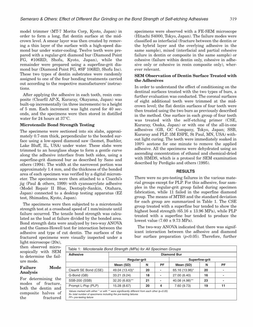
There were no pre-testing failures in the various mate-rial groups except for PLP. For this adhesive, four sam-ples in the regular-grit group failed during specimenfabrication, while 11 failed in the superfine diamondgroup. The means of MTBS and the standard deviationfor each group are summarized in Table 1. The CSEgroup treated with a superfine bur tended to show thehighest bond strength (65.16 ± 13.96 MPa), while PLPtreated with a superfine bur tended to produce thelowest value (7.60 ± 9.73 MPa).
The two-way ANOVA indicated that there was signif-icant interaction between the adhesive and diamondbur surface preparation (p<0.05). Therefore, further
Adhesive Diamond Bur
Regular-grit Superfine-grit
Mean (SD) N PF Mean (SD) N PF
Clearfil SE Bond (CSE) 49.04 (13.43)* 20 - 65.16 (13.96)* 20 -
Values marked with either * or with ** were significantly different from each other (p<0.05)N= total number of specimens including the pre-testing failuresPF= pre-testing failure
Table 1: Microtensile Bond Strength (MPa) for All Specimen Groups
Semeraro & Others: Effect of Different Bur Grinding on the Bond Strength of Self-etching Adhesives
320 Operative Dentistry
analysis for significant differences between groups wascarried out using the Games-Howell test.
Significant differences were found between MTBS todentin cut with a regular-grit diamond (lower values)bur and dentin cut with a superfine-grit diamond buronly for the CSE and SSB adhesives (p<0.05). For bothGB and PLP, no significant differences in MTBS valueswere shown between the specimens prepared with reg-ular-grit burs compared to those prepared withsuperfine-grit burs.
Fractographical analysis of the specimens using SEMrevealed a mixed mode of failure in all groups exceptPLP. The specimens of CSE bonded to dentin cut withthe regular-grit diamond bur showed failures at dif-ferent levels of the adhesive in the same sample, thatis, interfacial between the hybrid layer and the adhe-sive, within the adhesive as well as the interfacialbetween the adhesive and the overlying resin com-posite. In the CSE group bonded to dentin cut with thesuperfine-grit diamond bur, the mixed fracturesoccurred not only within the adhesive, but they werealso interfacial between the dentin and the adhesive.
For the GB group, the mixed fractures for both spec-imens cut with a superfine and a regular-grit diamondbur were interfacial (between the hybrid layer and theadhesive) and also occurred at different planes in theadhesive within the same sample; the adhesive alsoshowed the presence of “blisters” (Figure 1) in bothgroups.
In the SSB samples cut with a regular diamond, themixed failures in each sample were both interfacialbetween the hybrid layer and the adhesive, as well as
within the adhesive. Those prepared with a superfine-grit bur displayed fractures at different planes withinthe adhesive—cohesive failure within the adhesive.
In the PLP samples treated with both types of burs,all the samples showed failures that were cohesivewithin the adhesive only (Figure 2).
SEM observations of the conditioned dentin surfaceswith the two types of burs revealed differences with thevarious adhesives. Dentin prepared with regular-gritburs treated with CSE showed partial removal of thesmear layer and the peritubular dentin appeared to beslightly etched (Figure 3A). Dentin prepared withsuperfine-grit burs displayed partial demineralizationof the smear layer. The peritubular dentin was alsocomparatively more etched, and the porosity of theintertubular dentin was slightly greater than the regu-lar grit prepared dentin (Figure 3B).
There were no distinct differences in terms of removalof the smear layer between the dentin cut with a regular-grit bur and dentin cut with a superfine diamond forGB. The smear layer was partially demineralized andthe remnants were attached to the dentin surface. Theonly distinct differences were dissolution of the smearplugs: the smear plugs in the regular-grit prepareddentin were more resistant to the acidity of the adhe-sive and therefore remained.
For SSB, removal of the smear layer was minimaland similar for both specimens cut with a regular-gritbur and those cut with a superfine diamond. The poros-ity of the intertubular dentin cut with a superfine-gritbur appeared to be relatively greater than that of regu-lar-grit burs.
A
B
Figure 1. SEM photomicrograph of a typical (both regular and superfine-grit) specimen of GB showing interfacial fracture (I) between the hybridlayer and the adhesive, as well as areas where the failure occurred with-in planes in the adhesive (A). In addition, “blisters” (arrowed) can be seenin the adhesive.
Figure 2. SEM image of a typical PLP (of both regular and superfine-grit)sample showing a cohesive fracture that occurred within the adhesivelayer.
321
The PLP samples cut with both a regular-grit and asuperfine-grit bur showed complete removal of thesmear layer and plugs. Additionally, complete dissolu-tion of peritubular dentin was noted in both. The inter-tubular dentin cut with the regular-grit bur appearedless aggressively etched compared to dentin cut withthe superfine-grit bur (Figures 4A and 4B).
DISCUSSION
This study assessed the effect of different smear layersgenerated by two different types of burs on themicrotensile bond strengths to dentin.
CSE performed significantly better with superfine cutdentin. CSE primer partially dissolves the smear layeras seen in Figures 3A and 3B. It was previously reported
Figure 4A. SEM appearance of a dentin surface cut with a regular-gritdiamond bur and conditioned with PLP, which was not light-cured, fol-lowed by rinsing with acetone for 1 minute.This shows complete removalof the smear layer and plugs. In addition, the complete dissolution of per-itubular dentin can be seen.
Figure 4B. SEM micrograph of a dentin surface cut with a superfine-gritdiamond bur and conditioned with PLP, which was not light-cured, fol-lowed by rinsing with acetone for 1 minute.This shows complete removalof the smear layer and plugs, as well as complete dissolution of per-itubular dentin as in Figure 4A. The intertubular dentin appears moreaggressively etched compared to that cut with a regular-grit bur in Figure4A.
Figure 3A. SEM image of a dentin surface cut with a regular-grit dia-mond bur, conditioned with CSE, which was not light-cured, followed byrinsing with acetone for 1 minute. The micrograph shows shows partialremoval of the smear layer and the peritubular dentin appears to beslightly etched.
Figure 3B. SEM micrograph of a similarly treated sample as in Figure 3A,but with the dentin cut with a superfine diamond bur. Partial demineral-ization of the smear layer is noted. The peritubular dentin appears com-paratively more etched, and the porosity of the intertubular dentin isslightly greater than that of regular-grit prepared dentin.
Semeraro & Others: Effect of Different Bur Grinding on the Bond Strength of Self-etching Adhesives
322 Operative Dentistry
that a hybridized smear layer and hybridized dentinwere observed when using CSE (Tay & others, 2000).CSE can penetrate the partially demineralized smearlayer and create a hybrid layer (Tay & others, 2000).The greater porosity of intertubular dentin observed inthis study with the superfine-grit preparation impliesmore channels of penetration of adhesive monomers.This could be the reason for the greater bond strengthsachieved with the superfine diamond bur.
For GB, there were no differences in bond strengthsbetween the dentin cut with a regular-grit bur anddentin cut with a superfine diamond bur. This may beattributed to the similar mild removal of the smearlayer in both. Another reason there were no differencesin bond strengths may be the presence of “blisters”within the adhesive resin. Blisters may create defectswithin the adhesive resin during tension and initiatethe propagation of cracks within the adhesive. Theelimination of blisters should be important for producinggood bonding between the resin and dentin. Furtherwork is required to determine why blisters occur.
SSB showed significantly higher bond strengths tosuperfine cut dentin compared to regular cut dentin.Although the smear layer removal for both groups isminimal, increased porosity within the intertubulardentin was found for the superfine grit. This mayexplain the higher bond strengths observed in thisgroup. Further work, such as using TEM, is necessaryto clarify the interaction between the resin and dentinof this system.
PLP was the only adhesive tested which had pre-testing failures in both groups. In addition, PLP tendedto show the lowest bond strengths for both groups andconsistently demonstrated cohesive failure within theadhesive. Although Figures 4A and 4B show completedissolution of the smear layer (due to the low pH of 0.8)to expose the collagen fibrils, whether or not the subse-quent penetration of resin monomers into the exposedcollagen web occurs is not clear. The authors speculatethat the relatively lower bond strengths with this adhe-sive suggest incomplete hybridization. Future studiesshould focus on the quality of the resin-dentin interfaceof this system.
CONCLUSIONS
This study showed that the type of bur used to preparethe dentinal surface may affect microtensile bondstrength when using some of the current all-in-one sys-tems. Hence, the null hypothesis that there are no dif-ferences in microtensile bond strengths to dentin pre-pared with either a coarse diamond bur or a superfinebur was rejected. In conclusion, when cutting dentin,selecting the proper bur type is important for improvingthe bond strength of some self-etching adhesive sys-tems.
(Received 8 October 2004)
References
Ayad MF, Rosenstiel SF & Hassan MM (1996) Surface roughnessof dentin after tooth preparation with different rotary instru-mentation Journal of Prosthetic Dentistry 75(2) 122–128.
Charbeneau GT, Peyton FA & Anthony DH (1957) Profile charac-teristics of cut tooth surfaces developed by rotating instru-ments Journal of Dental Research 957–964.
Eick JD, Wilko RA, Anderson CH & Sorensen SE (1970) Scanningelectron microscopy of cut tooth surfaces and identification ofdebris by use of the electron microprobe Journal of DentalResearch 49(6) 1359–1368.
Gilboe DB, Svare CW, Thayer KE & Drennon DG (1980) Dentinalsmearing: An investigation of the phenomenon Journal ofProsthetic Dentistry 44(3) 310–316.
Inoue H, Inoue S, Uno S, Takahashi A, Koase K & Sano H (2001)Microtensile bond strength of two single–step adhesive sys-tems to bur prepared dentin Journal of Adhesive Dentistry 2129-139.
Kanemura N, Sano H & Tagami J (1999) Tensile bond strengthand SEM evaluation of ground and intact enamel surfacesJournal of Dentistry 27 523-530.
Koase K, Inoue S, Noda M, Tanaka T, Kawamoto C, Takahashi A,Nakaoki Y & Sano H (2004) Effect of bur-cut dentin on bondstrength using two all-in-one and one two-step adhesive sys-tems Journal of Dentistry 6 97-104.
Latta MA, Barkmeier WW, Triolo PT, Cavel WT & Blankenau RJ(1997) One year clinical evaluation of the Clearfil Liner Bond2 system Journal of Dental Research 76 Abstract #1186 p 162.
Miyazaki M, Onose H & Moore K (2000) Effect of operator vari-ability on dentin bond strength of two-step bonding systemsAmerican Journal of Dentistry 13 101-104.
Nakabayashi N (1992) The hybrid layer: A resin-dentin compos-ite Proceedings of the Finnish Dental Society 88 321-329.
Nakabayashi N & Saimi Y (1996) Bonding to intact dentinJournal of Dental Research 75(9) 1706–1715.
Ogata M, Harada N, Yamaguchi S, Nakajima M, Pereira PN &Tagami J (2001) Effects of different burs on dentin bondstrengths of self-etching primer bonding systems OperativeDentistry 26(4) 375–382.
Pashley DH (1991) Dentin bonding: Overview of the substratewith respect to adhesive materials Journal of EstheticsDentistry 3 46-50.
Pashley DH, Ciucchi B, Sano H & Horner JA (1993) Permeabilityof dentin to adhesive agents Quintessence International 24618-631.
Paul SJ, Welter DA, Ghazi M & Pashley DH (1999) Nanoleakageat the dentin adhesive interface vs tensile bond strengthOperative Dentistry 24 181-188.
Perdigão J, Lambrechts P, Van Meerbeek B, Vanherle G & LopesAL (1995) Field emission SEM comparison of four postfixationdrying techniques for human dentin Journal BiomedicalMaterials Research 29(9) 1111-1120.
323
Prati C, Bigini G, Rizzoli C, Nucci C, Zucchini C & Montanari G(1990) Shear bond strength and SEM evaluation of dentinalbonding systems American Journal of Dentistry 3 283-288.
Sano H, Shono T, Sonoda H, Takatsu T, Ciucchi B, Carvalho R &Pashley DH (1994) Relationship between surface area foradhesion and tensile bond strength–evaluation of microtensilebond test Dental Materials 10 236-240.
Sano H, Kanemura N, Burrow MF, Inai N, Yamada T & TagamiJ (1998) Effect of operator variability on dentin adhesionDental Materials Journal 17 51-58.
Swift EJ Jr, Perdigão J & Heymann HO (1995) Bonding to enam-el and dentin: A brief history and state of the art QuintessenceInternational 26(2) 95–110.
Tagami J, Tao L, Pashley DH, Honoda H & Sano H (1991) Effectof high-speed cutting on dentin permeability and bondingDental Materials 7 234-239.
Tay FR, Carvalho R, Sano H & Pashley DH (2000) Effect of smearlayers on the bonding of a self-etching primer to dentinJournal of Adhesive Dentistry 2 99-116.
Toida T, Watanabe A & Nakabayashi N (1995) Effect of smearlayer on bonding to dentin prepared with the bur The Journalof the Japanese Society for Dental Materials and Devices 14109-116.
Watanabe I, Nakabayashi N & Pashley DH (1994a) Bonding toground dentin by a phenyl-P self-etching primer Journal ofDental Research 73(6) 1212–1220.
Watanabe I, Saimi Y & Nakabayashi N (1994b) Effect of smearlayer on bonding to ground dentin-relationship between grind-ing condition and tensile bond strength The Journal of theJapanese Society for Dental Materials and Devices 13 101-108.
Semeraro & Others: Effect of Different Bur Grinding on the Bond Strength of Self-etching Adhesives