The aim of the study was to evaluate dentinal tubule occlusion, measuring the dentin permeability (Lp) and using different desensitizing agents before and after abrasive/erosive challenge. Dentin discs from 42 healthy human third molars were obtained. Minimum Lp was measured after a smear layer simulation using #600 SiC paper and maximum Lp after an immersion in 0.5 M EDTA. The specimens were treated with different desensitizers: two varnishes (Clinpro XT Varnish-CV, Fluor Protector-FP), a paste (Desensibilize Nano P-NP) and a gel (Oxa Gel-OG). The Lp of each specimen was measured immediately after the desensitizers’ application. The discs were subjected to erosion/abrasion cycles for 7 days, with 0.5% citric acid solution (6x/day) and tooth brushing (3x/day). Lp was measured after the first, fourth and seventh day of the challenge. The data were analyzed by 3-way ANOVA with repeated measurements and by a Games-Howell test (α=5%). FP and CV did not show significant differences in Lp immediately after application until the 7th day (p<0.05). OG showed a significant increase in Lp after the 4th and 7th days. NP resulted in a significantly higher permeability compared to the other materials immediately after the application and after the 1st day of challenge. All the desensitizers reduced the dentin permeability immediately after application. However, only the varnishes were able to maintain the occlusive effect after the erosion/abrasion challenge. Effect of Erosion/Abrasion Challenge on the Dentin Tubule Occlusion Using Different Desensitizing Agents Gabriela D. Canali 1 , Rodrigo N. Rached 1 , Rui F. Mazur 1 , Evelise M. Souza 1,2 1 Graduate Program in Dentistry, School of Life Sciences, PUCPR - Pontifícia Universidade Católica do Paraná, Curitiba, PR, Brazil 2 Operative Dentistry, Department of Restorative Dentistry, UFPR - Universidade Federal do Paraná, Curitiba, PR, Brazil Correspondence: Evelise M. Souza, Rua Imaculada Conceição, 1155, 80215-901 Curitiba, PR, Brasil. Tel: +55-41-3271-1637. e-mail: [email protected]Key Words: dentin permeability, desensitizing agent, dental varnish, erosion, abrasion. ISSN 0103-6440 Brazilian Dental Journal (2017) 28(2): 216-224 http://dx.doi.org/10.1590/0103-6440201700811 Introduction Discomfort caused by dentin hypersensitivity is one of the most common dental complaints. Epidemiological studies demonstrated that the prevalence of dentin hypersensitivity varies from 2.8% to 74% depending on the studied population and the study design (1). Dentin hypersensitivity (DH) occurs when the dentinal tubules are exposed to the oral environment as result of enamel loss by abrasion, erosion, abfraction or any exposure of the root surface caused by gingival recession or periodontal treatments (2). Hypersensitivity is an exaggerated sensitivity, where the patient displays acute pain (variable intensity, short duration) in response to stimuli, like thermal, tactile, osmotic or chemical changes, that cannot be ascribed to any other dental condition (2). The most widely accepted theory for DH was proposed by Brännström in the 1960's (3). This hydrodynamic theory postulates that the application of a stimulus to the exposed dentin disturbs the fluid within the dentinal tubules, causing change in osmotic pressure inside the pulp chamber, which stimulates baroreceptors, leading to neural discharge. However, an alternative theory proposed later states that mechanically induced dentin deformation may directly trigger nerve impulses, or may exert mechanically induced dentinal fluid flow that triggers nerve activity (4). Home-based or in-office products may be employed for the treatment of dentin hypersensitivity by occluding the dentinal tubules. Home-based products include toothpaste and, to a lesser extent, gels and mouthwash solutions. Professionally applied desensitizing agents include sealers, varnishes, gels, pastes or adhesive systems. These products generally contain fluorides, oxalates, potassium nitrate and calcium phosphate, already investigated in vitro for their ability to occlude dentinal tubules and reduce dentin permeability (5-9). The precipitates or barriers formed by the desensitizing agents may be removed or dissolved between applications or shortly after (10). Therefore, the simulation of etiological factors like abrasive wear caused by tooth brushing or erosive loss due to an acidic diet is an important procedure to evaluate the stability of the precipitates and the consequent efficacy of desensitizing agents. Until now, just one in vitro study explored the effect of erosive and abrasive challenges on dentin permeability with different surface treatments (11). Some resin-containing desensitizers, such as resin- modified glass ionomer cements, act on the dentin structure infiltrating the dentinal tubules, thereby reducing the sensitivity of exposed dentin (8). Calcium phosphate- based products are able to occlude the tubules by forming an amorphous mineral similar to apatite, which causes
Transcript
The aim of the study was to evaluate dentinal tubule occlusion, measuring the dentin permeability (Lp) and using different desensitizing agents before and after abrasive/erosive challenge. Dentin discs from 42 healthy human third molars were obtained. Minimum Lp was measured after a smear layer simulation using #600 SiC paper and maximum Lp after an immersion in 0.5 M EDTA. The specimens were treated with different desensitizers: two varnishes (Clinpro XT Varnish-CV, Fluor Protector-FP), a paste (Desensibilize Nano P-NP) and a gel (Oxa Gel-OG). The Lp of each specimen was measured immediately after the desensitizers’ application. The discs were subjected to erosion/abrasion cycles for 7 days, with 0.5% citric acid solution (6x/day) and tooth brushing (3x/day). Lp was measured after the first, fourth and seventh day of the challenge. The data were analyzed by 3-way ANOVA with repeated measurements and by a Games-Howell test (α=5%). FP and CV did not show significant differences in Lp immediately after application until the 7th day (p<0.05). OG showed a significant increase in Lp after the 4th and 7th days. NP resulted in a significantly higher permeability compared to the other materials immediately after the application and after the 1st day of challenge. All the desensitizers reduced the dentin permeability immediately after application. However, only the varnishes were able to maintain the occlusive effect after the erosion/abrasion challenge.
Effect of Erosion/Abrasion Challenge on the Dentin Tubule Occlusion Using Different Desensitizing Agents
Gabriela D. Canali1, Rodrigo N. Rached1, Rui F. Mazur1, Evelise M. Souza1,2
1Graduate Program in Dentistry, School of Life Sciences, PUCPR - Pontifícia Universidade Católica do Paraná, Curitiba, PR, Brazil2Operative Dentistry, Department of Restorative Dentistry, UFPR - Universidade Federal do Paraná, Curitiba, PR, Brazil
Correspondence: Evelise M. Souza, Rua Imaculada Conceição, 1155, 80215-901 Curitiba, PR, Brasil. Tel: +55-41-3271-1637. e-mail: [email protected]
Introduction Discomfort caused by dentin hypersensitivity is one
of the most common dental complaints. Epidemiological studies demonstrated that the prevalence of dentin hypersensitivity varies from 2.8% to 74% depending on the studied population and the study design (1).
Dentin hypersensitivity (DH) occurs when the dentinal tubules are exposed to the oral environment as result of enamel loss by abrasion, erosion, abfraction or any exposure of the root surface caused by gingival recession or periodontal treatments (2). Hypersensitivity is an exaggerated sensitivity, where the patient displays acute pain (variable intensity, short duration) in response to stimuli, like thermal, tactile, osmotic or chemical changes, that cannot be ascribed to any other dental condition (2).
The most widely accepted theory for DH was proposed by Brännström in the 1960's (3). This hydrodynamic theory postulates that the application of a stimulus to the exposed dentin disturbs the fluid within the dentinal tubules, causing change in osmotic pressure inside the pulp chamber, which stimulates baroreceptors, leading to neural discharge. However, an alternative theory proposed later states that mechanically induced dentin deformation may directly trigger nerve impulses, or may exert mechanically induced dentinal fluid flow that triggers nerve activity (4).
Home-based or in-office products may be employed for
the treatment of dentin hypersensitivity by occluding the dentinal tubules. Home-based products include toothpaste and, to a lesser extent, gels and mouthwash solutions. Professionally applied desensitizing agents include sealers, varnishes, gels, pastes or adhesive systems. These products generally contain fluorides, oxalates, potassium nitrate and calcium phosphate, already investigated in vitro for their ability to occlude dentinal tubules and reduce dentin permeability (5-9).
The precipitates or barriers formed by the desensitizing agents may be removed or dissolved between applications or shortly after (10). Therefore, the simulation of etiological factors like abrasive wear caused by tooth brushing or erosive loss due to an acidic diet is an important procedure to evaluate the stability of the precipitates and the consequent efficacy of desensitizing agents. Until now, just one in vitro study explored the effect of erosive and abrasive challenges on dentin permeability with different surface treatments (11).
Some resin-containing desensitizers, such as resin-modified glass ionomer cements, act on the dentin structure infiltrating the dentinal tubules, thereby reducing the sensitivity of exposed dentin (8). Calcium phosphate-based products are able to occlude the tubules by forming an amorphous mineral similar to apatite, which causes
Braz Dent J 28(2) 2017
217
Eros
ion/
abra
sion
effec
t on
dent
in tu
bule
occ
lusi
on
a mechanical and physicochemical reduction in dentin tubule diameter (12). The use of potassium oxalate-based desensitizers causes neural depolarization, as well as occlusion of the tubules, by forming oxalate calcium crystals (13). Fluoride-based varnishes in contact with mineralized dental structures react with the calcium and phosphate ions of dentin, leading to precipitation of calcium fluoride crystals at the orifices of dentinal tubules. However, the crystals thus formed are unstable and therefore undergo rapid dissociation (14).
Regardless the chosen treatment method, the greatest challenge is achieving long-term effectiveness. Therefore, it must be evaluated the behavior of the precipitates formed by the application of these desensitizing agents when exposed to the varying conditions and pH of the oral environment.
The objective of this study was to evaluate the occlusive effect of dentin desensitizing agents subjected to abrasive wear and acid challenge by changes in dentin permeability. Additionally, scanning electron microscopy (SEM) showed the changes in morphology and dentin microstructure. The study tested two null hypotheses: 1) there would be no difference in dentin permeability when different desensitizing agents were applied and 2) there would be no difference in the permeability of dentin treated by the same desensitizing agent, before and after the erosion/abrasion challenge.
Material and MethodsSpecimen Preparation
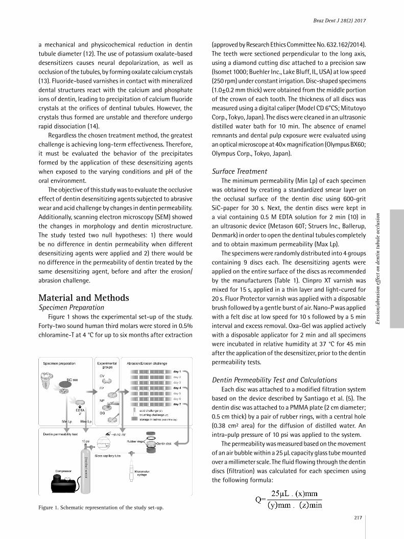
Figure 1 shows the experimental set-up of the study. Forty-two sound human third molars were stored in 0.5% chloramine-T at 4 °C for up to six months after extraction
(approved by Research Ethics Committee No. 632.162/2014). The teeth were sectioned perpendicular to the long axis, using a diamond cutting disc attached to a precision saw (Isomet 1000; Buehler Inc., Lake Bluff, IL, USA) at low speed (250 rpm) under constant irrigation. Disc-shaped specimens (1.0±0.2 mm thick) were obtained from the middle portion of the crown of each tooth. The thickness of all discs was measured using a digital caliper (Model CD 6”CS; Mitutoyo Corp., Tokyo, Japan). The discs were cleaned in an ultrasonic distilled water bath for 10 min. The absence of enamel remnants and dental pulp exposure were evaluated using an optical microscope at 40× magnification (Olympus BX60; Olympus Corp., Tokyo, Japan).
Surface TreatmentThe minimum permeability (Min Lp) of each specimen
was obtained by creating a standardized smear layer on the occlusal surface of the dentin disc using 600-grit SiC-paper for 30 s. Next, the dentin discs were kept in a vial containing 0.5 M EDTA solution for 2 min (10) in an ultrasonic device (Metason 60T; Struers Inc., Ballerup, Denmark) in order to open the dentinal tubules completely and to obtain maximum permeability (Max Lp).
The specimens were randomly distributed into 4 groups containing 9 discs each. The desensitizing agents were applied on the entire surface of the discs as recommended by the manufacturers (Table 1). Clinpro XT varnish was mixed for 15 s, applied in a thin layer and light-cured for 20 s. Fluor Protector varnish was applied with a disposable brush followed by a gentle burst of air. Nano-P was applied with a felt disc at low speed for 10 s followed by a 5 min interval and excess removal. Oxa-Gel was applied actively with a disposable applicator for 2 min and all specimens were incubated in relative humidity at 37 °C for 45 min after the application of the desensitizer, prior to the dentin permeability tests.
Dentin Permeability Test and CalculationsEach disc was attached to a modified filtration system
based on the device described by Santiago et al. (5). The dentin disc was attached to a PMMA plate (2 cm diameter; 0.5 cm thick) by a pair of rubber rings, with a central hole (0.38 cm² area) for the diffusion of distilled water. An intra-pulp pressure of 10 psi was applied to the system.
The permeability was measured based on the movement of an air bubble within a 25 µL capacity glass tube mounted over a millimeter scale. The fluid flowing through the dentin discs (filtration) was calculated for each specimen using the following formula:
Figure 1. Schematic representation of the study set-up.
Braz Dent J 28(2) 2017
218
G.D
. Can
ali e
t al.
Where Q is the dentin filtration rate, x is the distance traveled by the bubble in mm, y is the capillary length (130 mm) and z is the time in minutes.
The permeability (or hydraulic conductivity) of dentin, Lp (µL.min-1.cm H2O-1.cm-1), was calculated by the following formula:
Where Q is the filtration rate (μL.min-1), P the water pressure (703.1 cm H2O), and A is the area of exposed dentin (0.38 cm²). The permeability of each specimen was measured and expressed as a percentage of the fluid flow (% Lp) of the Lp max of the same specimen, allowing each specimen to be its own control. The permeability of the dentin discs was measured at each of the pre-established interval times, namely 5, 10 and 15 min, under constant pressure.
Erosion/Abrasion ChallengeAll specimens were subjected to an acid challenge
cycle for 7 days in 0.5% citric acid solution (pH 2.5). The samples were immersed in the solution six times a day for 2 min under agitation (15). Between immersion times, the specimens were stored in artificial saliva (CaCl2.2H2O, 0.167 g; KH2PO4, 0.123 g; KCl, 11.2 g; (HOCH2)3CNH2, 2.42 g; distilled water; pH adjusted to 7.0) at 37 °C. The specimens were brushed for 5 s with a slurry of toothpaste (Colgate Total 12; Colgate-Palmolive Company, Ontario, Canada) and distilled water (1:1) after the first, third and sixth erosion cycle of seven days, using an electric toothbrush (Oral-B Pro-Health Power; The Procter & Gamble Company, Cincinnati, OH, USA) attached to a fixed grip under a 200 g load. The discs remained in contact with the slurry for 2
min after brushing. Next, the discs were rinsed with distilled water in an ultrasound bath for 10 s and stored in artificial saliva at 37 °C. Dentin permeability was measured on the first, fourth, and seventh day, at 5, 10 and 15 min.
Scanning Electron Microscopy (SEM)Six sound human molars were additionally prepared for
morphological analysis of the dentin. The obtained dentin discs were sectioned into four parts. Each part underwent one of the dentin treatment stages, i.e., smear layer, tubule opening with EDTA and application of desensitizers at each evaluation time of the abrasion/erosion cycle. The specimens were fixated in 2.5% glutaraldehyde solution buffered with 0.1 M sodium phosphate (pH 7.4) at 4 °C for 24 h and rinsed in 0.2 M sodium phosphate solution (pH 7.4) for 1 h with three solution changes. The specimens were dehydrated in successive baths of increasing concentrations of ethanol (25%, 50%, 75%, 95% and 100%). The critical point dryer (EMS 850, Electron Microscopy Sciences, Hatfield, PA, USA) was used for final drying, with the samples transferred to the device chamber and covered with liquid carbon dioxide (CPD 030, Bal-Tec AG, Balzers, Liechtenstein). The samples were sputtered with a gold/palladium alloy and observed in a scanning electron microscope with a 5000x magnification (VEGA3 LMU, Tescan Orsay Holding, Brno-Kohoutovice, Czech Republic).
Statistical Analysis
The means and standard deviations of %Lp were calculated. The data were analyzed by Kolmogorov-Smirnov and Levene tests to determine the normality distribution and homogeneity of variance. Multiple comparisons between the groups were analyzed by three-way ANOVA with repeated measurements and the Games-Howell test. All tests were performed at a significance level of 5%. Data
Table 1. Description of the groups and composition of desensitizing agents used in this study
Group Material Composition Application Mode *
CVClinpro XT Varnish, 3M ESPE, St. Paul, MN, USA
Part A: glass particles of silanized fluoroaluminosilicate, HEMA, water,
BIS-GMA, and silanized silica.Part B: copolymer of polyalkenoic acid, water,
HEMA and calcium glycerophosphate.
Mixing both pastes for 15 s, application of the varnish in a thin
layer, light-curing for 20 s and surface cleaning with a moistened pellet
Nanometric calcium phosphate, sodium fluoride and potassium nitrate
Active application for 10 s with felt disc at low speed; 5 min waiting
time and excess removal.
OGOxa-Gel, Kota Imports Ltda.,
São Paulo, SP, BrazilPotassium oxalate monohydrate and
carboxymethyl cellulose gelActive application with a
disposable applicator for 2 min
* Manufacturer’s recommended protocol.
Braz Dent J 28(2) 2017
219
Eros
ion/
abra
sion
effec
t on
dent
in tu
bule
occ
lusi
on
were analyzed using the SPSS v.22.0 software (SPSS Inc., Chicago, IL, USA).
Results ANOVA failed to detect significant differences for the
variable “time”, as well as significant interactions between “time” and the other two variables. Therefore, the analysis was made only for the “material” and “experimental condition” factors, by grouping the three evaluation times (5, 10 and 15 min). Significant differences were found between groups based on the used material and experimental conditions, as well as a significant interaction between these factors (p<0.05). Table 2 presents the results for dentin permeability in percentage relative to maximum Lp.
Assessment of minimum permeability (Min Lp) revealed no statistically significant differences between groups (p>0.05). Immediately after the application and after the first day of challenge, Nano-P presented a significantly higher permeability compared to the other materials (p<0.05). The mean permeability of Fluor Protector and Clinpro XT were significantly lower from Oxa-Gel and Nano-P (p<0.05) after the 4th and 7th day of challenge.
The mean dentin permeability obtained with Fluor Protector and Clinpro XT desensitizers was similar to the minimum permeability, under all evaluated conditions (p>0.05). Compared to the minimum permeability, Oxa-Gel showed no difference in permeability immediately after application and after the first day of challenge (p>0.05). On the other hand, it presented a significant increase in permeability after the fourth and seventh day, compared to the period immediate after application (p<0.05). The application of Nano-P resulted in permeability similar to the minimum value (p>0.05). However, the permeability increased significantly relative to the minimum Lp after the first day of the erosion/abrasion challenge and it was maintained up to the seventh day (p<0.05).
Figures 2 to 6 show the SEM micrographs of dentin surfaces. Figure 2A shows the dentin surface covered with smear layer and Figure 2B shows the same surface after
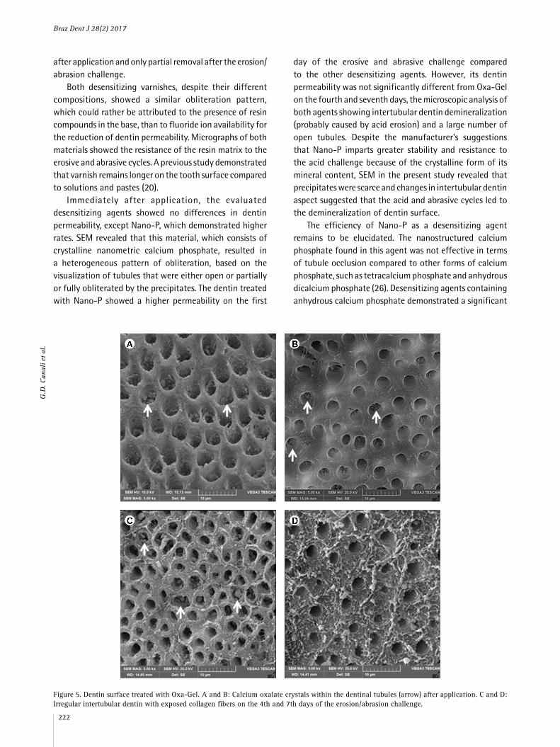
tubule opening with EDTA. The Fluor Protector varnish (Fig. 3) resulted in a partial obliteration of dentinal tubules after the application and showed an uneven layer gradually removed from the dentin surface after the erosion/abrasion challenge. Figure 4 shows dentin treated with Clinpro XT, with complete obliteration of the dentinal tubules, gradual degradation of the resin matrix and detachment of glass particles four days after the erosion/abrasion challenge. From the first to the seventh day, Oxa-Gel desensitizer (Fig. 5) caused only partial occlusion of the tubules with calcium oxalate crystals within and evidences of intertubular dentin demineralization. Dentin treated with Nano-P (Fig. 6) showed partially and completely occluded tubules with calcium phosphate particles that disappeared after the erosion/abrasion cycles.
DiscussionThe null hypotheses were rejected because significant
differences were found in dentin permeability between different dentin-desensitizing agents, and within the same group before and after the erosion/abrasion challenge.
The Clinpro XT Varnish and Fluor Protector desensitizing agents showed similar dentin permeability rates during all phases of the study and similar to dentin permeability obtained with smear layer. Clinpro XT Varnish is composed of polyalkenoic acid copolymer, also found in the resin modified glass ionomer cements Vitrebond and Vitremer, which provide chemical adhesion to dentin by ionic bonding with the hydroxyapatite calcium, the prevalent mineral component of dentin (16). Furthermore, Clinpro XT Varnish contains calcium glycerophosphate, which allows the increase of bioavailable calcium and phosphate in saliva (17). In this study, the samples were maintained in artificial saliva during the intervals of the erosion/abrasion challenge, which might have allowed the release of calcium and phosphate from the saliva to the dentin after each pH drop by the citric acid action in the erosion cycle.
Previous studies have evaluated the desensitizing activity of Clinpro XT Varnish (8,17-19). An in vitro comparison between Clinpro XT and a conventional
Table 2. Mean (SD) dentin permeability (%Lp) with smear layer (Lp min) and with desensitizers tested immediately after application and after 1, 4 and 7 days of erosive/abrasive challenge
Group Smear layer Immediate 1st day 4th day 7th day
CV 93.51 (4.47) Aa 94.45 (1.90) Aa 95.01 (3.39) Aa 95.78 (3.70) Aa 94.38 (5.82) Aa
FP 94.33 (2.35) Aa 91.21 (2.50) Aa 93.86 (2.29) Aa 93.23 (2.42) Aa 93.59 (1.92) Aa
OG 92.67 (3.37) Aa 95.48 (2.28) Aa 90.32 (6.73) ABa 85.36 (7.53) Bb 84.70 (6.37) Bb
Values of groups with the same uppercase letters were not significantly different in the same row, and those with the same lowercase letters were not significantly different in the same column (p>0.05).
Braz Dent J 28(2) 2017
220
G.D
. Can
ali e
t al.
Figure 2. A: Representative micrograph of the dentin surface with the smear layer obliterating dentinal tubules. B: Dentin treated with EDTA showing full opening of the dentinal tubules.
Figure 3. Dentin surface treated with Fluor Protector. A: After the application, an uneven layer is observed partially obliterating the dentinal tubules. B and C: 1st and 4th days of the erosion/abrasion cycle: discontinuation in the varnish layer as well as tubular exposure is observed. Precipitates suggesting the deposition of calcium fluoride (asterisk). D: 7th day of the erosion/abrasion cycle showing a visibly worn varnish layer with cracks and dentin exposure due to the aggressive nature of the challenge.
Braz Dent J 28(2) 2017
221
Eros
ion/
abra
sion
effec
t on
dent
in tu
bule
occ
lusi
on
Figure 4. Dentin surface treated with Clinpro XT Varnish. A: Fluoroaluminosilicate glass particles (asterisk) observed after the application of the varnish. B: Few particles with an exposed and flattened resin matrix on the 1st day of the erosion/abrasion cycle. C and D: Pores corresponding to the removal of filler particles (arrows) on the 4th and 7th days of the challenge.
adhesive system (Single Bond Plus) revealed that both reduced dentin permeability and sealed dentin, with or without the smear layer (8). Clinical trials comparing Clinpro XT Varnish to a glutaraldehyde-based desensitizer observed a greater reduction in hypersensitivity and a more extended action after 2 weeks (18) and 4 weeks (19). Clinpro XT Varnish maintained dentin permeability in all the phases of the present study, even after the erosion/abrasion challenge. This was suggested by the aspect of the dentin surface found in the SEM micrographs, which displayed a continuous layer obliterating the dentinal tubules over the duration of the cycles (seven days), despite the mild surface layer deterioration caused by the detachment of some load particles.
Fluor Protector varnish contains a polyurethane base that allows an intimate contact with the tooth surface after evaporation of the solvent. In addition, this varnish contains 1% silane fluoride, equivalent to 1,000 ppm (20). The action of a fluoride varnish is based on the reaction
between NaF and calcium ions resulting in the formation of small calcium fluoride crystals (0.05 µm) deposited in the open dentinal tubules (21). A recent study (22) observed that a 5.24% sodium fluoride varnish imparted similar permeability to the minimum value before and after acid challenge, which is in line with the results of the present study. Clinical studies have reported the efficacy of fluoride varnishes in reducing dentinal hypersensitivity (23) for up to 8 weeks (24).
The polyurethane base of Fluor Protector has an advantage of more efficient adhesion, compared to the synthetic resin base commonly seen in fluoride varnishes (25). This may explain the low permeability of Fluor Protector observed in this study. Despite a much lower fluoride concentration compared to the Duraphat fluoride varnish (23,600 ppm), an in vitro study (25) simulating an erosive challenge using wine proved the ability of Fluor Protector to remain on the dentin surface. Indeed, SEM showed a layer of varnish on the dentin surface immediately
Braz Dent J 28(2) 2017
222
G.D
. Can
ali e
t al.
Figure 5. Dentin surface treated with Oxa-Gel. A and B: Calcium oxalate crystals within the dentinal tubules (arrow) after application. C and D: Irregular intertubular dentin with exposed collagen fibers on the 4th and 7th days of the erosion/abrasion challenge.
after application and only partial removal after the erosion/abrasion challenge.
Both desensitizing varnishes, despite their different compositions, showed a similar obliteration pattern, which could rather be attributed to the presence of resin compounds in the base, than to fluoride ion availability for the reduction of dentin permeability. Micrographs of both materials showed the resistance of the resin matrix to the erosive and abrasive cycles. A previous study demonstrated that varnish remains longer on the tooth surface compared to solutions and pastes (20).
Immediately after application, the evaluated desensitizing agents showed no differences in dentin permeability, except Nano-P, which demonstrated higher rates. SEM revealed that this material, which consists of crystalline nanometric calcium phosphate, resulted in a heterogeneous pattern of obliteration, based on the visualization of tubules that were either open or partially or fully obliterated by the precipitates. The dentin treated with Nano-P showed a higher permeability on the first
day of the erosive and abrasive challenge compared to the other desensitizing agents. However, its dentin permeability was not significantly different from Oxa-Gel on the fourth and seventh days, the microscopic analysis of both agents showing intertubular dentin demineralization (probably caused by acid erosion) and a large number of open tubules. Despite the manufacturer’s suggestions that Nano-P imparts greater stability and resistance to the acid challenge because of the crystalline form of its mineral content, SEM in the present study revealed that precipitates were scarce and changes in intertubular dentin aspect suggested that the acid and abrasive cycles led to the demineralization of dentin surface.
The efficiency of Nano-P as a desensitizing agent remains to be elucidated. The nanostructured calcium phosphate found in this agent was not effective in terms of tubule occlusion compared to other forms of calcium phosphate, such as tetracalcium phosphate and anhydrous dicalcium phosphate (26). Desensitizing agents containing anhydrous calcium phosphate demonstrated a significant
Braz Dent J 28(2) 2017
223
Eros
ion/
abra
sion
effec
t on
dent
in tu
bule
occ
lusi
on
Figure 6. Dentin surface treated with Nano-P. A: Dentinal tubules partially (arrow) and completely (asterisk) occluded probably by calcium phosphate particles after the application. B: Open tubules on the 1st day of the erosion/abrasion cycle. C and D: Exposed dentinal tubules with irregular intertubular dentin on the 4th and 7th days.
reduction in dentin permeability even after an acid challenge and also crystal precipitations along the dentin and dentinal tubules after 4 weeks (9).
Potassium oxalate is considered to be an effective desensitizing agent for the reduction of dentin permeability (5,22,26,27). Studies evaluating the occlusive aspect of dentinal tubules demonstrated the penetration of potassium oxalate into the dentinal tubules and subsequent reaction with calcium ions to form calcium oxalate crystals (27,28). However, the stability of these crystals remains controversial. While some authors assert that calcium oxalate crystals are insoluble (22,28), others claim that the crystals formed as a result of the reaction between potassium oxalate and hydroxyapatite will be dissolved over time (9,29). The micrographs in this study demonstrate the limited occlusive effect of Oxa-Gel after application, as well as absence of oxalate crystals from the first up to the seventh day of the erosion/abrasion challenge. These results corroborate the results of a previous study (27),
which confirmed the dissolution of calcium oxalate crystals and open tubules after erosive challenge with citric acid.
The most common acid challenge used for in vitro studies is based on a single immersion in citric acid for 1 to 5 min (5,9,22,27). On the opposite, the erosion/abrasion challenge in the present study aimed to simulate high consumption of acidic beverages combined with regular tooth brushing. Thus, acid solution was applied during 2 min for 6 times per day and tooth brushing was carried out during 5 s, 3 times per day, both for 7 days. The time each specimen was brushed was based on the amount of time recommended to brush all teeth, which is about 2 min. Moreover, toothbrushing duration is not considered an important risk factor for DH, but the frequency and technique, including excessive force and direction (30).
The results of dentin permeability tests should not be extrapolated directly to clinical situations. Randomized clinical trials are the best way to verify the efficacy of desensitizing agents under physiological intraoral
Braz Dent J 28(2) 2017
224
G.D
. Can
ali e
t al.
conditions, when subjected to mechanical and chemical challenges.
Within the limitations of this in vitro study, it was concluded that all desensitizers reduced the dentin permeability immediately after application. However, only the varnishes were able to maintain the occlusion effect after the erosion/abrasion challenge.
Resumo O objetivo do estudo foi avaliar a permeabilidade da dentina (Lp) usando diferentes agentes dessensibilizantes antes e depois de um desafio abrasivo/erosivo. Discos de dentina foram obtidos a partir 42 terceiros molares humanos. Lp mínima foi medida após uma simulação de smear layer usando lixa de SiC # 600 e a Lp máxima foi medida após imersão em EDTA 0,5 M. As amostras foram tratadas com diferentes dessensibilizantes: dois vernizes (Fluor Protector-FP, Clinpro XT Varnish-CV), uma pasta (Desensibilize Nano P-NP) e um gel (Oxa Gel-OG). A Lp de cada amostra foi medida imediatamente após a aplicação dos dessensibilizantes. Os discos foram submetidos a um ciclo de erosão/abrasão durante 7 dias, com uma solução de 0,5% de ácido cítrico (6x/dia) e escovação (3x/dia). Lp foi medida do primeiro ao sétimo dia do desafio. Os dados foram analisados por ANOVA a 3 critérios com medidas repetidas e teste de Games-Howell (α=5%). FP e CV demonstraram Lp semelhante imediatamente depois da aplicação até 7 dias (p<0,05). OG apresentou um aumento significativo na Lp depois do 4º e 7º dias. NP resultou numa permeabilidade significativamente mais elevada comparada com a dos outros materiais imediatamente após a aplicação e após o primeiro dia de desafio. Todos os dessensibilizantes reduziram a permeabilidade da dentina imediatamente após a aplicação. No entanto, apenas os vernizes foram capazes de manter o efeito oclusivo após o desafio da erosão/abrasão.
AcknowledgementsThe authors would like to thank Prof. Paulo César Soares Jr. for the scanning electron microscopy analysis.
References 1. Lin PY, Cheng YW, Chu CY, Chien KL, Lin CP, Tu YK. In-office treatment
for dentin hypersensitivity: a systematic review and network meta-analysis. J Clin Periodontol 2013;40:53-64.
2. Bissada NF. Symptomatology and clinical features of hypersensitive teeth. Arch Oral Biol 1994;39(Suppl):31S-32S.
3. Brännström M. Sensitivity of dentin. Oral Surge Oral Med Oral Pathol 1966;2:517-526.
4. Linsuwanont P, Versluis A, Palamara JE, Messer HH. Thermal stimulation causes tooth deformation: a possible alternative to the hydrodynamic theory? Arch Oral Biol 2008;53:261-272.
5. Santiago SL, Pereira JC, Martineli AC. Effect of commercially available and experimental potassium oxalate-based dentin desensitizing agents in dentin permeability: influence of time and filtration system. Braz Dent J. 2006;17:300-305.
6. Ishihata H, Kanehira M, Nagai T, Finger WJ, Shimauchi H, Komatsu M. Effect of desensitizing agents on dentin permeability. Am J Dent 2009;22:143-146.
7. Pinto SC, Batitucci RG, Pinheiro MC, Zandim DL, Spin-Neto R, Sampaio JE. Effect of an acid diet allied to sonic toothbrushing on root dentin permeability: an in vitro study. Braz Dent J 2010;21:390-395.
8. Rusin RP, Agee K, Suchko M, Pashley DH. Effect of a new desensitizing material on human dentin permeability. Dent Mater 2010;26:600-607.
9. Thanatvarakorn O, Nakashima S, Sadr A, Prasansuttiporn T, Ikeda M, Tagami J. In vitro evaluation of dentinal hydraulic conductance and tubule sealing by a novel calcium-phosphate desensitizer. J Biomed Mater Res B Appl Biomater 2013;101:303-309.
10. Petersson LG. The role of fluoride in the preventive management
of dentin hypersensitivity and root caries. Clin Oral Investig 2013;17(Suppl):63S-71S.
11. Esteves SR, Huhtala MF, Gomes AP, Ye Q, Spencer P, De Paiva Gonçalves SE. Longitudinal effect of surface treatments modified by NaOCl-induced deproteinization and Nd:yag laser on dentin permeability. Photomed Laser Surg 2016;34:68-75.
12. Suge T, Ishikawa K, Kawasaki A, Suzuki K, Matsuo T, Noiri Y, et al.. Calcium phosphate precipitation method for the treatment of dentin hypersensitivity. Am J Dent. 2002;15:220-226.
13. Aranha AC, Pimenta LA, Marchi GM. Clinical evaluation of desensitizing treatments for cervical dentin hypersensitivity. Braz Oral Res 2009;23:333-339.
14. Sieck B, Takagi S, Chow LC. Assessment of loosely-bound and firmly-bound fluoride uptake by tooth enamel from topically applied fluoride treatments. J Dent Res 1990;69:1261-1265.
15. Schlueter N, Lussi A, Tolle A, Ganss C. Effects of erosion protocol design on erosion/abrasion study outcome and on active agent (NaF and SnF2) efficacy. Caries Res 2016;50:170-179.
16. Mitra SB, Lee CY, Bui HT, Tantbirojn D, Rusin RF. Long-term adhesion and mechanism of bonding of a paste-liquid resin-modified glass-ionomer. Dent Mater 2005;25:459-466.
17. Lynch RJ, Ten Cate JM. Effect of calcium glycerophosphate on demineralization in an in vitro biofilm model. Caries Res 2006;40:142-147.
18. Bhandary S, Hegde MN. A clinical comparison of in-office management of dentin hypersensitivity in a short-term treatment period. Int J of Biomed & Adv Res 2012;3:169-174.
19. Ding YJ, Yao H, Wang GH, Song H. A randomized double-blind placebo-controlled study of the efficacy of Clinpro XT varnish and Gluma dentin desensitizer on dentin hypersensitivity. Am J Dent 2014;27:79-83.
20. Beltrán-Aguilar ED, Goldstein JW, Lockwood SA. Fluoride varnishes: a review of their clinical use, cariostatic mechanism, efficacy and safety. J Am Dent Assoc 2000;131:589-596.
21. Pesevska S, Nakova M, Ivanovski K, Angelov N, Kesic L, Obradovic R, et al.. Dentinal hypersensitivity following scaling and root planing: comparison of low-level laser and topical fluoride treatment. Lasers Med Sci 2010;25:647-650.
22. Calabria M, Porfirio R, Fernandes S, Wang L, Buzalaf M, Pereira J, et al.. Comparative in vitro effect of TiF4 to NaF and potassium oxalate on reduction of dentin hydraulic conductance. Oper Dent 2014;39:427-432.
23. Ritter AV, De L Dias W, Miguez P, Caplan DJ, Swift EJ Jr. Treating cervical dentin hypersensitivity with fluoride varnish: a randomized clinical study. J Am Dent Assoc 2006;137:1013-1020.
24. Hoang-Dao BT, Hoang-Tu H, Tran-Thi NN, Koubi G, Camps J, About I. Clinical efficiency of a natural resin fluoride varnish (Shellac F) in reducing dentin hypersensitivity. J Oral Rehabil 2009;36:124-131.
25. Mok TB, McIntyre J, Hunt D. Dental erosion: in vitro model of wine assessor’s erosion. Aust Dent J. 2001;46:263-268.
26. Thanatvarakorn O, Nakashima S, Sadr A, Prasansuttiporn T, Thitthaweerat S, Tagami J. Effect of a calcium-phosphate based desensitizer on dentin surface characteristics. Dent Mater J 2013;32:615-621.
27. Pereira JC, Segala AD, Gillam DG. Effect of desensitizing agents on the hydraulic conductance of human dentin subjected to different surface pre-treatments - an in vitro study. Dent Mater 2005;21:129-138.
28. Sales-Peres SH, Reinato JV, Sales-Peres A de C, Marsicano JA. Effect of iron gel on dentin permeability. Braz Dent J 2011;22:198-202.
29. Pashley DH, Carvalho RM, Pereira JC, Villanueva R, Tay FR. The use of oxalate to reduce dentin permeability under adhesive restorations. Am J Dent 2001;14:89-94.
30. Scaramucci T, João-Souza SH, Lippert F, Eckert GJ, Aoki IV, Hara AT. Influence of tooth brushing on the anti-erosive effect of film-forming agents. Caries Res 2016;50:104-110.
Received February 11, 2016Accepted December 19, 2016